Review

Age of Antibiotic Resistance in MDR/XDR Clinical Pathogen of Pseudomonas aeruginosa

1
Department of Microbiology, All India Institute of Medical Sciences, Rishikesh 249203, India
2
Department of Dentistry, All India Institute of Medical Sciences, Rishikesh 249203, India
3
Department of Burn and Plastic Surgery, All India Institute of Medical Sciences, Rishikesh 249203, India
4
Department of Trauma Surgery, All India Institute of Medical Sciences, Rishikesh 249203, India
5
Department of Biochemistry, All India Institute of Medical Sciences, Rishikesh 249203, India
6
Department of Orthopaedics, All India Institute of Medical Sciences, Rishikesh 249203, India
7
Department of Cellular Biology and Anatomy, Augusta University, Augusta, GA 30912, USA
8
Department of Medical Oncology, All India Institute of Medical Sciences, Rishikesh 249203, India
9
Division of Cancer Biology, Central Drug Research Institute, Lucknow 226031, India
10
Department of Entomology, University of Kentucky, Lexington, KY 40503, USA
*
Authors to whom correspondence should be addressed.
Pharmaceuticals 2023, 16(9), 1230; https://doi.org/10.3390/ph16091230
Submission received: 8 July 2023 / Revised: 15 August 2023 / Accepted: 23 August 2023 / Published: 30 August 2023

Abstract

:
Antibiotic resistance in Pseudomonas aeruginosa remains one of the most challenging phenomena of everyday medical science. The universal spread of high-risk clones of multidrug-resistant/extensively drug-resistant (MDR/XDR) clinical P. aeruginosa has become a public health threat. The P. aeruginosa bacteria exhibits remarkable genome plasticity that utilizes highly acquired and intrinsic resistance mechanisms to counter most antibiotic challenges. In addition, the adaptive antibiotic resistance of P. aeruginosa, including biofilm-mediated resistance and the formation of multidrug-tolerant persisted cells, are accountable for recalcitrance and relapse of infections. We highlighted the AMR mechanism considering the most common pathogen P. aeruginosa, its clinical impact, epidemiology, and save our souls (SOS)-mediated resistance. We further discussed the current therapeutic options against MDR/XDR P. aeruginosa infections, and described those treatment options in clinical practice. Finally, other therapeutic strategies, such as bacteriophage-based therapy and antimicrobial peptides, were described with clinical relevance.

Graphical Abstract

1. Introduction

There are several reasons why multidrug-resistant (MDR) and extensively drug-resistant (XDR) P. aeruginosa strains represent a worldwide health threat. First, P. aeruginosa is extremely opportunistic and can cause serious infections in hospital settings, especially in immunocompromised people. Second, it has versatility in its antibiotic resistance and may transmit drug resistance through multiple routes [1]. High-risk clones are spreading worldwide, posing a public health issue that must be examined and addressed [2]. In 2017, the World Health Organization placed carbapenem-resistant P. aeruginosa in the “critical” group, for which new medicines are needed [3].
P. aeruginosa MDR and XDR strains have increased in frequency during the past several years, with rates varying between 15 and 30% in certain geographic regions [4,5]. However, combination medication resistance is also prevalent in P. aeruginosa MDR and XDR strains. A total of 5.5% of P. aeruginosa isolates that were resistant to all five antimicrobial groups under monitoring in 2015, as well as 13% of isolates that were resistant to more than three antimicrobial groups, were reported by the European Centers for Disease Prevention and Control [6]. Recent research has examined the in vitro effectiveness of polymyxin B in combination with 13 other antibiotics (amikacin, aztreonam, cefepime, chloramphenicol, ciprofloxacin, fosfomycin, linezolid, meropenem, minocycline, rifampin, temocillin, thiamphenicol, or trimethoprim) against four clinical strains of MDR Pseudomonas aeruginosa. Positive interactions were frequently observed with the tested combinations against strains that carried multiple mechanisms of drug resistance, and with antibiotics that are typically inactive against P. aeruginosa [7]. Another study involving 3184 clinical isolates collected from 71 US medical centers shows that Ceftazidime–avibactam (97.0% susceptible (S)), ceftolozane–tazobactam (98.0% S), imipenem––relebactam (97.3% S) and tobramycin (96.4% S) were the most active agents against the aggregate P. aeruginosa isolate collection, and retained good activity against piperacillin–tazobactam-non-susceptible, meropenem-non-susceptible and multidrug-resistant (MDR) isolates [8]. The remedies to this issue involve investing more money in fundamental research, clinical research, antimicrobial stewardship infection control, the creation of novel antimicrobials, and the optimum use of those that are already available. In this review article, we discussed the available treatments for MDR/XDR P. aeruginosa infections and examined the most recent research on its resistance. We also looked at the therapeutic alternatives applied in clinical practice to treat MDR/XDR P. aeruginosa infections. Last but not least, several treatment modalities with clinical applicability were discovered, including bacteriophage-based therapy and antimicrobial peptide therapy, that can be used in future research and medication.

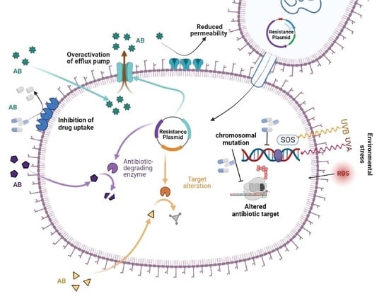
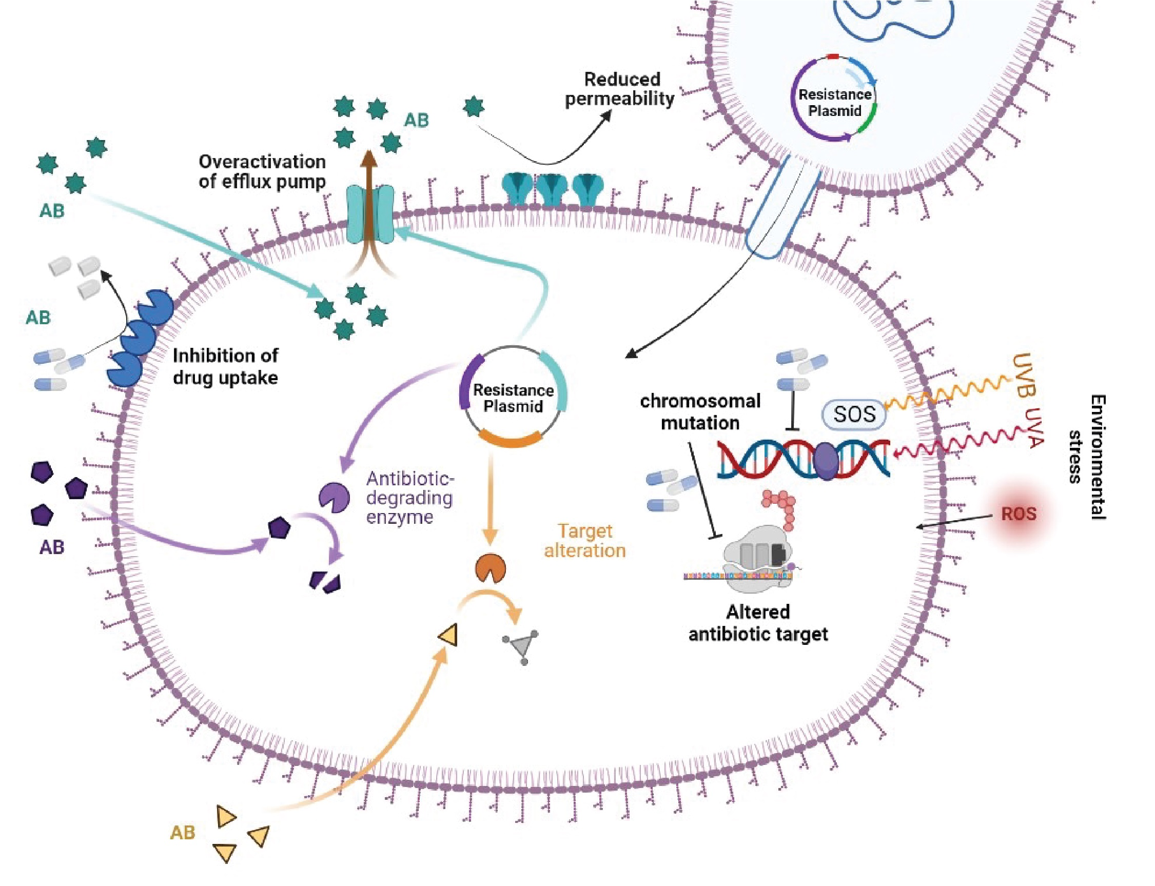
2. Molecular Mechanism of Drug Resistance in P. aeruginosa

2.1. Inherent Resistome

It is intriguing how P. aeruginosa possesses a peculiar assortment of drug-resistance mechanisms, such as multiple chromosomal-associated genes that confer resistance to antibiotics, as well as complex regulatory pathways involved in both inherent and adaptive resistance [9,10,11,12]. When compared to other Gram-negative bacteria, the development of inherent resistance in P. aeruginosa is primarily influenced by the expression of inducible AmpC cephalosporinase, the synthesis of constitutive and inducible efflux pumps, and the low permeability of its outer membrane. The synthesis of inducible beta-lactamase is of utmost importance in the inherent resistance of P. aeruginosa to aminopenicillins and cephalosporins, specifically cefoxitin. These antibiotics are potent inducers of AmpC expression, resulting in an overabundance of cephalosporinase [13]. The hydrolytic stability of Imipenem is slightly affected by its strong ability to induce enzymes. The expression of inducible AmpC is important in reducing the natural sensitivity of P. aeruginosa [14]. Additionally, the recently discovered imipenemase (IMP) PA5542 may also influence the inherent susceptibility of β-lactam antibiotics [15]. Nevertheless, additional research is needed to explore their involvement in innate or acquired resistance. The expression of efflux pump is crucial in reducing the inherent susceptibility to a wide range of β-lactam antibiotics and fluoroquinolones. In a similar manner, the inducible expression of Hyperproduction of efflux-mediated (MexXY) genes exerts a notable impact on the inherent, minimal resistance to aminoglycosides [16]. These efflux pump systems effectively expel various classes of antibiotics from the bacterial cell, thereby providing it with intrinsic resistance.

2.2. Mutational Resistome

In addition to its wide-ranging innate resistance repertoire, P. aeruginosa demonstrates remarkable proficiency in acquiring chromosomal mutations, leading to the emergence of novel antimicrobial-resistant superbugs [1], as summarized in Table 1. The β-lactam resistance mechanism driven by mutation has been observed in 20% of P. aeruginosa [12,17,18]. The deactivation of penicillin binding protein-4 (PBP-4) triggers the activation of the CreBC/BlrAB two-component system, which is responsive to carbon sources and contributes to β-lactam resistance. This activation leads to an additional increase in resistance levels [18]. The clinical strains were found to possess distinct mutations that affected the transcriptional regulator of AmpR, a protein responsible for regulating the overexpression of ampC and conferring resistance to beta-lactam antibiotics. The mutations under consideration encompass the R154H mutation, which is associated with the epidemic MDR/XDR ST175 high-risk clone, and the D135N mutation, observed in species other than P. aeruginosa [12]. Numerous genetic variations have been documented to enhance the amplification of ampC in various genetic sequences, encompassing those responsible for other amidases (AmpDh2/AmpDh3), PBP5 or PBP7, lytic transglycosylases (MltB and SltB1), MPL (UDP-N-acetylmuramate: L-alanyl—D-glutamyl-Meso-(NADH dehydrogenase I chain N) [13].
Findings from studies have demonstrated that genetic variations that modify the structure of AmpC can lead to the development of resistance against β-lactam antibiotics. This resistance includes the newly developed β-lactam–lactamase inhibitor, as well as the combinations of ceftolozane–tazobactam and ceftazidime–avibactam. This is in addition to the phenomenon of AmpC hyperproduction, as previously reported [19,20,21,22]. The development of resistance to ceftolozane–tazobactam and ceftazidime–avibactam was observed in a specific group of P. aeruginosa clinical isolates [23]. This resistance was linked to various changes in the amino acid composition of AmpC. Recent findings have unveiled the existence of more than 300 distinct variations of cephalosporinase derived from the Pseudomonas genus. Notably, certain variations have been observed to confer increased resistance to ceftolozane–tazobactam and ceftazidime–avibactam. There is a growing body of evidence suggesting that alterations in penicillin-binding proteins (PBPs), specifically mutations in PBP-3, contribute to the development of resistance to β-lactam antibiotics, alongside β-lactamases. Recent data obtained from individuals diagnosed with cystic fibrosis [24,25], as well as from strains of bacteria causing epidemics [26,27], and laboratory experiments conducted in controlled environments [28,29], have provided evidence indicating that specific alterations in penicillin-binding protein-3 play a role in the emergence of resistance to -lactam antibiotics. The R504C/R504H and F533L mutations, located in the domains responsible for stabilizing the -lactam–penicillin-binding protein-3 inactivation complex, have been frequently documented in the scientific literature [30]. The presence of inhibitory deletion/insertion sequences within the OprD gene, as well as distant mutations that enhance the activity of efflux pump systems MexEF-OprN or CzcCBA while simultaneously reducing the expression of OprD, can result in the loss of the carbapenem-specific porin—OprD. The inactivation of OprD often leads to resistance against all conventional anti-pseudomonal β-lactams in a synergistic fashion when combined with AmpC overexpression [31]. Another pivotal determinant in resistance is the mutational upregulation of one of the four primary efflux pumps in P. aeruginosa [32,33]. The prevalence of MexAB-OprM and MexXY overexpression in clinical isolates ranges from 10% to 30%, while the overexpression of MexCD-OprJ and MexEF-OprN is less-frequently observed, occurring in approximately 5% of cases. The OprD porin exhibits either inactivity or downregulation, leading to reduced sensitivity to Meropenem and the inducible synthesis of AmpC. The simultaneous upregulation of MexAB-OprM and downregulation of OprD is a significant determinant of Meropenem resistance in clinical strains [34].
More than 20% of isolates frequently demonstrate resistance to imipenem, and the majority of these isolates are deficient in OprD [34]. The MexAB-OprM efflux pump exhibits the most extensive substrate specificity, and its upregulation due to mutations leads to decreased susceptibility to all -lactams and fluoroquinolones (with the exception of imipenem). The mutation-driven overexpression of MexXY is a common contributing factor in the resistance of clinical strains to cefepime, in addition to its primary role in intrinsic aminoglycoside resistance [35]. The hyperproduction of MexEF-OprN is not commonly observed and primarily impacts fluoroquinolone antibiotics. However, mutations in mexT/mexS genes that lead to MexEF-OprN hyperproduction also result in resistance to imipenem by repressing OprD gene expression [36]. In spite of exhibiting heightened resistance to various β-lactams and aminoglycosides, the upregulation of MexCD-OprJ, a phenomenon frequently observed in persistent infections, additionally plays a role in conferring resistance to cefepime [37].
Fluoroquinolone resistance in P. aeruginosa often arises due to the overexpression of efflux pumps, as well as mutations occurring in type IV topoisomerases (ParC and ParE) and DNA-gyrases (GyrA and GyrB) [38]. From a geographical perspective, the prevalence of fluoroquinolone resistance is observed to be the dominant trait, ranging from 30% to 40% in multiple countries. Recent scientific investigations have elucidated that genetic mutations occurring in the fusA1 gene, responsible for encoding the elongation factor G, have the potential to induce resistance to aminoglycoside antibiotics. This resistance mechanism operates in conjunction with the overexpression of the MexXY genes and the acquisition of genetic pathways through horizontal gene transfer. Indeed, empirical evidence has shown that certain mutations in the FusA1 gene can result in resistance to aminoglycoside antibiotics in laboratory settings [39,40] and in clinical cases of P. aeruginosa infection, especially in individuals with cystic fibrosis [41].
In essence, although the occurrence of colistin resistance is still relatively limited (5%), there has been a recent surge, likely due to its heightened utilization as a final option against infections caused by multidrug resistant/extensively drug resistant bacterial strains. The development of colistin resistance frequently occurs due to alterations in the lipid A component of lipopolysaccharide (LPS) following the addition of 4-amino-4-deoxy-L-arabinose [42]. The mutations observed are often associated with the regulatory systems PmrAB and PhoPQ, which result in the activation of the arnBCADTEF operon. In recent studies, it has been demonstrated that mutations in the ParRS two-component regulator play a crucial role in driving colistin resistance. These mutations activate the arnBCATEF genes, leading to an MDR profile. Furthermore, they upregulate the MexXY genes while downregulating the OprD gene [11]. The ColRS and CprRS systems have also been implicated in the development of polymyxin resistance [43].

2.3. Horizontally Acquired Resistome

Novel antibiotics of the latest generation have been developed with the specific purpose of selectively inhibiting crucial cellular proteins involved in DNA replication and repair, protein synthesis, and the production of components for the cell membrane [44]. The primary strategies employed to address acquired resistance involve implementing chemical modifications to preexisting antibiotics. The rate of antibiotic production has experienced a notable decrease in recent years, despite the fact that a considerable number of antibiotics are presently undergoing their third or fourth round of modifications. Furthermore, due to the evolutionary adaptability of bacteria, the efficacy of antibiotic therapy has progressively diminished over a period of time [45]. Furthermore, bacteria have developed a complex regulatory evolutionary adaptation by acquiring resistance genes primarily through conjugation and, to a lesser degree, through spontaneous transformation and transduction [46]. Despite the perceived insignificance of transformation, recent research suggests that its importance may be greater than previously hypothesized [47]. Recent research examined the effectiveness of horizontal gene transfer (HGT) through conjugation and examined the MDR phenotypes of numerous clinical and environmental bacterial strains from various sources. Along with examining the effects of medications and heavy metal (arsenic), conjugation efficiency between clinical and environmental strains was also examined. They discovered that using 2-HDA as a COIN prevented HGT between strains that were obtained in hospitals and those that naturally exist [48].
One aspect of mutational resistance that is of significant interest is the transferable type of P. aeruginosa resistance, which occurs relatively frequently and contributes to the overall accumulation of concern. Bacterial conjugation serves as the fundamental mechanism for both intra- and inter-species HGTs. It plays a crucial role in expediting the dissemination of antibiotic resistance genes [49]. Certainly, the prevalence of highly hazardous transferable β-lactamases, including ESBLs and carbapenemases (specifically class B carbapenemases, also known as Metallo β-lactamases), is steadily increasing on a global scale. Nevertheless, their distribution exhibits inconsistency and displays variation across hospitals and regions, ranging from less than 1% to approximately 50% [50]. Moreover, the occurrence of transferable β-lactamases in P. aeruginosa might have been underestimated in several locations due to the challenges associated with their detection [51]. Integrons belonging to Class 1 generally encompass determinants of resistance to aminoglycosides, as well as the genes responsible for extended-spectrum beta-lactamases (ESBLs) and carbapenemases. While the involvement of conjugative elements is now more commonly recognized, these integrons are often inserted into transposable elements located on the bacterial chromosome [51,52,53,54]. A recent study was conducted to review the distribution of spreadable β-lactamases in P. aeruginosa [55]. Frequently documented extended-spectrum beta-lactamases (ESBLs) in P. aeruginosa encompass class D enzymes, specifically OXA-2 or OXA-10 variants, as well as class A enzymes including PER, VEB, GES, BEL, and PME variants. Metallo β-lactamases (MBLs) are the predominant carbapenemases found in P. aeruginosa. Among these MBLs, the VIM and IMP variants are the most prevalent and widely distributed across different geographical regions. In Brazil, the prevalence of the SPM MBL gene is extensive, while the NDM, GIM, and FIM genes are sporadically detected. The prevalence of Class A carbapenemases in P. aeruginosa is relatively low on a global scale, even though GES and KPC enzymes have been identified in multiple countries [54].
The transferability resistance of aminoglycosides is influenced by the presence of aminoglycoside-modifying enzymes that are encoded within Class 1 integrons. The acetyltransferases frequently observed in P. aeruginosa are AAC 3′ gentamicin and AAC 6′ tobramycin, as well as the nucleotidyltransferase ANT 2′-I gentamicin and tobramycin. Nevertheless, there are significant emerging concerns associated with 16S rRNA methyltransferases, such as Rmt or Arm, as they confer resistance to all commercially available aminoglycosides, including the recently developed plazomicin [54]. Intermittently, it has been observed that the prevalence of transferable resistance to fluoroquinolones is primarily influenced by Qnr determinants, such as QnrVC1 [56]. In a recent scientific study, it has been demonstrated that a novel phosphotransferase, known as CrpP, is responsible for facilitating plasmid-mediated quinolone resistance [57].
Ceftolozane–tazobactam and Ceftazidime–avibactam, two recently developed combinations, exhibit a notable degree of resistance to AmpC hydrolysis [58,59]. This resistance is attributed to the inhibitory effect of avibactam on AmpC in the case of ceftazidime–avibactam, and the ability of ceftolozane to remain stable against hydrolysis by AmpC in the case of ceftolozane–tazobactam. Nevertheless, based on existing in vitro and in vivo research, it appears that the emergence of resistance to both drugs could be attributed to a combination of genetic mutations, leading to increased production of AmpC and alterations in its structure [19,20,22]. The empirical evidence obtained from experiments conducted in living organisms (in vivo) and in controlled laboratory conditions (in vitro) suggests that particular genetic alterations in penicillin-binding protein-3 have the potential to reduce the vulnerability to the aforementioned combinations. The susceptibility of ceftazidime–avibactam seems to be more influenced by the overexpression of different efflux pumps compared to ceftolozane–tazobactam [23,60]. Both ceftolozane–tazobactam and ceftazidime–avibactam have demonstrated a lack of efficacy against strains that produce acquired β-lactamases. Ceftazidime–avibactam, but not ceftolozane–tazobactam, exhibits potential activity against isolates that generate class A carbapenemases, such as GES enzymes [61]. In a similar vein, the effectiveness of ceftolozane–tazobactam and ceftazidime–avibactam against strains of P. aeruginosa that produce extended-spectrum beta-lactamase (ESBL) exhibits variability, albeit with a generally favorable outcome for ceftazidime–avibactam. Ultimately, the development of resistance to both pharmaceutical agents can arise due to the presence of extended-spectrum mutations in horizontally transferred OXA-type β-lactamases [62,63].

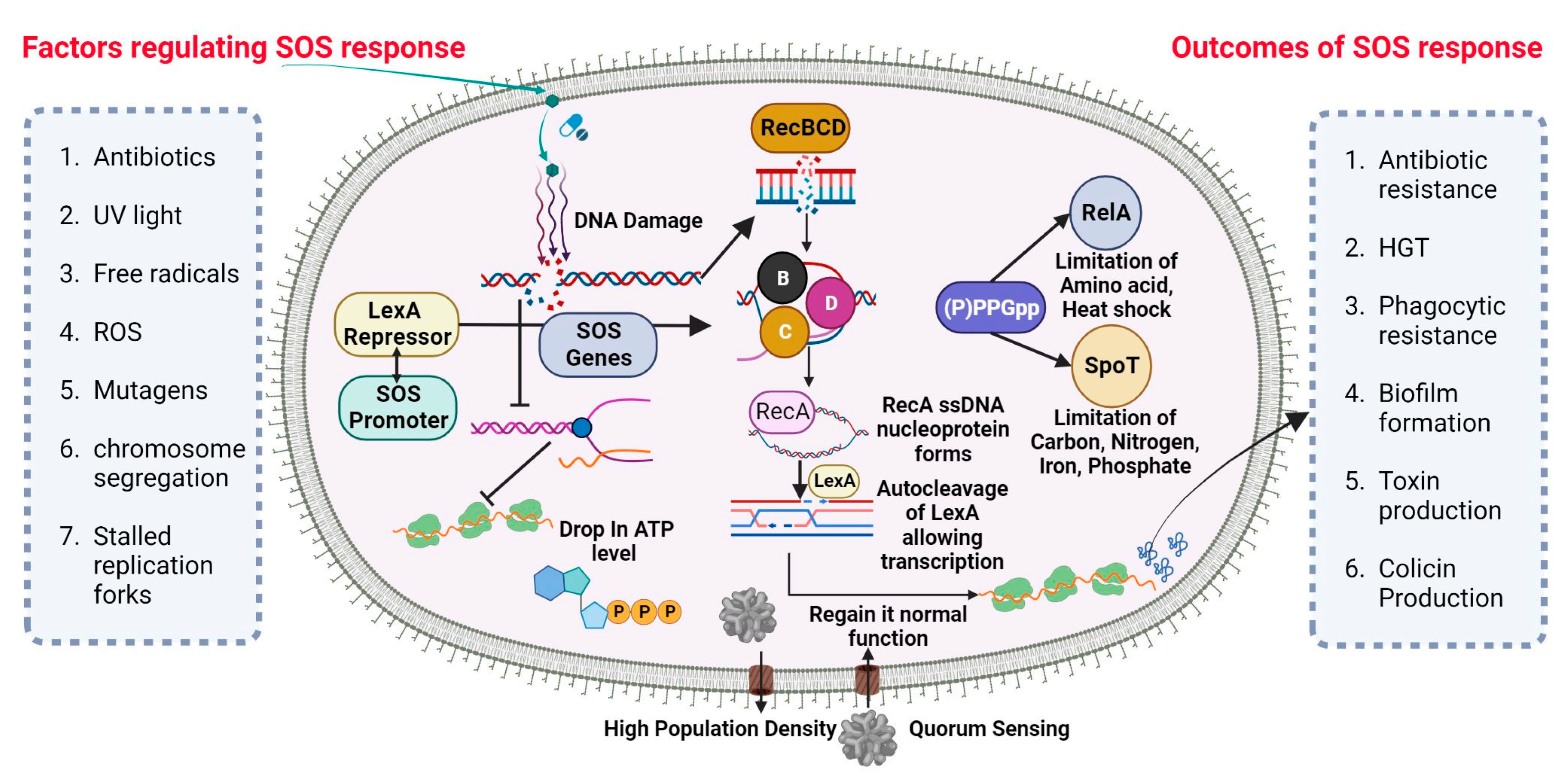
2.4. Antibiotic Resistance by SOS Response

A universally preserved bacterial stress response is primarily triggered by DNA damage. The SOS response initiates and coordinates various biological processes, including DNA repair mechanisms, bacterial cell division arrest, and latent bacteriophage induction. The SOS response is characterized by the activation of DNA polymerases IV and V. This occurs when the DNA damage is prolonged and significant. (Figure 1) depicts the visual representation of the data or information being discussed. Bacterial cultures cultivated in an artificial environment were employed in nearly all studies pertaining to the SOS response and the development of resistance to antibiotics. Prior studies have established a correlation between the mutator phenotype and the ability of bacteria, such as P. aeruginosa, to cause chronic infections in individuals with cystic fibrosis [64,65]. Research examining the genetic alterations in consecutively recovered strains has provided evidence supporting the activation of the SOS response in vivo [66]. Additionally, other indirect evidence has also been reported [67,68].

SOS-Dependent Mutagenesis and Resistance

The cellular SOS response is a captivating bacterial defense mechanism through which bacteria can acquire drug resistance, induce mutagenesis, and undergo genome reorganization [69]. The Lexi promoter-binding repressor protein regulates the SOS system. Upon binding to the RecA filament, the LexA protein undergoes self-cleavage, resulting in a reduction in LexA protein levels within the cell. This process subsequently triggers the activation of over 40 genes in bacterial cells, including the recA gene [70]. The proteins associated with the SOS response play a crucial role in regulating various metabolic processes within bacterial cells [71]. Furthermore, mutagenesis is initiated during the advanced phases of the SOS response. The PolV polymerase has been identified as a key driver of SOS-dependent mutagenesis in Escherichia coli (E. coli) [72]. The polymerases known for their high error rates in DNA synthesis, which are notorious for their low accuracy, encompass PolV polymerase within their category. One of the causes of induced mutagenesis can be attributed to the activity of PolV, which leads to the insertion of an erroneous nucleotide into the DNA molecule. The formation of a RecA by PolV polymerase is expected to impose certain constraints on the potential diversity of recombinases during the process of selection [73]. The process of replicating damaged DNA, known as translesion synthesis (TLS), entails the utilization of PolV polymerase to bypass DNA lesions.
While pol II, pol IV, and polV polymerase are involved in TLS, they also exert an inhibitory effect on RecA-dependent recombination. Achieving equilibrium between these two strategies is of utmost importance. The TLS phenomenon accounts for approximately 1% to 2% of the occurrences in the absence of the SOS response. According to the TLS mechanism, it has been observed that when subjected to stress, TLS has the potential to increase by up to 40% [74]. If recombination is performed by specific RecA variants that are efficient in polymerizing onto single-stranded DNA but somewhat impaired in strand invasion, the ratio may also significantly shift in favor of the TLS mechanism [75]. Simultaneously, bacteria experience significant detrimental effects due to the rise in mutations. As recombination decreases, there is a subsequent decrease in the size of the bacterial population [75]. Moreover, there exists a possibility that moderately unfavorable mutations may undergo fixation when the magnitude of the bacterial population is significantly diminished due to stochastic genetic drift. DNA recombination plays a crucial role in impeding detrimental mutations, as it establishes the boundaries that prevent the occurrence of a “mutational catastrophe” [76].
There exist alternative mechanisms for induced mutagenesis, notwithstanding the fact that PolV polymerase (UmuD2C) is the conventional origin of mutations for the purpose of evolutionary selection. While the RecA protein does not engage in interactions to generate a mutasome, an additional error-prone E. coli Pol IV polymerase is synthesized during the SOS response [77]. Both polymerases are widely distributed among the majority of bacterial species and belong to the Y family [78]. In spite of the substantial diversity observed within the Y family, it is noteworthy that the majority of polymerases share a conserved sequence of 30 residues at their C-terminus. Induced mutagenesis has been observed to occur through the activity of closely related families of polymerases in various bacterial taxa. DnaE2 polymerase, classified as a member of the C family of polymerases, plays a crucial role in the emergence of evolutionary resistance in the bacterium Mycobacterium tuberculosis [79].

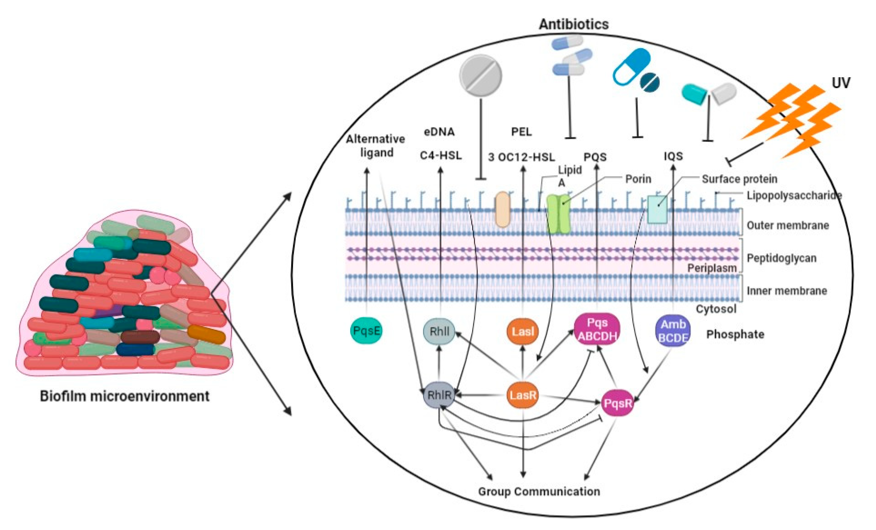
2.5. Biofilm-Mediated Resistome

The sensitivity of pseudomonas cells cultivated in biofilms is comparatively lower to antimicrobial agents and host immune responses when compared to cells grown in free aqueous suspension [80]. When bacteria proliferate within a biofilm, even those lacking protective mutations or innate resistance mechanisms can exhibit a reduced susceptibility to antibiotics [81]. When bacteria experience a loss of biofilm protection, there is a rapid restoration of antibiotic sensitivity. This suggests that the resistance to antibiotics mediated by biofilms is not a result of genetic changes or an adaptive mechanism [82]. The overarching mechanisms underlying biofilm-mediated resistance involve impeding the penetration of antibiotics, creating a modified microenvironment that hinders the growth of biofilm cells, triggering an adaptive stress response, and promoting the differentiation of persister cells. These processes collectively serve to safeguard bacteria from the detrimental effects of antibiotic treatment [81].
P. aeruginosa synthesizes DNA, proteins, and exopolysaccharides, which are utilized for the formation of a biofilm on the surfaces of lung epithelial cells, leading to the development of persistent lung infections [83]. The development of P. aeruginosa biofilms is regulated by multiple factors, primarily including quorum-sensing systems, the GacS/GacA and RetS/LadS two-component regulatory systems, exopolysaccharides, and c-di-GMP [84]. Bacterial communication, also known as quorum sensing, regulates the expression of genes in response to changes in the number of cells present [85]. P. aeruginosa exhibits three prominent quorum-sensing systems, namely LasI-LasR, RhlI-RhlR, and PQS-MvfR, which collectively contribute to the formation of fully developed and specialized biofilms [86]. The GacS/GacA system was found to play a beneficial regulatory role in biofilm development, as evidenced by a tenfold reduction in biofilm generation in a GacA-deficient strain of P. aeruginosa (PA14) compared to the wild-type PA14 strain (Figure 2) [87]. The acidification of the environment and upregulation of genes controlled by the PhoPQ and PmrAB two-component regulatory systems were observed as a result of the presence of P. aeruginosa’s environmental DNA (eDNA). This led to a notable increase in aminoglycoside resistance, indicating a previously unknown function of eDNA [88]. The intracellular molecule known as c-di-GMP serves as a nucleotide second messenger in the process of signal transduction [89]. It plays a role in increasing the levels of c-di-GMP within cells, and these levels are associated with the development of biofilms. In contrast, a diminished level of c-di-GMP has been found to be associated with the presence of planktonic cells [90].
Throughout the process of biofilm formation, the bacterium P. aeruginosa undergoes numerous changes in its physiological and phenotypic characteristics [91]. For example, strains of P. aeruginosa undergo a transformation into a mucoid phenotype during chronic infection in individuals with cystic fibrosis (CF). This transformation is characterized by an enhanced production of alginate, which is stimulated by the specific conditions present in the CF environment. This increased alginate synthesis facilitates the formation of biofilm colonies by the bacteria [92]. The periplasmic cyclic β-(1,3)-glucans, with which tobramycin had physical interaction and were sequestered in the periplasm prior to reaching its target site, were produced through the activity of the glucosyltransferase encoded by the ndvB gene [93]. An operon encompassing the gene PA14 40260-40230 encodes a novel efflux pump. The resistance of P. aeruginosa to gentamicin and ciprofloxacin in biofilm was observed to decrease upon deletion of the specific operon [94]. The regulation of Type VI secretion in P. aeruginosa is governed by the tssC1 gene, which exhibits a high level of expression within biofilm structures [95].

3. MDR/XDR P. aeruginosa Epidemiology

The MDR phenomenon is characterized by the lack of susceptibility to at least one agent in a minimum of three antibiotic classes. XDR is defined as the lack of susceptibility to at least one agent in all but one or two antibiotic classes. Pan-drug resistance (PDR) is the state of being unable to be susceptible to all agents in all antibiotic classes. The subsequent suggested antibiotics for testing antipseudomonal antibiotics are cephalosporins (ceftazidime and cefepime), antipseudomonal penicillin’s combined with β-lactamase inhibitors, monobactams, antipseudomonal carbapenems, aminoglycosides, fluoroquinolones, phosphonic acids, and polymyxins. Although the aforementioned suggestion undeniably holds value in terms of standardizing the descriptions of P. aeruginosa resistance profiles, it is imperative to consider various additional factors. The outcome will exhibit variability based on the utilization of either EUCAST or CLSI breakpoints, even when employing a singular definition. The extensive implementation of the suggested definition is constrained by the absence of clinical breakpoints (as defined by CLSI and EUCAST) for one of the substances (fosfomycin). In a similar vein, it was previously believed that EUCAST breakpoints for aztreonam were not applicable to criteria for MDR, XDR, and PDR bacteria due to acquired resistance. This was due to the inherent resistance of P. aeruginosa to aztreonam. The current classification does not consider newly developed antipseudomonal medications such as ceftazidime–avibactam and ceftolozane–tazobactam [56,96]. Moreover, a significant number of MDR strains meet the criteria for extensively XDR strains, thereby further constraining the spectrum of viable therapeutic options. In a study conducted in 2017, it was found that 26% of P. aeruginosa infections in Spain exhibited MDR. Furthermore, 65% of these MDR isolates (equivalent to 17% of all isolates) met the criteria for XDR classification. The investigation was carried out on a significant scale, encompassing 51 hospitals, utilizing a multicenter approach. The vast majority of isolates exhibited susceptibility to either amikacin or colistin, with a total of 102 isolates falling into this category. Indeed, the prevalence of colistin-only sensitive (COS) profiles is high in numerous hospitals worldwide, and the existence of pan-drug resistance has already been documented [97,98]. Nevertheless, the presence of resistance to novel antipseudomonal drugs did not play a significant role in the majority of research studies. The geographical distribution of acquired beta-lactamases, including ESBLs or carbapenemases, exhibits significant variation, despite the overall low prevalence of resistance to these innovative therapeutic options, typically below 10% [98,99,100,101].

4. Clinical Impact of MDR P. aeruginosa

Selecting the optimal empirical antibiotic for the management of MDR pathogens can pose a challenging task. Individuals afflicted with MDR/XDR infections exhibit a heightened propensity towards receiving inadequate initial antimicrobial therapy [102,103]. In individuals afflicted with bloodstream infections caused by P. aeruginosa, the postponement of commencing appropriate antibiotic treatment has been found to be associated with unfavorable outcomes and increased mortality rates [104,105,106]. In the context of these infections, the presence of MDR/XDR patterns is associated with an increased likelihood of receiving inadequate empirical treatment based on evidence [107,108,109,110]. In a similar vein, antimicrobial medications that are administered as second or third-line treatments are commonly utilized as directed therapy for infections caused by MDR/XDR pathogens. As a result, their performance is inferior to that of medications utilized in the treatment of infections caused by susceptible strains [111]. However, the causal relationship between multidrug resistance and clinical outcomes remains unclear. The poorer prognosis can be attributed to the fact that the colonization and infection of MDR/XDR P. aeruginosa usually manifests in patients with multiple underlying medical conditions [112,113]. The mortality of individuals may be attributed to elevated levels of pre-existing comorbidities. There is a growing body of research focused on investigating the biological impacts of antibiotic resistance on the pathogenicity of P. aeruginosa. In the scientific community, there is a prevailing belief that the emergence of resistance mechanisms is associated with fitness expenses that diminish the pathogenicity of MDR/XDR strains [114]. Nevertheless, specific resistance mutations have also been observed to exhibit no discernible correlation with reductions in fitness [115,116]. Based on scientific research, it has been observed that MDR strains have the ability to develop suppressor or compensatory changes. These changes allow them to restore their original level of fitness, thereby preventing them from experiencing a decline in their virulence over time [115,117]. As previously stated, P. aeruginosa possesses numerous virulence factors [118,119]. The secretion system plays a crucial role in determining the pathogenicity of microorganisms [120,121]. For instance, the type III secretion system (T3SS), also known as the T3SS, is responsible for injecting effector cytotoxins (ExoS, ExoT, ExoU, and ExoT) into host cells [119,121]. The upregulation of ExoU, the most potent among the four identified effector exotoxins, has been correlated with an unfavorable prognosis [122]. The presence of the exoU+ genotype was found to be associated with increased early mortality in a recent clinical trial involving individuals diagnosed with P. aeruginosa bacteremia. It was postulated that this genotype could potentially serve as a predictive biomarker for P. aeruginosa infections [4]. Additional virulence factors of P. aeruginosa have been recently discovered, including the toxin ExlA. This toxin induces rupture of the plasma membrane of host cells, thereby enhancing the pathogenicity of the bacterium [123]. In their study, Pea et al. [4] identified a correlation between certain type III secretion system (TTSS) genotypes and patterns of antibiotic resistance. Specifically, they found that the exoU+ genotype was less prevalent in MDR strains of P. aeruginosa. This finding sheds light on the impact of multidrug resistance on the pathogenicity of P. aeruginosa [4]. Furthermore, multiple studies indicate a correlation between specific resistance profiles and the Type Three Secretion System (TTSS) [122]. The exoU+ genotype is exclusively found in one out of the three most high-risk clones globally, namely ST235 [4]. Nevertheless, numerous experimental and clinical studies indicate a potential decrease in virulence in multidrug resistant/P. aeruginosa [4,110,124]. In vitro investigations have demonstrated that MDR strains exhibit a slower development rate and display deficiencies in virulence determinants, such as bacterial motility or pigment synthesis [125]. Resent study show the role of pigment production in antibiotic resistance, Yellow pigment-producing P. aeruginosa strains posed a significant problem, which may be associated with the development of multidrug resistance [126]. P. aeruginosa MDR/XDR strains have demonstrated reduced capacity compared to susceptible bacteria in terms of causing infection, eliciting an inflammatory response, and resulting in mortality in animal models conducted in vivo [125,127]. The reduced pathogenicity of MDR P. aeruginosa strains is supported by clinical research [4,110,124]. As previously mentioned, it has been observed that at least one of the international XDR high-risk clone strains exhibits sustained high virulence irrespective of its resistance profile. Consequently, it is advisable to exercise caution when interpreting the data. In order to enhance clarity, further investigation is necessary.

5. Antibiotic Agents for of MDR P. aeruginosa

5.1. Polymyxins

Colistin (polymyxin E) and polymyxin B are the two polymyxins that are commonly employed in clinical environments. However, a significant amount of preclinical and clinical data pertaining to these “ancient medications” has recently been revealed [128,129,130,131]. The antibacterial efficacy of polymyxins is contingent upon their chemical composition. Moreover, the hydrophobic regions of polymyxins have the ability to interact with the lipopolysaccharide (LPS) [132]. The interactions mentioned [132,133] lead to the disruption of the bacterial cell membrane.
Nevertheless, the exact mechanism behind bacterial cell death remains to be fully comprehended [134]. Recent scientific investigations on P. aeruginosa have presented findings that question the established belief regarding the mechanism of action of colistin, which was previously thought to eliminate bacteria by causing damage to the cytoplasmic membrane [135,136,137]. Additional hypotheses, such as the proposition that bacterial demise occurs through phospholipid interchange between the outer and cytoplasmic membranes, impeding respiratory enzymes and generating reactive oxygen species, warrant further exploration in subsequent scientific investigations [137].
Colistin is administered in the form of an inactive prodrug known as colistin methane sulfonate (CMS), while Polymyxin B is directly administered as an active antibiotic. However, it should be noted that after injection, Polymyxin B needs to undergo a conversion process to become colistin [138]. The frequency of usage of a specific polymyxin may vary depending on the geographical region. After administering a loading dosage of 9 million international units (IU), several clinical investigations were conducted to evaluate the efficacy of parenteral colistin at higher doses (4.5 IU administered every 12 h) [139,140]. The clinical outcomes of patients who received doses calculated using the Garonzik et al. equation have not been supported by any clinical data [129]. In 2016, Nation et al. [131] revised this equation, and Sorl et al. conducted a study to examine the impact of colistin plasma concentrations on clinical outcomes in 91 patients with infections caused by MDR P. aeruginosa. The aim of their study was to apply pharmacokinetics and pharmacodynamics (PK/PD) information to clinical practice [141].
In summary, the absence of clinical trials assessing the outcomes of individuals adhering to updated guidelines [131,142] leaves a gap in evaluating the efficacy of higher colistin doses, as suggested by PK/PD studies. Colistin serves as a viable alternative for the treatment of urinary tract infections due to the presence of substantial amounts of recently synthesized colistin in urine [142]. Nevertheless, there are a lack of clinical data to substantiate this decision. Colistin has been the focus of numerous published clinical investigations for the management of MDR/XDR P. aeruginosa infections. The majority of studies consist of single-center retrospective series with small patient populations, with the exception of two outliers that each involve more than 100 patients [143,144]. In the context of MDR/XDR P. aeruginosa infections, the utilization of colistin in combination with antibiotic therapy has been demonstrated to be a more favorable treatment modality [145]. Another crucial inquiry regarding the utilization of polymyxins in the management of multidrug-resistant/P. aeruginosa infections is whether the concurrent administration of multiple therapies can improve patient outcomes.
The clinical use of polymyxin has just been resumed. SPR741 is a polymyxin B derivative with less nephrotoxicity. SPR741 does not directly kill bacteria, but it improves the efficiency of antibiotics that are co-administered because they would not otherwise reach their intracellular targets [146]. A total of 64 healthy adult volunteers were included in a phase I randomized control experiment to assess the safety, tolerability, and pharmacokinetics of both single and multiple intravenous doses of SPR741 (NCT03022175) [147].

5.2. Carbapenems

Various in vitro and in vivo investigations have established that pharmacokinetics/pharmacodynamics (PK/PD) is the most reliable method for determining the proportion of the dosing interval during which the concentration of unbound or free drug in the bloodstream surpasses the minimum inhibitory concentration (MIC) required to combat the pathogen. A pharmacodynamics study employed Monte Carlo simulation techniques to assess various dosage regimens of meropenem, administered through intermittent or extended infusions, in their effectiveness against Enterobacteriaceae and P. aeruginosa strains with varying susceptibilities. A total of 276 isolates of P. aeruginosa were included in the study, with 22.1% of them exhibiting minimum inhibitory concentration (MIC) values greater than 4 mg/L. To achieve a 50% probability of maintaining a free drug concentration above the minimum inhibitory concentration (MIC) required to effectively combat all susceptible P. aeruginosa isolates (with MIC values of 4 mg/L or lower), a dosage of 1 g of meropenem every 8 h in extended infusion or 2 g every 8 h in intermittent/extended infusion is necessary. Nevertheless, in the case of organisms that are classified as intermediate-resistant to meropenem (with a minimum inhibitory concentration of 8 mg/L), it was observed that only the higher-dose regimen of 2 g administered every 8 h through extended infusion was able to provide sufficient bactericidal exposure. The researchers proposed that the administration of meropenem at the highest dose of 2 g/8 h through extended infusion could be an effective treatment for P. aeruginosa strains that are classified as intermediate or resistant [148]. A study was conducted using an in vitro infection model and Monte Carlo simulation to assess the most effective dosage of imipenem in combination with tobramycin against clinical isolates of P. aeruginosa that are resistant to carbapenems and aminoglycosides. The optimal antibacterial activity of imipenem was observed at simulated doses of 4 or 5 g/day administered via continuous infusion, in combination with tobramycin [149]. The adequacy of this dosage regimen in a neutropenic mouse thigh model of XDR P. aeruginosa infection was confirmed by the same group [149]. In a single study, a total of 237 cases of bloodstream infections caused by P. aeruginosa with reduced susceptibility to carbapenem were analyzed. The study aimed to evaluate the effectiveness of different dosing regimens of carbapenem antibiotics, specifically imipenem, meropenem, and doripenem. The dosing regimens tested included imipenem administered at a dose range of 0.5 to 1 g every 6 h using both 0.5- and 3 h infusion durations, meropenem administered at a dose range of 1 to 2 g every 8 h using both 0.5- and 3 h infusion durations, and doripenem administered at a dose range of 0.5 to 2 g every 8 h using both 1- and 4 h infusion durations. A T > MIC value of 40% was determined to be the optimal pharmacokinetic/pharmacodynamic ratio. The findings indicated that the administration of meropenem at a dosage of 2 g every 8 h over a 3 h infusion period, and doripenem at a dosage of 1 g every 8 h over a 4 h infusion period, exhibited the highest effectiveness in combating P. aeruginosa strains that displayed decreased susceptibility to carbapenem antibiotics [150]. In a case report involving a critically ill patient who underwent a double-lung transplant and developed pneumonia caused by multidrug resistant P. aeruginosa with a meropenem minimum inhibitory concentration (MIC) of 32 mg/L, the patient was treated with a continuous infusion of meropenem at a dosage of 8 g per 24 h. Remarkably, this treatment approach resulted in the successful resolution of the patient’s clinical condition [151]. The occurrence of carbapenem-resistant P. aeruginosa (CRPA) primarily arises due to the presence of carbapenem-hydrolyzing enzymes. Metallo-beta-lactamases (MβLs), such as Verona integron-encoded metallo-beta-lactamases (VIMs), IMPs, and New Delhi metallo-beta-lactamases (NDMs), are widely recognized as crucial enzymes responsible for antibiotic resistance in clinical pathogens of P. aeruginosa.

5.3. Antipseudomonal β-Lactams

Limited data are currently accessible regarding the efficacy of various conventional antipseudomonal β-lactams, including cefepime, ceftazidime, piperacillin–tazobactam, and aztreonam, as standalone treatments for MDR/XDR P. aeruginosa infections. Aztreonam therapy could potentially serve as a viable treatment for Ambler class B metallo-beta-lactamase (MBL)-producing Gram-negative bacteria (GNB), including P. aeruginosa. A study was conducted to assess the clinical efficacy of a particular series in treating infections caused by P. aeruginosa that produce metallo-beta-lactamase (MBL). The observed mortality rate in the conducted trial was determined to be 30%. The sample size utilized in the study was insufficient to establish definitive conclusions, with the majority of cases [152] involving the administration of combination therapy. Out of the nine patients infected with MBL-producing Pseudomonas who were treated with intravenous colistin in combination with either aztreonam or piperacillin–tazobactam, a total of seven patients (77.8%) experienced favorable outcomes and survived [153]. According to a clinical case study [154], a patient with a compromised immune system and a wound infection caused by multidrug resistant P. aeruginosa was effectively treated using a continuous infusion of high-dose aztreonam at a rate of 8.4 g per day. A critically immunocompromised patient with multidrug resistant P. aeruginosa bacteremia and a ceftazidime minimum inhibitory concentration (MIC) of 64 mg/L [155] was effectively administered a continuous infusion of high-dose ceftazidime ranging from 6.5 to 9.6 g per day. As previously mentioned, certain in vitro combination assays, specifically those involving cefepime–tobramycin [156] and cefepime–aztreonam [157], have demonstrated additive or synergistic effects against P. aeruginosa. Based on the specific strain, resistance phenotype, and genotype, a potential approach in individual cases may involve the utilization of high doses of certain drugs delivered through prolonged infusion as part of a combination therapeutic regimen.
Relebactam, a newly developed beta-lactamase inhibitor, specifically targets various beta-lactamase enzymes in multidrug-resistant bacteria [158]. Imipenem with relebactam against P. aeruginosa was introduced. As a result, relebactam’s effectiveness against P. aeruginosa has been the subject of numerous clinical trials (NCT02493764, NCT02452047, NCT05561764, NCT03583333, and NCT05204563. In a subsequent phase III clinical trial (NCT02493764 NCT03583333), patients with ventilator-associated or hospital-acquired bacterial pneumonia were randomized to receive either piperacillin/tazobactam intravenously every six hours for seven to fourteen days, or imipenem/cilastatin/relebactam. Treatment included either the fixed-dose combination of imipenem/relebactam/cilastatin, or the fixed-dose combination of piperacillin/tazobactam. The 264 patients who received imipenem/cilastatin/relebactam and the 267 patients who received piperacillin/tazobactam both showed similar improvements in mortality, morbidity, and clinical symptoms. Imipenem/cilastatin/relebactam can effectively treat patients with Gram-negative bacterial pathogen infections, including P. aeruginosa, even in critically ill, high-risk patients, according to the study’s overall findings [159]. Additionally, nacubactam, a different new beta-lactamase antibiotic, has demonstrated encouraging outcomes as a strong antibiotic against P. aeruginosa infections (NCT02134834, NCT02972255).

5.4. Aminoglycosides

Certain aminoglycosides [158,159] are administered in conjunction with other antimicrobial agents to address highly resistant infections caused by multidrug-resistant/P. aeruginosa. Multiple pharmacodynamics, in vitro and in vivo investigations have confirmed that aminoglycosides exhibit antibacterial activity that is dependent on the concentration, and a peak concentration of ≥8 to 10 is the most reliable predictor of efficacy in terms of pharmacokinetics/pharmacodynamics (PK/PD) [157]. Regarding clinical isolates of P. aeruginosa that exhibited resistance to carbapenem and aminoglycosides, a previously mentioned pharmacokinetic (PK) model evaluated the optimal dosage of tobramycin and imipenem [156]. The administration of aminoglycosides at significantly elevated dosages, in conjunction with continuous renal clearance techniques, was employed as a therapeutic approach to address infections caused by XDR P. aeruginosa, with the aim of mitigating the risk of renal toxicity. The results exhibited noteworthy rates of survival, despite the limited sample size of [160,161] individuals. In instances of severe or profound infections induced by MDR/XDR P. aeruginosa, alternative approaches for delivering aminoglycosides may be utilized. This is particularly applicable to conditions like pneumonia or meningitis. The administration of amikacin through inhalation for the treatment of pneumonia enables the attainment of elevated drug concentrations specifically at the infection site, such as the epithelial lining fluid (ELF). This approach also prevents excessive systemic exposure to the drug, which could potentially lead to systemic toxicity. Nevertheless, the utilization of inhaled antibiotics (specifically polymyxins or aminoglycosides) is recommended solely as an adjunctive therapy for infections caused by Gram-negative bacilli that exhibit susceptibility exclusively to aminoglycosides or polymyxins when used in conjunction with other drugs administered systemically [162]. Meningitis is a challenging infection to manage. The effectiveness of intravenous aminoglycosides is constrained by their limited ability to penetrate the central nervous system, resulting in insufficient and suboptimal concentrations at the infection site. In instances of this nature, the administration of intraventricular aminoglycosides may be necessary. A case of post-surgical meningitis caused by PDR P. aeruginosa was effectively managed through the administration of intravenous cefepime via continuous infusion, in combination with intravenous and intraventricular amikacin [163]. Despite the fact that the strain exhibited a minimum inhibitory concentration (MIC) of 32 mg/L for amikacin, the attainment of concentrations as high as 200 mg/L in the central nervous system proved to be effective in resolving the infection. Enzymatic modification of aminoglycosides through aminoglycoside-acetyltransferases (AAC), aminoglycoside-adenyltransferases (AAD), and aminoglycoside-phosphotransferases (APH) represents the prevailing resistance mechanism in P. aeruginosa for the aminoglycoside class of antibiotics. Furthermore, these enzymes can also be encoded on mobile genetic elements, thereby facilitating their widespread dissemination. A study was conducted to analyze 137 P. aeruginosa isolates collected from the University Hospital in Cumana, Venezuela. The most commonly observed genes in these isolates were aphA1, aadB, and aac (6′)-Ib. Among these genes, the last enzyme, aac (6′)-Ib, was found to catalyze N-acetylation at the 6′ position, which is a common mechanism employed by P. aeruginosa to modify aminoglycosides. The mean resistance observed for gentamicin was 32.6%, amikacin exhibited a resistance of 24.6%, and tobramycin demonstrated a resistance of 29.9% [164]. Previous research conducted in Latin American nations has revealed a prevalence of resistant strains ranging from 27.8% to 42.0% for gentamicin and 16.3% to 28.9% for amikacin [165].

5.5. Fosfomycin

The reemergence of intravenous fosfomycin as a treatment for infections caused by MDR bacteria is attributed to its remarkable bactericidal activity against various species, including MDR P. aeruginosa [166,167], as demonstrated in laboratory studies. Several additional experiments have evaluated the utilization of fosfomycin in conjunction with β-lactams, aminoglycosides, or colistin. In addition, this purportedly “ancient” antibiotic has been linked to more contemporary antibiotics such as ceftazidime–avibactam and ceftolozane–tazobactam [155,168]. The patient with XDR P. aeruginosa meningitis [169] was effectively treated by administering a combination of Ceftolozane–tazobactam and fosfomycin at a dosage of 4 g every 6 h.
Several studies have been conducted to evaluate different dosage regimens of fosfomycin in combination with carbapenem for the treatment of non-MDR and MDR P. aeruginosa clinical isolates, based on the attainment of the pharmacokinetic/pharmacodynamic (PK/PD) target. One study utilized a Monte Carlo simulation to assess the probability of achieving the desired outcome for different doses and durations of carbapenem and fosfomycin administration [170]. In the case of non-MDR P. aeruginosa isolates, the administration of a carbapenem and fosfomycin through continuous infusion at a rate ranging from 16 to 24 g per day resulted in the most favorable pharmacokinetic/pharmacodynamic ratios. The clinical isolates examined in this study, which took place in Thailand, exhibited remarkably elevated fosfomycin minimum inhibitory concentration (MIC) values. Therefore, it is not possible to extrapolate these results to other settings [170]. Further clinical series and trials are necessary to determine the future role of fosfomycin in these infections, encompassing optimal dosage and potential combinations. The effectiveness of fosfomycin IV against recurrent P. aeruginosa infections was studied in a phase I clinical study. Fosfomycin IV interferes with the formation of cell walls by inhibiting peptidoglycan assembly. A total of 30 healthy participants between the ages of 18 and 45 were recruited for the trial, and they were randomized to one of three treatment sequences, each lasting between 18 and 26 days (NCT02178254).

5.6. Newly Discovered Antimicrobial Agents

When present, murepavadin prevents Gram-negative bacteria from transporting LPS, as shown in a clinical trial (NCT02096315), and represents new class of antibiotics known as outer membrane protein targeted antibiotics (ompTAs) [171]. Murepavadin attaches to the outer membrane protein lipopolysaccharide transport protein D (LptD), which is important in the production of lipopolysaccharides in Gram-negative bacteria. By inhibiting LptD’s ability to transport LPS, it alters lipopolysaccharides and ultimately leads to cell death [171]. Murepavadin’s potent bactericidal properties were shown in vitro with 1219 P. aeruginosa isolates, many of which were multidrug resistant, collected from 112 hospitals in the US, China, and Europe. These tests determined that murepavadin’s MIC50 against numerous isolates of P. aeruginosa was 0.12 mg/L [172].

6. Therapeutic Strategies for P. aeruginosa Treatment

The process of developing novel antibiotics is highly time-consuming and constrained, thereby impeding the progress of therapeutic strategies for challenging P. aeruginosa infections. These therapeutic approaches can function independently or in conjunction to counteract infections. They encompass the suppression of quorum sensing and bacterial lectins, phage therapy, vaccine strategies, and antimicrobial photodynamic therapy [171,173]. Table 2 displays the treatment options for varying degrees of drug resistance in P. aeruginosa.
Despite the perplexing resistance mechanisms exhibited by P. aeruginosa, this pathogen remains a significant threat in clinical settings. However, there is optimism that through the utilization of emerging knowledge and its practical implementation, we can effectively combat this pathogen. Several emerging therapeutic approaches currently being investigated include combinatory therapy, the utilization of antimicrobial peptides (AMPs), bacteriophage therapy, and nanoparticles. Table 3 displays the therapeutic approaches for multidrug resistant MDR and extensively drug-resistant XDR P. aeruginosa infections, along with various mechanisms of action utilized.

6.1. Combinatory Therapy

The utilization of various drug combinations offers a promising approach to address the issue of antimicrobial resistance. This strategy involves employing a multi-targeted attack, taking advantage of their synergistic effects, and reducing the required dosage and associated side effects. In the case of P. aeruginosa, which exhibits multiple mechanisms of antibiotic resistance, combinatory therapy has the potential to be more effective, as indicated in Table 3 and Figure 3.
Ceftolozane–tazobactam (5th generation cephalosporin combined with a β-lactamase inhibitor)
The efficacy of this combination has been demonstrated in effectively combating P. aeruginosa, including MDR/XDR strains, by inhibiting the activity of penicillin-binding proteins (PBPs) [180]. The unique three-dimensional structure of the compound makes it more resistant to hydrolysis by AmpC β-lactamase in comparison to commonly employed cephalosporins, resulting in increased stability. Despite the emergence of resistance mechanisms involving class D β-lactamases and oxacillinases (OXA), as well as reduced membrane permeability due to OprD porin mutation and increased expression of efflux pumps [101,181], Ceftolozane–tazobactam remains a viable treatment option for multidrug-resistant/P. aeruginosa infections. Additionally, it can be considered as the preferred initial drug for carbapenem-resistant P. aeruginosa (CRPA) infections [7]. The combination of treatments has demonstrated positive results in severe infections occurring in intensive care units [182], as well as infections caused by P. aeruginosa in individuals with hematologic malignancies [183]. It has also shown effectiveness in treating complicated urinary tract infections and intra-abdominal infections [184], particularly when there is a high prevalence of P. aeruginosa in the microbiological intent-to-treat population. For uncomplicated urinary tract infections (UTIs) caused by drug-resistant P. aeruginosa DTRPA strain, it is recommended to administer a high and immediate dose therapy of this combination [185]. Severe infections induced by CRPA have demonstrated significant advantages when treated with CT (combinatory therapy) in comparison to the utilization of polymyxin or other combinations based on aminoglycosides. The European Society of Clinical Microbiology and Infectious Diseases (ESCMID) guidelines recommend the use of CT if the strain exhibits susceptibility in vitro. Caution should be exercised when determining the ideal dosage, particularly in cases of renal impairment [186] and in situations involving high-inoculum infections where the development of resistance may occur [13].
Ceftazidime–avibactam
Avibactam exhibits potent activity against resistance mechanisms, including extended-spectrum beta-lactamases (ESBLs), AmpC cephalosporinases, and OXA enzymes, thereby enhancing the efficacy of this combination. This combination has undergone testing in complex urinary tract infections (UTIs) and hospital-acquired pneumonia (HAP), demonstrating a notable degree of safety and effectiveness in the treatment of infections caused by 4 to 35% of isolates classified as MDR/XDR, as well as nearly all drug-resistant P. aeruginosa DTRPA strains [187]. Following the administration of colistin, it was observed that this particular combination exhibited the highest efficacy against subsets of pathogens that had developed resistance in patients hospitalized with pneumonia in Western Europe during the year 2020 [8]. The resistance to this combination may be ascribed to alterations in porin or efflux pump activity, or it could be a result of acquired cross-resistance against the ceftolozane–tazobactam combination. While the efficacy of this drug combination appears to be promising, it is advisable to exercise caution when administering it in cases of severe infections.
Imipenem–Cilastin–Relebactam (carbapenem + dehydropeptidase inhibitor + non-β lactam bicyclic diazabicyclooctane β-lactamase inhibitor)
This combination exhibits efficacy in strains that possess resistance against porins, as its mechanism of action is not reliant on the frequently implicated multidrug efflux pumps (MexA-MexB-OprM) [188]. Based on the findings of the SMART European surveillance, it was observed that there was imipenem susceptibility detected in isolates that were initially deemed non-susceptible [189]. The strain of P. aeruginosa, which was obtained from respiratory tract infections in intensive care unit settings, intra-abdominal injuries, and urinary tract infections, exhibited a significantly higher susceptibility to the combination treatment compared to imipenem alone. Despite exhibiting lower activity compared to ceftolozane–tazobactam, the imipenem–cilastin–relebactam combination may prove beneficial in the treatment of infections that have shown resistance to the ceftolozane–tazobactam combination [190]. Given that the majority of data regarding the effectiveness of this drug combination are derived from in vitro studies, its definitive role in clinical practice remains to be fully established.
Meropenem–Vaborbactam (Carbapenem + cyclic boronic acid β-lactamase inhibitor)
The distinguishing characteristic of this combination resides in its strong attraction to serine residues. Consequently, it forms covalent bonds with the β-lactamase enzyme, thereby competitively inhibiting its hydrolytic activity. The combination has been observed to enhance the sensitivity to meropenem in strains that exhibit lower sensitivity to meropenem alone. This finding highlights the potential significance of the combination in treating P. aeruginosa infections [191]. In certain instances, characterized by resistance mediated by KPC (Klebsiella pneumonia carbapenemase), this particular combination has demonstrated its efficacy against MDR/XDR strains [192]. However, its activity against MBL or OXA is reduced, and the combination of meropenem and vaborbactam does not exhibit a wider range of effectiveness compared to meropenem alone. This is because the resistance mechanisms of MBL or OXA are not overcome by the addition of vaborbactam.
Cefepime–Taniborbactam (4th generation cephalosporin + Boronic acid β-lactamase inhibitor)
P. aeruginosa has undergone evolutionary changes that have led to the acquisition of a notable resistance to cefepime through the upregulation of efflux pumps and increased synthesis of chromosomal AmpC enzymes. To enhance the effectiveness of cefepime, researchers have introduced a new compound called taniborbactam. This combination is currently being tested in phase 1 and phase 2 trials to determine if it is at least as effective as existing treatments. The trials involve 211 participants. Interestingly, taniborbactam is the first beta-lactamase inhibitor (BLI) that can competitively inhibit all metallo-beta-lactamases (MBLs), except for the IMP-type. It achieves this by directly inhibiting Ambler class A, B, C, and D enzymes. Therefore, it can be regarded as a potential combination in the production of CRPA and MBL isolates [193].
Cefepime–Zidebactam (4th generation cephalosporin + β-lactam enhancer antibiotic)
The observed activity of this combination is achieved by inhibiting the activity of penicillin-binding protein 3 (PBP3) through cefepime, and penicillin-binding protein 2 (PBP2) through zidebactam. The compound has demonstrated significant efficacy against P. aeruginosa isolates, including those that exhibit high levels of efflux pump production. This phenomenon can be attributed to the unaffected activity of the organism in question, regardless of the expression of ESBLs (Extended-Spectrum Beta-Lactamases), OXA-48-like carbapenemases, and MBL (Metallo-Beta-Lactamase) carbapenemases. While in vitro studies have shown resistance against this combination, it remains a promising choice for isolates where local resistance mechanisms are of greater concern [194].
Meropenem–Nacubactam (Carbapenem + diazabicyclooctane (DBO) type β- lactamase inhibitor)
While this combination exhibits the inhibition of penicillin-binding protein 2 (PBP2) and demonstrates activity against AmpC hyperproducing and KPC-producing P. aeruginosa, its efficacy is more pronounced against Enterobacterales [195]. The following Table 4 presents the effectiveness of different drug combinations, considering their performance in in vitro time-killing (TK) or pharmacokinetic/pharmacodynamic (PK/PD) studies. The combinatorial ratios, as determined by the minimum inhibitory concentration (MIC) of individual strains, were assessed using in vitro models. The level of synergy was classified as low, moderate, or high. It is crucial to take into account the diminished impact of both combination therapies and monotherapies as a result of the continuous release of planktonic cells from biofilms in in vivo situations. Consequently, the effectiveness of any drug combination therapy is significantly influenced by the maturity of the biofilm, necessitating clinical trials to optimize therapeutic outcomes.

6.2. Antimicrobial Peptides

Antimicrobial peptides (AMPs) are bioactive substances that are remarkably biocompatible and less likely to cause bacterial resistance to evolve [211,212]. AMPs encompass a wide range of naturally derived or artificially synthesized small peptides that exert their effects on relatively non-specific targets, primarily within cells. These peptides exhibit a reduced propensity to induce the development of resistance [213]. This emerging therapeutic modality exhibits numerous advantages, including a high bactericidal potency at micromolar concentrations [214], a mechanism of action that targets multiple sites, and a rapid onset of effect [215]. Propelled by the emergence of antimicrobial peptides (AMPs), a novel category of peptides exhibiting remarkable target specificity and heightened responsiveness, known as “selectively targeted AMPs” (STAMPs), have been devised and are currently garnering escalating significance [216]. Antimicrobial peptides (AMPs), including human cathelicidin peptide (LL-37), colistin (derived from Bacillus polymyxa var colistinus), and colistin-derived AMPs (AA139 and SET-M33), have been suggested as potential therapeutic agents for combating drug-resistant P. aeruginosa infections. These substances exhibit bactericidal properties without promoting the development of resistance, and they also demonstrate activity against biofilms [217].

6.3. Phage Therapy

Bacteriophage therapy is a recently developed and highly innovative therapeutic approach aimed at addressing the issue of antibiotic resistance. Among the two classifications of bacteriophages, namely lytic and temperate, it is only the lytic phage that can be utilized in clinical scenarios. The substance exhibits specific binding affinity towards the external membrane of the bacterial cells, facilitating the transfer of its genetic material into the recipient cell. The phage replicates within the host cell by utilizing host proteins, and the resulting offspring phages migrate and exhibit bactericidal activity towards specific targets, such as flagella and pili organs of the bacteria. These substances can be administered via systemic injection or applied topically to wounds to exert their bactericidal properties. Various combinations of phages, referred to as phage cocktails, are currently undergoing testing. These experiments have shown enhanced effectiveness in treating P. aeruginosa strains that are resistant to conventional treatments (as indicated in Table 5). Experiments are being conducted to evaluate the efficacy of bacteriophages that target the virulent factors of P. aeruginosa strains. P. aeruginosa releases a variety of extracellular toxins that have been documented to induce significant harm by disrupting blood clotting and causing tissue death. Bacteriophages capable of modifying these exotoxins and inducing their inactivation are currently under development [218,219,220]. Bacteriophages have the potential to eliminate P. aeruginosa biofilms through mechanisms such as extracellular matrix degradation, enhanced permeability of the inner biofilm layer, and suppression of quorum-sensing activity, as documented in a study [221]. Pili and fimbriae serve as the primary factors for bacterial adherence. Bacteriophages are being developed to inhibit the expression of these factors [222]. Mono-phage, phages-cocktail, phage-derived enzyme (lysin), bio-engineered phage, and phage in combination with antibiotics are all forms of phage therapy [223]. A thorough search of the literature revealed several case study reports and compassionate uses for severely ill patients at specialized facilities, but despite encouraging outcomes, recent clinical trials based on evidence will be required. Due to PP1131’s lack of efficacy vs. SOC, the first randomized controlled trial using a combination of natural lytic P. aeruginosa phages for the topical treatment of infected burn wound patients was discontinued in January 2017 [224]. Phage therapy poses significant challenges due to safety considerations, including the presence of phage-neutralizing antibodies and immune responses, as well as the potential for resistance development via the CRISPR-Cas system.

7. Conclusions

MDR/XDR Infections caused by P. aeruginosa pose a significant challenge within healthcare environments. The antibiotic resistance observed in P. aeruginosa is attributed to mechanisms that are either innate, acquired, or adaptive in nature. P. aeruginosa is a remarkably adaptable pathogenic microorganism possessing an intricate regulatory network, which is among the most intricate in the bacterial kingdom. The presence of interference can potentially lead to secondary effects on the cellular physiology and result in complications for infected individuals. A more comprehensive understanding of the host bacteria as a cohesive system in its response to treatment is imperative in order to formulate innovative therapeutic approaches to combat this exceptionally formidable human pathogen. Multiple antibiotic resistance mechanisms play a role in the emergence of strains, rendering traditional antibiotics ineffective in the treatment of MDR/XDR P. aeruginosa infections. Moreover, the process of biofilm formation in P. aeruginosa persister cells is accountable for the persistence and resistance of infections in individuals with cystic fibrosis.
Nevertheless, clinical P. aeruginosa exhibits a remarkable ability to evolve novel resistance mechanisms against both established and emerging antibiotics. This poses significant challenges and potential threats to public health. The non-antibiotic therapeutic strategies, specifically phage therapy and Antimicrobial peptides, have demonstrated noteworthy antimicrobial properties against antibiotic-resistant strains of P. aeruginosa in laboratory settings or animal experiments. Future investigations should prioritize the exploration of innovative methodologies aimed at mitigating adverse impacts and enhancing the levels of safety and effectiveness observed in clinical trials. P. aeruginosa utilizes a diverse antibiotic resistance strategy. The optimal approach for future treatments is expected to involve combinational therapies targeting this highly virulent pathogen. This strategy entails the integration of novel treatments with conventional antibiotics to achieve the successful eradication of the pathogen in immunocompromised patients who are particularly susceptible to its detrimental effects.

Author Contributions

Conceptualization, A.K.; writing, A.K., R.K., B.J.O., N.J., G.M. and S.K.; editing, B.J.O., A.P., V.M., M.U. and R.B.K. All authors have read and agreed to the published version of the manuscript.

Funding

This work was supported by SERB-DST core research grant No. 2018/001317. A.K. was supported by ICMR SRF/RA fellowship grant No. 2021-13568.

Institutional Review Board Statement

Not applicable.

Informed Consent Statement

Not applicable.

Data Availability Statement

Data sharing is not applicable.

Conflicts of Interest

The authors declare no conflict of interest.

References

  1. Breidenstein, E.B.; de la Fuente-Núñez, C.; Hancock, R.E. Pseudomonas aeruginosa: All roads lead to resistance. Trends Microbiol. 2011, 19, 419–426. [Google Scholar] [CrossRef]
  2. Oliver, A.; Mulet, X.; López-Causapé, C.; Juan, C. The increasing threat of Pseudomonas aeruginosa high-risk clones. Drug Resist. Updat. 2015, 21–22, 41–59. [Google Scholar] [CrossRef]
  3. Tacconelli, E.; Carrara, E.; Savoldi, A.; Harbarth, S.; Mendelson, M.; Monnet, D.L.; Pulcini, C.; Kahlmeter, G.; Kluytmans, J.; Carmeli, Y.; et al. Discovery, research, and development of new antibiotics: The WHO priority list of antibiotic-resistant bacteria and tuberculosis. Lancet Infect. Dis. 2018, 18, 318–327. [Google Scholar] [CrossRef]
  4. Peña, C.; Cabot, G.; Gómez-Zorrilla, S.; Zamorano, L.; Ocampo-Sosa, A.; Murillas, J.; Almirante, B.; Pomar, V.; Aguilar, M.; Granados, A.; et al. Influence of Virulence Genotype and Resistance Profile in the Mortality of Pseudomonas aeruginosa Bloodstream Infections. Clin. Infect. Dis. 2015, 60, 539–548. [Google Scholar] [CrossRef]
  5. Walkty, A.; Lagace-Wiens, P.; Adam, H.; Baxter, M.; Karlowsky, J.; Mulvey, M.R.; McCracken, M.; Zhanel, G.G. Antimicrobial susceptibility of 2906 Pseudomonas aeruginosa clinical isolates obtained from patients in Canadian hospitals over a period of 8 years: Results of the Canadian Ward surveillance study (CANWARD), 2008–2015. Diagn. Microbiol. Infect. Dis. 2017, 87, 60–63. [Google Scholar] [CrossRef]
  6. Solomon, S.L.; Oliver, K.B. Antibiotic resistance threats in the United States: Stepping back from the brink. Am. Fam. Physician 2014, 89, 938–941. [Google Scholar]
  7. Pogue, J.M.; Kaye, K.S.; Veve, M.P.; Patel, T.S.; Gerlach, A.T.; Davis, S.L.; Puzniak, L.A.; File, T.M.; Olson, S.; Dhar, S.; et al. Ceftolozane/Tazobactam vs Polymyxin or Aminoglycoside-based Regimens for the Treatment of Drug-resistant Pseudomonas aeruginosa. Clin. Infect. Dis. 2020, 71, 304–310. [Google Scholar] [CrossRef]
  8. Sader, H.S.; Carvalhaes, C.G.; Shortridge, D.; Castanheira, M. Comparative activity of newer β-lactam/β-lactamase inhibitor combinations against Pseudomonas aeruginosa from patients hospitalized with pneumonia in European medical centers in 2020. Eur. J. Clin. Microbiol. Infect. Dis. 2022, 41, 319–324. [Google Scholar] [CrossRef]
  9. Lister, P.D.; Wolter, D.J.; Hanson, N.D. Antibacterial-Resistant Pseudomonas aeruginosa: Clinical Impact and Complex Regulation of Chromosomally Encoded Resistance Mechanisms. Clin. Microbiol. Rev. 2009, 22, 582–610. [Google Scholar] [CrossRef]
  10. Skiada, A.; Markogiannakis, A.; Plachouras, D.; Daikos, G.L. Adaptive resistance to cationic compounds in Pseudomonas aeruginosa. Int. J. Antimicrob. Agents 2011, 37, 187–193. [Google Scholar] [CrossRef]
  11. Muller, C.; Plésiat, P.; Jeannot, K. A two-component regulatory system interconnects resistance to polymyxins, aminoglycosides, fluoroquinolones, and β-lactams in Pseudomonas aeruginosa. Antimicrob. Agents Chemother. 2011, 55, 1211–1221. [Google Scholar] [CrossRef] [PubMed]
  12. Juan, C.; Torrens, G.; González-Nicolau, M.; Oliver, A. Diversity and regulation of intrinsic β-lactamases from non-fermenting and other Gram-negative opportunistic pathogens. FEMS Microbiol. Rev. 2017, 41, 781–815. [Google Scholar] [CrossRef]
  13. Horcajada, J.P.; Montero, M.; Oliver, A.; Sorlí, L.; Luque, S.; Gómez-Zorrilla, S.; Benito, N.; Grau, S. Epidemiology and Treatment of Multidrug-Resistant and Extensively Drug-Resistant Pseudomonas aeruginosa Infections. Clin. Microbiol. Rev. 2019, 32, e00031-19. [Google Scholar] [CrossRef] [PubMed]
  14. Girlich, D.; Naas, T.; Nordmann, P. Biochemical Characterization of the Naturally Occurring Oxacillinase OXA-50 of Pseudomonas aeruginosa. Antimicrob. Agents Chemother. 2004, 48, 2043–2048. [Google Scholar] [CrossRef]
  15. Fajardo, A.; Hernando-Amado, S.; Oliver, A.; Ball, G.; Filloux, A.; Martinez, J.L. Characterization of a novel Zn2+-dependent intrinsic imipenemase from Pseudomonas aeruginosa. J. Antimicrob. Chemother. 2014, 69, 2972–2978. [Google Scholar] [CrossRef]
  16. Li, X.-Z.; Plésiat, P.; Nikaido, H. The Challenge of Efflux-Mediated Antibiotic Resistance in Gram-Negative Bacteria. Clin. Microbiol. Rev. 2015, 28, 337–418. [Google Scholar] [CrossRef]
  17. Cabot, G.; Ocampo-Sosa, A.A.; Tubau, F.; Macia, M.D.; Rodríguez, C.; Moya, B.; Zamorano, L.; Suárez, C.; Peña, C.; Martínez-Martínez, L.; et al. Overexpression of AmpC and Efflux Pumps in Pseudomonas aeruginosa Isolates from Bloodstream Infections: Prevalence and Impact on Resistance in a Spanish Multicenter Study. Antimicrob. Agents Chemother. 2011, 55, 1906–1911. [Google Scholar] [CrossRef]
  18. Moya, B.; Dötsch, A.; Juan, C.; Blázquez, J.; Zamorano, L.; Haussler, S.; Oliver, A. Beta-lactam resistance response triggered by inactivation of a nonessential penicillin-binding protein. PLoS Pathog. 2009, 5, e1000353. [Google Scholar] [CrossRef]
  19. Cabot, G.; Bruchmann, S.; Mulet, X.; Zamorano, L.; Moyà, B.; Juan, C.; Haussler, S.; Oliver, A. Pseudomonas aeruginosa Ceftolozane-Tazobactam Resistance Development Requires Multiple Mutations Leading to Overexpression and Structural Modification of AmpC. Antimicrob. Agents Chemother. 2014, 58, 3091–3099. [Google Scholar] [CrossRef]
  20. Fraile-Ribot, P.A.; Cabot, G.; Mulet, X.; Periañez, L.; Martín-Pena, M.L.; Juan, C.; Pérez, J.L.; Oliver, A. Mechanisms leading to in vivo ceftolozane/tazobactam resistance development during the treatment of infections caused by MDR Pseudomonas aeruginosa. J. Antimicrob. Chemother. 2018, 73, 658–663. [Google Scholar] [CrossRef]
  21. Lahiri, S.D.; Alm, R.A. Identification of Novel VEB β-Lactamase Enzymes and Their Impact on Avibactam Inhibition. Antimicrob. Agents Chemother. 2016, 60, 3183–3186. [Google Scholar] [CrossRef] [PubMed]
  22. Haidar, G.; Philips, N.J.; Shields, R.K.; Snyder, D.; Cheng, S.; Potoski, B.A.; Doi, Y.; Hao, B.; Press, E.G.; Cooper, V.S.; et al. Ceftolozane-Tazobactam for the Treatment of Multi-drug-Resistant Pseudomonas aeruginosa Infections: Clinical Effectiveness and Evolution of Resistance. Clin. Infect. Dis. 2017, 65, 110–120. [Google Scholar] [CrossRef]
  23. Berrazeg, M.; Jeannot, K.; Enguéné, V.Y.N.; Broutin, I.; Loeffert, S.; Fournier, D.; Plésiat, P. Mutations in β-Lactamase AmpC Increase Resistance of Pseudomonas aeruginosa Isolates to Antipseudomonal Cephalosporins. Antimicrob. Agents Chemother. 2015, 59, 6248–6255. [Google Scholar] [CrossRef] [PubMed]
  24. Caballero, J.D.; Clark, S.T.; Coburn, B.; Zhang, Y.; Wang, P.W.; Donaldson, S.L.; Tullis, D.E.; Yau, Y.C.W.; Waters, V.J.; Hwang, D.M.; et al. Selective Sweeps and Parallel Pathoadaptation Drive Pseudomonas aeruginosa Evolution in the Cystic Fibrosis Lung. mBio 2015, 6, e00981-15. [Google Scholar] [CrossRef] [PubMed]
  25. López-Causapé, C.; Sommer, L.M.; Cabot, G.; Rubio, R.; Ocampo-Sosa, A.A.; Johansen, H.K.; Figuerola, J.; Cantón, R.; Kidd, T.J.; Molin, S.; et al. Evolution of the Pseudomonas aeru-ginosa mutational resistome in an international Cystic Fibrosis clone. Sci. Rep. 2017, 7, 5555. [Google Scholar] [CrossRef] [PubMed]
  26. Cabot, G.; López-Causapé, C.; Ocampo-Sosa, A.A.; Sommer, L.M.; Domínguez, M.; Zamorano, L.; Juan, C.; Tubau, F.; Rodríguez, C.; Moyà, B.; et al. Deciphering the Resistome of the Widespread Pseudomonas aeruginosa Sequence Type 175 International High-Risk Clone through Whole-Genome Sequencing. Antimicrob. Agents Chemother. 2016, 60, 7415–7423. [Google Scholar] [CrossRef]
  27. Del Barrio-Tofiño, E.; López-Causapé, C.; Cabot, G.; Rivera, A.; Benito, N.; Segura, C.; Montero, M.M.; Sorlí, L.; Tubau, F.; Gómez-Zorrilla, S.; et al. Genomics and Susceptibility Profiles of Extensively Drug-Resistant Pseudomonas aeruginosa Isolates from Spain. Antimicrob. Agents Chemother. 2017, 61, e01589-17. [Google Scholar] [CrossRef]
  28. Cabot, G.; Zamorano, L.; Moyà, B.; Juan, C.; Navas, A.; Blázquez, J.; Oliver, A. Evolution of Pseudomonas aeruginosa Antimicrobial Resistance and Fitness under Low and High Mutation Rates. Antimicrob. Agents Chemother. 2016, 60, 1767–1778. [Google Scholar] [CrossRef]
  29. Cabot, G.; Florit-Mendoza, L.; Sánchez-Diener, I.; Zamorano, L.; Oliver, A. Deciphering β-lactamase-independent β-lactam resistance evolution trajectories in Pseudomonas aeruginosa. J. Antimicrob. Chemother. 2018, 73, 3322–3331. [Google Scholar] [CrossRef]
  30. Han, S.; Zaniewski, R.P.; Marr, E.S.; Lacey, B.M.; Tomaras, A.P.; Evdokimov, A.; Miller, J.R.; Shanmugasundaram, V. Structural basis for effectiveness of sidero-phore-conjugated monocarbams against clinically relevant strains of Pseudomonas aeruginosa. Proc. Natl. Acad. Sci. USA 2010, 107, 22002–22007. [Google Scholar] [CrossRef]
  31. Moyá, B.; Beceiro, A.; Cabot, G.; Juan, C.; Zamorano, L.; Alberti, S.; Oliver, A. Pan-β-lactam resistance development in Pseudomonas aeruginosa clinical strains: Molecular mechanisms, penicillin-binding protein profiles, and binding affinities. Antimicrob. Agents Chemother. 2012, 56, 4771–4778. [Google Scholar] [CrossRef] [PubMed]
  32. Hocquet, D.; Berthelot, P.; Roussel-Delvallez, M.; Favre, R.; Jeannot, K.; Bajolet, O.; Marty, N.; Grattard, F.; Mariani-Kurkdjian, P.; Bingen, E.; et al. Pseudomonas aeruginosa may accumulate drug resistance mechanisms without losing its ability to cause bloodstream infections. Antimicrob. Agents Chemother. 2007, 51, 3531–3536. [Google Scholar] [CrossRef] [PubMed]
  33. Solé, M.; Fabrega, A.; Cobos-Trigueros, N.; Zamorano, L.; Ferrer-Navarro, M.; Ballesté-Delpierre, C.; Reustle, A.; Castro, P.; Nicolás, J.M.; Oliver, A.; et al. In vivo evolution of resistance of Pseudomonas aeruginosa strains isolated from patients admitted to an intensive care unit: Mechanisms of resistance and antimicrobial exposure. J. Antimicrob. Chemother. 2015, 70, 3004–3013. [Google Scholar] [CrossRef] [PubMed]
  34. Riera, E.; Cabot, G.; Mulet, X.; García-Castillo, M.; del Campo, R.; Juan, C.; Cantón, R.; Oliver, A. Pseudomonas aeruginosa carbapenem resistance mechanisms in Spain: Impact on the activity of imipenem, meropenem and doripenem. J. Antimicrob. Chemother. 2011, 66, 2022–2027. [Google Scholar] [CrossRef] [PubMed]
  35. Guénard, S.; Muller, C.; Monlezun, L.; Benas, P.; Broutin, I.; Jeannot, K.; Plésiat, P. Multiple mutations lead to MexXY-OprM-dependent aminoglycoside resistance in clinical strains of Pseudomonas aeruginosa. Antimicrob. Agents Chemother. 2014, 58, 221–228. [Google Scholar] [CrossRef] [PubMed]
  36. Köhler, T.; Epp, S.F.; Curty, L.K.; Pechère, J.C. Characterization of MexT, the regulator of the MexE-MexF-OprN multidrug efflux system of Pseudomonas aeruginosa. J. Bacteriol. 1999, 181, 6300–6305. [Google Scholar] [CrossRef] [PubMed]
  37. Mulet, X.; Moyá, B.; Juan, C.; Macià, M.D.; Pérez, J.L.; Blázquez, J.; Oliver, A. Antagonistic Interactions of Pseudomonas aeruginosa Antibiotic Resistance Mechanisms in Planktonic but Not Biofilm Growth. Antimicrob. Agents Chemother. 2011, 55, 4560–4568. [Google Scholar] [CrossRef]
  38. Bruchmann, S.; Dötsch, A.; Nouri, B.; Chaberny, I.F.; Häussler, S. Quantitative Contributions of Target Alteration and Decreased Drug Accumulation to Pseudomonas aeruginosa Fluoroquinolone Resistance. Antimicrob. Agents Chemother. 2013, 57, 1361–1368. [Google Scholar] [CrossRef]
  39. Feng, Y.; Jonker, M.J.; Moustakas, I.; Brul, S.; ter Kuile, B.H. Dynamics of Mutations during Development of Resistance by Pseudomonas aeruginosa against Five Antibiotics. Antimicrob. Agents Chemother. 2016, 60, 4229–4236. [Google Scholar] [CrossRef]
  40. López-Causapé, C.; Cabot, G.; del Barrio-Tofiño, E.; Oliver, A. The Versatile Mutational Resistome of Pseudomonas aeruginosa. Front. Microbiol. 2018, 9, 685. [Google Scholar] [CrossRef]
  41. Greipel, L.; Fischer, S.; Klockgether, J.; Dorda, M.; Mielke, S.; Wiehlmann, L.; Cramer, N.; Tümmler, B. Molecular Epidemiology of Mutations in Antimicrobial Resistance Loci of Pseudomonas aeruginosa Isolates from Airways of Cystic Fibrosis Patients. Antimicrob. Agents Chemother. 2016, 60, 6726–6734. [Google Scholar] [CrossRef]
  42. Olaitan, A.O.; Morand, S.; Rolain, J.-M. Mechanisms of polymyxin resistance: Acquired and intrinsic resistance in bacteria. Front. Microbiol. 2014, 5, 643. [Google Scholar] [CrossRef] [PubMed]
  43. Gutu, A.D.; Sgambati, N.; Strasbourger, P.; Brannon, M.K.; Jacobs, M.A.; Haugen, E.; Kaul, R.K.; Johansen, H.K.; Høiby, N.; Moskowitz, S.M. Polymyxin Resistance of Pseudomonas aeruginosa phoQ Mutants Is Dependent on Additional Two-Component Regulatory Systems. Antimicrob. Agents Chemother. 2013, 57, 2204–2215. [Google Scholar] [CrossRef] [PubMed]
  44. Cirz, R.T.; Chin, J.K.; Andes, D.R.; de Crécy-Lagard, V.; Craig, W.A.; Romesberg, F.E. Inhibition of Mutation and Combating the Evolution of Antibiotic Resistance. PLoS Biol. 2005, 3, e176. [Google Scholar] [CrossRef]
  45. McKenzie, G.J.; Harris, R.S.; Lee, P.L.; Rosenberg, S.M. The SOS response regulates adaptive mutation. Proc. Natl. Acad. Sci. USA 2000, 97, 6646–6651. [Google Scholar] [CrossRef]
  46. Panda, A.; Drancourt, M.; Tuller, T.; Pontarotti, P. Genome-wide analysis of horizontally acquired genes in the genus Mycobacterium. Sci. Rep. 2018, 8, 14817. [Google Scholar] [CrossRef] [PubMed]
  47. Prudhomme, M.; Attaiech, L.; Sanchez, G.; Martin, B.; Claverys, J.P. Antibiotic stress induces genetic transformability in the human pathogen Streptococcus pneumoniae. Science 2006, 313, 89–92. [Google Scholar] [CrossRef] [PubMed]
  48. Kothari, A.; Kumar, P.; Gaurav, A.; Kaushal, K.; Pandey, A.; Yadav, S.R.M.; Jain, N.; Omar, B.J. Association of antibiotics and heavy metal arsenic to horizontal gene transfer from multidrug-resistant clinical strains to antibiotic-sensitive environmental strains. J. Hazard Mater. 2023, 443, 130260. [Google Scholar] [CrossRef]
  49. Halary, S.; Leigh, J.W.; Cheaib, B.; Lopez, P.; Bapteste, E. Network analyses structure genetic diversity in independent genetic worlds. Proc. Natl. Acad. Sci. USA 2010, 107, 127–132. [Google Scholar] [CrossRef]
  50. Patel, G.; Bonomo, R.A. Status report on carbapenemases: Challenges and prospects. Expert Rev. Anti-Infect. Ther. 2011, 9, 555–570. [Google Scholar] [CrossRef]
  51. Juan, C.; Conejo, M.C.; Tormo, N.; Gimeno, C.; Pascual, Á.; Oliver, A. Challenges for accurate susceptibility testing, detection and interpretation of β-lactam resistance phenotypes in Pseudomonas aeruginosa: Results from a Spanish multicentre study. J. Anti-Microb. Chemother. 2013, 68, 619–630. [Google Scholar] [CrossRef] [PubMed]
  52. Botelho, J.; Grosso, F.; Quinteira, S.; Mabrouk, A.; Peixe, L. The complete nucleotide sequence of an IncP-2 megaplasmid unveils a mosaic architecture comprising a putative novel blaVIM-2-harbouring transposon in Pseudomonas aeruginosa. J. Antimicrob. Chemother. 2017, 72, 2225–2229. [Google Scholar] [CrossRef] [PubMed]
  53. Van der Zee, A.; Kraak, W.B.; Burggraaf, A.; Goessens, W.H.F.; Pirovano, W.; Ossewaarde, J.M.; Tommassen, J. Spread of Carbapenem Resistance by Transposition and Conjugation Among Pseudomonas aeruginosa. Front. Microbiol. 2018, 9, 2057. [Google Scholar] [PubMed]
  54. Botelho, J.; Grosso, F.; Peixe, L. Unravelling the genome of a Pseudomonas aeruginosa isolate belonging to the high-risk clone ST235 reveals an integrative conjugative element housing a blaGES-6 carbapenemase. J. Antimicrob. Chemother. 2018, 73, 77–83. [Google Scholar] [CrossRef] [PubMed]
  55. Potron, A.; Poirel, L.; Nordmann, P. Emerging broad-spectrum resistance in Pseudomonas aeruginosa and Acinetobacter baumannii: Mechanisms and epidemiology. Int. J. Antimicrob. Agents 2015, 45, 568–585. [Google Scholar]
  56. Del Barrio-Tofiño, E.; Zamorano, L.; Cortes-Lara, S.; López-Causapé, C.; Sánchez-Diener, I.; Cabot, G.; Bou, G.; Martínez-Martínez, L.; Oliver, A.; Galán, F.; et al. Spanish nationwide survey on Pseudomonas aeruginosa antimicrobial resistance mechanisms and epidemiology. J. Antimicrob. Chemother. 2019, 74, 1825–1835. [Google Scholar]
  57. Chávez-Jacobo, V.M.; Hernández-Ramírez, K.C.; Romo-Rodríguez, P.; Pérez-Gallardo, R.V.; Campos-García, J.; Gutiérrez-Corona, J.F.; García-Merinos, J.P.; Meza-Carmen, V.; Silva-Sánchez, J.; Ramírez-Díaz, M.I. CrpP Is a Novel Ciprofloxacin-Modifying Enzyme Encoded by the Pseudomonas aeruginosa pUM505 Plasmid. Antimicrob. Agents Chemother. 2018, 62, e02629-17. [Google Scholar] [CrossRef]
  58. Moya, B.; Zamorano, L.; Juan, C.; Pérez, J.L.; Ge, Y.; Oliver, A. Activity of a new cephalosporin, CXA-101 (FR264205), against be-ta-lactam-resistant Pseudomonas aeruginosa mutants selected in vitro and after antipseudomonal treatment of intensive care unit patients. Antimicrob. Agents Chemother. 2010, 54, 1213–1217. [Google Scholar] [CrossRef]
  59. Torrens, G.; Cabot, G.; Ocampo-Sosa, A.A.; Conejo, M.C.; Zamorano, L.; Navarro, F.; Pascual, Á.; Martínez-Martínez, L.; Oliver, A. Activity of Ceftazidime-Avibactam against Clinical and Isogenic Laboratory Pseudomonas aeruginosa Isolates Expressing Combinations of Most Relevant β-Lactam Resistance Mechanisms. Antimicrob. Agents Chemother. 2016, 60, 6407–6410. [Google Scholar] [CrossRef]
  60. Sanz-García, F.; Hernando-Amado, S.; Martínez, J.L. Mutation-Driven Evolution of Pseudomonas aeruginosa in the Presence of either Ceftazidime or Ceftazidime-Avibactam. Antimicrob. Agents Chemother. 2018, 62, e01379-18. [Google Scholar] [CrossRef]
  61. Recio, R.; Villa, J.; Viedma, E.; Orellana, M.; Lora-Tamayo, J.; Chaves, F. Bacteraemia due to extensively drug-resistant Pseudomonas aeruginosa sequence type 235 high-risk clone: Facing the perfect storm. Int. J. Antimicrob. Agents 2018, 52, 172–179. [Google Scholar] [CrossRef]
  62. Díaz-Cañestro, M.; Periañez, L.; Mulet, X.; Martin-Pena, M.L.; Fraile-Ribot, P.A.; Ayestarán, I.; Colomar, A.; Nuñez, B.; Maciá, M.; Novo, A.; et al. Ceftolozane/tazobactam for the treatment of multidrug resistant Pseudomonas aeruginosa: Experience from the Balearic Islands. Eur. J. Clin. Microbiol. Infect. Dis. 2018, 37, 2191–2200. [Google Scholar] [PubMed]
  63. Fraile-Ribot, P.A.; Mulet, X.; Cabot, G.; del Barrio-Tofiño, E.; Juan, C.; Pérez, J.L.; Oliver, A. In Vivo Emergence of Resistance to Novel Cephalosporin–β-Lactamase Inhibitor Combinations through the Duplication of Amino Acid D149 from OXA-2 β-Lactamase (OXA-539) in Sequence Type 235 Pseudomonas aeruginosa. Antimicrob. Agents Chemother. 2017, 61, e01117-17. [Google Scholar] [PubMed]
  64. Watson, M.E.; Burns, J.L.; Smith, A.L. Hypermutable Haemophilus influenzae with mutations in mutS are found in cystic fibrosis sputum. Microbiology 2004, 150, 2947–2958. [Google Scholar] [CrossRef] [PubMed]
  65. Oliver, A.; Cantón, R.; Campo, P.; Baquero, F.; Blázquez, J. High Frequency of Hypermutable Pseudomonas aeruginosa in Cystic Fibrosis Lung Infection. Science 2000, 288, 1251–1253. [Google Scholar] [CrossRef]
  66. Hocquet, D.; Llanes, C.; Thouverez, M.; Kulasekara, H.D.; Bertrand, X.; Plésiat, P.; Mazel, D.; Miller, S.I. Evidence for induction of integron-based an-tibiotic resistance by the SOS response in a clinical setting. PLoS Pathog. 2012, 8, e1002778. [Google Scholar] [CrossRef]
  67. Breidenstein, E.B.M.; Bains, M.; Hancock, R.E.W. Involvement of the Lon Protease in the SOS Response Triggered by Ciprofloxacin in Pseudomonas aeruginosa PAO1. Antimicrob. Agents Chemother. 2012, 56, 2879–2887. [Google Scholar]
  68. Rossi, E.; La Rosa, R.; Bartell, J.A.; Marvig, R.L.; Haagensen, J.A.J.; Sommer, L.M.; Molin, S.; Johansen, H.K. Pseudomonas aeruginosa adaptation and evolution in patients with cystic fibrosis. Nat. Rev. Microbiol. 2020, 19, 331–342. [Google Scholar]
  69. Smith, P.A.; Romesberg, F.E. Combating bacteria and drug resistance by inhibiting mechanisms of persistence and adaptation. Nat. Chem. Biol. 2007, 3, 549–556. [Google Scholar] [CrossRef]
  70. Horii, T.; Ogawa, T.; Nakatani, T.; Hase, T.; Matsubara, H.; Ogawa, H. Regulation of SOS functions: Purification of E. coli LexA protein and determination of its specific site cleaved by the RecA protein. Cell 1981, 27, 515–522. [Google Scholar] [CrossRef]
  71. Courcelle, J.; Khodursky, A.; Peter, B.; Brown, P.O.; Hanawalt, P.C. Comparative gene expression profiles following UV exposure in wild-type and SOS-deficient Escherichia coli. Genetics 2001, 158, 41–64. [Google Scholar] [CrossRef] [PubMed]
  72. Sutton, M.D.; Smith, B.T.; Godoy, V.G.; Walker, G.C. The SOS response: Recent insights into umuDC-dependent mutagenesis and DNA damage tolerance. Annu. Rev. Genet. 2000, 34, 479–497. [Google Scholar] [PubMed]
  73. Gruber, A.J.; Erdem, A.L.; Sabat, G.; Karata, K.; Jaszczur, M.M.; Vo, D.D.; Olsen, T.M.; Woodgate, R.; Goodman, M.F.; Cox, M.M. A RecA Protein Surface Required for Activation of DNA Polymerase V. PLoS Genet. 2015, 11, e1005066. [Google Scholar] [CrossRef]
  74. Naiman, K.; Pagès, V.; Fuchs, R.P. A defect in homologous recombination leads to increased translesion synthesis in E. coli. Nucleic Acids Res. 2016, 44, 7691–7699. [Google Scholar] [CrossRef]
  75. Yakimov, A.; Bakhlanova, I.; Baitin, D. Targeting evolution of antibiotic resistance by SOS response inhibition. Comput. Struct. Biotechnol. J. 2021, 19, 777–783. [Google Scholar]
  76. Sniegowski, P.D.; Gerrish, P.J.; Lenski, R.E. Evolution of high mutation rates in experimental populations of E. coli. Nature 1997, 387, 703–705. [Google Scholar] [CrossRef]
  77. Pomerantz, R.T.; Kurth, I.; Goodman, M.F.; O’Donnell, M.E. Preferential D-loop extension by a translesion DNA polymerase underlies error-prone recombination. Nat. Struct. Mol. Biol. 2013, 20, 748–755. [Google Scholar]
  78. Ohmori, H.; Friedberg, E.C.; Fuchs, R.P.; Goodman, M.F.; Hanaoka, F.; Hinkle, D.; Kunkel, T.A.; Lawrence, C.W.; Livneh, Z.; Nohmi, T.; et al. The Y-family of DNA polymerases. Mol. Cell 2001, 8, 7–8. [Google Scholar] [CrossRef]
  79. Boshoff, H.I.M.; Reed, M.B.; Barry, C.E.; Mizrahi, V. DnaE2 polymerase contributes to in vivo survival and the emergence of drug resistance in Mycobacterium tuberculosis. Cell 2003, 113, 183–193. [Google Scholar] [CrossRef]
  80. Stewart, P.S.; Costerton, J.W. Antibiotic resistance of bacteria in biofilms. Lancet 2001, 358, 135–138. [Google Scholar] [CrossRef]
  81. Stewart, P.S. Mechanisms of antibiotic resistance in bacterial biofilms. Int. J. Med. Microbiol. 2002, 292, 107–113. [Google Scholar] [CrossRef] [PubMed]
  82. Walters, M.C.; Roe, F.; Bugnicourt, A.; Franklin, M.J.; Stewart, P.S. Contributions of antibiotic penetration, oxygen limitation, and low metabolic activity to tolerance of Pseudomonas aeruginosa biofilms to ciprofloxacin and tobramycin. Antimicrob. Agents Chemother. 2003, 47, 317–323. [Google Scholar] [CrossRef] [PubMed]
  83. Taylor, P.K.; Yeung, A.T.; Hancock, R.E. Antibiotic resistance in Pseudomonas aeruginosa biofilms: Towards the development of novel anti-biofilm therapies. J. Biotechnol. 2014, 191, 121–130. [Google Scholar] [CrossRef]
  84. Rasamiravaka, T.; Labtani, Q.; Duez, P.; El Jaziri, M. The Formation of Biofilms by Pseudomonas aeruginosa: A Review of the Natural and Synthetic Compounds Interfering with Control Mechanisms. Biomed Res. Int. 2015, 2015, 759348. [Google Scholar] [CrossRef]
  85. Miller, M.B.; Bassler, B.L. Quorum Sensing in Bacteria. Annu. Rev. Microbiol. 2001, 55, 165–199. [Google Scholar] [CrossRef]
  86. Kang, D.; Turner, K.E.; Kirienko, N.V. PqsA Promotes Pyoverdine Production via Biofilm Formation. Pathogens 2017, 7, 3. [Google Scholar] [CrossRef]
  87. Parkins, M.D.; Ceri, H.; Storey, D.G. Pseudomonas aeruginosa GacA, a factor in multihost virulence, is also essential for biofilm for-mation. Mol. Microbiol. 2001, 40, 1215–1226. [Google Scholar] [CrossRef] [PubMed]
  88. Wilton, M.; Charron-Mazenod, L.; Moore, R.; Lewenza, S. Extracellular DNA Acidifies Biofilms and Induces Aminoglycoside Resistance in Pseudomonas aeruginosa. Antimicrob. Agents Chemother. 2016, 60, 544–553. [Google Scholar] [CrossRef]
  89. Hengge, R. Principles of c-di-GMP signalling in bacteria. Nat. Rev. Microbiol. 2009, 7, 263–273. [Google Scholar] [CrossRef]
  90. Ha, D.-G.; O’Toole, G.A.; Ghannoum, M.; Parsek, M.; Whiteley, M.; Mukherjee, P.K. c-di-GMP and its Effects on Biofilm Formation and Dispersion: A Pseudomonas aeruginosa Review. Microbiol. Spectr. 2015, 3, MB-0003-2014. [Google Scholar] [CrossRef]
  91. Drenkard, E. Antimicrobial resistance of Pseudomonas aeruginosa biofilms. Microbes Infect. 2003, 5, 1213–1219. [Google Scholar] [CrossRef]
  92. Pritt, B.; O’Brien, L.; Winn, W. Mucoid Pseudomonas in cystic fibrosis. Am. J. Clin. Pathol. 2007, 128, 32–34. [Google Scholar] [CrossRef]
  93. Mah, T.-F.; Pitts, B.; Pellock, B.; Walker, G.C.; Stewart, P.S.; O’Toole, G.A. A genetic basis for Pseudomonas aeruginosa biofilm antibiotic resistance. Nature 2003, 426, 306–310. [Google Scholar] [CrossRef] [PubMed]
  94. Zhang, L.; Mah, T.-F. Involvement of a Novel Efflux System in Biofilm-Specific Resistance to Antibiotics. J. Bacteriol. 2008, 190, 4447–4452. [Google Scholar] [CrossRef] [PubMed]
  95. Zhang, L.; Hinz, A.J.; Nadeau, J.P.; Mah, T.F. Pseudomonas aeruginosa tssC1 links type VI secretion and biofilm-specific antibiotic resistance. J. Bacteriol. 2011, 193, 5510–5513. [Google Scholar] [CrossRef] [PubMed]
  96. Sader, H.S.; Castanheira, M.; Duncan, L.R.; Flamm, R.K. Antimicrobial Susceptibility of Enterobacteriaceae and Pseudomonas aeruginosa Isolates from United States Medical Centers Stratified by Infection Type: Results from the International Network for Optimal Resistance Monitoring (INFORM) Surveillance Program, 2015–2016. Diagn. Microbiol. Infect. Dis. 2018, 92, 69–74. [Google Scholar]
  97. Souli, M.; Galani, I.; Giamarellou, H. Emergence of extensively drug-resistant and pandrug-resistant Gram-negative bacilli in Europe. Eurosurveillance 2008, 13, 19045. [Google Scholar] [CrossRef]
  98. Viedma, E.; Juan, C.; Acosta, J.; Zamorano, L.; Otero, J.R.; Sanz, F.; Chaves, F.; Oliver, A. Nosocomial spread of colistin-only-sensitive sequence type 235 Pseudomonas aeruginosa isolates producing the extended-spectrum beta-lactamases GES-1 and GES-5 in Spain. Antimicrob. Agents Chemother. 2009, 53, 4930–4933. [Google Scholar] [CrossRef]
  99. Flamm, R.K.; Farrell, D.J.; Sader, H.S.; Jones, R.N. Ceftazidime/avibactam activity tested against Gram-negative bacteria isolated from bloodstream, pneumonia, intra-abdominal and urinary tract infections in US medical centres (2012). J. Antimicrob. Chemother. 2014, 69, 1589–1598. [Google Scholar] [CrossRef]
  100. Giani, T.; Arena, F.; Pollini, S.; Di Pilato, V.; D’Andrea, M.M.; Henrici De Angelis, L.; Bassetti, M.; Rossolini, G.M. Italian nationwide survey on Pseudomonas aeruginosa from invasive infections: Activity of ceftolozane/tazobactam and comparators, and molecular epidemiology of car-bapenemase producers. J. Antimicrob. Chemother. 2018, 73, 664–671. [Google Scholar] [CrossRef]
  101. Livermore, D.M.; Meunier, D.; Hopkins, K.L.; Doumith, M.; Hill, R.; Pike, R.; Staves, P.; Woodford, N. Activity of ceftazidime/avibactam against problem Enterobacteriaceae and Pseudomonas aeruginosa in the UK, 2015–2016. J. Antimicrob. Chemother. 2017, 73, 648–657. [Google Scholar] [CrossRef]
  102. Paterson, D.L.; Rice, L.B. Empirical antibiotic choice for the seriously ill patient: Are minimization of selection of resistant organisms and maximization of individual outcome mutually exclusive? Clin. Infect. Dis. 2003, 36, 1006–1012. [Google Scholar] [CrossRef] [PubMed]
  103. Guillamet, C.V.; Vazquez, R.; Noe, J.; Micek, S.T.; Kollef, M.H. A cohort study of bacteremic pneumonia: The importance of antibiotic resistance and appropriate initial therapy? Medicine 2016, 95, e4708. [Google Scholar] [CrossRef] [PubMed]
  104. Kang, C.; Kim, S.; Kim, H.; Park, S.; Choe, Y.; Oh, M.; Kim, E.; Choe, K. Pseudomonas aeruginosa Bacteremia: Risk Factors for Mortality and Influence of Delayed Receipt of Effective Antimicrobial Therapy on Clinical Outcome. Clin. Infect. Dis. 2003, 37, 745–751. [Google Scholar] [CrossRef] [PubMed]
  105. Micek, S.T.; Lloyd, A.E.; Ritchie, D.J.; Reichley, R.M.; Fraser, V.J.; Kollef, M.H. Pseudomonas aeruginosa Bloodstream Infection: Importance of Appropriate Initial Antimicrobial Treatment. Antimicrob. Agents Chemother. 2005, 49, 1306–1311. [Google Scholar] [CrossRef] [PubMed]
  106. Park, S.Y.; Park, H.J.; Moon, S.M.; Park, K.H.; Chong, Y.P.; Kim, M.N.; Kim, S.H.; Lee, S.O.; Kim, Y.S.; Woo, J.H.; et al. Impact of adequate empirical combination therapy on mortality from bacteremic Pseudomonas aeruginosa pneumonia. BMC Infect. Dis. 2012, 12, 308. [Google Scholar] [CrossRef]
  107. Hirsch, E.B.; Tam, V.H. Impact of multidrug-resistant Pseudomonas aeruginosa infection on patient outcomes. Expert Rev. Phar-macoecon. Outcomes Res. 2010, 10, 441–451. [Google Scholar] [CrossRef]
  108. Gómez-Zorrilla, S.; Morandeira, F.; Castro, M.J.; Tubau, F.; Periche, E.; Cañizares, R.; Dominguez, M.A.; Ariza, J.; Peña, C. Acute Inflammatory Response of Patients with Pseudomonas aeruginosa Infections: A Prospective Study. Microb. Drug Resist. 2017, 23, 523–530. [Google Scholar] [CrossRef]
  109. Planquette, B.; Timsit, J.F.; Misset, B.Y.; Schwebel, C.; Azoulay, E.; Adrie, C.; Vesin, A.; Jamali, S.; Zahar, J.R.; Allaouchiche, B.; et al. Pseudomonas aeruginosa ventilator-associated pneu-monia. predictive factors of treatment failure. Am. J. Respir. Crit. Care Med. 2013, 188, 69–76. [Google Scholar] [CrossRef]
  110. Kaminski, C.; Timsit, J.-F.; Dubois, Y.; Zahar, J.-R.; Garrouste-Orgeas, M.; Vesin, A.; Azoulay, E.; Feger, C.; Dumenil, A.-S.; Adrie, C.; et al. Impact of ureido/carboxypenicillin resistance on the prognosis of ventilator-associated pneumonia due to Pseudomonas aeruginosa. Crit. Care 2011, 15, R112. [Google Scholar] [CrossRef]
  111. Juan, C.; Peña, C.; Oliver, A. Host and Pathogen Biomarkers for Severe Pseudomonas aeruginosa Infections. J. Infect. Dis. 2017, 215 (Suppl. S1), S44–S51. [Google Scholar] [CrossRef]
  112. McCarthy, K.; Paterson, D. Long-term mortality following Pseudomonas aeruginosa bloodstream infection. J. Hosp. Infect. 2017, 95, 292–299. [Google Scholar] [CrossRef]
  113. Gómez-Zorrilla, S.; Camoez, M.; Tubau, F.; Periche, E.; Cañizares, R.; Dominguez, M.A.; Ariza, J.; Peña, C. Antibiotic Pressure Is a Major Risk Factor for Rectal Colonization by Multidrug-Resistant Pseudomonas aeruginosa in Critically Ill Patients. Antimicrob. Agents Chemother. 2014, 58, 5863–5870. [Google Scholar] [CrossRef] [PubMed]
  114. Blot, S.; Vandewoude, K.; De Bacquer, D.; Colardyn, F. Nosocomial Bacteremia Caused by Antibiotic-Resistant Gram-Negative Bacteria in Critically Ill Patients: Clinical Outcome and Length of Hospitalization. Clin. Infect. Dis. 2002, 34, 1600–1606. [Google Scholar] [CrossRef] [PubMed]
  115. Olivares Pacheco, J.; Alvarez-Ortega, C.; Alcalde Rico, M.; Martínez, J.L. Metabolic Compensation of Fitness Costs Is a General Outcome for Antibiotic-Resistant Pseudomonas aeruginosa Mutants Overexpressing Efflux Pumps. mBio 2017, 8, e00500-17. [Google Scholar] [CrossRef]
  116. Skurnik, D.; Roux, D.; Cattoir, V.; Danilchanka, O.; Lu, X.; Yoder-Himes, D.R.; Han, K.; Guillard, T.; Jiang, D.; Gaultier, C.; et al. Enhanced in vivo fitness of carbapenem-resistant oprD mutants of Pseudomonas aeruginosa revealed through high-throughput sequencing. Proc. Natl. Acad. Sci. USA 2013, 110, 20747–20752. [Google Scholar] [CrossRef]
  117. Andersson, D.I. The biological cost of mutational antibiotic resistance: Any practical conclusions? Curr. Opin. Microbiol. 2006, 9, 461–465. [Google Scholar] [CrossRef]
  118. McCarthy, K.; Wailan, A.; Jennison, A.; Kidd, T.; Paterson, D.P. aeruginosa blood stream infection isolates: A “full house” of virulence genes in isolates associated with rapid patient death and patient survival. Microb. Pathog. 2018, 119, 81–85. [Google Scholar] [CrossRef]
  119. Cuevas, D.A.; Edirisinghe, J.; Henry, C.S.; Overbeek, R.; O’connell, T.G.; Edwards, R.A. From DNA to FBA: How to Build Your Own Genome-Scale Metabolic Model. Front. Microbiol. 2016, 7, 907. [Google Scholar] [CrossRef]
  120. Engel, J.; Balachandran, P. Role of Pseudomonas aeruginosa type III effectors in disease. Curr. Opin. Microbiol. 2009, 12, 61–66. [Google Scholar] [CrossRef]
  121. El-Solh, A.A.; Hattemer, A.B.; Hauser, A.R.; Alhajhusain, A.; Vora, H. Clinical outcomes of type III Pseudomonas aeruginosa bacteremia. Crit. Care Med. 2012, 40, 1157–1163. [Google Scholar] [CrossRef] [PubMed]
  122. Hauser, A.R. Pseudomonas aeruginosa virulence and antimicrobial resistance: Two sides of the same coin? Crit. Care Med. 2014, 42, 201–202. [Google Scholar] [CrossRef] [PubMed]
  123. Reboud, E.; Elsen, S.; Bouillot, S.; Golovkine, G.; Basso, P.; Jeannot, K.; Attrée, I.; Huber, P. Phenotype and toxicity of the recently discovered exlA-positive Pseudomonas aeruginosa strains collected worldwide. Environ. Microbiol. 2016, 18, 3425–3439. [Google Scholar] [CrossRef]
  124. Gómez-Zorrilla, S.; Camoez, M.; Tubau, F.; Cañizares, R.; Periche, E.; Dominguez, M.A.; Ariza, J.; Peña, C. Prospective Observational Study of Prior Rectal Colonization Status as a Predictor for Subsequent Development of Pseudomonas aeruginosa Clinical Infections. Antimicrob. Agents Chemother. 2015, 59, 5213–5219. [Google Scholar] [CrossRef] [PubMed]
  125. Gómez-Zorrilla, S.; Juan, C.; Cabot, G.; Camoez, M.; Tubau, F.; Oliver, A.; Dominguez, M.A.; Ariza, J.; Peña, C. Impact of multidrug resistance on the pathogenicity of Pseudomonas aeruginosa: In vitro and in vivo studies. Int. J. Antimicrob. Agents 2016, 47, 368–374. [Google Scholar] [CrossRef]
  126. Kothari, A.; Kumar, S.K.; Singh, V.; Kumar, P.; Kaushal, K.; Pandey, A.; Jain, N.; Omar, B.J. Association of multidrug resistance behavior of clinical Pseudomonas aeruginosa to pigment coloration. Eur. J. Med. Res. 2022, 27, 120. [Google Scholar] [CrossRef]
  127. Giamarellos-Bourboulis, E.J.; Koussoulas, V.; Panagou, C.; Adamis, T.; Baziaka, F.; Skiadas, I.; Perrea, D.; Dionyssiou-Asteriou, A.; Giamarellou, H. Experimental sepsis using Pseu-domonas aeruginosa: The significance of multi-drug resistance. Int. J. Antimicrob. Agents 2004, 24, 357–361. [Google Scholar] [CrossRef]
  128. Nation, R.L.; Li, J.; Cars, O.; Couet, W.; Dudley, M.N.; Kaye, K.S.; Mouton, J.W.; Paterson, D.L.; Tam, V.H.; Theuretzbacher, U.; et al. Framework for optimisation of the clinical use of colistin and polymyxin B: The Prato polymyxin consensus. Lancet Infect. Dis. 2015, 15, 225–234. [Google Scholar] [CrossRef]
  129. Garonzik, S.M.; Li, J.; Thamlikitkul, V.; Paterson, D.L.; Shoham, S.; Jacob, J.; Silveira, F.P.; Forrest, A.; Nation, R.L. Population pharmacokinetics of colistin methanesul-fonate and formed colistin in critically ill patients from a multicenter study provide dosing suggestions for various categories of patients. Antimicrob. Agents Chemother. 2011, 55, 3284–3294. [Google Scholar] [CrossRef]
  130. Nation, R.L.; Garonzik, S.M.; Thamlikitkul, V.; Giamarellos-Bourboulis, E.J.; Forrest, A.; Paterson, D.L.; Li, J.; Silveira, F.P. Dosing guidance for in-travenous colistin in critically-ill patients. Clin. Infect. Dis. 2017, 64, 565–571. [Google Scholar]
  131. Nation, R.L.; Garonzik, S.M.; Li, J.; Thamlikitkul, V.; Giamarellos-Bourboulis, E.J.; Paterson, D.L.; Turnidge, J.D.; Forrest, A.; Silveira, F.P. Updated US and European Dose Recommendations for Intravenous Colistin: How Do They Perform? Clin. Infect. Dis. 2016, 62, 552–558. [Google Scholar] [CrossRef] [PubMed]
  132. Velkov, T.; Roberts, K.D.; Nation, R.L.; Thompson, P.E.; Li, J. Pharmacology of polymyxins: New insights into an “old” class of anti-biotics. Future Microbiol. 2013, 8, 711–724. [Google Scholar] [CrossRef]
  133. Poirel, L.; Jayol, A.; Nordmann, P. Polymyxins: Antibacterial Activity, Susceptibility Testing, and Resistance Mechanisms Encoded by Plasmids or Chromosomes. Clin. Microbiol. Rev. 2017, 30, 557–596. [Google Scholar] [CrossRef] [PubMed]
  134. Nation, R.L.; Velkov, T.; Li, J. Colistin and polymyxin B: Peas in a pod, or chalk and cheese? Clin Infect Dis. 2014, 59, 88–94. [Google Scholar] [CrossRef]
  135. Zhang, L.; Dhillon, P.; Yan, H.; Farmer, S.; Hancock, R.E.W. Interactions of Bacterial Cationic Peptide Antibiotics with Outer and Cytoplasmic Membranes of Pseudomonas aeruginosa. Antimicrob. Agents Chemother. 2000, 44, 3317–3321. [Google Scholar] [CrossRef] [PubMed]
  136. Alhanout, K.; Malesinki, S.; Vidal, N.; Peyrot, V.; Rolain, J.M.; Brunel, J.M. New insights into the antibacterial mechanism of action of squalamine. J. Antimicrob. Chemother. 2010, 65, 1688–1693. [Google Scholar] [CrossRef] [PubMed]
  137. O’Driscoll, N.H.; Cushnie, T.P.T.; Matthews, K.H.; Lamb, A.J. Colistin causes profound morphological alteration but minimal cyto-plasmic membrane perforation in populations of Escherichia coli and Pseudomonas aeruginosa. Arch. Microbiol. 2018, 200, 793–802. [Google Scholar] [CrossRef] [PubMed]
  138. Bergen, P.J.; Li, J.; Rayner, C.R.; Nation, R.L. Colistin methanesulfonate is an inactive prodrug of colistin against Pseudomonas ae-ruginosa. Antimicrob. Agents Chemother. 2006, 50, 1953–1958. [Google Scholar] [CrossRef]
  139. Dalfino, L.; Puntillo, F.; Mosca, A.; Monno, R.; Spada, M.L.; Coppolecchia, S.; Miragliotta, G.; Bruno, F.; Brienza, N. High-dose, extended-interval colistin administration in critically ill patients: Is this the right dosing strategy? A preliminary study. Clin. Infect. Dis. 2012, 54, 1720–1726. [Google Scholar] [CrossRef]
  140. Paul, M.; Daikos, G.L.; Durante-Mangoni, E.; Yahav, D.; Carmeli, Y.; Benattar, Y.D.; Skiada, A.; Andini, R.; Eliakim-Raz, N.; Nutman, A.; et al. Colistin alone versus colistin plus meropenem for treatment of severe infections caused by carbapenem-resistant Gram-negative bacteria: An open-label, randomised controlled trial. Lancet Infect. Dis. 2018, 18, 391–400. [Google Scholar] [CrossRef]
  141. Sorlí, L.; Luque, S.; Segura, C.; Campillo, N.; Montero, M.; Esteve, E.; Herrera, S.; Benito, N.; Alvarez-Lerma, F.; Grau, S.; et al. Impact of colistin plasma levels on the clinical outcome of patients with infections caused by extremely drug-resistant Pseudomonas aeruginosa. BMC Infect. Dis. 2017, 17, 11. [Google Scholar] [CrossRef] [PubMed]
  142. Tsuji, B.T.; Pogue, J.M.; Zavascki, A.P.; Paul, M.; Daikos, G.L.; Forrest, A.; Giacobbe, D.R.; Viscoli, C.; Giamarellou, H.; Karaiskos, I.; et al. International Consensus Guidelines for the Optimal Use of the Polymyxins: Endorsed by the American College of Clinical Pharmacy (ACCP), European Society of Clinical Microbiology and Infectious Diseases (ESCMID), Infectious Diseases Society of America (IDSA), International Society for Anti-infective Pharmacology (ISAP), Society of Critical Care Medicine (SCCM), and Society of Infectious Diseases Pharmacists (SIDP). Pharmacother. J. Hum. Pharmacol. Drug Ther. 2019, 39, 10–39. [Google Scholar]
  143. Montero, M.; Horcajada, J.P.; Sorlí, L.; Alvarez-Lerma, F.; Grau, S.; Riu, M.; Sala, M.; Knobel, H. Effectiveness and safety of colistin for the treatment of multidrug-resistant Pseudomonas aeruginosa infections. Infection 2009, 37, 461–465. [Google Scholar] [CrossRef]
  144. Sorlí, L.; Luque, S.; Grau, S.; Berenguer, N.; Segura, C.; Montero, M.M.; Álvarez-Lerma, F.; Knobel, H.; Benito, N.; Horcajada, J.P. Trough colistin plasma level is an independent risk factor for nephrotoxicity: A prospective observational cohort study. BMC Infect. Dis. 2013, 13, 380. [Google Scholar] [CrossRef]
  145. Kothari, A.; Jain, N.; Kumar, S.K.; Kumar, A.; Kaushal, K.; Kaur, S.; Pandey, A.; Gaurav, A.; Omar, B.J. Potential Synergistic Antibiotic Combinations against Fluoroquinolone-Resistant Pseudomonas aeruginosa. Pharmaceuticals 2022, 15, 243. [Google Scholar] [CrossRef]
  146. French, S.; Farha, M.; Ellis, M.J.; Sameer, Z.; Côté, J.P.; Cotroneo, N.; Lister, T.; Rubio, A.; Brown, E.D. Potentiation of Antibiotics against Gram-Negative Bacteria by Polymyxin B Analogue SPR741 from Unique Perturbation of the Outer Membrane. ACS Infect Dis. 2020, 6, 1405–1412. [Google Scholar] [CrossRef]
  147. Eckburg, P.B.; Lister, T.; Walpole, S.; Keutzer, T.; Utley, L.; Tomayko, J.; Kopp, E.; Farinola, N.; Coleman, S. Safety, Tolerability, Pharmacokinetics, and Drug Interaction Potential of SPR741, an Intravenous Potentiator, after Single and Multiple Ascending Doses and When Combined with β-Lactam Antibiotics in Healthy Subjects. Antimicrob. Agents Chemother. 2019, 63, 10–1128. [Google Scholar] [CrossRef]
  148. Kuti, J.L.; Dandekar, P.K.; Nightingale, C.H.; Nicolau, D.P. Use of Monte Carlo Simulation to Design an Optimized Pharmacodynamic Dosing Strategy for Meropenem. J. Clin. Pharmacol. 2003, 43, 1116–1123. [Google Scholar] [CrossRef]
  149. Yadav, R.; Bulitta, J.B.; Wang, J.; Nation, R.L.; Landersdorfer, C.B. Evaluation of Pharmacokinetic/Pharmacodynamic Model-Based Optimized Combination Regimens against Multidrug-Resistant Pseudomonas aeruginosa in a Murine Thigh Infection Model by Using Humanized Dosing Schemes. Antimicrob. Agents Chemother. 2017, 61, e01268-17. [Google Scholar] [CrossRef]
  150. Lim, T.-P.; Wang, R.; Poh, G.Q.; Koh, T.-H.; Tan, T.-Y.; Lee, W.; Teo, J.Q.-M.; Cai, Y.; Tan, T.-T.; Ee, P.L.R.; et al. Integrated pharmacokinetic–pharmacodynamic modeling to evaluate empiric carbapenem therapy in bloodstream infections. Infect. Drug Resist. 2018, 11, 1591–1596. [Google Scholar] [CrossRef]
  151. Domenig, C.; Traunmüller, F.; Kozek, S.; Wisser, W.; Klepetko, W.; Steininger, R.; Spiss, C.; Thalhammer, F. Continuous beta-lactam antibiotic therapy in a double-lung transplanted patient with a multidrug-resistant Pseudomonas aeruginosa infection. Transplantation 2001, 71, 744–745. [Google Scholar] [CrossRef] [PubMed]
  152. Zavascki, A.P.; Barth, A.L.; Goldani, L.Z. Nosocomial bloodstream infections due to metallo-beta-lactamase-producing Pseudomonas aeruginosa. J. Antimicrob. Chemother. 2008, 61, 1183–1185. [Google Scholar] [CrossRef]
  153. Sabuda, D.M.; Laupland, K.; Pitout, J.; Dalton, B.; Rabin, H.; Louie, T.; Conly, J. Utilization of Colistin for Treatment of Multidrug-Resistant Pseudomonas aeruginosa. Can. J. Infect. Dis. Med. Microbiol. 2008, 19, 413–418. [Google Scholar] [CrossRef] [PubMed]
  154. Moriyama, B.; Henning, S.A.; Childs, R.; Holland, S.M.; Anderson, V.L.; Morris, J.C.; Wilson, W.H.; Drusano, G.L.; Walsh, T.J. High-dose continuous infusion beta-lactam antibiotics for the treatment of resistant Pseudomonas aeruginosa infections in immunocompromised patients. Ann. Pharmacother. 2010, 44, 929–935. [Google Scholar] [CrossRef]
  155. Apisarnthanarak, A.; Mundy, L.M. Carbapenem-resistant Pseudomonas aeruginosa pneumonia with intermediate minimum in-hibitory concentrations to doripenem: Combination therapy with high-dose, 4-h infusion of doripenem plus fosfomycin versus intravenous colistin plus fosfomycin. Int. J. Antimicrob. Agents 2012, 39, 271–272. [Google Scholar] [CrossRef] [PubMed]
  156. Drusano, G.L.; Bonomo, R.A.; Bahniuk, N.; Bulitta, J.B.; VanScoy, B.; DeFiglio, H.; Fikes, S.; Brown, D.; Drawz, S.M.; Kulawy, R.; et al. Resistance Emergence Mechanism and Mechanism of Resistance Suppression by Tobramycin for Cefepime for Pseudomonas aeruginosa. Antimicrob. Agents Chemother. 2012, 56, 231–242. [Google Scholar] [CrossRef]
  157. Lister, P.D.; Sanders, W.E.; Sanders, C.C. Cefepime-aztreonam: A unique double beta-lactam combination for Pseudomonas aeru-ginosa. Antimicrob. Agents Chemother. 1998, 42, 1610–1619. [Google Scholar] [CrossRef]
  158. Walkty, A.; Adam, H.; Baxter, M.; Lagacé-Wiens, P.; Karlowsky, J.A.; Hoban, D.J.; Zhanel, G.G. In vitro activity of ceftolozane/tazobactam versus antimicrobial non-susceptible Pseudomonas aeruginosa clinical isolates including MDR and XDR isolates obtained from across Canada as part of the CANWARD study, 2008–2016. J. Antimicrob. Chemother. 2018, 73, 703–708. [Google Scholar] [CrossRef]
  159. Pfaller, M.A.; Shortridge, D.; Sader, H.S.; Castanheira, M.; Flamm, R.K. Ceftolozane/tazobactam activity against drug-resistant Enterobacteriaceae and Pseudomonas aeruginosa causing healthcare-associated infections in the Asia-Pacific region (minus China, Australia and New Zealand): Report from an Antimicrobial Surveillance Programme (2013–2015). Int. J. Antimicrob. Agents. 2018, 51, 181–189. [Google Scholar] [CrossRef]
  160. Buehrle, D.J.; Shields, R.K.; Chen, L.; Hao, B.; Press, E.G.; Alkrouk, A.; Potoski, B.A.; Kreiswirth, B.N.; Clancy, C.J.; Nguyen, M.H. Evaluation of the In Vitro Activity of Ceftazidime-Avibactam and Ceftolozane-Tazobactam against Meropenem-Resistant Pseudomonas aeruginosa Isolates. Antimicrob. Agents Chemother. 2016, 60, 3227–3231. [Google Scholar] [CrossRef]
  161. Craig, W.A.; Andes, D.R. In Vivo Activities of Ceftolozane, a New Cephalosporin, with and without Tazobactam against Pseudomonas aeruginosa and Enterobacteriaceae, Including Strains with Extended-Spectrum β-Lactamases, in the Thighs of Neutropenic Mice. Antimicrob. Agents Chemother. 2013, 57, 1577–1582. [Google Scholar] [CrossRef] [PubMed]
  162. Farrell, D.J.; Flamm, R.K.; Sader, H.S.; Jones, R.N. Antimicrobial Activity of Ceftolozane-Tazobactam Tested against Enterobacteriaceae and Pseudomonas aeruginosa with Various Resistance Patterns Isolated in U.S. Hospitals (2011–2012). Antimicrob. Agents Chemother. 2013, 57, 6305–6310. [Google Scholar] [CrossRef]
  163. Molinaro, M.; Morelli, P.; De Gregori, M.; De Gregori, S.; Giardini, I.; Tordato, F.; Monzillo, V.; Pocaterra, D.; Casari, E. Efficacy of intraventricular amikacin treatment in pan-resistant Pseudomonas aeruginosa postsurgical meningitis. Infect. Drug Resist. 2018, 11, 1369–1372. [Google Scholar] [CrossRef] [PubMed]
  164. Teixeira, B.; Rodulfo, H.; Carreño, N.; Guzmán, M.; Salazar, E.; DE Donato, M. Aminoglycoside resistance genes in Pseudomonas aeruginosa isolates from cumana, venezuela. Rev. Inst. De Med. Trop. De São Paulo 2016, 58, 13. [Google Scholar] [CrossRef]
  165. Jones, R.N.; Guzman-Blanco, M.; Gales, A.C.; Gallegos, B.; Castro, A.L.L.; Martino, M.D.V.; Vega, S.; Zurita, J.; Cepparulo, M.; Castanheira, M. Susceptibility rates in Latin American nations: Report from a regional resistance surveillance program (2011). Braz. J. Infect. Dis. 2013, 17, 672–681. [Google Scholar] [CrossRef] [PubMed]
  166. Falagas, M.E.; Kastoris, A.C.; Karageorgopoulos, D.E.; Rafailidis, P.I. Fosfomycin for the treatment of infections caused by multi-drug-resistant non-fermenting Gram-negative bacilli: A systematic review of microbiological, animal and clinical studies. Int. J. Antimicrob. Agents 2009, 34, 111–120. [Google Scholar] [CrossRef] [PubMed]
  167. Michalopoulos, A.S.; Livaditis, I.G.; Gougoutas, V. The revival of fosfomycin. Int. J. Infect. Dis. 2011, 15, e732–e739. [Google Scholar] [CrossRef]
  168. Samonis, G.; Maraki, S.; Karageorgopoulos, D.E.; Vouloumanou, E.K.; Falagas, M.E. Synergy of fosfomycin with carbapenems, colistin, netilmicin, and tigecycline against multidrug-resistant Klebsiella pneumoniae, Escherichia coli, and Pseudomonas aeruginosa clinical isolates. Eur. J. Clin. Microbiol. Infect. Dis. 2012, 31, 695–701. [Google Scholar] [CrossRef]
  169. Frattari, A.; Savini, V.; Polilli, E.; Cibelli, D.; Talamazzi, S.; Bosco, D.; Consorte, A.; Fazii, P.; Parruti, G. Ceftolozane-tazobactam and Fosfomycin for rescue treatment of otogenous meningitis caused by XDR Pseudomonas aeruginosa: Case report and review of the literature. IDCases 2018, 14, e00451. [Google Scholar] [CrossRef]
  170. Asuphon, O.; Montakantikul, P.; Houngsaitong, J.; Kiratisin, P.; Sonthisombat, P. Optimizing intravenous fosfomycin dosing in combination with carbapenems for treatment of Pseudomonas aeruginosa infections in critically ill patients based on pharmaco-kinetic/pharmacodynamic (PK/PD) simulation. Int. J. Infect. Dis. 2016, 50, 23–29. [Google Scholar] [CrossRef]
  171. Hurley, M.N.; Cámara, M.; Smyth, A.R. Novel approaches to the treatment of Pseudomonas aeruginosa infections in cystic fibrosis. Eur. Respir. J. 2012, 40, 1014–1023. [Google Scholar] [CrossRef] [PubMed]
  172. Sader, H.S.; Dale, G.E.; Rhomberg, P.R.; Flamm, R.K. Antimicrobial Activity of Murepavadin Tested against Clinical Isolates of Pseudomonas aeruginosa from the United States, Europe, and China. Antimicrob. Agents Chemother. 2018, 62, 10–1128. [Google Scholar] [CrossRef] [PubMed]
  173. Qadri, H.; Shah, A.H.; Mir, M. Novel Strategies to Combat the Emerging Drug Resistance in Human Pathogenic Microbes. Curr. Drug Targets 2021, 22, 1424–1436. [Google Scholar]
  174. Zhao, X.-L.; Chen, Z.-G.; Yang, T.-C.; Jiang, M.; Wang, J.; Cheng, Z.-X.; Yang, M.-J.; Zhu, J.-X.; Zhang, T.-T.; Li, H.; et al. Glutamine promotes antibiotic uptake to kill multidrug-resistant uropathogenic bacteria. Sci. Transl. Med. 2021, 13, eabj0716. [Google Scholar] [CrossRef]
  175. Wang, L.; Di Luca, M.; Tkhilaishvili, T.; Trampuz, A.; Moreno, M.G. Synergistic Activity of Fosfomycin, Ciprofloxacin, and Gentamicin Against Escherichia coli and Pseudomonas aeruginosa Biofilms. Front. Microbiol. 2019, 10, 2522. [Google Scholar] [CrossRef] [PubMed]
  176. Hancock, R.E.W.; Haney, E.F.; Gill, E.E. The immunology of host defence peptides: Beyond antimicrobial activity. Nat. Rev. Immunol. 2016, 16, 321–334. [Google Scholar] [CrossRef]
  177. Martinez, M.; Gonçalves, S.; Felício, M.R.; Maturana, P.; Santos, N.C.; Semorile, L.; Hollmann, A.; Maffía, P.C. Synergistic and antibiofilm activity of the an-timicrobial peptide P5 against carbapenem-resistant Pseudomonas aeruginosa. Biochim. Biophys. Acta Biomembr. 2019, 1861, 1329–1337. [Google Scholar] [CrossRef]
  178. Vandenheuvel, D.; Lavigne, R.; Brüssow, H. Bacteriophage Therapy: Advances in Formulation Strategies and Human Clinical Trials. Annu. Rev. Virol. 2015, 2, 599–618. [Google Scholar] [CrossRef]
  179. Pei, R.; Lamas-Samanamud, G.R. Inhibition of Biofilm Formation by T7 Bacteriophages Producing Quorum-Quenching Enzymes. Appl. Environ. Microbiol. 2014, 80, 5340–5348. [Google Scholar] [CrossRef]
  180. Giacobbe, D.R.; Bassetti, M.; De Rosa, F.G.; Del Bono, V.; Grossi, P.A.; Menichetti, F.; Pea, F.; Rossolini, G.M.; Tumbarello, M.; Viale, P.; et al. Ceftolozane/tazobactam: Place in therapy. Expert Rev. Anti-Infect. Ther. 2018, 16, 307–320. [Google Scholar] [CrossRef]
  181. Gomis-Font, M.A.; Pitart, C.; del Barrio-Tofiño, E.; Zboromyrska, Y.; Cortes-Lara, S.; Mulet, X.; Marco, F.; Vila, J.; López-Causapé, C.; Oliver, A. Emergence of Resistance to Novel Cephalosporin-β-Lactamase Inhibitor Combinations through the Modification of the Pseudomonas aeruginosa MexCD-OprJ Efflux Pump. Antimicrob. Agents Chemother. 2021, 65, e0008921. [Google Scholar] [CrossRef] [PubMed]
  182. Balandin, B.; Ballesteros, D.; de Luna, R.R.; López-Vergara, L.; Pintado, V.; Sancho-González, M.; Soriano-Cuesta, C.; Pérez-Pedrero, M.J.; Asensio-Martín, M.J.; Fernández-Simón, I.; et al. Multicenter study of ceftolozane/tazobactam for treatment of Pseudomonas aeruginosa infections in critically ill patients. Int. J. Antimicrob. Agents 2021, 57, 106270. [Google Scholar] [CrossRef]
  183. Fernández-Cruz, A.; Alba, N.; Semiglia-Chong, M.A.; Padilla, B.; Rodríguez-Macías, G.; Kwon, M.; Cercenado, E.; Chamorro-de-Vega, E.; Machado, M.; Pérez-Lago, L.; et al. A Case-Control Study of Re-al-Life Experience with Ceftolozane-Tazobactam in Patients with Hematologic Malignancy and Pseudomonas aeruginosa Infection. Antimicrob, Agents Chemother. 2019, 63, e02340-18. [Google Scholar] [CrossRef]
  184. Solomkin, J.; Hershberger, E.; Miller, B.; Popejoy, M.; Friedland, I.; Steenbergen, J.; Yoon, M.; Collins, S.; Yuan, G.; Barie, P.S.; et al. Ceftolozane/Tazobactam Plus Metronidazole for Complicated Intra-abdominal Infections in an Era of Multidrug Resistance: Results from a Randomized, Double-Blind, Phase 3 Trial (ASPECT-cIAI). Clin. Infect. Dis. 2015, 60, 1462–1471. [Google Scholar] [CrossRef] [PubMed]
  185. Tamma, P.D.; Aitken, S.L.; Bonomo, R.A.; Mathers, A.J.; van Duin, D.; Clancy, C.J. Infectious Diseases Society of America Guidance on the Treatment of Extended-Spectrum β-lactamase Producing Enterobacterales (ESBL-E), Carbapenem-Resistant Enterobacterales (CRE), and Pseudomonas aeruginosa with Difficult-to-Treat Resistance (DTR-). Clin. Infect. Dis. 2021, 72, 1109–1116. [Google Scholar] [CrossRef] [PubMed]
  186. Bassetti, M.; Castaldo, N.; Cattelan, A.; Mussini, C.; Righi, E.; Tascini, C.; Menichetti, F.; Mastroianni, C.M.; Tumbarello, M.; Grossi, P.; et al. Ceftolozane/tazobactam for the treatment of serious Pseudomonas aeruginosa infections: A multicentre nationwide clinical experience. Int. J. Antimicrob. Agents 2019, 53, 408–415. [Google Scholar] [CrossRef]
  187. De Jonge, M.C.; Kornelis, J.A.; van den Berg, J.W. Electrically induced evacuation of the neurogenic bladder. Acute and long-term experiments (2 years) with paraplegic dogs. Acta Neurol. Scand. 1966, 42, 155–173. [Google Scholar] [CrossRef]
  188. Losito, A.R.; Raffaelli, F.; Del Giacomo, P.; Tumbarello, M. New Drugs for the Treatment of Pseudomonas aeruginosa Infections with Limited Treatment Options: A Narrative Review. Antibiotics 2022, 11, 579. [Google Scholar] [CrossRef]
  189. Lob, S.H.; Karlowsky, J.A.; Young, K.; Motyl, M.R.; Hawser, S.; Kothari, N.D.; Sahm, D.F. In vitro activity of imipenem-relebactam against resistant phenotypes of Enterobacteriaceae and Pseudomonas aeruginosa isolated from intraabdominal and urinary tract infection samples—SMART Surveillance Europe 2015–2017. J. Med. Microbiol. 2020, 69, 207–217. [Google Scholar] [CrossRef]
  190. Lob, S.H.; DePestel, D.D.; DeRyke, C.A.; Kazmierczak, K.M.; Young, K.; Motyl, M.R.; Sahm, D.F. Ceftolozane/tazobactam and imipenem/relebactam cross-susceptibility among clinical isolates of Pseudomonas aeruginosa from patients with respiratory tract infections in ICU and non-ICU wards—SMART United States 2017–2019. Open Forum Infect. Dis. 2021, 8, ofab320. [Google Scholar] [CrossRef]
  191. Noval, M.; Banoub, M.; Claeys, K.C.; Heil, E. The Battle Is on: New Beta-Lactams for the Treatment of Multidrug-Resistant Gram-Negative Organisms. Curr. Infect. Dis. Rep. 2020, 22, 1. [Google Scholar] [CrossRef]
  192. Shortridge, D.; Carvalhaes, C.; Deshpande, L.; Castanheira, M. Activity of meropenem/vaborbactam and comparators against Gram-negative isolates from Eastern and Western European patients hospitalized with pneumonia including ventila-tor-associated pneumonia (2014-19). J. Antimicrob. Chemother. 2021, 76, 2600–2605. [Google Scholar] [CrossRef]
  193. Dowell, J.A.; Marbury, T.C.; Smith, W.B.; Henkel, T. Safety and Pharmacokinetics of Taniborbactam (VNRX-5133) with Cefepime in Subjects with Various Degrees of Renal Impairment. Antimicrob. Agents Chemother. 2022, 66, e0025322. [Google Scholar] [CrossRef] [PubMed]
  194. Guo, Y.; Han, R.; Jiang, B.; Ding, L.; Yang, F.; Zheng, B.; Yang, Y.; Wu, S.; Yin, D.; Zhu, D.; et al. In Vitro Activity of New β-Lactam–β-Lactamase Inhibitor Combinations and Comparators against Clinical Isolates of Gram-Negative Bacilli: Results from the China Antimicrobial Surveillance Network (CHINET) in 2019. Microbiol. Spectr. 2022, 10, e0185422. [Google Scholar] [CrossRef] [PubMed]
  195. Wagenlehner, F.M.; Umeh, O.; Steenbergen, J.; Yuan, G.; Darouiche, R. Ceftolozane-tazobactam compared with levofloxacin in the treatment of complicated urinary-tract infections, including pyelonephritis: A randomised, double-blind, phase 3 trial (ASPECT-cUTI). Lancet 2015, 385, 1949–1956. [Google Scholar] [CrossRef] [PubMed]
  196. Wilhelm, C.M.; Nunes, L.d.S.; Martins, A.F.; Barth, A.L. In vitro antimicrobial activity of imipenem plus amikacin or polymyxin B against carbapenem-resistant Pseudomonas aeruginosa isolates. Diagn. Microbiol. Infect. Dis. 2018, 92, 152–154. [Google Scholar] [CrossRef] [PubMed]
  197. McGrath, B.J.; Lamp, K.C.; Rybak, M.J. Pharmacodynamic effects of extended dosing intervals of imipenem alone and in combination with amikacin against Pseudomonas aeruginosa in an in vitro model. Antimicrob. Agents Chemother. 1993, 37, 1931–1937. [Google Scholar] [CrossRef]
  198. Yadav, R.; Bulitta, J.B.; Nation, R.L.; Landersdorfer, C.B. Optimization of Synergistic Combination Regimens against Carbapenem- and Aminoglycoside-Resistant Clinical Pseudomonas aeruginosa Isolates via Mechanism-Based Pharmacokinetic/Pharmacodynamic Modeling. Antimicrob. Agents Chemother. 2017, 61, 10–1128. [Google Scholar] [CrossRef]
  199. Giamarellos-Bourboulis, E.J.; Grecka, P.; Giamarellou, H. Comparative in vitro interactions of ceftazidime, meropenem, and imipenem with amikacin on multiresistant Pseudomonas aeruginosa. Diagn. Microbiol. Infect. Dis. 1997, 29, 81–86. [Google Scholar] [CrossRef]
  200. Lister, P.D.; Wolter, D.J.; Wickman, P.A.; Reisbig, M.D. Levofloxacin/imipenem prevents the emergence of high-level resistance among Pseudomonas aeruginosa strains already lacking susceptibility to one or both drugs. J. Antimicrob. Chemother. 2006, 57, 999–1003. [Google Scholar] [CrossRef]
  201. Gunderson, B.W.; Ibrahim, K.H.; Hovde, L.B.; Fromm, T.L.; Reed, M.D.; Rotschafer, J.C. Synergistic Activity of Colistin and Ceftazidime against Multiantibiotic-Resistant Pseudomonas aeruginosa in an In Vitro Pharmacodynamic Model. Antimicrob. Agents Chemother. 2003, 47, 905–909. [Google Scholar] [CrossRef] [PubMed]
  202. Bergen, P.J.; Forrest, A.; Bulitta, J.B.; Tsuji, B.T.; Sidjabat, H.E.; Paterson, D.L.; Li, J.; Nation, R.L. Clinically Relevant Plasma Concentrations of Colistin in Combination with Imipenem Enhance Pharmacodynamic Activity against Multidrug-Resistant Pseudomonas aeruginosa at Multiple Inocula. Antimicrob. Agents Chemother. 2011, 55, 5134–5142. [Google Scholar] [CrossRef] [PubMed]
  203. Bergen, P.J.; Tsuji, B.T.; Bulitta, J.B.; Forrest, A.; Jacob, J.; Sidjabat, H.E.; Paterson, D.L.; Nation, R.L.; Li, J. Synergistic killing of multidrug-resistant Pseudomonas aeru-ginosa at multiple inocula by colistin combined with doripenem in an in vitro pharmacokinetic/pharmacodynamic model. Antimicrob. Agents Chemother. 2011, 55, 5685–5695. [Google Scholar] [CrossRef] [PubMed]
  204. Gómez-Junyent, J.; Benavent, E.; Sierra, Y.; El Haj, C.; Soldevila, L.; Torrejón, B.; Rigo-Bonnin, R.; Tubau, F.; Ariza, J.; Murillo, O. Efficacy of ceftolozane/tazobactam, alone and in combination with colistin, against multidrug-resistant Pseudomonas aeruginosa in an in vitro biofilm pharmacodynamic model. Int. J. Antimicrob. Agents 2019, 53, 612–619. [Google Scholar] [CrossRef]
  205. Montero, M.M.; Domene Ochoa, S.; López-Causapé, C.; Luque, S.; Sorlí, L.; Campillo, N.; López Montesinos, I.; Padilla, E.; Prim, N.; Angulo-Brunet, A.; et al. Time-Kill Evaluation of Antibiotic Combinations Containing Ceftazidime-Avibactam against Extensively Drug-Resistant Pseudomonas aeruginosa and Their Potential Role against Ceftazidime-Avibactam-Resistant Isolates. Microbiol. Spectr. 2021, 9, e0058521. [Google Scholar] [CrossRef] [PubMed]
  206. Scudeller, L.; Righi, E.; Chiamenti, M.; Bragantini, D.; Menchinelli, G.; Cattaneo, P.; Giske, C.G.; Lodise, T.; Sanguinetti, M.; Piddock, L.J.; et al. Systematic review and meta-analysis of in vitro efficacy of antibiotic combination therapy against carbapenem-resistant Gram-negative bacilli. Int. J. Antimicrob. Agents 2021, 57, 106344. [Google Scholar] [CrossRef]
  207. Galani, I.; Papoutsaki, V.; Karantani, I.; Karaiskos, I.; Galani, L.; Adamou, P.; Deliolanis, I.; Kodonaki, A.; Papadogeorgaki, E.; Markopoulou, M.; et al. In vitro activity of ceftolozane/tazobactam alone and in combination with amikacin against MDR/XDR Pseudomonas aeruginosa isolates from Greece. J. Antimicrob. Chemother. 2020, 75, 2164–2172. [Google Scholar] [CrossRef]
  208. Monogue, M.L.; Nicolau, D.P. Antibacterial activity of ceftolozane/tazobactam alone and in combination with other antimicrobial agents against MDR Pseudomonas aeruginosa. J. Antimicrob. Chemother. 2017, 73, 942–952. [Google Scholar] [CrossRef]
  209. Sader, H.S.; Rhomberg, P.R.; Jones, R.N. In vitro activity of beta-lactam antimicrobial agents in combination with aztreonam tested against metallo-beta-lactamase-producing Pseudomonas aeruginosa and Acinetobacter baumannii. J. Chemother. 2005, 17, 622–627. [Google Scholar] [CrossRef]
  210. Gaurav, A.; Kothari, A.; Omar, B.J.; Pathania, R. Assessment of polymyxin B-doxycycline in combination against Pseudomonas aeru-ginosa in vitro and in a mouse model of acute pneumonia. Int. J. Antimicrob. Agents 2020, 56, 106022. [Google Scholar] [CrossRef]
  211. Saini, M.; Gaurav, A.; Kothari, A.; Omar, B.J.; Gupta, V.; Bhattacharjee, A.; Pathania, R. Small Molecule IITR00693 (2-Aminoperimidine) Synergizes Polymyxin B Activity against Staphylococcus aureus and Pseudomonas aeruginosa. ACS Infect Dis. 2023, 9, 692–705. [Google Scholar] [CrossRef] [PubMed]
  212. Ridyard, K.E.; Elsawy, M.; Mattrasingh, D.; Klein, D.; Strehmel, J.; Beaulieu, C.; Wong, A.; Overhage, J. Synergy between Human Peptide LL-37 and Polymyxin B against Planktonic and Biofilm Cells of Escherichia coli and Pseudomonas aeruginosa. Antibiotics 2023, 12, 389. [Google Scholar] [CrossRef] [PubMed]
  213. Wang, G. Database-Guided Discovery of Potent Peptides to Combat HIV-1 or Superbugs. Pharmaceuticals 2013, 6, 728–758. [Google Scholar] [CrossRef]
  214. Aoki, W.; Ueda, M. Characterization of Antimicrobial Peptides toward the Development of Novel Antibiotics. Pharmaceuticals 2013, 6, 1055–1081. [Google Scholar] [CrossRef]
  215. Lei, J.; Sun, L.; Huang, S.; Zhu, C.; Li, P.; He, J.; Mackey, V.; Coy, D.H.; He, Q. The antimicrobial peptides and their potential clinical applications. Am. J. Transl. Res. 2019, 11, 3919–3931. [Google Scholar]
  216. Chung, P.Y.; Khanum, R. Antimicrobial peptides as potential anti-biofilm agents against multidrug-resistant bacteria. J. Microbiol. Immunol. Infect. 2017, 50, 405–410. [Google Scholar] [CrossRef] [PubMed]
  217. León-Buitimea, A.; Garza-Cárdenas, C.R.; Garza-Cervantes, J.A.; Lerma-Escalera, J.A.; Morones-Ramírez, J.R. The Demand for New Antibiotics: Antimicrobial Peptides, Nanoparticles, and Combinatorial Therapies as Future Strategies in Antibacterial Agent Design. Front. Microbiol. 2020, 11, 1669. [Google Scholar] [CrossRef]
  218. Kim, S.; Rahman, M.; Seol, S.Y.; Yoon, S.S.; Kim, J. Pseudomonas aeruginosa Bacteriophage PA1Ø Requires Type IV Pili for Infection and Shows Broad Bactericidal and Biofilm Removal Activities. Appl. Environ. Microbiol. 2012, 78, 6380–6385. [Google Scholar] [CrossRef]
  219. Liu, P.V. Extracellular toxins of Pseudomonas aeruginosa. J. Infect. Dis. 1974, 130, S94–S99. [Google Scholar] [CrossRef]
  220. Shiau, J.-W.; Tang, T.-K.; Shih, Y.-L.; Tai, C.; Sung, Y.-Y.; Huang, J.-L.; Yang, H.-L. Mice immunized with DNA encoding a modified Pseudomonas aeruginosa exotoxin A develop protective immunity against exotoxin intoxication. Vaccine 2000, 19, 1106–1112. [Google Scholar] [CrossRef]
  221. Chang, C.; Yu, X.; Guo, W.; Guo, C.; Guo, X.; Li, Q.; Zhu, Y. Bacteriophage-Mediated Control of Biofilm: A Promising New Dawn for the Future. Front. Microbiol. 2022, 13, 825828. [Google Scholar] [CrossRef] [PubMed]
  222. Burrows, L.L. The Therapeutic Pipeline for Pseudomonas aeruginosa Infections. ACS Infect. Dis. 2018, 4, 1041–1047. [Google Scholar] [CrossRef]
  223. Patil, A.; Banerji, R.; Kanojiya, P.; Koratkar, S.; Saroj, S. Bacteriophages for ESKAPE: Role in pathogenicity and measures of control. Expert Rev. Anti-Infect. Ther. 2021, 19, 845–865. [Google Scholar] [CrossRef] [PubMed]
  224. Jault, P.; Leclerc, T.; Jennes, S.; Pirnay, J.P.; Que, Y.-A.A.; Resch, G.; Rousseau, A.F.; Ravat, F.; Carsin, H.; Le Floch, R.; et al. Efficacy and tolerability of a cocktail of bacteriophages to treat burn wounds infected by Pseudomonas aeruginosa (PhagoBurn): A randomised, controlled, double-blind phase 1/2 trial. Lancet Infect. Dis. 2019, 19, 35–45. [Google Scholar] [CrossRef] [PubMed]
Figure 1. SOS DNA repair mechanism in P. aeruginosa. Nucleic acid inhibitor antibiotic nitration can stimulate the SOS response in P. aeruginosa via the synthesis of the RecA gene from RecBCD subunit and upregulating Lex-containing TisAB, leading to an ATP level drop and the downregulation of important cellular functions. RecA filaments merge and trigger the SOS response. When LexA and RecA-ssDNA nucleoprotein filament connect, LexA’s latent protease activity is activated, leading to LexA’s autocleavage. After LexA is autocleaved and rendered inactive, the SOS gene’s transcription is triggered, inducing a global transcriptional response.
Pharmaceuticals 16 01230 g001
Figure 2. The P. aeruginosa employs four interwoven quorum-sensing loops for biofilm formation using LasI and LasR, RhlI, PqsE and RhlR, PqsABCDH and PqsR, and AmbBCDE.
Pharmaceuticals 16 01230 g002
Figure 3. Diagrammatic representation showing mechanisms of action of single antibiotics and antibiotic with combinations.
Pharmaceuticals 16 01230 g003
Table 1. Key genes that are known to be associated in P. aeruginosa mutational antibiotic resistance [2,11].
Responsible GenesMechanism of ResistanceAssociated Antibiotics
gyrATarget modification of Quinolones (DNA gyrase)Fluoroquinolones
gyrBTarget modification of Quinolones (DNA gyrase)Fluoroquinolones
parCTarget modification of Quinolones (DNA topoisomerase IV)Fluoroquinolones
parETarget modification of Quinolones (DNA topoisomerase IV)Fluoroquinolones
phoQ, cprS, colR, colS, pmrA, pmrBModification of Lipopolysaccharide (addition, 4-amino-4-deoxy-L-arabinose moiety to the lipid A portion)Polymyxins
parRModification of Lipopolysaccharide (addition, 4-amino-4-deoxy-L-arabinose moiety to the lipid A portion)Polymyxins
Hyperproduction of efflux-mediated genes (MexEF-OprN)Fluoroquinolones
Downregulation of OprDImipenem, meropenem
Hyperproduction of efflux-mediated genes (MexXY)Aminoglycosides, cefepime
parSModification of Lipopolysaccharide (addition, 4-amino-4-deoxy-L-arabinose moiety to the lipid A portion)Polymyxins
Downregulation of OprDImipenem, meropenem
Hyperproduction of efflux-mediated genes (MexEF-OprN)Fluoroquinolones
Hyperproduction of efflux-mediated genes (MexXY)Fluoroquinolones
mexRHyperproduction of efflux-mediated genes (MexAB-OprM)Fluoroquinolones
nfxBHyperproduction of efflux-mediated genes (MexCD-OprJ)Fluoroquinolones, cefepime
mexSHyperproduction of efflux-mediated genes (MexEF-OprN)Fluoroquinolones
Downregulation of OprDImipenem, meropenem
mexTHyperproduction of efflux-mediated genes (MexEF-OprN)Fluoroquinolones
cmrA, mvaT, PA3271Hyperproduction of efflux-mediated genes (MexEF-OprN)Fluoroquinolones
mexZ, PA5471.1, amgSHyperproduction of efflux-mediated genes (MexXY)MexXY hyperproduction
oprDInactivation of Porin channelsImipenem, meropenem
ampD, ampDh2, ampDh3, ampR, dacB, mplHyperproduction of AmpCCeftazidime, cefepime, piperacillin–tazobactam
ftsITarget modification (PBP3)Ceftazidime, cefepime, piperacillin–tazobactam
fusA1Target modification of Aminoglycoside (elongation factor G)Aminoglycosides
glpTTransporter protein inactivation GlpTFosfomycin
rpoBRifampin target modification, RNA polymerase β-chainRifampin
Table 2. Treatment options of different levels of drug resistant P. aeruginosa.
Treatment OptionsMDRPA (Multidrug Resistant P. aeruginosa)DTRPA (Difficult-to-Treat Resistant P. aeruginosa)XDRPA (Extensively Drug Resistant P. aeruginosa)CRPA (Carbapenem Resistant P. aeruginosa)MBL-Producing-CRPA (Metallo-β-lactamase)P. aeruginosa Resistant to Ceftolozane–tazobactam Combination
COMBINATORY THERAPY
Ceftolozane–tazobactam
(High, immediate dose)

(1st line drug)
Ceftazidime–avibactam
Imipenem–Cilastin–Relebactam
Meropenem–Vaborbactam
Cefepime–Zidebactam
Cefepime–Taniborbactam
Fosfomycin (along with other combinations)
NEWER DRUGS
Cefiderocol
ANTIMICROBIAL PEPTIDES (AMPs)
AMPs–colistin
Table 3. Summary of the strategies discovered in recent years for the treatment of MDR/XDR P. aeruginosa infections and different mechanisms of action.
Therapeutic StrategiesMechanismsAdvantagesReference
Antibiotic combinationsCombinations with antibiotic to antibiotic or other substances to destroy biofilms and prevent the antibiotic resistance.combinations provide an excellent solution to the resistance to antimicrobials as it explores the usage of multi-targeted attack, synergism, and reduced dosage and side effects[174,175]
Antimicrobial peptides (AMPs)Induce membrane disruption, leading to cell lysis and death. AMPs enter into the cells without membrane disruption and inhibiting intracellular function by binding to nucleic acid.Inhibits the quorum-sensing system rapid killing kinetics, low levels of induced resistance, low toxicity to host[176,177]
Phage therapyViral assembly, destroy extracellular matrix by encode enzymes.Highly specific, replication at infection site without effects commensal flora. Easy administration, easy delivery.[178,179]
Table 4. Summaries of antibiotic combination therapeutic strategies for treatment of P. aeruginosa.
SR NO.Drug CombinationNumder of PA StrainsSynergySource
1.Carbapenems + AminoglycosidesImipenem + Amikacin27
87
Low (TK)
High (PK/PD)
[196,197]
Imipenem + Tobramycin3Moderate (TK)[198]
Meropenem + Amikacin63
3
Moderate
(TK)
[130,199]
2.Carbapenems + FluoroquinolonesImipenem + Levofloxacin5Moderate[200]
Meropenem + Ciprofloxacin17Low[201]
3.Fluoroquinolones + PolymyxinsCiprofloxacin + Colistin2
17
No synergy (PD)
High
[145,201]
4.Polymyxins + CarbapenemsColistin + Doripenem3Moderate[202,203]
Colistin + Imipenem2Moderate (TK)[202]
Colistin + Meropenem7
17
Moderate
Low
[145,204]
5.Cephalosporin + AminoglycosideCeftazidime/avibactam + Amikacin3
21
Low
Moderate (TK)
[205]
Ceftolozane/tazobactam + Amikacin4
20
No synergy
Moderate (TK)
[206,207]
6.Cephalosporin + PolymyxinCeftolozane/tazobactam + Colistin4Moderate[201,208]
Ceftazidime + Colistin2Moderate (PD)
7.Cephalosporin + MonobactamCeftolozane/Tazobactam + Aztreonam4No synergy[209]
8.Polymyxin B + TetracyclinePolymyxin B + Doxcycline3Moderate synergy (TK)[210]
Table 5. Clinical trials in humans of phage therapy to treat resistant P. aeruginosa strains.
Trial NumberPhase/ParticipantsStudy DesignType of InfectionTest TherapyResult
NCT03140085P2,3/113Cohort (Randomized, Double-blinded, Parallel 3 arm)Urinary tract infections in patients undergoing trans-urethral resection of the prostate.Intravesical bacteriophage treatment with PYO phage.Intravesical bacteriophage therapy was non-inferior to standard-of-care antibiotic treatment, but was not superior to placebo bladder irrigation in treating UTIs in patients undergoing TURP. Bacteriophage safety profile seems to be favorable.
NCT04803708P I/IIa/20Cohort (Randomized, Double-blinded)Non-infected and infected diabetic foot ulcers with P. aeruginosa, Staphylococcus aureus and/or Acinetobacter baumanni.Topical bacteriophage cocktail (TP-102).(Not published)
NCT01818206-/60InterventionalCystic fibrosis with P. aeruginosa infection.A cocktail of 10 bacteriophages.(Not published)
Disclaimer/Publisher’s Note: The statements, opinions and data contained in all publications are solely those of the individual author(s) and contributor(s) and not of MDPI and/or the editor(s). MDPI and/or the editor(s) disclaim responsibility for any injury to people or property resulting from any ideas, methods, instructions or products referred to in the content.

