
Antibacterial Activity against Clinical Strains of a Natural Polyphenolic Extract from Albariño White Grape Marc
 , , ,
, , ,  ,
,  and
and
Abstract
:1. Introduction
2. Results
2.1. Sensitivity Study of the Isolate Strains
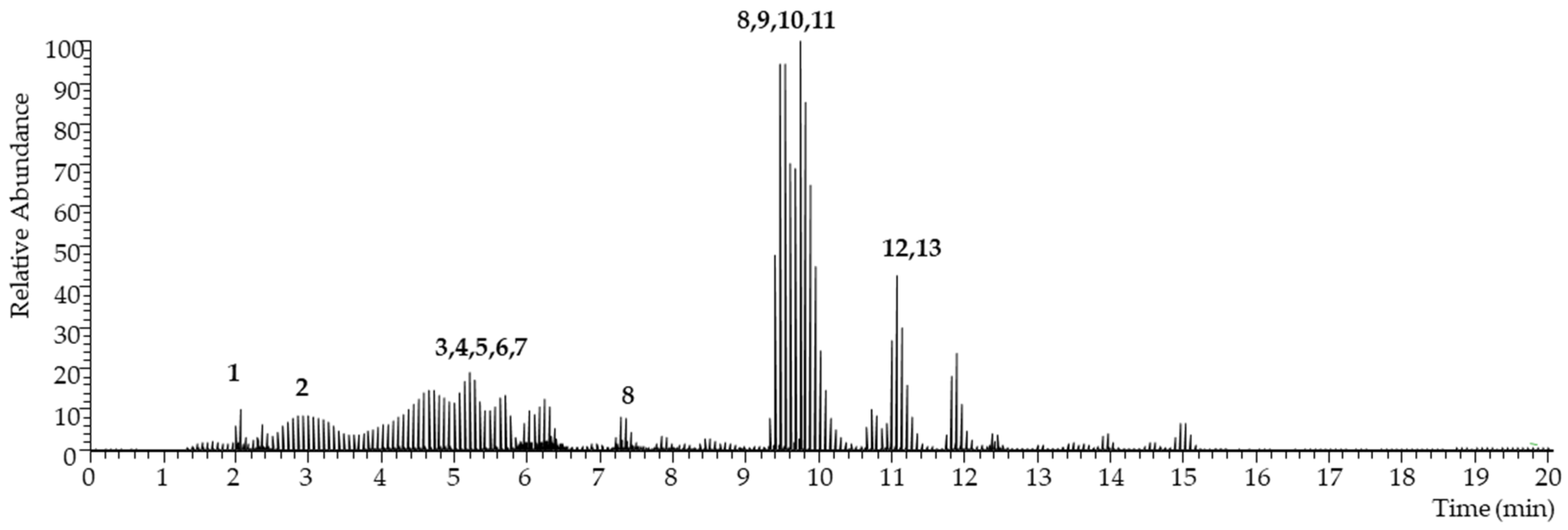
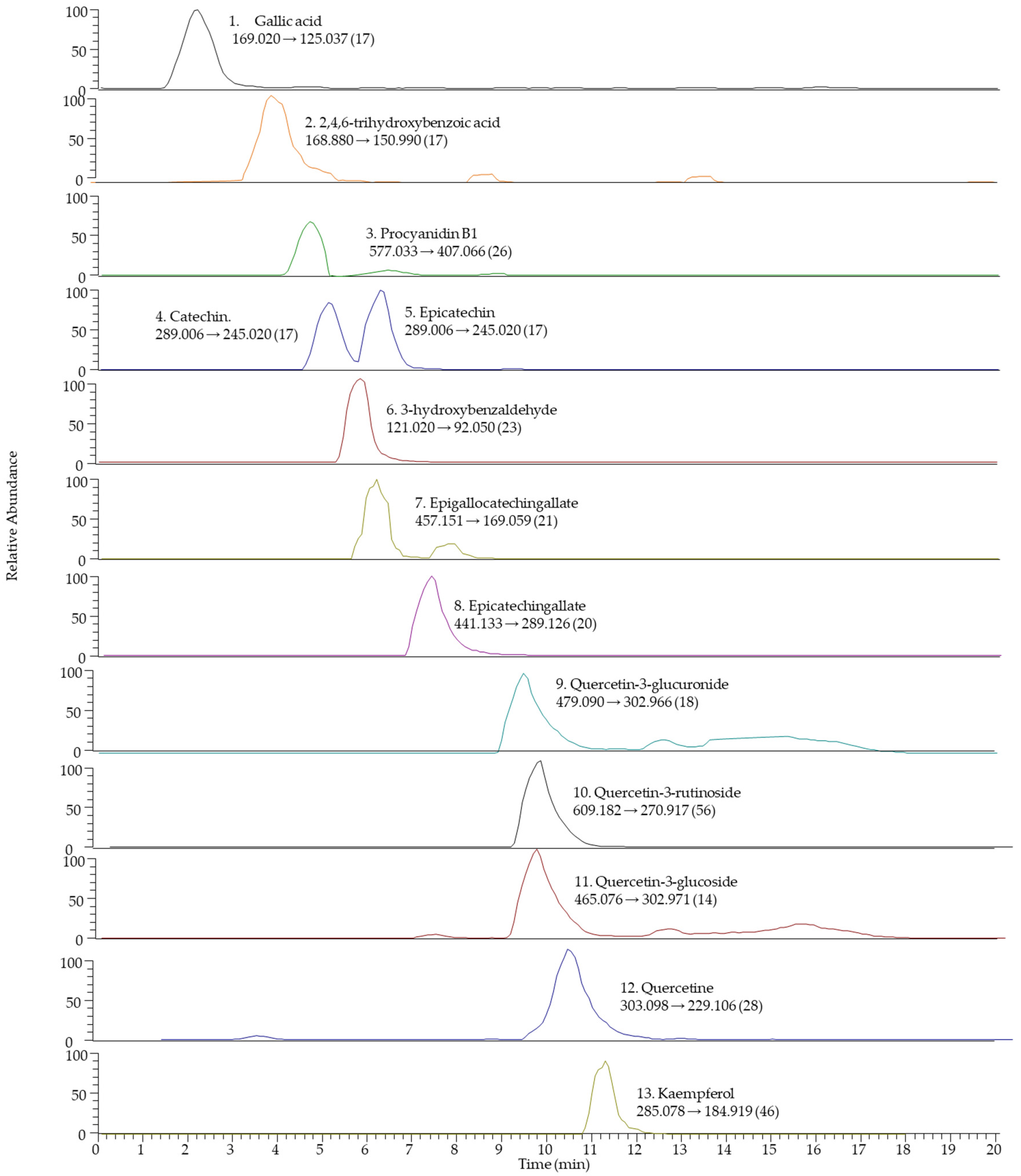
2.2. Chemical–Analytical Characterization of the Extract
2.3. In Vitro Methods for the Evaluation of the Antibacterial Activity of the Extract
3. Discussion
4. Materials and Methods
4.1. Clinical Strains
4.2. Extract
4.3. Obtaining the Extract
4.4. Total Polyphenolic Index of the Extract (TPI)
4.5. Antioxidant Activity (AA) of the Extract
4.6. Characterization of Polyphenols Using Liquid Chromatography Coupled to a Tandem Mass Spectrometer (LC-MS/MS)
4.7. Determination of Antibacterial Activity from the Viable Cell Count Method with Fluorometric Reading
5. Conclusions
Author Contributions
Funding
Institutional Review Board Statement
Informed Consent Statement
Data Availability Statement
Conflicts of Interest
References
- Fleming, A. On the antibacterial action of cultures of a penicillium, with special reference to their use in the isolation of B. influenzae. 1929. Bull. World Health Organ. 2001, 79, 780–790. [Google Scholar]
- Calvo, J.; Martínez-Martínez, L. Mecanismos de acción de los antimicrobianos. Enferm. Infecc Microbiol. Clínica 2009, 27, 44–52. [Google Scholar] [CrossRef]
- AMC|European Centre for Disease Prevention and Control. Available online: https://qap.ecdc.europa.eu/public/extensions/AMC2_Dashboard/AMC2_Dashboard.html#geo-distribution-tab (accessed on 20 January 2023).
- Plan Nacional Frente a la Resistencia a los Antibióticos|PRAN. Available online: https://www.resistenciaantibioticos.es/es/publicaciones/plan-nacional-frente-la-resistencia-los-antibioticos (accessed on 4 March 2023).
- Magiorakos, A.-P.; Srinivasan, A.; Carey, R.B.; Carmeli, Y.; Falagas, M.E.; Giske, C.G.; Harbarth, S.; Hindler, J.F.; Kahlmeter, G.; Olsson-Liljequist, B.; et al. Multidrug-resistant, extensively drug-resistant and pandrug-resistant bacteria: An international expert proposal for interim standard definitions for acquired resistance. Clin. Microbiol. Infect. 2012, 18, 268–281. [Google Scholar] [CrossRef] [Green Version]
- Resistencia a los Antimicrobianos. Available online: https://www.who.int/es/news-room/fact-sheets/detail/antimicrobial-resistance (accessed on 24 August 2022).
- Plan de Acción Mundial Sobre la Resistencia a los Antimicrobianos. Available online: https://www.who.int/es/publications/i/item/9789241509763 (accessed on 24 August 2022).
- Prestinaci, F.; Pezzotti, P.; Pantosti, A. Antimicrobial resistance: A global multifaceted phenomenon. Pathog. Glob. Health 2015, 109, 309–318. [Google Scholar] [CrossRef] [Green Version]
- Los Líderes Mundiales Reunidos en la Asamblea General de las Naciones Unidas se Comprometen a Adoptar una Estrategia Contra la Resistencia a los Antibióticos. Agencia Española de Medicamentos y Productos Sanitarios. Available online: https://www.aemps.gob.es/informa/notasinformativas/laaemps/2016/ni-aemps_11-2016-reunion-onu-antibioticos/ (accessed on 4 March 2023).
- About EARS-NET. European Centre for Disease Prevention and Control. Available online: https://www.ecdc.europa.eu/en/about-us/networks/disease-networks-and-laboratory-networks/ears-net-about (accessed on 6 September 2022).
- Coantibio 2: Plan National de Réduction des Risques D’antibiorésistance en Médecine Vétérinaire (2017–2022). Ministère de l’Agriculture et de la Souveraineté Alimentaire. Available online: https://agriculture.gouv.fr/le-plan-ecoantibio-2-2017-2022 (accessed on 4 March 2023).
- DART 2020: Fighting Antibiotic Resistance for the Good of Both Humans and Animals. Available online: https://www.bmel.de/SharedDocs/Downloads/EN/Publications/DART2020.html (accessed on 4 March 2023).
- StAR. Swiss Antibiotic Resistance Report 2020. Available online: https://www.star.admin.ch/star/en/home/star/Newsletter-Beitraege/swiss-antibiotic-resistance-report-2020.html (accessed on 4 March 2023).
- La OMS Publica la Lista de Las Bacterias para las que se Necesitan Urgentemente Nuevos Antibióticos. Available online: https://www.who.int/es/news/item/27-02-2017-who-publishes-list-of-bacteria-for-which-new-antibiotics-are-urgently-needed (accessed on 24 August 2022).
- Rice, L.B. Federal Funding for the Study of Antimicrobial Resistance in Nosocomial Pathogens: No ESKAPE. J. Infect. Dis. 2008, 197, 1079–1081. [Google Scholar] [CrossRef] [PubMed]
- Mulani, M.S.; Kamble, E.E.; Kumkar, S.N.; Tawre, M.S.; Pardesi, K.R. Emerging Strategies to Combat ESKAPE Pathogens in the Era of Antimicrobial Resistance: A Review. Front. Microbiol. 2019, 10, 539. [Google Scholar] [CrossRef]
- World Health Organization. Antibacterial Agents in Clinical Development: An Analysis of the Antibacterial Clinical Development Pipeline, Including Tuberculosis; Report No.: WHO/EMP/IAU/2017.11; World Health Organization: Geneva, Switzerland, 2017. Available online: https://apps.who.int/iris/handle/10665/258965 (accessed on 6 September 2022).
- Terreni, M.; Taccani, M.; Pregnolato, M. New Antibiotics for Multidrug-Resistant Bacterial Strains: Latest Research Developments and Future Perspectives. Molecules 2021, 26, 2671. [Google Scholar] [CrossRef] [PubMed]
- Zhu, L.; Zhang, Y.; Lu, J. Phenolic Contents and Compositions in Skins of Red Wine Grape Cultivars among Various Genetic Backgrounds and Originations. Int. J. Mol. Sci. 2012, 13, 3492–3510. [Google Scholar] [CrossRef] [PubMed] [Green Version]
- Yu, J.; Ahmedna, M. Functional components of grape pomace: Their composition, biological properties and potential applications. Int. J. Food Sci. Technol. 2013, 48, 221–237. [Google Scholar] [CrossRef]
- Mattos, G.N.; Tonon, R.V.; AL Furtado, A.; MC Cabral, L. Grape by-product extracts against microbial proliferation and lipid oxidation: A review. J. Sci. Food Agric. 2017, 97, 1055–1064. [Google Scholar] [CrossRef] [PubMed]
- Aguín, M.L.; Jares, C.G.; Casas, M.Á.; Llompart, M. Polyphenol Extract from White-Grape Residue. European Patent EP2875822A1, 27 May 2015. [Google Scholar]
- Quideau, S.; Deffieux, D.; Douat-Casassus, C.; Pouységu, L. Plant Polyphenols: Chemical Properties, Biological Activities, and Synthesis. Angew. Chem. Int. Ed. 2011, 50, 586–621. [Google Scholar] [CrossRef]
- Garcia-Salas, P.; Morales-Soto, A.; Segura-Carretero, A.; Fernández-Gutiérrez, A. Phenolic-Compound-Extraction Systems for Fruit and Vegetable Samples. Molecules 2010, 15, 8813–8826. [Google Scholar] [CrossRef]
- Manso, T.; Lores, M.; de Miguel, T. Antimicrobial Activity of Polyphenols and Natural Polyphenolic Extracts on Clinical Isolates. Antibiotics 2021, 11, 46. [Google Scholar] [CrossRef] [PubMed]
- Brown, J.C.; Huang, G.; Haley-Zitlin, V.; Jiang, X. Antibacterial Effects of Grape Extracts on Helicobacter pylori. Appl. Environ. Microbiol. 2009, 75, 848–852. [Google Scholar] [CrossRef] [Green Version]
- Calvo, J.; Cantón, R.; Fernandez Cuenca, F.; Mirelis, B.; Navarro, F. Detección Fenotípica de Mecanismos de Resistencia en Gramnegativos (38). In Procedimientos en Microbiología Clínica; Cercenado, E., Cantón, R., Eds.; Sociedad Española de Enfermedades Infecciosas y Microbiología Clínica (SEIMC): Santiago, Spain, 2011. [Google Scholar]
- Ardanuy, C.; Cercenado, E.; Morosini, M.I.; Torres, C. Detección Fenotípica de Mecanismos de Resistencia en Grampositivos (39). In Procedimientos en Microbiología Clínica; Cercenado, E., Cantón, R., Eds.; Sociedad Española de Enfermedades Infecciosas y Microbiología Clínica (SEIMC): Santiago, Spain, 2011. [Google Scholar]
- Methods for the determination of susceptibility of bacteria to antimicrobial agents. Terminol. Clin. Microbiol. Infect. 1998, 4, 291–296. [CrossRef] [Green Version]
- IC50 Calculator, AAT Bioquest. Available online: https://www.aatbio.com/tools/ic50-calculator (accessed on 22 March 2023).
- Álvarez-Casas, M.; Pájaro, M.; Lores, M.; Garcia-Jares, C. Characterization of grape marcs from native and foreign white varieties grown in northwestern Spain by their polyphenolic composition and antioxidant activity. Eur. Food Res. Technol. 2016, 242, 655–665. [Google Scholar] [CrossRef]
- Garcia-Jares, C.; Vazquez, A.; Lamas, J.P.; Pajaro, M.; Alvarez-Casas, M.; Lores, M. Antioxidant White Grape Seed Phenolics: Pressurized Liquid Extracts from Different Varieties. Antioxidants 2015, 4, 737–749. [Google Scholar] [CrossRef] [PubMed] [Green Version]
- Rama, J.-L.R.; Mallo, N.; Biddau, M.; Fernandes, F.; de Miguel, T.; Sheiner, L.; Choupina, A.; Lores, M. Exploring the powerful phytoarsenal of white grape marc against bacteria and parasites causing significant diseases. Environ. Sci. Pollut. Res. 2021, 28, 24270–24278. [Google Scholar] [CrossRef] [Green Version]
- Kua, Y.L.; Gan, S.; Morris, A.; Ng, H.K. Ethyl lactate as a potential green solvent to extract hydrophilic (polar) and lipophilic (non-polar) phytonutrients simultaneously from fruit and vegetable by-products. Sustain. Chem. Pharm. 2016, 4, 21–31. [Google Scholar] [CrossRef]
- Anastas, P.; Eghbali, N. Green Chemistry: Principles and Practice. Chem. Soc. Rev. 2010, 39, 301–312. [Google Scholar] [CrossRef]
- López-Lorente, I.; Pena-Pereira, F.; Pedersen-Bjergaard, S.; Zuin, V.G.; Ozkan, S.A.; Psillakis, E. The ten principles of green sample preparation. TrAC Trends Anal. Chem. 2022, 148, 116530. [Google Scholar] [CrossRef]
- Gato, E.; Rosalowska, A.; Martínez-Guitián, M.; Lores, M.; Bou, G.; Pérez, A. Anti-adhesive activity of a Vaccinium corymbosum polyphenolic extract targeting intestinal colonization by Klebsiella pneumoniae. Biomed. Pharmacother. 2020, 132, 110885. [Google Scholar] [CrossRef] [PubMed]
- Lores, M.; Pájaro, M.; Álvarez-Casas, M.; Domínguez, J.; García-Jares, C. Use of ethyl lactate to extract bioactive compounds from Cytisus scoparius: Comparison of pressurized liquid extraction and medium scale ambient temperature systems. Talanta 2015, 140, 134–142. [Google Scholar] [CrossRef]
- Castillo, A.; Celeiro, M.; Rubio, L.; Bañobre, A.; Otero-Otero, M.; Garcia-Jares, C.; Lores, M. Optimization of bioactives extraction from grape marc via a medium scale ambient temperature system and stability study. Front. Nutr. 2022, 9, 1008457. [Google Scholar] [CrossRef]
- Bañobre Pena, A. Extracción y Caracterización Analítica de Compuestos Bioactivos de Origen Natural. Máster Universitario en Investigación Química y Química Industrial; Universidade de Santiago de Compostela: Santiago, Spain, 2021. [Google Scholar]
- CFR Part 172–Food Additives Permitted for Direct Addition to Food for Human Consumption. Available online: https://www.ecfr.gov/current/title-21/chapter-I/subchapter-B/part-172 (accessed on 3 March 2023).
- Luchian, C.E.; Cotea, V.V.; Vlase, L.; Toiu, A.M.; Colibaba, L.C.; Răschip, I.E.; Nadăș, G.; Gheldiu, A.M.; Tuchiluș, C.; Rotaru, L. Antioxidant and antimicrobial effects of grape pomace extracts. BIO Web. Conf. 2019, 15, 04006. [Google Scholar] [CrossRef] [Green Version]
- Re, R.; Pellegrini, N.; Proteggente, A.; Pannala, A.; Yang, M.; Rice-Evans, C. Antioxidant activity applying an improved ABTS radical cation decolorization assay. Free Radic. Biol. Med. 1999, 26, 1231–1237. [Google Scholar] [CrossRef] [PubMed]
- Trošt, K.; Klančnik, A.; Vodopivec, B.M.; Lemut, M.S.; Novšak, K.J.; Raspor, P.; Možina, S.S. Polyphenol, antioxidant and antimicrobial potential of six different white and red wine grape processing leftovers. J. Sci. Food Agric. 2016, 96, 4809–4820. [Google Scholar] [CrossRef] [PubMed]
- González-Centeno, M.R.; Jourdes, M.; Femenia, A.; Simal, S.; Rosselló, C.; Teissedre, P.-L. Characterization of Polyphenols and Antioxidant Potential of White Grape Pomace Byproducts (Vitis vinifera L.). J. Agric. Food Chem. 2013, 61, 11579–11587. [Google Scholar] [CrossRef] [PubMed]
- Pandey, K.B.; Rizvi, S.I. Plant polyphenols as dietary antioxidants in human health and disease. Oxid. Med. Cell. Longev. 2009, 2, 270–278. [Google Scholar] [CrossRef] [Green Version]
- Molinari, R.; Merendino, N.; Costantini, L. Polyphenols as modulators of pre-established gut microbiota dysbiosis: State-of-the-art. Biofactors 2021, 48, 255–273. [Google Scholar] [CrossRef]
- Cardona, F.; Andrés-Lacueva, C.; Tulipani, S.; Tinahones, F.J.; Queipo-Ortuño, M.I. Benefits of polyphenols on gut microbiota and implications in human health. J. Nutr. Biochem. 2013, 24, 1415–1422. [Google Scholar] [CrossRef] [PubMed] [Green Version]
- Piekarska-Radzik, L.; Klewicka, E. Mutual influence of polyphenols and Lactobacillus spp. bacteria in food: A review. Eur. Food Res. Technol. 2021, 247, 9–24. [Google Scholar] [CrossRef]
- Zhang, H.; Tsao, R. Dietary polyphenols, oxidative stress and antioxidant and anti-inflammatory effects. Curr. Opin. Food Sci. 2016, 8, 33–42. [Google Scholar] [CrossRef]
- Pop, A.; Bogdan, C.; Fizesan, I.; Iurian, S.; Carpa, R.; Bacali, C.; Vlase, L.; Benedec, D.; Moldovan, M.L. In Vitro Evaluation of Biological Activities of Canes and Pomace Extracts from Several Varieties of Vitis vinifera L. for Inclusion in Freeze-Drying Mouthwashes. Antioxidants 2022, 11, 218. [Google Scholar] [CrossRef] [PubMed]
- Efenberger-Szmechtyk, M.; Nowak, A.; Czyzowska, A. Plant extracts rich in polyphenols: Antibacterial agents and natural preservatives for meat and meat products. Crit. Rev. Food Sci. Nutr. 2021, 61, 149–178. [Google Scholar] [CrossRef]
- Lores, M.; Iglesias-Estévez, M.; Álvarez-Casas, M.; Llompart, M.; García, C. Extraction of bioactive polyphenols from grape marc by a matrix solid-phase dispersion method. Recur. Rurais 2012, 8, 39–47. [Google Scholar]
- Miyasaki, Y.; Rabenstein, J.D.; Rhea, J.; Crouch, M.-L.; Mocek, U.M.; Kittell, P.E.; Morgan, M.A.; Nichols, W.S.; Van Benschoten, M.M.; Hardy, W.D.; et al. Isolation and Characterization of Antimicrobial Compounds in Plant Extracts against Multidrug-Resistant Acinetobacter baumannii. PLoS ONE 2013, 8, e61594. [Google Scholar] [CrossRef] [Green Version]
- Papadopoulou, C.; Soulti, K.; Roussis, I. Potential Antimicrobial Activity of Red and White Wine Phenolic Extracts against Strains of Staphylococcus aureus, Escherichia coli and Candida albicans. Food Technol. Biotechnol. 2005, 43, 41–46. [Google Scholar]
- Olszewska, M.A.; Gędas, A.; Simões, M. Antimicrobial polyphenol-rich extracts: Applications and limitations in the food industry. Food Res. Int. 2020, 134, 109214. [Google Scholar] [CrossRef]
- Zhang, Q.; Zhang, J.; Zhang, J.; Xu, D.; Li, Y.; Liu, Y.; Zhang, X.; Zhang, R.; Wu, Z.; Weng, P. Antimicrobial Effect of Tea Polyphenols against Foodborne Pathogens: A Review. J. Food Prot. 2021, 84, 1801–1808. [Google Scholar] [CrossRef]
- Álvarez-Martínez, F.J.; Barrajón-Catalán, E.; Encinar, J.A.; Rodríguez-Díaz, J.C.; Micol, V. Antimicrobial Capacity of Plant Polyphenols against Gram-positive Bacteria: A Comprehensive Review. Curr. Med. Chem. 2020, 27, 2576–2606. [Google Scholar] [CrossRef] [PubMed]
- Xu, C.; Yagiz, Y.; Hsu, W.-Y.; Simonne, A.; Lu, J.; Marshall, M.R. Antioxidant, Antibacterial, and Antibiofilm Properties of Polyphenols from Muscadine Grape (Vitis rotundifolia Michx.) Pomace against Selected Foodborne Pathogens. J. Agric. Food Chem. 2014, 62, 6640–6649. [Google Scholar] [CrossRef]
- Su, P.-W.; Yang, C.-H.; Yang, J.-F.; Su, P.-Y.; Chuang, L.-Y. Antibacterial Activities and Antibacterial Mechanism of Polygonum cuspidatum Extracts against Nosocomial Drug-Resistant Pathogens. Molecules 2015, 20, 11119–11130. [Google Scholar] [CrossRef] [Green Version]
- Marinaş, I.C.; Chifiriuc, C.; Oprea, E.; Lazăr, V. Antimicrobial and antioxidant activities of alcoholic extracts obtained from vegetative organs of A. retroflexus. Rom. Arch. Microbiol. Immunol. 2014, 7, 35–42. [Google Scholar]
- Betts, J.W.; Hornsey, M.; Higgins, P.G.; Lucassen, K.; Wille, J.; Salguero, F.J.; Seifert, H.; La Ragione, R.M. Restoring the activity of the antibiotic aztreonam using the polyphenol epigallocatechin gallate (EGCG) against multidrug-resistant clinical isolates of Pseudomonas aeruginosa. J. Med. Microbiol. 2019, 68, 1552–1559. [Google Scholar] [CrossRef] [PubMed]
- Jiamboonsri, P.; Pithayanukul, P.; Bavovada, R.; Chomnawang, M.T. The Inhibitory Potential of Thai Mango Seed Kernel Extract against Methicillin-Resistant Staphylococcus Aureus. Molecules 2011, 16, 6255–6270. [Google Scholar] [CrossRef] [Green Version]
- Mollica, A.; Scioli, G.; Della Valle, A.; Cichelli, A.; Novellino, E.; Bauer, M.; Kamysz, W.; Llorent-Martínez, E.J.; Córdova, M.L.F.-D.; Castillo-López, R.; et al. Phenolic Analysis and In Vitro Biological Activity of Red Wine, Pomace and Grape Seeds Oil Derived from Vitis vinifera L. cv. Montepulciano d’Abruzzo. Antioxidants 2021, 10, 1704. [Google Scholar] [CrossRef]
- Radulescu, C.; Buruleanu, L.C.; Nicolescu, C.M.; Olteanu, R.L.; Bumbac, M.; Holban, G.C.; Simal-Gandara, J. Phytochemical Profiles, Antioxidant and Antibacterial Activities of Grape (Vitis vinifera L.) Seeds and Skin from Organic and Conventional Vineyards. Plants 2020, 9, 1470. [Google Scholar] [CrossRef]
- Simonetti, G.; Brasili, E.; Pasqua, G. Antifungal Activity of Phenolic and Polyphenolic Compounds from Different Matrices of Vitis vinifera L. against Human Pathogens. Molecules 2020, 25, 3748. [Google Scholar] [CrossRef]
- Squillaci, G.; Zannella, C.; Carbone, V.; Minasi, P.; Folliero, V.; Stelitano, D.; La Cara, F.; Galdiero, M.; Franci, G.; Morana, A. Grape Canes from Typical Cultivars of Campania (Southern Italy) as a Source of High-Value Bioactive Compounds: Phenolic Profile, Antioxidant and Antimicrobial Activities. Molecules 2021, 26, 2746. [Google Scholar] [CrossRef]
- Dias, C.; Domínguez-Perles, R.; Aires, A.; Teixeira, A.; Rosa, E.; Barros, A.; Saavedra, M.J. Phytochemistry and activity against digestive pathogens of grape (Vitis vinifera L.) stem’s (poly)phenolic extracts. LWT Food Sci. Technol. 2015, 61, 25–32. [Google Scholar] [CrossRef]
- Leal, C.; Santos, R.A.; Pinto, R.; Queiroz, M.; Rodrigues, M.; Saavedra, M.J.; Barros, A.; Gouvinhas, I. Recovery of bioactive compounds from white grape (Vitis vinifera L.) stems as potential antimicrobial agents for human health. Saudi J. Biol. Sci. 2020, 27, 1009–1015. [Google Scholar] [CrossRef]
- Singleton, V.L.; Orthofer, R.; Lamuela-Raventós, R.M. Analysis of total phenols and other oxidation substrates and antioxidants by means of folin-ciocalteu reagent. Methods Enzymol. 1999, 299, 152–178. [Google Scholar]
- Zhang, Q.; Zhang, J.; Shen, J.; Silva, A.; Dennis, D.A.; Barrow, C.J. A Simple 96-Well Microplate Method for Estimation of Total Polyphenol Content in Seaweeds. J. Appl. Phycol. 2006, 18, 445–450. [Google Scholar] [CrossRef] [Green Version]
- Xiao, F.; Xu, T.; Lu, B.; Liu, R. Guidelines for antioxidant assays for food components. Food Front. 2020, 1, 60–69. [Google Scholar] [CrossRef] [Green Version]
- Skoko, A.-M.G.; Šarkanj, B.; Lores, M.; Celeiro, M.; Babojelić, M.S.; Kamenjak, D.; Flanjak, I.; Jozinović, A.; Kovač, T.; Lončarić, A. Identification and Quantification of Polyphenols in Croatian Traditional Apple Varieties. Plants 2022, 11, 3540. [Google Scholar] [CrossRef] [PubMed]
- Lancaster, M.V.; Fields, R.D. Antibiotic and Cytotoxic Drug Susceptibility Assays using Resazurin and Poising Agents. U.S. Patent No. 5,501,959, 26 March 1996. [Google Scholar]
- Elshikh, M.; Ahmed, S.; Funston, S.; Dunlop, P.; McGaw, M.; Marchant, R.; Banat, I.M. Resazurin-based 96-well plate microdilution method for the determination of minimum inhibitory concentration of biosurfactants. Biotechnol. Lett. 2016, 38, 1015–1019. [Google Scholar] [CrossRef] [Green Version]
- Baker, C.N.; Tenover, F.C. Evaluation of Alamar colorimetric broth microdilution susceptibility testing method for staphylococci and enterococci. J. Clin. Microbiol. 1996, 34, 2654–2659. [Google Scholar] [CrossRef] [Green Version]
- Sarker, S.D.; Nahar, L.; Kumarasamy, Y. Microtitre plate-based antibacterial assay incorporating resazurin as an indicator of cell growth, and its application in the in vitro antibacterial screening of phytochemicals. Methods 2007, 42, 321–324. [Google Scholar] [CrossRef]
- Abiodun, O.; Sood, S.; A Osiyemi, O.; Agnihotri, V.K.; Gulati, A.; O Ajaiyeoba, E.; Singh, B. In vitro antimicrobial activity of crude ethanol extracts and fractions of Terminalia catappa and Vitex doniana. Afr. J. Med. Med. Sci. 2015, 44, 21–26. [Google Scholar]
- Alamarblue Assay. Available online: https://tools.thermofisher.com/content/sfs/manuals/PI-DAL1025-1100_TI%20alamarBlue%20Rev%201.1.pdf (accessed on 6 March 2023).


| Number and Layout of Carbon Atoms | Polyphenol Class or Family (Polyphenol Subclass) | Main Representatives in White Grapes |
|---|---|---|
| C6-C1 | Benzoic acids and derivatives | Gallic acid 2,4,6-trihydroxibenzoic acid 3-hydrobenzaldehyde Protocatechuic acid Syringic acid |
| C6-C3 | Cinnamic acids | Caftaric acid Caffeic acid |
| Coumarins | ||
| C6-C2-C6 | Stilbens | |
| C6-C3-C6 Flavonoids | Flavonols | Quercetin-3-glucuronide (miquelianin) Quercetin-3-O-rutinoside (rutin) Quercetin-3-O-glucoside (isoquercitrin) Quercetin Kaempferol Quercetin-3-rhamnoside (quercitrin) |
| Flavan-3-ols | Procyanidins Catechin Epicatechin Epigallocatequingalate Epicatequingalate | |
| Flavanones | ||
| Flavones | Luteolin | |
| Isoflavones | ||
| Anthocyani(di)ns | ||
| (C6-C1)n | Hydrolyzable tannins | |
| Ellagitannins | ||
| Gallotanins |
| Strain Code | Bacteria | Acquired Resistance Mechanism | Clinical Sample | Culture Medium for Their Isolation |
|---|---|---|---|---|
| Pflu | Pseudomonas fluorescens | VIM carbapenemase | Urine | CLED |
| Paer | Pseudomonas aeruginosa | Wound | BA | |
| Bcep | Burkholderia cepacia | Catheter | BA | |
| Smal | Stenotrophomonas. maltophilia | Wound | BA | |
| Kpne | Klebsiella pneumoniae | OXA-48 carbapenemase | Urine | CLED |
| Pmir | Proteus mirabilis | ESBL | Urine | CLED |
| Ecol | Escherichia coli | ESBL | Urine | CLED |
| Cfre | Citrobacter freundii | ESBL | Urine | CLED |
| Eclo | Enterobacter cloacae | Urine | CLED | |
| Apun | Aeromonas punctata (caviae) | Feces | CIN | |
| Yent | Yersinia enterocolitica | Feces | CIN | |
| Sent | Salmonella enteritidis | ESBL | Feces | SS |
| Strain Code | Bacteria | Acquired Resistance Mechanism | Clinical Sample | Culture Medium for Their Isolation |
|---|---|---|---|---|
| Saur | Staphylococcus aureus | Methicillin resistant | Blood culture | BA |
| Sepi | Staphylococcus epidermidis | Methicillin resistant | Wound | BA |
| Ssap | Staphylococcus saprophyticus | Urine | CLED | |
| Saga | Streptococcus agalactiae | Blood culture | BA | |
| Spyo | Streptococcus pyogenes | Blood culture | BA | |
| Efae | Enterococcus faecalis | Urine | CLED | |
| Efac | Enterococcus faecium | Blood culture | BA |
| AK | AM | AS | AUG | AZT | C/T | CZA | CAZ | CFE | CFT | CFX | CL | CP | CPE | CRM | ETR | FD | FOS | GM | IMP | LVX | MER | NXN | PI | PT | TS | TI | TO | |
|---|---|---|---|---|---|---|---|---|---|---|---|---|---|---|---|---|---|---|---|---|---|---|---|---|---|---|---|---|
| Pflu | S | R | R | R | R | S | R | R | R | R | R | R | R | R | R | R | ||||||||||||
| Paer | S | R | S | R | R | S | R | R | S | R | R | R | R | R | R | R | R | |||||||||||
| Bcep | R | R | S | S | ||||||||||||||||||||||||
| Smal | R | R | R | R | R | R | R | R | R | S | R | R | S | R | R | |||||||||||||
| Kpne | R | R | R | R | S | R | R | R | R | S | R | R | R | R | R | R | R | R | R | R | R | R | R | R | ||||
| Pmir | S | R | S | R | R | R | S | R | R | R | R | S | R | R | S | I | S | R | S | S | R | S | ||||||
| Ecol | S | R | S | R | R | R | S | S | R | R | R | S | S | S | S | S | R | R | S | R | R | S | ||||||
| Cfre | R | R | R | R | R | R | R | S | R | R | R | S | S | S | R | S | R | R | R | R | R | R | ||||||
| Eclo | S | R | R | R | R | S | R | R | R | R | S | R | R | R | R | R | S | R | S | R | S | R | R | R | R | R | ||
| Apun | S | S | S * | S | S | S | S | S | S | S | ||||||||||||||||||
| Yent | S | R | R | S | S * | R | S | S | S | S | S | S | ||||||||||||||||
| Sent | R | R | R | R | R * | R | R | R | S | S | R | S | S | R |
| AK | AM | AUG | CD | CP | DAP | E | ES1000 | FA | FD | FOS | GM | GM500 | LVX | LZD | MUP | OX | P | RIF | SYN | TS | TE | TEI | TO | VA | |
|---|---|---|---|---|---|---|---|---|---|---|---|---|---|---|---|---|---|---|---|---|---|---|---|---|---|
| Saur | S | R | S | R | S | S | S | S | S | R | S | S | R | R | S | S | S | S | S | S | |||||
| Sepi | S | R | S | R | S | S | S | S | R | R | S | R | R | R | S | R | I | S | R | S | |||||
| Ssap | R | S | S | S | S | R | S | R | S | S | S | S | R | S | S | S | S | ||||||||
| Saga | S | S | S | S | S | R | S | S | R | S | |||||||||||||||
| Spyo | S | S | S | S | S | ||||||||||||||||||||
| Efae | S | R | R | S | R | S | S | R | S | R | S | S | R | R | S | S | |||||||||
| Efac | R | R | R | S | R | R | S | R | S | R | s | S | S | S |
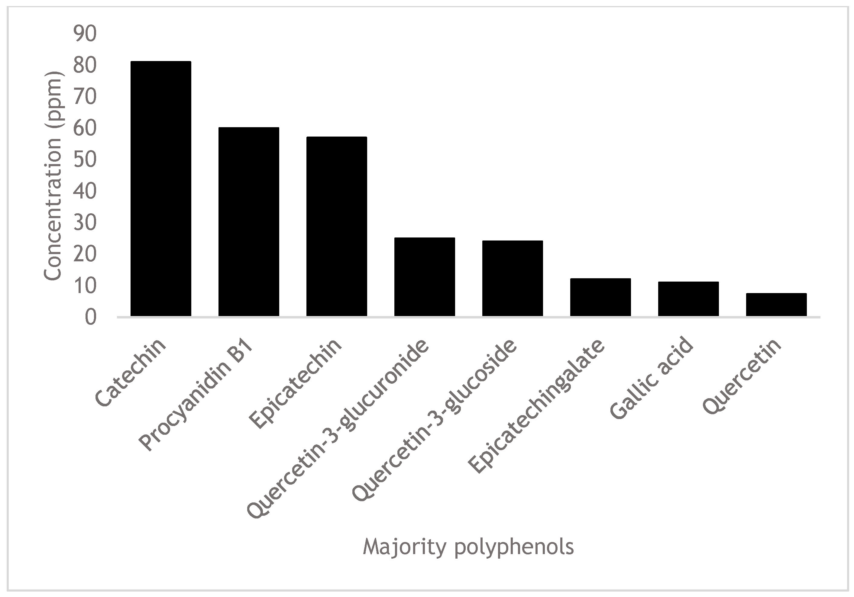
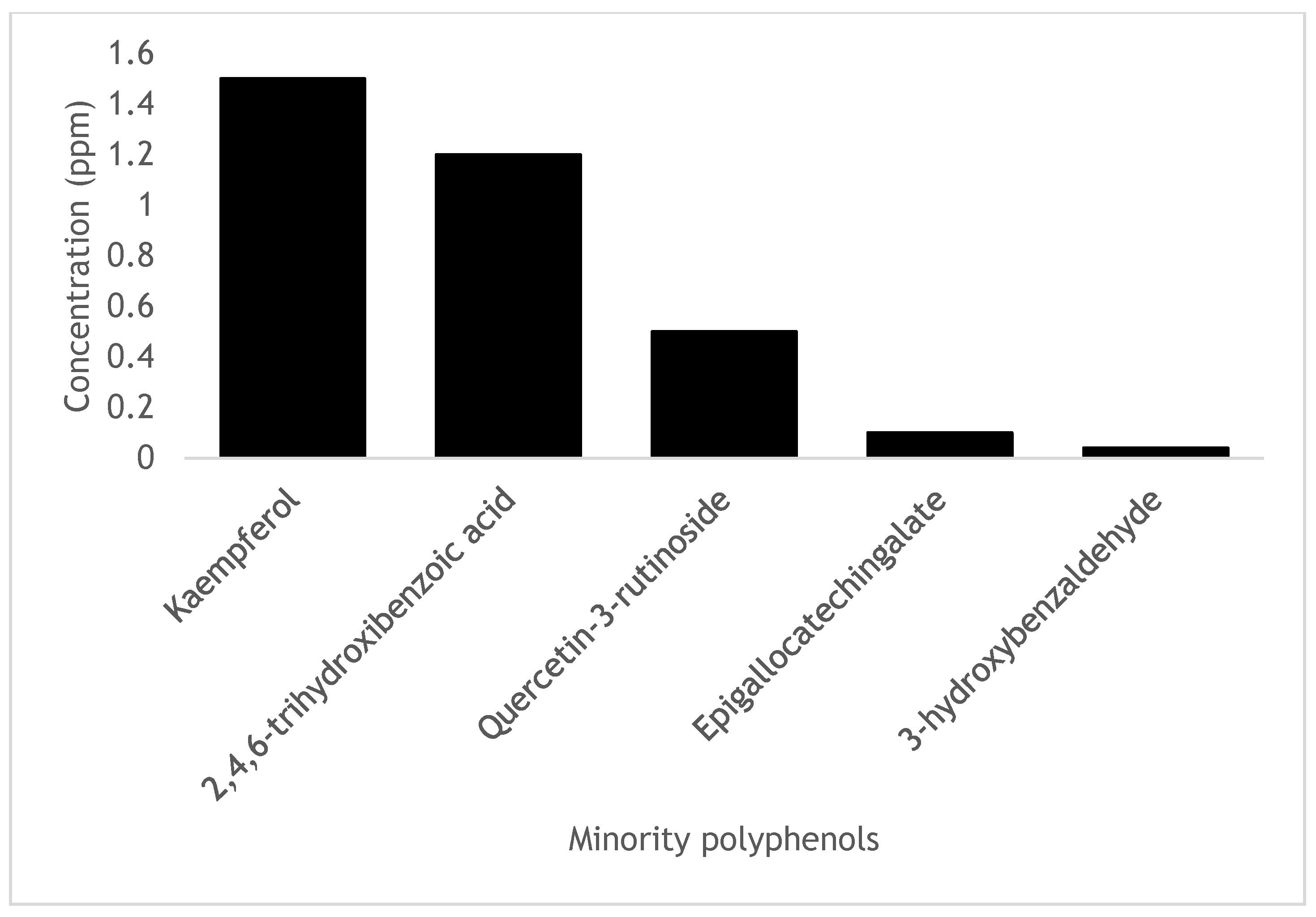
| Polyphenol | Concentration (ppm) |
|---|---|
| Gallic acid | 11 ± 1 |
| 2,4,6-trihydroxybenzoic acid | 1.2 ± 0.08 |
| Procyanidin B1 | 60 ± 5 |
| Catechin | 81 ± 7 |
| 3-hydroxibenzoaldehide | 0.04 ± 0.007 |
| Epicatechin | 57 ± 4 |
| Epigallocatequingalate | 0.1 ± 0.008 |
| Epicatechingalate | 12 ± 2 |
| Quercetin-3-glucuronide | 25 ± 5 |
| Quercetin-3-rutinoside | 0.5 ± 0.17 |
| Quercetin-3-glucoside | 24 ± 3 |
| Quercetin | 7.3 ± 0.9 |
| Kaempferol | 1.5 ± 0.2 |
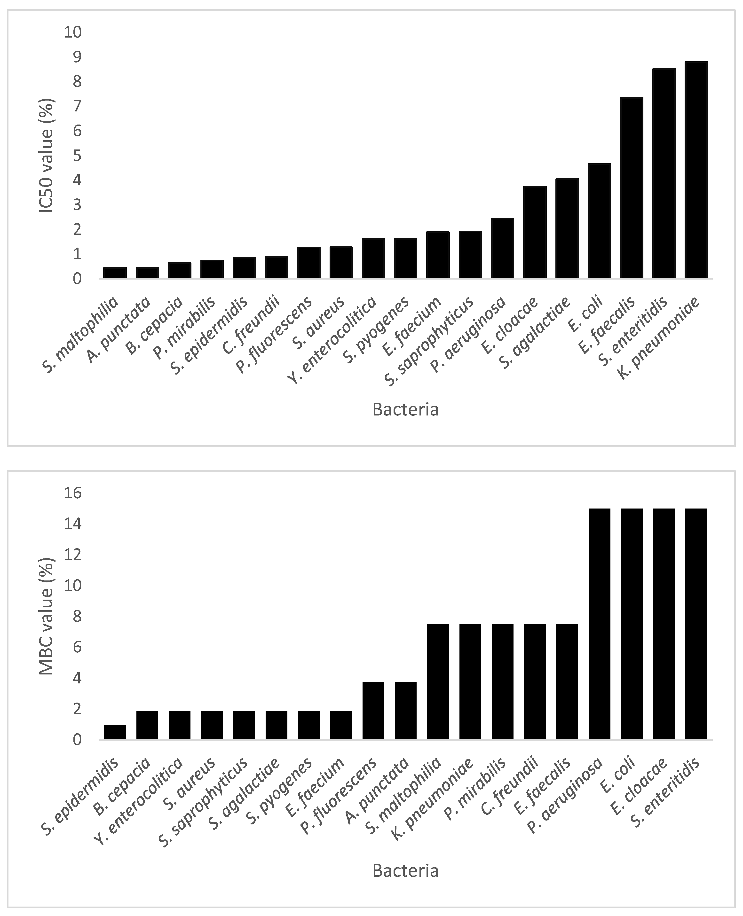
| Strain Code | IC50 (% (v/v)) | MBC (% (v/v)) |
|---|---|---|
| Pflu | 1.27 | 3.75 |
| Paer | 2.44 | 15 |
| Bcep | 0.63 | 1.87 |
| Smal | 0.45 | 7.5 |
| Kpne | 8.79 | 7.5 |
| Pmir | 0.74 | 7.5 |
| Ecol | 4.65 | 15 |
| Cfre | 0.89 | 7.5 |
| Eclo | 3.74 | 15 |
| Apun | 0.45 | 3.75 |
| Yent | 1.61 | 1.87 |
| Sent | 8.52 | 15 |
| Saur | 1.28 | 1.87 |
| Sepi | 0.86 | 0.97 |
| Ssap | 1.92 | 1.87 |
| Saga | 4.05 | 1.87 |
| Spyo | 1.63 | 1.87 |
| Efae | 7.34 | 7.5 |
| Efac | 1.89 | 1.87 |
| Polyphenols | Purity | Company | CAS |
|---|---|---|---|
| Gallic acid | 99.9 | SIGMA a | 149-91-7 |
| 2,4,6-trihydroxybenzoic acid | 98.4 | SIGMA a | 487-70-7 |
| Procyanidin B1 | 96.7 | EXTRAS b | 20315-25-7 |
| Catechin | 98.0 | SIGMA a | 18829-70-4 |
| 3-hydroxybenzaldehyde | 99.0 | SIGMA a | 90-02-8 |
| Epicatechin | 90.0 | SIGMA a | 490-46-0 |
| Epigallocatechingalate | 99.1 | SIGMA a | 989-51-5 |
| Epicatechingallate | 98.0 | SIGMA a | 1257-08-5 |
| Quercetin-3-glucuronide | 98.5 | SIGMA a | 27253-19-6 |
| Quercetin-3-rutinoside | 99.1 | SIGMA a | 115888-40-9 |
| Quercetin-3-glucoside | 98.0 | SIGMA a | 21637-25-2 |
| Quercetin | 96.0 | SIGMA a | 117-39-5 |
| Kaempferol | 99.3 | SIGMA a | 520-18-3 |
| Polyphenols | Rt (min) | Molecular Mass (g/mol) | I a | Precursor Ion (m/z) b | Product Ions (m/z) b | Collision Energy (eV) b | Linear Range (mg/L) | R2 |
|---|---|---|---|---|---|---|---|---|
| Gallic acid | 2.35 | 170.12 | - | 169.020 | 125.037 153.10 | 17 15 | 0.5–5 | 0.9972 |
| 2,4,6-trihydroxybenzoic acid | 3.88 | 170.11 | - | 168.88 | 150.99 83.02 107.02 | 17 23 22 | 0.5–5 | 0.9960 |
| Procyanidin B1 | 5.30 | 578.52 | - | 577.033 | 407.066 288.93 424.98 | 26 25 26 | 0.5–5 | 0.9960 |
| Catechin | 5.34 | 290.27 | - | 289.006 | 245.020 203.12 | 17 22 | 0.5–5 | 0.9995 |
| 3-hydroxy-benzaldehyde | 5.77 | 122.12 | - | 121.02 | 92.05 93.05 120.04 | 23 20 19 | 0.5–5 | 0.9954 |
| Epicatechin | 6.50 | 290.27 | - | 289.006 | 245.020 203.12 | 17 22 | 0.5–5 | 0.9980 |
| Epigallocatechingalate | 6.80 | 458.4 | - | 457.151 | 169.059 125.09 305.09 | 21 42 21 | 0.5–5 | 0.9902 |
| Epicatechingalate | 7.29 | 442.4 | - | 441.133 | 289.126 125.08 169.05 | 20 42 24 | 0.5–5 | 0.9901 |
| Quercetin-3-glucuronide | 9.54 | 478.36 | + | 479.090 | 302.966 461.50 | 18 14 | 0.5–5 | 0.9998 |
| Quercetin-3-rutinoside | 9.72 | 610.518 | - | 609.182 | 270.917 178.87 | 56 44 | 0.5–5 | 0.9987 |
| Quercetin-3-glucoside | 9.75 | 464.376 | + | 465.076 | 302.971 256.90 | 14 41 | 0.5–5 | 0.9920 |
| Quercetin | 10.72 | 302.23 | + | 303.098 | 229.106 153.05 | 28 33 | 0.5–5 | 0.9976 |
| Kaempferol | 11.89 | 286.24 | - | 285.078 | 184.919 239.13 | 46 35 | 0.5–5 | 0.9957 |
Disclaimer/Publisher’s Note: The statements, opinions and data contained in all publications are solely those of the individual author(s) and contributor(s) and not of MDPI and/or the editor(s). MDPI and/or the editor(s) disclaim responsibility for any injury to people or property resulting from any ideas, methods, instructions or products referred to in the content. |
© 2023 by the authors. Licensee MDPI, Basel, Switzerland. This article is an open access article distributed under the terms and conditions of the Creative Commons Attribution (CC BY) license (https://creativecommons.org/licenses/by/4.0/).
Share and Cite
Manso, T.; Lores, M.; Rama, J.L.R.; Villarino, R.-A.; Calvo, L.G.; Castillo, A.; Celeiro, M.; de Miguel, T. Antibacterial Activity against Clinical Strains of a Natural Polyphenolic Extract from Albariño White Grape Marc. Pharmaceuticals 2023, 16, 950. https://doi.org/10.3390/ph16070950
Manso T, Lores M, Rama JLR, Villarino R-A, Calvo LG, Castillo A, Celeiro M, de Miguel T. Antibacterial Activity against Clinical Strains of a Natural Polyphenolic Extract from Albariño White Grape Marc. Pharmaceuticals. 2023; 16(7):950. https://doi.org/10.3390/ph16070950
Chicago/Turabian StyleManso, Tamara, Marta Lores, José Luis R. Rama, Rosa-Antía Villarino, Lorena G. Calvo, Aly Castillo, María Celeiro, and Trinidad de Miguel. 2023. "Antibacterial Activity against Clinical Strains of a Natural Polyphenolic Extract from Albariño White Grape Marc" Pharmaceuticals 16, no. 7: 950. https://doi.org/10.3390/ph16070950