Article

Alkaloid from Geissospermum sericeum Benth. & Hook.f. ex Miers (Apocynaceae) Induce Apoptosis by Caspase Pathway in Human Gastric Cancer Cells

by
Mirian Letícia Carmo Bastos
1,2,†,
João Victor Silva-Silva
3,†,
Jorddy Neves Cruz
2,
Amanda Roberta Palheta da Silva
4,
Alexandre Augusto Bentaberry-Rosa
4,
Gisele da Costa Ramos
5,
José Edson de Sousa Siqueira
5,
Márlia Regina Coelho-Ferreira
6,
Sandro Percário
1,
Patrícia Santana Barbosa Marinho
5,
Andrey Moacir do Rosario Marinho
5,
Marcelo de Oliveira Bahia
7 and
Maria Fâni Dolabela
1,2,4,8,*
1
Post-Graduate Program in Biodiversity and Biotechnology, Federal University of Pará, Belém 66075-110, PA, Brazil
2
Post-Graduate Program in Pharmaceutical Sciences, Federal University of Pará, Belém 66075-110, PA, Brazil
3
Laboratory of Medicinal and Computational Chemistry, Institute of Physics of São Carlos, University of São Paulo, São Carlos 13563-120, SP, Brazil
4
Faculty of Pharmacy, Federal University of Pará, Belém 66075-110, PA, Brazil
5
Post-Graduate Program in Chemistry, Federal University of Pará, Belém 66075-110, PA, Brazil
6
Emílio Goeldi Paraense Museum, Coordination of Botany, Ministry of Science, Technology, Innovation and Communications, Belém 66077-830, PA, Brazil
7
Laboratory of Human Cytogenetic, Institute of Biological Sciences, Federal University of Pará, Belém 66075-110, PA, Brazil
8
Post-Graduate Program in Pharmaceutical Innovation, Federal University of Pará, Belém 66075-110, PA, Brazil
*
Author to whom correspondence should be addressed.
These authors contributed equally to this work.
Pharmaceuticals 2023, 16(5), 765; https://doi.org/10.3390/ph16050765
Submission received: 9 April 2023 / Revised: 30 April 2023 / Accepted: 5 May 2023 / Published: 18 May 2023
(This article belongs to the Special Issue Anticancer Compounds in Medicinal Plants 2023)

Abstract

:
Gastric cancer is among the major causes of death from neoplasia leading causes of death worldwide, with high incidence rates and problems related to its treatment. Here, we outline how Geissospermum sericeum exerts antitumor activity on the ACP02 cell line (human gastric adenocarcinoma) and the mechanism of cell death. The ethanol extract and fractions, neutral fraction and alkaloid fraction, were characterized by thin-layer chromatography and HPLC-DAD, yielding an alkaloid (geissoschizoline N4-methylchlorine) identified by NMR. The cytotoxicity activity of the samples (ethanol extract, neutral fraction, alkaloid fraction, and geissoschizoline N4-methylchlorine) in HepG2 and VERO cells was determined by MTT. The ACP02 cell line was used to assess the anticancer potential. Cell death was quantified with the fluorescent dyes Hoechst 33342, propidium iodide, and fluorescein diacetate. The geissoschizoline N4-methylchlorine was evaluated in silico against caspase 3 and 8. In the antitumor evaluation, there was observed a more significant inhibitory effect of the alkaloid fraction (IC50 18.29 µg/mL) and the geissoschizoline N4-methylchlorine (IC50 12.06 µg/mL). However, geissoschizoline N4-methylchlorine showed lower cytotoxicity in the VERO (CC50 476.0 µg/mL) and HepG2 (CC50 503.5 µg/mL) cell lines, with high selectivity against ACP02 cells (SI 39.47 and 41.75, respectively). The alkaloid fraction showed more significant apoptosis and necrosis in 24 h and 48 h, with increased necrosis in higher concentrations and increased exposure time. For the alkaloid, apoptosis and necrosis were concentration- and time-dependent, with a lower necrosis rate. Molecular modeling studies demonstrated that geissoschizoline N4-methylchlorine could occupy the active site of caspases 3 and 8 energetically favorably. The results showed that fractionation contributed to the activity with pronounced selectivity for ACP02 cells, and geissoschizoline N4-methylchlor is a promising candidate for caspase inhibitors of apoptosis in gastric cancer. Thus, this study provides a scientific basis for the biological functions of Geissospermum sericeum, as well as demonstrates the potential of the geissoschizoline N4-methylchlorine in the treatment of gastric cancer.

1. Introduction

Cancer is one of the leading public health problems worldwide, responsible for about one in six deaths [1,2]. Cancer incidence rates increase with population aging and exposure to risk factors such as smoking, excessive alcohol consumption, lack of physical activity, and obesity [1]. Estimates point to approximately 29 million cancer cases worldwide in 2040, an increase by 47% compared to 2020. Countries in transition are the most affected due to demographic changes, increased associated risk factors to globalization, and a growing economy [3]. Once diagnosed, patients have a long way to go and may undergo surgical procedures, radiotherapy, and/or systemic therapy (chemotherapy, hormone treatments, and targeted biological therapies) [2].
Gastric cancer (adenocarcinoma) is the fourth leading cause of cancer death, constituting 5.6% of all new cases, with the fifth highest incidence among cancers worldwide, and with more than one million new cases diagnosed annually [1]. Treatment selection considers the stage of gastric cancer, with chemotherapy used in advanced tumor cases [4]. However, there are certain problems associated with chemotherapy treatment, such as tumor resistance to treatment [5], high toxicity [6], and high cost [7].
In this context, plants can be a valuable contribution to the search for new therapeutic alternatives, especially plant species that produce alkaloids [8]. These molecules have a low molecular weight and account for approximately 20% of the secondary metabolites found in plants [9]. Traditionally, the bark decoction of Geissospermum has been used in the Amazon region to treat malaria, fever, and stomach disorders [10]. Extracts from the bark and leaves of Geissospermum, including ethanolic [11], methanolic [10], and hydroalcoholic [12], have been tested for biological activities, including antitumor activity [13]. One species that synthesized promising alkaloids for antitumor activity is Geissospermum sericeum Benth. & Hook.f. ex Miers (Apocynaceae). The MeOH−H2O (9:1) extract of the bark of G. sericeum yielded geissoschizoline, geissoschizoline N4-oxide, 1,2-dehydrogeissoschizoline, and flavopereirine alkaloids (Figure 1) [12].
The flavopereirine, dihydroflavopereirine, and PB-100 (mixture of flavopereirine and dihydroflavopereirine) were submitted to evaluate antitumor activity using different tumor and standard strains. The 100 µg/mL concentration of PB-100 inhibited 16 other tumor cell lines, including drug-resistant ones, and did not inhibit the multiplication of normal cells (maintained 95% of normal cells viable). Flavopereirine and dihydroflavopereirine showed similar antitumor activity, confirming the hypothesis that these two alkaloids are responsible for the activity of PB-100 [14]. Flavopereirin is a β-carboline alkaloid, and according to Beljanski and Beljanski [15], alkaloids belonging to the β-carboline class can inhibit DNA synthesis of cancer cells while leaving healthy DNA unaltered, potentially providing a cancer treatment with reduced side effects [13]. Different mechanisms may be involved in the antitumor activity of alkaloids, such as the interaction with microtubules [16]; induction of apoptosis [17]; induction of apoptosis by activating caspases, and altering the Bcl-2/Bax ratio [18].
Despite studies on isolating alkaloids from Geissospermum sericeum, none of the approaches addresses the isolation of geissoschizoline N4-methylchlorine and its activity in gastric cancer. The purpose of this study is to describe for the first time the isolation of geissoschizoline N4-methylchlorine in this species. It investigates whether apoptosis may be the mechanism involved in the activity of geissoschizoline N4-methylchloride. Furthermore, it evaluates the impact of the fractionation of the ethanolic extract of G. sericeum on antitumor activity and cytotoxicity.

2. Results

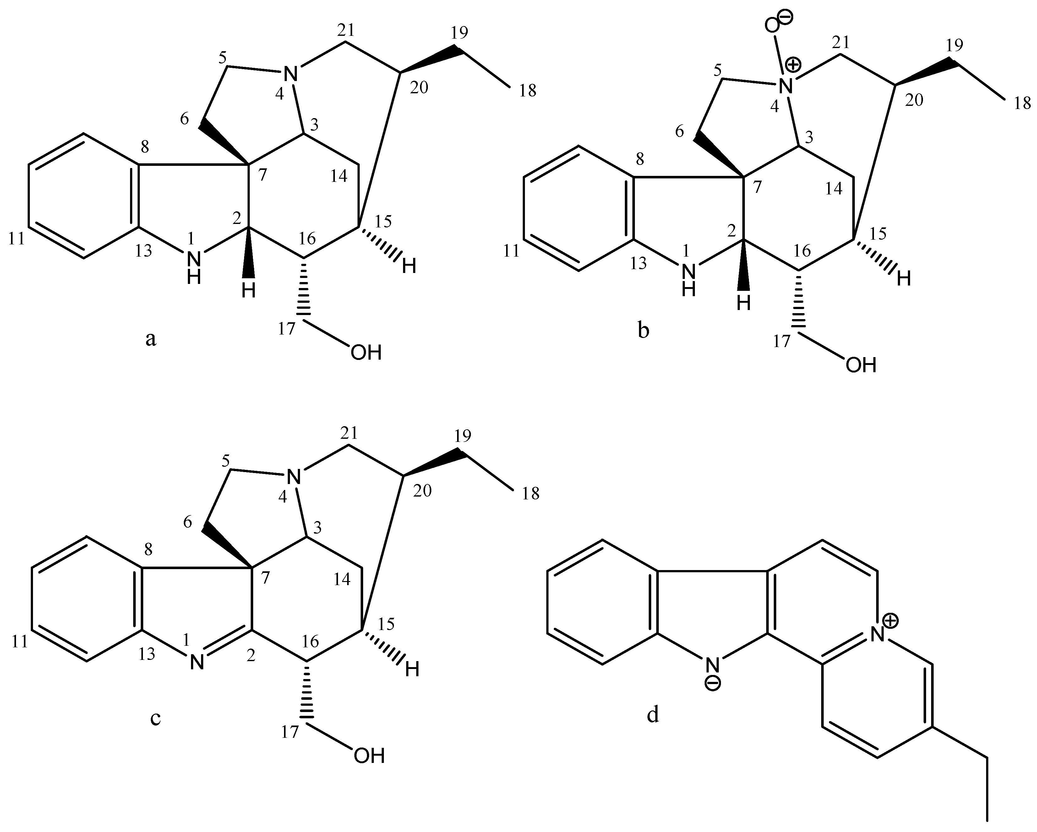
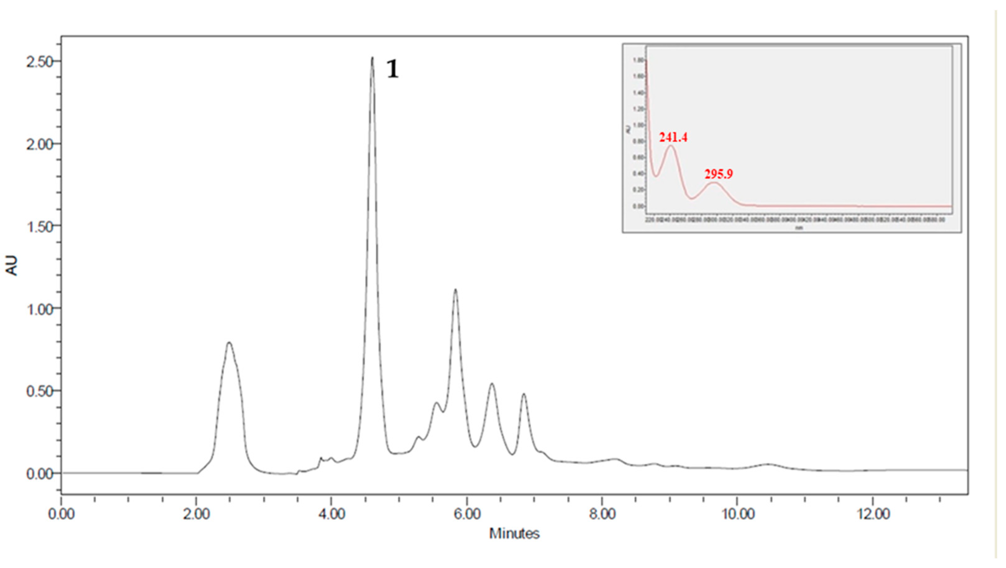
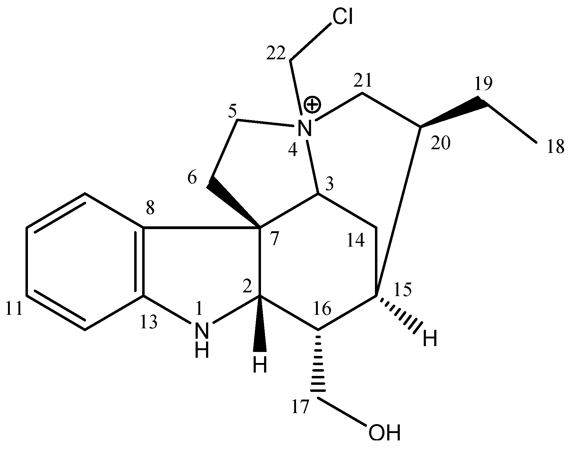
2.1. Phytochemical Analysis

The friction process made it possible to obtain a 2.2% yield of ethanol extract (EE) from the barks of G. sericeum (Table 1). This extract was subjected to fractionation by acid-base partition, obtaining a neutral fraction (NF, 17.3%) and a fraction of alkaloids (AF, 10.7%; Table 1).
Studies in thin-layer chromatography suggest that EE, NF, and AF must contain alkaloids. Due to less complexity, the AF was subjected to fractionation on a Sephadex LH-20 column, giving rise to 18 subfractions. Semi-preparative HPLC analyzed subfraction 6 (F6AF), and the chromatogram obtained showed a peak around 4.5 min with absorption at λ = 241 and 295 nm, compatible with the indole system. Then the height was collected to isolate compound 1 (Figure 2 and Figure 3; Table 2).
Through electrospray ionization mass spectrometry (ESIMS) and nuclear magnetic resonance (NMR) data, it was determined that its molecular formula was C20H28N2OCl (Supplementary Materials Figures S1–S3). In the 13C NMR spectrum of compound 1, 20 carbon signals, three signals from no hydrogenated carbon atoms, nine from methine, seven from methylene, and one from a methyl carbon atom were observed according to DEPT experiments. In the 1H NMR spectrum of compound 1, signals typical of an indole alkaloid [12] at δ 7.29 (d, 7.5, 1H, H-9), δ 6.80 (t, 7.5, 1H, H-10), δ 7.08 (t, 7.5, 1H, H-11) and δ 6.65 (d, 7.7, 1H, H-12) were observed. The signal observed for H-2 at 4.03 (d, 5.5), together with the HMBC correlations observed from H-9 and H-2 to a quaternary carbon atom at δ 53.4 (C-7), indicated that compound 1 is a dihydroindole derivative with an extended fused ring system. Moreover, the presence of characteristic resonances was observed for C-17 CH2OH (δ 3.64 and 3.79; δC 65.6) and C-19–C-18 Et group (δ 1.02 (CH3) and 1.33 (CH2a) and 1.46 (CH2b); δC 11.5 and 23.6, respectively). The CH2OH group was attached at C-16 by the HMBC correlations of H-17a and H-17b with carbons C-2, C-16, and C-15 signals. The Et group was localized at C-20 by the HMBC correlations of H-19 with C-15, C-18, C-20, and C-21 signals. The NMR data suggest that compound 1 is structurally similar to the indole alkaloid geissoschizoline, with some chemical shift changes to carbons C-3, C-5, and C-21 [12].
However, the MS spectrum showed 49 additional mass units for compound 1, compatible with the CH2Cl group. Through the mass pattern characteristic for chlorine, its presence was confirmed in compound 1, m/z 347 [MCl35] (100) and m/z 349 [MCl37] (30). The pattern of HMBC correlations observed for 1 was identical to geissoschizoline, with changes observed due to the additional CH2Cl group. The CH2Cl group was localized at N-4 through HMBC correlation of CH2Cl (δ 5.59 and 5.40) with the carbons C-5 (δ 64.2), C-21 (δ 54.5), and C-3 (δ 77.0). NMR data to compound 1 are shown in Table 2. Thus, the compound 1 was identified as geissoschizoline N4-methylchlorine (Figure 3).

2.2. Antitumor Activity, Cytotoxicity, and Selectivity Index

Gastric cancer cells (ACP02) were submitted to treatment with ethanol extract (EE); neutral fraction (NF); alkaloid fraction (AF), and the alkaloid geissoschizoline N4-methychlorine (compound 1). It was witnessed that the fractionation of EE led to obtaining AF, where alkaloid 1 showed similar significant activities in this cell line (Table 3).
The standard renal cell line (VERO) was also treated in the same manner as the ACP02. In contrast to what was observed for the tumor cell, alkaloid 1 showed a lower cytotoxic effect for the VERO cells, and its selectivity was high for the tumor cell. It should be considered that the fractionation contributed significantly to the increase in the selectivity index. Furthermore, the EE, NF, AF, and alkaloid 1 cytotoxicity were evaluated in human hepatoma (HepG2), showing the lower toxicity of the alkaloid and higher selectivity for activity in gastric tumor cells (Table 3).

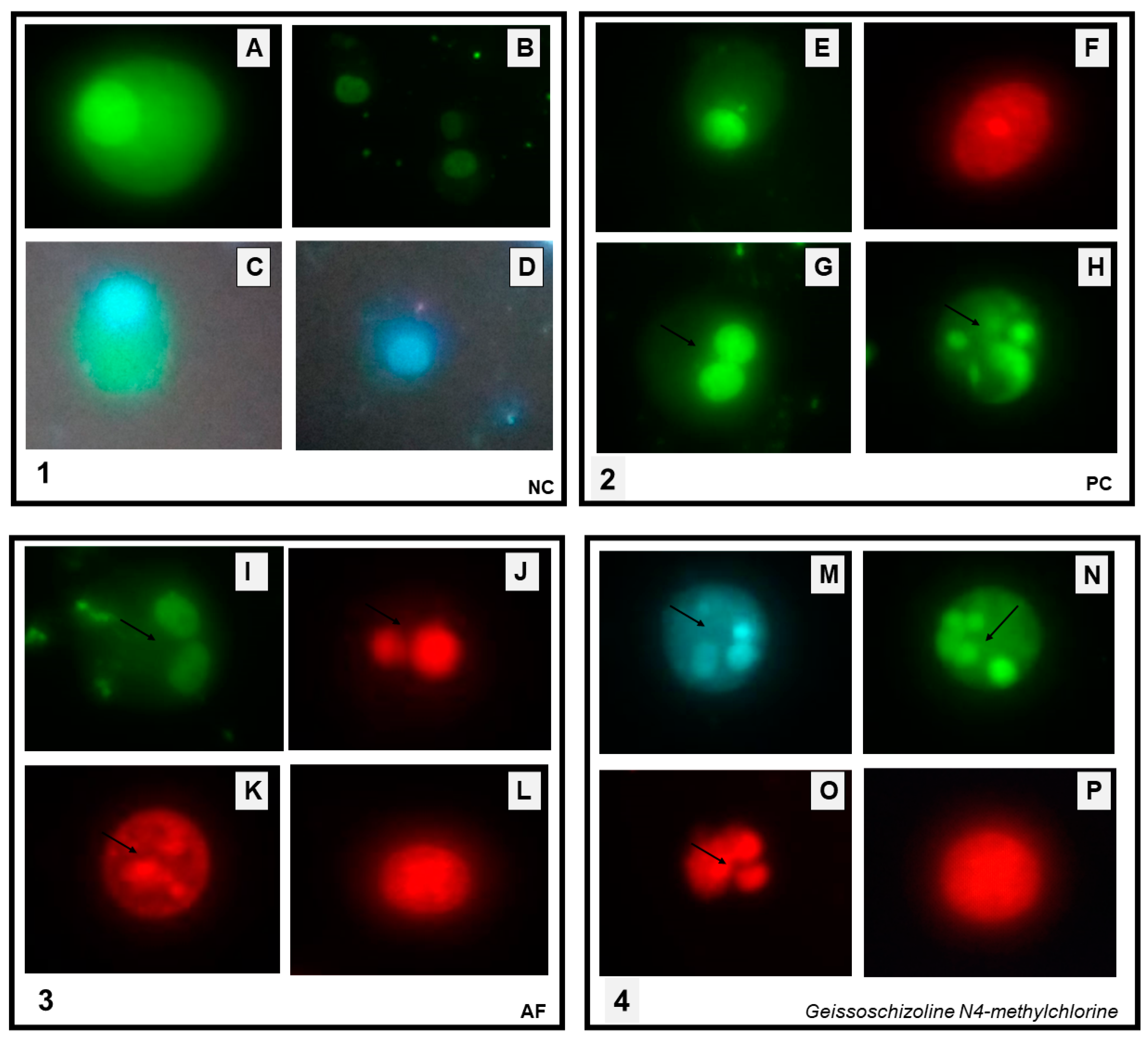
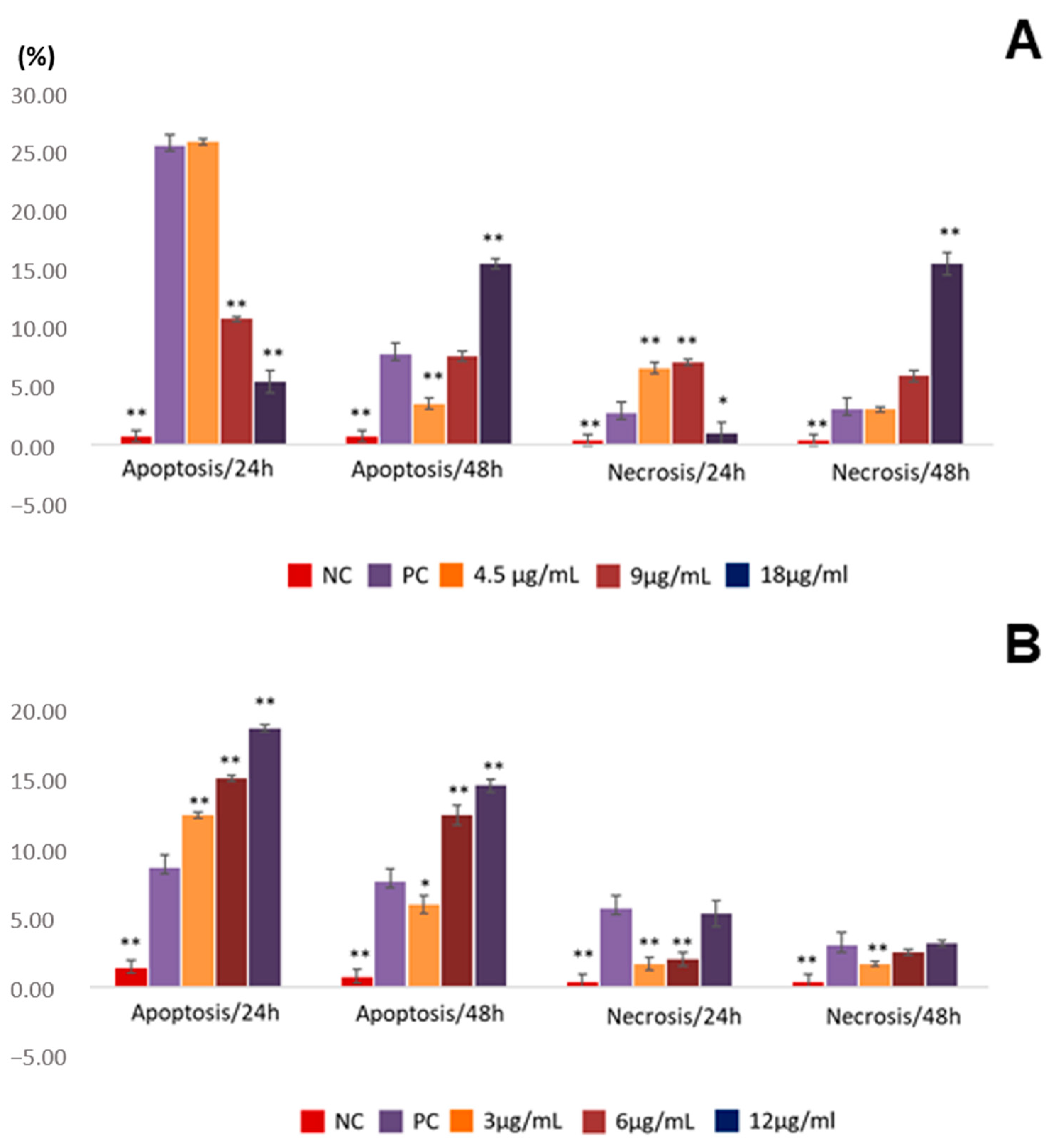
2.3. Assessment of Apoptosis Induction
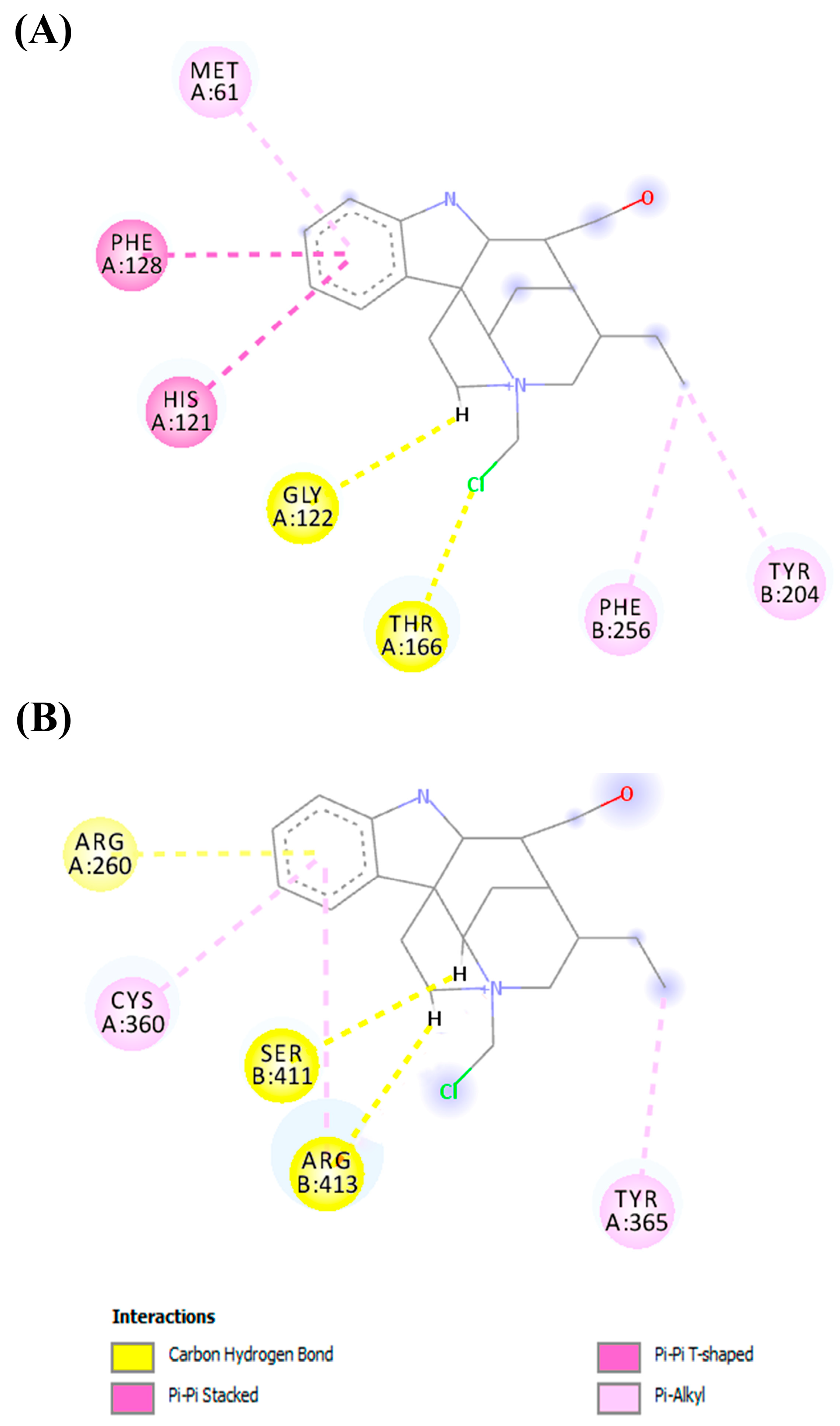
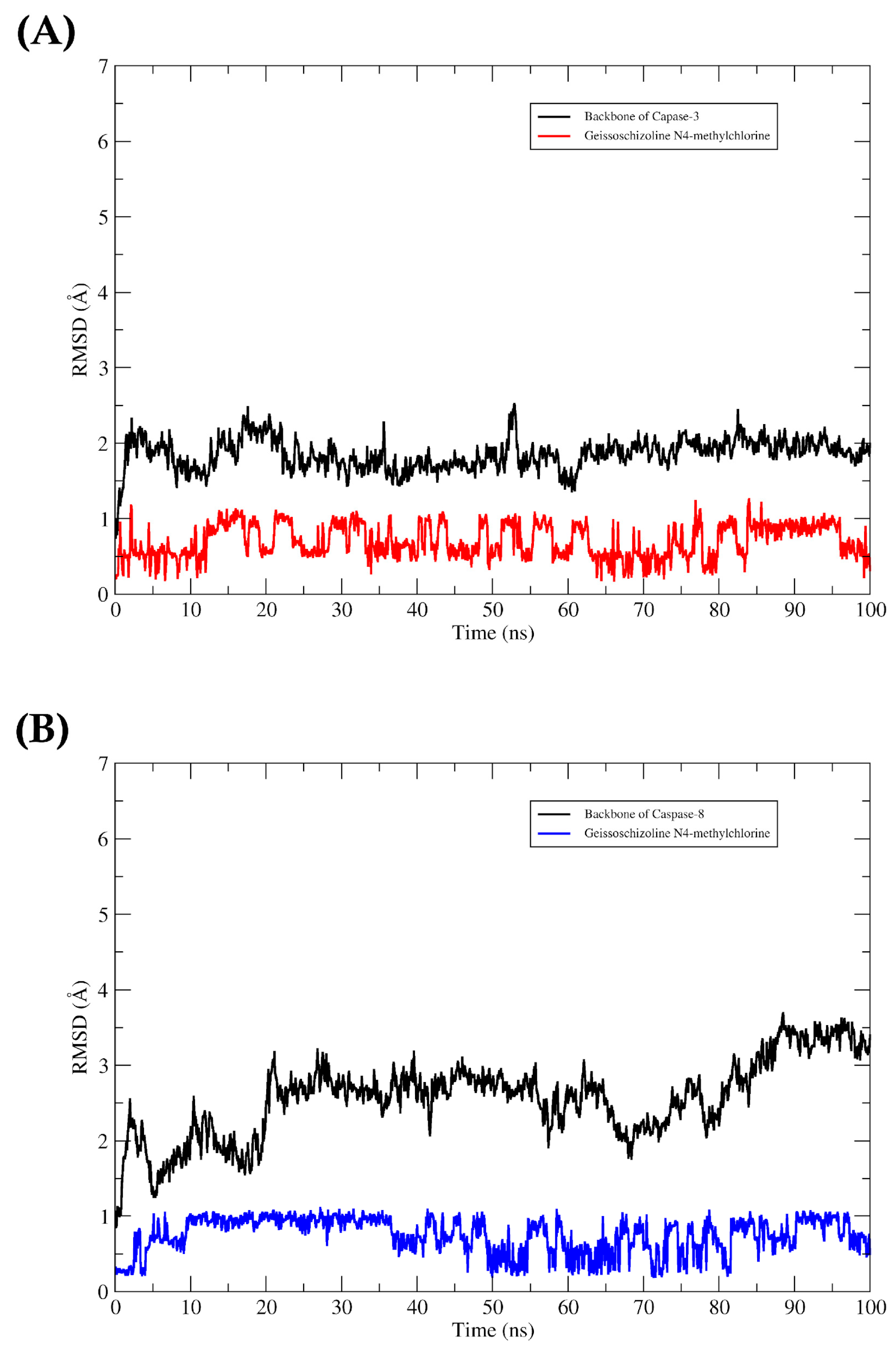
AF and alkaloid 1 were more active in ACP02, so they were submitted to the assay to evaluate cell death by apoptosis or necrosis using doxorubicin as a positive control. Cell death by apoptosis was observed in both AF and alkaloid 1 treated cells.
Apoptotic bodies were visualized in AF-treated cells (Figure 4I–K). Whereas some cells showed no change in the cell membrane, suggesting an early stage of apoptosis (Figure 4I), others already showed membrane changes, possibly in the final stage of apoptosis (Figure 4K). It was also possible to observe permeability changes in some of the cells (Figure 4, quadrant 3 J). In cells treated with alkaloid 1, the following changes were observed: apoptotic bodies (Figure 4M–O); changes in the cell membrane and permeability (Figure 4O); no membrane alteration suggesting apoptosis in the early stages. Similar changes were seen in doxorubicin-treated ACP02 cells (Figure 4G,H).
The lowest concentration of AF induced apoptosis in a proportion equal to that of doxorubicin, approximately 25%, with no statistically significant difference (Figure 5). With increasing concentration, apoptosis and necrosis decrease. With an increase in exposure time (48 h), there is a reduction in apoptosis and necrosis at the lowest concentration and an increase in the proportion of apoptosis and necrosis at the highest concentration (Figure 5).
Differently from the alkaloid fraction, the proportion of apoptosis induced by geissoschizoline N4-methylchlorine at times of 24 and 48 h was concentration-dependent, higher at 12 µg/mL. The highest percentage of apoptosis was observed after 24 h of exposure. The induction of cell death by necrosis was less pronounced at both analyzed times (Figure 5).

2.4. In Silico

The favorable binding mode of the geissoschizoline molecule N4-methylchlorine with the pocket binding of caspase-3 and 8 was chosen according to the Moldock score results. The Moldock score values are summarized in Table 4.
Figure 6 shows the interactions in the complex between the geissoschizoline N4-methylchlorine and caspases 3 and 8. In the binding cavity of the two molecular targets, the ligand interacted with its residues. With caspase 3, the ligand interacted through hydrophobic interactions with the residues of Met61, Phe128, His121, Phe256, and Tyr204. The interactions were Pi-Pi stacked with Phe128, His121, and Pi-Pi T-shaped with Phe256 and Tyr204. In addition, hydrogen bonds were established with Gly122 and Thr166. In the complex formed between geissoschizoline N4-methylchlorine and caspase-8, hydrophobic interactions of the Pi-Alkyl type were formed with Cys360 and Tyr365. Another three hydrogen bonds were formed with Arg260, Ser411, and Arg413.
The RMSD plots showed conformational stabilities of caspases-3 and 8 to the geissoschizoline ligand N4-methylchlorine throughout the 100 ns trajectory, as the complexes remained formed. It was observed that throughout the entire trajectory, the complexes remained formed. The ligand in the two proteins’ binding cavity did not show large deviations in their plots. The time series of RMSD values are shown in Figure 7.
We used the MM-GBSA method to evaluate the system affinity energy after the MD simulations. It was observed that the van der Waals, electrostatic, and non-polar interactions were the ones that most contributed to the formation of the complex. Of these energies, the van der Waals interactions were the ones that most collaborated, reaching values of ΔEvdW = −32.15 and ΔEvdW = −30.64 kcal/mol for the caspase-3 and caspase-8 systems. The sum of all these energies also demonstrates that the complexes formed spontaneously, as observed in the values of ΔGbind = −35.62 kcal/mol and ΔGbind = −30.69 kcal/mol, for caspase-3 and 8, respectively. The values obtained are shown in Table 5.

3. Discussion

In the study of plants, whether the fractionation of the extract influences the biological and cytotoxic activity is considered. This study investigated the antitumor activity of EE, NF, AF, and alkaloid geissoschizoline N4-methylchlorine (compound 1) samples against gastric cancer cells (ACP02), as well as the cytotoxic activity against VERO cells and HepG2 hepatoma cells.
The results showed that, in general, the VERO cell was more sensitive to the deleterious effect of G. sericeum than the HepG2 cells. On the other hand, the fractionation of the ethanol extract contributed to the increase in biological activity, especially of geissoschizoline N4-methylchlorin, with reduced cytotoxicity and increased selectivity index. This factor is significant because cancer chemotherapy often includes drugs that cause cytotoxic effects [19] causing adverse effects, and decreasing patient adherence to treatment, indicating the potential use of this compound in future clinical trials.
Literature data report other indole alkaloids such as geissoschizoline, geissoschizoline N4-oxide, and 1,2-dehydrogeissoschizoline, as well as a β-carboline alkaloid flavopereirine, obtained from the bark of G. sericeum showed activity for the human KB cell line. For the N4 oxide of geissoschizolina and geissoschizolin, the IC50 was >40 μM, while the IC50 of the 1,2-deiidrogeissoschizolina derivative was 40 μM. Flavoperericin also showed moderate cytotoxic activity (IC50 10.7 μM); this alkaloid was the most active in this study. The cytotoxicity of flavopereirin has been cited previously due to its ability to selectively inhibit deoxyribonucleic acid (DNA) synthesis in tumor cells compared to healthy cells [12].
The alkaloid fraction obtained from the shells of G. sericeum made it possible to isolate compound 1, with a high selectivity index for the gastric tumor cell. The results suggest that it is a promising compound for antitumor activity. This may occur because the fractionation of the extract impacts its activity and cytotoxicity [11]. When compound 1 is compared with the alkaloid geissoschizoline N4-oxide [12], it is observed that the oxide group was replaced by methylchlorine. Other studies have shown that halogenated substances contain a biological potential. These substances act as antibacterial, antifungal, antiviral, anti-inflammatory, antiproliferative, anti-fouling, anti-food, cytotoxic, ichthyotoxic, and insecticidal [20,21,22].
Studies of the antitumor activity of Geissospermum are still scarce. However, a report in the literature of an in vitro assay performed with G. vellosii (EGv) extract shows a dose- and time-dependent induction of apoptosis and selectively inhibiting ovarian cancer cell growth [13]. In another study, the evaluation of EGv in metastatic castration-resistant prostate cancer (CRPC) cells showed a concentration and time-dependent response of apoptosis induction and cell cycle arrest. The EGv-induced cell cycle inhibitors, p21 and p27, repressed PCNA, cyclin A, and cyclin D1. Furthermore, it induced upregulation of Bax (pro-apoptotic), reduction of Bcl-2 (anti-apoptotic), Bcl-xL, and XIAP expression, which are associated with cleavage of poly (ADP-ribose) polymerase [23]. Several indole alkaloids such as geissolosimine, geissospermine, geissoschizoline, geissoschizone, and vellosiminol have already been isolated with an elucidated structure from extracts of this species [10], which suggests a possible contribution to this activity.
Alkaloids used in cancer treatment, vinblastine and vincristine, can induce cell death by apoptosis in various tumor cells [24]. In the present study, AF and geissoschizoline N4-methylchlorine induced tumor cell death through apoptosis, and it has been observed that this occurs mainly within 24 h of exposure.
Naturally, the cell cycle is regulated by variable mechanisms because the cells are influenced by the tissue in which they are inserted. Gastric cells, for example, always renew themselves continuously in the cell cycle. However, this factor can be modified in cancer development [25]. This action may not be related only to the cell cycle of tumor cells since cancer cells’ molecular makeup and biological properties can vary significantly, even within the same tumor [26].
In our study, we used the exposure time factor to verify the influence of this aspect on the biological action and defense mechanisms of the cell. Based on its hypothesis, the lowest concentrations of the geissoschizoline N4-methylchlorine alkaloid activate the apoptosis pathway using tools of action similar to specific cyclo-cellular natural products, such as the vinca alkaloids.
The mechanisms involved in cell death by apoptosis are complex, and there are two interconnected signaling pathways: the extrinsic (or cell death receptor pathway) and the intrinsic pathway (or mitochondrial pathway), and molecules of one of them can influence the other [27]. One of the main protein sets responsible for apoptosis is caspases [28], and their activation leads to rapid cell death. Caspases that are involved in the process of apoptosis have been classified into two subtypes based on their mechanism of action: initiator caspases (caspase-2, -8, -9, and -10) and executioner caspases (caspase-3, -6, and -7) [29,30]. It has been reported that activation of caspase-8 and caspase-3 is involved in the mechanism of apoptosis induction in gastric cancer [31].
Several studies have reported the success of molecular docking methodology in investigating the interaction of compounds of natural origin with molecular targets of pharmacological interest (Costa et al., 2020; da Silva Júnior et al., 2021; Leão et al., 2020; Santos et al., 2020). In order to assess whether caspases activation is involved in the antitumor activity of geissoschizoline N4-methylchlorine, docking and molecular dynamics simulations were performed, using caspases 3 and 8 as enzymatic targets, as they are fundamental mediators of the intrinsic and extrinsic pathway of apoptosis, respectively [32].
Our molecular docking results showed that the compounds interact favorably with caspases 3 and 8. The investigation showed hydrophobic interactions between geissoschizoline N4-methylchlorine and caspase-3, with Met61, Phe128, His121, Phe256, and Tyr204. For caspase 3 the Met residue is part of a hydrophobic recess, formed together with Phe55 and Phe128, which harbors His121, and any change in these residues affects the enzyme’s activity [33]. Furthermore, this analysis revealed the selectivity of the alkaloid for caspases, as it interacted with hydrophobic residues in the S2 pocket (Tyr204, Trp206, and Phe256) that are unique to caspases 3 and 7 [34]. The interaction of geissoschizoline N4-methylchlorine with caspase 8 also formed hydrophobic interactions but with the residues of Cys360 and Tyr365. The catalytic triad residues Cys360, His317, and the carbonyl oxygen atom of Arg258 are present in the active site of caspase 8 [35,36]. Interactions with active site residues, separately or together, can generate consequent changes in the enzymatic mechanism [37]. In addition, hydrogen interactions also involved other residues in stabilizing the alkaloid and caspase-3 complexes further, as well as the alkaloid and caspase-8 complexes. In order to take into account the protein flexibility and evaluate the dynamic stability of the predicted interactions between the alkaloid and the caspases, the lowest energy conformations obtained from the coupling were submitted to MD simulations for 100 ns. The RMSD values showed that the complexes remained formed throughout the entire trajectory, and the scoring function analysis highlights the contribution of van der Waals interactions to the binding energy (alkaloid/caspases complex). These data implied the involvement of the intrinsic pathway accompanied by the activation of the extrinsic pathway in the induction of apoptosis during the treatment step with geissoschizoline N4-methylchlorine.
Previous studies have shown that alkaloids show binding affinity with the enzymes caspase-3 [38] and caspase-8 [39] in an in silico study, and that in vitro indole alkaloids induce apoptosis through activation of caspase-3 and 8 [40]. According to these reports, our results show that geissoschizoline N4-methylchlorine can interact reversibly with residues from the active site of caspases 3 and 8 and trigger an inhibitory response in gastric cancer cells, a hypothesis that corroborates the results obtained in vitro.

4. Materials and Methods

4.1. Reagents

Methanol-d4 was purchased from Cambridge Isotope Laboratories Inc. (Miami, FL, USA). Ethanol was purchased from Tedia (São Paulo, SP, Brazil). Roswell Park Memorial Institute 1640 medium (RPMI 1640), [3- (4,5-dimethylthiazol-2-yl) -2,5-diphenyltetrazolium] bromide solution (MTT), dimethylsulfoxide (DMSO), doxorubicin hydrochloride and fluorescein diacetate (FDA) were purchased from Sigma-Aldrich (St. Louis, MO, USA). Fetal bovine serum, phosphate buffered saline (PBS), and Trypsin were purchased from GIBCOTM (made in Brazil), while propidium iodide and Hoechst 33342 were purchased from Invitrogen™ (Carlsbad, CA, USA).

4.2. Equipment

CO2 incubator (Thermo Fisher Scientific, Waltham, MA, USA), microplate spectrophotometer reader (Thermo Fisher Scientific, Waltham, MA, USA), benchtop centrifuge (Benchtop Mikro 220R centrifuge, Hettich, Bäch, Switzerland), fluorescence microscope (Olympus BX41, Olympus Corporation, Tokyo, Japan). HPLC-DAD system (column SunFireTM C18 5 μm; 4.6 × 150 mm; Waters Co., Milford, MA, USA), LC-MS (ACQUITY TQD SYSTEM, Waters Corporation, Milford, MA, USA), and NMR Varian Mercury 300 (Varian, Oxford, UK), Sephadex LH-20 column (Sigma-Aldrich, St. Louis, MO, USA).

4.3. Plant Material, Extraction Procedure and Isolation

The stem barks from Geissospermum sericeum Benth. & Hook.f. ex Miers were collected at Vitória do Xingu region, State of Pará, Brazil (S 2°52′50.5″ W 52°00′29.1″) in 26 January 2013. The species was identified by technician L.C. Batista Lobato of the Emilio Goeldi Museum of Pará, and voucher specimen was incorporated in MG Herbarium (MG209580) of this institution.
The bark powder was macerated in an ethanolic solution, and the maceration product was concentrated in a rotary evaporator giving rise to the ethanolic extract. Then, the ethanolic extract was acid–base partitioned, obtaining the neutral fraction (FN) and the alkaloid fraction (FA). The AF underwent further fractionation in a chromatography column, where Sephadex LH-20 was used as the stationary phase, giving rise to 18 subfractions (F1AF-F18AF). The F6AF subfraction showed a chromatographic profile for alkaloids by TLC analysis. It was selected for alkaloid isolation by high-performance liquid chromatography in preparative mode on an HPLC-PAD Binary pump 1525 (Waters) equipped with PDA (2698 Waters), SunFire™ Prep. C18 OBD™ 5 µm 19 × 150 mm column, using flow rate = 10.0 mL/min, temperature 18 °C, mobile phase water/acetonitrile 8:2 for 12 min, resulting in the isolation of indolic alkaloid geissoschizoline N4-methylchlorine.

4.4. NMR and MS Analysis

The mass spectrum was obtained by direct infusion using negative ion mode on an electrospray ionization source (ESI). To record the NMR spectra, the sample was dissolved in methanol-d4. The chemical shifts are presented in delta (δ) values and the coupling constants (J) in Hertz (Hz).

4.5. Cells

For cytotoxic tests, used tumor lineage of primary gastric adenocarcinoma (ACP02), hepatocellular carcinoma (HepG2), and kidney cells of African green monkey kidney cells Cercopithecus aethiops (VERO) were used. These cells were supplied by the Human Cytogenetics Laboratory (LCH) of the Institute of Biological Sciences of the Federal University of Pará (UFPA). For the apoptosis assay, the tumor line ACP02 was used.

4.6. Antitumor Activity and Cytotoxicity through the Cell Viability Assay (MTT)

The cell viability assay was carried out according to the methodology described by Mosmann et al. [41]. Using 96-well plates, VERO cells were seeded (8 × 10³ cells/mL medium RPMI-1640); ACP02 (8 × 10³ cells/mL medium RPMI-1640), and HepG2 (1 × 10⁴ cells/mL medium RPMI-1640). The plates were incubated at 37 °C in a humid atmosphere with 5% CO2.
After incubation (24 h), the cells were treated in triplicate with seven concentrations (500–7.812 μg/mL) of the ethanol extract, neutral fraction, alkaloid fraction, and geissoschizoline N4-methylchlorine. A triplicate for negative control was also performed, containing only 10% fetal bovine serum. The plates were returned to the oven at 37 °C and 5% CO2 humidity for further incubation for 24 h. Then [3-(4,5-dimethylthiazol-2-yl) -2,5-diphenyltetrazolium bromide] (MTT, 5 mg/mL) solution was added to the wells containing the treatment samples. They were subsequently incubated at 37 °C at 5% CO2 for 4 h. The supernatant was discarded, and 100 µL of dimethylsulfoxide (DMSO) was added to all wells to dissolve formazan crystals. The plates were homogenized for the complete dissolution of the crystals. Around 1 h later, the absorbances of the wells were quantified in a microplate spectrophotometer reader working with a reference wavelength of 570 nm. Cell viability was expressed as a percentage of the control absorbance in the untreated cells after subtracting the appropriate background. Inhibitory concentration (IC50 for ACP02 cells) and cytotoxic concentration (CC50 for VERO and HepG2 cells) were determined by linear regression [11]. The selectivity index (SI) was obtained from the ratio between the IC50 values (Vero or HepG2) and the IC50 of the ACP02 cell.

4.7. Investigation of Necrosis and Apoptosis by Quantifying Cell Death Patterns

The cell lines from primary gastric adenocarcinoma (ACP02) (2.5 × 10⁵ cells) were incubated for 24 h in a 5% CO2 and 37 °C. After, the cells were treated with AF (4.5 μg/mL, 9 μg/mL and 18 μg/mL) and indole alkaloid (geissoschizoline N4-methylchlorine; 3 μg/mL, 6 μg/mL and 12 μg/mL), doxorubicin was used as possible control (1 μM and 1.84 µg/mL) and negative control only cells (2.5 × 10⁵) in culture medium. The cells were incubated again for 24 and 48 h in 5% CO2 at 37 °C. After that, the cells were washed with 3 mL phosphate-buffered saline (PBS; 2×), suspended in 3.0 mL of trypsin, and then centrifuged at 1500 rpm for 5 min. The supernatant was discarded, and the dye mix was prepared with a final volume of 100 μL (25 μL propidium iodide—PI; 50 μL fluorescein diacetate—FDA; 10 μL Hoechst 33342—HO and 15 μL PBS), whose concentrations were 5 μg/mL for PI, 15 μg/mL for FAD, and 2 μg/mL for HO. Then, 100 μL of the cell suspension was mixed into 2 μL of the dye mix, and the mixture was placed in a water bath at 37 °C for 5 min. Subsequently, in one slide, 15 μL of the cell suspension of each sample was added and covered with a coverslip. The cells were analyzed using an OLYMPUS BX41 fluorescence microscope containing three filters (DAPI/FITC/TRITC) by quantifying 300 cells per group. Cells were evaluated according to staining and morphology: in viable cells—green cytoplasm and intact blue nucleus; apoptotic cells—condensed and fragmented blue nucleus, condensed and fragmented red nucleus, without differentiation between early and late apoptosis; and necrotic cells—intact red nucleus. Viability cell determination was performed by dividing viable cells by total cells multiplied by 100.

4.8. Molecular Docking

Our studies used caspases 3 and 8 as target proteins for geissoschizoline N4-methylchlorine. The compound’s molecular structure was optimized with B3LYP/6-31G* [42] in Gaussian 09 [43].
To perform the docking simulation, we used the Molegro Virtual Docker software (MVD, version 5.5) [44] and the 3D structure used as the molecular target can be found in the PDB using ID: 2XYP (caspase 3) [45] and 3KJN (caspase 8) [46]. The MolDock Score (GRID) function was used with a grid resolution of 0.30 Å and a radius of 5 Å encompassing the entire connection cavity. A description of the method can be found in [47].

4.9. Molecular Dynamics (MD) Simulations

Ligand RESP loads were obtained with HF/6-31G [48], and parameters were created using Antechamber [49] and described by GAFF [50]. The MD simulations were performed using the Amber 18 [51,52,53,54]. The tLEaP module was used to add the missing hydrogens to the protein structures. The PDB2PQR server [55] was used to determine the protonation state of the residuals of caspases 3 and 8. The MD simulations were performed using the 14SB force field [56]. TIP3P water molecules [57] were used to solvate the systems in an octahedron periodic box. The distance for the shear radius was 12 Å for all directions. Counterions were added to neutralize the system charges.
Adapted energy minimization protocols, heating, and md production simulations can be found in [58]. The Particle Mesh Ewald method [59] was used to calculate the electrostatic interactions, and the bonds involving hydrogen atoms were restricted with the SHAKE algorithm [60]. The temperature control was performed with the Langevin thermostat [61] within a collision frequency of 2 ps−1.

4.10. Binding Affinities Calculations

For the estimation of the receptor–ligand affinity, energy was calculated with the molecular mechanics/generalized born surface area (MM/GBSA) method according to [62,63,64]. We used the calculations of the last 10 ns of the MD simulation.
The free energy was estimated according to Equation (1):
ΔGbind = ΔEMM + ΔGsolv − TΔS

4.11. Statistical Analysis

The results were submitted to the analysis of variance test (ANOVA), followed by the Tukey test of the Bioestat 5.0 program to compare the various parameters. In all analyses, the level of significance was p < 0.05. The cytotoxic concentration of 50% (CC50) and the inhibitory concentration of 50% (IC50) were calculated using the program GraphPad Prism version 6.0.

5. Conclusions

Bioguided investigation on the plant of G. sericeum showed that the ethanol extract was less active in inhibiting APC02 cells. However, fractionation improved the antitumor response, with the activity and selectivity observed for the alkaloid fraction. Notably, bioguided monitoring also resulted in the isolation of an indole alkaloid geissoschizoline N4-methylchlorine, for the first time described in the literature, with selective cytotoxicity in gastric tumor cells and involvement of apoptosis in the cell death process. Moreover, the study exploring possible mechanisms showed that the induction of cell death might be related to its interactions when coupled to caspase 3 and 8, presented in the docking results and maintained throughout the molecular dynamics. This observation suggests that the geissoschizoline N4-methylchlorine alkaloid may act as a caspase inhibitor, contributing to its antitumor activity.
However, additional validation studies utilizing in vivo models are necessary to substantiate the findings obtained in the present study. In addition, we highlight the scientific community’s interest in investigating natural products for developing new drugs, highlighting the role of the Amazon as a source of actives, especially from the Apocynaceae family, used in traditional medicine in the Amazon.

Supplementary Materials

The following supporting information can be downloaded at: https://www.mdpi.com/article/10.3390/ph16050765/s1. Figure S1: 1H NMR spectrum of compound 1 (300 MHz, MeOD). Figure S2: 13C NMR spectrum of compound 1 (75 MHz, MeOD). Figure S3: Full scan MS spectrum of compound 1 ESI(+).

Author Contributions

Conceptualization, M.L.C.B., J.V.S.-S., M.d.O.B. and M.F.D.; data curation, M.L.C.B., J.V.S.-S., S.P., A.M.d.R.M., M.d.O.B. and M.F.D.; formal analysis, M.L.C.B., J.V.S.-S., S.P., A.M.d.R.M., M.d.O.B. and M.F.D.; funding acquisition, S.P. and M.F.D.; investigation, M.L.C.B., J.V.S.-S., J.N.C., A.R.P.d.S., A.A.B.-R., G.d.C.R., J.E.d.S.S., M.R.C.-F., P.S.B.M., A.M.d.R.M., M.d.O.B. and M.F.D.; methodology, M.L.C.B., J.V.S.-S., A.M.d.R.M., M.d.O.B. and M.F.D.; project administration, M.L.C.B., J.V.S.-S., M.d.O.B. and M.F.D.; resources, M.L.C.B., J.V.S.-S., S.P., A.M.d.R.M., M.d.O.B. and M.F.D.; supervision, A.M.d.R.M., M.d.O.B. and M.F.D.; validation, M.L.C.B., J.V.S.-S., M.R.C.-F., S.P., M.d.O.B. and M.F.D.; visualization, M.L.C.B., J.V.S.-S., A.M.d.R.M., M.d.O.B. and M.F.D.; writing—original draft, M.L.C.B., J.V.S.-S., J.N.C., A.R.P.d.S., A.A.B.-R., G.d.C.R., J.E.d.S.S., M.R.C.-F., S.P., P.S.B.M., A.M.d.R.M., M.d.O.B. and M.F.D.; writing—review and editing, M.L.C.B., J.V.S.-S., J.N.C., A.R.P.d.S., A.A.B.-R., G.d.C.R., J.E.d.S.S., M.R.C.-F., S.P., P.S.B.M., A.M.d.R.M., M.d.O.B. and M.F.D. All authors have read and agreed to the published version of the manuscript.

Funding

This work was supported by Edital Universal 2018 (Processo 432458/2018.2), Coordenação de Aperfeiçoamento de Pessoal de Nível Superior—CAPES, Brazil. PROPESP/UFPA support the publication of this article.

Institutional Review Board Statement

Not applicable.

Informed Consent Statement

Not applicable.

Data Availability Statement

Data is contained within the article and supplementary material.

Conflicts of Interest

The authors declare no conflict of interest.

References

  1. Sung, H.; Ferlay, J.; Siegel, R.L.; Laversanne, M.; Soerjomataram, I.; Jemal, A.; Bray, F. Global Cancer Statistics 2020: GLOBOCAN Estimates of Incidence and Mortality Worldwide for 36 Cancers in 185 Countries. CA Cancer J. Clin. 2021, 71, 209–249. [Google Scholar] [CrossRef] [PubMed]
  2. World Health Organisation. Cancer—Key Facts. 2018. Available online: https://www.who.int/news-room/fact-sheets/detail/cancer (accessed on 9 April 2022).
  3. World Health Organisation. Stomach—Global Cancer Observatory. 2020. Available online: https://gco.iarc.fr/today/data/factsheets/cancers/7-Stomach-fact-sheet.pdf (accessed on 27 April 2023).
  4. Ajani, J.A.; D’Amico, T.A.; Almhanna, K.; Bentrem, D.J.; Chao, J.; Das, P.; Denlinger, C.S.; Fanta, P.; Farjah, F.; Fuchs, C.S.; et al. Gastric Cancer, Version 3.2016, NCCN Clinical Practice Guidelines in Oncology. J. Natl. Compr. Cancer Netw. 2016, 14, 1286–1312. [Google Scholar] [CrossRef] [PubMed]
  5. Barati, N.; Momtazi-Borojeni, A.A.; Majeed, M.; Sahebkar, A. Potential Therapeutic Effects of Curcumin in Gastric Cancer. J. Cell. Physiol. 2019, 234, 2317–2328. [Google Scholar] [CrossRef] [PubMed]
  6. Schirrmacher, V. From Chemotherapy to Biological Therapy: A Review of Novel Concepts to Reduce the Side Effects of Systemic Cancer Treatment (Review). Int. J. Oncol. 2019, 54, 407–419. [Google Scholar] [CrossRef] [PubMed]
  7. Siddiqui, M.; Rajkumar, S.V. The High Cost of Cancer Drugs and What We Can Do about It. Mayo Clin. Proc. 2012, 87, 935–943. [Google Scholar] [CrossRef] [PubMed]
  8. Habli, Z.; Toumieh, G.; Fatfat, M.; Rahal, O.N.; Gali-Muhtasib, H. Emerging Cytotoxic Alkaloids in the Battle against Cancer: Overview of Molecular Mechanisms. Molecules 2017, 22, 250. [Google Scholar] [CrossRef]
  9. Kaur, R.; Arora, S. Alkaloids—Important Therapeutic Secondary Metabolites of Plant Origin. J. Crit. Rev. 2015, 2, 1–8. [Google Scholar]
  10. Mbeunkui, F.; Grace, M.H.; Lategan, C.; Smith, P.J.; Raskin, I.; Lila, M.A. In Vitro Antiplasmodial Activity of Indole Alkaloids from the Stem Bark of Geissospermum Vellosii. J. Ethnopharmacol. 2012, 139, 471–477. [Google Scholar] [CrossRef]
  11. Silva, J.V.d.S.e.; Brigido, H.P.C.; de Albuquerque, K.C.O.; Carvalho, J.M.; Reis, J.F.; Faria, L.V.; Coelho-Ferreira, M.R.; Silveira, F.T.; Carneiro, A.d.S.; Percário, S.; et al. Flavopereirine—An Alkaloid Derived from Geissospermum Vellosii—Presents Leishmanicidal Activity In Vitro. Molecules 2019, 24, 785. [Google Scholar] [CrossRef]
  12. Steele, J.C.P.; Veitch, N.C.; Kite, G.C.; Simmonds, M.S.J.; Warhurst, D.C. Indole and β-Carboline Alkaloids from Geissospermum s Ericeum. J. Nat. Prod. 2002, 65, 85–88. [Google Scholar] [CrossRef]
  13. Yu, J.; Chen, Q. The Plant Extract of Pao Pereira Potentiates Carboplatin Effects against Ovarian Cancer. Pharm. Biol. 2014, 52, 36–43. [Google Scholar] [CrossRef] [PubMed]
  14. Beljanski, M. The Anticancer Agent PB-100, Selectively Active on Malignant Cells, Inhibits Multiplication of Sixteen Malignant Cell Lines, Even Multidrug Resistant. Genet. Mol. Biol. 2000, 23, 29–33. [Google Scholar] [CrossRef]
  15. Beljanski, M.; Beljanski, M.S. Selective Inhibition of in Vitro Synthesis of Cancer DNA by Alkaloids of β-Carboline Class. Pathobiology 1982, 50, 79–87. [Google Scholar] [CrossRef]
  16. Slaninová, I.; Pěnčíková, K.; Urbanová, J.; Slanina, J.; Táborská, E. Antitumour Activities of Sanguinarine and Related Alkaloids. Phytochem. Rev. 2014, 13, 51–68. [Google Scholar] [CrossRef]
  17. Kim, S.; Lee, T.-J.; Leem, J.; Choi, K.S.; Park, J.-W.; Kwon, T.K. Sanguinarine-Induced Apoptosis: Generation of ROS, down-Regulation of Bcl-2, c-FLIP, and Synergy with TRAIL. J. Cell. Biochem. 2008, 104, 895–907. [Google Scholar] [CrossRef] [PubMed]
  18. Tsukamoto, H.; Kondo, S.; Mukudai, Y.; Nagumo, T.; Yasuda, A.; Kurihara, Y.; Kamatani, T.; Shintani, S. Evaluation of Anticancer Activities of Benzo[c]Phenanthridine Alkaloid Sanguinarine in Oral Squamous Cell Carcinoma Cell Line. Anticancer Res. 2011, 31, 2841–2846. [Google Scholar] [PubMed]
  19. Luqmani, Y.A. Mechanisms of Drug Resistance in Cancer Chemotherapy. Med. Princ. Pract. 2005, 14, 35–48. [Google Scholar] [CrossRef]
  20. Abdo, N.Y.M.; Kamel, M.M. Synthesis and Anticancer Evaluation of 1,3,4-Oxadiazoles, 1,3,4-Thiadiazoles, 1,2,4-Triazoles and Mannich Bases. Chem. Pharm. Bull 2015, 63, 369–376. [Google Scholar] [CrossRef]
  21. Yar, M.S.; Akhter, M.W. Synthesis and Anticonvulsant Activity of Substituted Oxadiazole and Thiadiazole Derivatives. Acta Pol. Pharm. 2009, 66, 393–397. [Google Scholar]
  22. Blunt, J.W.; Copp, B.R.; Hu, W.-P.; Munro, M.H.G.; Northcote, P.T.; Prinsep, M.R. Marine Natural Products. Nat. Prod. Rep. 2009, 26, 170–244. [Google Scholar] [CrossRef]
  23. Chang, C.; Zhao, W.; Xie, B.; Deng, Y.; Han, T.; Cui, Y.; Dai, Y.; Zhang, Z.; Gao, J.; Guo, H.; et al. Pao Pereira Extract Suppresses Castration-Resistant Prostate Cancer Cell Growth, Survival, and Invasion through Inhibition of NFκB Signaling. Integr. Cancer Ther. 2014, 13, 249–258. [Google Scholar] [CrossRef] [PubMed]
  24. Fan, M.; Goodwin, M.E.; Birrer, M.J.; Chambers, T.C. The C-Jun NH(2)-Terminal Protein Kinase/AP-1 Pathway Is Required for Efficient Apoptosis Induced by Vinblastine. Cancer Res. 2001, 61, 4450–4458. [Google Scholar] [PubMed]
  25. Bianchi, J.J.; Zhao, X.; Mays, J.C.; Davoli, T. Not All Cancers Are Created Equal: Tissue Specificity in Cancer Genes and Pathways. Curr. Opin. Cell Biol. 2020, 63, 135–143. [Google Scholar] [CrossRef] [PubMed]
  26. Kumar, V.; Abbas, A.K.; Aster, J.C. Robbins y Cotran. Patología Estructural y Funcional; Elsevier Health Sciences: Amsterdam, The Netherlands, 2021; ISBN 84-13-82111-8. [Google Scholar]
  27. Igney, F.H.; Krammer, P.H. Death and Anti-Death: Tumour Resistance to Apoptosis. Nat. Rev. Cancer 2002, 2, 277–288. [Google Scholar] [CrossRef]
  28. D’Arcy, M.S. Cell Death: A Review of the Major Forms of Apoptosis, Necrosis and Autophagy. Cell Biol. Int. 2019, 43, 582–592. [Google Scholar] [CrossRef]
  29. McIlwain, D.R.; Berger, T.; Mak, T.W. Caspase Functions in Cell Death and Disease. Cold Spring Harb. Perspect. Biol. 2013, 5, a008656. [Google Scholar] [CrossRef]
  30. Shalini, S.; Dorstyn, L.; Dawar, S.; Kumar, S. Old, New and Emerging Functions of Caspases. Cell Death Differ. 2015, 22, 526–539. [Google Scholar] [CrossRef]
  31. Gu, Q.; De Wang, J.; Xia, H.H.; Lin, M.C.; He, H.; Zou, B.; Tu, S.P.; Yang, Y.; Liu, X.G.; Lam, S.K.; et al. Activation of the Caspase-8/Bid and Bax Pathways in Aspirin-Induced Apoptosis in Gastric Cancer. Carcinogenesis 2005, 26, 541–546. [Google Scholar] [CrossRef]
  32. Desai, T.H.; Joshi, S.V. In Silico Evaluation of Apoptogenic Potential and Toxicological Profile of Triterpenoids. Indian J. Pharmacol. 2019, 51, 181–207. [Google Scholar] [CrossRef]
  33. Daniel, A.G.; Peterson, E.J.; Farrell, N.P. The Bioinorganic Chemistry of Apoptosis: Potential Inhibitory Zinc Binding Sites in Caspase-3. Angew. Chem. Int. Ed. 2014, 53, 4098–4101. [Google Scholar] [CrossRef]
  34. Lee, D.; Long, S.A.; Adams, J.L.; Chan, G.; Vaidya, K.S.; Francis, T.A.; Kikly, K.; Winkler, J.D.; Sung, C.-M.; Debouck, C.; et al. Potent and Selective Nonpeptide Inhibitors of Caspases 3 and 7 Inhibit Apoptosis and Maintain Cell Functionality. J. Biol. Chem. 2000, 275, 16007–16014. [Google Scholar] [CrossRef]
  35. Jamal, S.; Grover, A.; Grover, S. Machine Learning from Molecular Dynamics Trajectories to Predict Caspase-8 Inhibitors against Alzheimer’s Disease. Front. Pharmacol. 2019, 10, 780. [Google Scholar] [CrossRef] [PubMed]
  36. Watt, W.; Koeplinger, K.A.; Mildner, A.M.; Heinrikson, R.L.; Tomasselli, A.G.; Watenpaugh, K.D. The Atomic-Resolution Structure of Human Caspase-8, a Key Activator of Apoptosis. Structure 1999, 7, 1135–1143. [Google Scholar] [CrossRef]
  37. Carter, P.; Wells, J.A. Dissecting the Catalytic Triad of a Serine Protease. Nature 1988, 332, 564–568. [Google Scholar] [CrossRef]
  38. Cole, E.R.; de Andrade, J.P.; Filho, J.F.A.; Schmitt, E.F.P.; Alves-Araújo, A.; Bastida, J.; Endringer, D.C.; de S. Borges, W.; Lacerda, V. Cytotoxic and Genotoxic Activities of Alkaloids from the Bulbs of Griffinia gardneriana and Habranthus itaobinus (Amaryllidaceae). Anticancer Agents Med. Chem. 2019, 19, 707–717. [Google Scholar] [CrossRef] [PubMed]
  39. Kumar, P.; Rawat, A.; Keshari, A.K.; Singh, A.K.; Maity, S.; De, A.; Samanta, A.; Saha, S. Antiproliferative Effect of Isolated Isoquinoline Alkaloid from Mucuna pruriens Seeds in Hepatic Carcinoma Cells. Nat. Prod. Res. 2016, 30, 460–463. [Google Scholar] [CrossRef] [PubMed]
  40. Fadaeinasab, M.; Karimian, H.; Omar, H.; Taha, H.; Khorasani, A.; Banisalam, B.; Ketuly, K.A.; Abdullah, Z. Reflexin A, a New Indole Alkaloid from Rauvolfia reflexa Induces Apoptosis against Colon Cancer Cells. J. Asian Nat. Prod. Res. 2020, 22, 474–488. [Google Scholar] [CrossRef]
  41. Mosmann, T. Rapid Colorimetric Assay for Cellular Growth and Survival: Application to Proliferation and Cytotoxicity Assays. J. Immunol. Methods 1983, 65, 55–63. [Google Scholar] [CrossRef]
  42. Becke, A.D. Density-functional Thermochemistry. III. The Role of Exact Exchange. J. Chem Phys. 1993, 98, 5648–5652. [Google Scholar] [CrossRef]
  43. Frisch, M.J.; Trucks, G.W.; Schlegel, H.B.; Scuseria, G.E.; Robb, M.A.; Cheeseman, J.R.; Scalmani, G.; Barone, V.; Mennucci, B.; Petersson, G.A.; et al. Gaussian 09; Gaussian Inc.: Wallingford, CT, USA, 2009. [Google Scholar]
  44. Thomsen, R.; Christensen, M.H. MolDock: A New Technique for High-Accuracy Molecular Docking. J. Med. Chem. 2006, 49, 3315–3321. [Google Scholar] [CrossRef]
  45. Ganesan, R.; Jelakovic, S.; Mittl, P.R.E.; Caflisch, A.; Grütter, M.G. In Silico Identification and Crystal Structure Validation of Caspase-3 Inhibitors without a P1 Aspartic Acid Moiety. Acta Crystallogr. Sect. F Struct. Biol. Cryst. Commun. 2011, 67, 842–850. [Google Scholar] [CrossRef] [PubMed]
  46. Wang, Z.; Watt, W.; Brooks, N.A.; Harris, M.S.; Urban, J.; Boatman, D.; McMillan, M.; Kahn, M.; Heinrikson, R.L.; Finzel, B.C.; et al. Kinetic and Structural Characterization of Caspase-3 and Caspase-8 Inhibition by a Novel Class of Irreversible Inhibitors. Biochim. Et Biophys. Acta (BBA) Proteins Proteom. 2010, 1804, 1817–1831. [Google Scholar] [CrossRef] [PubMed]
  47. Cruz, J.N.; Silva, S.G.; Pereira, D.S.; Filho, A.P.d.S.S.; de Oliveira, M.S.; Lima, R.R.; Andrade, E.H.d.A. In Silico Evaluation of the Antimicrobial Activity of Thymol—Major Compounds in the Essential Oil of Lippia thymoides Mart. & Schauer (Verbenaceae). Molecules 2022, 27, 4768. [Google Scholar] [CrossRef] [PubMed]
  48. Cornell, W.D.; Cieplak, P.; Bayly, C.I.; Kollman, P.A. Application of RESP Charges to Calculate Conformational Energies, Hydrogen Bond Energies, and Free Energies of Solvation. J. Am. Chem. Soc. 1993, 115, 9620–9631. [Google Scholar] [CrossRef]
  49. Wang, J.; Wang, W.; Kollman, P.A.; Case, D.A. Automatic Atom Type and Bond Type Perception in Molecular Mechanical Calculations. J. Mol. Graph. Model. 2006, 25, 247–260. [Google Scholar] [CrossRef] [PubMed]
  50. Wang, J.; Wolf, R.M.; Caldwell, J.W.; Kollman, P.A.; Case, D.A. Development and Testing of a General Amber Force Field. J. Comput. Chem. 2004, 25, 1157–1174. [Google Scholar] [CrossRef]
  51. Araújo, P.H.F.; Ramos, R.S.; da Cruz, J.N.; Silva, S.G.; Ferreira, E.F.B.; de Lima, L.R.; Macêdo, W.J.C.; Espejo-Román, J.M.; Campos, J.M.; Santos, C.B.R. Identification of Potential COX-2 Inhibitors for the Treatment of Inflammatory Diseases Using Molecular Modeling Approaches. Molecules 2020, 25, 4183. [Google Scholar] [CrossRef]
  52. Neto, R.d.A.M.; Santos, C.B.R.; Henriques, S.V.C.; Machado, L.d.O.; Cruz, J.N.; da Silva, C.H.T.d.P.; Federico, L.B.; Oliveira, E.H.C.d.; de Souza, M.P.C.; da Silva, P.N.B.; et al. Novel Chalcones Derivatives with Potential Antineoplastic Activity Investigated by Docking and Molecular Dynamics Simulations. J. Biomol. Struct. Dyn. 2022, 40, 2204–2216. [Google Scholar] [CrossRef]
  53. Lima, A.d.M.; Siqueira, A.S.; Möller, M.L.S.; Souza, R.C.d.; Cruz, J.N.; Lima, A.R.J.; Silva, R.C.d.; Aguiar, D.C.F.; Junior, J.L.d.S.G.V.; Gonçalves, E.C. In Silico Improvement of the Cyanobacterial Lectin Microvirin and Mannose Interaction. J. Biomol. Struct. Dyn. 2022, 40, 1064–1073. [Google Scholar] [CrossRef]
  54. de Oliveira, M.S.; da Cruz, J.N.; da Costa, W.A.; Silva, S.G.; Brito, M.d.P.; de Menezes, S.A.F.; Neto, A.M.d.J.C.; Andrade, E.H.d.A.; Junior, R.N.d.C. Chemical Composition, Antimicrobial Properties of Siparuna Guianensis Essential Oil and a Molecular Docking and Dynamics Molecular Study of Its Major Chemical Constituent. Molecules 2020, 25, 3852. [Google Scholar] [CrossRef]
  55. Dolinsky, T.J.; Nielsen, J.E.; McCammon, J.A.; Baker, N.A. PDB2PQR: An Automated Pipeline for the Setup of Poisson-Boltzmann Electrostatics Calculations. Nucleic Acids Res. 2004, 32, W665–W667. [Google Scholar] [CrossRef] [PubMed]
  56. Maier, J.A.; Martinez, C.; Kasavajhala, K.; Wickstrom, L.; Hauser, K.E.; Simmerling, C. Ff14SB: Improving the Accuracy of Protein Side Chain and Backbone Parameters from Ff99SB. J. Chem. Theory Comput. 2015, 11, 3696–3713. [Google Scholar] [CrossRef] [PubMed]
  57. Jorgensen, W.L.; Chandrasekhar, J.; Madura, J.D.; Impey, R.W.; Klein, M.L. Comparison of Simple Potential Functions for Simulating Liquid Water. J. Chem. Phys. 1983, 79, 926–935. [Google Scholar] [CrossRef]
  58. Cruz, J.N.; De Oliveira, M.S.; Silva, S.G.; Filho, A.P.d.S.S.; Pereira, D.S.; Lima, E.A.H.L.; Andrade, E.H.d.A. Insight into the Interaction Mechanism of Nicotine, NNK, and NNN with Cytochrome P450 2A13 Based on Molecular Dynamics Simulation. J. Chem. Inf. Model. 2020, 60, 766–776. [Google Scholar] [CrossRef]
  59. Darden, T.; York, D.; Pedersen, L. Particle Mesh Ewald: An N⋅log(N) Method for Ewald Sums in Large Systems. J. Chem. Phys. 1993, 98, 10089–10092. [Google Scholar] [CrossRef]
  60. Ryckaert, J.-P.; Ciccotti, G.; Berendsen, H.J.C. Numerical Integration of the Cartesian Equations of Motion of a System with Constraints: Molecular Dynamics of n-Alkanes. J. Comput. Phys. 1977, 23, 327–341. [Google Scholar] [CrossRef]
  61. Izaguirre, J.A.; Catarello, D.P.; Wozniak, J.M.; Skeel, R.D. Langevin Stabilization of Molecular Dynamics. J. Chem. Phys. 2001, 114, 2090–2098. [Google Scholar] [CrossRef]
  62. Almeida, V.M.; Dias, Ê.R.; Souza, B.C.; Cruz, J.N.; Santos, C.B.R.; Leite, F.H.A.; Queiroz, R.F.; Branco, A. Methoxylated Flavonols from Vellozia dasypus Seub Ethyl Acetate Active Myeloperoxidase Extract: In Vitro and in Silico Assays. J. Biomol. Struct. Dyn. 2021, 40, 7574–7583. [Google Scholar] [CrossRef]
  63. Castro, A.L.G.; Cruz, J.N.; Sodré, D.F.; Correa-Barbosa, J.; Azonsivo, R.; de Oliveira, M.S.; Siqueira, J.E.d.S.; Galucio, N.C.D.R.; Bahia, M.D.O.; Burbano, R.M.R.; et al. Evaluation of the Genotoxicity and Mutagenicity of Isoeleutherin and Eleutherin Isolated from Eleutherine Plicata Herb. Using Bioassays and in Silico Approaches. Arab. J. Chem. 2021, 14, 103084. [Google Scholar] [CrossRef]
  64. Mascarenhas, A.M.S.; de Almeida, R.B.M.; Neto, M.F.d.A.; Mendes, G.O.; da Cruz, J.N.; dos Santos, C.B.R.; Botura, M.B.; Leite, F.H.A. Pharmacophore-Based Virtual Screening and Molecular Docking to Identify Promising Dual Inhibitors of Human Acetylcholinesterase and Butyrylcholinesterase. J. Biomol. Struct. Dyn. 2021, 39, 6021–6030. [Google Scholar] [CrossRef]
Figure 1. Alkaloids isolated from Geissospermum sericeum. (a) geissoschizoline, (b) geissoschizoline N4-oxide, (c) 1,2-dehydrogeissoschizoline, (d) flavopereirine.
Pharmaceuticals 16 00765 g001
Figure 2. Chromatographic HPLC profile of the subfraction F6AF obtained from the stem barks of G. sericeum. (1) geissoschizoline N4-methylchlorine isolated compound. Condition: SunFire™ Prep. C18 OBD™ 5 µm 19 × 150 mm column, flow rate = 10.0 mL/min, temperature 18 °C. Mobile phase—t = 0 min: 80% water and 20% acetonitrile; t = 10 min: 80% water and 20% acetonitrile.
Pharmaceuticals 16 00765 g002
Figure 3. Compound structure of geissoschizoline N4-methylchlorine (1).
Pharmaceuticals 16 00765 g003
Figure 4. Micrographs of ACP02 cells after exposure to doxorubicin, AF, and geissoschizoline N4-methylchlorine for 48 h. 1—Negative control (NC): (AD)—viable cells with intact nuclei and cytoplasm. Presence of cells stained with Hoechst 33342 (HO) that binds to DNA staining the nucleus in blue and with fluorescein diacetate (DAF) staining the cytoplasm in green. 2—Cells treated with the positive control (PC; doxorubicin, 1 µM) (E) viable cells, (F) necrotic cells stained red by propidium iodide (PI); (G,H) apoptotic cells with emphasis on the presence of apoptotic bodies (arrows). 3—Cells treated with the alkaloid fraction of G. sericeum (AF, 18 µg/mL): (IK)—apoptotic cells with emphasis on the presence of apoptotic bodies (arrows); (L)—necrotic cells stained red by propidium iodide. 4—Cells treated with isolated indole alkaloid (geissoschizoline N4-methylchlorine, 12 µg/mL): (MO)—apoptotic cells with the presence of apoptotic bodies (arrows), (P)—necrotic cells stained red by PI.
Pharmaceuticals 16 00765 g004
Figure 5. Frequency of apoptosis and necrosis in ACP02 cells exposed to AF and geissoschizoline N4-methylchlorine. (A) alkaloid fraction of G. sericeum (AF); (B) geissoschizoline N4-methylchlorine. NC, negative control; PC, positive control. * p< 0.05; ** p < 0.01. The data represent the mean ± standard deviation. * p  <  0.05 and ** p  <  0.001.
Pharmaceuticals 16 00765 g005
Figure 6. Interactions were established between geissoschizoline N4-methylchlorine and the binding pocket of (A) caspase-3 and (B) caspase 8.
Pharmaceuticals 16 00765 g006
Figure 7. RMSD values for 100 ns of MD simulation. Complexes formed between geissoschizoline N4-methylchlorine and (A) caspase-3 and (B) caspase 8.
Pharmaceuticals 16 00765 g007
Table 1. Phytochemical characterization of ethanol extract of G. sericeum and its fractions.
SamplesYield (%)TLC
UV LightDragendorff
EE2.2++
NF17.3+ +
AF10.7+ +
TLC, thin layer chromatographic; EE, ethanol extract; NF, neutral fraction; AF, alkaloid fraction.
Table 2. 1H- and 13C NMR data for F3F6FA (300 MHz, CD3OD).
Position1H-NMR13C-NMRHMBC
1
24.03 (d, 5.5)64.3C-3, C-7, C-15, C-16
33.72 (dd, 14.5, 4.2)77.0C-8, C-15, C-22
4
53.85 (dd, 12.0, 6.8)
3.96 (ddd, 12.0, 11.8, 8.1)
64.2C-3, C-7
62.66 (m)35.1C-7, C-8
7 53.4
8 134.2
97.29 (d, 7.5)123.7C-7, C-11, C-13
106.80 (t, 7.5)121.0C-8, C-12
117.08 (t, 7.5)130.4C-9, C-13
126.65 (d, 7.7)111.4C-8, C-10
13 151.0
142.43 (dd, 15.0, 2.6)
1.87 (dt, 15.0, 3.1)
23.3C-15
151.74 (br)28.7
162.16 (m)34.8C-17, C-20
173.64 (dd, 10.5, 6.8)
3.79 (dd, 10.5, 9,7)
65.6C-2, C-15, C-16
181.02 (t, 7.4)11.5C-19, C-20
191.33 (m)
1.46 (m)
23.6C-15, C-18, C-20, C-21
201.96 (m)39.2C-16
213.42 (t, 14.0)
3.71 (m)
54.2C-20, C-22
225.40 (d, 10.0)
5.59 (d, 10.0)
67.1C-3, C-5, C-21
The numbers in parentheses are J values in Hz.
Table 3. Antitumor activity (IC50), cytotoxicity (CC50), and selective index (SI) of G. sericeum.
SamplesACP02Vero CellsHepG2 Cells
IC50 (µg/mL)CC50 (µg/mL)SIACP02CC50 (µg/mL)SIACP02
EE131.55 ± 0.49113.65 ± 0.350.86472.55 ± 0.633.59
NF114.45 ± 0.21130.10 ± 0.421.13259.30 ± 0.422.27
AF18.29 ± 0.02173.3 ± 0.329.48299.45 ± 0.3516.37
Geissoschizoline N4-methylchlorine12.06 ± 0.04476.0 ± 0.5439.4750.5 ± 0.2841.75
Data represent mean ± SD of at least three experiments realized in triplicate. EE, ethanol extract; NF, neutral fraction; AF, alkaloid fraction; IC50: inhibitory concentration for 50% of cells; IC50: cytotoxic concentration for 50% of cells; SI = CC50 cells/IC50 cells.
Table 4. Moldock score results for the investigated complexes.
CompoundMolecular Target
Caspase-3Caspase-8
Geissoschizoline N4-methylchlorine−49.72−56.31
Table 5. Binding energy values and energy components in kcal/mol.
ComplexΔEvdWΔEeleΔGGBΔGNPΔGbind
Caspase-3−32.15−9.3410.38−4.51−35.62
Caspase-8−30.64−8.8412.42−3.63−30.69
ΔEvdW, contributions by van der Waals; ΔEele, electrostatic energy in the gas phase; ΔGGB, polar solving energy; ΔGnp, nonpolar solvation energy; ΔGbind, binding affinity.
Disclaimer/Publisher’s Note: The statements, opinions and data contained in all publications are solely those of the individual author(s) and contributor(s) and not of MDPI and/or the editor(s). MDPI and/or the editor(s) disclaim responsibility for any injury to people or property resulting from any ideas, methods, instructions or products referred to in the content.

