
Fluorescent Probes as a Tool in Diagnostic and Drug Delivery Systems
 , , and
Abstract
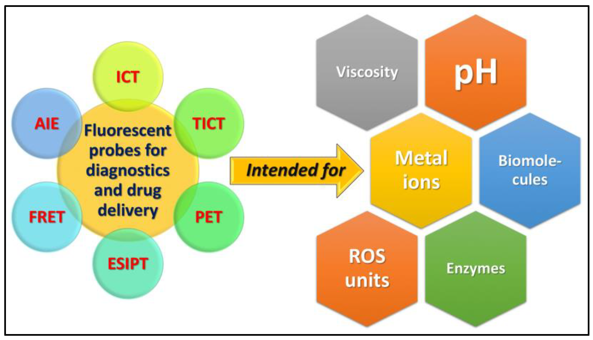
:1. Introduction
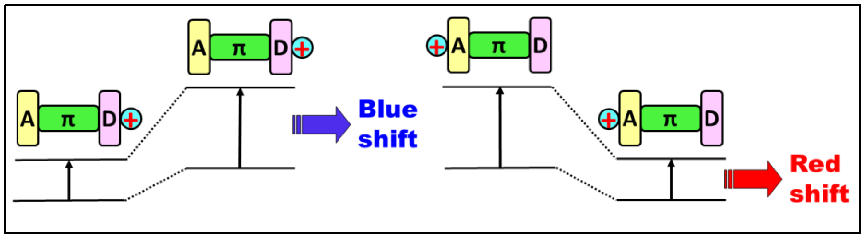
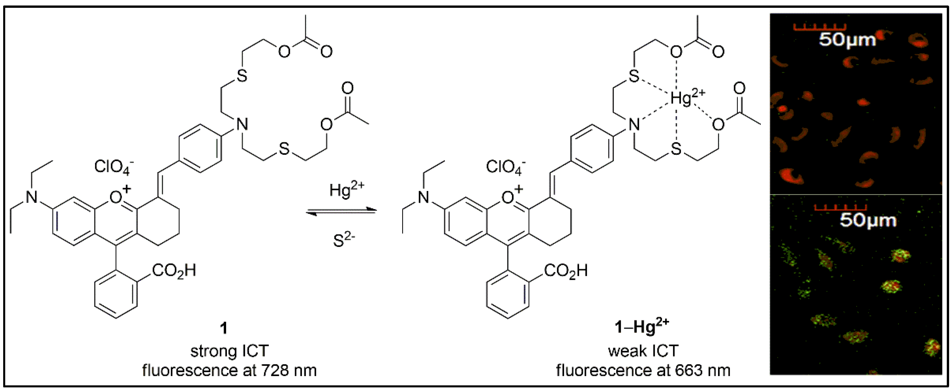
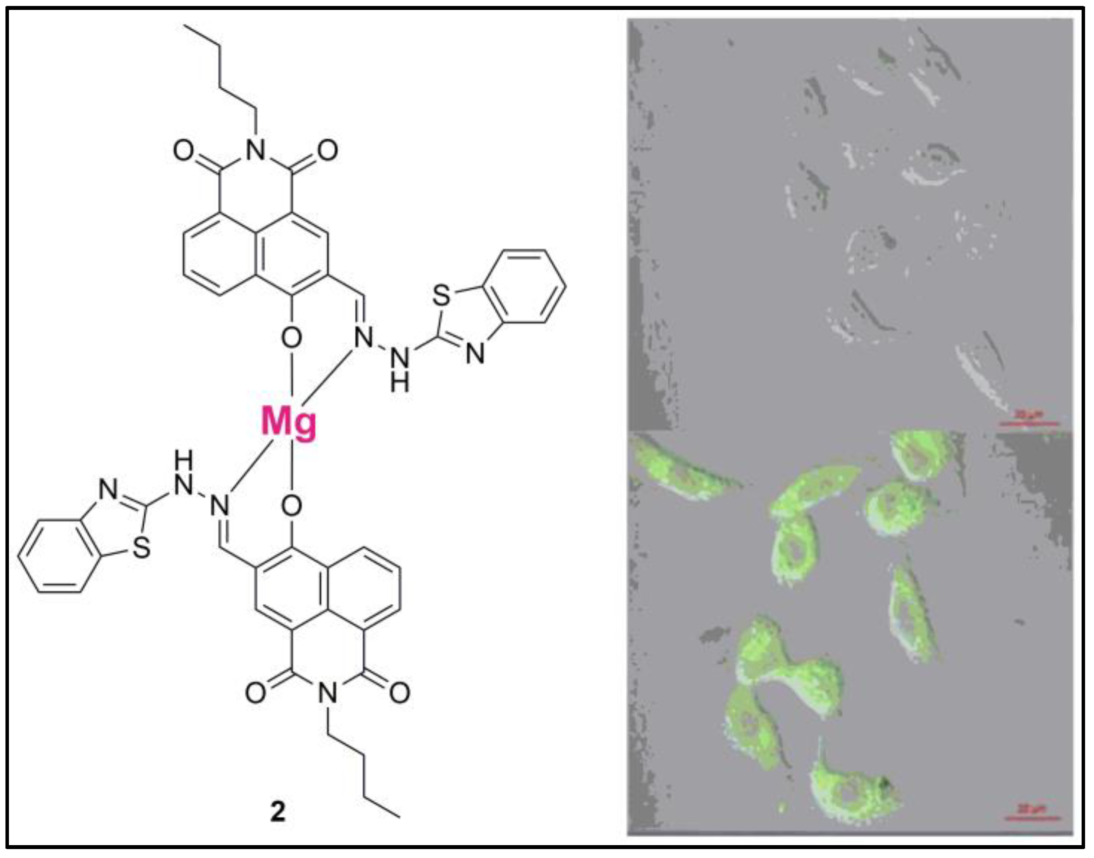
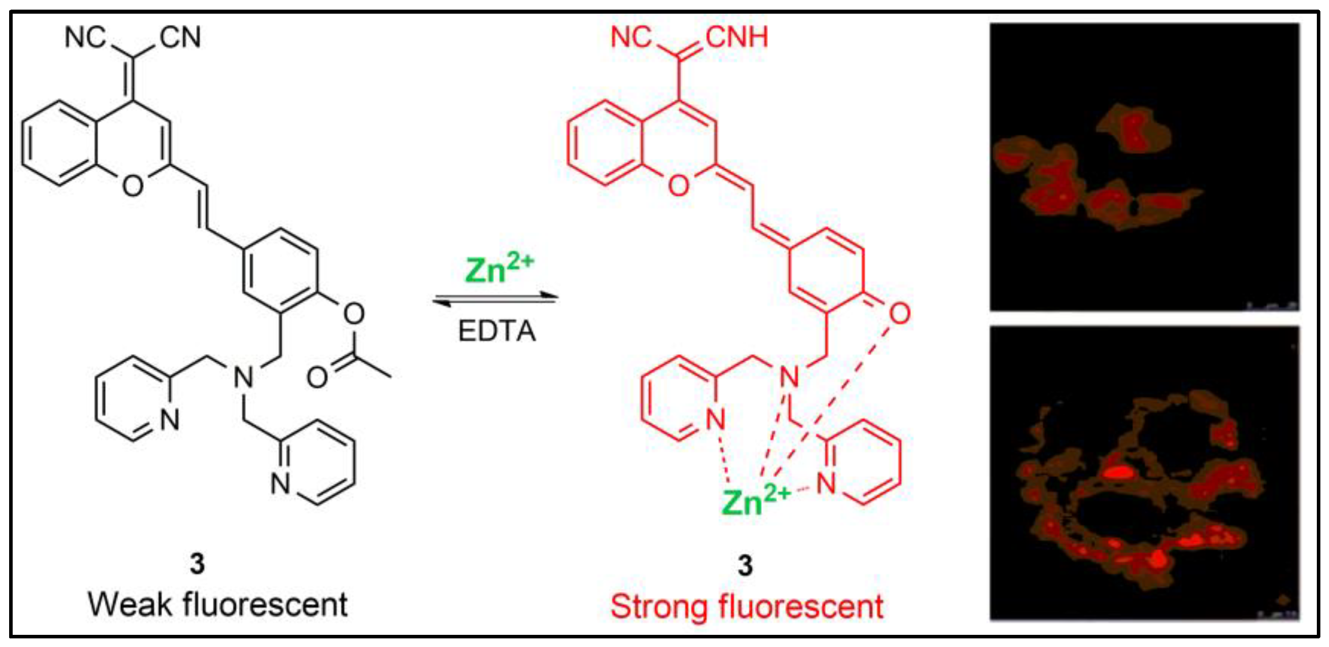
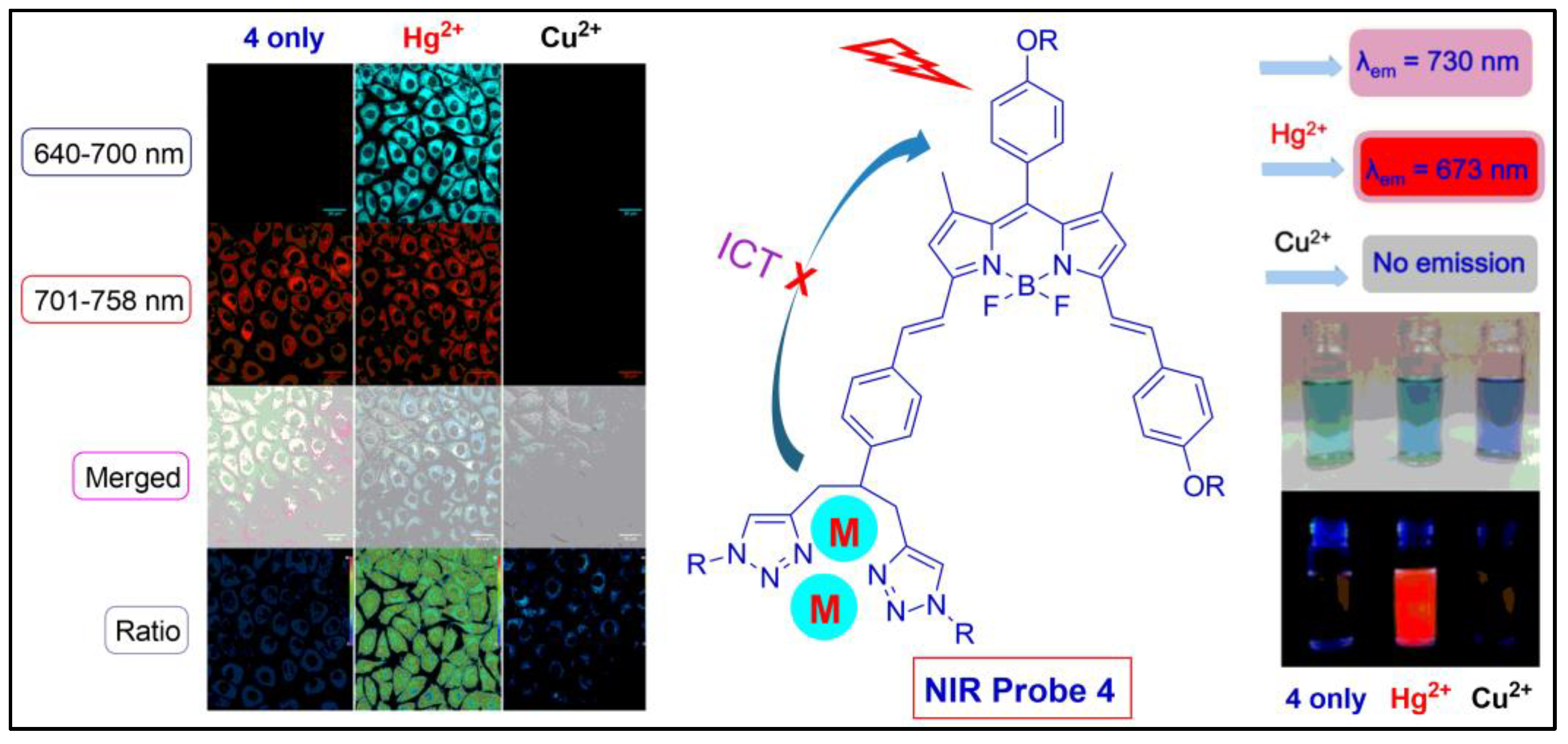
2. Fluorescent Probes Based on Intramolecular Charge Transfer (ICT)
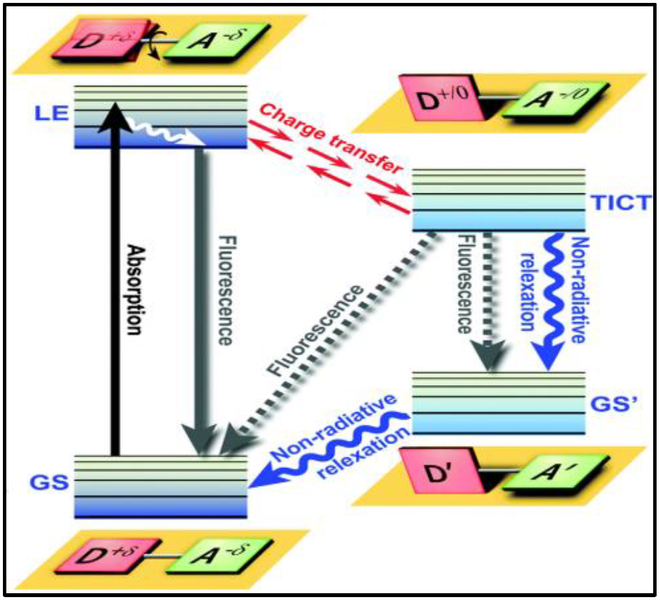
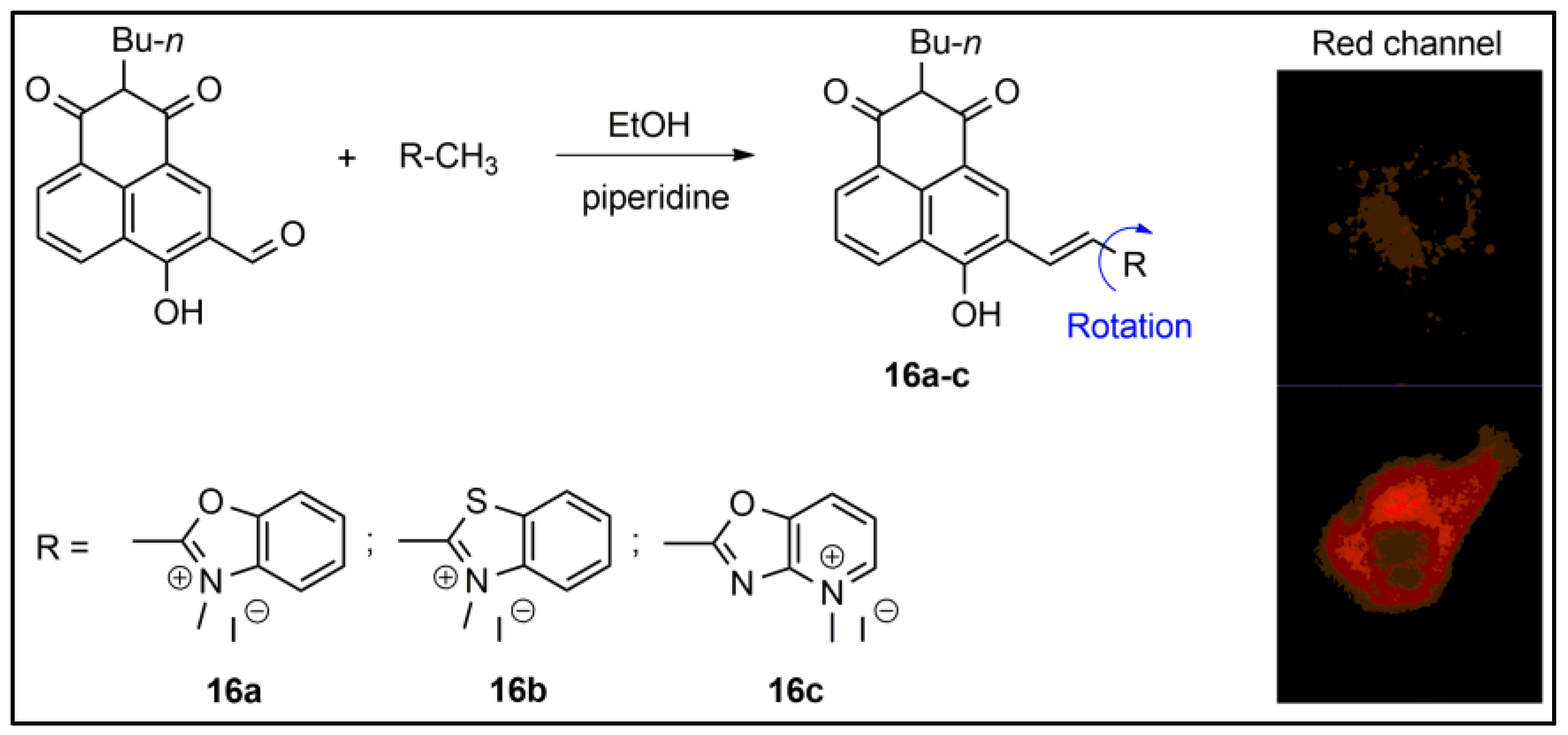
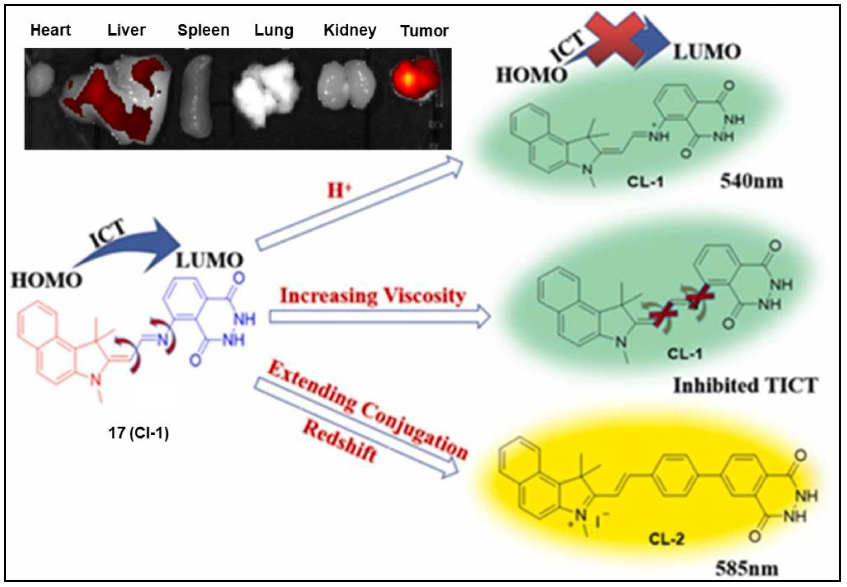
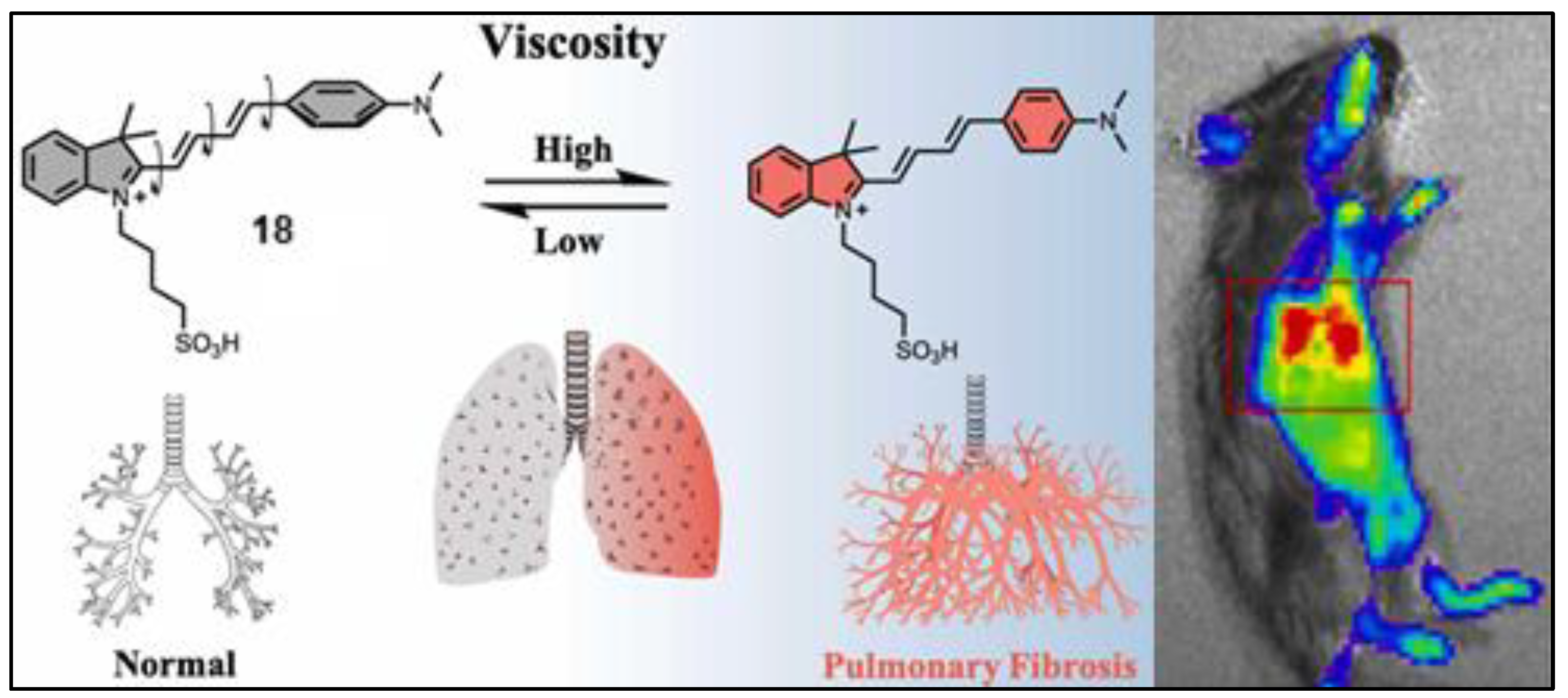
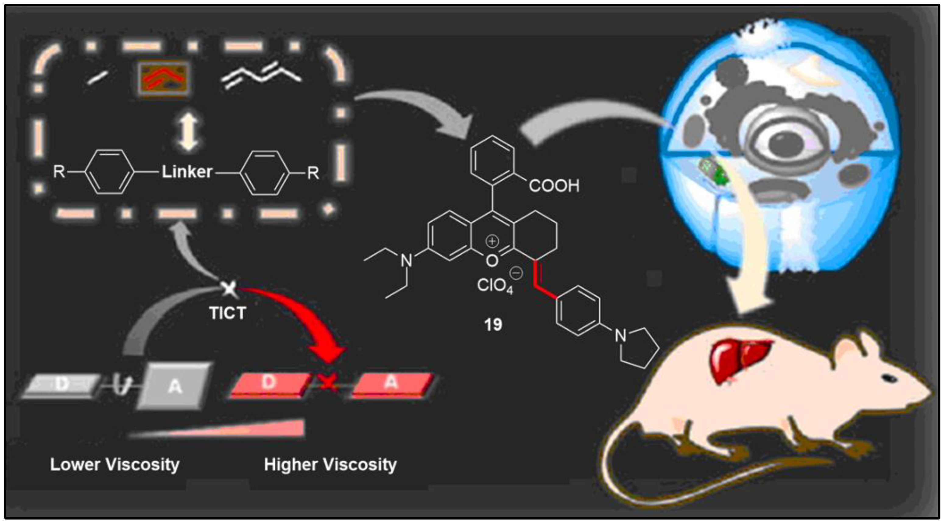
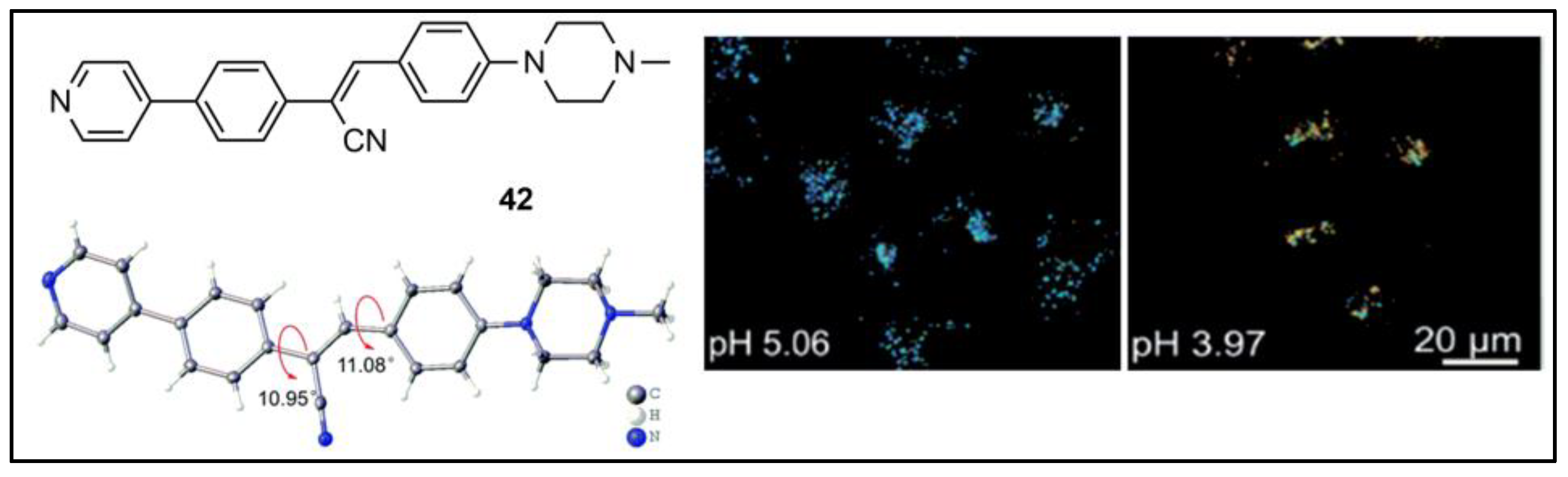
3. Fluorescent Probes Based on Twisted Intramolecular Charge Transfer (TICT)
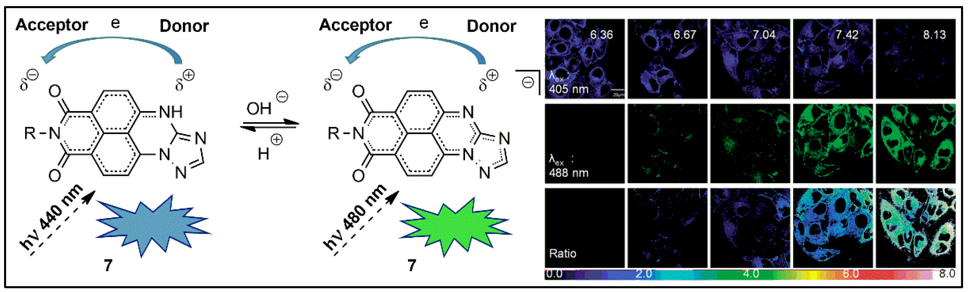
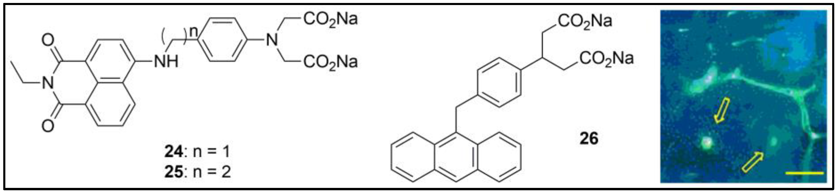
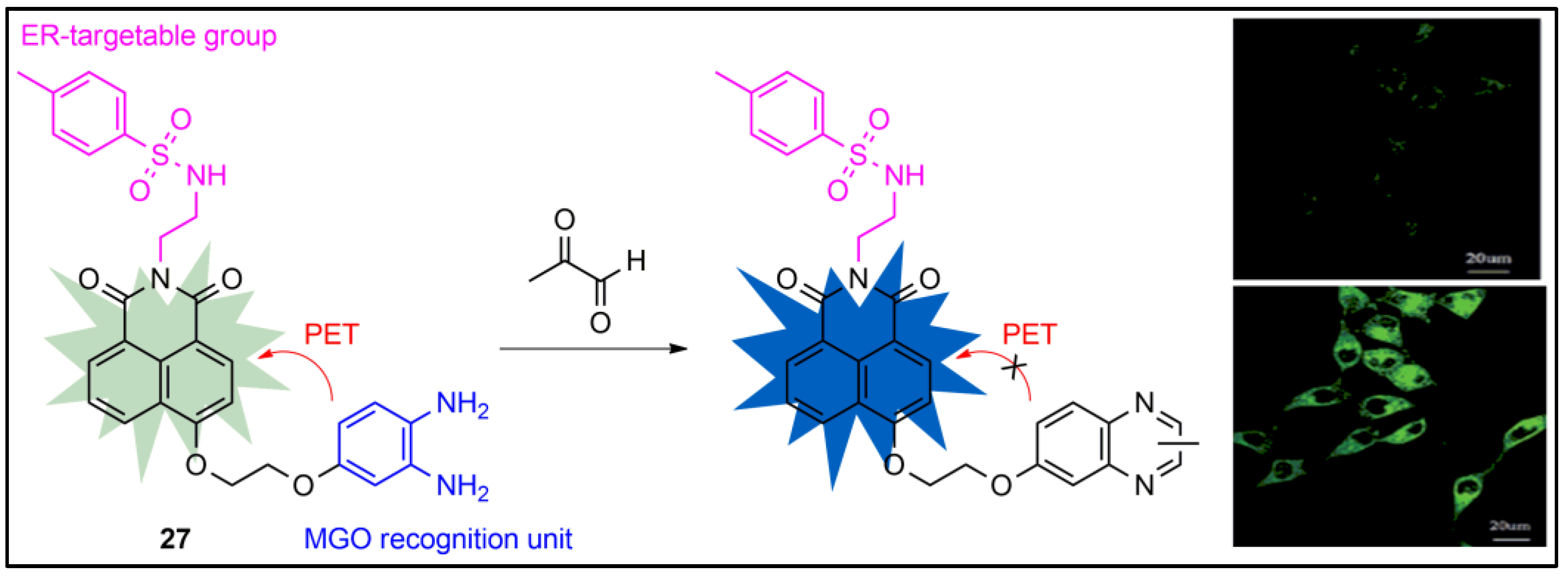
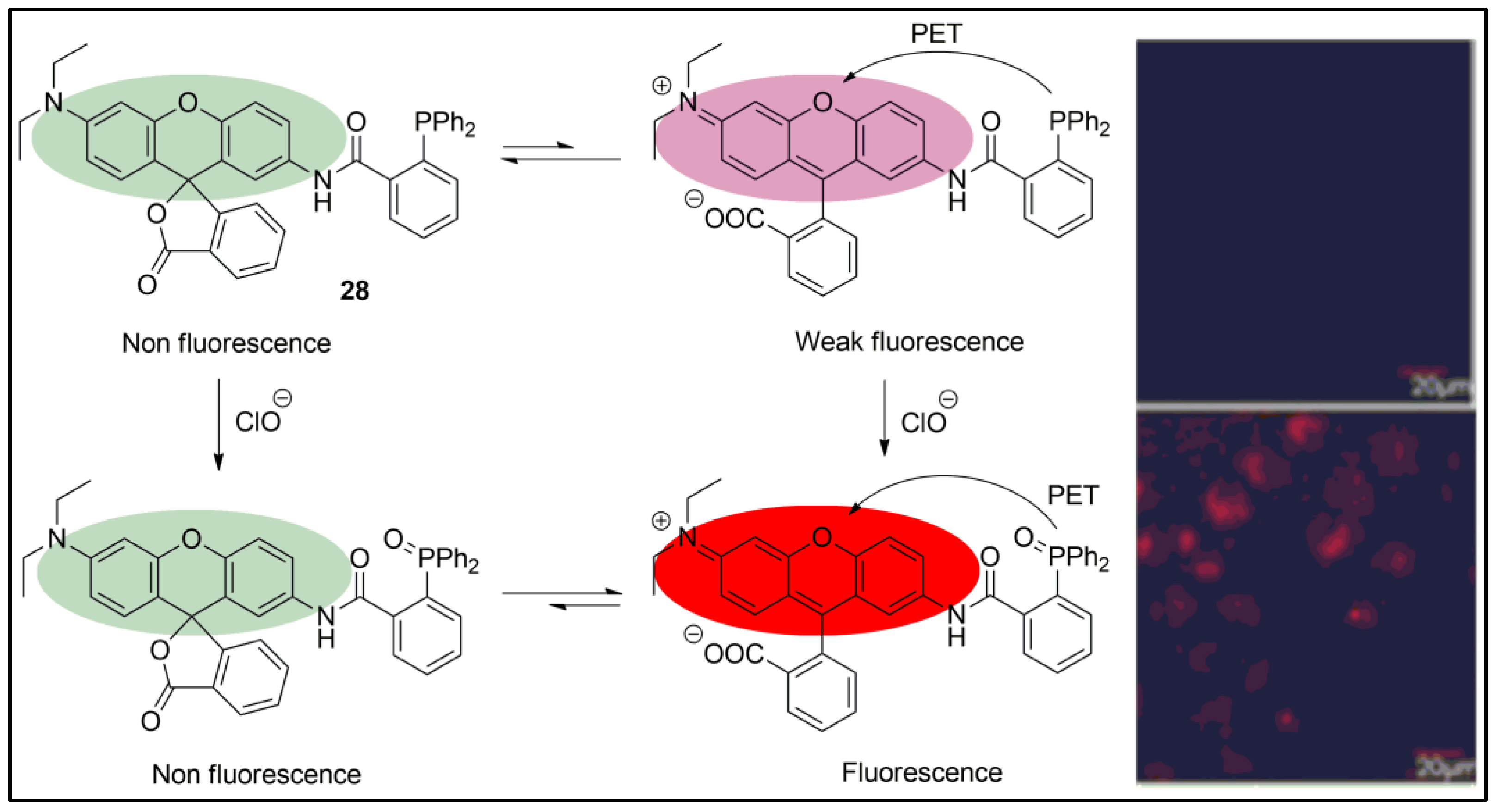
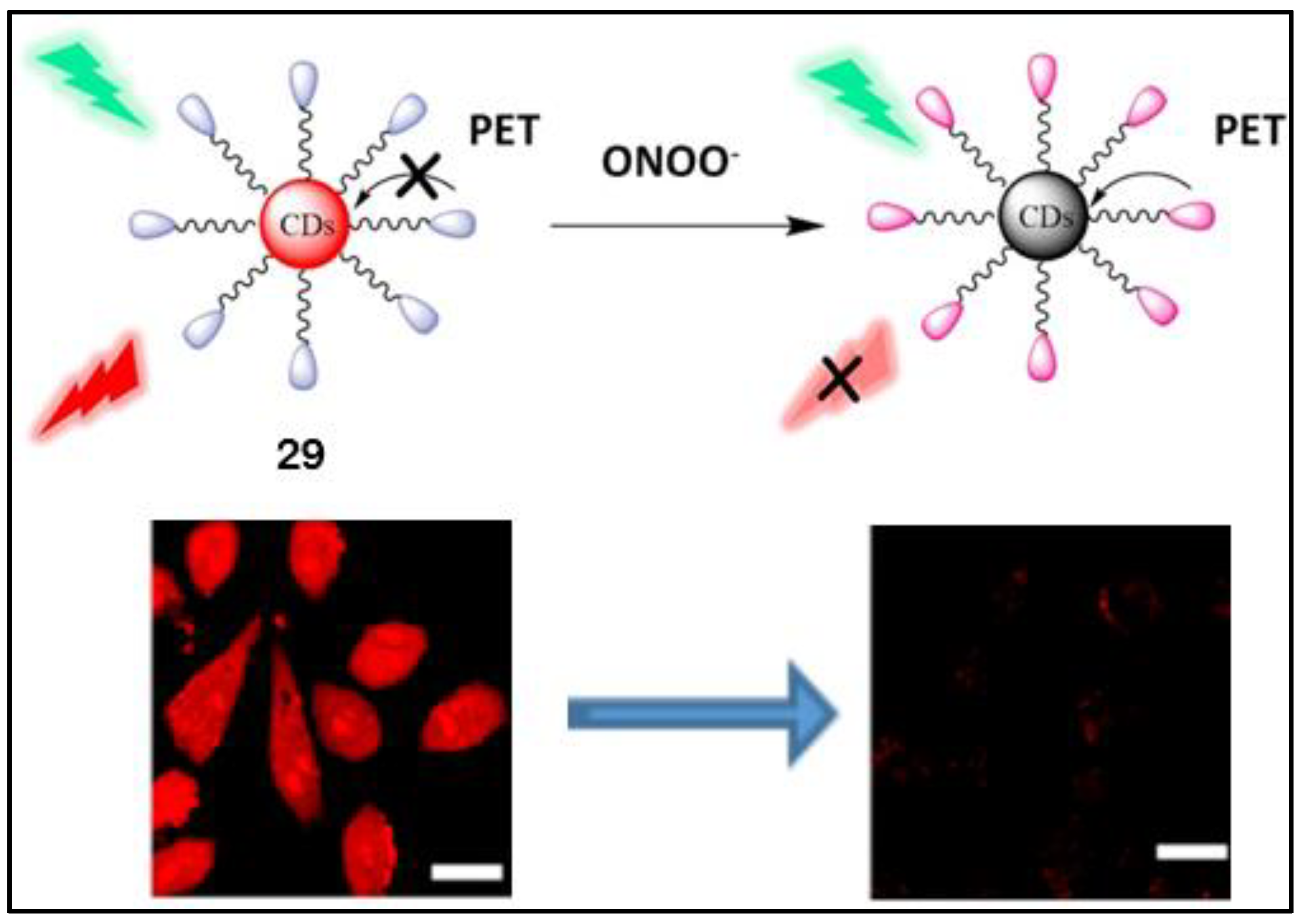
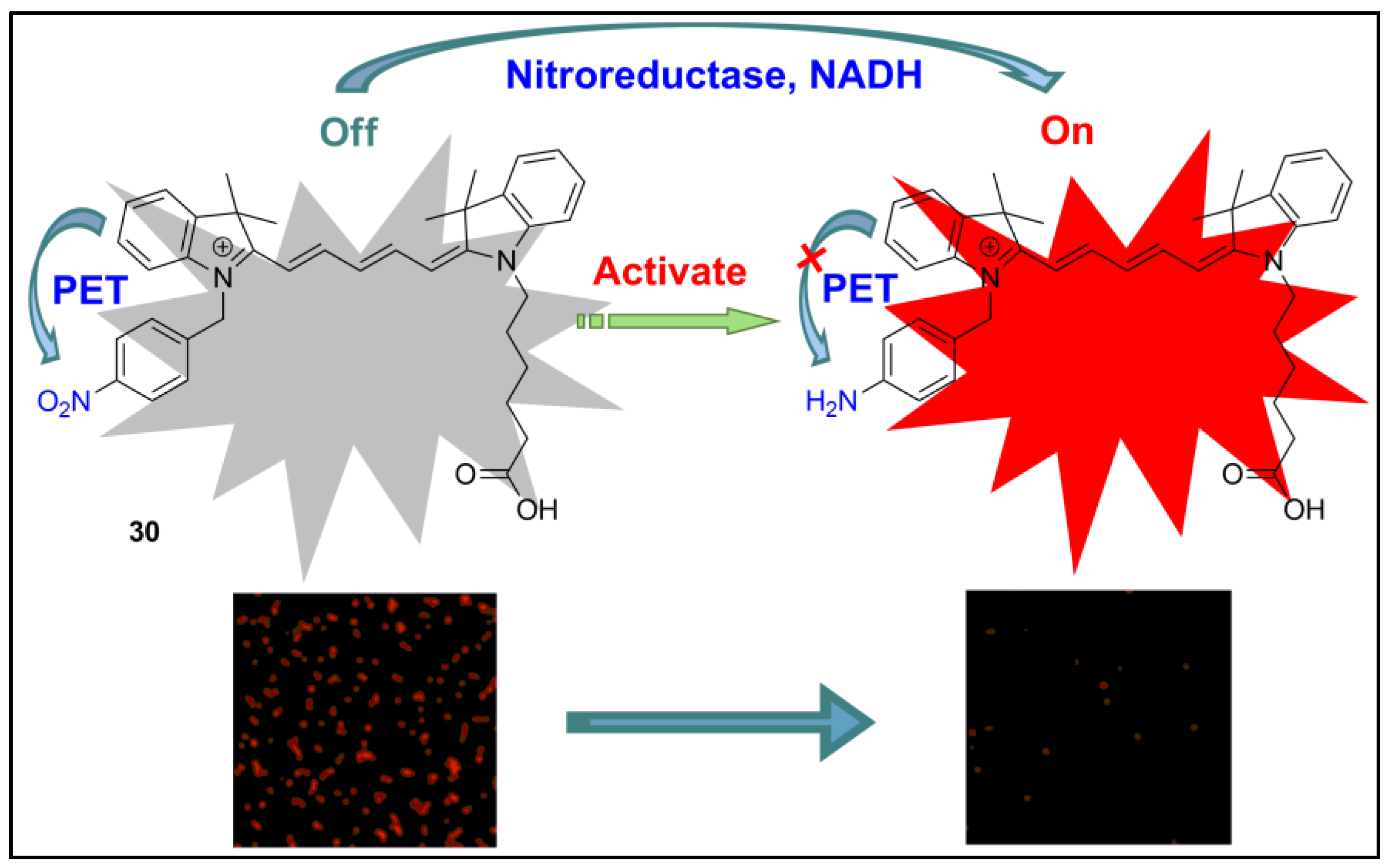
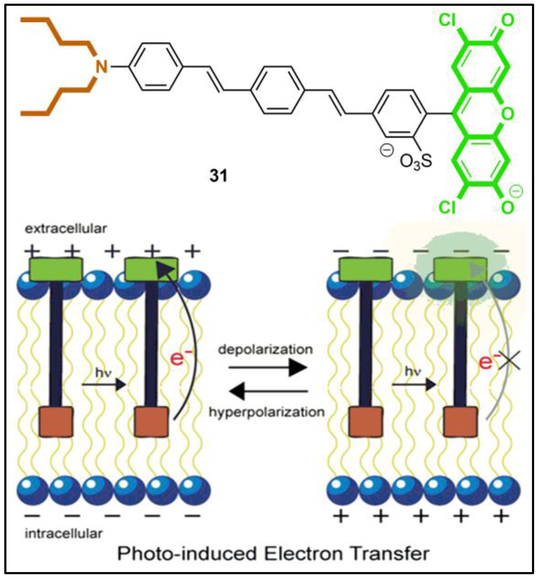
4. Fluorescent Probes Based on Photoinduced Electron Transfer (PET)
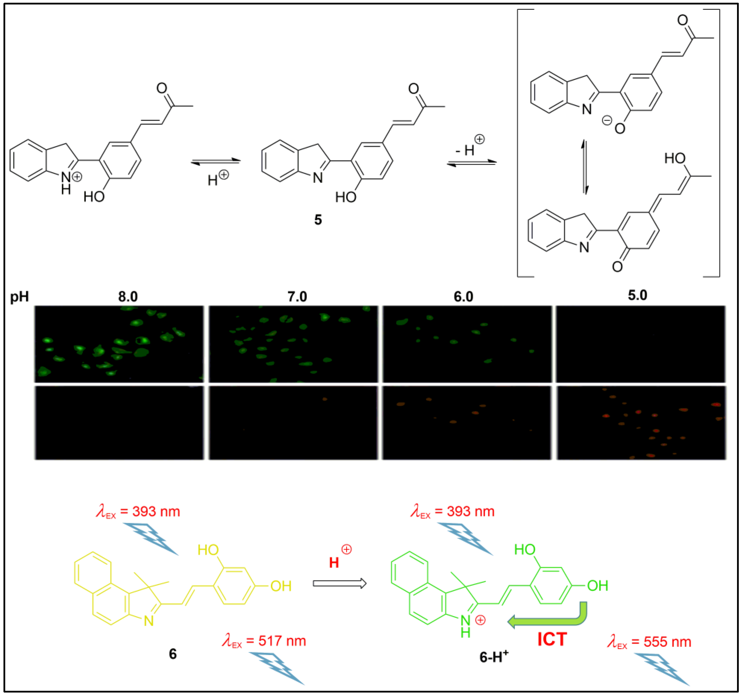
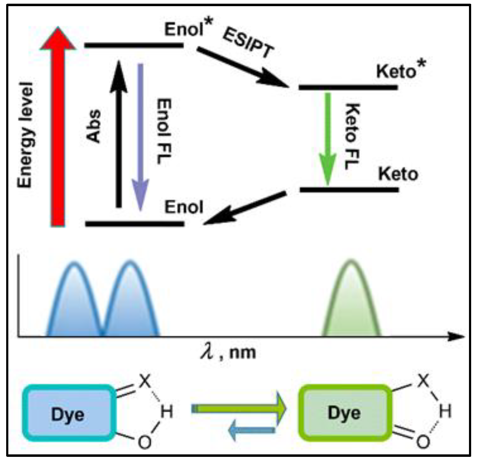
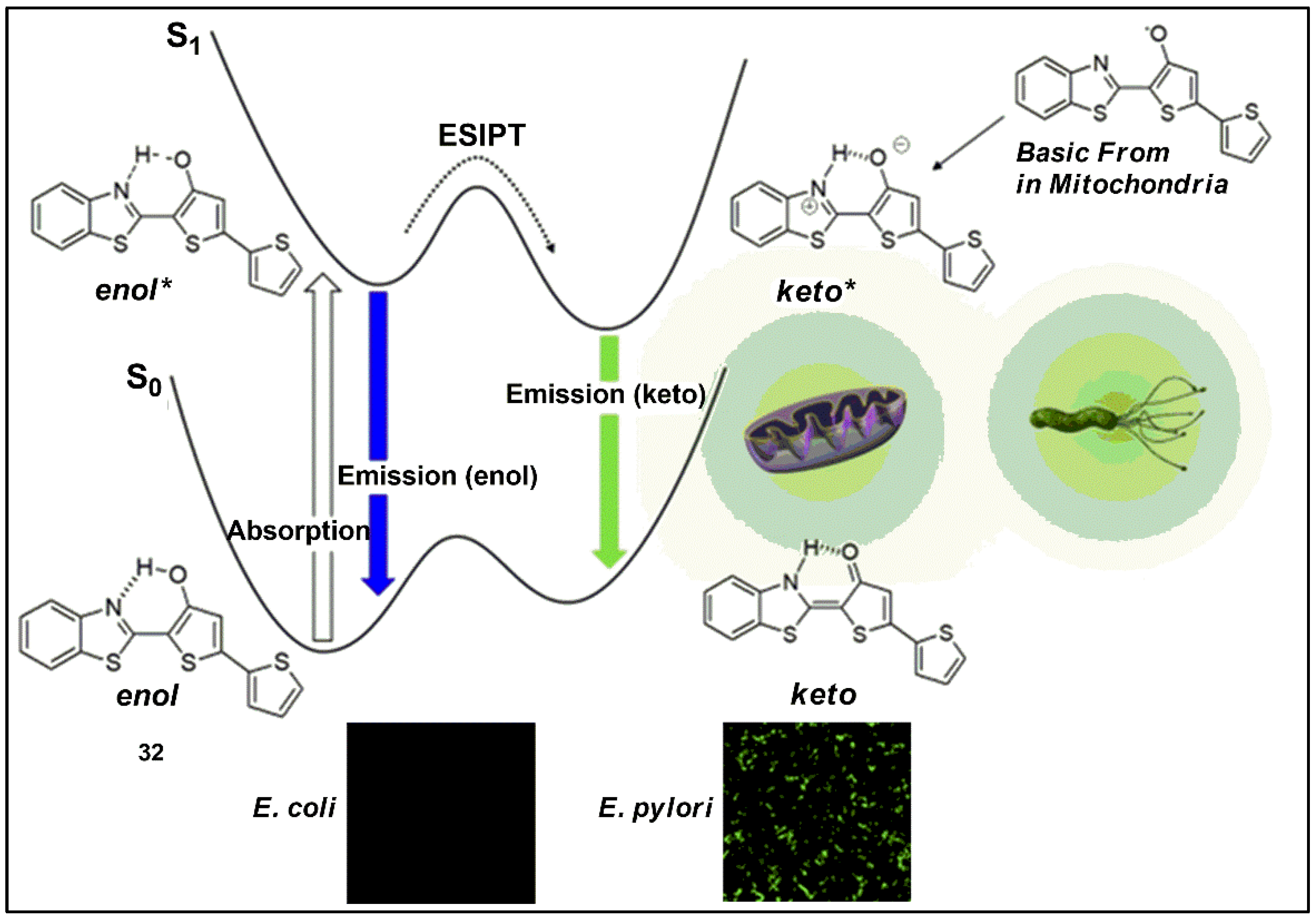
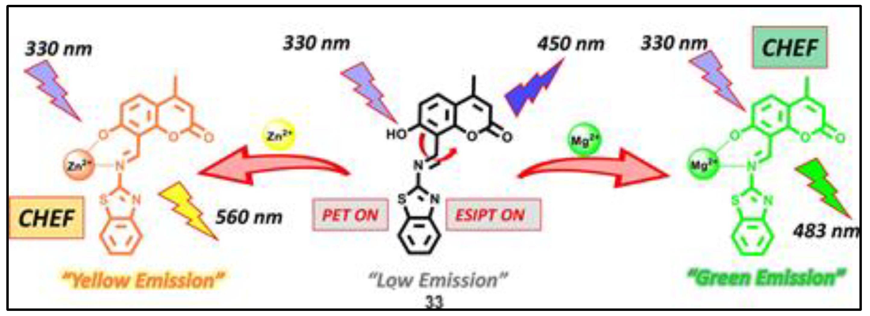
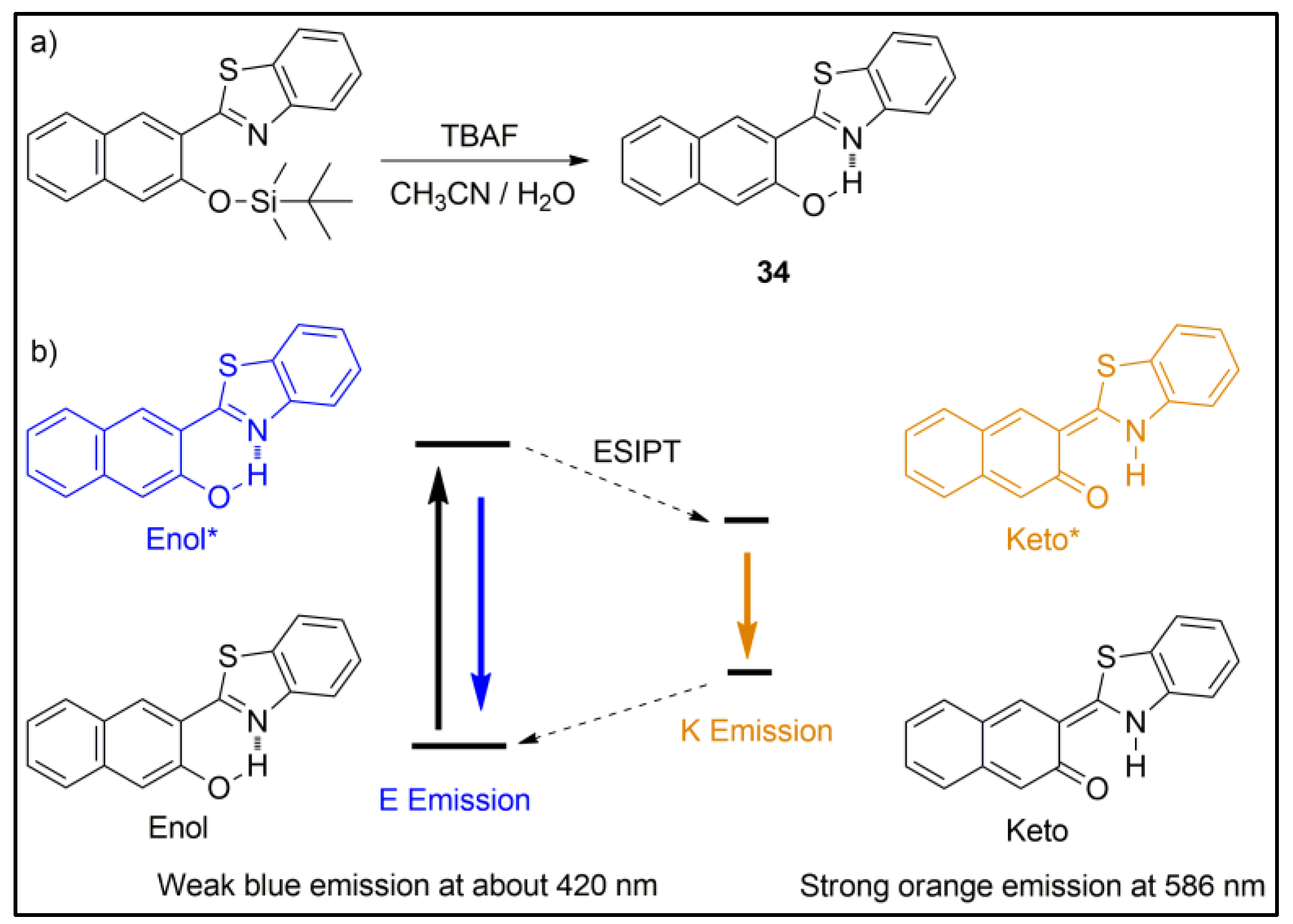
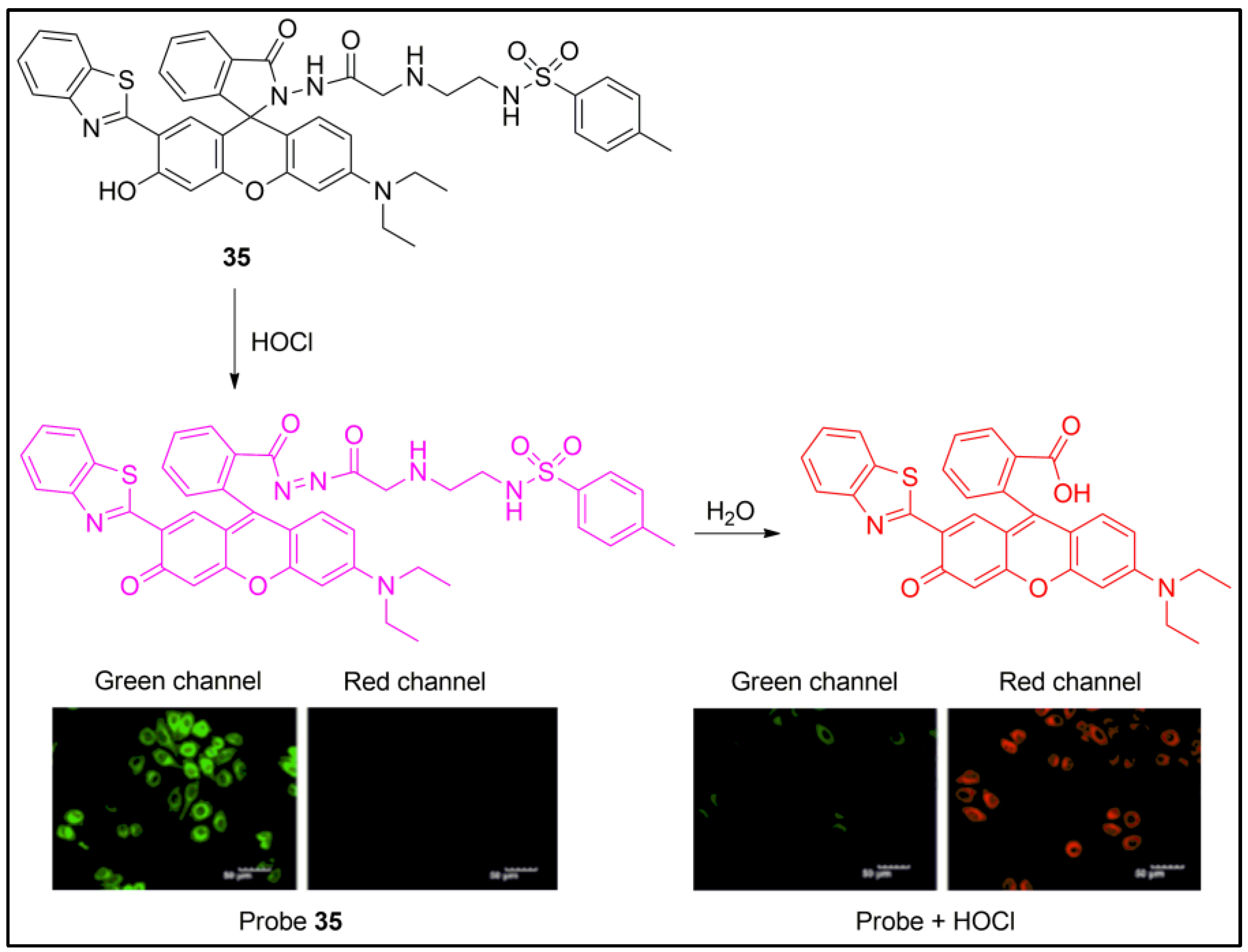
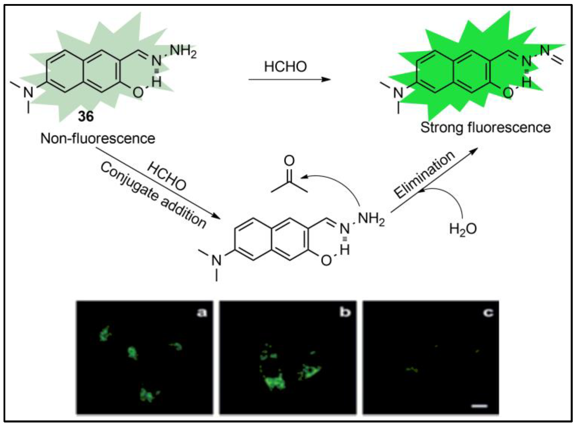
5. Fluorescent Probes Based on Excited-State Intramolecular Proton Transfer (ESIPT)
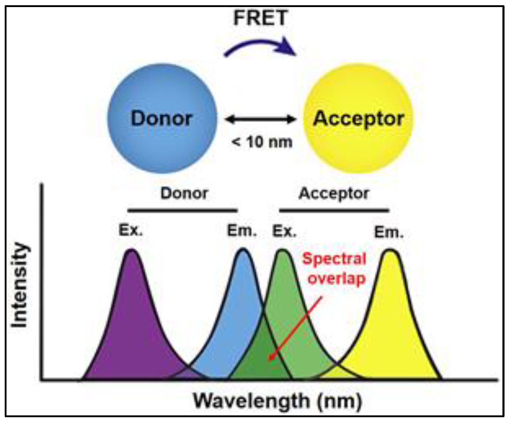
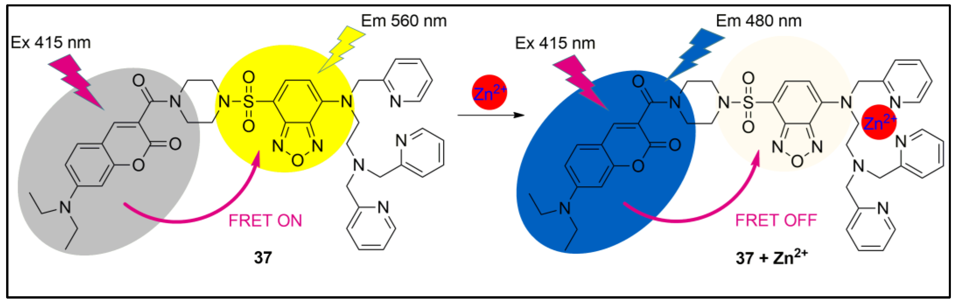
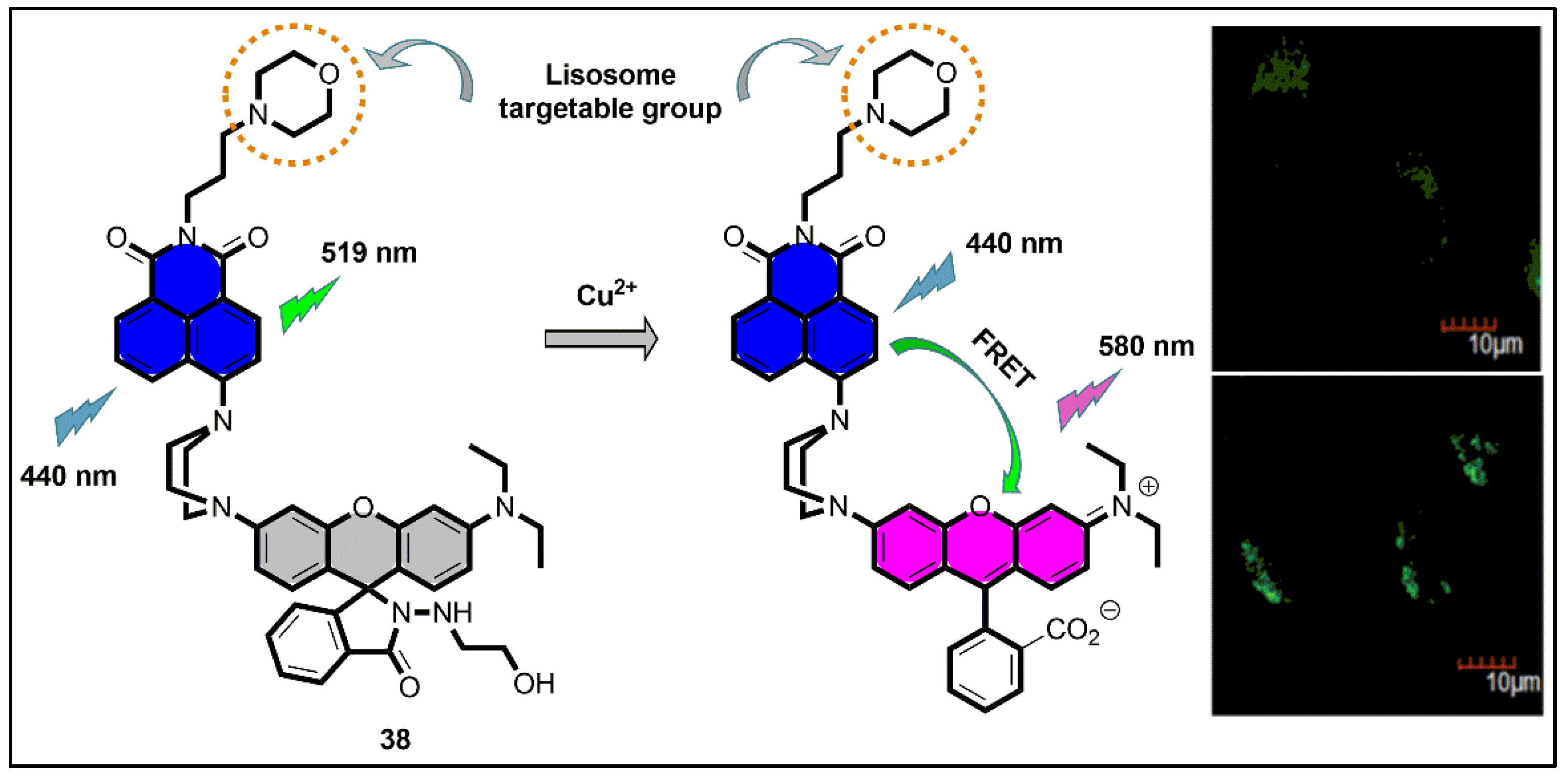
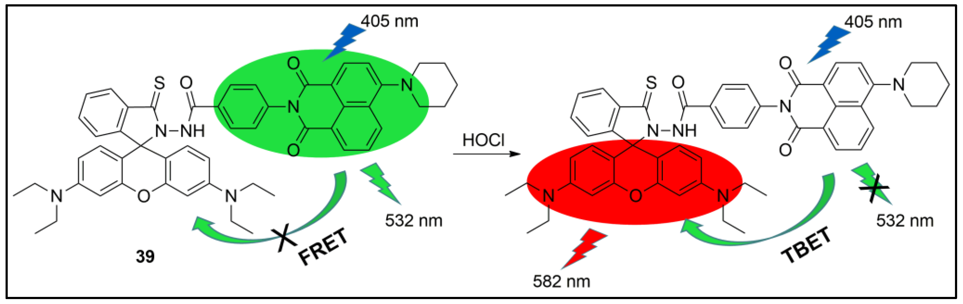
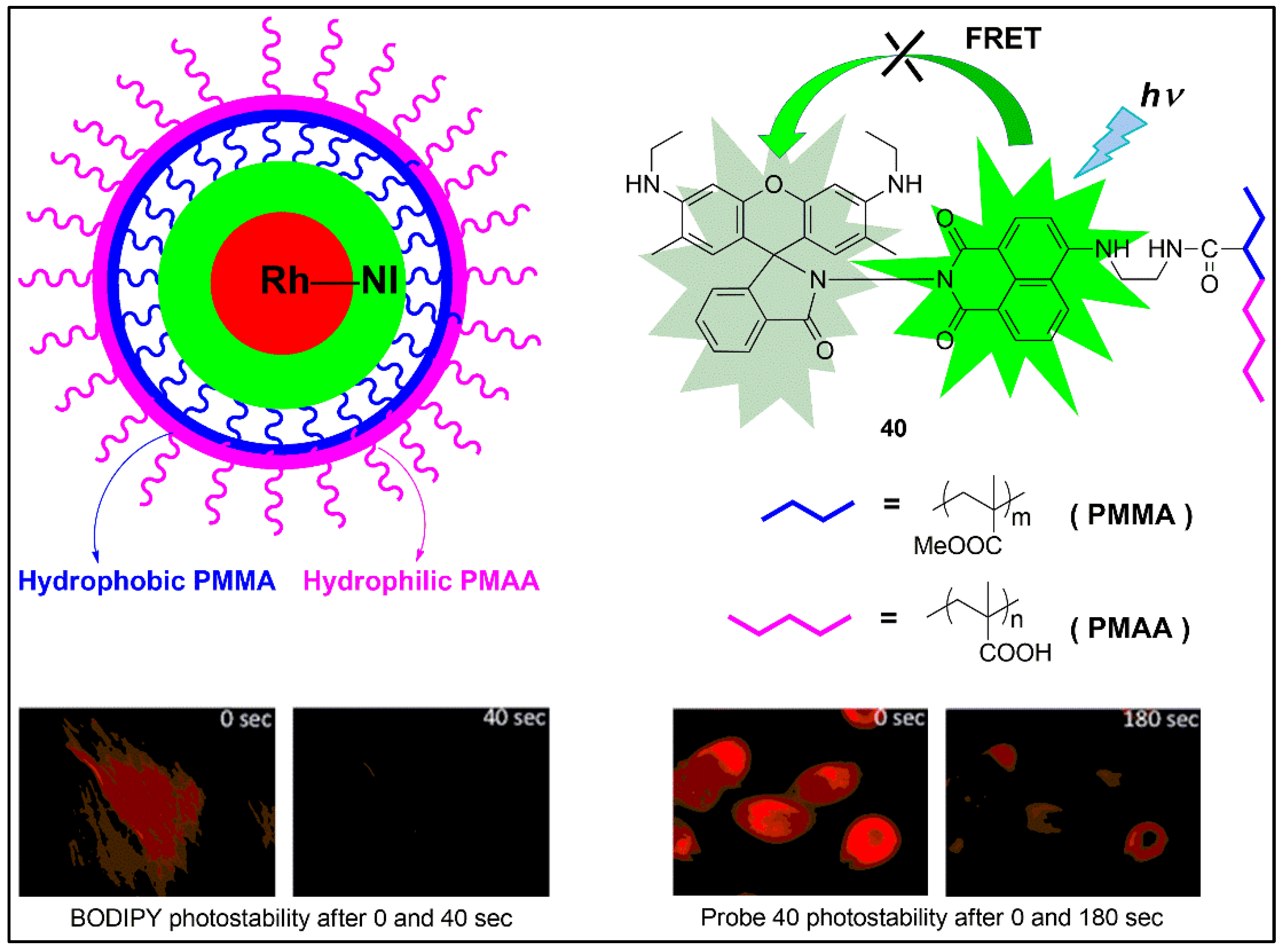
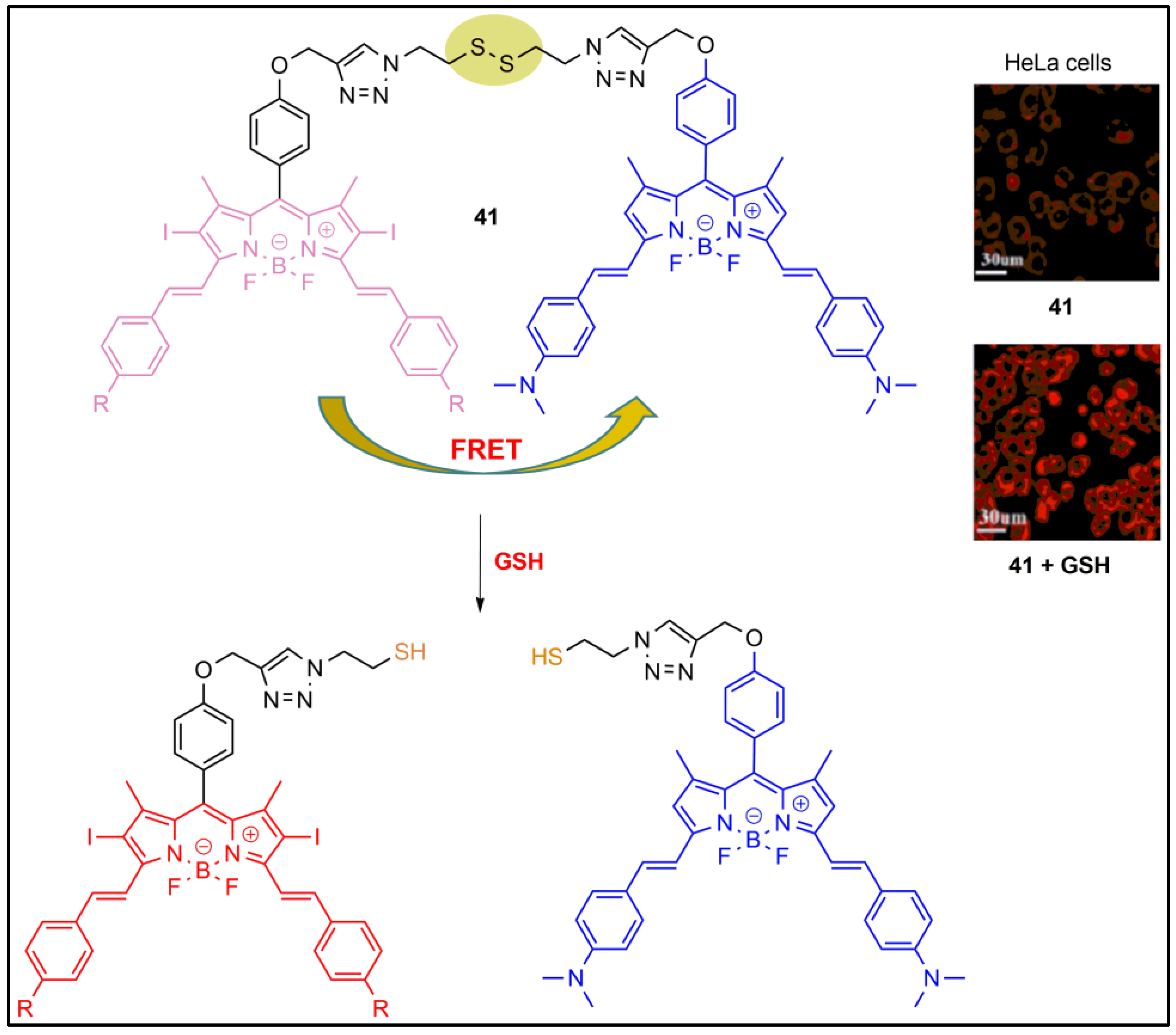
6. Fluorescent Probes Based on Fluorescent Resonance Energy Transfer (FRET)
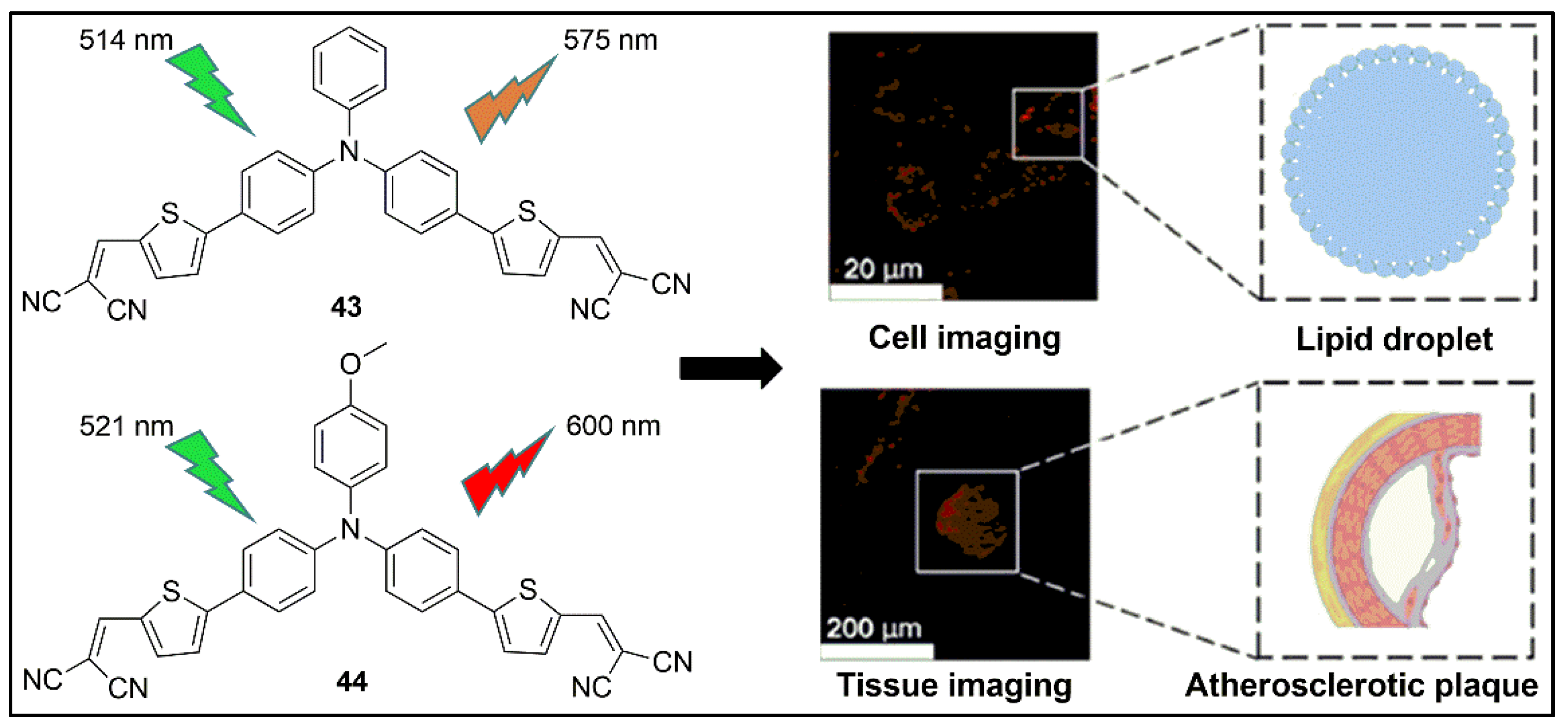
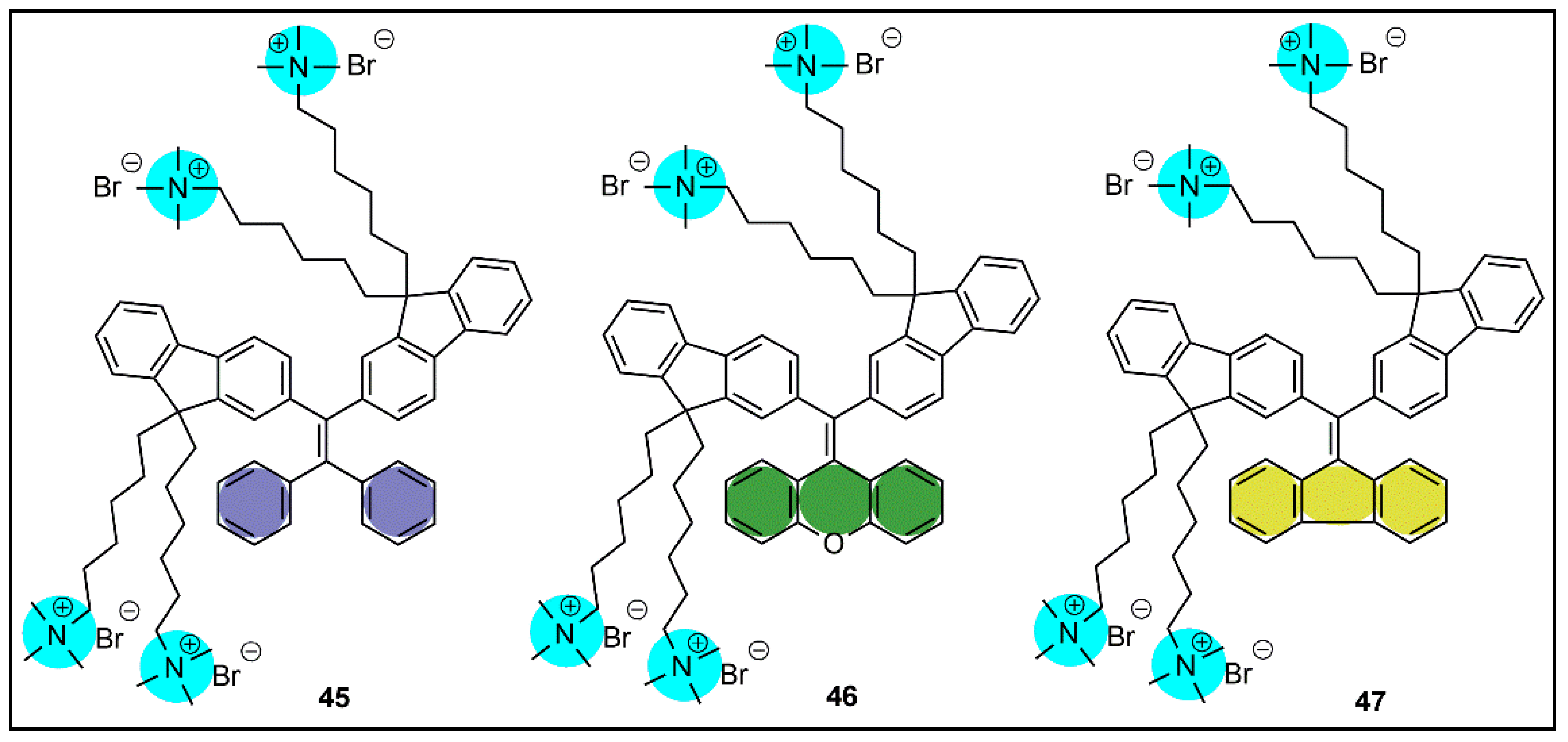
7. Fluorescent Probes Based on Aggregation-Induced Emission (AIE)
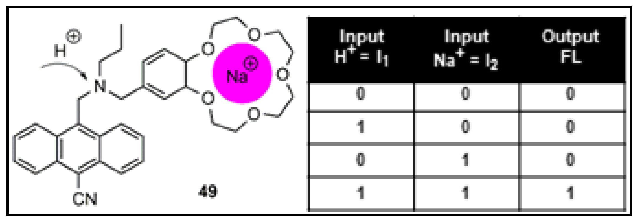
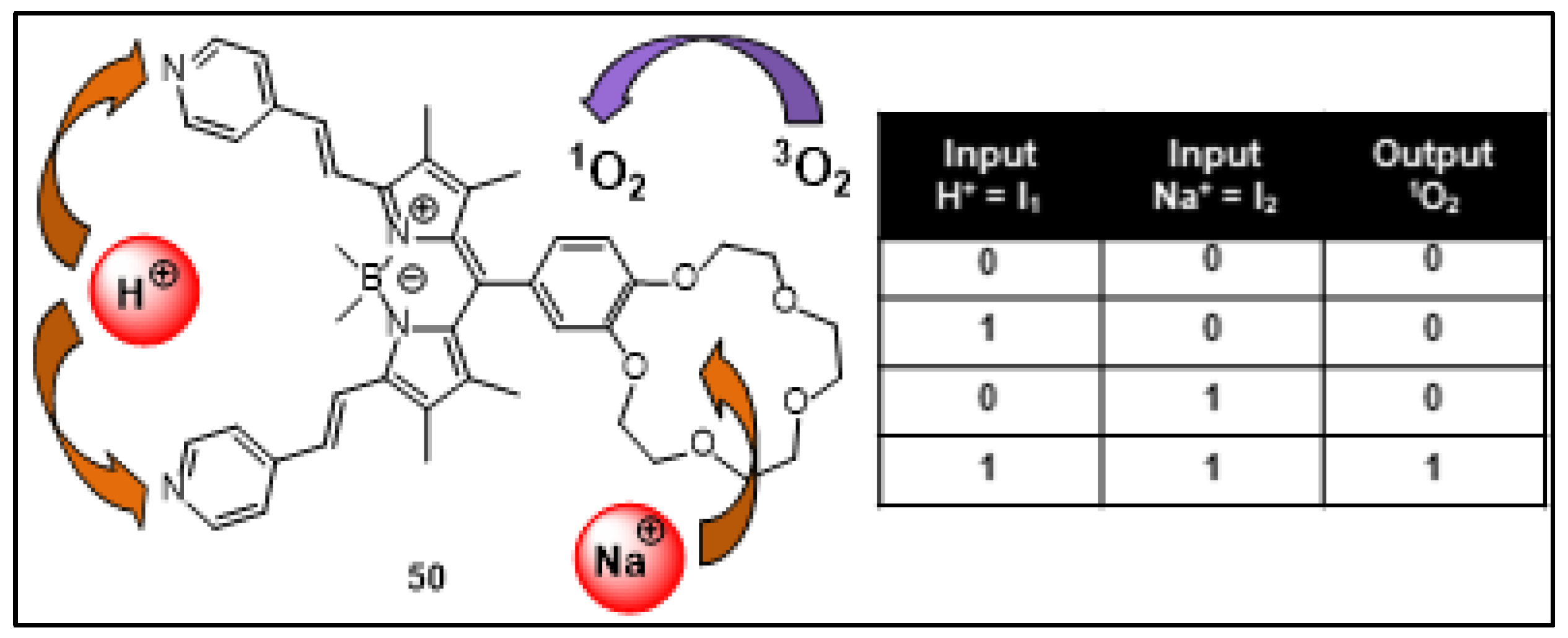
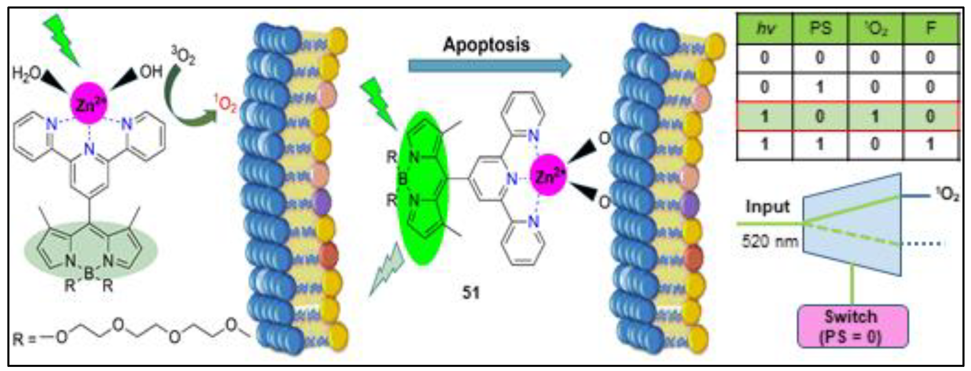
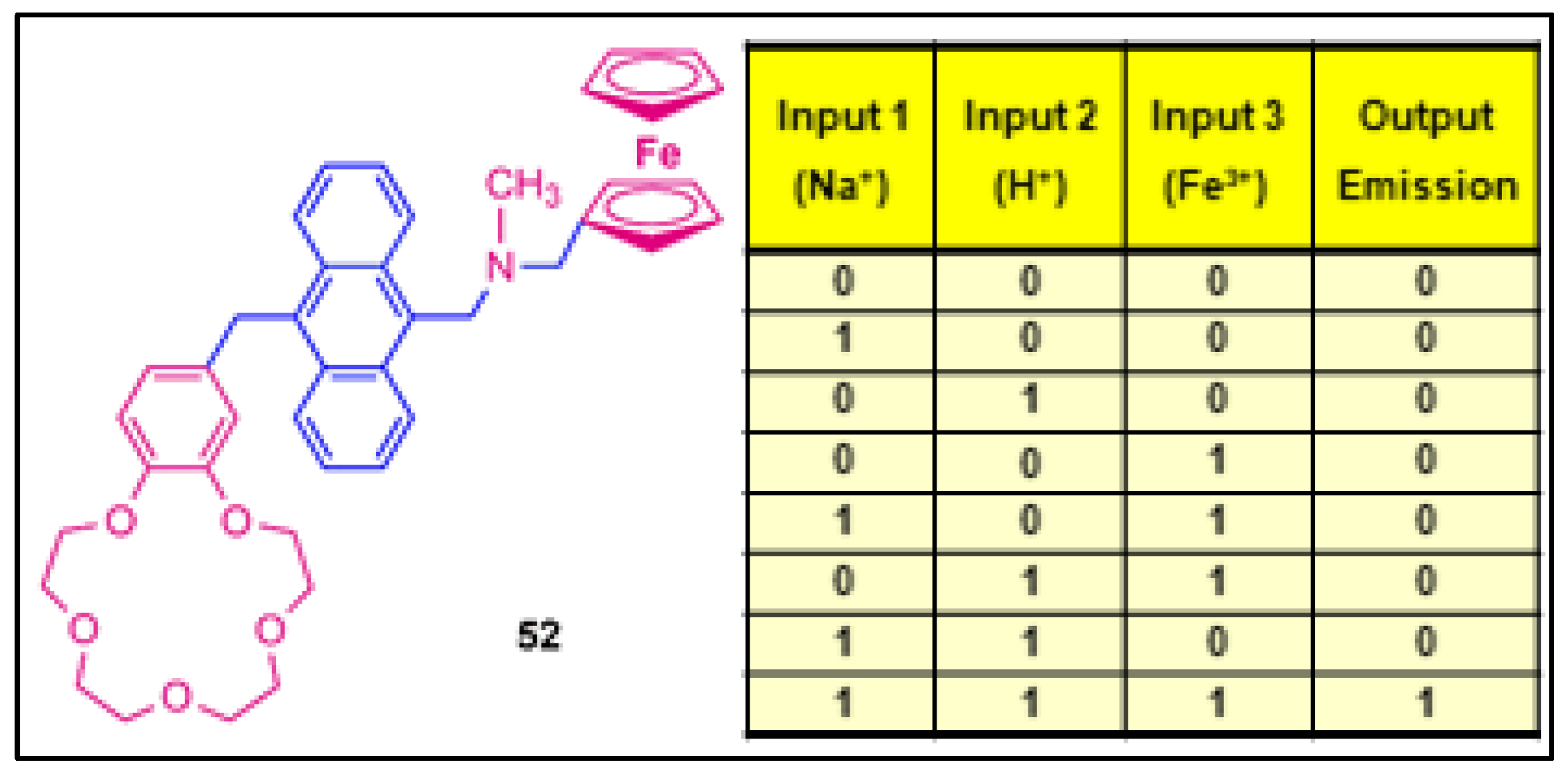
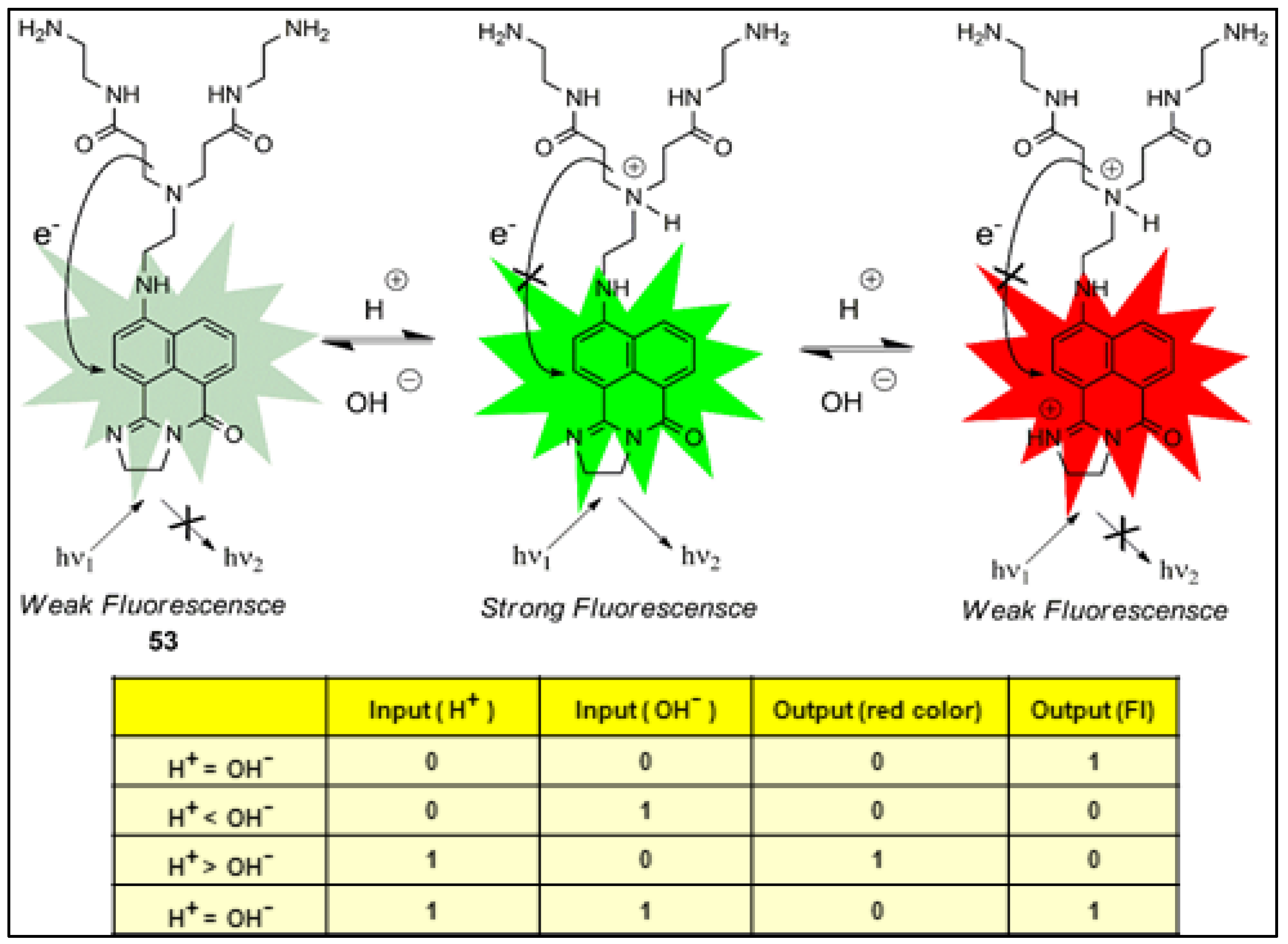
8. Molecular Logic Gates
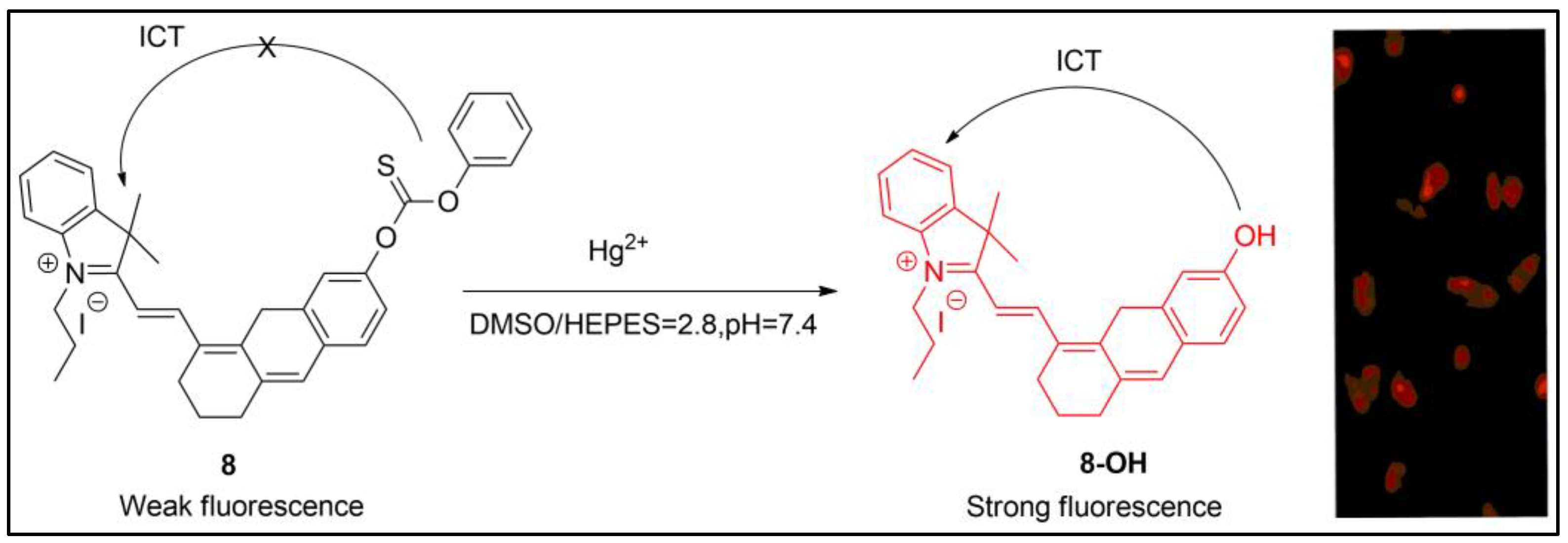
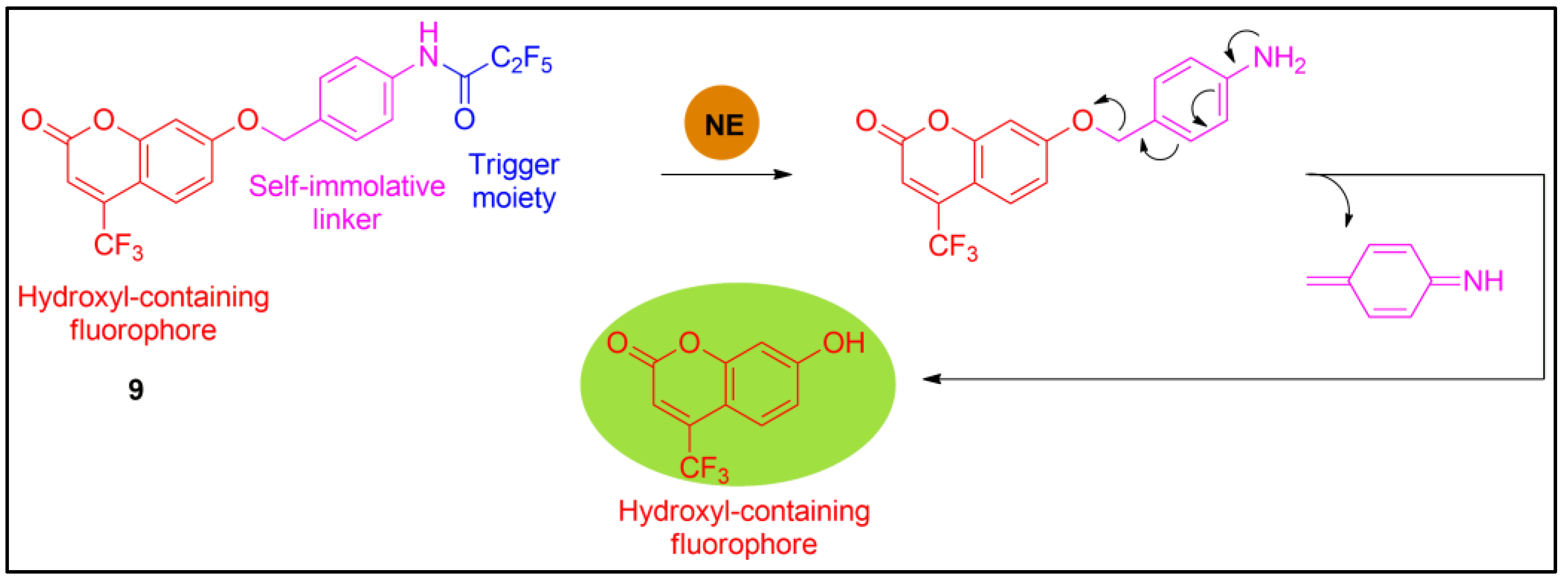
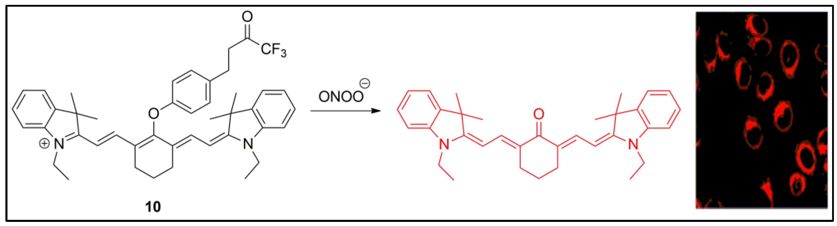
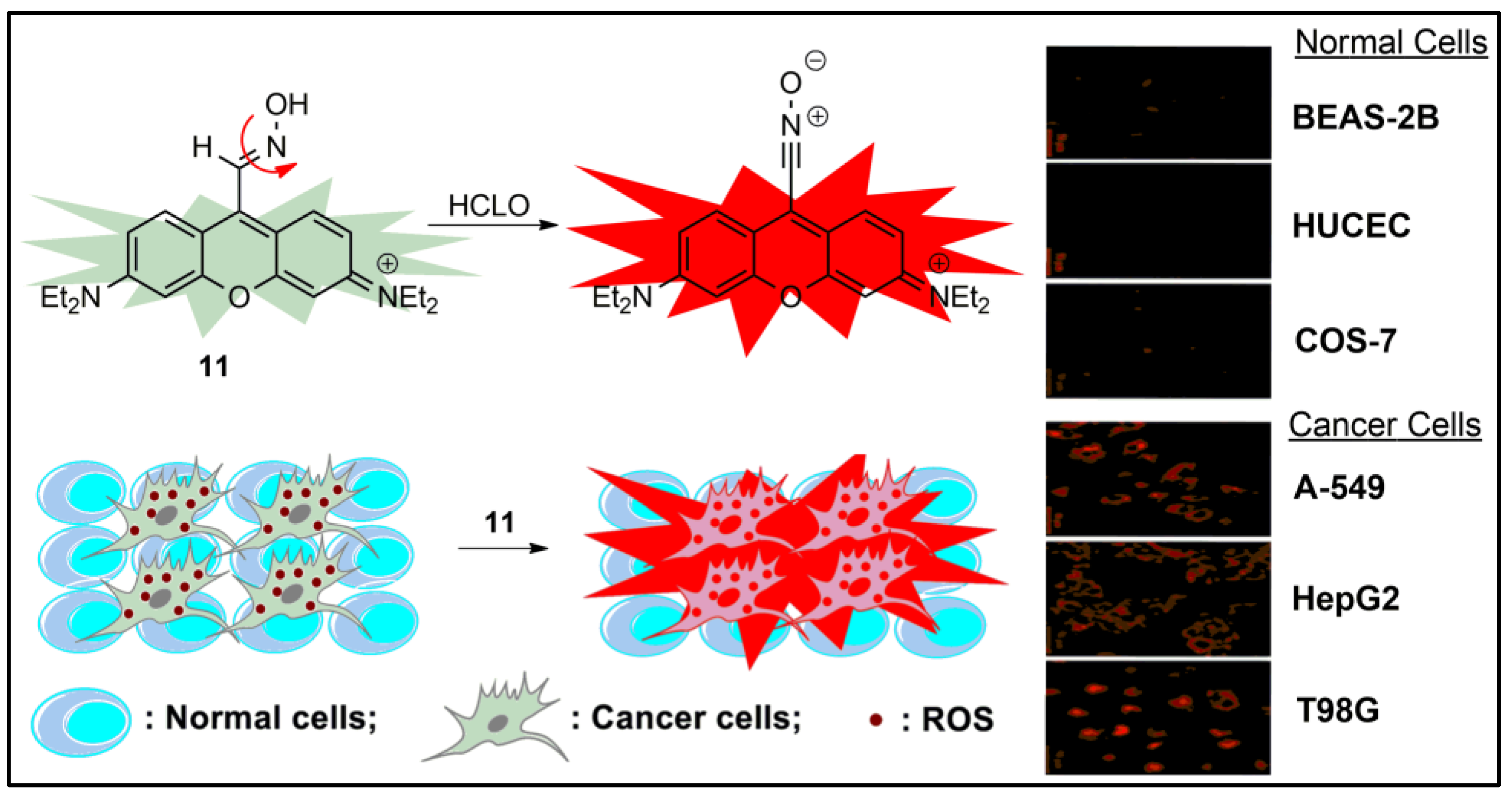
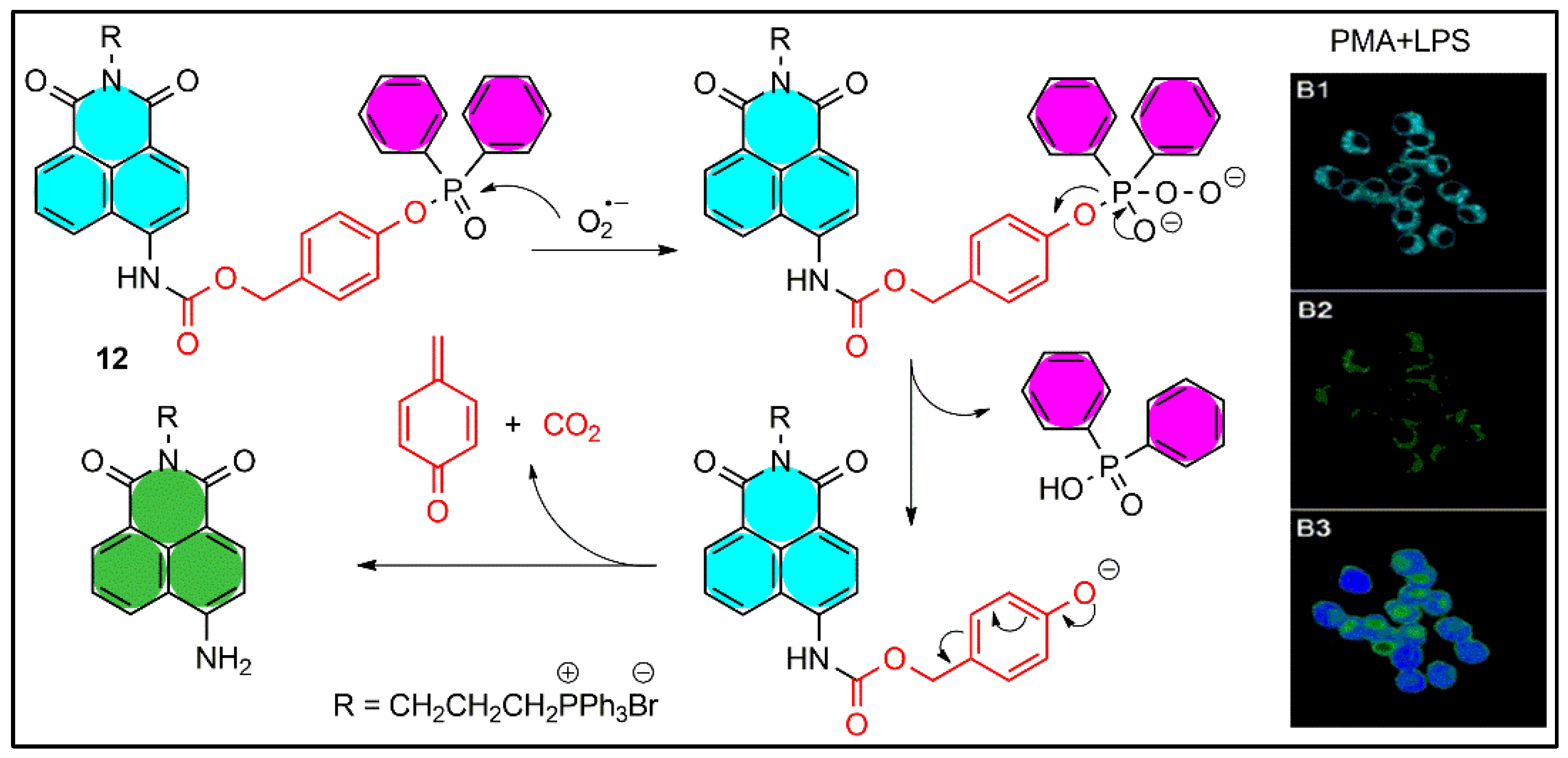
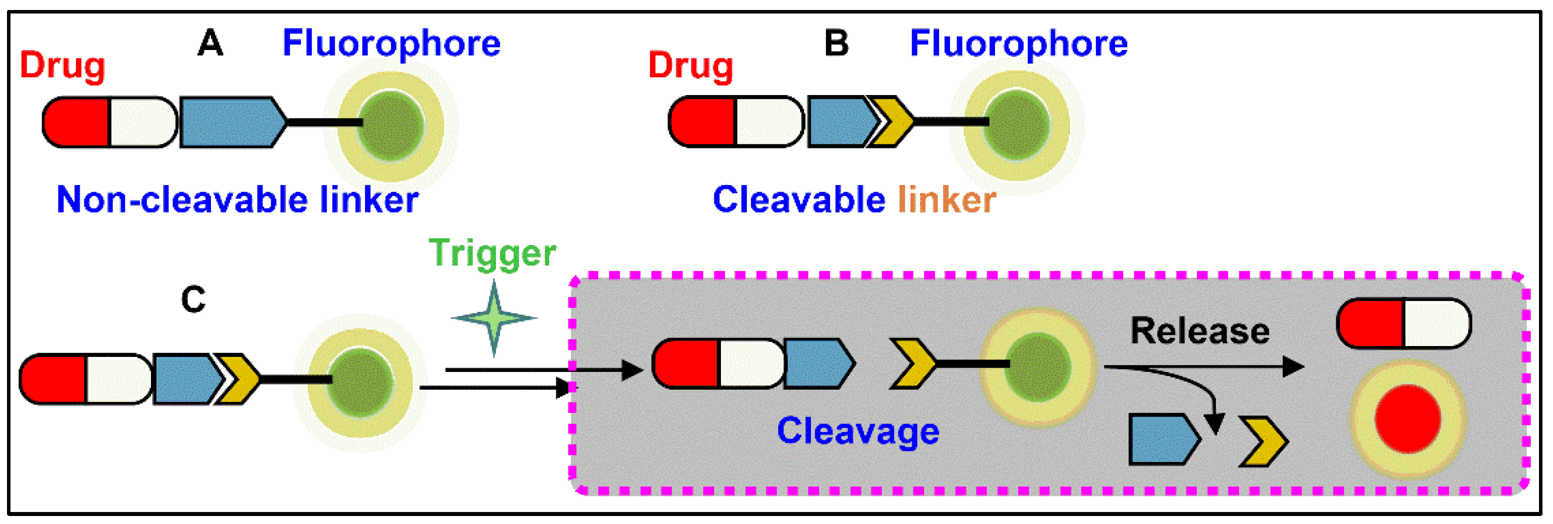
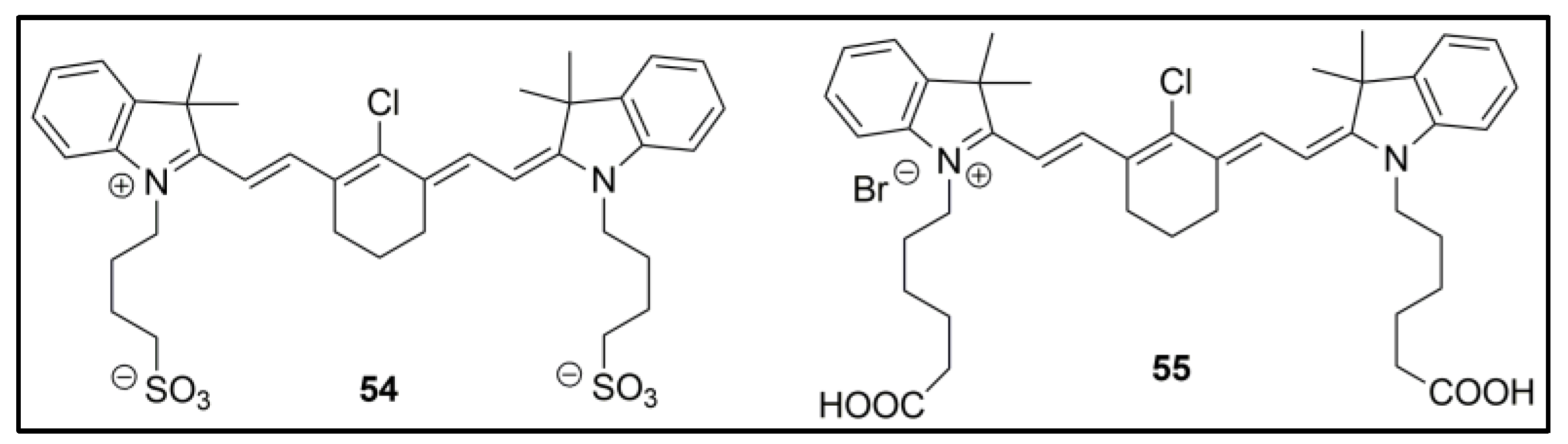
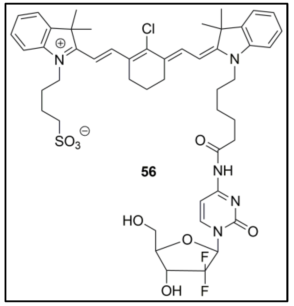
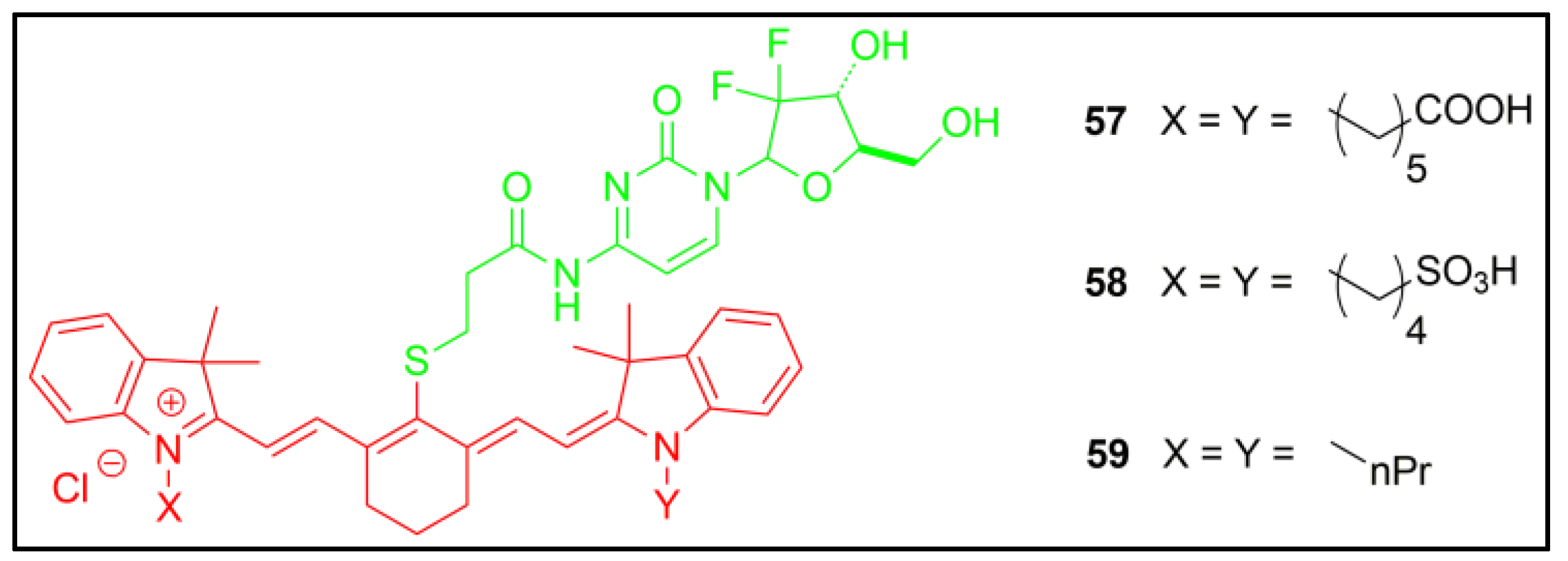
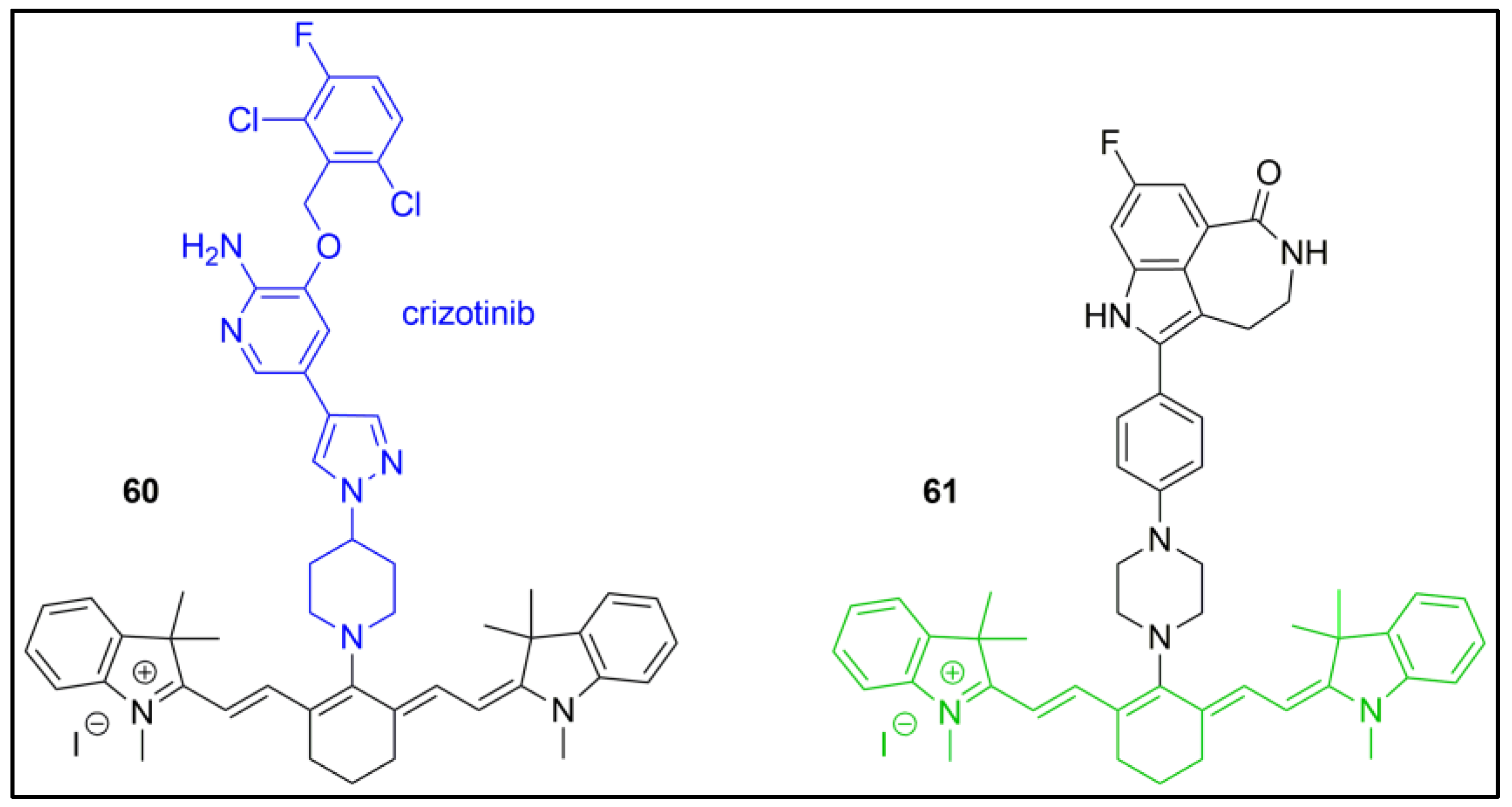
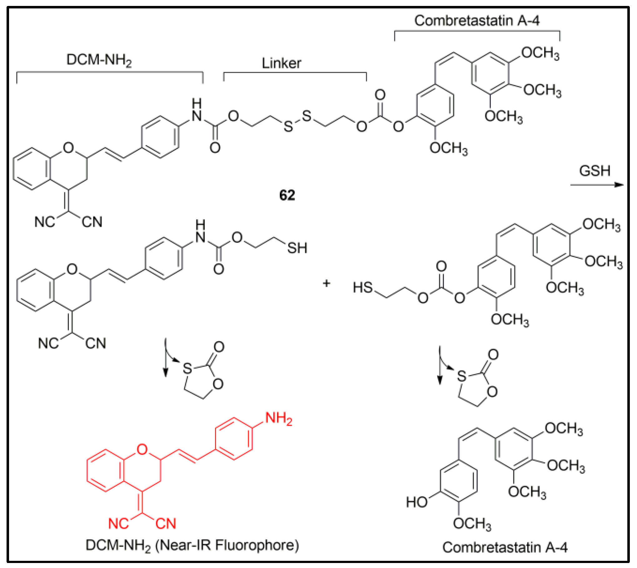
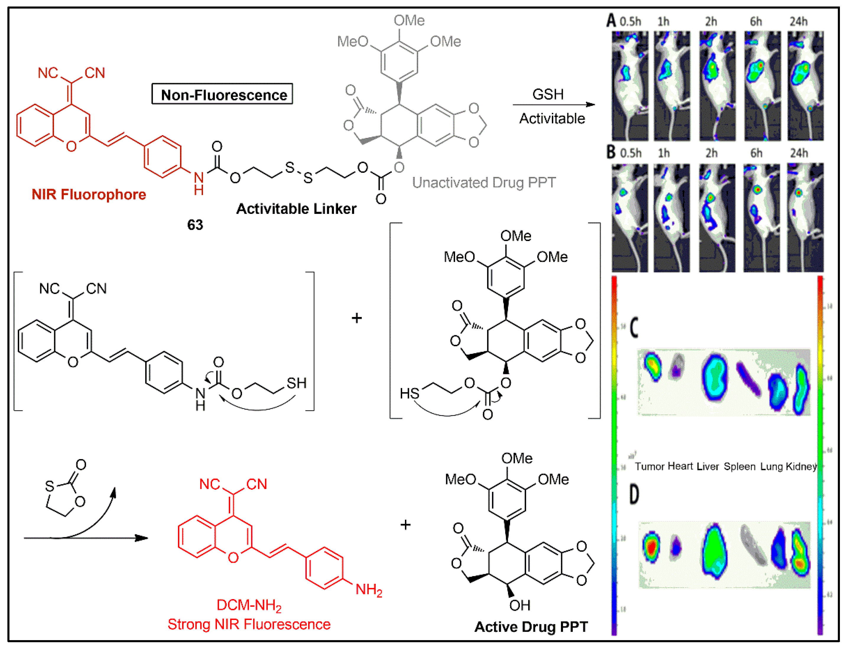
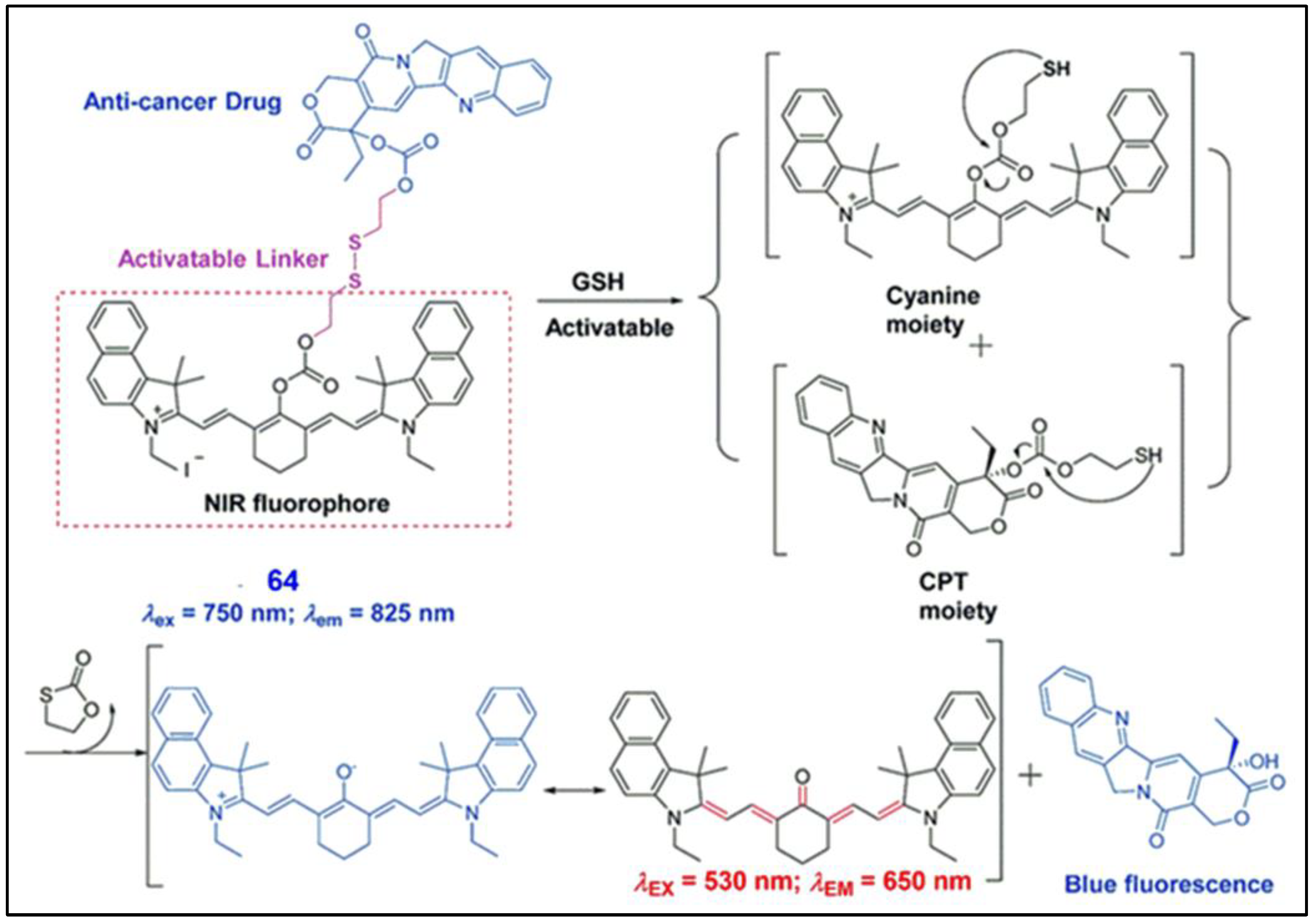
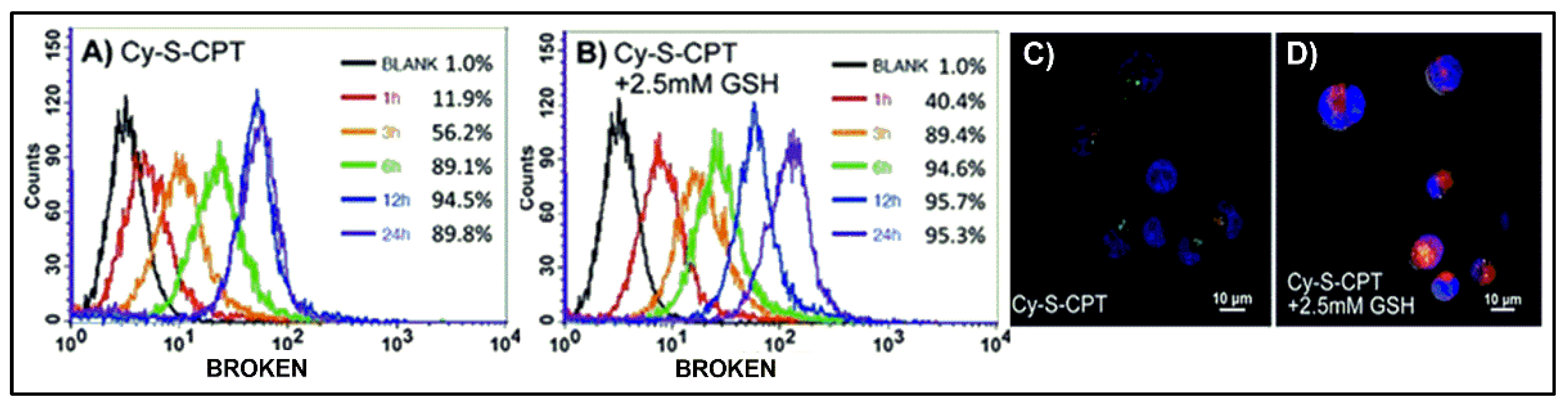
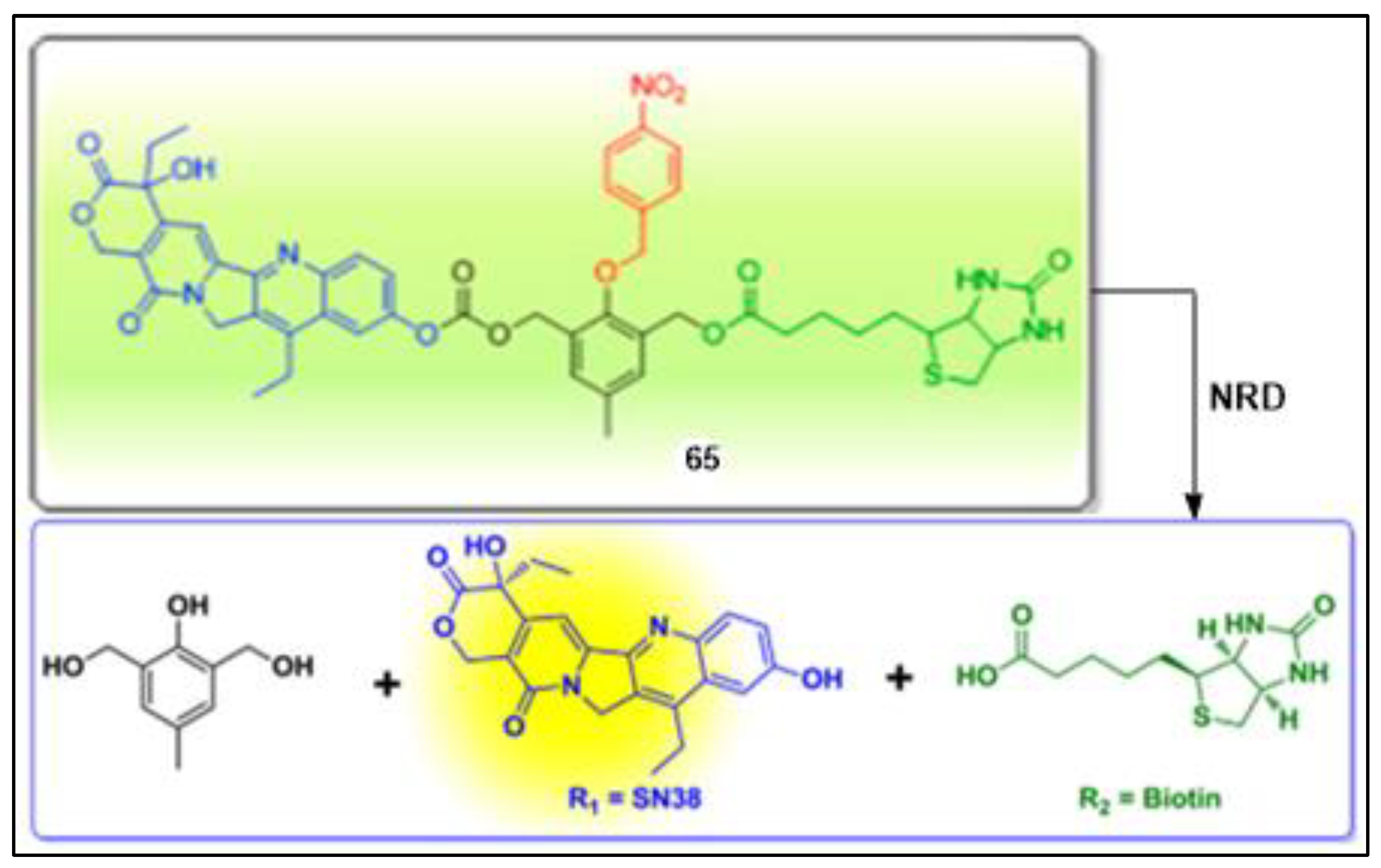
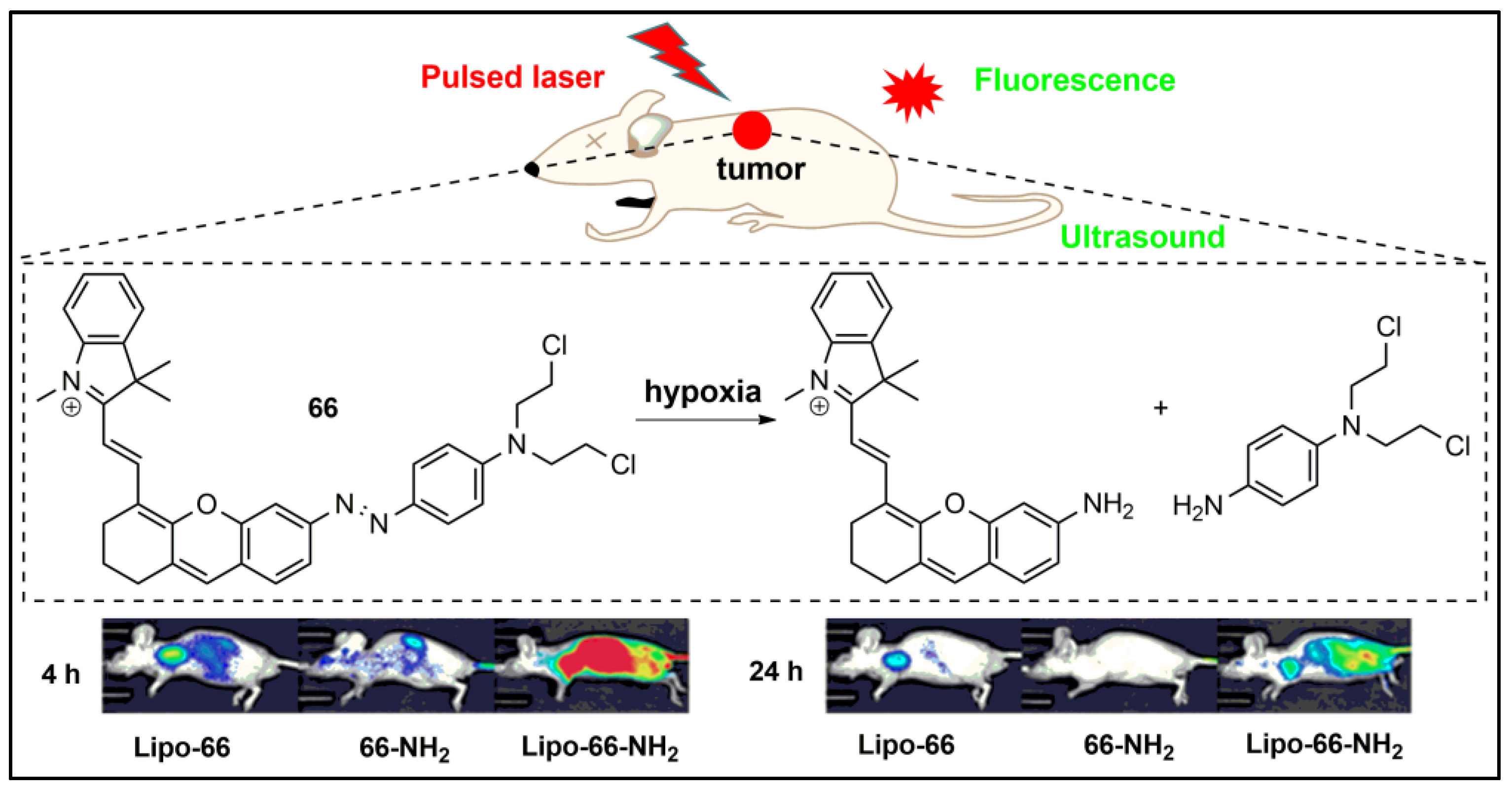
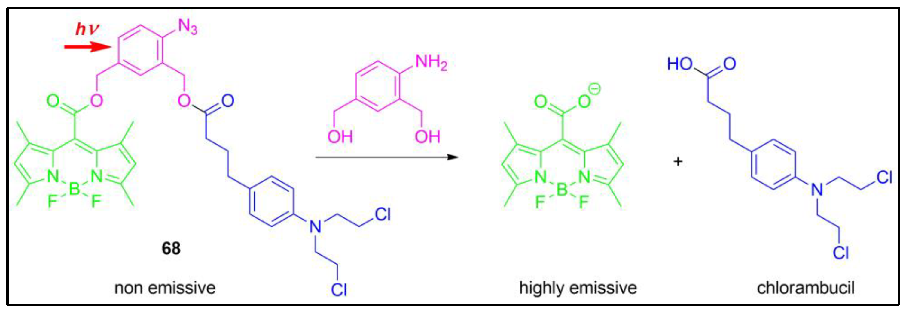
9. Fluorescent–Drug Conjugates for Diagnostic and Therapy
10. Conclusions and Perspectives
Author Contributions
Funding
Institutional Review Board Statement
Informed Consent Statement
Data Availability Statement
Conflicts of Interest
References
- Steinegger, A.; Wolfbeis, O.S.; Borisov, S.M. Optical sensing and imaging of pH values: Spectroscopies, materials, and applications. Chem. Rev. 2020, 120, 12357–12489. [Google Scholar] [CrossRef] [PubMed]
- Garcia, A.L.; Ochoa-Teran, A.; Tirado-Guizar, A.; Jara-Cortes, J.; Pina-Luis, G.; Ortega, H.S.; Labastida-Galvan, V.; Ordonezd, M.; Peon, J. Experimental and theoretical study of novel aminobenzamide–aminonaphthalimide fluorescent dyads with a FRET mechanism. RSC Adv. 2022, 12, 6192–6204. [Google Scholar] [CrossRef] [PubMed]
- Zhang, D.; Wang, S.; Yang, F.; Li, Z.; Huang, W. Visual inspection of acidic pH and bisulfite in white wine using a colorimetric and fluorescent probe. Food Chem. 2023, 408, 135200. [Google Scholar] [CrossRef]
- Mishra, S.; Singh, A.K. Optical sensors for water and humidity and their further applications. Coord. Chem. Rev. 2021, 445, 214063. [Google Scholar] [CrossRef]
- Dian, J.; Jindrich, J.; Jelinek, I. Functionalized materials with fluorescent dyes for chemosensor applications. Monatsh. Chem. 2017, 148, 1929–1935. [Google Scholar] [CrossRef]
- Zhao, J.; Guo, H.; Li, J.; Bandodkar, A.J.; Rogers, J.A. Body-interfaced chemical sensors for noninvasive monitoring and analysis of biofluids. Trends Chem. 2019, 1, 559–571. [Google Scholar] [CrossRef]
- Georgiev, N.; Krasteva, P.; Bojinov, V. A ratiometric 4-amido-1,8-naphthalimide fluorescent probe based on excimer-monomer emission for determination of pH and water content in organic solvents. J. Lumin. 2019, 212, 271–278. [Google Scholar] [CrossRef]
- Georgiev, N.I.; Bakov, V.V.; Bojinov, V.B. Photoinduced electron transfer and aggregation-induced emission in 1,8-naphthalimide probes as a platform for detection of acid/base vapors. Photonics 2022, 9, 994. [Google Scholar] [CrossRef]
- Georgiev, N.I.; Bakov, V.V.; Bojinov, V.B. A solid-state-emissive 1,8-naphthalimide probe based on photoinduced electron transfer and aggregation-induced emission. ChemistrySelect 2019, 4, 4163–4167. [Google Scholar] [CrossRef]
- Li, Y.; Dahal, D.; Abeywickrama, C.S.; Pang, Y. Progress in tuning emission of the excited-state intramolecular proton transfer (ESIPT)-based fluorescent probes. ACS Omega 2021, 6, 6547–6553. [Google Scholar] [CrossRef]
- Liu, H.W.; Chen, L.; Xu, C.; Li, Z.; Zhang, H.; Zhang, X.B.; Tan, W. Recent progresses in small-molecule enzymatic fluorescent probes for cancer imaging. Chem. Soc. Rev. 2018, 47, 7140–7180. [Google Scholar] [CrossRef] [PubMed]
- Oguz, A.; Oguz, M.; Kursunlu, A.N.; Yilmaz, M. A fully water-soluble calix[4]arene probe for fluorometric and colorimetric detection of toxic hydrosulfide and cyanide ions: Practicability in living cells and food samples. Food Chem. 2023, 401, 134132. [Google Scholar] [CrossRef] [PubMed]
- Shi, C.; Luo, J.; Wang, Y.; Ding, L.; Liang, Q.; Yang, Z.; Lu, J.; Wu, A. A water-soluble naphthalimide fluorescent probe for Cr2O72− and Fe3+ based on inner filter effect. Spectrochim. Acta Part A 2023, 289, 122245. [Google Scholar] [CrossRef] [PubMed]
- Arabahmadi, R.; Orojloo, M.; Amani, S. Three and four inputs combinational logic circuits based on a azo-azomethine chemosensor for the detection of Ni2+ and CN− /OAC− ions: Experimental and DFT studies. J. Photochem. Photobiol. A Chem. 2023, 434, 114231. [Google Scholar] [CrossRef]
- Paul, A.; Sahoo, A.; Bhattacharya, S.; Baitalik, S. Anion and temperature responsive molecular switches based on trimetallic complexes of Ru(II) and Os(II) that demonstrate advanced boolean and fuzzy logic functions. Inorg. Chem. 2022, 61, 3186–3201. [Google Scholar] [CrossRef]
- Tigoianu, R.; Airinei, A.; Georgescu, E.; Nicolescu, A.; Georgescu, F.; Isac, D.L.; Deleanu, C.; Oancea, F. Synthesis and solvent dependent fluorescence of some piperidine-substituted naphthalimide derivatives and consequences for water sensing. Int. J. Mol. Sci. 2022, 23, 2760. [Google Scholar] [CrossRef]
- Johnson, A.D.; Zammit, R.; Vella, J.; Valentino, M.; Buhagiar, J.A.; Magri, D.C. Aminonaphthalimide hybrids of mitoxantrone and amonafide as anticancer and fluorescent cellular imaging agents. Bioorg. Chem. 2019, 93, 103287. [Google Scholar] [CrossRef]
- Chen, T.; He, B.; Tao, J.; He, Y.; Deng, H.; Wang, X.; Zheng, Y. Application of Förster Resonance Energy Transfer (FRET) technique to elucidate intracellular and In Vivo biofate of nanomedicines. Adv. Drug Deliv. Rev. 2019, 143, 177–205. [Google Scholar] [CrossRef]
- Ji, L.; Zhou, X.; Liu, R.; Wang, L.; Hu, Y.; Gu, J.; Li, Z.; Li, C.; Huang, T.; Yu, Y. Design of a selective and water-soluble fluorescent probe targeting Tau fibrils for intracellular and in vivo imaging. Sens. Actuators B Chem. 2023, 380, 133415. [Google Scholar] [CrossRef]
- Gao, P.; Pan, W.; Li, N.; Tang, B. Fluorescent probes for organelle-targeted bioactive species imaging. Chem. Sci. 2019, 10, 6035–6071. [Google Scholar] [CrossRef] [Green Version]
- Liu, C.; Gao, X.; Yuan, J.; Zhang, R. Advances in the development of fluorescence probes for cell plasma membrane imaging. Trends Anal. Chem. 2020, 133, 116092. [Google Scholar] [CrossRef]
- Gao, M.; Yu, F.; Lv, C.; Choo, J.; Chen, L. Fluorescent chemical probes for accurate tumor diagnosis and targeting therapy. Chem. Soc. Rev. 2017, 46, 2237–2271. [Google Scholar] [CrossRef] [PubMed]
- Patsenker, L.; Gellerman, G. Fluorescent reporters for drug delivery monitoring. Isr. J. Chem. 2020, 60, 1–16. [Google Scholar] [CrossRef]
- Lang, W.; Yuan, C.; Zhu, L.; Du, S.; Qian, L.; Gea, J.; Yao, S.Q. Recent advances in construction of small molecule-based fluorophore-drug conjugates. J. Pharm. Anal. 2020, 10, 434–443. [Google Scholar] [CrossRef]
- Georgiev, N.I.; Sakr, A.R.; Bojinov, V.B. Design and synthesis of a novel PET and ICT based 1,8-naphthalimide FRET bichromophore as a four-input Disabled–Enabled-OR logic gate. Sens. Actuators B Chem. 2015, 221, 625–634. [Google Scholar] [CrossRef]
- Erbas-Cakmak, S.; Kolemen, S.; Sedgwick, A.C.; Gunnlaugsson, T.; James, T.D.; Yoon, J.; Akkaya, E.U. Molecular logic gates: The past, present and future. Chem. Soc. Rev. 2018, 47, 2228–2248. [Google Scholar] [CrossRef] [Green Version]
- Yao, C.; Lin, H.; Crory, H.; de Silva, A.P. Supra-molecular agents running tasks intelligently (SMARTI): Recent developments in molecular logic-based computation. Mol. Syst. Des. Eng. 2020, 5, 1325–1353. [Google Scholar] [CrossRef]
- Said, A.; Georgiev, N.; Bojinov, V. Synthesis of a single 1,8-naphthalimide fluorophore as a molecular logic lab for simultaneously detecting of Fe3+, Hg2+ and Cu2+. Spectrochim. Acta Part A 2018, 196, 76–82. [Google Scholar] [CrossRef]
- Daly, B.; Ling, J.; de Silva, A.P. What has supramolecular chemistry done for us? Supramol. Chem. 2015, 28, 201–203. [Google Scholar] [CrossRef]
- Konry, T.; Walt, D. Intelligent medical diagnostics via molecular logic. J. Am. Chem. Soc. 2009, 131, 13232–13233. [Google Scholar] [CrossRef] [Green Version]
- Etrych, T.; Janoušková, O.; Chytil, P. Fluorescence imaging as a tool in preclinical evaluation of polymer-based nano-DDS systems intended for cancer treatment. Pharmaceutics 2019, 11, 471. [Google Scholar] [CrossRef] [PubMed] [Green Version]
- Zingale, E.; Romeo, A.; Rizzo, S.; Cimino, C.; Bonaccorso, A.; Carbone, C.; Musumeci, T.; Pignatello, R. Fluorescent nanosystems for drug tracking and theranostics: Recent applications in the ocular field. Pharmaceutics 2022, 14, 955. [Google Scholar] [CrossRef] [PubMed]
- Wang, S.; Ren, W.X.; Hou, J.-T.; Won, M.; An, J.; Chen, X.; Shu, J.; Kim, J. Fluorescence imaging of pathophysiological microenvironments. Chem. Soc. Rev. 2021, 50, 8887–8902. [Google Scholar] [CrossRef] [PubMed]
- Han, H.-H.; Tian Jr., H.; Zang, Y.; Sedgwick, A.C.; Li, J.; Sessler, J.L.; He, X.-P.; James, T.D. Small-molecule fluorescence-based probes for interrogating major organ diseases. Chem. Soc. Rev. 2021, 50, 9391–9429. [Google Scholar] [CrossRef] [PubMed]
- Wang, Y.; Zhang, Y.; Wang, J.; Liang, X.-J. Aggregation-induced emission (AIE) fluorophores as imaging tools to trace the biological fate of nano-based drug delivery systems. Adv. Drug Deliv. Rev. 2019, 143, 161–176. [Google Scholar] [CrossRef]
- Ma, Y.; Chen, Q.; Pan, X.; Zhang, J. Insight into fluorescence imaging and bioorthogonal reactions in biological analysis. Top. Curr. Chem. 2021, 379, 10. [Google Scholar] [CrossRef]
- de Silva, A.P.; Gunaratne, H.Q.N.; Gunnlaugsson, T.; Huxley, A.J.M.; McCoy, C.P.; Rademacher, J.T.; Rice, T.E. Signaling recognition events with fluorescent sensors and switches. Chem. Rev. 1997, 97, 1515–1566. [Google Scholar] [CrossRef]
- Jiao, X.; Liu, C.; He, S.; Zhao, L.; Zeng, X. Highly selective and sensitive ratiometric near-infrared fluorescent probe for real-time detection of Hg2+ and its bioapplications in live cells. Dyes Pigments 2019, 160, 86–92. [Google Scholar] [CrossRef]
- Weisinger, J.R.; Bellorín-Font, E. Magnesium and phosphorus. Lancet 1998, 352, 391–396. [Google Scholar] [CrossRef]
- Abed, E.; Moreau, R. Importance of melastatin-like transient receptor potential 7 and magnesium in the stimulation of osteoblast proliferation and migration by platelet-derived growth factor. Am. J. Physiol. Cell Physiol. 2009, 297, C360–C368. [Google Scholar] [CrossRef] [Green Version]
- Gawaz, M.; Reininger, A.; Neumann, F.J. Platelet function and platelet-leukocyte adhesion in symptomatic coronary heart disease. Effects of intravenous magnesium. Thromb. Res. 1996, 83, 341–349. [Google Scholar] [CrossRef] [PubMed]
- Zhang, H.; Yin, C.; Liu, T.; Chao, J.; Zhang, Y.; Huo, F. Selective “off-on” detection of magnesium (II) ions using a naphthalimide-derived fluorescent probe. Dyes Pigments 2017, 146, 344–351. [Google Scholar] [CrossRef]
- Bojinov, V.; Panova, I.P. Novel 4-(2,2,6,6-tetramethylpiperidin-4-ylamino)-1,8-naphthalimide based yellow-green emitting fluorescence sensors for transition metal ions and protons. Dyes Pigments 2009, 80, 61–66. [Google Scholar] [CrossRef]
- Marinova, N.; Georgiev, N.; Bojinov, V. Design, synthesis and pH sensing properties of novel 1,8-naphtalimide-based bichromophoric system. J. Photochem. Photobiol. A Chem. 2011, 222, 132–140. [Google Scholar] [CrossRef]
- Panchenko, P.A.; Fedorov, Y.V.; Fedorova, O.A.; Jonusauskas, G. Comparative analysis of the PET and ICT sensor properties of 1,8-naphthalimides containing aza-15-crown-5 ether moiety. Dyes Pigments 2013, 98, 347–357. [Google Scholar] [CrossRef] [Green Version]
- Ahmed, N.; Zareen, W.; Zhang, D.; Yang, X.; Ye, Y. A DCM-based NIR sensor for selective and sensitive detection of Zn2+ in living cells. Spectrochim. Acta Part A 2020, 243, 118758. [Google Scholar] [CrossRef]
- Huang, Y.; Li, C.-F.; Shi, W.-J.; Tan, H.-Y.; He, Z.-Z.; Zheng, L.; Liu, F.; Yan, J.-W. A near-infrared BODIPY-based fluorescent probe for ratiometric and discriminative detection of Hg2+ and Cu2+ ions in living cells. Talanta 2019, 198, 390–397. [Google Scholar] [CrossRef] [PubMed]
- Sun, M.; Du, L.; Yu, H.; Zhang, K.; Liu, Y.; Wang, S. An intramolecular charge transfer process based fluorescent probe for monitoring subtle pH fluctuation in living cells. Talanta 2017, 162, 180–186. [Google Scholar] [CrossRef]
- Zhang, Y.; Bu, F.; Zhao, Y.; Zhao, B.; Wang, L.; Song, B. A hemicyanine fluorescent probe with intramolecular charge transfer (ICT) mechanism for highly sensitive and selective detection of acidic pH and its application in living cells. Anal. Chim. Acta 2020, 1098, 155–163. [Google Scholar] [CrossRef]
- Zhou, J.; Fang, C.; Chang, T.; Liu, X.; Shangguan, D. A pH sensitive ratiometric fluorophore and its application for monitoring the intracellular and extracellular pHs simultaneously. J. Mater. Chem. B 2013, 1, 661–667. [Google Scholar] [CrossRef]
- Hou, Y.; Zhou, J.; Gao, Z.; Sun, X.; Liu, C.; Shangguan, D.; Yang, W.; Gao, M. Protease-activated ratiometric fluorescent probe for pH mapping of malignant tumors. ACS Nano 2015, 9, 3199–3205. [Google Scholar] [CrossRef]
- Georgiev, N.; Dimitrova, M.; Todorova, Y.; Bojinov, V. Synthesis, chemosensing properties and logic behaviour of a novel ratiometric 1,8-naphthalimide probe based on ICT and PET. Dyes Pigments 2016, 131, 9–17. [Google Scholar] [CrossRef] [Green Version]
- Yuan, G.; Lv, H.; Liu, H.; He, H.; Sun, Q.; Zhang, X.; Wang, S. Hemicyanine-based colorimetric and near-infrared fluorescent off-on probe for Hg2+ detection and imaging in living cells and zebrafish. Dyes Pigments 2020, 183, 108674. [Google Scholar] [CrossRef]
- Li, X.; Guo, Y.; Qiu, Y.; Luo, X.; Liu, G.; Han, Y.; Sun, Q.; Dong, Q. A novel strategy of designing neutrophil elastase fluorescent probe based on self-immolative group and its application in bioimaging. Anal. Chim. Acta 2023, 1237, 340617. [Google Scholar] [CrossRef]
- Craven, T.H.; Walton, T.; Akram, A.R.; Scholefield, E.; McDonald, N.; Marshall, A.D.L.; Dhaliwal, K. Activated neutrophil fluorescent imaging technique for human lungs. Sci. Rep. 2021, 11, 976. [Google Scholar] [CrossRef] [PubMed]
- Tang, L.; Zhou, L.; Yan, X.; Zhong, K.; Gao, X.; Liu, X.; Li, J. A simple benzothiazole-based mitochondrial-targeting fluorescent probe for visualizing and monitoring viscosity in living cell, lung organ tissue, and living mice. Dyes Pigments 2020, 182, 108644. [Google Scholar] [CrossRef]
- Liu, H.; Zhang, H.; Lou, X.; Teng, L.; Yuan, J.; Yuan, L.; Zhang, X.; Tan, W. Imaging of peroxynitrite in drug-induced acute kidney injury with a near-infrared fluorescence and photoacoustic dual-modal molecular probe. Chem. Commun. 2020, 56, 8103–8106. [Google Scholar] [CrossRef]
- Schulz-Fincke, A.-C.; Blaut, M.; Braune, A.; Gütschow, M. A BODIPY-tagged phosphono peptide as activity-based probe for human leukocyte elastase. ACS Med. Chem. Lett. 2018, 9, 345–350. [Google Scholar] [CrossRef] [PubMed]
- Wiesman, H.; Halliwell, B. Damage to DNA by reactive oxygen and nitrogen species: Role in inflammatory disease and progression to cancer. Biochem. J. 1996, 313, 17–29. [Google Scholar] [CrossRef] [PubMed] [Green Version]
- Kossenjans, W.; Eis, A.; Sahay, R.; Brockman, D.; Myatt, L. Role of peroxynitrite in altered fetal-placental vascular reactivity in diabetes or preeclampsia. Am. J. Physiol. Heart Circ. Physiol. 2000, 278, H1311–H1319. [Google Scholar] [CrossRef] [PubMed] [Green Version]
- Supinski, G.; Stofan, D.; Callahan, L.A.; Nethery, D.; Nosek, T.M.; DiMarco, A. Peroxynitrite induces contractile dysfunction and lipid peroxidation in the diaphragm. J. Appl. Physiol. 1999, 87, 783–791. [Google Scholar] [CrossRef] [PubMed] [Green Version]
- White, C.R.; Brock, T.A.; Chang, L.Y.; Crapo, J.; Briscoe, P.; Ku, D.; Freeman, B.A. Superoxide and peroxynitrite in atherosclerosis. Proc. Natl. Acad. Sci. USA 1994, 91, 1044–1048. [Google Scholar] [CrossRef] [PubMed] [Green Version]
- Huang, Y.; Yu, L.; Fu, L.; Hou, J.; Wang, L.; Sun, M.; Wang, X.; Chen, L. Molecular fluorescent probes for imaging and evaluation of peroxynitrite fluctuations in living cells and in vivo under hypoxic stress. Sens. Actuators B Chem. 2022, 370, 132410. [Google Scholar] [CrossRef]
- Wang, L.; Liu, J.; Zhang, H.; Guo, W. Discrimination between cancerous and normal cells/tissues enabled by a near-infrared fluorescent HClO probe. Sens. Actuators B Chem. 2021, 334, 129602. [Google Scholar] [CrossRef]
- Barber, A.J. A new view of diabetic retinopathy: A neurodegenerative disease of the eye. Prog. Neuro-Psychopharmacol. Biol. Psychiatry 2003, 27, 283–290. [Google Scholar] [CrossRef]
- Mantovani, A.; Allavena, P.; Sica, A.; Balkwill, F. Cancer-related inflammation. Nature 2008, 454, 436–444. [Google Scholar] [CrossRef]
- Mortensen, S.A.; Mortensen, A.L. The mitochondria in heart failure: A target for coenzyme Q10 therapy? Clin. Pharmacol. Ther. 2014, 96, 645–647. [Google Scholar] [CrossRef]
- Tuppen, H.A.L.; Blakely, E.L.; Turnbull, D.M.; Taylor, R.W. Mitochondrial DNA mutations and human disease. Biochim. Biophys. Acta Bioenerg. 2010, 1797, 113–128. [Google Scholar] [CrossRef] [Green Version]
- Wallace, D.C. Mitochondrial diseases in man and mouse. Science 1999, 283, 1482–1488. [Google Scholar] [CrossRef] [Green Version]
- Wu, S.M.; Pizzo, S.V. alpha(2)-Macroglobulin from rheumatoid arthritis synovial fluid: Functional analysis defines a role for oxidation in inflammation. Arch. Biochem. Biophys. 2001, 391, 119–126. [Google Scholar] [CrossRef]
- Zhang, Z.; Fan, J.; Zhao, Y.; Kang, Y.; Du, J.; Peng, X. Mitochondria-accessing ratiometric fluorescent probe for imaging endogenous superoxide anion in live cells and daphnia magna. ACS Sens. 2018, 3, 735–741. [Google Scholar] [CrossRef]
- Tian, Y.; Xin, F.; Gao, C.; Jing, J.; Zhang, X. Ratiometric fluorescence imaging of endogenous selenocysteine in cancer cell matrix. J. Mater. Chem. B 2017, 5, 6890–6896. [Google Scholar] [CrossRef]
- Qin, H.-L.; Shang, Z.-P.; Jantan, I.; Tan, O.U.; Hussain, M.A.; Sher, M.; Bukhari, S.N.A. Molecular docking studies and biological evaluation of chalcone based pyrazolines as tyrosinase inhibitors and potential anticancer agents. RSC Adv. 2015, 5, 46330–46338. [Google Scholar] [CrossRef]
- Ando, H.; Kondoh, H.; Ichihashi, M.; Hearing, V.J. Approaches to Identify Inhibitors of melanin biosynthesis via the quality control of tyrosinase. J. Investig. Dermatol. 2007, 127, 751–761. [Google Scholar] [CrossRef] [PubMed] [Green Version]
- Fishman, P.; Merimski, O.; Baharav, E.; Shoenfeld, Y. Autoantibodies to tyrosinase: The bridge between melanoma and vitiligo. Cancer 1997, 79, 1461–1464. [Google Scholar] [CrossRef]
- Sidhu, J.S.; Singh, A.; Garg, N.; Kaur, N.; Singh, N. A highly selective naphthalimide based ratiometric fluorescence probe for recognition of tyrosinase and cellular imaging. Analyst 2018, 143, 4476–4483. [Google Scholar] [CrossRef] [PubMed]
- Li, H.; Wang, J.; Li, Y.; Chen, X.; Zhang, W.; Zhao, Y.; Liu, G.; Pan, J. Detection of Aβ oligomers in early Alzheimer’s disease diagnose by in vivo NIR-II fluorescence imaging. Sens. Actuators B Chem. 2022, 358, 131481. [Google Scholar] [CrossRef]
- Sasaki, S.; Drummen, G.; Konishi, G. Recent advances in twisted intramolecular charge transfer (TICT) fluorescence and related phenomena in materials chemistry. J. Mater. Chem. C 2016, 4, 2731–2743. [Google Scholar] [CrossRef] [Green Version]
- Bakov, V.; Georgiev, N.; Bojinov, V. A novel fluorescent probe for determination of pH and viscosity based on a highly water-soluble 1,8-naphthalimide rotor. Molecules 2022, 27, 7556. [Google Scholar] [CrossRef] [PubMed]
- Koenig, M.; Storti, B.; Bizzarri, R.; Guldi, D.; Brancato, G.; Bottari, G. A fluorescent molecular rotor showing vapochromism, aggregation-induced emission, and environmental sensing in living cells. J. Mater. Chem. C 2016, 4, 3018–3027. [Google Scholar] [CrossRef] [Green Version]
- Georgiev, N.; Marinova, N.; Bojinov, V. Design and synthesis of light-harvesting rotor based on 1,8-naphthalimide units. J. Photochem. Photobiol. A Chem. 2020, 401, 112733. [Google Scholar] [CrossRef]
- Wei, Y.-F.; Zhang, X.-Q.; Sun, R.; Xu, Y.-J.; Ge, J.-F. Fluorescent probes based 1,8-naphthalimide-nitrogen heterocyclic for monitoring the fluctuation of mitochondrial viscosity. Dyes Pigments 2021, 194, 109559. [Google Scholar] [CrossRef]
- Steinmark, I.E.; James, A.L.; Chung, P.H.; Morton, P.E.; Parsons, M.; Dreiss, C.A.; Lorenz, C.D.; Yahioglu, G.; Suhling, K. Targeted fluorescence lifetime probes reveal responsive organelle viscosity and membrane fluidity. PLoS ONE 2019, 14, e0211165. [Google Scholar] [CrossRef]
- Wang, X.; Wang, L.; Jin, T.; Sun, K.; Yang, J. pH/Viscosity dual-response fluorescent probes as highly selective tumor visualization tools. Sens. Actuators B Chem. 2023, 375, 132935. [Google Scholar] [CrossRef]
- Zhan, Z.; Wei, Z.; Ying, B.; Chai, L.; Yu, Q.; Yu, X.; Zhou, L.; Wan, C.; Li, F.; Huang, J.; et al. A near-infrared fluorescent probe for visualizing viscosity fluxes in live cells and idiopathic pulmonary fibrosis. Sens. Actuators B Chem. 2022, 371, 132575. [Google Scholar] [CrossRef]
- Song, H.; Zhang, W.; Zhang, Y.; Yin, C.; Huo, F. Viscosity activated NIR fluorescent probe for visualizing mitochondrial viscosity dynamic and fatty liver mice. Chem. Eng. J. 2022, 445, 136448. [Google Scholar] [CrossRef]
- Meng, L.; Jiang, S.; Song, M.; Yan, F.; Zhang, W.; Xu, B.; Tian, W. TICT-based near-infrared ratiometric organic fluorescent thermometer for intracellular temperature sensing. ACS Appl. Mater. Interfaces 2020, 12, 26842–26851. [Google Scholar] [CrossRef]
- Georgiev, N.; Dimitrova, M.; Mavrova, A.; Bojinov, V. Synthesis, fluorescence-sensing and molecular logic of two water-soluble 1,8-naphthalimides. Spectrochim. Acta Part A 2017, 183, 7–16. [Google Scholar] [CrossRef]
- Seraj, S.; Rouhani, S.; Faridbod, F. Naphthalimide-based optical turn-on sensor for monosaccharide recognition using boronic acid receptor. RSC Adv. 2019, 9, 17933–17940. [Google Scholar] [CrossRef] [Green Version]
- Panchenko, P.A.; Fedorov, Y.V.; Fedorova, O.A. Selective fluorometric sensing of Hg2+ in aqueous solution by the inhibition of PET from dithia-15-crown-5 ether receptor conjugated to 4-amino-1,8-naphthalimide fluorophore. J. Photochem. Photobiol. A Chem. 2018, 364, 124–129. [Google Scholar] [CrossRef]
- Cardona, M.A.; Mallia, C.J.; Baisch, U.; Magri, D.C. Water-soluble amino(ethanesulfonate) and [bis(ethanesulfonate)] anthracenes as fluorescent photoinduced electron transfer (PET) pH indicators and Fe3+ chemosensors. RSC Adv. 2016, 6, 3783–3791. [Google Scholar] [CrossRef]
- de Silva, A.P. Luminescent photoinduced electron transfer (PET) molecules for sensing and logic operations. J. Phys. Chem. Lett. 2011, 2, 2865–2871. [Google Scholar] [CrossRef]
- de Silva, A.P.; Gunaratne, H.Q.N.; Habib-Jiwan, J.-L.; McCoy, C.P.; Rice, T.E.; Soumillion, J.-P. New fluorescent model compounds for the study of photoinduced electron transfer: The influence of a molecular electric field in the excited state. Angew. Chem. Int. Ed. 1995, 34, 1728–1731. [Google Scholar] [CrossRef]
- Daffy, L.M.; de Silva, A.P.; Gunaratne, H.Q.M.; Huber, C.; Lynch, P.L.M.; Werner, T.; Wolfbeis, O.S. Arenedicarboximide building blocks for fluorescent photoinduced electron transfer pH sensors applicable with different media and communication wavelengths. Chem. Eur. J. 1998, 4, 1810–1815. [Google Scholar] [CrossRef]
- Georgiev, N.; Dimitrova, M.; Krasteva, P.V.; Bojinov, V. A novel water-soluble 1,8-naphthalimide as a fluorescent pH-probe and a molecular logic circuit. J. Lumin. 2017, 187, 383–391. [Google Scholar] [CrossRef]
- Gunnlaugsson, T.; McCoy, C.; Morrow, R.; Phelan, C.; Stomeo, F. Towards the development of controllable and reversible ‘on-off’ luminescence switchingin soft-matter; synthesis and spectroscopic investigation of 1,8-naphthalimide- based PET (photoinduced electron transfer) chemosensors for pH in water- permeable hydrogels. Arkivoc 2003, 7, 216–228. [Google Scholar]
- Georgiev, N.I.; Said, A.I.; Toshkova, R.A.; Tzoneva, R.D.; Bojinov, V.B. A novel water-soluble perylenetetracarboxylic diimide as a fluorescent pH probe: Chemosensing, biocompatibility and cell imaging. Dyes Pigments 2019, 160, 28–36. [Google Scholar] [CrossRef]
- Parkesh, R.; Lee, T.C.; Gunnlaugsson, T. Fluorescence imaging of bone cracks (microdamage) using visibly emitting 1,8-naphthalimide-based PET sensors. Tetrahedron Lett. 2009, 50, 4114–4116. [Google Scholar] [CrossRef]
- Yang, M.; Fan, J.; Zhang, J.; Du, J.; Peng, X. Visualization of methylglyoxal in living cells and diabetic mice model with a 1,8-naphthalimide based two-photon fluorescent probe. Chem. Sci. 2018, 9, 6758–6764. [Google Scholar] [CrossRef] [Green Version]
- Huang, K.; He, S.; Zeng, X. A fluoran-based fluorescent probe via a strategy of blocking the intramolecular photoinduced electron transfer (PET) process. Tetrahedron Lett. 2017, 58, 2004–2008. [Google Scholar] [CrossRef]
- Zhu, J.; Sun, S.; Jiang, K.; Wang, Y.; Liu, W.; Lin, H. A highly sensitive and selective fluorimetric probe for intracellular peroxynitrite based on photoinduced electron transfer from ferrocene to carbon dots. Biosens. Bioelectron. 2017, 97, 150–156. [Google Scholar] [CrossRef] [PubMed]
- Zhang, N.; Wang, Y.; Leng, S.; Xu, S.; Zhang, L.; Wang, Q.; Zhang, Q.; Hu, H.-Y. An efficient fluorescence sensor for nitroreductase selective imaging based on intramolecular photoinduced electron transfer. Talanta 2019, 205, 120133. [Google Scholar] [CrossRef] [PubMed]
- Miller, E.W.; Lin, J.Y.; Frady, E.P.; Steinbach, P.A.; Kristan, W.B.; Tsien, R.Y. Optically monitoring voltage in neurons by photoinduced electron transfer through molecular wires. Proc. Natl. Acad. Sci. USA 2012, 109, 2114–2119. [Google Scholar] [CrossRef] [PubMed] [Green Version]
- de Silva, A.P. Bright ideas. Nat. Chem. 2012, 4, 440–441. [Google Scholar] [CrossRef] [PubMed]
- Joshi, H.C.; Antonov, L. Excited-state intramolecular proton transfer: A short introductory review. Molecules 2021, 26, 1475. [Google Scholar] [CrossRef] [PubMed]
- Sedgwick, A.; Wu, L.; Han, H.-H.; Bull, S.; He, X.-P.; James, T.; Sessler, J.; Tang, B.Z.; Tian, H.; Yoon, J. Excited-state intramolecular proton-transfer (ESIPT) based fluorescence sensors and imaging agents. Chem. Soc. Rev. 2018, 47, 8842–8880. [Google Scholar] [CrossRef] [Green Version]
- Hong, K.-I.; Park, S.-H.; Lee, S.M.; Shin, I.; Jang, W.-D. A pH-sensitive excited state intramolecular proton transfer fluorescent probe for imaging mitochondria and Helicobacter pylori. Sens. Actuators B Chem. 2019, 286, 148–153. [Google Scholar] [CrossRef]
- Singh, D.; Tomar, S.; Singh, S.; Chaudhary, G.; Singh, A.P.; Gupta, R. A fluorescent pH switch probe for the ‘turn-on’ dual-channel discriminative detection of magnesium and zinc ions. J. Photochem. Photobiol. A Chem. 2023, 435, 114334. [Google Scholar] [CrossRef]
- Mahapatra, A.K.; Mondal, S.; Manna, S.K.; Maiti, K.; Maji, R.; Ali, S.S.; Mandal, D.; Uddin, M.R.; Mandal, S. Reaction-based sensing of fluoride ions using desilylation method for triggering excited-state intramolecular proton transfer. Supramol. Chem. 2015, 28, 693–706. [Google Scholar] [CrossRef]
- Xiong, X.Z.; Liu, J.L.; He, W.H.; Xia, T.; He, P.; Chen, X.M.; Yang, K.D.; Wang, A.G. Dose-effect relationship between drinking water fluoride levels and damage to liver and kidney functions in children. Environ. Res. 2007, 103, 112–116. [Google Scholar] [CrossRef]
- Kovacs, P.; Pallinger, E.; Csaba, G. Effects of sodium fluoride (NaF) on the cilia and microtubular system of Tetrahymena. Cell Biochem. Funct. 2008, 26, 591–597. [Google Scholar] [CrossRef] [PubMed]
- Lee, J.-H.; Jung, J.-Y.; Jeong, Y.-J.; Park, J.-H.; Yang, K.-H.; Choi, N.-K.; Kim, S.-H.; Kim, W.-J. Involvement of both mitochondrial- and death receptor-dependent apoptotic pathways regulated by Bcl-2 family in sodium fluoride-induced apoptosis of the human gingival fibroblasts. Toxicology 2008, 243, 340–347. [Google Scholar] [CrossRef] [PubMed]
- Jiang, C.; Xu, X.; Yao, C. A ratiometric fluorescence probe for imaging endoplasmic reticulum (ER) hypochlorous acid in living cells undergoing excited state intramolecular proton transfer. Spectrochim. Acta Part A 2022, 273, 121075. [Google Scholar] [CrossRef]
- Tsukada, Y.; Fang, J.; Erdjument-Bromage, H.; Warren, M.E.; Borchers, C.H.; Tempst, P.; Zhang, Y. Histone demethylation by a family of JmjC domain-containing proteins. Nature 2006, 439, 811–816. [Google Scholar] [CrossRef] [PubMed]
- Chan, J.; Dodani, S.C.; Chang, C. Reaction-based small-molecule fluorescent probes for chemoselective bioimaging. Nat. Chem. 2012, 4, 973–984. [Google Scholar]
- Chen, W.; Yang, M.; Luo, N.; Wang, F.; Yu, R.-Q.; Jiang, J.-H. An intramolecular charge transfer and excited state intramolecular proton transfer based fluorescent probe for highly selective detection and imaging of formaldehyde in living cells. Analyst 2019, 144, 6922–6927. [Google Scholar] [CrossRef]
- Zhang, W.; Liu, X.; Li, P.; Zhang, W.; Wang, H.; Tang, B. Cellular fluorescence imaging based on resonance energy transfer. Trends Anal. Chem. 2020, 123, 115742. [Google Scholar] [CrossRef]
- Demchenko, A. Practical aspects of wavelength ratiometry in the studies of intermolecular interactions. J. Molec. Str. 2014, 1077, 51–67. [Google Scholar] [CrossRef]
- Xu, H.; Zhu, C.; Chen, Y.; Bai, Y.; Han, Z.; Yao, S.; Jiao, Y.; Yuan, H.; He, W.; Guo, Z. A FRET-based fluorescent Zn2+ sensor: 3D ratiometric imaging, flow cytometric tracking and cisplatin-induced Zn2+ fluctuation monitoring. Chem. Sci. 2020, 11, 11037–11041. [Google Scholar] [CrossRef]
- Kaler, S.G. ATP7A-related copper transport diseases-emerging concepts and future trends. Nat. Rev. Neurol. 2011, 7, 15–29. [Google Scholar] [CrossRef] [Green Version]
- Lutsenko, S.; Gupta, A.; Burkhead, J.L.; Zuzel, V. Cellular multitasking: The dual role of human Cu-ATPases in cofactor delivery and intracellular copper balance. Arch. Biochem. Biophys. 2008, 476, 22–32. [Google Scholar] [CrossRef] [PubMed] [Green Version]
- Barceloux, D.G. Copper. Clin. Toxicol. 1999, 37, 217–230. [Google Scholar] [CrossRef] [PubMed]
- Liu, C.; Jiao, X.; He, S.; Zhao, L.; Zeng, X. A highly selective and sensitive fluorescent probe for Cu2+ based on a novel naphthalimide-rhodamine platform and its application in live cell imaging. Org. Biomol. Chem. 2017, 15, 3947–3954. [Google Scholar] [CrossRef] [PubMed]
- Shen, S.-L.; Ning, J.-Y.; Zhang, X.-F.; Mia, J.-Y.; Zhao, B.-X. Through-bond energy transfer-based ratiometric fluorescent probe for the imaging of HOCl in living cells. Sens. Actuators B Chem. 2017, 244, 907–913. [Google Scholar] [CrossRef]
- Georgiev, N.; Bryaskova, R.; Tzoneva, R.; Ugrinova, I.; Detrembleur, C.; Miloshev, S.; Asiri, A.; Qusti, A.; Bojinov, V. A novel pH sensitive water soluble fluorescent nanomicellar sensor for potential biomedical applications. Bioorg. Med. Chem. 2013, 21, 6292–6302. [Google Scholar] [CrossRef]
- Cao, J.-J.; Zhang, M.-S.; Li, X.-Q.; Yang, D.-C.; Xu, G.; Liu, J.-Y. A glutathione-responsive photosensitizer with fluorescence resonance energy transfer characteristics for imaging-guided targeting photodynamic therapy. Eur. J. Med. Chem. 2020, 193, 112203. [Google Scholar] [CrossRef]
- Gamcsik, M.P.; Kasibhatla, M.S.; Teeter, S.D.; Colvin, O.M. Glutathione levels in human tumors. Biomarkers 2012, 17, 671–691. [Google Scholar] [CrossRef] [Green Version]
- Mei, J.; Leung, N.L.C.; Kwok, R.T.K.; Lam, J.W.Y.; Tang, B.Z. Aggregation-induced emission: Together we shine, united we soar! Chem. Rev. 2015, 115, 11718–11940. [Google Scholar] [CrossRef]
- Mei, J.; Hong, Y.; Lam, J.W.Y.; Qin, A.; Tang, Y.; Tang, B.Z. Aggregation-induced emission: The whole Is more brilliant than the parts. Adv. Mater. 2014, 26, 5429–5479. [Google Scholar] [CrossRef]
- Hong, Y.; Lam, J.W.Y.; Tang, B.Z. Aggregation-induced emission. Chem. Soc. Rev. 2011, 40, 5361–5388. [Google Scholar] [CrossRef] [Green Version]
- Gopikrishna, P.; Meher, N.; Iyer, P.K. Functional 1,8-naphthalimide AIE/AIEEgens: Recent advances and prospects. ACS Appl. Mater. Interfaces 2018, 10, 12081–12111. [Google Scholar] [CrossRef]
- Zhang, Y.; Huang, W.; Tan, X.; Wang, J.; Zhao, Y.; Hu, J.; Wang, S. A mitochondria-targeted dual-functional aggregation-induced emission luminogen for intracellular mitochondrial imaging and photodynamic therapy. Biomater. Sci. 2021, 9, 1232–1236. [Google Scholar] [CrossRef] [PubMed]
- Shi, X.; Yan, N.; Niu, G.; Sung, S.H.P.; Liu, Z.; Liu, J.; Kwok, R.T.K.; Lam, J.W.Y.; Wang, W.X.; Sung, H.H.-Y.; et al. In vivo monitoring of tissue regeneration using a ratiometric lysosomal AIE probe. Chem. Sci. 2020, 11, 3152–3163. [Google Scholar] [CrossRef] [Green Version]
- Gluchowski, N.L.; Becuwe, M.; Walther, T.C.; Farese, R.V. Lipid droplets and liver disease: From basic biology to clinical implications. Nat. Rev. Gastroenterol. Hepatol. 2017, 14, 343–355. [Google Scholar] [CrossRef] [PubMed]
- Bäck, M.; Yurdagul, A.; Tabas, I.; Öörni, K.; Kovanen, P.T. Inflammation and its resolution in atherosclerosis: Mediators and therapeutic opportunities. Nat. Rev. Cardiol. 2019, 16, 389–406. [Google Scholar] [CrossRef] [PubMed]
- Pennetta, G.; Welte, M.A. Emerging links between lipid droplets and motor neuron diseases. Dev. Cell. 2018, 45, 427–432. [Google Scholar] [CrossRef] [Green Version]
- Liu, Q.; Luo, Q.; Halim, A.; Song, G. Targeting lipid metabolism of cancer cells: A promising therapeutic strategy for cancer. Cancer Lett. 2017, 401, 39–45. [Google Scholar] [CrossRef]
- Wang, D.; Tang, L.; Wang, J.; Zheng, Z.; Cai, H.; Li, L.; Gan, X.; Zhou, H. Three polarity-sensitive fluorescence probe possessing AIE activity and its application on lipid droplets imaging. Dyes Pigments 2023, 211, 111082. [Google Scholar] [CrossRef]
- Sheng, W.; Guo, X.; Tang, B.; Bu, W.; Zhang, F.; Hao, E.; Jiao, L. Hybridization of triphenylamine to BODIPY dyes at the 3,5,8-positions: A facile strategy to construct near infra-red aggregation-induced emission luminogens with intramolecular charge transfer for cellular imaging. Spectrochim. Acta Part A 2023, 285, 121902. [Google Scholar] [CrossRef]
- Wang, L.; Chen, X.; Ran, X.; Tang, H.; Cao, D. Recent advance of lipid droplets fluorescence imaging with aggregation-induced emission luminogens (AIEgens). Dyes Pigments 2022, 203, 110332. [Google Scholar] [CrossRef]
- Zhang, Y.; Zhuang, W.; Chen, J.; Li, C.; Li, S.; Chen, M. Aggregation-induced emission fluorescent probes for lipid droplets-specific bioimaging of cells and atherosclerosis plaques. Spectrochim. Acta Part A 2023, 286, 122017. [Google Scholar] [CrossRef]
- Jack, C.R.; Bennett, D.A.; Blennow, K.; Carrillo, M.C.; Dunn, B.; Haeberlein, S.B.; Holtzman, D.M.; Jagust, W.; Jessen, F.; Karlawish, J.; et al. NIA-AA research framework: Toward a biological definition of Alzheimer’s disease. Alzheimers Dement. 2018, 14, 535–562. [Google Scholar] [CrossRef]
- Yakubovich, S.; Israeli-Korn, S.; Halperin, O.; Yahalom, G.; Hassin-Baer, S.; Zaidel, A. Visual self-motion cues are impaired yet overweighted during visual–vestibular integration in Parkinson’s disease. Brain Commun. 2020, 2, fcaa035. [Google Scholar] [CrossRef] [PubMed] [Green Version]
- Reinhard, H.; Jacobsen, P.K.; Lajer, M.; Pedersen, N.; Billestrup, N.; Mandrup- Poulsen, T.; Parving, H.H.; Rossing, P. Multifactorial treatment increases endothelial progenitor cells in patients with type 2 diabetes. Diabetologia 2010, 53, 2129–2133. [Google Scholar] [CrossRef] [PubMed] [Green Version]
- Mei, L.-J.; Fan, C.; Xu, C.-R.; Yu, Q.; Li, C.; Wang, Y.-L.; Zhu, M.-Q. Cationic molecular probes based on aggregation-induced emission for fluorescent sensing and super-resolution imaging of insulin fibrosis. Chem. Eng. J. 2023, 451, 139027. [Google Scholar] [CrossRef]
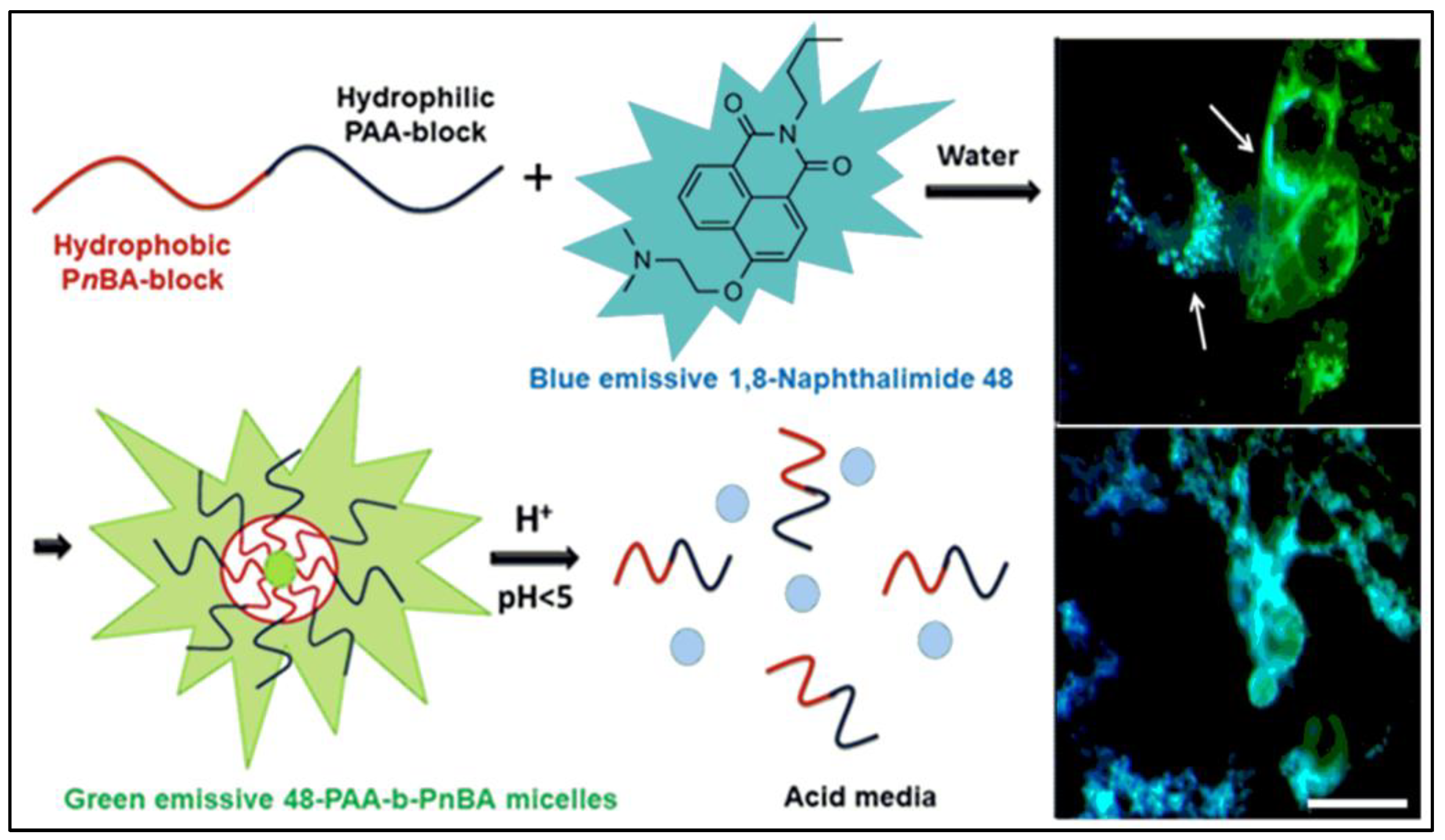
- Georgiev, N.I.; Bryaskova, R.G.; Ismail, S.R.; Philipova, N.D.; Uzunova, V.P.; Bakov, V.V.; Tzoneva, R.D.; Bojinov, V.B. Aggregation induced emission in 1,8-naphthalimide embedded nanomicellar architecture as a platform for fluorescent ratiometric pH-probe with biomedical applications. J. Photochem. Photobiol. 2021, 418, 113380. [Google Scholar] [CrossRef]
- Wright, G.D.; Yao, C.-Y.; Moody, T.S.; de Silva, A.P. Fluorescent molecular logic gates based on photoinduced electron transfer (PET) driven by a combination of atomic and biomolecular inputs. Chem. Commun. 2020, 56, 6838–6841. [Google Scholar] [CrossRef] [PubMed]
- Andréasson, J.; Pischel, U. Molecules with a sense of logic: A progress report. Chem. Soc. Rev. 2015, 44, 1053–1069. [Google Scholar] [CrossRef] [Green Version]
- Kaur, N. Supramolecular switches-advanced molecular logic and computation molecular logic gates. Curr. Org. Chem. 2014, 18, 2892–2909. [Google Scholar] [CrossRef]
- de Silva, A.P.; Gunaratne, H.; McCoy, C. A molecular photoionic AND gate based on fluorescent signaling. Nature 1993, 364, 42–44. [Google Scholar] [CrossRef]
- Pais, V.; Remon, P.; Collado, D.; Andréasson, J.; Perez-Inestrosa, E.; Pischel, U. OFF-ON-OFF fluorescence switch with T-latch function. Org. Lett. 2011, 13, 5572–5575. [Google Scholar] [CrossRef]
- Ozlem, S.; Akkaya, E.U. Thinking outside the silicon box: Molecular AND logic as an additional layer of selectivity in singlet oxygen generation for photodynamic therapy. J. Am. Chem. Soc. 2009, 131, 48–49. [Google Scholar] [CrossRef] [PubMed]
- Turan, I.S.; Gunaydin, G.; Ayan, S.; Akkaya, E.U. Molecular demultiplexer as a terminator automaton. Nat. Commun. 2018, 9, 805. [Google Scholar] [CrossRef] [PubMed] [Green Version]
- Magri, D.; Fava, M.; Mallia, C. A sodium-enabled “Pourbaix sensor”: A three-input AND logic gate as a “lab-on-a-molecule” for monitoring Na+, pH and pE. Chem. Commun. 2014, 50, 1009–1011. [Google Scholar] [CrossRef] [PubMed]
- Huan, X. Iron overload and its association with cancer risk in humans: Evidence for iron as a carcinogenic metal. Mutat. Res. 2003, 533, 153–171. [Google Scholar] [CrossRef] [PubMed]
- Georgiev, N.I.; Yaneva, I.S.; Surleva, A.R.; Asiri, A.M.; Bojinov, V.B. Synthesis, sensor activity and logic behavior of a highly water-soluble naphthalimide derivative. Sens. Actuators B Chem. 2013, 184, 54–63. [Google Scholar] [CrossRef]
- Zhang, J.; Ning, L.; Huang, J.; Zhang, C.; Pu, K. Activatable molecular agents for cancer theranostics. Chem. Sci. 2020, 11, 618–630. [Google Scholar] [CrossRef] [PubMed] [Green Version]
- Lee, M.H.; Sharma, A.; Chang, M.J.; Lee, J.; Son, S.; Sessler, J.L.; Kang, C.; Kim, J.S. Fluorogenic reaction-based prodrug conjugates as targeted cancer theranostics. Chem. Soc. Rev. 2018, 47, 28–52. [Google Scholar] [CrossRef]
- Cooper, E.; Choi, P.J.; William, A.; Denny, W.A.; Jose, J.; Dragunow, M.; Park, T. The use of heptamethine cyanine dyes as drug-conjugate systems in the treatment of primary and metastatic brain tumors. Front. Oncol. 2021, 11, 654921. [Google Scholar] [CrossRef]
- Yang, X.; Shi, C.; Tong, R.; Qian, W.; Zhau, H.E.; Wang, R.; Zhu, G.; Cheng, J.; Yang, V.W.; Cheng, T.; et al. Near IR hepta-methine cyanine dye-mediated cancer imaging. Clin. Cancer Res. 2010, 16, 2833–2844. [Google Scholar] [CrossRef] [Green Version]
- Wu, J.B.; Shi, C.; Chu, G.C.; Xu, Q.; Zhang, Y.; Li, Q.; Yu, J.S.; Zhau, H.E.; Chung, L.W.K. Biomaterials near infrared fluorescence heptamethine carbocyanine dyes mediate imaging and targeted drug delivery for human brain tumor. Biomaterials 2015, 67, 1–10. [Google Scholar] [CrossRef] [PubMed] [Green Version]
- Tao, X.M.; Wang, J.C.; Wang, J.B.; Feng, Q.; Gao, S.Y.; Zhang, L.R.; Zhang, Q. Enhanced anticancer activity of gemcitabine coupling with conjugated linoleic acid against human breast cancer in vitro and in vivo. Eur. J. Pharm. Biopharm. 2012, 82, 401–409. [Google Scholar] [CrossRef] [PubMed]
- Jiang, Z.; Pflug, K.; Usama, S.M.; Kuai, D.; Yan, X.; Sitcheran, R.; Burgess, K. Cyanine gemcitabine conjugates as targeted theranostic agents for glioblastoma tumor cells. J. Med. Chem. 2019, 62, 9236–9245. [Google Scholar] [CrossRef]
- Choi, P.J.; Cooper, E.; Schweder, P.; Mee, E.; Faull, R.; Denny, W.A.; Dragunow, M.; Park, T.I.-H.; Jose, J. The synthesis of a novel crizotinib heptamethine cyanine dye conjugate that potentiates the cytostatic and cytotoxic effects of crizotinib in patient-derived glioblastoma cell lines. Bioorg. Med. Chem. Lett. 2019, 29, 2617–2621. [Google Scholar] [CrossRef] [PubMed]
- Choi, P.J.; Cooper, E.; Schweder, P.; Mee, E.; Turner, C.; Faull, R.; Denny, W.A.; Dragunow, M.; Park, T.I.-H.; Jose, J. PARP inhibitor cyanine dye conjugate with enhanced cytotoxic and antiproliferative activity in patient derived glioblastoma cell lines. Bioorg. Med. Chem. Lett. 2020, 30, 127252. [Google Scholar] [CrossRef] [PubMed]
- Kong, Y.; Smith, J.; Li, K.; Cui, J.; Han, J.; Hou, S.; Brown, M.L. Development of a novel near-infrared fluorescent theranostic combretastain A-4 analogue, YK-5-252, to target triple negative breast cancer. Bioorg. Med. Chem. 2017, 25, 2226–2233. [Google Scholar] [CrossRef] [PubMed]
- Liu, Y.; Zhu, S.; Gu, K.; Guo, Z.; Huang, X.; Wang, M.; Amin, H.M.; Zhu, W.; Shi, P. GSH-activated NIR fluorescent prodrug for podophyllotoxin delivery. ACS Appl. Mater. Interfaces 2017, 9, 29496–29504. [Google Scholar] [CrossRef]
- Ye, M.; Wang, X.; Tang, J.; Guo, Z.; Shen, Y.; Tian, H.; Zhu, W.-H. Dual-channel NIR activatable theranostic prodrug for in vivo spatiotemporal tracking thiol-triggered chemotherapy. Chem. Sci. 2016, 7, 4958–4965. [Google Scholar] [CrossRef] [Green Version]
- Li, Y.; Zhao, L.; Li, X.-F. Targeting hypoxia: Hypoxia-activated prodrugs in cancer therapy. Front. Oncol. 2021, 11, 700407. [Google Scholar] [CrossRef]
- Wu, J.; Sha, J.; Zhang, C.; Liu, W.; Zheng, X.; Wang, P. Recent advances in theranostic agents based on natural products for photodynamic and sonodynamic therapy. View 2020, 1, 20200090. [Google Scholar] [CrossRef]
- Kumar, R.; Kim, E.J.; Han, J.; Lee, H.; Shin, W.S.; Kim, H.M.; Bhuniya, S.; Kim, J.S.; Hong, K.S. Hypoxia-directed and activated theranostic agent: Imaging and treatment of solid tumor. Biomaterials 2016, 104, 119–128. [Google Scholar] [CrossRef]
- Huang, J.; Wu, Y.; Zeng, F.; Wu, S. An activatable near-infrared chromophore for multispectral optoacoustic imaging of tumor hypoxia and for tumor inhibition. Theranostics 2019, 9, 7313–7324. [Google Scholar] [CrossRef] [PubMed]
- Zhang, C.; Yuan, Q.; Zhang, Z.; Tang, Y. A pH-responsive drug delivery system based on conjugated polymer for effective synergistic chemo-/photodynamic therapy. Molecules 2023, 28, 399. [Google Scholar] [CrossRef] [PubMed]
- Jang, Y.; Kim, T.-I.; Kim, H.; Choi, Y.; Kim, Y. Photoactivatable BODIPY platform: Light-triggered anticancer drug release and fluorescence monitoring. ACS Appl. Biol. Mater. 2019, 2, 2567–2572. [Google Scholar] [CrossRef] [PubMed]


Disclaimer/Publisher’s Note: The statements, opinions and data contained in all publications are solely those of the individual author(s) and contributor(s) and not of MDPI and/or the editor(s). MDPI and/or the editor(s) disclaim responsibility for any injury to people or property resulting from any ideas, methods, instructions or products referred to in the content. |
© 2023 by the authors. Licensee MDPI, Basel, Switzerland. This article is an open access article distributed under the terms and conditions of the Creative Commons Attribution (CC BY) license (https://creativecommons.org/licenses/by/4.0/).
Share and Cite
Georgiev, N.I.; Bakov, V.V.; Anichina, K.K.; Bojinov, V.B. Fluorescent Probes as a Tool in Diagnostic and Drug Delivery Systems. Pharmaceuticals 2023, 16, 381. https://doi.org/10.3390/ph16030381
Georgiev NI, Bakov VV, Anichina KK, Bojinov VB. Fluorescent Probes as a Tool in Diagnostic and Drug Delivery Systems. Pharmaceuticals. 2023; 16(3):381. https://doi.org/10.3390/ph16030381
Chicago/Turabian StyleGeorgiev, Nikolai I., Ventsislav V. Bakov, Kameliya K. Anichina, and Vladimir B. Bojinov. 2023. "Fluorescent Probes as a Tool in Diagnostic and Drug Delivery Systems" Pharmaceuticals 16, no. 3: 381. https://doi.org/10.3390/ph16030381