
Synthesis of Coumarin and Homoisoflavonoid Derivatives and Analogs: The Search for New Antifungal Agents

 , and
, and 
Abstract
:1. Introduction
2. Results
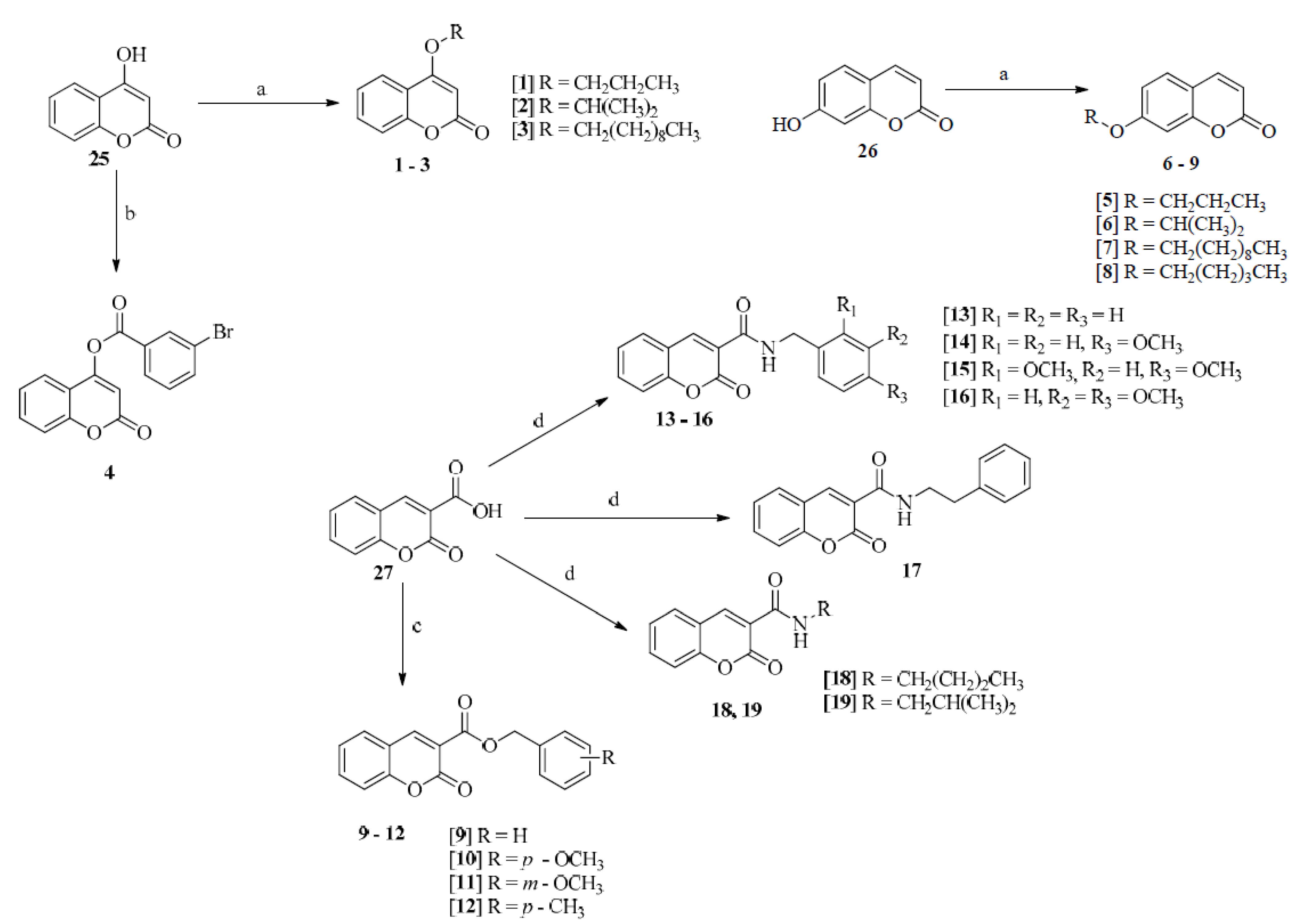
2.1. Chemistry of Compounds 1–24
2.2. Antifungal Activity of Compounds 1–24
2.2.1. Verification of the Mode of Action on the Fungal Cell Wall and Membrane
2.2.2. Evaluation of the Antimicrobial Activity of Compound 8 on the Reduction of Fungal Biofilm
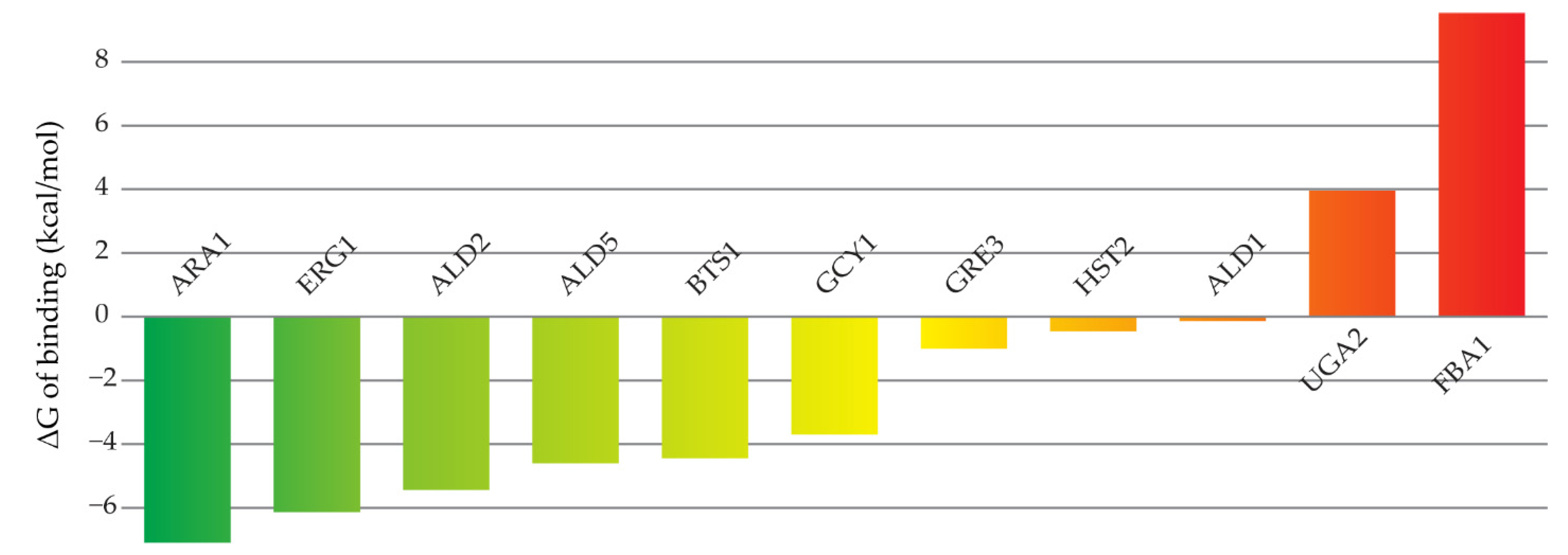
2.3. Molecular Modeling
2.4. ADMET Predictions
3. Discussion
4. Materials and Methods
4.1. Chemistry
4.1.1. Methodology for Obtaining Ethers Derived from 4-Hydroxycoumarin (1–3) and 7-Hydroxycoumarin (5–8)
4.1.2. Methodology for Obtaining 4-Hydroxycoumarin Derivative 4
4.1.3. Methodology for Obtaining Esters 9–12 Derived from Coumarin-3-Carboxylic Acid
4.1.4. Methodology for Obtaining Amides 13–19 Derived from Coumarin-3-Carboxylic Acid
4.1.5. Methodology for Obtaining Chalcone 20
4.1.6. Methodology for Obtaining Derivatives 21–24 from 4-Chromanone
4.2. Antifungal Activity
4.2.1. Determination of Minimum Inhibitory Concentration (MIC)
4.2.2. Determination of Minimum Fungicide Concentration (MFC)
4.2.3. Verification of Mode of Activity on the Fungal Cell Wall and Membrane
Ergosterol Test
Sorbitol Assay
4.2.4. Evaluation of the Antimicrobial Activity of Compound 8 on the Reduction of Fungal Biofilm
4.3. Molecular Modeling Study
4.3.1. Targets Selection
4.3.2. Molecular Docking
4.3.3. Molecular Dynamics Simulations and Estimation of Free Energies of Binding
4.4. ADMET Predictions
5. Conclusions
Supplementary Materials
Author Contributions
Funding
Institutional Review Board Statement
Informed Consent Statement
Data Availability Statement
Acknowledgments
Conflicts of Interest
References
- Aldardeer, N.F.; Albar, H.; Attas, M.-A.; Eldali, A.; Qutub, M.; Hassanien, A.; Alraddadi, B. Antifungal resistance in patients with Candidaemia: A retrospective cohort study. BMC Infect Dis. 2020, 20, 55. [Google Scholar] [CrossRef] [PubMed] [Green Version]
- Pappas, P.G.; Lionakis, M.S.; Arendrup, M.C.; Zeichner, L.O.; Kulberg, B.J. Invasive candidiasis. Nat. Rev. Dis. Primers 2018, 4, 18026. [Google Scholar] [CrossRef] [PubMed]
- Bartlett, J.G. Nosocomial bloodstream infections in US hospitals: Analysis of 24,179 cases from a prospective nationwide surveillance study. Infect. Dis. Clin. Pract. 2004, 12, 309–317. [Google Scholar] [CrossRef]
- McCarty, T.P.; Pappas, P.G. Invasive Candidiasis. Infect. Dis. Clin. N. Am. 2015, 30, 103–124. [Google Scholar] [CrossRef] [PubMed]
- Lazić, J.; Ajdacic, V.; Vojnovic, S.; Zlatovic, M.; Pekmezovic, M.; Mogavero, S.; Opsenica, I.; Nikodinovic-Runic, J. Bis-guanylhydrazones as efficient anti-Candida compounds through DNA interaction. Appl. Microbiol. Biotechnol. 2018, 102, 1889–1901. [Google Scholar] [CrossRef]
- Serra, S.; Chicca, A.; Delogu, G.; Vásquez-Ródriguez, S.; Santana, L.; Uriarte, E.; Casu, L.; Gertsch, J. Synthesis and cytotoxic activity of non-naturally substituted 4-oxycoumarin derivatives. Bioorg. Med. Chem. Lett. 2012, 22, 5791–5794. [Google Scholar] [CrossRef]
- An, J.Y.; Lee, H.-H.; Shin, J.-S.; Yoo, H.-S.; Park, J.S.; Son, S.W.; Kim, S.W.; Yu, J.; Lee, J.; Lee, K.-T.; et al. Identification and structure activity relationship of novel flavone derivatives that inhibit the production of nitric oxide and PGE2 in LPS-induced RAW 264.7 cells. Bioorg. Med. Chem. Lett. 2017, 27, 2613–2616. [Google Scholar] [CrossRef]
- Kirsch, G.; Abdelwahab, A.B.; Chaimbault, P. Natural and synthetic coumarins with effects on inflammation. Molecules 2016, 21, 1322. [Google Scholar] [CrossRef]
- Hussain, H.; Green, I.R. A patent review of the therapeutic potential of isoflavones (2012–2016). Expert Opin. Ther. Pat. 2017, 27, 1135–1146. [Google Scholar] [CrossRef]
- Phutdhawong, W.; Chuenchid, A.; Taechowisan, T.; Sirirak, J.; Phutdhawong, W.S. Synthesis and biological evaluation of coumarin-3-carboxamide derivatives. Molecules 2021, 26, 1653. [Google Scholar] [CrossRef]
- Weng, K.G.; Yuan, Y.L. Synthesis and evaluation of coumarin derivatives against human lung cancer cell lines. Braz. J. Med. Biol. Res. 2017, 50, 502–507. [Google Scholar] [CrossRef] [PubMed] [Green Version]
- Kadhum, A.A.H.; Al-Amiery, A.A.; Musa, A.Y.; Mohamed, A.B. The antioxidant activity of new coumarin derivatives. Int. J. Mol. Sci. 2011, 12, 5747–5761. [Google Scholar] [CrossRef] [Green Version]
- Golfakhrabadi, F.; Abdollahi, M.; Reza, M.; Ardakni, S.; Saeidnia, S.; Akbarzadeh, T.; Ebrahini, A.; Yousefbeyk, F.; Hassanzadeh, A.; Khanavi, M. Anticoagulant activity of isolated coumarins (suberosin and suberenol) and toxicity evaluation of Ferulago carduchorum in rats. Pharm. Biol. 2014, 52, 1335–1340. [Google Scholar] [CrossRef] [PubMed]
- Chiang, C.-C.; Cheng, M.-J.; Peng, C.-F.; Huang, H.-Y.; Chen, I.-S. A novel dimeric coumarin analog and antimycobacterial constituents from Fatoua pilosa. Chem. Biodivers. 2010, 7, 1728–1736. [Google Scholar] [CrossRef] [PubMed]
- Neyts, J.; De Clercq, E.; Singha, R.; Chang, Y.H.; Das, A.R.; Chakraborty, S.K.; Hong, S.C.; Tsay, S.C.; Hsu, M.-H.; Hwu, J.R. Structure-activity relationship of new anti-hepatitis C virus agents: Heterobicycle-coumarin conjugates. J. Med. Chem. 2009, 52, 1486–1490. [Google Scholar] [CrossRef] [PubMed]
- Jia, C.; Zhang, J.; Yu, L.; Wang, C.; Yang, Y.; Rong, X.; Xu, K.; Chu, M. Antifungal activity of coumarin against Candida albicans is related to apoptosis. Front. Cell. Infect. Microbiol. 2019, 8, 445. [Google Scholar] [CrossRef] [Green Version]
- Desideri, N.; Monaco, L.P.; Fioravanti, M.B.; Yanez, M.; Alcaro, S.; Ortuso, F. (E)-3-Heteroarylidenechroman-4-ones as potent and selective monoamine oxidase-B inhibitors. Eur. J. Med. Chem. 2016, 117, 292–300. [Google Scholar] [CrossRef] [Green Version]
- Lin, L.-G.; Liu, Q.-Y.; Ye, Y. Naturally Occurring Homoisoflavonoids and Their Pharmacological Activities. Plant. Med. 2014, 80, 1053–1066. [Google Scholar] [CrossRef] [Green Version]
- Ragab, F.A.; Yahya, T.A.A.; El-Naa, M.M.; Arafa, R.K. Design, synthesis and structure-activity relationship of novel semi-synthetic flavonoids as antiproliferative agents. Eur. J. Med. Chem. 2014, 82, 506–520. [Google Scholar] [CrossRef]
- Tian, S.S.; Jiang, F.-S.; Zhang, K.; Zhu, X.-X.; Jin, B.; Lu, J.-J.; Ding, Z.-S. Flavonoids from the leaves of Carya cathayensis Sarg. inhibit vascular endothelial growth factor-induced angiogenesis. Fitoterapia 2014, 92, 34–40. [Google Scholar] [CrossRef]
- Zhang, X.; Huang, H.; Zhao, X.; Lv, Q.; Sunc, C.; Li, X.; Chen, K. Effects of flavonoids-rich Chinese bayberry (Myrica rubra Sieb. et Zucc.) pulp extracts on glucose consumption in human HepG2 cells. J. Funct. Foods. 2015, 14, 144–153. [Google Scholar] [CrossRef]
- Peralta, M.A.; Silva, M.A.; Ortega, M.G.; Cabrera, J.L.; Parage, M.G. Antifungal activity of a prenylated flavonoid from Dalea elegans against Candida albicans biofilms. Phytomedicine. 2015, 22, 975–980. [Google Scholar] [CrossRef] [PubMed]
- Wang, Y.H.; Dong, H.-H.; Zhao, F.; Wang, J.; Yan, F.; Jiang, Y.-Y.; Jin, Y.-S. The synthesis and synergistic antifungal effects of chalcones against drug resistant Candida albicans. Bioorg. Med. Chem. Lett. 2016, 26, 3098–3102. [Google Scholar] [CrossRef] [PubMed]
- Jin, Y.S. Recent advances in natural antifungal flavonoids and their derivatives. Bioorg. Med. Chem. Lett. 2019, 29, 126589. [Google Scholar] [CrossRef]
- Singh, H.; Kumar, M.; Nepali, K.; Gupta, M.K.; Saxena, A.K.; Sharma, S.; Bedi, P.M. Triazole tethered C5-curcuminoid-coumarin based molecular hybrids as novel antitubulin agents: Design, synthesis, biological investigation and docking studies. Eur. J. Med. Chem. 2016, 116, 102–115. [Google Scholar] [CrossRef]
- Hugo, A.G.; Jimena, M.M.; Glady, M.C.; Carlos, E.T.; Carlos, R.P. Inhibition of reverse transcriptase and Taq DNA polymerase by compounds possessing the coumarin framework. Bioorg. Med. Chem. Lett. 2014, 24, 760–764. [Google Scholar] [CrossRef]
- Lutjen, A.B.; Quirk, M.A.; Barbera, A.M.; Kolonko, E.M. Synthesis of (E)-cinnamyl ester derivatives via a greener Steglich esterification. Bioorg. Med. Chem. 2018, 26, 5291–5298. [Google Scholar] [CrossRef]
- Rajan, P.; Vedernikova, I.; Cos, P.; vanden Berghe, D.; Augustyns, K.; Haemers, A. Synthesis and evaluation of caffeic acid amides as antioxidants. Bioorg. Med. Chem. Lett. 2001, 11, 215–217. [Google Scholar] [CrossRef]
- Yu, X.; Teng, P.; Zhang, Y.L.; Xu, Z.J.; Zhang, M.Z.; Zhang, W.H. Design, synthesis and antifungal activity evaluation of coumarin-3-carboxamide derivatives. Fitoterapia 2018, 127, 387–395. [Google Scholar] [CrossRef]
- Badavath, V.N.; Jadav, S.S.; Boris, P.; de Xavier, L.; Barij, N.S.; Venkatesan, J. Synthesis and Antiviral Activity of 2-aryl-4H-chromen-4-one Derivatives against Chikungunya Virus. Lett. Drug Des. Discov. 2016, 13, 1019–1024. [Google Scholar] [CrossRef] [Green Version]
- da Alves, D.N.; Ferreira, A.R.; Duarte, A.B.S.; Melo, A.K.V.; de Sousa, D.P.; de Castro, R.D. Breakpoints for the Classification of Anti-Candida Compounds in Antifungal Screening. BioMed Res. Int. 2021, 2021, 6653311. [Google Scholar] [CrossRef] [PubMed]
- Escalante, A.; Gattuso, M.; Pérez, P.; Zacchino, S. Evidence of the mechanism of action of the antifungal phytolaccoside B isolated from Phytolacca tetramera Hauman. J. Nat. Prod. 2008, 71, 1720–1725. [Google Scholar] [CrossRef] [PubMed]
- de Freires, I.A.; Murata, R.M.; Furletti, V.F.; Sartoratto, A.; de Alencar, S.M.; Figueira, G.M.; de Oliveira Rodrigues, J.A.; Duarte, M.C.T.; Rosalen, P.L. Coriandrum sativum L. (Coriander) Essential Oil: Antifungal Activity and Mode of Action on Candida spp., and Molecular Targets Affected in Human Whole-Genome Expression. PLoS ONE 2014, 9, e99086. [Google Scholar] [CrossRef] [PubMed] [Green Version]
- Guterres, H.; Im, W. Improving Protein-Ligand Docking Results with High-Throughput Molecular Dynamics Simulations. J. Chem. Inf. Model. 2020, 60, 2189–2198. [Google Scholar] [CrossRef]
- Wang, J.; Morin, P.; Wang, W.; Kollman, P.A. Use of MM-PBSA in Reproducing the Binding Free Energies to HIV-1 RT of TIBO Derivatives and Predicting the Binding Mode to HIV-1 RT of Efavirenz by Docking and MM-PBSA. J. Am. Chem. Soc. 2001, 123, 5221–5230. [Google Scholar] [CrossRef]
- Katsila, T.; Spyroulias, G.A.; Patrinos, G.P.; Matsoukas, M.-T. Computational approaches in target identification and drug discovery. Comput. Struct. Biotechnol. J. 2016, 14, 177–184. [Google Scholar] [CrossRef] [Green Version]
- Lemos, A.S.O.; Florêncio, J.R.; Pinto, N.C.C.; Campos, L.M.; Silva, T.P.; Grazul, R.M.; Pinto, P.F.; Tavares, G.D.; Scio, E.; Apolônio, A.C.M.; et al. Antifungal Activity of the Natural Coumarin Scopoletin against Planktonic Cells and Biofilms from a Multidrug-Resistant Candida tropicalis Strain. Front. Microbiol. 2020, 11, 1525. [Google Scholar] [CrossRef]
- Tiwari, S.; Seijas, J.; Vazquez-Tato, M.; Sarkate, A.; Karnik, K.; Nikalje, A. Facile Synthesis of Novel Coumarin Derivatives, Antimicrobial Analysis, Enzyme Assay, Docking Study, ADMET Prediction and Toxicity Study. Molecules 2017, 22, 1172. [Google Scholar] [CrossRef] [Green Version]
- Pettersen, E.F.; Goddard, T.D.; Huang, C.C.; Couch, G.S.; Greenblatt, D.M.; Meng, E.C.; Ferrin, T.E. UCSF Chimera—A visualization system for exploratory research and analysis. J. Comput. Chem. 2004, 25, 1605–1612. [Google Scholar] [CrossRef] [Green Version]
- Laskowski, R.A.; Swindells, M.B. LigPlot+: Multiple Ligand–Protein Interaction Diagrams for Drug Discovery. J. Chem. Inf. Model. 2011, 51, 2778–2786. [Google Scholar] [CrossRef]
- Shannon, P.; Markiel, A.; Ozier, O.; Baliga, N.S.; Wang, J.T.; Ramage, D.; Amin, N.; Schwikowski, B.; Ideker, T. Cytoscape: A Software Environment for Integrated Models of Biomolecular Interaction Networks. Genome Res. 2003, 13, 2498–2504. [Google Scholar] [CrossRef] [PubMed]
- Emami, S.; Ghanbarimasir, Z. Recent advances of chroman-4-one derivatives: Synthetic approaches and bioactivities. Eur. J. Med. Chem. 2015, 93, 539–563. [Google Scholar] [CrossRef] [PubMed]
- Jamiu, A.T.; Albertyn, J.; Sebolai, O.M.; Pohl, C.H. Update on Candida krusei, a potential multidrug-resistant pathogen. Med. Mycol. 2021, 59, 14–30. [Google Scholar] [CrossRef] [PubMed]
- Pan, L.; Lei, D.; Jin, L.; He, Y.; Yang, Q. Promising Fungicides from Allelochemicals: Synthesis of Umbelliferone Derivatives and Their Structure–Activity Relationships. Molecules 2018, 23, 3002. [Google Scholar] [CrossRef] [Green Version]
- Chu, L.L.; Pandey, R.P.; Lim, H.N.; Jung, H.J.; Thuan, N.H.; Kim, T.-S.; Sohng, J.K. Synthesis of umbelliferone derivatives in Escherichia coli and their biological activities. J. Biol. Eng. 2017, 11, 15. [Google Scholar] [CrossRef] [Green Version]
- Zaki, M.A.; Nanayakkara, N.P.D.; Hetta, M.H.; Jacob, M.R.; Khan, S.I.; Mohammed, R.; Ibrahim, M.A.; Samoylenko, V.; Coleman, C.; Fronczek, F.R.; et al. Bioactive Formylated Flavonoids from Eugenia rigida: Isolation, Synthesis, and X-ray Crystallography. J. Nat. Prod. 2016, 79, 2341–2349. [Google Scholar] [CrossRef]
- Farhadri, F.; Khameneh, B.; Iranshahi, M.; Milad, I. Antibacterial activity of flavonoids and their structure-activity relationship: An update review. Phytother. Res. 2019, 33, 13–40. [Google Scholar] [CrossRef] [Green Version]
- Ullah Mughal, E.; Ayaz, M.; Hussain, Z.; Hasan, A.; Sadiq, A.; Riaz, M.; Malik, A.; Hussain, S.; Choudhary, M.I. Synthesis and antibacterial activity of substituted flavones, 4-thioflavones and 4-iminoflavones. Bioorg. Med. Chem. 2006, 14, 4704–4711. [Google Scholar] [CrossRef]
- Noushini, S.; Alipour, E.; Emami, S.; Safavi, M.; Ardestani, S.K.; Gohari, A.R.; Shafiee, A.; Foroumadi, A. Synthesis and cytotoxic properties of novel (E)-3-benzylidene-7-methoxychroman-4-one derivatives. DARU J. Pharm. Sci. 2013, 21, 31. [Google Scholar] [CrossRef] [Green Version]
- Siddaiah, V.; Rao, C.V.; Venkateswarlu, S.; Krishnaraju, A.V.; Subbaraju, G. Synthesis, stereochemical assignments, and biological activities of homoisoflavonoids. Bioorg. Med. Chem. 2006, 14, 2545–2551. [Google Scholar] [CrossRef]
- Das, U.; Lorand, T.; Dimmock, S.G.; Perjesi, P.; Dimmock, J.R. 3-Benzylidene-4-chromanones: A novel cluster of anti-tubercular agents. J. Enzyme Inhib. Med. 2015, 30, 259–263. [Google Scholar] [CrossRef] [PubMed]
- Tait, S.; Salvati, A.L.; Desideri, N.; Fiore, L. Antiviral activity of substituted homoisoflavonoids on enteroviruses. Antivir. Res. 2006, 72, 252–255. [Google Scholar] [CrossRef] [PubMed]
- Das, B.; Thirupathi, P.; Ravikanth, B.; Kumar, R.A.; Sarma, A.V.S.; Basha, S.J. Isolation, Synthesis, and Bioactivity of Homoisoflavonoids from Caesalpinia pulcherrima. Chem. Pharm. Bull. 2009, 57, 1139–1141. [Google Scholar] [CrossRef] [PubMed] [Green Version]
- Chowdhury, S.; Chanda, T.; Gupta, A.; Koley, S.; Ramulu, B.J.; Jones, R.C.F.; Singh, M.S. Indium(0)-Mediated C sp 3-S/O Cross-Coupling Approach Towards the Regioselective Alkylation of α-Enolic Esters/Dithioesters: A Mechanistic Insight. Eur. J. Org. Chem. 2014, 2014, 2964–2971. [Google Scholar] [CrossRef]
- de Araújo, R.; de Guerra, F.O.; Lima, E.; de Simone, C.; Tavares, J.; Scotti, L.; Scotti, M.; de Aquino, T.; de Moura, R.; Mendonça, F.; et al. Synthesis, Structure-Activity Relationships (SAR) and in Silico Studies of Coumarin Derivatives with Antifungal Activity. Int. J. Mol. Sci. 2013, 14, 1293–1309. [Google Scholar] [CrossRef]
- Maresca, A.; Temperini, C.; Pochet, L.; Masereel, B.; Scozzafava, A.; Supuran, C.T. Deciphering the Mechanism of Carbonic Anhydrase Inhibition with Coumarins and Thiocoumarins. J. Med. Chem. 2010, 53, 335–344. [Google Scholar] [CrossRef] [Green Version]
- Adfa, M.; Hattori, Y.; Yoshimura, T.; Koketsu, M. Antitermite activity of 7-alkoxycoumarins and related analogs against Coptotermes formosanus Shiraki. Int. Biodeterior. Biodegrad. 2012, 74, 129–135. [Google Scholar] [CrossRef]
- Kuang, Y.; Liu, X.; Chang, L.; Wang, M.; Lin, L.; Feng, X. Catalytic Asymmetric Conjugate Allylation of Coumarins. Org. Lett. 2011, 13, 3814–3817. [Google Scholar] [CrossRef]
- Nikoofar, K.; Yielzoleh, F.M. Novel nano-titania embedded on graphite (nano-TiO2@Cg) as an efficient, eco-friendly, and recyclable catalyst for one-pot, solvent-free synthesis of 4-aryl-3,4-dihydroquinolin-2(1H)-ones, 3-methyl-4-aryl/alkyl-2,4,5,7-tetrahydropyrazolo [3,4-b]pyridin-6-ones, and coumarin-3-carboxylic esters. Res. Chem. Intermed. 2018, 44, 7353–7367. [Google Scholar] [CrossRef]
- Espinosa, M.A.; Tamariz, J.; Padilla-Martínez, I.I.; Martínez-Martínez, F.J. Síntesis y estudio estructural por RMN de 1H y 13C de la N-[4-[2-(2-oxo-2H-1-benzopiranil-3-carboxamidil)etil]bencensulfonil]-N’-ciclohexilurea y de la N-[4-[2-(4-nitrobenzamidil)etil]-bencensulfonil]-N’-ciclohexilurea’. Version. Soc. Quím. Mex. 2001, 45, 214–217. [Google Scholar]
- Sonam Shinde, V.N.; Kumar, A. KPF6—Mediated Esterification and Amidation of Carboxylic Acids. J. Org. Chem. 2022, 87, 2651–2661. [Google Scholar] [CrossRef] [PubMed]
- Ohkatsu, Y.; Satoh, T. Antioxidant and Photo-antioxidant Activities of Chalcone Derivatives. J. Jpn. Pet. 2008, 51, 298–308. [Google Scholar] [CrossRef] [Green Version]
- Mandal, T.K.; Pal, R.; Mondal, R.; Mallik, A.K. Facile Condensation of Aromatic Aldehydes with Chroman-4-ones and 1-Thiochroman-4-ones Catalysed by Amberlyst-15 under Microwave Irradiation Condition. E-J. Chem. 2011, 8, 863–869. [Google Scholar] [CrossRef]
- Biju, A.T.; Wurz, N.E.; Glorius, F. N-Heterocyclic Carbene-Catalyzed Cascade Reaction Involving the Hydroacylation of Unactivated Alkynes. J. Am. Chem. Soc. 2010, 132, 5970–5971. [Google Scholar] [CrossRef] [PubMed]
- Gopaul, K.; Shaikh, M.; Ramjugernath, D.; Koorbanally, N.A.; Omondi, B. 3-(3-Methoxybenzylidene)chroman-4-one. Acta Crystallogr. Sect. E Struct. Rep. Online 2012, 68, o1006. [Google Scholar] [CrossRef] [Green Version]
- CLSI. Reference Method for Broth Dilution Antifungal Susceptibility Testing of Yeasts: Approved Standard-Third Edition, 3rd ed.; CLSI document M27-A3; Clinical and Laboratory Standards Institute: Wayne, PA, USA, 2008. [Google Scholar]
- Siddiqui, Z.N.; Farooq, F.; Musthafa, T.N.M.; Ahmad, A.; Khan, A.U. Synthesis, characterization and antimicrobial evaluation of novel halopyrazole derivatives. J. Saudi Chem. Soc. 2013, 17, 237–243. [Google Scholar] [CrossRef] [Green Version]
- Lima, I.O.; de Pereira, F.O.; de Oliveira, W.A.; de Lima, E.O.; Menezes, E.A.; Cunha, F.A.; de Diniz, M.F.F.M. Antifungal activity and mode of action of carvacrol against Candida albicans strains. J. Essent. Oil Res. 2013, 25, 138–142. [Google Scholar] [CrossRef]
- Djordjevic, D.; Wiedmann, M.; McLandsborough, L.A. Microtiter Plate Assay for Assessment of Listeria monocytogenes biofilm formation. Appl. Environ. Microbiol. 2002, 68, 2950–2958. [Google Scholar] [CrossRef] [Green Version]
- Chaves, G.M.; Diniz, M.G.; da Silva-Rocha, W.P.; de Souza, L.B.F.C.; Gondim, L.A.M.; Ferreira, M.A.F.; Svidzinski, T.I.E.; Milan, E.P. Species distribution and virulence factors of Candida spp. isolated from the oral cavity of kidney transplant recipients in Brazil. Mycopathologia 2013, 175, 255–263. [Google Scholar] [CrossRef]
- Lopes, S.P.; Yepes, L.M.; Pérez-Castillo, Y.; Robledo, S.M.; de Sousa, D.P. Alkyl and Aryl Derivatives Based on p-Coumaric Acid Modification and Inhibitory Action against Leishmania braziliensis and Plasmodium falciparum. Molecules 2020, 25, 3178. [Google Scholar] [CrossRef]
- Araújo, M.O.; Pérez-Castillo, Y.; Oliveira, L.H.G.; Nunes, F.C.; de Sousa, D.P. Larvicidal Activity of Cinnamic Acid Derivatives: Investigating Alternative Products for Aedes aegypti L. Control. Molecules 2020, 26, 61. [Google Scholar] [CrossRef] [PubMed]
- Keiser, M.J.; Roth, B.L.; Armbruster, B.N.; Ernsberger, P.; Irwin, J.J.; Shoichet, B.K. Relating protein pharmacology by ligand chemistry. Nat. Biotechnol. 2007, 25, 197–206. [Google Scholar] [CrossRef] [PubMed] [Green Version]
- Altschul, S. Gapped BLAST and PSI-BLAST: A new generation of protein database search programs. Nucleic Acids Res. 1997, 25, 3389–3402. [Google Scholar] [CrossRef] [PubMed] [Green Version]
- OMEGA. OpenEye Scientific Software. Santa Fe, NM, USA. Available online: http://www.eyesopen.com (accessed on 20 December 2021).
- Hawkins, P.C.D.; Skillman, A.G.; Warren, G.L.; Ellingson, B.A.; Stahl, M.T. Conformer Generation with OMEGA: Algorithm and Validation Using High Quality Structures from the Protein Databank and Cambridge Structural Database. J. Chem. Inf. Model. 2010, 50, 572–584. [Google Scholar] [CrossRef]
- QUACPAC. OpenEye Scientific Software. Santa Fe, NM, USA. Available online: http://www.eyesopen.com (accessed on 20 December 2021).
- Jumper, J.; Evans, R.; Pritzel, A.; Green, T.; Figurnov, M.; Ronneberger, O.; Tunyasuvunakool, K.; Bates, R.; Žídek, A.; Potapenko, A.; et al. Highly accurate protein structure prediction with AlphaFold. Nature 2021, 596, 583–589. [Google Scholar] [CrossRef]
- Bienert, S.; Waterhouse, A.; de Beer, T.A.P.; Tauriello, G.; Studer, G.; Bordoli, L.; Schwede, T. The SWISS-MODEL Repository—new features and functionality. Nucleic Acids Res. 2017, 45, D313–D319. [Google Scholar] [CrossRef] [Green Version]
- Jones, G.; Willett, P.; Glen, R.C.; Leach, A.R.; Taylor, R. Development and validation of a genetic algorithm for flexible docking. J. Mol. Biol. 1997, 267, 727–748. [Google Scholar] [CrossRef] [Green Version]
- Perez-Castillo, Y.; Lima, T.C.; Ferreira, A.R.; Silva, C.R.; Campos, R.S.; Neto, J.B.A.; Magalhães, H.I.F.; Cavalcanti, B.C.; Júnior, H.V.N.; de Sousa, D.P. Bioactivity and Molecular Docking Studies of Derivatives from Cinnamic and Benzoic Acids. Biomed. Res. Int. 2020, 2020, 6345429. [Google Scholar] [CrossRef]
- Lopes, S.P.; Castillo, Y.P.; Monteiro, M.L.; de Menezes, R.R.P.P.B.; Almeida, R.N.; Martins, A.M.C.; de Sousa, D.P. Trypanocidal Mechanism of Action and in silico Studies of p-Coumaric Acid Derivatives. Int. J. Mol. Sci. 2019, 20, 5916. [Google Scholar] [CrossRef] [Green Version]
- Case, D.A.; Ben-Shalom, I.Y.; Brozell, S.R.; Cerutti, D.S.; Cheatham, T.E., III; Cruzeiro, V.W.D.; Darden, T.A.; Duke, R.E.; Ghoreishi, D.; Gilson, M.K.; et al. AMBER 2021; University of California: San Francisco, CA, USA, 2021. [Google Scholar]
- de Morais, M.C.; Perez-Castillo, Y.; Silva, V.R.; de Santos, L.S.; Soares, M.B.P.; Bezerra, D.P.; de Castro, R.D.; de Sousa, D.P. Cytotoxic and Antifungal Amides Derived from Ferulic Acid: Molecular Docking and Mechanism of Action. Biomed. Res. Int. 2021, 2021, 3598000. [Google Scholar] [CrossRef]
- Pires, D.E.V.; Blundell, T.L.; Ascher, D.B. pkCSM: Predicting Small-Molecule Pharmacokinetic and Toxicity Properties Using Graph-Based Signatures. J. Med. Chem. 2015, 58, 4066–4072. [Google Scholar] [CrossRef] [PubMed]
- ul Hassan, S.S.; Abbas, S.Q.; Ali, F.; Ishaq, M.; Bano, I.; Hassan, M.; Jin, H.-Z.; Bungau, S.G. A Comprehensive In Silico Exploration of Pharmacological Properties, Bioactivities, Molecular Docking, and Anticancer Potential of Vieloplain F from Xylopia vielana Targeting B-Raf Kinase. Molecules 2022, 27, 917. [Google Scholar] [CrossRef] [PubMed]


| Compounds | C. albicans ATCC 90028 | C. albicans ATCC 60193 | C. tropicalis ATCC 13803 | C. krusei ATCC 6258 | C. parapsilosis ATCC 22019 | C. glabrata ATCC 90030 | ||||||
|---|---|---|---|---|---|---|---|---|---|---|---|---|
| MIC (µg/mL) | MIC (µmol/mL) | MIC (µg/mL) | MIC (µmol/mL) | MIC (µg/mL) | MIC (µmol/mL) | MIC (µg/mL) | MIC (µmol/mL) | MIC (µg/mL) | MIC (µmol/mL) | MIC (µg/mL) | MIC (µmol/mL) | |
| 1 | 1000 | 4.89 | - | - | - | - | - | - | - | - | - | - |
| 2 | - | - | - | - | - | - | - | - | - | - | - | - |
| 3 | - | - | - | - | - | - | 31.25 | 0.103 | - | - | - | - |
| 4 | - | - | - | - | - | - | - | - | - | - | - | - |
| 5 | 125 | 0.612 | 500 | 2.44 | 500 | 2.44 | 250 | 1.22 | 500 | 2.44 | 500 | 2.44 |
| 6 | 62.5 | 0.306 | - | - | 125 | 0.612 | 125 | 0.612 | - | - | - | - |
| 7 | - | - | - | - | - | - | - | - | - | - | - | - |
| 8 | 15.62 | 0.067 | 250 | 1.07 | 15.62 | 0.067 | 62.50 | 0.269 | 250 | 1.07 | 500 | 2.15 |
| 9 | - | - | - | - | - | - | - | - | - | - | - | - |
| 10 | - | - | - | - | - | - | - | - | - | - | - | - |
| 11 | - | - | - | - | - | - | - | - | - | - | - | - |
| 12 | - | - | - | - | - | - | - | - | - | - | - | - |
| 13 | - | - | - | - | - | - | - | - | - | - | - | - |
| 14 | - | - | - | - | - | - | - | - | - | - | - | - |
| 15 | - | - | - | - | - | - | - | - | - | - | - | - |
| 16 | - | - | 125 | 0.368 | - | - | - | - | 250 | 0.736 | 500 | 1.47 |
| 17 | - | - | - | - | - | - | - | - | - | - | - | - |
| 18 | - | - | 250 | 1.01 | - | - | - | - | - | - | - | - |
| 19 | 250 | 1.02 | 1000 | 4.07 | 125 | 1.02 | - | - | - | - | - | - |
| 20 | 250 | 0.925 | - | - | 62.5 | 0.231 | 500 | 1.84 | - | - | - | - |
| 21 | 62.5 | 0.264 | - | - | 250 | 1.06 | 62.5 | 0.264 | - | - | - | - |
| 22 | 1000 | 3.75 | - | - | 62.5 | 0.234 | - | - | - | - | - | - |
| 23 | 62.5 | 0.234 | 500 | 1.87 | 62.5 | 0.234 | 62.5 | 0.234 | 500 | 1.87 | 500 | 1.87 |
| 24 | 62.5 | 0.234 | 250 | 0.938 | 62.5 | 0.234 | 500 | 1.87 | 500 | 1.87 | 500 | 1.87 |
| 25 | 1000 | 6.17 | - | - | 250 | 1.54 | - | - | - | - | - | - |
| 26 | 500 | 3.08 | - | - | 125 | 0.770 | - | - | - | - | - | - |
| 27 | 500 | 2.63 | - | - | 1000 | 5.26 | - | - | - | - | - | - |
| 28 | 1000 | 6.75 | - | - | 1000 | 6.75 | 1000 | 6.75 | - | - | - | - |
| 29 | 1000 | 7.34 | 1000 | 7.34 | - | - | - | - | 1000 | 7.34 | 1000 | 7.34 |
| Nystatin | 1.5 | 0.0016 | 1.5 | 0.0016 | 1.5 | 0.0016 | 1.5 | 0.0016 | 1.5 | 0.0016 | 1.5 | 0.0016 |
| Ketoconazole | 0.5 | 0.00094 | 0.5 | 0.00094 | 4 | 0.0078 | 0.5 | 0.00094 | 0.5 | 0.0078 | 0.5 | 0.00094 |
| DMSO | - | - | - | - | - | - | - | - | - | - | - | - |
| Compounds | C. albicans ATCC 90028 | C. albicans ATCC 60193 | C. tropicalis ATCC 13803 | C. krusei ATCC 6258 | C. parapsilosis ATCC 22019 | C. glabrata ATCC 90030 | ||||||
|---|---|---|---|---|---|---|---|---|---|---|---|---|
| MFC | MFC/MIC * | MFC | MFC/MIC * | MFC | MFC/MIC * | MFC | MFC/MIC * | MFC | MFC/MIC * | MFC | MFC/MIC * | |
| 1 | >4.89 | 1 | - | - | - | - | - | - | - | - | - | - |
| 2 | - | - | - | - | - | - | - | - | - | - | - | - |
| 3 | - | - | - | - | - | - | 0.206 | 2 | - | - | - | - |
| 4 | - | - | - | - | - | - | - | - | - | - | - | - |
| 5 | 1.22 | 2 | - | - | >4.89 | - | 4.89 | 4 | - | - | - | - |
| 6 | 0.306 | 1 | - | - | 1.22 | 2 | 0.612 | 1 | - | - | - | - |
| 7 | - | - | - | - | - | - | - | - | - | - | - | - |
| 8 | 0.134 | 2 | - | - | 0.067 | 1 | 0.269 | 1 | - | - | - | - |
| 9 | - | - | - | - | - | - | - | - | - | - | - | - |
| 10 | - | - | - | - | - | - | - | - | - | - | - | - |
| 11 | - | - | - | - | - | - | - | - | - | - | - | - |
| 12 | - | - | - | - | - | - | - | - | - | - | - | - |
| 13 | - | - | - | - | - | - | - | - | - | - | - | - |
| 14 | - | - | - | - | - | - | - | - | - | - | - | - |
| 15 | - | - | - | - | - | - | - | - | - | - | - | - |
| 16 | - | - | 1.47 | 4 | - | - | - | - | 1.47 | 2 | 1.47 | 2 |
| 17 | - | - | - | - | - | - | - | - | - | - | - | - |
| 18 | - | - | - | - | - | - | - | - | - | - | - | - |
| 19 | 1.02 | 1 | 4.07 | 1 | 2.04 | 2 | - | - | - | - | - | - |
| 20 | 3.69 | 4 | - | - | 0.231 | 1 | 1.84 | 1 | - | - | - | - |
| 21 | 0.264 | 1 | - | - | 1.06 | 1 | 0.264 | 1 | - | - | - | - |
| 22 | 3.75 | 1 | - | - | 0.469 | 2 | - | - | - | - | - | - |
| 23 | 0.234 | 1 | 3.75 | 2 | 0.234 | 1 | 0.234 | 1 | 3.75 | 2 | 3.75 | 2 |
| 24 | 0.234 | 1 | 3.75 | 4 | 0.469 | 2 | 3.75 | 2 | 3.75 | 2 | 3.75 | 2 |
| 25 | 6.17 | 1 | - | - | 1.54 | 1 | - | - | - | - | - | - |
| 26 | 6.17 | 2 | - | - | 0.770 | 1 | - | - | - | - | - | - |
| 27 | 2.63 | 1 | - | - | 5.26 | 1 | - | - | - | - | - | - |
| 28 | 6.75 | 1 | - | - | 6.75 | 1 | 6.75 | 1 | - | - | - | - |
| 29 | 7.34 | 1 | - | - | - | - | - | - | - | - | - | - |
| Nystatin | 0.0016 | 1 | 0.0016 | 1 | 0.0016 | 1 | 0.0016 | 1 | 0.0016 | 1 | 0.0016 | 1 |
| Ketoconazole | 0.00094 | 1 | 0.00094 | 1 | 0.0078 | 1 | 0.00094 | 1 | 0.00094 | 1 | 0.00094 | 1 |
| DMSO | - | - | - | - | - | - | - | - | - | - | - | - |
| C. albicans ATCC 90028 | ||||||||
|---|---|---|---|---|---|---|---|---|
| 21 | 8 | Nistatin | ||||||
| Concentration (μmol/mL) | Without ergosterol | With ergosterol | Concentration (μmol/mL) | Without ergosterol | With ergosterol | Concentration (μmol/mL) | Without ergosterol | With ergosterol |
| 4.23 | - | - | 4.30 | - | - | 0.051 | - | - |
| 2.11 | - | - | 2.15 | - | - | 0.025 | - | + |
| 1.05 | - | - | 1.07 | - | - | 0.012 | - | + |
| 0.52 | - | - | 0.53 | - | - | 0.006 | - | + |
| 0.26 | - | - | 0.26 | - | - | 0.003 | - | + |
| 0.13 | + | + | 0.13 | - | - | 0.0016 | - | + |
| 0.065 | + | + | 0.067 | - | - | 0.0008 | + | + |
| 0.032 | + | + | 0.033 | + | + | 0.0004 | + | + |
| C. albicans ATCC 90028 | ||||||||
|---|---|---|---|---|---|---|---|---|
| 21 | 8 | Caspofungin | ||||||
| Concentration (μmol/mL) | Without sorbitol | With sorbitol | Concentration (μmol/mL) | Without sorbitol | With sorbitol | Concentration (μmol/mL) | Without sorbitol | With sorbitol |
| 4.23 | - | - | 4.30 | - | - | 0.0036 | - | - |
| 2.11 | - | - | 2.15 | - | - | 0.0018 | - | - |
| 1.05 | - | - | 1.07 | - | - | 0.00091 | - | + |
| 0.52 | - | - | 0.53 | - | - | 0.00045 | - | + |
| 0.26 | - | - | 0.26 | - | - | 0.00022 | - | + |
| 0.13 | + | + | 0.13 | - | - | 0.00011 | + | + |
| 0.065 | + | + | 0.067 | - | - | 0.000056 | + | + |
| 0.032 | + | + | 0.033 | + | + | 0.000028 | + | + |
| UniProt Accession | ID | Description |
|---|---|---|
| A0A1D8PNK3 | GRE3 | D-xylose reductase |
| Q5ADT3 | ALD2 | Aldo-keto reductase |
| Q5ADT4 | GCY1 | Glycerol 2-dehydrogenase |
| A0A1D8PI24 | ARA1 | D-arabinose 1-dehydrogenase |
| A0A1D8PGT5 | ALD5 | Aldehyde dehydrogenase, mitochondrial |
| A0A1D8PSW6 | ALD1 | Aldehyde dehydrogenase |
| Q59T88 | UGA2 | Succinate-semialdehyde dehydrogenase |
| Q9URB4 | FBA1 | Fructose-bisphosphate aldolase |
| Q92206 | ERG1 | Squalene monooxygenase |
| A0A1D8PNS6 | BTS1 | Farnesyltranstransferase |
| Q5A985 | HST2 | NAD-dependent protein deacetylase |
| Parameters | Compound 8 | Ketoconazole |
|---|---|---|
| Physicochemical properties | ||
| Molecular weight (g/mol) | 232.28 | 531.43 |
| Rotatable bonds | 5 | 8 |
| H-bond acceptors | 3 | 5 |
| H-bond donors | 0 | 0 |
| Fraction Csp3 | 0.36 | 0.38 |
| TPSA (A3) | 39.44 | 0.38 |
| Lipophilicity (Log Po/w) | ||
| iLOGP | 3.06 | 3.96 |
| XLOGP3 | 3.92 | 4.34 |
| MLOGP | 2.45 | 2.47 |
| Consensus | 3.33 | 3.56 |
| Absorption | ||
| Water solubility (log(mol/L)) | −3.476 | −3.464 |
| Gastrointestinal absorption (%) | 96.494 | 94.465 |
| Skin permeability (log(Kp)) | −2.064 | −2.736 |
| Distribution | ||
| Blood-brain barrier permeability (log(BB)) | 0.112 | −1.5 |
| CNS permeability (log(PS)) | −2.092 | −2.512 |
| VDss (human, log(L/kg)) | 0.205 | 0.216 |
| Metabolism | ||
| CYP1A2 inhibitor | Yes | Yes |
| CYP2C9 inhibitor | No | Yes |
| CYP2C19 inhibitor | Yes | Yes |
| CYP3A4 inhibitor | No | Yes |
| CYP2D6 inhibitor | No | No |
| Excretion | ||
| Total Clearance (log(mL/min/kg)) | 1.077 | 0.587 |
| Renal OCT2 substrate | No | Yes |
| Toxicity | ||
| AMES toxicity | No | No |
| Max. tolerated dose (human, log(mg/kg/day)) | 0.505 | 0.949 |
| hERG I inhibitor | No | No |
| hERG II inhibitor | No | Yes |
| Oral Rat Acute Toxicity (LD50, mol/kg) | 2.144 | 3.174 |
| Oral Rat Chronic Toxicity (LOAEL, log(mg/kg_bw/day)) | 2.251 | 0.677 |
| Hepatotoxicity | No | Yes |
| Skin Sensitization | No | No |
Publisher’s Note: MDPI stays neutral with regard to jurisdictional claims in published maps and institutional affiliations. |
© 2022 by the authors. Licensee MDPI, Basel, Switzerland. This article is an open access article distributed under the terms and conditions of the Creative Commons Attribution (CC BY) license (https://creativecommons.org/licenses/by/4.0/).
Share and Cite
Ferreira, A.R.; Alves, D.d.N.; de Castro, R.D.; Perez-Castillo, Y.; de Sousa, D.P. Synthesis of Coumarin and Homoisoflavonoid Derivatives and Analogs: The Search for New Antifungal Agents. Pharmaceuticals 2022, 15, 712. https://doi.org/10.3390/ph15060712
Ferreira AR, Alves DdN, de Castro RD, Perez-Castillo Y, de Sousa DP. Synthesis of Coumarin and Homoisoflavonoid Derivatives and Analogs: The Search for New Antifungal Agents. Pharmaceuticals. 2022; 15(6):712. https://doi.org/10.3390/ph15060712
Chicago/Turabian StyleFerreira, Alana R., Danielle da N. Alves, Ricardo D. de Castro, Yunierkis Perez-Castillo, and Damião P. de Sousa. 2022. "Synthesis of Coumarin and Homoisoflavonoid Derivatives and Analogs: The Search for New Antifungal Agents" Pharmaceuticals 15, no. 6: 712. https://doi.org/10.3390/ph15060712