Article

Development and Biological Evaluation of the First Highly Potent and Specific Benzamide-Based Radiotracer [18F]BA3 for Imaging of Histone Deacetylases 1 and 2 in Brain

by
Oliver Clauß
1,*,
Linda Schäker-Hübner
2,3,
Barbara Wenzel
1,
Magali Toussaint
1,
Winnie Deuther-Conrad
1,
Daniel Gündel
1,
Rodrigo Teodoro
1,
Sladjana Dukić-Stefanović
1,
Friedrich-Alexander Ludwig
1,
Klaus Kopka
1,4,
Peter Brust
1,
Finn K. Hansen
2 and
Matthias Scheunemann
1,*
1
Department of Neuroradiopharmaceuticals, Institute of Radiopharmaceutical Cancer Research, Research Site Leipzig, Helmholtz-Zentrum Dresden-Rossendorf, 04318 Leipzig, Germany
2
Pharmaceutical and Cell Biological Chemistry, Pharmaceutical Institute, University of Bonn, 53121 Bonn, Germany
3
Institute for Drug Discovery, Medical Faculty, Leipzig University, 04103 Leipzig, Germany
4
Faculty of Chemistry and Food Chemistry, School of Science, Technical University Dresden, 01062 Dresden, Germany
*
Authors to whom correspondence should be addressed.
Pharmaceuticals 2022, 15(3), 324; https://doi.org/10.3390/ph15030324
Submission received: 1 February 2022 / Revised: 25 February 2022 / Accepted: 3 March 2022 / Published: 8 March 2022
(This article belongs to the Special Issue Drug Insight: Histone Deacetylase (HDAC) Inhibitors)

Abstract

:
The degree of acetylation of lysine residues on histones influences the accessibility of DNA and, furthermore, the gene expression. Histone deacetylases (HDACs) are overexpressed in various tumour diseases, resulting in the interest in HDAC inhibitors for cancer therapy. The aim of this work is the development of a novel 18F-labelled HDAC1/2-specific inhibitor with a benzamide-based zinc-binding group to visualize these enzymes in brain tumours by positron emission tomography (PET). BA3, exhibiting high inhibitory potency for HDAC1 (IC50 = 4.8 nM) and HDAC2 (IC50 = 39.9 nM), and specificity towards HDAC3 and HDAC6 (specificity ratios >230 and >2080, respectively), was selected for radiofluorination. The two-step one-pot radiosynthesis of [18F]BA3 was performed in a TRACERlab FX2 N radiosynthesizer by a nucleophilic aliphatic substitution reaction. The automated radiosynthesis of [18F]BA3 resulted in a radiochemical yield of 1%, a radiochemical purity of >96% and a molar activity between 21 and 51 GBq/µmol (n = 5, EOS). For the characterization of BA3, in vitro and in vivo experiments were carried out. The results of these pharmacological and pharmacokinetic studies indicate a suitable inhibitory potency of BA3, whereas the applicability for non-invasive imaging of HDAC1/2 by PET requires further optimization of the properties of this compound.

Graphical Abstract

1. Introduction

Epigenetics describe inheritable and reversible regulation mechanisms of genes expression without altering the DNA sequence. It plays a pivotal role in gene expression by enzyme-mediated post-translational modifications (PTMs) of proteins, such as formylation, methylation, ubiquitination and acetylation. Two of the most studied modifications are the processes of deacetylation and acetylation of lysine side chains on histones, which are regulated by the opposing enzymes histone deacetylases (HDACs) and histone acetyltransferases (HATs). The HDAC family consists of 18 isoforms, which are subdivided into four classes. The enzymes of classes I, II and IV are zinc-dependent enzymes, whereas class III enzymes, also known as sirtuins, are NAD+-dependent (Table 1) [1,2,3]. The enzymatic deacetylation of lysine residues by HDACs leads to a positive charge on histones, and consequently modulates the chromatin structure by increasing the interaction of histones with the negatively charged phosphate groups of the DNA. The condensed chromatin structure restricts access of transcription factors to the DNA, and thus results in transcriptional repression. Conversely, the acetylation of histones by HATs is associated with a more open, and relaxed chromatin conformation and induced transcriptional activation. Over the past years, accumulating evidence showed that dysregulations of the histone deacetylation mechanism impact, among others, cell proliferation, apoptosis, cell differentiation and inflammation processes. These are associated with the progression of pathophysiological states, such as cancer, neurodegenerative disorders or memory impairment [2,3,4,5]. Furthermore, the upregulation of HDACs1/2 was discovered in several types of cancers, including gastric, colorectal, and prostate cancer as well as glioblastoma and several haematological malignancies. Both HDACs share a sequence similarity of around 93% and are usually co-expressed in several large co-repressor complexes, such as NuRD and MiDAC [6,7,8,9]. HDACs1/2 were also found to play a critical role in tumourigenesis and are inversely correlated with disease-free intervals and overall survival [4,10,11,12,13,14,15]. Given their regulatory activity in multiple processes involved in cancer progression and resistance to radiotherapy and chemotherapy, the development of HDAC-targeted therapies using single agents or drug combinations is very attractive for tumour entities presenting a poor prognosis, and treatment resistance such as glioblastoma [16,17,18]. For these reasons, the use of HDAC inhibitors in the management of gliomas is studied in clinical trials using pan-inhibitors: valproic acid (NCT00302159), panobinostat (NCT00859222) or vorinostat (NCT01236560) as adjuvant therapy to radiotherapy and/or chemotherapy. However, the development of such a personalized therapy would benefit from a non-invasive imaging tool to help (1) assessing the drug targeting potential and (2) to gain more fundamental understanding of the HDAC1/2 density and role in healthy and pathogenic brain tissue, which is still not well understood.
Therefore, we aim to develop a radiotracer for the non-invasive imaging of HDACs1/2 in the brain via positron emission tomography (PET). The evaluation of the expression and distribution patterns in vivo will help to understand the significance of these enzymes in neuronal processes and pathologies such as glioblastoma.
In the last two decades, numerous HDAC inhibitors and radiotracers have been developed, but only a few were able to cross the blood–brain barrier (BBB) and exhibit suitable pharmacokinetics for brain imaging. HDAC inhibitors contain generally a zinc-binding group (ZBG), a linker and a cap group, visualized in Figure 1 on the structures of published inhibitors. Over the years hydroxamic acids and benzamides have emerged as the most frequently used ZBGs.
Hydroxamic-acid-based radiotracers such as [18F]SAHA [19] and [18F]FAHA [20,21,22,23,24,25], structurally based on the FDA-approved drug vorinostat (SAHA) [26], showed either poor BBB penetration ([18F]SAHA) or acted as a substrate for HDACs and thus was metabolized rapidly in vivo ([18F]FAHA). Characterized as pan-HDAC inhibitors, these compounds notoriously lack selectivity towards specific HDAC isoforms or classes. In 2014, the cinnamoyl hydroxamic acid [11C]martinostat was developed as an HDAC1, 2, 3 and 6-specific radiotracer with good BBB penetration [27,28,29,30]. In contrast, benzamide-based inhibitors such as tacedinaline and entinostat (see Figure 1) generally demonstrate selectivity towards class I HDACs and do not show genotoxicity [31,32,33,34]. Consequently, radiolabelled benzamide inhibitors have emerged as an alternative to hydroxamic acids for the diagnostic imaging of class I HDACs in brain via PET. Nevertheless, there are only a few benzamide-based radiotracers published. Additionally, both the 11C-radiolabelled HDAC1-3-specific inhibitors tacedinaline [11C]CI-994 [35] and entinostat [11C]MS-275 [36] showed a low brain uptake in vivo. However, while developing a series of benzamide-based brain-penetrable radiotracers, it was discovered that a benzylic amine, e.g., in [11C]I (Figure 1), is responsible for an improved brain uptake [35]. Furthermore, the specificity towards HDAC1, as well as HDAC2, can be improved by a substitution on the 5-position of the 2-aminoanilide structure (see Figure 1) with a (hetero)aromatic moiety. Such bulky, lipophilic moieties are able to engage the so-called “foot pocket”, an internal cavity in these enzymes [37,38,39,40,41].
The aim of this work is the development of a novel 18F-labelled HDAC1/2-specific inhibitor with a benzamide-based ZBG for targeting these enzymes in brain tumours. Therefore, a series of fluorinated compounds, based on entinostat and tacedinaline, bearing bulky, lipophilic moieties on the 5-position of the ortho-aminoanilide scaffold, to increase the specificity towards the HDACs1/2, was designed. Subsequently, the designed compounds were synthesized and their inhibitory potencies against HDAC1, 2, 3 and 6 were determined. The most potent compound was radiofluorinated and biologically evaluated regarding its in vitro cell toxicity in different brain tumour cell lines as well as its in vivo metabolism and pharmacokinetics.

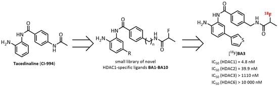
2. Results and Discussion

2.1. Organic Chemistry

A small library of new compounds (BA1BA10) was synthesized using a six-step synthesis route (Scheme 1). As fluorine-bearing moiety the 2-fluoropropanamide was used instead of the apparently obvious fluoroacetamide moiety. The [18F]fluoroacetamido group is known to suffer from limited stability in vivo and to carry the risk of defluorination [22,23,42,43]. Both processes would be disadvantageous for imaging by PET. In contrast, the 2-[18F]fluoropropanoyl group is used frequently for PET tracer design, due to higher metabolic stability [44,45,46,47]. Additionally, it is expected to have a low impact on biological activity due to its small size.
Starting from 4-bromo-2-nitroaniline, the Boc protection of the aromatic amino group was performed in two steps [48]. First of all, the amino group was treated with di-tert-butyl dicarbonate (Boc2O) and catalytic amounts of N,N-dimethyl-4-aminopyridine (DMAP) to afford the bis-Boc-protected intermediate followed by cleavage of one Boc group using trifluoroacetic acid (TFA) to give tert-butyl (4-bromo-2-nitrophenyl)carbamate (1) in a total yield of 95%. Under these conditions it was possible to achieve higher yields with this two-step synthesis compared with the reported yields [49,50,51,52,53] using a one-step synthesis. Subsequently, 1 was reacted with 2-/3-thienyl-, 2-/3-furanyl- or para-fluoro-phenylboronic acid in a Suzuki cross coupling reaction with tetrakis(triphenylphosphine)palladium(0) (Pd(PPh3)4) in 1,4-dioxane/water (7:3, v/v) to provide the corresponding compounds 2ae in very good yields (90–96%). In the first approach, the reduction in the aromatic nitro group was carried out under hydrogen atmosphere with palladium on carbon and afforded 3ac in quantitative yields [49]. Using the same procedure for the 3-furan-containing compound 2e, the desired aniline product could not be obtained. The NMR analysis showed that both, the aromatic nitro group and the 3-furan, were entirely reduced to form the corresponding aniline derivative substituted with 3-tetrahydrofuran instead of 3-furan. Therefore, a reduction by sodium dithionite was carried out as a milder alternative, which gave the desired products 3d and 3e in good to moderate yields (83% and 40%, respectively) [54]. Subsequently, the amide couplings of 3a and 3b with 4-(Fmoc-aminomethyl)benzoic acid were performed in a straight forward synthesis under standard conditions using (benzotriazol-1-yloxy)tripyrrolidinophosphonium hexafluorophosphate (PyBOP) as coupling reagent. After cleaving the Fmoc protecting group with a solution of 20% piperidine in N,N-dimethylformamide (DMF), the resulting amines were coupled with 2-fluoropropanoic acid under the same reaction conditions and led to the compounds 4a and 4b (23% and 40%, respectively, over three steps). To bypass the use of protecting groups and increase the yield of the amide coupling, we synthesized the 4-[(2-fluoropropanamido)methyl]- and 4-(2-fluoropropanamido)benzoic acids 7 and 8 by acylation of the amino group of 4-(aminomethyl)-(PAMBA) and 4-aminobenzoic acid (PABA) with 2-fluoropropanoyl chloride 6 in good yields (63% and 79%, respectively). With the compounds 7 and 8 we were then able to perform the final amide coupling in one instead of three reaction steps to synthesize the corresponding compounds 4ae and 5ae in better yields (57–95%). Finally, the Boc protecting group was removed with a solution of 30% TFA in dichloromethane (DCM) to give the final products BA1–BA10 after precipitation in moderate to good yields (15–91%).

2.2. Determination of the In Vitro Inhibitory Potency

All prepared compounds as well as the reference HDAC inhibitors vorinostat, entinostat and tacedinaline were tested regarding their inhibitory activity against the class I enzymes HDAC1-3. Additionally, HDAC6 was used as a control isoform to evaluate the selectivity towards class IIb HDACs. The IC50 values are shown in Table 2.
The HDAC inhibitors containing (hetero)aryl substituted benzamides as ZBG showed, as expected, a high inhibitory activity against HDAC1 with IC50 values ranging from 4.8 nM to 24.2 nM for BA1BA10. In contrast, negligibly low inhibitions of HDAC3 and HDAC6 were observed. The screening also revealed that the compounds with the PAMBA linker (n = 1, BA1/BA3/BA5/BA7), compared with the PABA linker (n = 0, BA2/BA4/BA6/BA8), have a higher inhibitory potency towards HDAC1. The only exceptions are BA9 and BA10, which both bear a 3-furan substitution and exhibit no substantial differences in the IC50 values against HDAC1. Compared with the approved hydroxamic acid-based HDAC inhibitor vorinostat and the benzamides tacedinaline and entinostat, the inhibitory potencies of the new compounds are significantly improved towards HDAC1. It is known that the use of a foot pocket-targeting (hetero)aromatic substituent at position 5 of the aminoanilide moiety leads to an increase in the specificity for HDAC1 and HDAC2 over HDAC3 [37,38,39,40,41]. This could be proven by the specificity ratios (HDAC1/HDAC3) determined for the synthesized compounds ranging from >74 (BA6) to 479 (BA1) compared with nearly no specificity of the reference compounds vorinostat, entinostat or tacedinaline, respectively. Out of the series, BA1 and BA3 showed the best inhibitory activities against HDAC1. BA3 was selected for radiolabelling as the most active inhibitor of HDAC1 and HDAC2.

2.3. Precursor- and Radiosynthesis

2.3.1. Manual Radiosynthesis

For a direct radiofluorination of [18F]BA3, the corresponding Boc-protected bromine precursor 9 was synthesized as shown in Scheme 1 in an overall yield of 34%. The radiolabelling process was investigated under thermal heating using 2–3 mg of precursor 9 and different (i) polar aprotic solvents, (ii) bases, (iii) different precursor to base ratios, (iv) temperatures, (v) reaction times and (vi) azeotropically dried 18F-fluorination agents. First, the conventional K[18F]F-K2.2.2 was used for radiofluorination of the intermediate Boc-[18F]BA3 in DMF, dimethyl sulfoxide (DMSO) and acetonitrile (MeCN) as solvents. The reactions were carried out at 150 °C (DMF/DMSO) or 100 °C (MeCN) for up to 20 min. Aliquots of the reaction mixtures were taken every 5 min and analysed by radio-thin layer chromatography (radio-TLC) and/or radio-reversed phase high-performance liquid chromatography (radio-RP-HPLC) to determine the radiochemical yield (RCY) of the labelling process. In DMF or DMSO with K2CO3 as base, the radiosynthesis of Boc-[18F]BA3 was not possible. According to radio-RP-HPLC, under these reaction conditions a polar radioactive by-product was formed in high amounts (30% in DMF, 45% in DMSO). When using MeCN as a solvent, it was possible to obtain Boc-[18F]BA3 in labelling yields between 1% and 2% (with KHCO3 or K2CO3, respectively), but the same radioactive by-product (~10%) was formed as shown by radio-RP-HPLC. The use of K2C2O4 instead of KHCO3/K2CO3 and 18-crown-6 instead of K2.2.2 did not result in an increase in the radiochemical yield of Boc-[18F]BA3. Finally, the additional impact of microwave irradiation, which was investigated in DMSO, DMF and MeCN, respectively, did not improve the radiofluorination. For all syntheses, the (radio-)RP-HPLC chromatograms showed a decomposition of precursor 9 and Boc-[18F]BA3 already after 5 min, as well as a large amount of unreacted [18F]fluoride.
In general, these conditions are strongly basic, limiting the synthetic utility for base-sensitive precursors since various side reactions, such as eliminations, can occur [56]. Assuming that the reaction conditions described above are not suitable for radiosynthesis of Boc-[18F]BA3, the use of [18F]tetra-n-butylammonium fluoride ([18F]TBAF) was investigated as a milder reagent [57]. The impact of (i) different solvents, (ii) temperatures, and (iii) reaction times were studied. The radiofluorination of Boc-[18F]BA3 was investigated first in MeCN at 100 °C achieving labelling yields up to 7% after 15 min, which were not increasing with further reaction time. Then, the influence of tert-butanol (tert-BuOH) as a solvent at 100 °C was investigated and a labelling yield of 5% was achieved after 20 min. Due to the fact that Boc-[18F]BA3 was obtained in highest labelling yields with MeCN, the use of this solvent was kept for further evaluation. The second step after the radiofluorination of Boc-[18F]BA3 was the cleavage of the Boc protecting group, which was performed by addition of an aqueous solution of 2 M hydrochloric acid (HClaq) to the crude mixture of Boc-[18F]BA3 and heating at 80 and 100 °C. At 80 °C the reaction was already completed after 5 min as detected by radio-TLC. Finally, the novel radioligand [18F]BA3 could be prepared in a two-step one-pot reaction by a direct aliphatic nucleophilic substitution, starting with the precursor 9 and subsequent Boc deprotection under the optimized reaction conditions as shown in Scheme 2.

2.3.2. Automated Radiosynthesis

An automated radiosynthesis of [18F]BA3 was established based on the optimized reaction conditions using the radiosynthesis module TRACERlab FX2 N. The detailed configuration is shown in the Supplemental part (Figure S1). Briefly, [18F]fluoride (starting activities of 7–32 GBq) was trapped on a QMA cartridge and eluted into the reactor with TBAHCO3 dissolved in MeCN/H2O and dried via azeotropic distillation. The nucleophilic substitution of the bromine precursor 9 (4 mg) was performed using [18F]TBAF in MeCN at 100 °C for 15 min. The subsequent Boc deprotection was carried out by adding an aqueous 2 M HCl solution and stirring for 5 min at 80 °C. After neutralization with a 1 M solution of aqueous NaHCO3, the radiolabelled product [18F]BA3 was isolated via semi-preparative RP-HPLC (Figure 2) and purified by solid-phase extraction (SPE) on a C18 cartridge. Subsequent elution with ethanol (EtOH) and formulation in sterile 0.9% saline containing 10% of EtOH reproducibly provided [18F]BA3 in a total synthesis time of approximately 85 min with RCYs of 0.9 ± 0.2% (EOB, n = 5; see Figure S2 for UV- and radio-RP-HPLC-chromatograms of formulated [18F]BA3). The identity of [18F]BA3 was confirmed by analytical radio- and UV-RP-HPLC of the final product, co-injected with the reference compound BA3 (Figure 2). Finally, the radiotracer was obtained with a radiochemical purity of >96% and a molar activity in the range of 21–51 GBq/µmol (EOS, n = 5).

2.4. Lipophilicity and Radiochemical Stability of [18F]BA3

The distribution coefficient (logD7.4) was experimentally determined by the shake-flask method in the n-octanol/PBS system. The logD7.4 value of 1.75 ± 0.16 (n = 8) for [18F]BA3 suggests that a moderate passive diffusion through the BBB is most likely [58,59,60]. The radiotracer [18F]BA3 was stable in n-octanol, as well as in the saline formulation containing 10% EtOH at room temperature for more than two hours (radiochemical purity > 99%).

2.5. In Vivo PET/MRI Studies of [18F]BA3

The pharmacokinetics of [18F]BA3 was investigated by dynamic PET scans on healthy CD-1 mice in order to evaluate the passage across the BBB, nonspecific binding and thus the expected background level. The time–activity curve (TAC) of the brain (peak standardized uptake value (SUV) at 0.8 min of ~0.16) demonstrates a low brain uptake (see Figure 3 and Figure S4). Further radiotracer accumulation reaches a plateau at 15 min (~0.22 SUV) followed by a slow washout reaching a SUV of ~0.16 at 60 min after i.v. administration. Notably, only 21% of the parent compound was found in the brain at 30 min p.i., suggesting that the major part of the measured activity originates from the nonspecific accumulation of radiometabolites.
Although these first results do not support favourable imaging features of [18F]BA3, the evaluation of its potential affinity to efflux drug transporter present at the BBB could help to predict the features of other derivatives of this class of compound. Thus, pre-administration of cyclosporine A, an inhibitor of the P-glycoprotein (P-gp) efflux transporter, increased the initial uptake of [18F]BA3 by about factor two (0.18 vs. 0.42 SUV) indicating that [18F]BA3 is a substrate of P-gp.
Evaluation of the whole-body distribution derived from PET imaging revealed a low accumulation of tracer and/or metabolites in spleen, kidney, muscle, liver, and small intestines (initial uptake: SUV5 min < 4), as well as a relatively constant activity in blood circulation (SUV5 min to SUV60 min: 1.47 vs. 1.10). An elevated accumulation occurred in the gallbladder and the bladder, indicating both urinary and hepatobiliary excretion (Figure S5, Figure S6 and Table S1).
In conclusion, although [18F]BA3 presents a high in vitro inhibitory potency towards HDAC1 (IC50 = 4.8 ± 0.6 nM) and HDAC2 (IC50 = 39.9 ± 3.2 nM), and an excellent specificity of HDAC1 over HDAC3 and HDAC6 (specificity ratios >230 and >2080, respectively), its low passage across the BBB and the presence of radiometabolites in brain show the need for further ligand optimization.

2.6. In Vivo Metabolism of [18F]BA3

The in vivo metabolism of [18F]BA3 was investigated in the plasma, brain homogenate and urine obtained from mice at 30 min after injection into the tail vein. The radiometabolites and parent compound were analysed by (i) analytical radio-RP-HPLC and (ii) radio-micellar chromatography (radio-MLC) [61]. Prior to the analysis with radio-RP-HPLC, the plasma and brain samples were treated with MeCN/H2O (9:1, v/v) as extraction solvent. The recovery of activity in plasma and brain samples was 97% and 95%, respectively. Radio-RP-HPLC analysis revealed that the intact radiotracer accounted for about 5% and 21% (mean, n = 2) of the total activity in plasma and brain, respectively. Similar results were obtained with MLC (about 6% and 24% (mean, n = 2) in plasma and brain, respectively), where the samples are directly injected without previous protein precipitation (for corresponding MLC chromatograms see Figure S3). In urine intact radiotracer was not detected by neither radio-RP-HPLC nor radio-MLC. In the corresponding radio-chromatograms (Figure 4) five radiometabolites ([18F]M1-[18F]M5) were detected in brain at 30 min p.i. In contrast, in plasma one single major radiometabolite, [18F]M4, amounting to 74%, was found However, a conclusion, whether the radiometabolites in the brain originate from the plasma and pass the blood–brain barrier or are formed within the brain itself, was not possible, as only a single time point was investigated.

2.7. Expression of HDAC1 in F98 and U251-MG Cells

The expression of HDAC1 was evaluated by immunofluorescence microscopy of the glioblastoma cell lines F98 and U251-MG to check whether they are suitable for the assessment of the toxicity of HDAC inhibitors. As shown in Figure 5, both cell lines possess a HDAC1 nucleic localization, proved by the merged signals of the HDAC1 (green) enzyme and the nuclei (blue). Interestingly, the HDAC1 signal is exactly co-localized with the nuclei in the F98 cells but additionally presents a slight cytoplasmic localization in U251-MG cells, as also previously found by Seto et al. [62].

2.8. The New HDAC Inhibitor BA3 Has Potential to Reduce the Proliferation of Cancer Cells

Inhibition of HDAC activity has been shown to hinder the proliferation of cancer cells. To assess the cytotoxic potential of BA3, the effect of this compound on the proliferation of the rat glioma F98 cells and the human glioblastoma U251-MG cells in comparison with the well-established HDAC inhibitors tacedinaline, vorinostat, and entinostat was investigated by an MTS assay (Table 3). Although solubility problems detected only after completion of the study (slight precipitation of BA3 at 50 µM in cell culture medium supplemented with 0.5% DMSO) preclude the direct comparison of the cytotoxic potency between the established HDAC inhibitors and BA3, the similar strongly diminished viability of F98 and U251-MG cells induced by BA3 at a concentration of even lower than 50 µM indicate nevertheless a high cytotoxic potential of BA3. Thus, although differently designed studies would be needed to determine proper cytotoxic IC50 values and even though not relevant for imaging studies performed with radiotracers at low to sub-nanomolar concentrations, the presented data suggest, that the pharmacological potency of BA3 is at least comparable if not even higher than that of established and clinically tested HDAC inhibitors.

3. Materials and Methods

3.1. General

Chemicals were purchased from commercial suppliers in analytical grade and used without further purification. Solvents were dried before use, if required. Air- and moisture-sensitive reactions were carried out under inert gas atmosphere using argon. The reaction monitoring was carried out by (radio-)TLC on pre-coated silica gel plates (Alugram® Xtra SIL G/UV254; Polygram® SIL G/UV254) purchased from Macherey-Nagel (Düren, Germany). The compounds were visualized with UV light at λ = 254 nm and 365 nm and/or by staining with aqueous KMnO4 solution or ninhydrin solution.
The UV purity of the final compounds BA1BA10 was determined by LC-MS on an UltiMate 3000 UHPLC System (Thermo Scientific, Germering, Germany) including a DAD detector (DAD-3000RS) coupled to an MSQ Plus Single Quadrupole Mass Spectrometer (Thermo Scientific, Austin, TX, USA). LC-MS analyses were performed on a ReproSil-Pur 120 C18-AQ-column (150 mm x 3 mm, 3 µm; Dr. Maisch GmbH, Ammerbuch, Germany) equipped with an appropriate precolumn in gradient mode (0–1.5 min: 5% MeCN/20 mM NH4OAc, 1.5–8 min: 5% → 80% MeCN/20 mM NH4OAc, 8–12 min: 80% MeCN/20 mM NH4OAc, 12–12.5 min: 80% → 5% MeCN/20 mM NH4OAc, 12.5–15 min: 5% MeCN/20 mM NH4OAc) and a flow of 0.7 mL/min. UV detection was performed at a wavelength of 254 nm and used for determination of UV purity by calculation of the relative peak area. MSQ Plus single quadrupole mass spectrometer was operated in positive electrospray ionization mode: probe temperature 500 °C, needle voltage 5 V and cone voltage 75 V.
NMR spectra (1H, 13C, 19F) were recorded on Mercury 300/Mercury 400 (Varian, Palo Alto, CA, USA) or Fourier 300/Avance DRX 400 Bruker (Billerica, MA, USA) instruments. The hydrogenated residue of deuterated solvents and/or tetramethylsilane (TMS) were used as internal standards for 1H-NMR (CDCl3, δ = 7.26; DMSO-d6, δ = 2.50) and 13C-NMR (CDCl3, δ = 77.16; DMSO-d6, δ = 39.52). The chemical shifts (δ) are reported in ppm (s, singlet; brs, broad singlet; d, doublet; t, triplet; q, quartet; m, multiplet) and the corresponding coupling constants (J) are reported in Hz. High resolution mass spectra (ESI +/−) were recorded on an Esquire 3000Plus instrument (Bruker Daltonics, Billerica, MA, USA; equipped with ion trap). The 1H-, 13C- and 19F-NMR spectra and LC-MS chromatograms for compounds BA1BA10 are displayed in Figures S7–S46

3.2. General Procedures

3.2.1. Suzuki Coupling A1

An aqueous solution of K2CO3 (2.6 eq.) was added to a solution of 1 (1 eq.) and the respective boronic acid (1.2 eq.) in 1,4-dioxane under argon atmosphere. After addition of tetrakis(triphenylphosphine)palladium(0) (0.05 eq.), the mixture was stirred under reflux until the complete consumption of 2. The 1,4-dioxane was evaporated; the crude product was dissolved in ethyl acetate (EA) and washed with water and brine. After drying over Na2SO4 the solvent was evaporated and the product purified by flash chromatography.

3.2.2. Nitro Reduction with Hydrogen A2a

The nitro compound was dissolved in EA and diluted with MeOH (1:1, v/v). After flushing with argon, Pd/C (0.05 eq., 10% wt/wt) was added and the mixture was stirred under hydrogen atmosphere at room temperature for 12 h. The solvent was removed, the crude product was adsorbed on silica and purified by flash chromatography.

3.2.3. Nitro Reduction with Sodium Dithionite A2b

To a solution of nitro compound (1 eq.) in 1,4-dioxane/water (5:1, v/v), sodium dithionite (4 eq.) and sodium bicarbonate (10 eq.) were added. The heterogeneous mixture was stirred vigorously under reflux until the complete consumption of the nitro compound. The salts were filtered off and 1,4-dioxane evaporated. The organic compounds were transferred in a separation funnel with EA and H2O, extracted twice with EA, washed with brine, and dried over Na2SO4. After evaporation, the crude product was purified by flash chromatography.

3.2.4. Amide Coupling A3

To a solution of the carboxylic acid and N,N-diisopropylethylamine (DIPEA) in DMF, PyBOP was added in small portions and subsequently stirred for 30 min at room temperature. In the meantime, a solution of the respective amine in DMF was prepared and then added to the stirring solution. After stirring overnight most of the DMF was evaporated, the residue was dissolved in EA, washed with water and brine, and dried over Na2SO4. After adsorbing the crude product on silica, it was purified by flash chromatography.

3.2.5. Boc Cleavage A4

For the cleavage of the Boc group the corresponding compound (1 eq.) was dissolved in a 30% TFA (20 eq.) solution in DCM and stirred for 2–4 h. The solution was diluted with EA, washed extensively with a 5% aqueous solution of NaHCO3 and dried over Na2SO4. The EA was removed until the product started to precipitate. Ice-cold heptane was added, and the product was filtered off and dried under reduced pressure.

3.3. Compounds

3.3.1. Synthesis of tert-butyl (4-bromo-2-nitrophenyl)carbamate (1)

To a solution of 4-bromo-2-nitroaniline (10 g, 46 mmol, 1 eq.) in 150 mL THF, TEA (15.9 mL, 115 mmol, 2.5 eq.) and DMAP (1.1 g, 9.2 mmol, 0.2 eq.) were added successively. While stirring at room temperature, the first portion Boc2O (10 g, 46 mmol, 1 eq.) was added until it was completely dissolved. Then the second portion Boc2O (11.6 g, 53 mmol, 1.15 eq.) was added in smaller portions and the solution was stirred at room temperature for 4 h. The reaction volume was reduced to a third and an aqueous solution of citric acid (29 g, 151 mmol) was added. After extraction of the compound with EA, the organic layer was dried over Na2SO4 and the solvent was evaporated to give a light-yellow solid. The crude product was dissolved in a solution of TFA (5.3 mL, 69 mmol, 1.5 eq.) in 140 mL DCM and stirred for 6 h. The reaction was monitored by TLC and after completion an aqueous solution of NaHCO3 (6.7 g, 80 mmol) was added, the product was extracted twice with DCM, washed with brine, and dried over Na2SO4. It was purified by flash chromatography (silica, gradient PE/EA 20:1 → 18:1 → 16:1) to give 1 as yellow solid (13.93 g, 95%).
1H-NMR (400 MHz, CDCl3) δ 9.60 (s, 1H), 8.51 (d, J = 9.2 Hz, 1H), 8.33 (d, J = 2.4 Hz, 1H), 7.68 (dd, J = 9.2, 2.4 Hz, 1H), 1.54 (s, 9H); 13C-NMR (101 MHz, CDCl3) δ 152.09, 138.67, 136.32, 135.28, 128.40, 122.35, 113.88, 82.45, 28.32 (in accordance with literature [53]).

3.3.2. Synthesis of tert-butyl [2-nitro-4-(thiophen-2-yl)phenyl]carbamate (2a)

Compound 1 (2 g, 6.3 mmol) and 2-thienylboronic acid in 25 mL 1,4-dioxane, K2CO3 in 11 mL H2O and catalyst were reacted according to the general procedure A1 to give after purification by flash chromatography (silica, gradient PE/EA: 25:1 → 20:1 → 15:1) compound 2a (1.88 g, 93%) as yellow solid.
1H-NMR (400 MHz, CDCl3) δ 9.65 (s, 1H), 8.59 (d, J = 9.0 Hz, 1H), 8.40 (d, J = 2.3 Hz, 1H), 7.82 (dd, J = 8.9, 2.3 Hz, 1H), 7.37–7.30 (m, 2H), 7.10 (dd, J = 5.1, 3.6 Hz, 1H), 1.56 (s, 9H); 13C-NMR (101 MHz, CDCl3) δ 152.27, 141.49, 136.20, 134.96, 133.07, 128.88, 128.46, 125.81, 124.08, 122.52, 121.35, 82.17, 28.36 (in accordance with literature [53]).

3.3.3. Synthesis of tert-butyl [2-nitro-4-(thiophen-3-yl)phenyl]carbamate (2b)

Compound 1 (3 g, 9.46 mmol) and 3-thienylboronic acid in 38 mL 1,4-dioxane, K2CO3 in 17 mL H2O and catalyst were reacted according to the general procedure A1 to give after purification by flash chromatography (silica, gradient PE/EA: 24:1 → 19:1 → 14:1 → 9:1) compound 2b (2.87 g, 95%) as yellow solid.
1H-NMR (400 MHz, CDCl3) δ 9.65 (s, 1H), 8.60 (d, J = 8.9 Hz, 1H), 8.40 (d, J = 2.2 Hz, 1H), 7.83 (dd, J = 8.9, 2.3 Hz, 1H), 7.50 (dd, J = 2.9, 1.4 Hz, 1H), 7.45–7.35 (m, 2H), 1.56 (s, 9H); 13C-NMR (101 MHz, CDCl3) δ 152.33, 139.55, 136.25, 134.78, 133.62, 130.16, 127.16, 125.90, 123.08, 121.27, 121.21, 82.07, 28.36.

3.3.4. Synthesis of tert-butyl (4′-fluoro-3-nitro-[1,1′-biphenyl]-4-yl)carbamate (2c)

Compound 1 (1 g, 3.15 mmol) and 4-fluorophenylboronic acid in 15 mL 1,4-dioxane, K2CO3 in 5 mL H2O and catalyst were reacted according to the general procedure A1 to give after purification by flash chromatography (silica, gradient PE/EA: 25:1 → 11.5:1 → 4:1) compound 2c (1.0 g, 96%) as white solid.
1H-NMR (400 MHz, CDCl3) δ 9.66 (s, 1H), 8.63 (d, J = 8.9 Hz, 1H), 8.37 (d, J = 2.3 Hz, 1H), 7.79 (dd, J = 8.9, 2.3 Hz, 1H), 7.59–7.50 (m, 2H), 7.21–7.10 (m, 2H), 1.56 (s, 9H); 19F-NMR (377 MHz, CDCl3) δ −114.12 (ddd, J = 13.8, 8.6, 5.2 Hz); 13C-NMR (76 MHz, CDCl3) δ 162.98 (d, J = 247.9 Hz), 152.35, 136.34, 135.08, 134.51 (d, J = 3.2 Hz), 134.28, 134.12, 128.55 (d, J = 8.2 Hz), 123.73, 121.41, 116.21 (d, J = 21.7 Hz), 82.16, 28.36 (in accordance with literature [55]).

3.3.5. Synthesis of tert-butyl [4-(furan-2-yl)-2-nitrophenyl]carbamate (2d)

Compound 1 (2 g, 6.3 mmol) and 2-furanylboronic acid in 25 mL 1,4-dioxane, K2CO3 in 11 mL H2O and catalyst were reacted according to the general procedure A1 to give after purification by flash chromatography (silica, gradient PE/EA: 30:1 → 20:1 → 10:1) compound 2d (1.83 g, 95%) as orange solid.
1H-NMR (400 MHz, CDCl3) δ 9.68 (s, 1H), 8.62 (d, J = 8.9 Hz, 1H), 8.48 (d, J = 2.1 Hz, 1H), 7.88 (dd, J = 8.9, 2.2 Hz, 1H), 7.52 (dd, J = 1.8, 0.7 Hz, 1H), 6.72 (dd, J = 3.4, 0.8 Hz, 1H), 6.52 (dd, J = 3.4, 1.8 Hz, 1H), 1.58 (s, 9H); 13C-NMR (101 MHz, CDCl3) δ 152.28, 151.53, 142.87, 136.25, 134.77, 130.92, 125.37, 121.19, 120.66, 112.08, 106.12, 82.12, 28.36.

3.3.6. Synthesis of tert-butyl [4-(furan-3-yl)-2-nitrophenyl]carbamate (2e)

Compound 1 (2 g, 6.3 mmol) and 3-furanylboronic acid in 25 mL 1,4-dioxane, K2CO3 in 11 mL H2O and catalyst were reacted according to the general procedure A1 to give after purification by flash chromatography (silica, gradient PE/EA: 30:1 → 20:1 → 10:1) compound 2d (1.73 g, 90%) as orange solid.
1H-NMR (400 MHz, CDCl3) δ 9.63 (s, 1H), 8.57 (d, J = 8.9 Hz, 1H), 8.27 (d, J = 2.2 Hz, 1H), 7.77 (t, J = 1.2 Hz, 1H), 7.70 (dd, J = 8.9, 2.2 Hz, 1H), 7.53–7.48 (m, 1H), 6.70 (dd, J = 2.0, 0.9 Hz, 1H), 1.55 (s, 9H); 13C-NMR (101 MHz, CDCl3) δ 152.35, 144.35, 139.07, 136.30, 134.70, 133.17, 126.96, 124.39, 122.54, 121.34, 108.61, 82.09, 28.37.

3.3.7. Synthesis of tert-butyl [2-amino-4-(thiophen-2-yl)phenyl]carbamate (3a)

The reduction in compound 2a (1.5 g, 4.7 mmol) was performed according to the general procedure A2a in 60 mL EA/MeOH (1:1, v/v) to give after purification by flash chromatography (silica, gradient PE/EA: 6:1 → 4:1 → 3:1 → 1:1) compound 3a (1.37 g) as white-beige solid in quantitative yield.
1H-NMR (400 MHz, DMSO-d6) δ 8.34 (s, 1H), 7.44 (dd, J = 5.1, 1.2 Hz, 1H), 7.32–7.24 (m, 2H), 7.08 (dd, J = 5.1, 3.6 Hz, 1H), 6.98 (d, J = 2.2 Hz, 1H), 6.84 (dd, J = 8.2, 2.2 Hz, 1H), 5.01 (s, 2H), 1.47 (s, 9H); 13C-NMR (101 MHz, DMSO-d6) δ 153.48, 144.01, 141.20, 130.18, 128.21, 124.55, 123.51, 122.42, 113.65, 112.40, 78.83, 28.15 (in accordance with literature [49]).

3.3.8. Synthesis of tert-butyl [2-amino-4-(thiophen-3-yl)phenyl]carbamate (3b)

The reduction in compound 2b (1.0 g, 3.12 mmol) was performed according to the general procedure A2a in 50 mL EA/MeOH (1:1, v/v) to give after purification by flash chromatography (silica, gradient PE/EA: 6:1 → 4:1 → 3:1 → 1:1) compound 3b (890 mg, 98%) as white-beige solid.
1H-NMR (400 MHz, DMSO-d6) δ 8.32 (s, 1H), 7.66–7.54 (m, 2H), 7.38 (dd, J = 5.0, 1.4 Hz, 1H), 7.25 (d, J = 8.2 Hz, 1H), 7.01 (d, J = 2.1 Hz, 1H), 6.88 (dd, J = 8.2, 2.1 Hz, 1H), 4.90 (s, 2H), 1.47 (s, 9H); 13C-NMR (101 MHz, DMSO-d6) δ 153.54, 141.82, 141.12, 131.75, 126.70, 125.99, 124.47, 123.09, 119.54, 114.46, 113.18, 78.73, 28.16.

3.3.9. Synthesis of tert-butyl (3-amino-4′-fluoro-[1,1′-biphenyl]-4-yl)carbamate (3c)

The reduction in compound 2c (1.0 g, 3.0 mmol) was performed according to the general procedure A2a in 50 mL EA/MeOH (1:1, v/v) to give after purification by flash chromatography (silica, gradient PE/EA: 4:1 → 1.5:1 → 1:1) compound 3c (895 mg) as white-beige solid in quantitative yield.
1H-NMR (300 MHz, CDCl3) δ 7.52–7.43 (m, 2H), 7.34 (d, J = 7.8 Hz, 1H), 7.13–7.03 (m, 2H), 6.99–6.90 (m, 2H), 6.24 (s, 1H), 3.85 (s, 2H), 1.53 (s, 9H); 19F-NMR (282 MHz, CDCl3) δ −116.04 (1H decoupled); 13C-NMR (75 MHz, CDCl3) δ 162.94 (d, J = 246.1 Hz), 153.96, 140.23, 138.41, 137.15, 128.59 (d, J = 8.02 Hz), 125.11, 124.31, 118.56, 116.25, 115.63 (d, J = 21.38 Hz), 80.86, 28.48 (in accordance with literature [55]).

3.3.10. Synthesis of tert-butyl [2-amino-4-(furan-2-yl)phenyl]carbamate (3d)

The reduction in compound 2d (1.78 g, 5.85 mmol) was performed according to the general procedure A2b in 42 mL 1,4-dioxane/H2O (5:1, v/v) to give after purification by flash chromatography (silica, gradient PE/EA: 4:1 → 2:1 → 1:1) compound 3d (1.34 g, 83%) as white-beige solid.
1H-NMR (400 MHz, CDCl3) δ 7.42 (dd, J = 1.8, 0.8 Hz, 1H), 7.32 (d, J = 8.3 Hz, 1H), 7.14–7.07 (m, 2H), 6.55 (dd, J = 3.4, 0.8 Hz, 1H), 6.44 (dd, J = 3.4, 1.8 Hz, 1H), 6.26 (s, 1H), 3.77 (s, 2H), 1.52 (s, 9H); 13C-NMR (101 MHz, CDCl3) δ 153.90, 153.86, 141.88, 139.86, 128.80, 124.70, 124.46, 115.71, 113.03, 111.71, 104.73, 80.80, 28.47 (in accordance with literature [63]).

3.3.11. Synthesis of tert-butyl [2-amino-4-(furan-3-yl)phenyl]carbamate (3e)

The reduction in compound 2e (1.68 g, 5.52 mmol) was performed according to the general procedure A2b in 42 mL 1,4-dioxane/H2O (5:1, v/v) to give after purification by flash chromatography (silica, gradient PE/EA: 4:1 → 2:1 → 1:1) compound 3e (611 mg, 40%) as white-beige solid.
1H-NMR (400 MHz, CDCl3) δ 7.65 (dd, J = 1.6, 0.9 Hz, 1H), 7.45–7.43 (m, 1H), 7.27 (d, J = 8.0 Hz, 1H), 6.95–6.86 (m, 2H), 6.63 (dd, J = 1.9, 0.9 Hz, 1H), 6.23 (s, 1H), 3.42 (brs, 2H), 1.52 (s, 9H); 13C-NMR (101 MHz, CDCl3) δ 153.98, 143.63, 140.27, 138.45, 130.55, 126.25, 125.21, 123.91, 117.52, 115.06, 109.04, 80.77, 28.47.

3.3.12. Synthesis of tert-butyl (2-{4-[(2-fluoropropanamido)methyl]benzamido}-4-[thiophen-2-yl]phenyl)carbamate (4a)

Compound 7 (20 mg, 0.089 mmol, 1 eq.), compound 3a (29 mg, 0.098 mmol, 1.1 eq.), DIPEA (34 µL, 0.2 mmol, 2.3 eq.) and PyBOP (56 mg, 0.107 mmol, 1.2 eq.) in 1 mL DMF were reacted according to the general procedure A3 to give after purification by flash chromatography (silica, gradient PE/EA: 3:1 → 2:1) compound 4a (40 mg, 90%) as brownish foam.
1H-NMR (400 MHz, CDCl3) δ 9.35 (s, 1H), 7.96–7.87 (m, 3H), 7.35–7.25 (m, 4H), 7.24–7.19 (m, 2H), 7.16 (s, 1H), 7.05–6.98 (m, 1H), 6.82 (d, J = 5.5 Hz, 1H), 5.06 (dq, J = 49.3, 6.8 Hz, 1H), 4.51 (d, J = 6.0 Hz, 2H), 1.61 (dd, J = 24.8, 6.8 Hz, 3H), 1.48 (s, 9H); 19F-NMR (377 MHz, DMSO-d6) δ −181.13 (dqd, J = 51.5, 25.6, 19.3 Hz); 13C-NMR (101 MHz, CDCl3) δ 171.00 (d, J = 19.2 Hz), 165.68, 154.69, 143.34, 141.89, 133.40, 132.10, 130.88, 129.64, 128.10, 128.07, 127.76, 124.96, 123.55, 123.45, 122.98, 88.98 (d, J = 182.9 Hz), 81.44, 42.62, 28.38, 18.65 (d, J = 21.5 Hz); HRFT-MS (ESI+): m/z = 995.3654 (calc. 995.3642 for [2M+H]+).

3.3.13. Synthesis of tert-butyl (2-{4-[(2-fluoropropanamido)methyl]benzamido}-4-[thiophen-3-yl]phenyl)carbamate (4b)

Compound 7 (12 mg, 0.053 mmol, 1 eq.), compound 3b (17 mg, 0.058 mmol, 1.1 eq.), DIPEA (21 µL, 0.122 mmol, 2.3 eq.) and PyBOP (34 mg, 0.064 mmol, 1.2 eq.) in 1 mL DMF were reacted according to the general procedure A3 to give after a rough purification by flash chromatography (silica, gradient PE/EA: 3:1 → 2:1) compound 4b (24 mg, 91%) as brownish foam.
1H-NMR (400 MHz, CDCl3) δ 9.34 (s, 1H), 7.95–7.86 (m, 3H), 7.36–7.22 (m, 7H), 7.08 (s, 1H), 6.79 (d, J = 5.4 Hz, 1H), 5.05 (dq, J = 49.3, 6.8 Hz, 1H), 4.50 (d, J = 6.0 Hz, 2H), 1.60 (dd, J = 24.8, 6.8 Hz, 3H), 1.47 (s, 9H); 19F-NMR (377 MHz, DMSO-d6) δ −181.16 (tdd, J = 53.1, 44.7, 25.2 Hz); 13C-NMR (101 MHz, CDCl3) δ 170.98 (d, J = 19.3 Hz), 165.60, 154.77, 141.89, 141.27, 133.62, 133.49, 130.97, 129.22, 128.05, 127.79, 126.36, 126.27, 124.95, 123.99, 123.62, 120.53, 88.99 (d, J = 182.8 Hz), 81.43, 42.63, 28.40, 18.65 (d, J = 21.5 Hz); HRFT-MS (ESI+): m/z = 1017.3456 (calc. 1017.3461 for [2M+Na]+).

3.3.14. Synthesis of tert-butyl (4′-fluoro-3-{4-[(2-fluoropropanamido)methyl]benzamido}-[1,1′-biphenyl]-4-yl)carbamate (4c)

Compound 7 (100 mg, 0.444 mmol, 1 eq.), compound 3c (174 mg, 0.577 mmol, 1.3 eq.), DIPEA (184 µL, 1.021 mmol, 2.3 eq.) and PyBOP (277 mg, 0.533 mmol, 1.2 eq.) in 12 mL DMF were reacted according to the general procedure A3 to give after a rough purification by flash chromatography (silica, gradient PE/EA: 9:1 → 4:1 → 1:1) the crude compound 4c (300 mg, 132% mass balance). It was used without further purification for following reactions.
1H-NMR (300 MHz, DMSO-d6) δ 9.89 (s, 1H), 8.79 (d, J = 9.7 Hz, 2H), 7.98–7.89 (m, 2H), 7.84 (d, J = 2.2 Hz, 1H), 7.76–7.60 (m, 3H), 7.50 (dd, J = 8.5, 2.2 Hz, 1H), 7.42 (d, J = 8.3 Hz, 2H), 7.34–7.25 (m, 2H), 5.09 (dq, J = 48.9, 6.7 Hz, 1H), 4.40 (d, J = 6.1 Hz, 2H), 1.47 (dd, J = 24.5, 6.7 Hz, 3H), 1.47 (s, 9H); 19F-NMR (282 MHz, DMSO-d6) δ −115.65 (ddd, J = 14.1, 9.0, 5.3 Hz), −181.12 (dd, J = 48.9, 24.3 Hz); 13C-NMR (75 MHz, DMSO-d6) δ 170.02 (d, J = 20.2 Hz), 165.29, 161.78 (d, J = 244.3 Hz), 153.41, 143.34, 135.81 (d, J = 3.0 Hz), 134.82, 132.79, 131.09, 129.95, 128.34 (d, J = 8.3 Hz), 127.69, 127.11, 124.07, 124.03, 123.72, 115.75 (d, J = 21.3 Hz), 87.96 (d, J = 179.2 Hz), 79.79, 41.49, 28.03, 18.46 (d, J = 21.9 Hz); HRFT-MS (ESI+): m/z = 1019.4312 (calc. 1019.4325 for [2M+H]+).

3.3.15. Synthesis of tert-butyl (2-{4-[(2-fluoropropanamido)methyl]benzamido}-4-[furan-2-yl]phenyl)carbamate (4d)

Compound 7 (130 mg, 0.577 mmol, 1 eq.), compound 3d (206 mg, 0.75 mmol, 1.3 eq.), DIPEA (238 µL, 1.327 mmol, 2.3 eq.) and PyBOP (360 mg, 0.692 mmol, 1.2 eq.) in 15 mL DMF were reacted according to the general procedure A3 to give after a rough purification by flash chromatography (silica, gradient PE/EA: 10:1 → 8:1 → 4:1 → 1:1) the crude compound 4d (290 mg, 104%). It was used without further purification for following reactions.
1H-NMR (400 MHz, DMSO-d6) δ 9.87 (s, 1H), 8.84–8.78 (m, 1H), 8.75 (s, 1H), 7.94 (d, J = 8.3 Hz, 2H), 7.88 (d, J = 2.0 Hz, 1H), 7.73 (dd, J = 1.8, 0.8 Hz, 1H), 7.62 (d, J = 8.6 Hz, 1H), 7.54 (dd, J = 8.5, 2.1 Hz, 1H), 7.42 (d, J = 8.3 Hz, 2H), 6.88 (dd, J = 3.4, 0.8 Hz, 1H), 6.59 (dd, J = 3.4, 1.8 Hz, 1H), 5.09 (dq, J = 48.9, 6.7 Hz, 1H), 4.40 (d, J = 6.2 Hz, 2H), 1.47 (dd, J = 24.5, 6.7 Hz, 3H), 1.46 (s, 9H); 19F-NMR (377 MHz, DMSO-d6) δ −176.35 (dq, J = 49.0, 24.6 Hz); 13C-NMR (101 MHz, DMSO-d6) δ 170.03 (d, J = 20.3 Hz), 165.32, 153.34, 152.47, 143.34, 142.74, 132.76, 131.05, 129.89, 127.73, 127.11, 126.27, 123.98, 120.99, 120.86, 112.10, 105.46, 87.97 (d, J = 179.2 Hz), 79.80, 41.50, 28.03, 18.47 (d, J = 21.9 Hz); HRFT-MS (ESI+): m/z = 963.4101 (calc. 963.4099 for [2M+H]+).

3.3.16. Synthesis of tert-butyl (2-{4-[(2-fluoropropanamido)methyl]benzamido}-4-[furan-3-yl]phenyl)carbamate (4e)

Compound 7 (130 mg, 0.577 mmol, 1 eq.), compound 3e (206 mg, 0.75 mmol, 1.3 eq.), DIPEA (238 µL, 1.327 mmol, 2.3 eq.) and PyBOP (360 mg, 0.692 mmol, 1.2 eq.) in 15 mL DMF were reacted according to the general procedure A3 to give after a rough purification by flash chromatography (silica, gradient PE/EA: 10:1 → 8:1 → 4:1 → 1:1) the crude compound 4e (265 mg, 95%). It was used without further purification for following reactions.
1H-NMR (400 MHz, DMSO-d6) δ 9.86 (s, 1H), 8.84–8.74 (m, 1H), 8.65 (s, 1H), 8.14 (dd, J = 1.6, 0.9 Hz, 1H), 8.02–7.86 (m, 2H), 7.77–7.69 (m, 2H), 7.60–7.50 (m, 1H), 7.49–7.33 (m, 3H), 6.91 (dd, J = 1.9, 0.9 Hz, 1H), 5.09 (ddd, J = 48.8, 6.7, 5.0 Hz, 1H), 4.43–4.34 (m, 2H), 1.52–1.40 (m, 12H); 19F-NMR (377 MHz, DMSO-d6) δ −181.13 (d, J = 17.6 Hz); 13C-NMR (101 MHz, DMSO-d6) δ 170.03 (d, J = 20.2 Hz), 165.24, 153.39, 144.31, 143.32, 139.10, 132.74, 130.77, 129.92, 129.38, 127.69, 127.10, 125.18, 123.95, 123.23, 122.99, 108.64, 87.96 (d, J = 179.4 Hz), 79.69, 41.50, 28.03, 18.46 (d, J = 21.7 Hz); HRFT-MS (ESI+): m/z = 504.1935 (calc. 504.1905 for [M+Na]+).

3.3.17. Synthesis of tert-butyl {2-[4-(2-fluoropropanamido)benzamido]-4-(thiophen-2-yl)phenyl}carbamate (5a)

Compound 8 (300 mg, 1.42 mmol, 1 eq.), compound 3a (578 mg, 1.988 mmol, 1.4 eq.), DIPEA (612 µL, 3.408 mmol, 2.4 eq.) and PyBOP (887 mg, 1.704 mmol, 1.2 eq.) in 15 mL DMF were reacted according to the general procedure A3 to give after purification by flash chromatography (silica, gradient PE/EA: 5:1 → 4:1 → 3:1) the compound 5a (500 mg, 73%) as brownish foam.
1H-NMR (400 MHz, DMSO-d6) δ 10.38 (d, J = 1.7 Hz, 1H), 9.87 (s, 1H), 8.74 (s, 1H), 8.02–7.94 (m, 2H), 7.91–7.85 (m, 2H), 7.84 (d, J = 2.2 Hz, 1H), 7.61 (d, J = 8.5 Hz, 1H), 7.54–7.49 (m, 2H), 7.46 (dd, J = 3.6, 1.2 Hz, 1H), 7.13 (dd, J = 5.1, 3.6 Hz, 1H), 5.23 (dq, J = 48.5, 6.7 Hz, 1H), 1.56 (dd, J = 24.5, 6.7 Hz, 3H), 1.46 (s, 9H); 19F-NMR (377 MHz, DMSO-d6) δ −179.83 (1H decoupled); 13C-NMR (101 MHz, DMSO-d6) δ 168.92 (d, J = 20.7 Hz), 164.91, 153.33, 142.68, 141.42, 131.17, 130.07, 129.64, 129.19, 128.56, 128.49, 125.42, 124.15, 123.42, 122.82, 122.59, 119.30, 87.78 (d, J = 179.1 Hz), 79.81, 28.02, 18.25 (d, J = 22.1 Hz); HRFT-MS (ESI+): m/z = 967.3345 (calc. 967.3329 for [2M+H]+).

3.3.18. Synthesis of tert-butyl {2-[4-(2-fluoropropanamido)benzamido]-4-(thiophen-3-yl)phenyl}carbamate (5b)

Compound 8 (164 mg, 0.774 mmol, 1.5 eq.), compound 3b (150 mg, 0.516 mmol, 1 eq.), DIPEA (360 µL, 2.064 mmol, 4 eq.) and PyBOP (540 mg, 1.032 mmol, 2 eq.) in 10 mL DMF were reacted according to the general procedure A3 to give after purification by flash chromatography (silica, gradient PE/EA: 5:1 → 4:1 → 3:1) the compound 5b (220 mg, 88%) as brownish foam.
1H-NMR (400 MHz, DMSO-d6) δ 10.38 (d, J = 1.7 Hz, 1H), 9.85 (s, 1H), 8.69 (s, 1H), 8.01–7.94 (m, 2H), 7.89–7.84 (m, 3H), 7.81 (dd, J = 2.9, 1.4 Hz, 1H), 7.64 (dd, J = 5.0, 2.9 Hz, 1H), 7.60 (d, J = 8.5 Hz, 1H), 7.56 (dd, J = 8.5, 2.0 Hz, 1H), 7.52 (dd, J = 5.0, 1.4 Hz, 1H), 5.23 (dq, J = 48.5, 6.7 Hz, 1H), 1.55 (dd, J = 24.6, 6.7 Hz, 3H), 1.46 (s, 9H); 19F-NMR (377 MHz, DMSO-d6) δ −179.84 (1H decoupled); 13C-NMR (101 MHz, DMSO-d6) δ 168.91 (d, J = 20.7 Hz), 164.83, 153.38, 141.37, 140.66, 131.10, 130.88, 129.94, 129.24, 128.50, 127.14, 126.01, 123.93, 123.70, 123.37, 120.56, 119.30, 87.77 (d, J = 179.1 Hz), 79.73, 28.03, 18.25 (d, J = 22.2 Hz); HRFT-MS (ESI+): m/z = 989.3136 (calc. 989.3148 for [2M+Na]+).

3.3.19. Synthesis of tert-butyl {4′-fluoro-3-[4-(2-fluoropropanamido)benzamido]-[1,1′-biphenyl]-4-yl}carbamate (5c)

Compound 8 (100 mg, 0.47 mmol, 1 eq.), compound 3c (187 mg, 0.616 mmol, 1.3 eq.), DIPEA (196 µL, 1.09 mmol, 2.3 eq.) and PyBOP (296 mg, 0.569 mmol, 1.2 eq.) in 12 mL DMF were reacted according to the general procedure A3 to give after purification by flash chromatography (silica, gradient PE/EA: 8:1 → 7:1 → 6:1 → 4:1 → 2:1 → 1:2) the compound 5c (185 mg, 79%) as brownish foam.
1H-NMR (400 MHz, DMSO-d6) δ 10.38 (s, 1H), 9.86 (s, 1H), 8.78 (s, 1H), 8.01–7.94 (m, 2H), 7.88–7.77 (m, 3H), 7.73–7.60 (m, 3H), 7.49 (dd, J = 8.5, 2.2 Hz, 1H), 7.33–7.24 (m, 2H), 5.23 (dq, J = 48.5, 6.7 Hz, 1H), 1.55 (dd, J = 24.6, 6.7 Hz, 3H), 1.47 (s, 9H); 19F-NMR (377 MHz, DMSO-d6) δ −115.63 (tt, J = 9.9, 5.4 Hz), −179.85 (dq, J = 49.1, 24.4 Hz); 13C-NMR (101 MHz, DMSO-d6) δ 168.92 (d, J = 20.5 Hz), 164.85, 161.78 (d, J = 244.2 Hz), 153.43, 141.40, 135.83, 134.83, 131.10, 130.26, 130.03, 129.26, 128.50, 128.34 (d, J = 8.1 Hz), 124.08, 123.68, 119.32, 115.75 (d, J = 21.3 Hz), 87.77 (d, J = 179.1 Hz), 79.81, 28.03, 18.25 (d, J = 22.1 Hz); HRFT-MS (ESI+): m/z = 991.3996 (calc. 991.4012 for [2M+H]+).

3.3.20. Synthesis of tert-butyl {2-[4-(2-fluoropropanamido)benzamido]-4-(furan-2-yl)phenyl}carbamate (5d)

Compound 8 (130 mg, 0.616 mmol, 1 eq.), compound 3d (220 mg, 0.801 mmol, 1.3 eq.), DIPEA (255 µL, 1.417 mmol, 2.3 eq.) and PyBOP (385 mg, 0.739 mmol, 1.2 eq.) in 15 mL DMF were reacted according to the general procedure A3 to give after purification by flash chromatography (silica, gradient PE/EA: 12:1 → 10:1 → 8:1 → 6:1 → 4:1) the compound 5d (260 mg, 90%) as brownish foam.
1H-NMR (400 MHz, DMSO-d6) δ 10.38 (s, 1H), 9.84 (s, 1H), 8.75 (s, 1H), 8.01–7.93 (m, 2H), 7.90–7.82 (m, 3H), 7.75–7.70 (m, 1H), 7.62 (d, J = 8.5 Hz, 1H), 7.54 (dd, J = 8.5, 2.1 Hz, 1H), 6.88 (dd, J = 3.4, 0.8 Hz, 1H), 6.58 (dd, J = 3.4, 1.8 Hz, 1H), 5.23 (dq, J = 48.5, 6.7 Hz, 1H), 1.56 (dd, J = 24.5, 6.7 Hz, 3H), 1.46 (s, 9H); 19F-NMR (377 MHz, DMSO-d6) δ −179.85 (dq, J = 48.9, 24.6 Hz); 13C-NMR (101 MHz, DMSO-d6) δ 168.92 (d, J = 20.6 Hz), 164.88, 153.36, 152.48, 142.73, 141.40, 131.06, 129.97, 129.24, 128.53, 126.29, 124.00, 121.03, 120.80, 119.32, 112.10, 105.45, 87.78 (d, J = 179.2 Hz), 79.81, 28.03, 18.26 (d, J = 22.1 Hz); HRFT-MS (ESI+): m/z = 935.3795 (calc. 935.3786 for [2M+H]+).

3.3.21. Synthesis of tert-butyl {2-[4-(2-fluoropropanamido)benzamido]-4-(furan-3-yl)phenyl}carbamate (5e)

Compound 8 (177 mg, 0.84 mmol, 1 eq.), compound 3e (300 mg, 1.09 mmol, 1.3 eq.), DIPEA (347 µL, 1.93 mmol, 2.3 eq.) and PyBOP (525 mg, 1 mmol, 1.2 eq.) in 20 mL DMF were reacted according to the general procedure A3 to give after purification by flash chromatography (silica, gradient PE/EA: 8:2 → 7:3 → 6:4) the compound 5e (224 mg, 57%) as brownish foam.
1H-NMR (300 MHz, DMSO-d6) δ 10.37 (s, 1H), 9.84 (s, 1H), 8.65 (s, 1H), 8.16–8.14 (m, 1H), 8.02–7.94 (m, 2H), 7.88–7.84 (m, 2H), 7.75–7.72 (m, 2H), 7.57 (d, J = 8.4 Hz, 1H), 7.46 (dd, J = 8.4, 2.1 Hz, 1H), 6.92 (dd, J = 1.9, 0.9 Hz, 1H), 5.23 (dq, J = 48.5, 6.7 Hz, 1H), 1.56 (dd, J = 24.5, 6.7 Hz, 3H), 1.46 (s, 9H); 19F-NMR (282 MHz, DMSO-d6) δ −179.85 (dq, J = 48.9, 24.5 Hz); 13C-NMR (75 MHz, DMSO-d6) δ 169.38 (d, J = 20.7 Hz), 165.27, 153.85, 144.77, 141.84, 139.57, 131.25, 130.44, 129.68, 128.96, 128.34, 125.65, 124.40, 123.75, 123.41, 119.76, 109.10, 88.24 (d, J = 179.2 Hz), 80.16, 28.50, 18.71 (d, J = 22.1 Hz); HRFT-MS (ESI+): m/z = 368.1418 (calc. 368.1405 for [M+H]+ Boc deprotection).

3.3.22. Synthesis of 2-fluoropropanoyl chloride (6)

Sodium 2-fluoropropanoate (3 g, 26.3 mmol, 1 eq.) was poured in a two-necked flask, cooled down to 0 °C in an ice bath and stirred vigorously. Phosphorus pentachloride PCl5 (6 g, 28.9 mmol, 1.1 eq.) was then added in small portions. After the complete addition of PCl5 the mixture was stirred further at room temperature for 3 h. After distillation (10 cm column, Tbath = 95–105 °C, TB = 75–80 °C; TB,Lit = 80.5–81 °C [64]) compound 6 (1.7 g, 58%) was isolated as a clear liquid.
Refractive index nD20 = 1.384 (nD20Lit = 1.384 [64]); 1H-NMR (400 MHz, CDCl3) δ 5.14 (dq, J = 48.6, 6.9 Hz, 1H), 1.70 (dd, J = 22.8, 6.9 Hz, 3H); 13C-NMR (101 MHz, CDCl3) δ 172.57 (d, J = 27.5 Hz), 90.33 (d, J = 194.0 Hz), 17.70 (d, J = 22.2 Hz) (according to literature [65]).

3.3.23. Synthesis of 4-[(2-fluoropropanamido)methyl]benzoic acid (7)

To an ice-cold suspension of 6 (1 g, 9 mmol, 1.2 eq.) and 4-(aminomethyl)benzoic acid (1.14 g, 7.5 mmol, 1 eq.) in 13 mL chloroform, pyridine (730 µL, 9 mmol, 1.2 eq.) was added dropwise. After stirring at room temperature overnight, most of the chloroform was evaporated and the residue dissolved in an aqueous solution of Na2CO3 (2.12 g, 20 mmol in 20 mL H2O). After washing twice with MTBE, the aqueous solution was acidified with 10 mL of a 6 M HCl solution, and the product started to precipitate. After stirring for a while, the solid was filtered off and dried under reduced pressure to give compound 7 (1.06 g, 63%) as white solid.
1H-NMR (400 MHz, DMSO-d6) δ 12.86 (s, 1H), 8.82–8.74 (m, 1H), 7.92–7.88 (m, 2H), 7.38–7.34 (m, 2H), 5.08 (dq, J = 48.8, 6.7 Hz, 1H), 4.37 (d, J = 6.1 Hz, 2H), 1.46 (dd, J = 24.5, 6.7 Hz, 3H); 19F-NMR (377 MHz, DMSO-d6) δ −181.15 (1H decoupled); 13C-NMR (101 MHz, DMSO-d6) δ 170.04 (d, J = 20.2 Hz), 167.16, 144.32, 129.39, 129.34, 127.14, 87.96 (d, J = 179.2 Hz), 41.53, 18.46 (d, J = 21.9 Hz).

3.3.24. Synthesis of 4-(2-fluoropropanamido)benzoic acid (8)

To an ice-cold suspension of 6 (1 g, 9 mmol, 1.2 eq.) and 4-aminobenzoic acid (1.03 g, 7.5 mmol, 1.2 eq.) in 13 mL chloroform, pyridine (730 µL, 9 mmol, 1.2 eq.) was added dropwise. After stirring at room temperature overnight, most of the chloroform was evaporated and the residue dissolved in an aqueous solution of Na2CO3 (2.12 g, 20 mmol in 20 mL H2O). After washing twice with MTBE, the aqueous solution was acidified with 10 mL of a 6 M HCl solution and the product started to precipitate. After stirring for a while, the solid was filtered off and dried under reduced pressure to give compound 8 (1.25 g, 79%) as white solid.
1H-NMR (400 MHz, DMSO-d6) δ 12.74 (s, 1H), 10.36 (s, 1H), 7.97–7.85 (m, 2H), 7.84–7.76 (m, 2H), 5.21 (dq, J = 48.5, 6.7 Hz, 1H), 1.53 (dd, J = 24.5, 6.7 Hz, 3H); 19F-NMR (377 MHz, DMSO-d6) δ −179.89 (1H decoupled); 13C-NMR (101 MHz, DMSO-d6) δ 168.95 (d, J = 20.7 Hz), 166.86, 142.20, 130.27, 125.88, 119.30, 87.73 (d, J = 179.0 Hz), 18.27 (d, J = 22.2 Hz).

3.3.25. Synthesis of tert-butyl (2-{4-[(2-bromopropanamido)methyl]benzamido}-4-[thiophen-3-yl]phenyl)carbamate (9)

To a solution of 4-(Fmoc-aminomethyl)benzoic acid (350 mg, 0.94 mmol, 1 eq.) and DIPEA (337 µL, 1.87 mmol, 2 eq.) in 7 mL DMF, PyBOP (536 mg, 1.03 mmol, 1.1 eq.) was added in small portions. After stirring the mixture for 30 min, a solution of 3b (355 mg, 1.22 mmol, 1.3 eq.) in 3 mL DMF was added and the solution was stirred at r.t. overnight. The amount of the DMF was reduced by evaporation, the residue was dissolved in EA, washed with water and brine and the organic phase was dried over Na2SO4. A filtration over a silica pad (silica, gradient PE/EA: 10:0 → 9:1 → 4:1) gave the crude Fmoc-protected compound (830 mg, 137%, mass balance) which was used without further analytics. The Fmoc-protected compound (830 mg, 0.94 mmol) was dissolved in 5 mL of a 20% piperidine solution in DMF and stirred at r.t. for 4 h. The amount of the DMF was reduced by evaporation, the residue was dissolved in EA, washed with water and brine and the organic phase dried over Na2SO4. After flash chromatography (silica, gradient DCM/MeOH: 100:0 → 99:1 → 95:5 → 90:10) the amine (240 mg, 60%) was used directly for the next reaction. To a solution of the amine (240 mg, 0.57 mmol, 1 eq.) and TEA (87 µL, 0.62 mmol, 1.1 eq.) in 4 mL DCM, 2-bromopropanoyl bromide (135 mg, 0.62 mmol, 1.1 eq.) was added. After stirring the mixture for 2.5 h, the solvent was evaporated and the crude product was purified by flash chromatography (silica, gradient PE/EA: 8:2 → 7:3 → 6:4 → 5:5) to give compound 9 (180 mg, 57%, 34% overall yield) as a white-beige solid.
1H-NMR (400 MHz, CDCl3) δ 9.38 (s, 1H), 7.89 (d, J = 1.8 Hz, 1H), 7.85–7.78 (m, 2H), 7.38–7.17 (m, 8H), 7.10 (t, J = 6.0 Hz, 1H), 4.52–4.33 (m, 3H), 1.85 (d, J = 7.0 Hz, 3H), 1.47 (s, 9H); 13C-NMR (101 MHz, CDCl3) δ 169.82, 165.83, 154.73, 141.99, 141.19, 133.45, 133.16, 130.75, 129.32, 127.93, 127.51, 126.34, 126.29, 124.93, 124.03, 123.51, 120.49, 81.41, 44.60, 43.54, 28.40, 22.89; HRFT-MS (ESI+): m/z = 580.0858 and 582.0850 (calc. 580.0876 and 582.0856 for [M + Na]+).

3.3.26. Synthesis of N-[2-amino-5-(thiophen-2-yl)phenyl]-4-[(2-fluoropropanamido)methyl]benzamide (BA1)

Compound 4a (140 mg, 0.28 mmol) was synthesized according to the general procedure A4 to give after filtration compound BA1 (53 mg, 53%) as light-brown solid.
UV purity: 98% (tR = 8.69 min); 1H-NMR (400 MHz, DMSO-d6) δ 9.70 (s, 1H), 8.84–8.76 (m, 1H), 7.96 (d, J = 8.2 Hz, 2H), 7.48 (d, J = 2.2 Hz, 1H), 7.39 (d, J = 8.2 Hz, 2H), 7.35 (dd, J = 5.1, 1.2 Hz, 1H), 7.30 (dd, J = 8.3, 2.2 Hz, 1H), 7.24 (dd, J = 3.6, 1.2 Hz, 1H), 7.05 (dd, J = 5.1, 3.6 Hz, 1H), 6.82 (d, J = 8.3 Hz, 1H), 5.19–4.99 (m, 3H), 4.39 (d, J = 6.1 Hz, 2H), 1.47 (dd, J = 24.5, 6.7 Hz, 3H); 19F-NMR (377 MHz, DMSO-d6) δ −181.06 (1H decoupled); 13C-NMR (101 MHz, DMSO-d6) δ 169.99 (d, J = 20.2 Hz), 165.29, 144.23, 142.99, 142.82, 133.07, 128.20, 127.90, 126.90, 123.90, 123.40, 123.21, 122.24, 121.01, 116.36, 87.96 (d, J = 179.1 Hz), 41.50, 18.47 (d, J = 22.0 Hz); HRFT-MS (ESI+): m/z = 398.1341 (calc. 398.1333 for [M+H]+).

3.3.27. Synthesis of N-[2-amino-5-(thiophen-2-yl)phenyl]-4-(2-fluoropropanamido)benzamide (BA2)

Compound 5a (200 mg, 0.414 mmol) was synthesized according to the general procedure A4 to give after filtration compound BA2 (144 mg, 91%) as greyish solid.
UV purity: 99% (tR = 8.95 min); 1H-NMR (400 MHz, DMSO-d6) δ 10.34 (s, 1H), 9.67 (s, 1H), 8.03–7.96 (m, 2H), 7.86–7.78 (m, 2H), 7.48 (d, J = 2.2 Hz, 1H), 7.35 (dd, J = 5.1, 1.1 Hz, 1H), 7.30 (dd, J = 8.3, 2.2 Hz, 1H), 7.25 (dd, J = 3.5, 1.2 Hz, 1H), 7.05 (dd, J = 5.1, 3.6 Hz, 1H), 6.82 (d, J = 8.3 Hz, 1H), 5.23 (dq, J = 48.5, 6.6 Hz, 1H), 5.14 (s, 2H), 1.55 (dd, J = 24.6, 6.7 Hz, 3H); 19F-NMR (377 MHz, DMSO) δ −179.76 (1H decoupled); 13C-NMR (101 MHz, DMSO-d6) δ 168.85 (d, J = 20.6 Hz), 164.78, 142.59, 141.69, 140.96, 129.69, 128.63, 126.54, 125.75, 124.57, 124.46, 123.89, 123.49, 119.17, 117.65, 116.33, 87.79 (d, J = 179.0 Hz), 18.28 (d, J = 22.2 Hz); HRFT-MS (ESI+): m/z = 384.1196 (calc. 384.1177 for [M+H]+).

3.3.28. Synthesis of N-[2-amino-5-(thiophen-3-yl)phenyl]-4-[(2-fluoropropanamido)methyl]benzamide (BA3)

Compound 4b (530 mg, 1.06 mmol) was synthesized according to the general procedure A4 to give after filtration compound BA3 (244 mg, 58%) as beige solid.
UV purity: 99% (tR = 8.60 min); 1H-NMR (400 MHz, DMSO-d6) δ 9.71 (s, 1H), 8.80 (dt, J = 6.3, 3.3 Hz, 1H), 7.97 (d, J = 8.0 Hz, 2H), 7.60–7.50 (m, 3H), 7.45–7.33 (m, 4H), 6.82 (d, J = 8.3 Hz, 1H), 5.20–4.96 (m, 3H), 4.39 (d, J = 6.1 Hz, 2H), 1.47 (dd, J = 24.5, 6.7 Hz, 3H); 19F-NMR (377 MHz, DMSO-d6) δ −181.06 (dq, J = 49.0, 24.5 Hz); 13C-NMR (101 MHz, DMSO-d6) δ 169.99 (d, J = 20.3 Hz), 165.22, 142.76, 142.55, 141.68, 133.14, 127.86, 126.90, 126.55, 125.74, 124.54, 124.50, 123.88, 123.41, 117.66, 116.32, 87.96 (d, J = 179.0 Hz), 41.51, 18.47 (d, J = 21.9 Hz); HRFT-MS (ESI+): m/z = 420.1163 (calc. 420.1152 for [M+Na]+).

3.3.29. Synthesis of N-[2-amino-5-(thiophen-3-yl)phenyl]-4-(2-fluoropropanamido)benzamide (BA4)

Compound 5b (100 mg, 0.207 mmol) was synthesized according to the general procedure A4 to give after filtration compound BA4 (60 mg, 76%) as light-brown solid.
UV purity: 99% (tR = 8.86 min); 1H-NMR (400 MHz, DMSO-d6) δ 10.34 (s, 1H), 9.67 (s, 1H), 8.04–7.97 (m, 2H), 7.87–7.80 (m, 2H), 7.49 (d, J = 2.2 Hz, 1H), 7.35 (dd, J = 5.1, 1.1 Hz, 1H), 7.30 (dd, J = 8.3, 2.2 Hz, 1H), 7.25 (dd, J = 3.6, 1.2 Hz, 1H), 7.05 (dd, J = 5.1, 3.6 Hz, 1H), 6.82 (d, J = 8.3 Hz, 1H), 5.32–5.09 (m, 3H), 1.56 (dd, J = 24.5, 6.7 Hz, 3H); 19F-NMR (377 MHz, DMSO-d6) δ −179.75 (1H decoupled); 13C-NMR (101 MHz, DMSO-d6) δ 168.86 (d, J = 20.5 Hz), 164.85, 144.25, 143.03, 141.00, 129.62, 128.67, 128.21, 123.92, 123.88, 123.48, 123.20, 122.27, 121.01, 119.18, 116.38, 87.80 (d, J = 179.1 Hz), 18.28 (d, J = 22.1 Hz); HRFT-MS (ESI+): m/z = 384.1184 (calc. 384.1177 for [M + H]+).

3.3.30. Synthesis of N-(4-amino-4′-fluoro-[1,1′-biphenyl]-3-yl)-4-[(2-fluoropropanamido)methyl]benzamide (BA5)

Compound 4c (255 mg, 0.4 mmol) was synthesized according to the general procedure A4 to give after filtration compound BA5 (104 mg, 64%) as white solid.
UV purity: 99% (tR = 8.90 min); 1H-NMR (400 MHz, DMSO-d6) δ 9.70 (s, 1H), 8.80 (t, J = 6.4 Hz, 1H), 7.96 (d, J = 7.9 Hz, 2H), 7.58 (dd, J = 8.5, 5.4 Hz, 2H), 7.53–7.46 (m, 1H), 7.39 (d, J = 7.9 Hz, 2H), 7.30 (dd, J = 8.3, 2.3 Hz, 1H), 7.21 (t, J = 8.7 Hz, 2H), 6.86 (d, J = 8.3 Hz, 1H), 5.09 (s, 2H), 5.09 (dq, J = 49.0, 6.7 Hz, 1H), 4.39 (d, J = 6.1 Hz, 2H), 1.47 (dd, J = 24.5, 6.7 Hz, 3H); 19F-NMR (377 MHz, DMSO-d6) δ −117.39–−117.60 (m), −181.07 (ddd, J = 47.4, 28.4, 14.1 Hz); 13C-NMR (101 MHz, DMSO-d6) δ 170.00 (d, J = 20.3 Hz), 165.30, 161.01 (d, J = 242.6 Hz), 142.79, 142.70, 136.75, 133.18, 127.88, 127.30 (d, J = 7.7 Hz), 127.16, 126.92, 124.73, 124.63, 123.59, 116.51, 115.50 (d, J = 21.3 Hz), 87.97 (d, J = 179.3 Hz), 41.51, 18.48 (d, J = 22.1 Hz); HRFT-MS (ESI+): m/z = 410.1704 (calc. 410.1675 for [M + H]+).

3.3.31. Synthesis of N-(4-amino-4′-fluoro-[1,1′-biphenyl]-3-yl)-4-(2-fluoropropanamido)benzamide (BA6)

Compound 5c (160 mg, 0.323 mmol) was synthesized according to the general procedure A4 to give after filtration compound BA6 (86 mg, 67%) as white solid.
UV purity: 95% (tR = 9.15 min); 1H-NMR (400 MHz, DMSO-d6) δ 10.34 (s, 1H), 9.67 (s, 1H), 8.03–7.97 (m, 2H), 7.83 (d, J = 8.7 Hz, 2H), 7.61–7.55 (m, 2H), 7.50 (d, J = 2.2 Hz, 1H), 7.30 (dd, J = 8.3, 2.3 Hz, 1H), 7.25–7.16 (m, 2H), 6.87 (d, J = 8.4 Hz, 1H), 5.23 (dq, J = 48.6, 6.7 Hz, 1H), 5.09 (s, 2H), 1.55 (dd, J = 24.5, 6.7 Hz, 3H); 19F-NMR (377 MHz, DMSO-d6) δ −117.50 (tt, J = 9.0, 5.3 Hz), −179.77 (dq, J = 48.9, 24.4 Hz); 13C-NMR (101 MHz, DMSO-d6) δ 168.85 (d, J = 20.6 Hz), 164.84, 161.01 (d, J = 242.7 Hz), 142.73, 140.97, 136.73 (d, J = 3.1 Hz), 129.71, 128.64, 127.30 (d, J = 7.9 Hz), 127.16, 124.76, 124.59, 123.66, 119.18, 116.51, 115.50 (d, J = 21.2 Hz), 87.79 (d, J = 179.1 Hz), 18.27 (d, J = 22.2 Hz); HRFT-MS (ESI+): m/z = 396.1540 (calc. 396.1518 for [M+H]+).

3.3.32. Synthesis of N-[2-amino-5-(furan-2-yl)phenyl]-4-[(2-fluoropropanamido)methyl]benzamide (BA7)

Compound 4d (260 mg, 0.54 mmol) was synthesized according to the general procedure A4 to give after filtration compound BA7 (39 mg, 19%) beige solid.
UV purity: 95% (tR = 8.41 min); 1H-NMR (400 MHz, DMSO-d6) δ 9.67 (s, 1H), 8.84–8.76 (m, 1H), 7.99–7.93 (m, 2H), 7.60 (dd, J = 1.9, 0.8 Hz, 1H), 7.54 (d, J = 2.0 Hz, 1H), 7.39 (d, J = 8.0 Hz, 2H), 7.33 (dd, J = 8.3, 2.1 Hz, 1H), 6.82 (d, J = 8.4 Hz, 1H), 6.61 (dd, J = 3.3, 0.8 Hz, 1H), 6.51 (dd, J = 3.4, 1.8 Hz, 1H), 5.20–4.98 (m, 3H), 4.39 (d, J = 6.2 Hz, 2H), 1.47 (dd, J = 24.5, 6.7 Hz, 3H); 19F-NMR (377 MHz, DMSO-d6) δ −181.06 (dq, J = 49.1, 24.5 Hz); 13C-NMR (101 MHz, DMSO-d6) δ 169.99 (d, J = 20.3 Hz), 165.31, 153.79, 142.92, 142.79, 141.21, 133.12, 127.88, 126.95, 126.91, 123.24, 122.27, 119.18, 116.18, 111.78, 102.39, 87.96 (d, J = 179.1 Hz), 41.51, 18.47 (d, J = 22.0 Hz); HRFT-MS (ESI+): m/z = 382.1563 (calc. 382.1561 for [M+H]+).

3.3.33. Synthesis of N-[2-amino-5-(furan-2-yl)phenyl]-4-(2-fluoropropanamido)benzamide (BA8)

Compound 5d (240 mg, 0.513 mmol) was synthesized according to the general procedure A4 to give after filtration compound BA8 (29 g, 15%) as orange solid.
UV purity: 95% (tR = 8.77 min); 1H-NMR (400 MHz, DMSO-d6) δ 10.35 (s, 1H), 9.65 (s, 1H), 8.03–7.95 (m, 2H), 7.86–7.79 (m, 2H), 7.60 (d, J = 1.8 Hz, 1H), 7.53 (d, J = 2.1 Hz, 1H), 7.33 (dd, J = 8.3, 2.1 Hz, 1H), 6.82 (d, J = 8.4 Hz, 1H), 6.61 (d, J = 3.4 Hz, 1H), 6.50 (dd, J = 3.3, 1.8 Hz, 1H), 5.34–5.09 (m, 3H), 1.55 (dd, J = 24.6, 6.7 Hz, 3H); 19F-NMR (377 MHz, DMSO-d6) δ −179.78 (dq, J = 48.9, 24.6 Hz); 13C-NMR (101 MHz, DMSO-d6) δ 168.84 (d, J = 20.5 Hz), 164.86, 153.80, 142.94, 141.19, 140.98, 129.66, 128.63, 123.33, 122.30, 122.20, 119.20, 119.16, 116.19, 111.78, 102.38, 87.77 (d, J = 179.1 Hz), 18.27 (d, J = 22.2 Hz); HRFT-MS (ESI+): m/z = 368.1408 (calc. 368.1405 for [M+H]+).

3.3.34. Synthesis of N-[2-amino-5-(furan-3-yl)phenyl]-4-[(2-fluoropropanamido)methyl]benzamide (BA9)

Compound 4e (234 mg, 0.486 mmol) was synthesized according to the general procedure A4 to give after filtration compound BA9 (50 mg, 27%) as beige solid.
UV purity: 96% (tR = 8.24 min); 1H-NMR (400 MHz, DMSO-d6) δ 9.70 (s, 1H), 8.84–8.76 (m, 1H), 8.00–7.93 (m, 3H), 7.66 (t, J = 1.7 Hz, 1H), 7.42–7.35 (m, 3H), 7.24 (dd, J = 8.3, 2.1 Hz, 1H), 6.86–6.77 (m, 2H), 5.09 (dq, J = 48.9, 6.7 Hz, 1H), 4.97 (s, 2H), 4.39 (d, J = 6.1 Hz, 2H), 1.47 (dd, J = 24.5, 6.7 Hz, 3H); 19F-NMR (377 MHz, DMSO-d6) δ −181.05 (dq, J = 49.0, 24.5 Hz); 13C-NMR (101 MHz, DMSO-d6) δ 170.00 (d, J = 20.1 Hz), 165.19, 143.82, 142.76, 142.42, 137.34, 133.10, 127.84, 126.90, 125.93, 124.15, 124.09, 123.46, 120.22, 116.34, 108.53, 87.96 (d, J = 179.1 Hz), 41.50, 18.47 (d, J = 21.9 Hz); HRFT-MS (ESI+): m/z = 382.1564 (calc. 382.1561 for [M+H]+).

3.3.35. Synthesis of N-[2-amino-5-(furan-3-yl)phenyl]-4-(2-fluoropropanamido)benzamide (BA10)

Compound 5e (150 g, 0.32 mmol) was synthesized according to the general procedure A4 to give after filtration compound BA10 (104 mg, 88%) as light-brown solid.
UV purity: 96% (tR = 8.49 min); 1H-NMR (400 MHz, DMSO-d6) δ 10.35 (s, 1H), 9.68 (s, 1H), 8.04–7.94 (m, 3H), 7.87–7.78 (m, 2H), 7.66 (t, J = 1.7 Hz, 1H), 7.40 (d, J = 2.1 Hz, 1H), 7.24 (dd, J = 8.2, 2.1 Hz, 1H), 6.84–6.77 (m, 2H), 5.23 (dq, J = 48.5, 6.7 Hz, 1H), 4.98 (s, 2H), 1.56 (dd, J = 24.5, 6.7 Hz, 3H); 19F-NMR (377 MHz, DMSO-d6) δ −179.77 (1H decoupled); 13C-NMR (101 MHz, DMSO-d6) δ 168.86 (d, J = 20.7 Hz), 164.75, 143.82, 142.47, 140.97, 137.35, 129.65, 128.62, 125.95, 124.20, 124.06, 123.54, 120.24, 119.17, 116.35, 108.54, 87.79 (d, J = 179.0 Hz), 18.28 (d, J = 22.1 Hz); HRFT-MS (ESI+): m/z = 368.1416 (calc. 368.1405 for [M+H]+).

3.4. Radiosynthesis

3.4.1. Manual Radiosynthesis of [18F]BA3

No-carrier-added (n.c.a.) [18F]fluoride was produced via the 18O(p,n)18F nuclear reaction by irradiation of the conical shaped [18O]H2O (Hyox 18 enriched water; Rotem Industries Ltd., Mishor Yamin, Israel) target Nirta® Conical 5 (IBA RadioPharma Solutions, Louvain-la-Neuve, Belgium) on a Cyclone 18/9 (IBA RadioPharma Solutions, Louvain-la-Neuve, Belgium) with 18 MeV proton beam or [18O]H2O recycled by the established in-house method [66]. [18F]Fluoride in 1.5 mL of H2O was trapped on a Sep-Pak® Accell QMA light cartridge (sorbent weight: 46 mg; preconditioned with 10 mL of an aqueous 0.5 M NaHCO3 solution and 10 mL water, Waters, Milford, MA, USA). The activity was eluted with 390 µL of an aqueous solution of K2CO3 (1.8 mg, 13 μmol), KHCO3 (1.35 mg, 13 μmol) or K2C2O4 (1.84 mg, 10 μmol), respectively, into a 5 mL microwave V-vial (CEM® Corporation, Matthews, NC, USA) and K2.2.2 (11 mg, 29 μmol) or 18-crown-6 (10 mg, 38 µmol), respectively, in 1 mL of MeCN was added. For investigations of the precursor to base ratio, the amount of potassium carbonate was accordingly reduced or increased. For labelling with [18F]TBAF, the tetra-n-butylammonium hydrogen carbonate (TBAHCO3) solution (150 µL, 0.075 M, ABX, Radeberg, Germany) was directly placed into the V-vial containing [18F]fluoride and 1 mL of MeCN. The solution was azeotropically dried under vacuum and argon flow in the microwave cavity (Discover PETwave microwave CEM® corporation, 50–60 °C, 75 W) for 8–10 min. Additional aliquots of MeCN (2 × 1.0 mL) were added during the drying process and the final complex was dissolved in an appropriate volume of labelling solvent and used directly or divided in several portions. Thereafter, a solution containing the bromo precursor 9 (2–3 mg, 3.6–5.4 µmol) in 300 µL of an appropriate solvent was added, and 18F-labelling was performed at different temperatures in dependence of the solvent used (80, 100 and 150 °C). The reaction was monitored in different time points (up to 20 min) via radio-thin layer chromatography (radio-TLC) and (radio-)reversed phase high-performance liquid chromatography ((radio)-RP-HPLC, see quality control).

3.4.2. Automated Radiosynthesis of [18F]BA3

Remotely controlled radiosynthesis was performed using a TRACERlab FX2 N synthesizer (GE Healthcare, Waukesha, WI, USA) equipped with a Laboport vacuum pump N810.3FT.18 (KNF Neuburger GmbH, Freiburg, Germany), a BlueShadow UV detector 10D (KNAUER GmbH, Berlin, Germany and the TRACERlab FX Software (Version 2.3.0, GE Healthcare, Waukesha, WI, USA) with the optimized radiolabelling conditions. [18F]TBAF complex was obtained after trapping [18F]fluoride (7–32 GBq) on a Sep-Pak® Accell QMA light cartridge (Figure S1, entry 1, sorbent weight: 46 mg; preconditioned with 10 mL of an aqueous 0.5 M NaHCO3 solution and 10 mL water, Waters, Milford, MA, USA) and eluted into the reactor with a solution of 150 µL of TBAHCO3 (entry 2, 0.075 M ABX GmbH, Radeberg, Germany), 300 µL of H2O and 600 µL of MeCN. After azeotropic drying at 50 °C for 3 min, 2 mL MeCN (entry 3) were added and azeotropic drying was continued for further 3 min at 70 °C and 1 min at 40 °C. Thereafter, the nucleophilic aliphatic radiofluorination proceeded by adding the bromo precursor 9 (4 mg, 7.2 µmol) dissolved in 800 µL of MeCN (entry 4) and stirring the reaction mixture at 100 °C for 15 min. The reaction mixture was cooled to 40 °C and the Boc deprotection was carried out by adding 800 µL of a 2 M HClaq solution (entry 5) and subsequent heating at 80 °C for another 5 min. After neutralization with a mixture of 1.6 mL of an aqueous 1 M NaHCO3 solution and 1.8 mL of 100 mM aqueous phosphate buffer (pH = 6, entry 6), the solution was transferred into the injection vial (entry 7). [18F]BA3 was isolated by semipreparative RP-HPLC (column: Reprosil-Pur C18-AQ, 250 × 10 mm, 10 µm; Dr. Maisch HPLC GmbH, Ammerbuch, Germany) with a solvent composition of 40% MeCN/20 mM NH4OAcaq at a flow rate of 4.0 mL/min (entry 8). The radiotracer [18F]BA3 was collected in the HPLC collection vial containing 40 mL of H2O (entry 9), trapped on a Sep-Pak® C18 light cartridge (entry 10, sorbent weight: 130 mg; preconditioned with 5 mL EtOH and 10 mL water, Waters, Milford, MA, USA) and followed by washing with 2 mL of H2O (entry 11) and elution of [18F]BA3 with 1.3 mL EtOH (entry 12) in the product vial (entry 13). The ethanolic solution was transferred outside of the hotcell by remote control, the solvent was reduced manually in a stream of argon at 70 °C and the desired radiotracer was reconstituted in a saline solution (NaCl 0.9%) containing max. 10% EtOH (v/v). [18F]BA3 was then ready for further biological characterization in a total synthesis time of about 85 min.

3.4.3. Quality Control and Analyses

Radio-thin layer chromatography (radio-TLC) of [18F]BA3 was performed on Alugram® SIL G/UV254 pre-coated plates (Macherey-Nagel, Düren, Germany) with DCM/MeOH (4:1, v/v). The plates were exposed to storage phosphor screens (BAS IP MS 2025 E, GE Healthcare Europe GmbH, Freiburg, Germany) and recorded using the Amersham Typhoon RGB Biomolecular Imager (GE Healthcare Life Sciences, Freiburg, Germany). Images were quantified with the ImageQuant TL8.1 software (Version 8.1, GE Healthcare Life Sciences, Freiburg, Germany). Analytical chromatographic separations were performed on a JASCO LC-2000 system, incorporating a PU-2080Plus pump, AS-2055Plus auto injector (100 µL sample loop), and a UV-2070Plus detector (JASCO Deutschland GmbH, Pfungstadt, Germany) coupled with a gamma radioactivity HPLC flow detector (Gabi Star, raytest Isotopenmessgeräte GmbH, Straubenhardt, Germany). Data analysis was performed with the Galaxie chromatography software (Agilent Technologies, Santa Clara, CA, USA) using the chromatograms obtained at 254 nm. Radiochemical yield from aliquots of the reaction mixture, radiochemical purities and in vivo metabolism analysis of plasma, brain and urine samples were assessed via reverse phase—high performance liquid chromatography (RP-HPLC) with a Reprosil-Pur C18-AQ column (250 × 4.6 mm, 5 μm) in gradient mode (0–5 min: 10% MeCN/20 mM NH4OAcaq, 5–25 min: 10% → 90% MeCN/20 mM NH4OAcaq, 25–29 min: 90% MeCN/20 mM NH4OAcaq, 29–30 min: 90% → 10% MeCN/20 mM NH4OAcaq, 30–35 min: 10% MeCN/20 mM NH4OAcaq) and a flow of 1.0 mL/min.
The molar activity was determined using analytical RP-HPLC with a Reprosil-Pur C18-AQ column (250 × 4.6 mm, 5 μm) and 38% MeCN/20 mM NH4OAcaq as eluent at a flow rate of 1.0 mL/min obtained at 254 nm.
The ammonium acetate concentrations stated as 20 mM NH4OAcaq correspond to the concentration in the aqueous component of an eluent mixture.

3.4.4. Determination of Radiochemical Stability and Lipophilicity (logD7.4)

Radiochemical stability of [18F]BA3 was investigated in 0.9% NaCl solution (containing 10% EtOH) and in n-octanol at room temperature for 2 h. Samples were analysed by radio-RP-HPLC (see quality control). The LogD7.4 value of [18F]BA3 was experimentally determined in n-octanol/phosphate-buffered saline (PBS, pH = 7.4) at room temperature by the shake-flask method as described previously [67]. Briefly, small tracer amounts (~800 kBq) were added to a mixture of 3 mL n-octanol and 3 mL PBS. After shaking for 20 min, the samples were centrifuged (10,000 rpm, 5 min) and 1 mL aliquots of the organic as well as aqueous layer were taken and measured in a gamma counter (1480 WIZARD, Perkin Elmer, Turku, Finland). Another 1 mL aliquot of the organic layer was mixed with 2 mL n-octanol and 3 mL PBS and subjected to the same procedure until constant partition coefficient values had been obtained. The measurement was performed twice in quadruplicate.

3.4.5. Inhibition Assay for HDAC1-3 and HDAC6

In vitro inhibitory activities against HDAC1−3 and HDAC6 were measured as previously published [68] with minor modifications. In short, 3-fold serial dilutions of test compounds and controls in assay buffer (50 mM Tris-HCl, pH = 8.0, 137 mM NaCl, 2.7 mM KCl, 1.0 mM MgCl2 × 6 H2O, 0.1 mg/mL BSA) were prepared, and 5.0 µL were transferred into black microplates (OptiPlate-96, PerkinElmer Inc., Waltham, MA, USA). 25 µL of assay buffer and 10 µL enzyme solution (human recombinant HDAC1 (BPS Bioscience Inc., San Diego, CA, USA, Catalog# 50051); HDAC2 (BPS Bioscience Inc., San Diego, CA, USA, Catalog# 50052); HDAC3/NcoR2 (BPS Bioscience Inc., San Diego, CA, USA, Catalog# 50003); HDAC6 (BPS Bioscience Inc., San Diego, CA, USA, Catalog# 50006) were added. Enzyme and inhibitor were preincubated at 25 °C for 60 min (HDAC1-3) or 15 min (HDAC6). Afterwards, the fluorogenic substrate Z-Lys(Ac)-AMC (ZMAL) [69] (10 µL; 75 µM in assay buffer) was added. The total assay volume of 50 µL (max. 1% DMSO) was incubated at 37 °C for 90 min. Subsequently, 50 µL trypsin solution (0.4 mg/mL trypsin in buffer: 50 mM Tris−HCl, pH = 8.0, 100 mM NaCl) was added, followed by additional 30 min of incubation at 37 °C. Fluorescence (excitation: 355 nm, emission: 460 nm) was measured using a Thermo ScientificTM Ascent FluoroskanTM microplate reader (Thermo Fisher Scientific, Waltham, MA, USA). All compounds were tested at least twice in duplicates. The 50% inhibitory concentration (IC50) was determined by plotting the dose response curves and nonlinear regression with GraphPad Prism (GraphPad Prism 9.3 for MacOS, GraphPad Software, LLC., San Diego, CA, USA).

3.5. Biological Experiments

All experimental work including animals has been conducted in accordance with the national legislation on the use of animals for research (Tierschutzgesetz [TierSchG], Tierschutz-Versuchstierverordnung [TierSchVersV]) and were approved by the responsible authorities of Saxony (Landesdirektion Sachsen, Referat 25—Veterinärwesen und Lebensmittelüberwachung; TVV 18/18, DD24.1-5131/446/19, valid until 23 June 2023). CD-1 mice (female, 8–15 weeks, 29–43 g) were obtained from the Medizinisch-Experimentelles Zentrum at the Medical Faculty of the Leipzig University (Leipzig, Germany).

3.5.1. Metabolite Analysis

The radiotracer [18F]BA3 (~30 MBq in 200 µL isotonic saline) was administered via the tail vein in non-anesthetized female CD-1 mice (n = 2). At 30 min p.i., the animals were anesthetized by isoflurane inhalation and blood was sampled from the retro-orbital plexus. After cervical dislocation, urine was obtained and the brain was isolated. Plasma was obtained by centrifugation of the blood sample (8000× g, 2 min), and the brain was homogenized in ~1 mL distilled water on ice using a glass/PTFE Potter Homogenizer (Potter S Homogenizer, B. Braun Melsungen AG, Melsungen, Germany).
Protein precipitation was performed by addition of an ice-cold MeCN/H2O mixture (9:1, v/v) in a ratio of 4:1 (v/v) of organic solvent to plasma or brain homogenate, respectively. The samples were vortexed for 3 min, equilibrated on ice for 3 min, and centrifuged for 10 min at 10,000 rpm. The precipitates were washed with 100 μL of the solvent mixture and subjected to the same procedure. The combined supernatants were concentrated at 70 °C under argon flow to a final volume of approximately 100 μL and analysed by analytical radio-RP-HPLC with a gradient system (see quality control section). The peak corresponding to the radiotracer in the radio-chromatogram was identified by co-injecting the sample with the reference compound BA3. To determine the percentage of activity in the supernatants compared with total activity, aliquots of each step as well as the precipitates were quantified by an automated gamma counter (1480 WIZARD, Perkin Elmer, Turku, Finland). For plasma and brain samples recoveries of 97% and 95%, respectively, of total activity were obtained.
For the preparation of the MLC samples, 20 µL of mouse plasma was dissolved in 200 µL of an aqueous 200 mM sodium dodecyl sulfate (SDSaq) solution and injected into the MLC system. For brain samples, 400 µL brain homogenate was dissolved in 800 µL 200 mM SDSaq, heated for 5 min at 70 °C and centrifuged for 5 min at 10,000 rpm. The MLC system was built up as earlier described [61]. Separations were performed by using a Reprosil-Pur C18-AQ column (250 × 4.6 mm, 10 µm + 10 mm pre-column) and an eluent mixture of EtOH/100 mM SDSaq/25 mM (NH4)2HPO4 in gradient mode (0–10 min at 100% 100 mM SDSaq, 10–15 min up to 10% EtOH, 15–20 min at 10% EtOH, 20–21 min up to 100% 100 mM SDSaq; 21–30 min at 100% 100 mM SDSaq) at a flow rate of 1.0 mL/min.
The sodium dodecyl sulfate concentrations stated as 100 mM SDSaq correspond to the concentration in the aqueous component of an eluent mixture.

3.5.2. Cell Culture

F98 cells (obtained from ATCC® Washington, DC, USA) and U251-MG cells (obtained from Drs A. Feldmann and N. Metwasi, Department of Radioimmunology, HZDR, Rossendorf, Germany) were cultivated in Dulbecco’s Modified Eagle Medium (DMEM, Gibco, Invitrogen, Dun Laoghaire, Ireland) supplemented with 10% heat inactivated fetal bovine serum (Gibco, Invitrogen, Dun Laoghaire, Ireland), and 100 U/mL penicillin and 100 µg/mL streptomycin (Gibco, Invitrogen, Dun Laoghaire, Ireland) in a humidified atmosphere in a CO2 incubator (37 °C, 5% CO2).

3.5.3. Immunofluorescence Staining

Cells were seeded at 5 × 10⁴/well in an 8 well micro-slide (Ibidi GmbH, Gräfeling, Germany) 48 h before staining. Cells were fixed in 2% PFA (paraformaldehyde) for 20 min at room temperature and then washed twice with PBS. After a 45 min permeabilization/blocking step (0.3 Triton X 100, 5% normal goat serum in PBS), the detection of the HDAC1 was performed by overnight incubation at 4 °C of the slides with the primary rabbit polyclonal antibody (1:500 in blocking buffer 5% normal goat serum, GTX100513, Biozol Diagnostica Vertrieb GmbH, Eching, Germany). After washing with a solution of 1% BSA (bovine serum albumin) in PBS, the slides were incubated for 1 h at room temperature with the goat polyclonal secondary antibody to rabbit IgG (1:200 in dilution buffer 1% BSA, Alexa Fluor® 488; ab150081, Abcam, Berlin, Germany). To finish, cells nuclei were counterstained with Hoechst, 10 min at room temperature (1:1000 in PBS, Hoechst 33258, Life Technologies, Carlsbad, CA, USA). After a step of washing and drying, slides were covered up with mounting medium (Aquapolymount, Polysciences Europe GmbH, Hirschberg an der Bergstrasse, Germany) for observation by fluorescence microscopy (Leica, DMi8, software Leica LASX, Leica Mikrosysteme Vertrieb GmbH, Wetzlar, Germany).

3.5.4. MTS Assay

Proliferation of F98 and U251-MG cells was determined using a commercially available MTS assay (CellTiter 96® AQueous One Solution Cell Proliferation Assay, Promega GmbH, Walldorf, Germany). Briefly, the cells were seeded into 96-well plates at a density of 5 × 10⁴ cells/well 24 h prior to the experiment at 37 °C in a humidified, 5% CO2 atmosphere. On the day of the experiment, the cell culture medium was replaced by fresh medium supplemented with either test compounds (working concentration: 50 µM; prepared by 1:200 dilution from a 10 mM stock solution in 100% DMSO with cell culture medium) or vehicle (0.5% DMSO in cell culture medium), and the cells were incubated for 72 h. Subsequently, 20 µL of the MTS solution was added per well and the plate was incubated in the CO2 incubator for another 2 h. Afterwards, the absorbance was measured at a wavelength of 490 nm using a microplate reader (Model 680 microplate reader, Bio-Rad Laboratories GmbH, Feldkirchen, Germany).

3.5.5. PET Studies

For the time of the experiments, female CD-1 mice (n = 6; 8–15 weeks; 29–43 g) were kept in a dedicated climatic chamber with free access to water and food under a 12:12 h dark:light cycle at a constant temperature (24 °C). The animals were anaesthetized (Anaesthesia Unit U-410, agntho’s, Lidingö, Sweden) with isoflurane (2.0%, 300 mL/min) delivered in a 60% oxygen/40% air mixture (Gas Blender 100 Series, MCQ instruments, Rome, Italy) and their body temperature maintained at 37 °C with a thermal bed system. For baseline studies [18F]BA3 was injected into the tail vein (n = 2; 6.0 ± 0.6 MBq in 150 µL isotonic saline; 9.2 ± 0.7 nmol/kg; Am: 21 GBq/μmol, EOS) followed by a dynamic 60 min PET/MR scan (Mediso nanoScan®, Budapest, Hungary). Permeability-glycoprotein efflux transporter studies consisted in pre-treatment via i.v. injection of cyclosporine A (50 mg/kg in NaCl/DMSO/kolliphor, 7:1:2, v/v; n = 2) or of vehicle (NaCl/DMSO/kolliphor, 7:1:2, v/v; n = 2) 30 min prior to [18F]BA3 (6.0 ± 0.9 MBq; 4.9 ± 0.7 nmol/kg; Am: 32–37 GBq/μmol, EOS). Each PET image was corrected for random coincidences, dead time, scatter and attenuation (AC), based on a whole body (WB) MR scan. The reconstruction parameters for the list mode data were 3D-ordered subset expectation maximization (OSEM), 4 iterations, 6 subsets, energy window: 400–600 keV, coincidence mode: 1–5, ring difference: 81. The mice were positioned prone in a special mouse bed (heated up to 37 °C), with the head fixed to a mouth piece for the anaesthetic gas supply with isoflurane in 40% air and 60% oxygen. The PET data were collected by a continuous WB scan during the entire investigation. Following the 60 min PET scan a T1 weighted WB gradient echo sequence (TR/TE: 20/6.4 ms, NEX: 1, FA: 25, FOV: 64 × 64 mm, Matrix: 128 × 128, STh: 0.5 mm) was performed for AC and anatomical orientation. Image registration and evaluation of the region of interest (ROI) was carried out with PMOD (PMOD Technologies LLC, v. 3.9, Zurich, Switzerland). The respective brain regions were identified using the mouse brain atlas template Ma-Benveniste-Mirrione-FDG. The activity data are expressed as mean SUV of the overall ROI.

4. Conclusions

A series of novel fluorinated benzamide-based HDAC inhibitors with high inhibition potency and specificity to HDAC1 and HDAC2 was synthesized. With IC50 values in the low nanomolar range against HDAC1 (4.8 nM each) and HDAC2 (64.3 and 39.9 nM, respectively), the inhibitors BA1 and BA3 emerged as the most promising compounds of the series. BA3, the most potent inhibitor of HDAC1 and HDAC2 was subsequently selected for radiofluorination. After optimizing the radiolabelling conditions, a two-step one-pot automated radiosynthesis of [18F]BA3 using the [18F]TBAF-complex and the TRACERlab FX2 N radiosynthesizer was successfully established. [18F]BA3 was finally obtained in a low radiochemical yield, but with a high radiochemical purity and good molar activities. The biological evaluation in mice revealed a low brain uptake along with a high amount of radiometabolites of [18F]BA3 in the brain. These results led to the conclusion that the radiotracer [18F]BA3 is not suitable for imaging HDAC1/2 in the brain. However, the pharmacological potency of BA3 in vitro is comparable to that of HDAC inhibitors possessing clinical efficacy in the treatment of cancer diseases, making BA3 a potential lead structure for the development of new HDAC inhibitors. Consequently, the scaffold of BA3 requires further modifications to optimize properties such as the metabolic stability and the brain penetrance.

Supplementary Materials

The following are available online at https://www.mdpi.com/article/10.3390/ph15030324/s1, Figure S1: Scheme of the synthesis module TRACERlab FX2 N for the radiosynthesis of [18F]BA3; Figure S2: Representative (A) UV- and (B) radio-RP-HPLC chromatograms of formulated [18F]BA3, Figure S3: Representative radio-MLC chromatograms of in vivo metabolism studies, Figure S4: Baseline time–activity curves (TACs) of CD-1 mice brain region after injection of [18F]BA3, Figure S5: Biodistribution of [18F]BA3 at different time points derived from PET imaging, Figure S6: Representative maximal intensity projection map of [18F]BA3, Figures S7–S46: 1H-, 13C- and 19F-NMR spectra and LC-MS chromatograms for compounds BA1-BA10, Table S1: Tissue biodistribution of radioactivity at different time point after i.v. injection of [18F]BA3 in CD-1 mice.

Author Contributions

Conceptualization, O.C., B.W., W.D.-C., P.B., F.K.H. and M.S.; methodology, O.C., L.S.-H., B.W., M.T., W.D.-C., F.-A.L., F.K.H. and M.S.; investigation, O.C., L.S.-H., B.W., M.T., W.D.-C., D.G., R.T., S.D.-S. and M.S.; writing—original draft preparation, O.C.; writing—review and editing, L.S.-H., B.W., M.T., W.D.-C., D.G., R.T., S.D.-S., F.-A.L., K.K., P.B., F.K.H. and M.S.; visualization, O.C., L.S.-H., B.W., M.T., W.D.-C., D.G., R.T., S.D.-S., F.-A.L., K.K., P.B., F.K.H. and M.S.; supervision, B.W., W.D.-C., K.K., P.B. and M.S.; project administration, O.C., P.B., F.K.H. and M.S. All authors have read and agreed to the published version of the manuscript.

Funding

The work of O.C. was funded by a stipend of the European Social Fund (ESF, grant 100343060).

Institutional Review Board Statement

All experimental work including animals has been conducted in accordance with the national legislation on the use of animals for research (Tierschutzgesetz [TierSchG], Tierschutz-Versuchstierverordnung [TierSchVersV]) and were approved by the responsible authorities of Saxony (Landesdirektion Sachsen, Referat 25—Veterinärwesen und Lebensmittelüberwachung; TVV 18/18, DD24.1-5131/446/19, valid until 23 June 2023).

Informed Consent Statement

Not applicable.

Data Availability Statement

Data is contained within the article or Supplementary Materials.

Acknowledgments

We thank Maik Icker and the staff of the MS Core Facility, Department of Chemistry and Mineralogy of University Leipzig (Leipzig, Germany), for NMR and HRFT-MS measurements, Karsten Franke and Steffen Fischer, Helmholtz-Zentrum Dresden-Rossendorf (HZDR), for providing [18F]fluoride and Tina Spalholz, HZDR, for technical assistance.

Conflicts of Interest

The authors declare no conflict of interest.

References

  1. Tago, T.; Toyohara, J. Advances in the Development of PET Ligands Targeting Histone Deacetylases for the Assessment of Neurodegenerative Diseases. Molecules 2018, 23, 300. [Google Scholar] [CrossRef] [PubMed] [Green Version]
  2. Lane, A.A.; Chabner, B.A. Histone deacetylase inhibitors in cancer therapy. J. Clin. Oncol. 2009, 27, 5459–5468. [Google Scholar] [CrossRef] [PubMed] [Green Version]
  3. Chuang, D.M.; Leng, Y.; Marinova, Z.; Kim, H.J.; Chiu, C.T. Multiple roles of HDAC inhibition in neurodegenerative conditions. Trends Neurosci. 2009, 32, 591–601. [Google Scholar] [CrossRef] [Green Version]
  4. West, A.C.; Johnstone, R.W. New and emerging HDAC inhibitors for cancer treatment. J. Clin. Investig. 2014, 124, 30–39. [Google Scholar] [CrossRef] [PubMed] [Green Version]
  5. Chen, H.P.; Zhao, Y.T.; Zhao, T.C. Histone deacetylases and mechanisms of regulation of gene expression. Crit. Rev. Oncog. 2015, 20, 35–47. [Google Scholar] [CrossRef]
  6. Wang, D.F.; Helquist, P.; Wiech, N.L.; Wiest, O. Toward selective histone deacetylase inhibitor design: Homology modeling, docking studies, and molecular dynamics simulations of human class I histone deacetylases. J. Med. Chem. 2005, 48, 6936–6947. [Google Scholar] [CrossRef]
  7. Ho, T.C.S.; Chan, A.H.Y.; Ganesan, A. Thirty Years of HDAC Inhibitors: 2020 Insight and Hindsight. J. Med. Chem. 2020, 63, 12460–12484. [Google Scholar] [CrossRef]
  8. Li, G.; Tian, Y.; Zhu, W.G. The Roles of Histone Deacetylases and Their Inhibitors in Cancer Therapy. Front. Cell Dev. Biol. 2020, 8, 576946. [Google Scholar] [CrossRef]
  9. Turnbull, R.E.; Fairall, L.; Saleh, A.; Kelsall, E.; Morris, K.L.; Ragan, T.J.; Savva, C.G.; Chandru, A.; Millard, C.J.; Makarova, O.V.; et al. The MiDAC histone deacetylase complex is essential for embryonic development and has a unique multivalent structure. Nat. Commun. 2020, 11, 3252. [Google Scholar] [CrossRef]
  10. Witt, O.; Deubzer, H.E.; Milde, T.; Oehme, I. HDAC family: What are the cancer relevant targets? Cancer Lett. 2009, 277, 8–21. [Google Scholar] [CrossRef]
  11. Wang, X.Q.; Bai, H.M.; Li, S.T.; Sun, H.; Min, L.Z.; Tao, B.B.; Zhong, J.; Li, B. Knockdown of HDAC1 expression suppresses invasion and induces apoptosis in glioma cells. Oncotarget 2017, 8, 48027–48040. [Google Scholar] [CrossRef] [PubMed]
  12. Lee, D.H.; Ryu, H.W.; Won, H.R.; Kwon, S.H. Advances in epigenetic glioblastoma therapy. Oncotarget 2017, 8, 18577–18589. [Google Scholar] [CrossRef] [PubMed] [Green Version]
  13. Shukla, S.; Tekwani, B.L. Histone Deacetylases Inhibitors in Neurodegenerative Diseases, Neuroprotection and Neuronal Differentiation. Front. Pharmacol. 2020, 11, 537. [Google Scholar] [CrossRef] [PubMed]
  14. Jenke, R.; Ressing, N.; Hansen, F.K.; Aigner, A.; Buch, T. Anticancer Therapy with HDAC Inhibitors: Mechanism-Based Combination Strategies and Future Perspectives. Cancers 2021, 13, 634. [Google Scholar] [CrossRef]
  15. Kunadis, E.; Lakiotaki, E.; Korkolopoulou, P.; Piperi, C. Targeting post-translational histone modifying enzymes in glioblastoma. Pharmacol. Ther. 2021, 220, 107721. [Google Scholar] [CrossRef]
  16. Camphausen, K.; Burgan, W.; Cerra, M.; Oswald, K.A.; Trepel, J.B.; Lee, M.J.; Tofilon, P.J. Enhanced radiation-induced cell killing and prolongation of gammaH2AX foci expression by the histone deacetylase inhibitor MS-275. Cancer Res. 2004, 64, 316–321. [Google Scholar] [CrossRef] [Green Version]
  17. Camphausen, K.; Scott, T.; Sproull, M.; Tofilon, P.J. Enhancement of xenograft tumor radiosensitivity by the histone deacetylase inhibitor MS-275 and correlation with histone hyperacetylation. Clin. Cancer Res. 2004, 10, 6066–6071. [Google Scholar] [CrossRef] [Green Version]
  18. Diss, E.; Nalabothula, N.; Nguyen, D.M.; Chang, E.T.; Kwok, Y.; Carrier, F. VorinostatSAHA Promotes Hyper-Radiosensitivity in Wild Type p53 Human Glioblastoma Cells. JSM Clin. Oncol. Res. 2014, 2, 1004. [Google Scholar]
  19. Hendricks, J.A.; Keliher, E.J.; Marinelli, B.; Reiner, T.; Weissleder, R.; Mazitschek, R. In vivo PET imaging of histone deacetylases by 18F-suberoylanilide hydroxamic acid (18F-SAHA). J. Med. Chem. 2011, 54, 5576–5582. [Google Scholar] [CrossRef] [Green Version]
  20. Mukhopadhyay, U.; Tong, W.P.; Gelovani, J.G.; Alauddin, M.M. Radiosynthesis of 6-([18F]fluoroacetamido)-1-hexanoicanilide ([18F]FAHA) for PET imaging of histone deacetylase (HDAC). J. Label. Compd. Radiopharm. 2006, 49, 997–1006. [Google Scholar] [CrossRef]
  21. Reid, A.E.; Hooker, J.; Shumay, E.; Logan, J.; Shea, C.; Kim, S.W.; Collins, S.; Xu, Y.; Volkow, N.; Fowler, J.S. Evaluation of 6-([18F]fluoroacetamido)-1-hexanoicanilide for PET imaging of histone deacetylase in the baboon brain. Nucl. Med. Biol. 2009, 36, 247–258. [Google Scholar] [CrossRef] [PubMed] [Green Version]
  22. Yeh, H.H.; Tian, M.; Hinz, R.; Young, D.; Shavrin, A.; Mukhapadhyay, U.; Flores, L.G.; Balatoni, J.; Soghomonyan, S.; Jeong, H.J.; et al. Imaging epigenetic regulation by histone deacetylases in the brain using PET/MRI with 18F-FAHA. Neuroimage 2013, 64, 630–639. [Google Scholar] [CrossRef] [PubMed]
  23. Li, F.; Cho, S.J.; Yu, L.; Hudson, R.H.; Luyt, L.; Pin, C.; Kovacs, M.; Koropatnick, J.; Lee, T.-Y. Evaluation of 6-([18F] fluoroacetamido)-1-hexanoic-anilide (18F-FAHA) as imaging probe in tumor xenograft mice model. SPIE Med. Imaging 2016, 9788, 978814. [Google Scholar] [CrossRef]
  24. Fukumitsu, N.; Yeh, S.H.; Flores Ii, L.G.; Mukhopadhyay, U.; Young, D.; Ogawa, K.; Jeong, H.J.; Tong, W.; Gelovani, J.G. In Vivo 6-([18F]Fluoroacetamido)-1-hexanoicanilide PET Imaging of Altered Histone Deacetylase Activity in Chemotherapy-Induced Neurotoxicity. Contrast Media Mol. Imaging 2018, 2018, 3612027. [Google Scholar] [CrossRef] [PubMed] [Green Version]
  25. Yeh, S.H.; Lin, M.H.; Leo Garcia, F., II; Mukhopadhyay, U.; Young, D.; Ogawa, K.; Jeong, J.H.; Tong, W.; Gelovani, J.G.; Fukumitsu, N. In Vivo Evaluation of the Combined Anticancer Effects of Cisplatin and SAHA in Nonsmall Cell Lung Carcinoma Using [18F]FAHA and [18F]FDG PET/CT Imaging. Mol. Imaging 2021, 2021, 6660358. [Google Scholar] [CrossRef] [PubMed]
  26. Marks, P.A.; Breslow, R. Dimethyl sulfoxide to vorinostat: Development of this histone deacetylase inhibitor as an anticancer drug. Nat. Biotechnol. 2007, 25, 84–90. [Google Scholar] [CrossRef] [PubMed]
  27. Wang, C.; Schroeder, F.A.; Wey, H.Y.; Borra, R.; Wagner, F.F.; Reis, S.; Kim, S.W.; Holson, E.B.; Haggarty, S.J.; Hooker, J.M. In vivo imaging of histone deacetylases (HDACs) in the central nervous system and major peripheral organs. J. Med. Chem. 2014, 57, 7999–8009. [Google Scholar] [CrossRef]
  28. Schroeder, F.A.; Wang, C.; Van de Bittner, G.C.; Neelamegam, R.; Takakura, W.R.; Karunakaran, A.; Wey, H.Y.; Reis, S.A.; Gale, J.; Zhang, Y.L.; et al. PET imaging demonstrates histone deacetylase target engagement and clarifies brain penetrance of known and novel small molecule inhibitors in rat. ACS Chem. Neurosci. 2014, 5, 1055–1062. [Google Scholar] [CrossRef]
  29. Wey, H.Y.; Wang, C.; Schroeder, F.A.; Logan, J.; Price, J.C.; Hooker, J.M. Kinetic Analysis and Quantification of [11C]Martinostat for in Vivo HDAC Imaging of the Brain. ACS Chem. Neurosci. 2015, 6, 708–715. [Google Scholar] [CrossRef] [Green Version]
  30. Wey, H.Y.; Gilbert, T.M.; Zurcher, N.R.; She, A.; Bhanot, A.; Taillon, B.D.; Schroeder, F.A.; Wang, C.; Haggarty, S.J.; Hooker, J.M. Insights into neuroepigenetics through human histone deacetylase PET imaging. Sci. Transl. Med. 2016, 8, 351ra106. [Google Scholar] [CrossRef] [Green Version]
  31. Skipper, P.L.; Tannenbaum, S.R.; Thilly, W.G.; Furth, E.E.; Bishop, W.W. Mutagenicity of Hydroxamic Acids and the Probable Involvement of Carbamoylation. Cancer Res. 1980, 40, 4704–4708. [Google Scholar]
  32. Subramanian, S.; Bates, S.E.; Wright, J.J.; Espinoza-Delgado, I.; Piekarz, R.L. Clinical Toxicities of Histone Deacetylase Inhibitors. Pharmaceuticals 2010, 3, 2751–2767. [Google Scholar] [CrossRef] [PubMed] [Green Version]
  33. Shen, S.; Kozikowski, A.P. Why Hydroxamates May Not Be the Best Histone Deacetylase Inhibitors—What Some May Have Forgotten or Would Rather Forget? ChemMedChem 2016, 11, 15–21. [Google Scholar] [CrossRef] [PubMed] [Green Version]
  34. Zhang, L.; Zhang, J.; Jiang, Q.; Zhang, L.; Song, W. Zinc binding groups for histone deacetylase inhibitors. J. Enzyme Inhib. Med. Chem. 2018, 33, 714–721. [Google Scholar] [CrossRef]
  35. Seo, Y.J.; Kang, Y.; Muench, L.; Reid, A.; Caesar, S.; Jean, L.; Wagner, F.; Holson, E.; Haggarty, S.J.; Weiss, P.; et al. Image-guided synthesis reveals potent blood-brain barrier permeable histone deacetylase inhibitors. ACS Chem. Neurosci. 2014, 5, 588–596. [Google Scholar] [CrossRef] [PubMed]
  36. Hooker, J.M.; Kim, S.W.; Alexoff, D.; Xu, Y.; Shea, C.; Reid, A.; Volkow, N.; Fowler, J.S. Histone deacetylase inhibitor, MS-275, exhibits poor brain penetration: PK studies of [11C]MS-275 using Positron Emission Tomography. ACS Chem. Neurosci. 2010, 1, 65–73. [Google Scholar] [CrossRef]
  37. Moradei, O.M.; Mallais, T.C.; Frechette, S.; Paquin, I.; Tessier, P.E.; Leit, S.M.; Fournel, M.; Bonfils, C.; Trachy-Bourget, M.C.; Liu, J.; et al. Novel aminophenyl benzamide-type histone deacetylase inhibitors with enhanced potency and selectivity. J. Med. Chem. 2007, 50, 5543–5546. [Google Scholar] [CrossRef]
  38. Methot, J.L.; Chakravarty, P.K.; Chenard, M.; Close, J.; Cruz, J.C.; Dahlberg, W.K.; Fleming, J.; Hamblett, C.L.; Hamill, J.E.; Harrington, P.; et al. Exploration of the internal cavity of histone deacetylase (HDAC) with selective HDAC1/HDAC2 inhibitors (SHI-1:2). Bioorg. Med. Chem. Lett. 2008, 18, 973–978. [Google Scholar] [CrossRef] [PubMed]
  39. Paris, M.; Porcelloni, M.; Binaschi, M.; Fattori, D. Histone deacetylase inhibitors: From bench to clinic. J. Med. Chem. 2008, 51, 1505–1529. [Google Scholar] [CrossRef]
  40. Bressi, J.C.; Jennings, A.J.; Skene, R.; Wu, Y.; Melkus, R.; De Jong, R.; O’Connell, S.; Grimshaw, C.E.; Navre, M.; Gangloff, A.R. Exploration of the HDAC2 foot pocket: Synthesis and SAR of substituted N-(2-aminophenyl)benzamides. Bioorg. Med. Chem. Lett. 2010, 20, 3142–3145. [Google Scholar] [CrossRef]
  41. Wambua, M.K.; Nalawansha, D.A.; Negmeldin, A.T.; Pflum, M.K. Mutagenesis studies of the 14 A internal cavity of histone deacetylase 1: Insights toward the acetate-escape hypothesis and selective inhibitor design. J. Med. Chem. 2014, 57, 642–650. [Google Scholar] [CrossRef]
  42. Rogers, G.A.; Stone-Elander, S.; Ingvar, M.; Eriksson, L.; Parsons, S.M.; Widén, L. 18F-labelled vesamicol derivatives: Syntheses and preliminary in vivo small animal positron emission tomography evaluation. Nucl. Med. Biol. 1994, 21, 219–230. [Google Scholar] [CrossRef]
  43. Sorger, D.; Scheunemann, M.; Grossmann, U.; Fischer, S.; Vercouille, J.; Hiller, A.; Wenzel, B.; Roghani, A.; Schliebs, R.; Brust, P.; et al. A new 18F-labeled fluoroacetylmorpholino derivative of vesamicol for neuroimaging of the vesicular acetylcholine transporter. Nucl. Med. Biol. 2008, 35, 185–195. [Google Scholar] [CrossRef]
  44. Hu, K.Z.; Wang, H.; Huang, T.; Tang, G.; Liang, X.; He, S.; Tang, X. Synthesis and biological evaluation of N-(2-[(18)F]Fluoropropionyl)-L-methionine for tumor imaging. Nucl. Med. Biol. 2013, 40, 926–932. [Google Scholar] [CrossRef]
  45. Hu, K.; Tang, X.; Tang, G.; Yao, S.; Yao, B.; Wang, H.; Nie, D.; Liang, X.; Tang, C.; He, S. 18F-FP-PEG2-beta-Glu-RGD2: A Symmetric Integrin alphavbeta3-Targeting Radiotracer for Tumor PET Imaging. PLoS ONE 2015, 10, e0138675. [Google Scholar] [CrossRef] [Green Version]
  46. Tang, C.; Tang, G.; Gao, S.; Liu, S.; Wen, F.; Yao, B.; Nie, D. Radiosynthesis and preliminary biological evaluation of N-(2-[18F]fluoropropionyl)-L-glutamine as a PET tracer for tumor imaging. Oncotarget 2016, 7, 34100–34111. [Google Scholar] [CrossRef] [PubMed]
  47. Wang, H.; Zhao, Q.; Dong, W.; Yang, L.; Lu, K.; Guo, X.; Liu, H.; Wei, H.; Cheng, Y.; Wu, Z.; et al. Radiosynthesis and evaluation of N(5)-(2-(18)F-fluoropropanyl) ornithine as a potential agent for tumor PET imaging. Nucl. Med. Biol. 2021, 94–95, 98–105. [Google Scholar] [CrossRef]
  48. Demont, E.H.; Bamborough, P.; Chung, C.W.; Craggs, P.D.; Fallon, D.; Gordon, L.J.; Grandi, P.; Hobbs, C.I.; Hussain, J.; Jones, E.J.; et al. 1,3-Dimethyl Benzimidazolones Are Potent, Selective Inhibitors of the BRPF1 Bromodomain. ACS Med. Chem. Lett. 2014, 5, 1190–1195. [Google Scholar] [CrossRef]
  49. Methot, J.L.; Hoffman, D.M.; Witter, D.J.; Stanton, M.G.; Harrington, P.; Hamblett, C.; Siliphaivanh, P.; Wilson, K.; Hubbs, J.; Heidebrecht, R.; et al. Delayed and Prolonged Histone Hyperacetylation with a Selective HDAC1/HDAC2 Inhibitor. ACS Med. Chem. Lett. 2014, 5, 340–345. [Google Scholar] [CrossRef] [Green Version]
  50. Witter, D.J.; Harrington, P.; Wilson, K.J.; Chenard, M.; Fleming, J.C.; Haines, B.; Kral, A.M.; Secrist, J.P.; Miller, T.A. Optimization of biaryl Selective HDAC1&2 Inhibitors (SHI-1:2). Bioorg. Med. Chem. Lett. 2008, 18, 726–731. [Google Scholar] [CrossRef] [PubMed]
  51. Li, X.; Zhang, Y.; Jiang, Y.; Wu, J.; Inks, E.S.; Chou, C.J.; Gao, S.; Hou, J.; Ding, Q.; Li, J.; et al. Selective HDAC inhibitors with potent oral activity against leukemia and colorectal cancer: Design, structure-activity relationship and anti-tumor activity study. Eur. J. Chem. 2017, 134, 185–206. [Google Scholar] [CrossRef] [PubMed]
  52. Gao, S.; Zang, J.; Gao, Q.; Liang, X.; Ding, Q.; Li, X.; Xu, W.; Chou, C.J.; Zhang, Y. Design, synthesis and anti-tumor activity study of novel histone deacetylase inhibitors containing isatin-based caps and o-phenylenediamine-based zinc binding groups. Bioorg. Med. Chem. 2017, 25, 2981–2994. [Google Scholar] [CrossRef] [PubMed]
  53. La, M.T.; Jeong, B.H.; Kim, H.K. Design and Synthesis of Novel N-(2-aminophenyl)benzamide Derivatives as Histone Deacetylase Inhibitors and Their Antitumor Activity Study. Bull. Korean Chem. Soc. 2021, 42, 740–743. [Google Scholar] [CrossRef]
  54. Kilchmann, F.; Marcaida, M.J.; Kotak, S.; Schick, T.; Boss, S.D.; Awale, M.; Gonczy, P.; Reymond, J.L. Discovery of a Selective Aurora A Kinase Inhibitor by Virtual Screening. J. Med. Chem. 2016, 59, 7188–7211. [Google Scholar] [CrossRef]
  55. Schäker-Hübner, L.; Haschemi, R.; Büch, T.; Kraft, F.B.; Brumme, B.; Schöler, A.; Jenke, R.; Meiler, J.; Aigner, A.; Bendas, G.; et al. Balancing Histone Deacetylase (HDAC) Inhibition and Drug-likeness: Biological and Physicochemical Evaluation of Class I Selective HDAC Inhibitors. ChemMedChem 2022, e202100755. [Google Scholar] [CrossRef] [PubMed]
  56. Bratteby, K.; Shalgunov, V.; Battisti, U.M.; Petersen, I.N.; van den Broek, S.L.; Ohlsson, T.; Gillings, N.; Erlandsson, M.; Herth, M.M. Insights into Elution of Anion Exchange Cartridges: Opening the Path toward Aliphatic (18)F-Radiolabeling of Base-Sensitive Tracers. ACS Pharmacol. Transl. Sci. 2021, 4, 1556–1566. [Google Scholar] [CrossRef] [PubMed]
  57. Cox, D.P.; Terpinski, J.; Lawrynowicz, W. Anhydrous Tetrabutylammonium Fluoride—A Mild but Highly Efficient Source of Nucleophilic Fluoride-Ion. J. Org. Chem. 1984, 49, 3216–3219. [Google Scholar] [CrossRef]
  58. Pike, V.W. PET radiotracers: Crossing the blood-brain barrier and surviving metabolism. Trends Pharmacol. Sci. 2009, 30, 431–440. [Google Scholar] [CrossRef] [PubMed] [Green Version]
  59. Van de Waterbeemd, H.; Camenisch, G.; Folkers, G.; Chretien, J.R.; Raevsky, O.A. Estimation of blood-brain barrier crossing of drugs using molecular size and shape, and H-bonding descriptors. J. Drug Target. 1998, 6, 151–165. [Google Scholar] [CrossRef]
  60. Waterhouse, R.N. Determination of lipophilicity and its use as a predictor of blood-brain barrier penetration of molecular imaging agents. Mol. Imaging Biol. 2003, 5, 376–389. [Google Scholar] [CrossRef]
  61. Lindemann, M.; Moldovan, R.P.; Hinz, S.; Deuther-Conrad, W.; Gundel, D.; Dukic-Stefanovic, S.; Toussaint, M.; Teodoro, R.; Juhl, C.; Steinbach, J.; et al. Development of a Radiofluorinated Adenosine A2B Receptor Antagonist as Potential Ligand for PET Imaging. Int. J. Mol. Sci. 2020, 21, 3197. [Google Scholar] [CrossRef]
  62. Seto, E.; Yoshida, M. Erasers of histone acetylation: The histone deacetylase enzymes. Cold Spring Harb. Perspect. Biol. 2014, 6, a018713. [Google Scholar] [CrossRef] [Green Version]
  63. Hiranaka, S.; Tega, Y.; Higuchi, K.; Kurosawa, T.; Deguchi, Y.; Arata, M.; Ito, A.; Yoshida, M.; Nagaoka, Y.; Sumiyoshi, T. Design, Synthesis, and Blood-Brain Barrier Transport Study of Pyrilamine Derivatives as Histone Deacetylase Inhibitors. ACS Med. Chem. Lett. 2018, 9, 884–888. [Google Scholar] [CrossRef]
  64. Yakubovich, A.Y.; Gitel, P.O. Some properties of α-fluoronitriles. J. Gen. Chem. USSR 1966, 36, 889–891. [Google Scholar]
  65. Banks, J.W.; O’Hagan, D. The enzymatic resolution of an α-fluoroamide by an acylase. J. Fluor. Chem. 2000, 102, 235–238. [Google Scholar] [CrossRef]
  66. Rotering, S.; Franke, K.; Zessin, J.; Brust, P.; Fuchtner, F.; Fischer, S.; Steinbach, J. Convenient recycling and reuse of bombarded [18O]H2O for the production and the application of [18F]F. Appl. Radiat. Isot. 2015, 101, 44–52. [Google Scholar] [CrossRef] [PubMed]
  67. Lindemann, M.; Hinz, S.; Deuther-Conrad, W.; Namasivayam, V.; Dukic-Stefanovic, S.; Teodoro, R.; Toussaint, M.; Kranz, M.; Juhl, C.; Steinbach, J.; et al. Radiosynthesis and in vivo evaluation of a fluorine-18 labeled pyrazine based radioligand for PET imaging of the adenosine A2B receptor. Bioorg. Med. Chem. 2018, 26, 4650–4663. [Google Scholar] [CrossRef] [PubMed]
  68. Schäker-Hübner, L.; Warstat, R.; Ahlert, H.; Mishra, P.; Kraft, F.B.; Schliehe-Diecks, J.; Scholer, A.; Borkhardt, A.; Breit, B.; Bhatia, S.; et al. 4-Acyl Pyrrole Capped HDAC Inhibitors: A New Scaffold for Hybrid Inhibitors of BET Proteins and Histone Deacetylases as Antileukemia Drug Leads. J. Med. Chem. 2021, 64, 14620–14646. [Google Scholar] [CrossRef] [PubMed]
  69. Heltweg, B.; Dequiedt, F.; Verdin, E.; Jung, M. Nonisotopic substrate for assaying both human zinc and NAD+-dependent histone deacetylases. Anal. Biochem. 2003, 319, 42–48. [Google Scholar] [CrossRef]
Figure 1. Pharmacophore model and structures of selected hydroxamic acids and benzamides as HDAC inhibitors and their corresponding PET radiotracers.
Pharmaceuticals 15 00324 g001
Scheme 1. Synthetic strategy. (i) (1) Boc2O, TEA, DMAP, THF, r.t., 4 h; (2) TFA, DCM, r.t., 6 h; (ii) boronic acid, Pd(PPh3)4, K2CO3, 1,4-dioxane/water 7:3 (v/v), 110 °C, 2.5 h; (iii) H2, 10% Pd/C, MeOH/EA 1:1 (v/v), r.t., 8 h; (iv) Na2S2O4, NaHCO3, dioxane/water 5:1 (v/v), 110 °C, 1 h; (v) for compounds 4ae, 5ae: 7/8, PyBOP, DIPEA, DMF, r.t., 12 h; for compound 9: (1) 4-(Fmoc-aminomethyl)benzoic acid, PyBOP, DIPEA, DMF, r.t., 12 h; (2) 20% piperidine in DMF, r.t., 4 h; (3) 2-bromopropanoyl bromide, TEA, DCM, r.t., 2.5 h; (vi) 30% TFA in DCM, r.t., 2 h.
Pharmaceuticals 15 00324 sch001
Scheme 2. Optimized reaction conditions for the radiosynthesis of [18F]BA3.
Pharmaceuticals 15 00324 sch002
Figure 2. (A) Analytical radio- and UV-RP-HPLC chromatograms of formulated [18F]BA3 spiked with the corresponding reference compound BA3 (ReproSil-Pur 120 C18-AQ column (250 × 4.6 mm, 5 µm), Gradient MeCN/20 mM NH4OAcaq. (see quality control), flow rate: 1 mL/min; red: radio-RP-HPLC chromatogram, black: UV-RP-HPLC chromatogram); and (B) Radio- and UV-RP-HPLC chromatograms obtained for the isolation of [18F]BA3 by semipreparative RP-HPLC (ReproSil-Pur 120 C18-AQ (250 × 10 mm, 10 µm), 40% MeCN/20 mM NH4OAcaq., flow rate: 4 mL/min; AU = absorption unit, a.u. = arbitrary unit; red: radio-RP-HPLC chromatogram, black: UV-RP-HPLC chromatogram).
Pharmaceuticals 15 00324 g002
Figure 3. (A) Time–activity curves (TACs) of CD-1 mice brain region after pre-administration of vehicle (n = 2; black line) or cyclosporine A (CsA; P-gp inhibitor, 50 mg/kg, n = 2; blue line) 30 min pre-injection of [18F]BA3. (B) Horizontal representative PET image of the brain after pre-administration of vehicle or CsA.
Pharmaceuticals 15 00324 g003
Figure 4. (A,B) Representative radio-RP-HPLC chromatograms of in vivo metabolism studies from brain and plasma samples obtained 30 min p.i. of [18F]BA3 in a CD-1 mouse ([18F]M1-[18F]M5: radiometabolites detected in brain; gamma-detection via radio-RP-HPLC: ReproSil-Pur 120 C18-AQ column (250 × 4.6 mm, 5 µm), Gradient MeCN/20 mM NH4OAcaq. (see quality control), flow: 1 mL/min) and (C) the amount of radiometabolites in percent (n = 2).
Pharmaceuticals 15 00324 g004
Figure 5. Immunofluorescent staining of HDAC1 enzyme. Representative images of the HDAC1 staining in (AC) F98 cells and (DF) U251 cells. (A,D) pictures displays the merged channels, (B,E) the green channel only and (C,F) the blue channel only. (Scale bar: 100 μm, magnification ×40, green channel: HDAC1 staining, blue channel: nuclei staining).
Pharmaceuticals 15 00324 g005
Table 1. Classification of HDACs and their distribution in human according to [2].
ClassEnzymesLocalizationExpression
IHDAC1, 2, 3, 8NucleusUbiquitous
IIaHDAC4, 5, 7, 9Nucleus and cytoplasmTissue specific
IIbHDAC6, 10CytoplasmTissue specific
IIISirtuins 1–7VariableVariable
IVHDAC11Nucleus and cytoplasmUbiquitous
Table 2. Determined inhibitory activities of BA1-BA10 against HDAC1-3 and 6.
CompoundRnIC50 [nM]
HDAC1HDAC2HDAC3HDAC6
BA12-thienyl14.8 ± 0.464.3 ± 4.52300 ± 190>10,000 a
BA22-thienyl08.3 ± 1.433.5 ± 1.5>1110 b>10,000 a
BA33-thienyl14.8 ± 0.639.9 ± 3.2>1110 b>10,000 a
BA43-thienyl011.2 ± 2.438.1 ± 4.5>3330 b>10,000 a
BA54-fluorophenyl110.2 ± 0.432.2 ± 5.51300 ± 70>10,000 a
BA64-fluorophenyl014.9 ± 0.456.6 ± 0.6>1110 b>10,000 a
BA72-furanyl120.4 ± 0.369.1 ± 1.23800 ± 180>10,000 a
BA82-furanyl024.2 ± 2.982.1 ± 0.14200 ± 80>10,000 a
BA93-furanyl119.8 ± 0.456.3 ± 0.22400 ± 250>10,000 a
BA103-furanyl017.0 ± 1.247.7 ± 2.93300 ± 480>10,000 a
vorinostat--124 ± 15197 ± 14129 ± 722 ± 3
entinostat c--426 ± 59354 ± 43311 ± 10>10,000 a
tacedinaline--636 ± 114696 ± 11263 ± 31>10,000 a
a < 10% inhibition at 10,000 nM; b < 30% inhibition at the given concentration; c data taken from reference [55].
Table 3. Effect of BA3 and reference HDAC inhibitors on cancer cell viability. Viability of rat F98 glioma cells and human U251-MG glioblastoma cells was measured by MTS assay (CellTiter 96® AQueous One Solution Cell Proliferation Assay; Promega GmbH, Walldorf, Germany) after incubation with <50 µM BA3 or 50 µM reference HDAC inhibitors for 72 h. The results are expressed in relation to vehicle (0.5% DMSO, considered as 100% viability) as the mean percentage ± standard deviation (n = 3; each experiment performed in quintuplicate).
Cell Viability [%] (Vehicle = 100%)
HDAC Inhibitor (conc.)U251-MGF98
BA3 (< 50 µM a)63.6 ± 20.736.3 ± 7.6
vorinostat (50 µM)36.9 ± 7.624.4 ± 7.9
entinostat (50 µM)55.0 ± 11.227.5 ± 4.4
tacedinaline (50 µM)28.5 ± 3.027.1 ± 3.0
a precipitation of BA3 at 50 µM visible under microscope, exact concentration unknown.
Publisher’s Note: MDPI stays neutral with regard to jurisdictional claims in published maps and institutional affiliations.

