
Effects of Ultramicronized Palmitoylethanolamide (um-PEA) in COVID-19 Early Stages: A Case–Control Study

 , , , ,
  , and
, and
Abstract
:1. Introduction
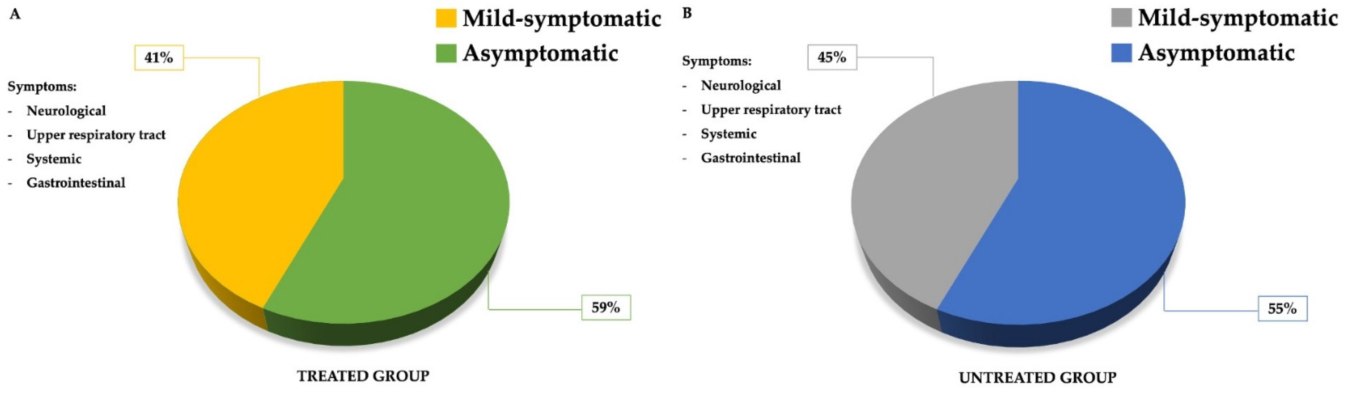
2. Results
3. Discussion
Limitations of the Study
4. Materials and Methods
4.1. Patients and Enrollment Criteria
4.2. m-PEA + um-PEA Treatment
4.3. Statistical Analysis
4.4. Laboratory Parameters
5. Conclusions
Author Contributions
Funding
Institutional Review Board Statement
Informed Consent Statement
Data Availability Statement
Acknowledgments
Conflicts of Interest
Abbreviations
| BBB | Blood–brain barrier |
| BMI | Body mass index |
| CNS | Central nervous system |
| CPK | Creatine kinase |
| COVID-19 | Coronavirus disease-19 |
| CRP | C-reactive protein |
| CS | Cytokine storm |
| ESR | Erythrocyte sedimentation rate |
| FORD | Free oxygen radical defense |
| FORT | Free oxygen radicals test |
| ICU | Intensive care unit |
| IL | Interleukin |
| mAbs | Monoclonal antibodies |
| NBCs | Natural bioactive compounds |
| NOS2 | Nitric oxide synthase-2 |
| OS | Oxidative stress |
| PCR | Polymerase chain reaction |
| PEA | Palmitoylethanolamide |
| PPARα | Proliferator-activated receptor alpha |
| PTV | Policlinico Tor Vergata |
| ROS | Reactive oxygen species |
| rRT | reverse transcriptase real time |
| SARS-CoV-2 | Severe acute respiratory syndrome coronavirus-2 |
| TLR | Toll-like receptor |
| TNF | Tumor necrosis factor |
| um-PEA | Ultramicronized palmitoylethanolamide |
References
- Renu, K.; Prasanna, P.L.; Valsala Gopalakrishnan, A. Coronaviruses pathogenesis, comorbidities and multi-organ damage—A review. Life Sci. 2020, 255, 117839. [Google Scholar] [CrossRef] [PubMed]
- Huang, C.; Wang, Y.; Li, X.; Ren, L.; Zhao, J.; Hu, Y.; Zhang, L.; Fan, G.; Xu, J.; Gu, X.; et al. Clinical features of patients infected with 2019 novel coronavirus in Wuhan, China. Lancet 2020, 395, 497–506. [Google Scholar] [CrossRef] [Green Version]
- Rokkas, T. Gastrointestinal involvement in COVID-19: A systematic review and meta-analysis. Ann. Gastroenterol. 2020, 33, 355–365. [Google Scholar] [CrossRef]
- Cheong, J.; Bartell, N.; Peeraphatdit, T.; Mosli, M.; Al-Judaibi, B. Gastrointestinal and liver manifestations of COVID-19. Saudi J. Gastroenterol. 2020, 26, 226–232. [Google Scholar] [CrossRef] [PubMed]
- Kolhe, N.V.; Fluck, R.J.; Selby, N.M.; Taal, M.W. Acute kidney injury associated with COVID-19: A retrospective cohort study. PLoS Med. 2020, 17, e1003406. [Google Scholar] [CrossRef]
- Ahmad, I.; Rathore, F.A. Neurological manifestations and complications of COVID-19: A literature review. J. Clin. Neurosci. 2020, 77, 8–12. [Google Scholar] [CrossRef]
- Daneshgaran, G.; Dubin, D.P.; Gould, D.J. Cutaneous Manifestations of COVID-19: An Evidence-Based Review. Am. J. Clin. Dermatol. 2020, 21, 627–639. [Google Scholar] [CrossRef]
- Li, Y.C.; Bai, W.Z.; Hashikawa, T. The neuroinvasive potential of SARS-CoV2 may play a role in the respiratory failure of COVID-19 patients. J. Med. Virol. 2020, 92, 552–555. [Google Scholar] [CrossRef]
- Kritas, S.K.; Ronconi, G.; Caraffa, A.; Gallenga, C.E.; Ross, R.; Conti, P. Mast cells contribute to coronavirus-induced inflammation: New anti-inflammatory strategy. J. Biol. Regul. Homeost. Agents 2020, 34, 9–14. [Google Scholar] [CrossRef]
- Xu, J.; Zhong, S.; Liu, J.; Li, L.; Li, Y.; Wu, X.; Li, Z.; Deng, P.; Zhang, J.; Zhong, N.; et al. Detection of severe acute respiratory syndrome coronavirus in the brain: Potential role of the chemokine mig in pathogenesis. Clin. Infect. Dis. 2005, 41, 1089–1096. [Google Scholar] [CrossRef] [Green Version]
- Menghini, R.; Campia, U.; Tesauro, M.; Marino, A.; Rovella, V.; Rodia, G.; Schinzari, F.; Tolusso, B.; di Daniele, N.; Federici, M.; et al. Toll-like receptor 4 mediates endothelial cell activation through NF-kappaB but is not associated with endothelial dysfunction in patients with rheumatoid arthritis. PLoS ONE 2014, 9, e99053. [Google Scholar] [CrossRef] [PubMed] [Green Version]
- Mehta, P.; McAuley, D.F.; Brown, M.; Sanchez, E.; Tattersall, R.S.; Manson, J.J.; Hlh Across Speciality Collaboration, U.K. COVID-19: Consider cytokine storm syndromes and immunosuppression. Lancet 2020, 395, 1033–1034. [Google Scholar] [CrossRef]
- Steardo, L., Jr.; Bronzuoli, M.R.; Iacomino, A.; Esposito, G.; Steardo, L.; Scuderi, C. Does neuroinflammation turn on the flame in Alzheimer’s disease? Focus on astrocytes. Front. Neurosci. 2015, 9, 259. [Google Scholar] [CrossRef] [PubMed] [Green Version]
- Scuderi, C.; Stecca, C.; Iacomino, A.; Steardo, L. Role of astrocytes in major neurological disorders: The evidence and implications. IUBMB Life 2013, 65, 957–961. [Google Scholar] [CrossRef] [PubMed]
- Morfopoulou, S.; Brown, J.R.; Davies, E.G.; Anderson, G.; Virasami, A.; Qasim, W.; Chong, W.K.; Hubank, M.; Plagnol, V.; Desforges, M.; et al. Human Coronavirus OC43 Associated with Fatal Encephalitis. N. Engl. J. Med. 2016, 375, 497–498. [Google Scholar] [CrossRef]
- Higgins, V.; Sohaei, D.; Diamandis, E.P.; Prassas, I. COVID-19: From an acute to chronic disease? Potential long-term health consequences. Crit. Rev. Clin. Lab. Sci. 2021, 58, 297–310. [Google Scholar] [CrossRef]
- Goertz, Y.M.J.; Van Herck, M.; Delbressine, J.M.; Vaes, A.W.; Meys, R.; Machado, F.V.C.; Houben-Wilke, S.; Burtin, C.; Posthuma, R.; Franssen, F.M.E.; et al. Persistent symptoms 3 months after a SARS-CoV-2 infection: The post-COVID-19 syndrome? ERJ Open Res. 2020, 6. [Google Scholar] [CrossRef]
- Center For Disease Control and Prevention “SymptomDuration and Risk Factors for Delayed Return to Usual Health Among Outpatients with COVID-19 in a Multistate Health Care Systems Network—United States, March–June 2020”. Available online: https://www.cdc.gov/mmwr/volumes/69/wr/mm6930e1.htm#References (accessed on 3 December 2021).
- Carfi, A.; Bernabei, R.; Landi, F.; Gemelli Against, C.-P.-A.C.S.G. Persistent Symptoms in Patients After Acute COVID-19. JAMA 2020, 324, 603–605. [Google Scholar] [CrossRef]
- Nalbandian, A.; Sehgal, K.; Gupta, A.; Madhavan, M.V.; McGroder, C.; Stevens, J.S.; Cook, J.R.; Nordvig, A.S.; Shalev, D.; Sehrawat, T.S.; et al. Post-acute COVID-19 syndrome. Nat. Med. 2021, 27, 601–615. [Google Scholar] [CrossRef]
- Saudou, F.; Hen, R. 5-Hydroxytryptamine receptor subtypes: Molecular and functional diversity. Adv. Pharmacol. 1994, 30, 327–380. [Google Scholar] [CrossRef]
- Garrigues, E.; Janvier, P.; Kherabi, Y.; Le Bot, A.; Hamon, A.; Gouze, H.; Doucet, L.; Berkani, S.; Oliosi, E.; Mallart, E.; et al. Post-discharge persistent symptoms and health-related quality of life after hospitalization for COVID-19. J. Infect. 2020, 81, e4–e6. [Google Scholar] [CrossRef] [PubMed]
- Huang, Y.; Tan, C.; Wu, J.; Chen, M.; Wang, Z.; Luo, L.; Zhou, X.; Liu, X.; Huang, X.; Yuan, S.; et al. Impact of coronavirus disease 2019 on pulmonary function in early convalescence phase. Respir. Res. 2020, 21, 163. [Google Scholar] [CrossRef] [PubMed]
- Zhao, Y.M.; Shang, Y.M.; Song, W.B.; Li, Q.Q.; Xie, H.; Xu, Q.F.; Jia, J.L.; Li, L.M.; Mao, H.L.; Zhou, X.M.; et al. Follow-up study of the pulmonary function and related physiological characteristics of COVID-19 survivors three months after recovery. eClinicalMedicine 2020, 25, 100463. [Google Scholar] [CrossRef]
- Esakandari, H.; Nabi-Afjadi, M.; Fakkari-Afjadi, J.; Farahmandian, N.; Miresmaeili, S.M.; Bahreini, E. A comprehensive review of COVID-19 characteristics. Biol. Proced. Online 2020, 22, 19. [Google Scholar] [CrossRef] [PubMed]
- Petrosino, S.; Schiano Moriello, A.; Cerrato, S.; Fusco, M.; Puigdemont, A.; De Petrocellis, L.; Di Marzo, V. The anti-inflammatory mediator palmitoylethanolamide enhances the levels of 2-arachidonoyl-glycerol and potentiates its actions at TRPV1 cation channels. Br. J. Pharmacol. 2016, 173, 1154–1162. [Google Scholar] [CrossRef] [Green Version]
- Gatti, A.; Lazzari, M.; Gianfelice, V.; Di Paolo, A.; Sabato, E.; Sabato, A.F. Palmitoylethanolamide in the treatment of chronic pain caused by different etiopathogenesis. Pain Med. 2012, 13, 1121–1130. [Google Scholar] [CrossRef] [Green Version]
- Aloe, L.; Leon, A.; Levi-Montalcini, R. A proposed autacoid mechanism controlling mastocyte behaviour. Agents Actions 1993, 39, C145–C147. [Google Scholar] [CrossRef]
- Haller, V.L.; Cichewicz, D.L.; Welch, S.P. Non-cannabinoid CB1, non-cannabinoid CB2 antinociceptive effects of several novel compounds in the PPQ stretch test in mice. Eur. J. Pharmacol. 2006, 546, 60–68. [Google Scholar] [CrossRef]
- D’Agostino, G.; La Rana, G.; Russo, R.; Sasso, O.; Iacono, A.; Esposito, E.; Mattace Raso, G.; Cuzzocrea, S.; Loverme, J.; Piomelli, D.; et al. Central administration of palmitoylethanolamide reduces hyperalgesia in mice via inhibition of NF-kappaB nuclear signalling in dorsal root ganglia. Eur. J. Pharmacol. 2009, 613, 54–59. [Google Scholar] [CrossRef]
- Luongo, L.; Guida, F.; Boccella, S.; Bellini, G.; Gatta, L.; Rossi, F.; de Novellis, V.; Maione, S. Palmitoylethanolamide reduces formalin-induced neuropathic-like behaviour through spinal glial/microglial phenotypical changes in mice. CNS Neurol. Disord. Drug Targets 2013, 12, 45–54. [Google Scholar] [CrossRef]
- Skaper, S.D.; Facci, L.; Fusco, M.; Della Valle, M.F.; Zusso, M.; Costa, B.; Giusti, P. Palmitoylethanolamide, a naturally occurring disease-modifying agent in neuropathic pain. Inflammopharmacology 2014, 22, 79–94. [Google Scholar] [CrossRef] [PubMed]
- LoVerme, J.; Russo, R.; La Rana, G.; Fu, J.; Farthing, J.; Mattace-Raso, G.; Meli, R.; Hohmann, A.; Calignano, A.; Piomelli, D. Rapid broad-spectrum analgesia through activation of peroxisome proliferator-activated receptor-alpha. J. Pharmacol. Exp. Ther. 2006, 319, 1051–1061. [Google Scholar] [CrossRef]
- Mazzari, S.; Canella, R.; Petrelli, L.; Marcolongo, G.; Leon, A. N-(2-hydroxyethyl)hexadecanamide is orally active in reducing edema formation and inflammatory hyperalgesia by down-modulating mast cell activation. Eur. J. Pharmacol. 1996, 300, 227–236. [Google Scholar] [CrossRef]
- Di Cesare Mannelli, L.; D’Agostino, G.; Pacini, A.; Russo, R.; Zanardelli, M.; Ghelardini, C.; Calignano, A. Palmitoylethanolamide is a disease-modifying agent in peripheral neuropathy: Pain relief and neuroprotection share a PPAR-alpha-mediated mechanism. Mediat. Inflamm. 2013, 2013, 328797. [Google Scholar] [CrossRef] [PubMed]
- Yoshihara, S.; Morimoto, H.; Ohori, M.; Yamada, Y.; Abe, T.; Arisaka, O. Endogenous cannabinoid receptor agonists inhibit neurogenic inflammations in guinea pig airways. Int. Arch. Allergy Immunol. 2005, 138, 80–87. [Google Scholar] [CrossRef]
- Helyes, Z.; Nemeth, J.; Than, M.; Bolcskei, K.; Pinter, E.; Szolcsanyi, J. Inhibitory effect of anandamide on resiniferatoxin-induced sensory neuropeptide release in vivo and neuropathic hyperalgesia in the rat. Life Sci. 2003, 73, 2345–2353. [Google Scholar] [CrossRef]
- Naderi, N.; Majidi, M.; Mousavi, Z.; Khoramian Tusi, S.; Mansouri, Z.; Khodagholi, F. The interaction between intrathecal administration of low doses of palmitoylethanolamide and AM251 in formalin-induced pain related behavior and spinal cord IL1-beta expression in rats. Neurochem. Res. 2012, 37, 778–785. [Google Scholar] [CrossRef]
- Brotini, S.; Schievano, C.; Guidi, L. Ultra-micronized Palmitoylethanolamide: An Efficacious Adjuvant Therapy for Parkinson’s Disease. CNS Neurol. Disord. Drug Targets 2017, 16, 705–713. [Google Scholar] [CrossRef]
- Evangelista, M.; Cilli; De Vitis, R.; Militerno, A.; Fanfani, F. Ultra-micronized Palmitoylethanolamide Effects on Sleep-wake Rhythm and Neuropathic Pain Phenotypes in Patients with Carpal Tunnel Syndrome: An Open-label, Randomized Controlled Study. CNS Neurol. Disord. Drug Targets 2018, 17, 291–298. [Google Scholar] [CrossRef]
- Del Giorno, R.; Skaper, S.; Paladini, A.; Varrassi, G.; Coaccioli, S. Palmitoylethanolamide in Fibromyalgia: Results from Prospective and Retrospective Observational Studies. Pain Ther. 2015, 4, 169–178. [Google Scholar] [CrossRef] [Green Version]
- Lunardelli, M.L.; Crupi, R.; Siracusa, R.; Cocuzza, G.; Cordaro, M.; Martini, E.; Impellizzeri, D.; Di Paola, R.; Cuzzocrea, S. Co-ultraPEALut: Role in Preclinical and Clinical Delirium Manifestations. CNS Neurol. Disord. Drug Targets 2019, 18, 530–554. [Google Scholar] [CrossRef] [PubMed]
- Bronzuoli, M.R.; Facchinetti, R.; Steardo, L., Jr.; Romano, A.; Stecca, C.; Passarella, S.; Steardo, L.; Cassano, T.; Scuderi, C. Palmitoylethanolamide Dampens Reactive Astrogliosis and Improves Neuronal Trophic Support in a Triple Transgenic Model of Alzheimer’s Disease: In Vitro and In Vivo Evidence. Oxid. Med. Cell. Longev. 2018, 2018, 4720532. [Google Scholar] [CrossRef] [PubMed] [Green Version]
- Rao, S.; Song, Y.; Peddie, F.; Evans, A.M. Particle size reduction to the nanometer range: A promising approach to improve buccal absorption of poorly water-soluble drugs. Int. J. Nanomed. 2011, 6, 1245–1251. [Google Scholar] [CrossRef] [Green Version]
- Sareen, S.; Mathew, G.; Joseph, L. Improvement in solubility of poor water-soluble drugs by solid dispersion. Int. J. Pharm. Investig. 2012, 2, 12–17. [Google Scholar] [CrossRef] [PubMed] [Green Version]
- Noce, A.; Albanese, M.; Marrone, G.; Di Lauro, M.; Pietroboni Zaitseva, A.; Palazzetti, D.; Guerriero, C.; Paolino, A.; Pizzenti, G.; Di Daniele, F.; et al. Ultramicronized Palmitoylethanolamide (um-PEA): A New Possible Adjuvant Treatment in COVID-19 patients. Pharmaceuticals 2021, 14, 336. [Google Scholar] [CrossRef]
- Putilina, M.V.; Grishin, D.V. SARS-CoV-2 (COVID-19) as a Predictor of Neuroinflammation and Neurodegeneration: Potential Treatment Strategies. Neurosci. Behav. Physiol. 2021, 51, 577–582. [Google Scholar] [CrossRef]
- Di Daniele, N.; Di Renzo, L.; Noce, A.; Iacopino, L.; Ferraro, P.M.; Rizzo, M.; Sarlo, F.; Domino, E.; De Lorenzo, A. Effects of Italian Mediterranean organic diet vs. low-protein diet in nephropathic patients according to MTHFR genotypes. J. Nephrol. 2014, 27, 529–536. [Google Scholar] [CrossRef]
- Chernyak, B.V.; Popova, E.N.; Prikhodko, A.S.; Grebenchikov, O.A.; Zinovkina, L.A.; Zinovkin, R.A. COVID-19 and Oxidative Stress. Biochemistry 2020, 85, 1543–1553. [Google Scholar] [CrossRef]
- Yaprak, E.; Sukur, Y.E.; Ozmen, B.; Sonmezer, M.; Berker, B.; Atabekoglu, C.; Aytac, R. Endometrial compaction is associated with the increased live birth rate in artificial frozen-thawed embryo transfer cycles. Hum. Fertil. 2021, 1–7. [Google Scholar] [CrossRef]
- Candi, E.; Tesauro, M.; Cardillo, C.; Lena, A.M.; Schinzari, F.; Rodia, G.; Sica, G.; Gentileschi, P.; Rovella, V.; Annicchiarico-Petruzzelli, M.; et al. Metabolic profiling of visceral adipose tissue from obese subjects with or without metabolic syndrome. Biochem. J. 2018, 475, 1019–1035. [Google Scholar] [CrossRef]
- Liu, X.; Shen, Y.; Wang, H.; Ge, Q.; Fei, A.; Pan, S. Prognostic Significance of Neutrophil-to-Lymphocyte Ratio in Patients with Sepsis: A Prospective Observational Study. Mediat. Inflamm. 2016, 2016, 8191254. [Google Scholar] [CrossRef] [PubMed] [Green Version]
- Guthrie, G.J.; Charles, K.A.; Roxburgh, C.S.; Horgan, P.G.; McMillan, D.C.; Clarke, S.J. The systemic inflammation-based neutrophil-lymphocyte ratio: Experience in patients with cancer. Crit. Rev. Oncol. Hematol. 2013, 88, 218–230. [Google Scholar] [CrossRef] [PubMed]
- Fu, J.; Kong, J.; Wang, W.; Wu, M.; Yao, L.; Wang, Z.; Jin, J.; Wu, D.; Yu, X. The clinical implication of dynamic neutrophil to lymphocyte ratio and D-dimer in COVID-19: A retrospective study in Suzhou China. Thromb. Res. 2020, 192, 3–8. [Google Scholar] [CrossRef] [PubMed]
- So, M.K.P.; Tiwari, A.; Chu, A.M.Y.; Tsang, J.T.Y.; Chan, J.N.L. Visualizing COVID-19 pandemic risk through network connectedness. Int. J. Infect. Dis. 2020, 96, 558–561. [Google Scholar] [CrossRef] [PubMed]
- Chiurchiu, V.; Leuti, A.; Smoum, R.; Mechoulam, R.; Maccarrone, M. Bioactive lipids ALIAmides differentially modulate inflammatory responses of distinct subsets of primary human T lymphocytes. FASEB J. 2018, 32, 5716–5723. [Google Scholar] [CrossRef]
- Rostami, M.; Mansouritorghabeh, H. D-dimer level in COVID-19 infection: A systematic review. Expert Rev. Hematol. 2020, 13, 1265–1275. [Google Scholar] [CrossRef]
- Magadum, A.; Engel, F.B. PPARbeta/delta: Linking Metabolism to Regeneration. Int. J. Mol. Sci. 2018, 19. [Google Scholar] [CrossRef] [Green Version]
- Noce, A.; Vidiri, M.F.; Marrone, G.; Moriconi, E.; Bocedi, A.; Capria, A.; Rovella, V.; Ricci, G.; De Lorenzo, A.; Di Daniele, N. Is low-protein diet a possible risk factor of malnutrition in chronic kidney disease patients? Cell Death Discov. 2016, 2, 16026. [Google Scholar] [CrossRef] [Green Version]


| Parameters | Cases (n = 45) | Controls (n = 45) | p-Value |
|---|---|---|---|
| Age (years) | 45.6 ± 13.7 | 55.8 ± 22.5 | n.s. |
| Male/female (n) | 17/28 | 22/23 | n.s. |
| Weight (kg) | 69.3 ± 6.9 | 70.4 ± 7.1 | n.s. |
| BMI (kg/m2) | 24.4 ± 3.4 | 25.6 ± 5.8 | n.s. |
| Parameters | Cases (n = 45) | Controls (n = 45) | p-Value | ||
|---|---|---|---|---|---|
| T0 (Mean ± SD) | T1 (Mean ± SD) | T0 (Mean ± SD) | T1 (Mean ± SD) | ||
| Red blood cell (104/µL) | 4.88 ± 0.52 | 4.64 ± 0.53 | 4.30 ± 0.78 | 4.21 ± 0.69 | n.s. |
| Hemoglobin (g/dL) | 13.98 ± 1.82 | 13.35 ± 1.74 | 12.54 ± 2.27 | 12.4 ± 2.00 | n.s. |
| Hematocrit (%) | 41.9 ± 5.2 | 40.1 ± 6.62 | 36.9 ± 8.08 | 37.3 ± 5.34 | n.s |
| MCV (fL) | 86.1 ± 6.1 | 86.7 ± 6.08 | 87.9 ± 5.96 | 88.3 ± 5.02 | n.s. |
| Neutrophil (103/µL) | 3.44 ± 2.04 | 3.4 ± 2.02 | 4.56 ± 2.60 | 4.43 ± 2.34 | n.s. |
| Lymphocytes (103/µL) | 1.64 ± 0.60 | 1.96 ± 0.53 | 1.32 ± 0.56 | 1.62 ± 0.74 | 0.02 |
| Neutrophil-to-Lymphocyte ratio | 2.43 ± 2.03 | 1.78 ± 3.8 | 3.45 ± 4.64 | 2.7 ± 3.16 | 0.04 |
| Platelets (103/µL) | 241.69 ± 74.7 | 267.8 ± 43.7 | 239.63 ± 95.4 | 240.75 ± 96.7 | n.s. |
| Myoglobin (mg/mL) | 43.02 ± 53.8 | 38.28 ± 45.2 | 110.75 ± 154.7 | 77.26 ± 93.3 | n.s. |
| D-Dimer (ng/mL) | 686.19 ± 1348.77 | 366.07 ± 230.76 | 1032 ± 1258.42 | 670.57 ± 507.08 | 0.0001 |
| PT (%) | 94.64 ± 18.62 | 97.35 ± 20.16 | 81.61 ± 15.76 | 88.40 ± 17.31 | n.s. |
| PT (INR) | 1.09 ± 0.15 | 1.32 ± 1.74 | 1.13 ± 0.14 | 1.08 ± 0.12 | n.s. |
| PT (s) | 13.41 ± 3.93 | 12.06 ± 2.05 | 13.83 ± 1.91 | 13.18 ± 1.72 | n.s. |
| Fibrinogen (mg/dL) | 298.5 ± 93.7 | 260.83 ± 100.0 | 455.14 ± 223.13 | 375.14 ± 167.3 | n.s. |
| Antithrombin III (%) | 107.25 ± 6.95 | 102.5 ± 4.43 | 98.54 ± 17.8 | 119.65 ± 123.4 | n.s. |
| Creatininemia (mg/dL) | 0.83 ± 0.19 | 0.87 ± 0.25 | 0.98 ± 0.84 | 0.95 ± 0.88 | n.s. |
| GFR (mL/min) | 87.56 ± 17.83 | 86.02 ± 19.82 | 94.81 ± 37.14 | 98.12 ± 35.12 | n.s. |
| Azotemia (md/dL) | 30.47 ± 8.44 | 32.44 ± 14.58 | 36.22 ± 16.54 | 35.16 ± 20.95 | n.s. |
| Vitamin D (ng/mL) | 32.0 ± 16.03 | 30.16 ± 15.41 | 21.57 ± 10.12 | 25.54 ± 13.20 | n.s. |
| ESR (mm/h) | 24.04 ± 20.37 | 14.89 ± 11.65 | 22.30 ± 17.60 | 15.90 ± 12.46 | n.s. |
| CRP (mg/dL) | 7.20 ± 12.95 | 1.55 ± 1.80 | 20.03 ± 24.59 | 9.79 ± 16.90 | 0.007 |
| TNF-α (pg/mL) | 19.23 ± 20.07 | 8.15 ± 8.69 | 50.00 ± 114.20 | 31.84 ± 84.69 | n.s. |
| IL-6 (pg/mL) | 11.22 ± 19.58 | 3.30 ± 1.54 | 24.20 ± 23.00 | 15.36 ± 19.90 | 0.0001 |
| GOT/AST (U/L) | 29.21 ± 10.94 | 24.7 ± 6.93 | 28.13 ± 14.76 | 25.11 ± 10.84 | n.s. |
| GPT/ALT (U/L) | 31.72 ± 20.09 | 26.82 ± 12.91 | 23.38 ± 15.22 | 21.0 ± 10.9 | n.s. |
| γ -GT (U/L) | 27.83 ± 38.7 | 20.55 ± 12.32 | 33.35 ± 44.6 | 24.86 ± 18.85 | n.s. |
| Creatine kinase (U/L) | 82.46 ± 57.65 | 92.46 ± 43.53 | 93.0 ± 127.57 | 56.59 ± 45.59 | n.s. |
| FORD (mmol/L Trolox) | 1.04 ± 0.34 | 1.49 ± 0.43 | 1.09 ± 0.35 | 1.23 ± 0.32 | n.s. |
| FORT (U) | 271.07 ± 156.82 | 222.02 ± 107.71 | 229.90 ± 143.98 | 283.30 ± 111.08 | 0.05 |
| CD3 + CD8 + absolute count | 526.98 ± 330.71 | 636.41 ± 325.26 | 497.13 ± 237.31 | 487.60 ± 196.81 | 0.0001 |
| Anti-SARS-CoV-2 IgG | NA | 4.37 ± 1.62 | NA | 2.89 ± 2.03 | 0.0001 |
| Composition NORMAST® MPS Microgranules | % |
|---|---|
| Ultramicronized Palmitoylethanolamide (um-PEA, 600 mg) | 48.48 |
| Micronized Palmitoylethanolamide (m-PEA,300 mg) | 24.24 |
| Fructose | 15.15 |
| Sorbitol | 9.33 |
| Polysorbate 80 | 0.36 |
| Palmitic esters of sucrose | 1.45 |
| Cross-linked sodium carboxymethylcellulose | 0.97 TOTAL 100.00 |
Publisher’s Note: MDPI stays neutral with regard to jurisdictional claims in published maps and institutional affiliations. |
© 2022 by the authors. Licensee MDPI, Basel, Switzerland. This article is an open access article distributed under the terms and conditions of the Creative Commons Attribution (CC BY) license (https://creativecommons.org/licenses/by/4.0/).
Share and Cite
Albanese, M.; Marrone, G.; Paolino, A.; Di Lauro, M.; Di Daniele, F.; Chiaramonte, C.; D’Agostini, C.; Romani, A.; Cavaliere, A.; Guerriero, C.; et al. Effects of Ultramicronized Palmitoylethanolamide (um-PEA) in COVID-19 Early Stages: A Case–Control Study. Pharmaceuticals 2022, 15, 253. https://doi.org/10.3390/ph15020253
Albanese M, Marrone G, Paolino A, Di Lauro M, Di Daniele F, Chiaramonte C, D’Agostini C, Romani A, Cavaliere A, Guerriero C, et al. Effects of Ultramicronized Palmitoylethanolamide (um-PEA) in COVID-19 Early Stages: A Case–Control Study. Pharmaceuticals. 2022; 15(2):253. https://doi.org/10.3390/ph15020253
Chicago/Turabian StyleAlbanese, Maria, Giulia Marrone, Agostino Paolino, Manuela Di Lauro, Francesca Di Daniele, Carlo Chiaramonte, Cartesio D’Agostini, Annalisa Romani, Alessandro Cavaliere, Cristina Guerriero, and et al. 2022. "Effects of Ultramicronized Palmitoylethanolamide (um-PEA) in COVID-19 Early Stages: A Case–Control Study" Pharmaceuticals 15, no. 2: 253. https://doi.org/10.3390/ph15020253