
Synthesis and Biological Evaluation of Novel Cinnamic Acid-Based Antimicrobials

 , , ,
, , ,  , and
, and
Abstract
:
1. Introduction
2. Results
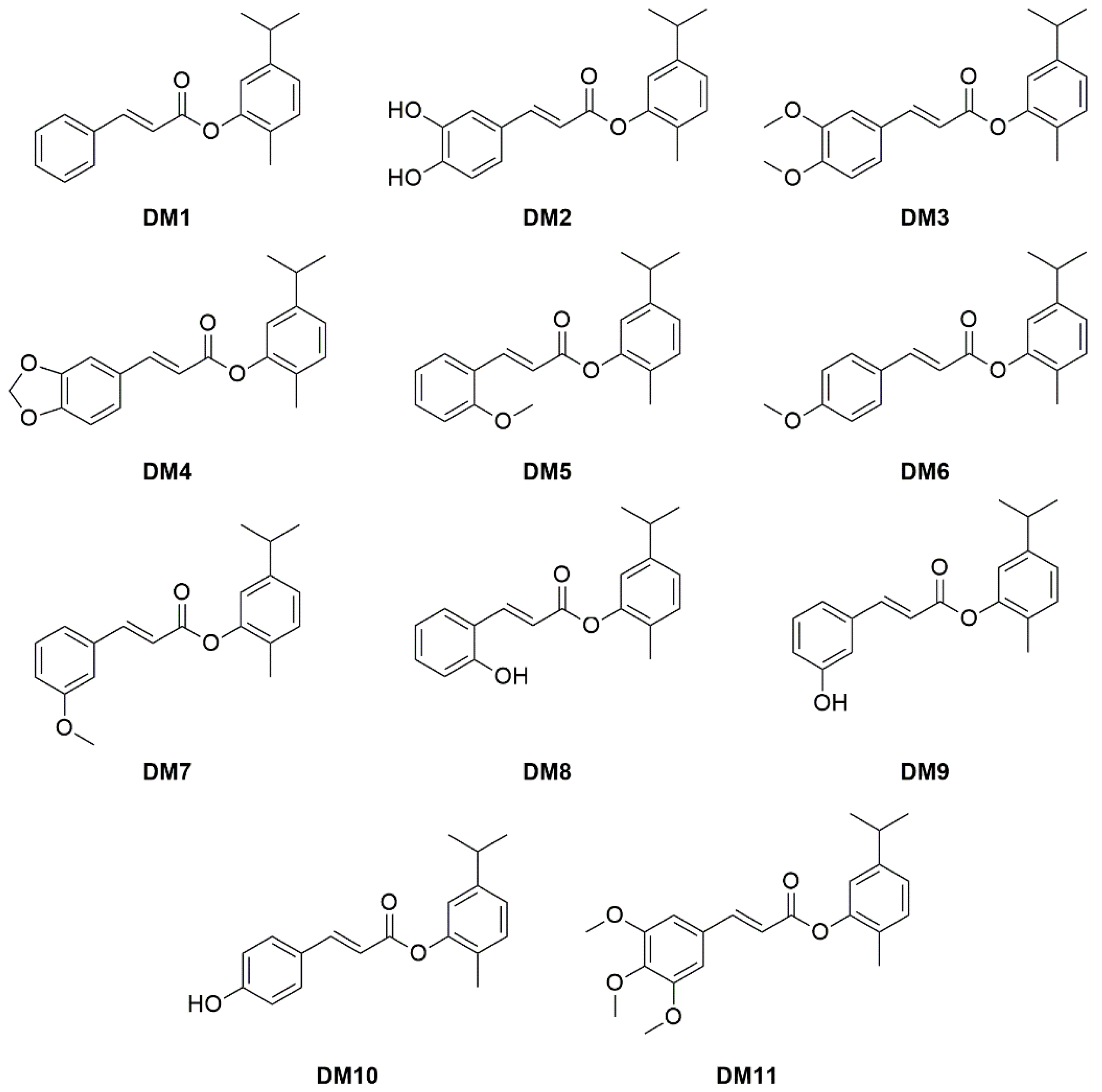
2.1. Chemistry
2.2. In Silico Skin Permeability Prediction Analysis
2.3. Antimicrobial and Antibiofilm Properties of DM1–11
- The presence of one, two, or three substituents on the ring structure of DM derivatives influenced the antimicrobial activity. The results showed that the introduction of one, two, or three methoxy groups, enhanced the lipophilicity of the molecule (Table 1), and negatively affected the antimicrobial activity.
- One or two hydroxyl groups on the ring structure of DM8 and DM2, respectively, enhanced the hydrophilia, ameliorating the interaction only with the cell wall of Gram-positive pathogens. The uptake of DM2 and DM8 into Gram-positive bacteria (S. aureus and epidermidis) was more efficient than into Gram-negative bacteria due to the differences in the cell walls. It is well known that the polar nature of the outer membrane of Gram-negative bacteria results in limited passive permeability of hydrophobic drugs. Moreover, the presence of promiscuous efflux pumps can hinder the entry of antimicrobials.
- DM2, having the lowest Log P (equal to 3.83) as compared with other DM derivatives, was found to be the most potent antimicrobial agent against S. aureus and epidermidis, which indicated that the presence of two withdrawing substituents on m- and p-position of the phenyl nucleus are important for the interaction with the bacterial cell wall.
- Despite DM8-10 having the same values as Log P, the position of the hydroxyl group in orto-, meta-, and para-position on the benzene ring of these molecules influenced the antimicrobial activity. The introduction of -OH moiety in meta- and para- in DM9 and DM10, respectively, caused a drastic loss of activity as compared with DM8, which had the OH- in orto;
- DM8 was the most active compound against E. faecium (MIC50% = 32 mg/L), suggesting that the hydroxyl group in o-position, as compared with DM1 that avoided substituents on the phenyl nucleus, is important for the interaction with the enterococcal cell wall.
2.4. Evaluation of the Wound Healing Effect of DM2
3. Cytotoxicity Studies
4. Materials and Methods
4.1. Chemistry
4.1.1. General Procedure for the Synthesis of DM1, DM3–7, and DM11
4.1.2. General Procedure for the Synthesis of DM8–10
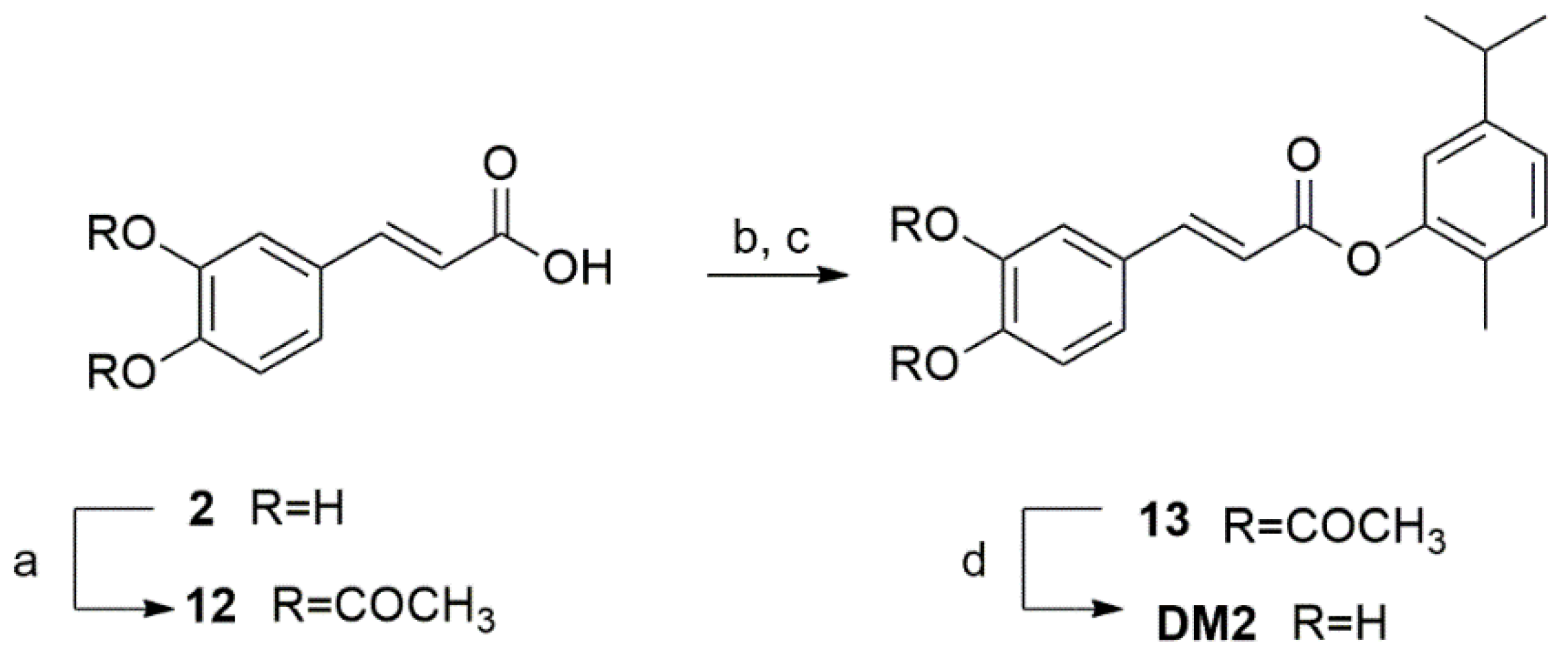
4.1.3. Synthesis of DM2
4.2. In Silico Skin Permeability Prediction Analysis
4.3. Bacterial Strains
4.4. Antimicrobial Activity
4.5. Antibiofilm Activity
4.6. Cell Cultures
4.7. Wound Scratch Assay
4.8. MTT Viability Assay
4.9. Statistical Analysis
5. Conclusions
Supplementary Materials
Author Contributions
Funding
Institutional Review Board Statement
Informed Consent Statement
Data Availability Statement
Conflicts of Interest
References
- WHO (World Health Organization). Available online: https://www.who.int/health-topics/antimicrobial-resistance (accessed on 20 May 2021).
- Nadeem, S.F.; Gohar, U.F.; Tahir, S.F.; Mukhtar, H.; Pornpukdeewattana, S.; Nukthamna, P.; Massa, S. Antimicrobial resistance: More than 70 years of war between humans and bacteria. Crit. Rev. Microbiol. 2020, 46, 578–599. [Google Scholar] [CrossRef] [PubMed]
- Cappiello, F.; Loffredo, M.R.; Plato, C.D.; Cammarone, S.; Casciaro, B.; Quaglio, D.; Ghirga, F. The revaluation of plant-derived terpenes to fight antibiotic-resistant infections. Antibiotics 2020, 9, 325. [Google Scholar] [CrossRef] [PubMed]
- Mulani, M.S.; Kamble, E.E.; Kumkar, S.N.; Tawre, M.S.; Pardesi, K.R. Emerging strategies to combat ESKAPE pathogens in the era of antimicrobial resistance: A review. Front. Microbiol. 2019, 10, 539. [Google Scholar] [CrossRef] [PubMed]
- Khan, H.A.; Kanwal Baig, F.; Mehboob, R. Nosocomial infections: Epidemiology, prevention, control, and surveillance. Asian Pac. J. Trop. Biomed. 2017, 7, 478–482. [Google Scholar] [CrossRef]
- Emily, R.M.; Sydnor, T.M.P. Hospital epidemiology and infection control in acute-care settings. Clin. Microbiol. Rev. 2011, 24, 141–173. [Google Scholar] [CrossRef] [Green Version]
- de Macedo, G.H.R.V.; Costa, C.D.E.; Oliveira, E.R.; Damasceno, G.V.; Mendonça, J.S.P.; Silva, L.S.; da Silva, L.C.N. Interplay between eskape pathogens and immunity in skin infections: An overview of the major determinants of virulence and antibiotic resistance. Pathogens 2021, 10, 148. [Google Scholar] [CrossRef]
- Weintrob, A.C.; Murray, C.K.; Xu, J.; Krauss, M.; Bradley, W.; Warkentien, T.E.; Tribble, D.R. Early infections complicating the care of combat casualties from Iraq and Afghanistan. Surg. Infect. 2018, 19, 286–297. [Google Scholar] [CrossRef]
- Arias, M.; Hassan-Reshat, S.; Newsholme, W. Retrospective analysis of diabetic foot osteomyelitis management and outcome at a tertiary care hospital in the UK. PLoS ONE 2019, 14, e0216701. [Google Scholar] [CrossRef] [Green Version]
- Annavajhala, M.K.; Gomez-Simmonds, A.; Uhlemann, A. Multidrug-resistant enterobacter cloacae complex emerging as a global, diversifying threat. Front. Microbiol. 2019, 10, 44. [Google Scholar] [CrossRef] [Green Version]
- Keen III, E.F.; Robinson, B.J.; Hospenthal, D.R.; Aldous, W.K.; Wolf, S.E.; Chung, K.K.; Murray, C.K. Incidence and bacteriology of burn infections at a military burn center. Burns 2010, 36, 461–468. [Google Scholar] [CrossRef]
- Davis, K.A.; Moran, K.A.; McAllister, C.K.; Gray, P.I. Multidrug-resistant acinetobacter extremity infections in soldiers. Emerg. Infect. Dis. 2005, 11, 1218–1224. [Google Scholar] [CrossRef] [PubMed]
- Haghi, F.; Zeighami, H.; Monazami, A.; Toutouchi, F.; Nazaralian, S.; Naderi, G. Diversity of virulence genes in multidrug resistant pseudomonas aeruginosa isolated from burn wound infections. Microb. Pathog. 2018, 115, 251–256. [Google Scholar] [CrossRef] [PubMed]
- Simonetti, O.; Cirioni, O.; Mocchegiani, F.; Cacciatore, I.; Silvestri, C.; Baldassarre, l.; Offidani, A. The efficacy of the quorum sensing inhibitor FS8 and tigecycline in preventing prosthesis biofilm in an animal model of staphylococcal infection. Int. J. Mol. Sci. 2013, 14, 16321–16332. [Google Scholar] [CrossRef] [PubMed] [Green Version]
- Simonetti, O.; Cirioni, O.; Cacciatore, I.; Baldassarre, L.; Orlando, F.; Pierpaoli, E.; Offidani, A. Efficacy of the quorum sensing inhibitor FS10 alone and in combination with tigecycline in an animal model of staphylococcal infected wound. PLoS ONE 2016, 11, e0151956. [Google Scholar] [CrossRef] [PubMed]
- Marinelli, L.; Fornasari, E.; Eusepi, P.; Ciulla, M.; Genovese, S.; Epifano, F.; Cacciatore, I. Carvacrol prodrugs as novel antimicrobial agents. Eur. J. Med. Chem. 2019, 178, 515–529. [Google Scholar] [CrossRef]
- Guzman, J.D. Natural cinnamic acids, synthetic derivatives, and hybrids with antimicrobial activity. Molecules 2014, 19, 19292–19349. [Google Scholar] [CrossRef]
- Sova, M. Antioxidant and antimicrobial activities of cinnamic acid derivatives. Mini-Rev. Med. Chem. 2012, 12, 749–767. [Google Scholar] [CrossRef]
- Ruwizhi, N.; Aderibigbe, B.A. Cinnamic acid derivatives and their biological efficacy. Int. J. Mol. Sci. 2020, 21, 5712. [Google Scholar] [CrossRef]
- Zhou, K.; Chen, D.; Li, B.; Zhang, B.; Miao, F.; Zhou, L. Bioactivity and structure-activity relationship of cinnamic acid esters and their derivatives as potential antifungal agents for plant protection. PLoS ONE 2017, 12, e0176189. [Google Scholar] [CrossRef]
- Ferro, T.A.F.; Souza, E.B.; Suarez, M.A.M.; Rodrigues, J.F.S.; Pereira, D.M.S.; Mendes, S.J.F.; Fernandes, E.S. Topical application of cinnamaldehyde promotes faster healing of skin wounds infected with pseudomonas aeruginosa. Molecules 2019, 24, 1627. [Google Scholar] [CrossRef] [Green Version]
- Eusepi, P.; Marinelli, L.; García-Villén, F.; Borrego-Sánchez, A.; Cacciatore, I.; Di Stefano, A.; Viseras, C. Carvacrol prodrugs with antimicrobial activity loaded on clay nanocomposites. Materials 2020, 13, 1793. [Google Scholar] [CrossRef] [PubMed] [Green Version]
- Cacciatore, I.; Fornasari, E.; Di Stefano, A.; Marinelli, L.; Cerasa, L.S.; Turkez, H.; Patruno, A. Development of glycine-α-methyl-proline-containing tripeptides with neuroprotective properties. Eur. J. Med. Chem. 2016, 108, 553–563. [Google Scholar] [CrossRef]
- Lipsky, B.A.; Hoey, C. Topical antimicrobial therapy for treating chronic wounds. Clin. Infect. Dis. 2009, 49, 1541–1549. [Google Scholar] [CrossRef] [PubMed] [Green Version]
- Monteiro-Neto, V.; de Souza, C.D.; Gonzaga, L.F.; da Silveira, B.C.; Sousa, N.C.F.; Pontes, J.P.; Fernandes, E.S. Cuminaldehyde potentiates the antimicrobial actions of ciprofloxacin against Staphylococcus aureus and Escherichia coli. PLoS ONE 2020, 15, e0232987. [Google Scholar] [CrossRef] [PubMed]
- Schneider, L.A.; Korber, A.; Grabbe, S.; Dissemond, J. Influence of pH on wound-healing: A new perspective for wound-therapy? Arch. Derm. Res. 2007, 298, 413–420. [Google Scholar] [CrossRef]
- Rajkumari, J.; Borkotoky, S.; Murali, A.; Suchiang, K.; Mohanty, S.K.; Busi, S. Cinnamic acid attenuates quorum sensing associated virulence factors and biofilm formation in pseudomonas aeruginosa PAO1. Biotechnol. Lett. 2018, 40, 1087–1100. [Google Scholar] [CrossRef] [PubMed]
- Rajkumari, J.; Borkotoky, S.; Murali, A.; Suchiang, K.; Mohanty, K.; Busi, S. Attenuation of quorum sensing controlled virulence factors and biofilm formation in Pseudomonas aeruginosa by pentacyclic triterpenes, betulin and betulinic acid. Microb. Pathog. 2018, 118, 48–60. [Google Scholar] [CrossRef]
- Simonetti, O.; Rizzetto, G.; Radi, G.; Molinelli, E.; Cirioni, O.; Giacometti, A.; Offidani, A. New perspectives on old and new therapies of staphylococcal skin infections: The role of biofilm targeting in wound healing. Antibiotics 2021, 10, 1377. [Google Scholar] [CrossRef]
- Wu, Y.; Cheng, N.; Cheng, C. Biofilms in chronic wounds: Pathogenesis and diagnosis. Trends Biotechnol. 2019, 37, 505–517. [Google Scholar] [CrossRef]
- Tarricone, A.; Mata, K.D.L.; Rothstein, M.; Soave, R.L. The effect of wound pH on healing rates in chronic wounds: A literature review. J. Am. Podiatr. Med. Assoc. 2020, 110, 1–6. [Google Scholar] [CrossRef]
- Scalise, A.; Bianchi, A.; Tartaglione, C.; Bolletta, E.; Pierangeli, M.; Torresetti, M.; Di Benedetto, G. Microenvironment and microbiology of skin wounds: The role of bacterial biofilms and related factors. Semin. Vasc. Surg. 2015, 28, 151–159. [Google Scholar] [CrossRef] [PubMed]
- Johnson, T.R.; Gómez, B.I.; McIntyre, M.K.; Dubick, M.A.; Christy, R.J.; Nicholson, S.E.; Burmeister, D.M. The cutaneous microbiome and wounds: New molecular targets to promote wound healing. Int. J. Mol. Sci. 2018, 19, 2699. [Google Scholar] [CrossRef] [PubMed] [Green Version]
- Omar, A.; Wright, J.B.; Schultz, G.; Burrell, R.; Nadworny, P. Microbial biofilms and chronic wounds. Microorganisms 2017, 5, 9. [Google Scholar] [CrossRef] [Green Version]
- Mori, Y.; Nakagami, G.; Kitamura, A.; Minematsu, T.; Kinoshita, M.; Suga, H.; Sanada, H. Effectiveness of biofilm-based wound care system on wound healing in chronic wounds. Wound Repair Regen. 2019, 27, 540–547. [Google Scholar] [CrossRef] [PubMed]
- Kirketerp-Møller, K.; Jensen, P.O.; Fazli, M.; Madsen, K.G.; Pedersen, J.; Moser, C.; Bjarnsholt, T. Distribution, organization, and ecology of bacteria in chronic wounds. J. Clin. Microbiol. 2008, 46, 2717–2722. [Google Scholar] [CrossRef] [Green Version]
- Pinnen, F.; Cacciatore, I.; Cornacchia, C.; Sozio, P.; Cerasa, L.S.; Iannitelli, A.; Di Stefano, A. Codrugs linking L-dopa and sulfur-containing antioxidants: New pharmacological tools against Parkinson’s disease. J. Med. Chem. 2009, 52, 559–563. [Google Scholar] [CrossRef]
- Cacciatore, I.; Di Giulio, M.; Fornasari, E.; Di Stefano, A.; Cerasa, L.S.; Marinelli, L.; Cellini, L. Carvacrol codrugs: A new approach in the antimicrobial plan. PLoS ONE 2015, 10, e0120937. [Google Scholar] [CrossRef] [Green Version]
- Marini, E.; Magi, G.; Mingoia, M.; Pugnaloni, A.; Facinelli, B. Antimicrobial and anti-virulence activity of capsaicin against erythromycin-resistant, cell-invasive Group A Streptococci. Front. Microbiol. 2015, 6, 1281. [Google Scholar] [CrossRef] [Green Version]
- Stepanovic, S.; Vukovic, D.; Dakic, I.; Savic, B.; Svabic-Vlahovic, M. A modified microtiter-plate test for quantification of staphylococcal biofilm formation. J. Microbiol. Methods 2000, 40, 175–179. [Google Scholar] [CrossRef]
- Pagano, S.; Lombardo, G.; Costanzi, E.; Balloni, S.; Bruscoli, S.; Flamini, S.; Marinucci, L. Morpho-functional effects of different universal dental adhesives on human gingival fibroblasts: An in vitro study. Odontology 2021, 109, 524–539. [Google Scholar] [CrossRef]
- Taticchi, A.; Urbani, S.; Albi, E.; Servili, M.; Codini, M.; Traina, G.; Conte, C. In vitro anti-inflammatory effects of phenolic compounds from moraiolo virgin olive oil (MVOO) in brain cells via regulating the TLR4/NLRP3 axis. Molecules 2019, 24, 4523. [Google Scholar] [CrossRef] [PubMed] [Green Version]


| Compound | MW a | Log P b | Log Kp c |
|---|---|---|---|
| DM1 | 280.26 | 4.60 | −4.28 |
| DM2 | 312.36 | 3.83 | −5.01 |
| DM3 | 340.41 | 4.56 | −4.69 |
| DM4 | 324.37 | 4.38 | −4.69 |
| DM5 | 310.39 | 4.61 | −4.49 |
| DM6 | 310.39 | 4.62 | −4.49 |
| DM7 | 310.39 | 4.61 | −4.49 |
| DM8 | 296.36 | 4.20 | −4.64 |
| DM9 | 296.36 | 4.22 | −4.64 |
| DM10 | 296.36 | 4.21 | −4.64 |
| DM11 | 370.44 | 4.57 | −4.90 |
| Strain (n) a | MIC (mg/L) b | DM2 | DM8 | DM9 | CAR |
|---|---|---|---|---|---|
| S. aureus (6) | range | 16–64 | 16–512 | 256–>512 | 128–256 |
| 50% | 32 | 64 | >512 | 256 | |
| 90% | 64 | 256 | >512 | 256 | |
| S. epidermidis (6) | range | 64–128 | 256 | >512 | 256 |
| 50% | 128 | >256 | >512 | 256 | |
| 90% | 128 | >256 | >512 | 256 | |
| E. faecalis (10) | range | 64–512 | 32–>512 | 512–>512 | 128–512 |
| 50% | 256 | 256 | >512 | 256 | |
| 90% | 512 | >512 | >512 | 256 | |
| E. faecium (10) | range | 32–512 | 16–512 | 512->512 | 256 |
| 50% | 128 | 32 | >512 | 256 | |
| 90% | 256 | 512 | >512 | 256 | |
| S. pyogenes (6) | range | 128–>512 | 512–>512 | >512 | 64–256 |
| 50% | 512 | >512 | >512 | 128 | |
| 90% | 512 | >512 | >512 | 256 | |
| S. agalactiae (10) | range | 512–>512 | 512–>512 | 512–>512 | 256–512 |
| 50% | >512 | >512 | >512 | 256 | |
| 90% | >512 | >512 | >512 | 512 | |
| E. coli (6) | range | >512 | >512 | >512 | 256 |
| 50% | >512 | >512 | >512 | 256 | |
| 90% | >512 | >512 | >512 | 256 | |
| K. pneumoniae (6) | range | 512–>512 | >512 | >512 | 512 |
| 50% | >512 | >512 | >512 | 512 | |
| 90% | >512 | >512 | >512 | 512 | |
| P. aeruginosa (6) | range | 512–>512 | >512 | >512 | 512–>512 |
| 50% | >512 | >512 | >512 | >512 | |
| 90% | >512 | >512 | >512 | >512 | |
| A. baumannii (10) | range | 256–>512 | >512 | >512 | 64–256 |
| 50% | 512 | >512 | >512 | 128 | |
| 90% | >512 | >512 | >512 | 256 | |
| Enterobacter spp. (6) | range | 512 | >512 | >512 | 512 |
| 50% | 512 | >512 | >512 | 512 | |
| 90% | 512 | >512 | >512 | 512 |
Publisher’s Note: MDPI stays neutral with regard to jurisdictional claims in published maps and institutional affiliations. |
© 2022 by the authors. Licensee MDPI, Basel, Switzerland. This article is an open access article distributed under the terms and conditions of the Creative Commons Attribution (CC BY) license (https://creativecommons.org/licenses/by/4.0/).
Share and Cite
Mingoia, M.; Conte, C.; Di Rienzo, A.; Dimmito, M.P.; Marinucci, L.; Magi, G.; Turkez, H.; Cufaro, M.C.; Del Boccio, P.; Di Stefano, A.; et al. Synthesis and Biological Evaluation of Novel Cinnamic Acid-Based Antimicrobials. Pharmaceuticals 2022, 15, 228. https://doi.org/10.3390/ph15020228
Mingoia M, Conte C, Di Rienzo A, Dimmito MP, Marinucci L, Magi G, Turkez H, Cufaro MC, Del Boccio P, Di Stefano A, et al. Synthesis and Biological Evaluation of Novel Cinnamic Acid-Based Antimicrobials. Pharmaceuticals. 2022; 15(2):228. https://doi.org/10.3390/ph15020228
Chicago/Turabian StyleMingoia, Marina, Carmela Conte, Annalisa Di Rienzo, Marilisa Pia Dimmito, Lorella Marinucci, Gloria Magi, Hasan Turkez, Maria Concetta Cufaro, Piero Del Boccio, Antonio Di Stefano, and et al. 2022. "Synthesis and Biological Evaluation of Novel Cinnamic Acid-Based Antimicrobials" Pharmaceuticals 15, no. 2: 228. https://doi.org/10.3390/ph15020228