
Aspartames Alter Pharmacokinetics Parameters of Erlotinib and Gefitinib and Elevate Liver Enzymes in Wistar Rats
 , , ,
, , , 
Abstract
:1. Introduction
2. Results
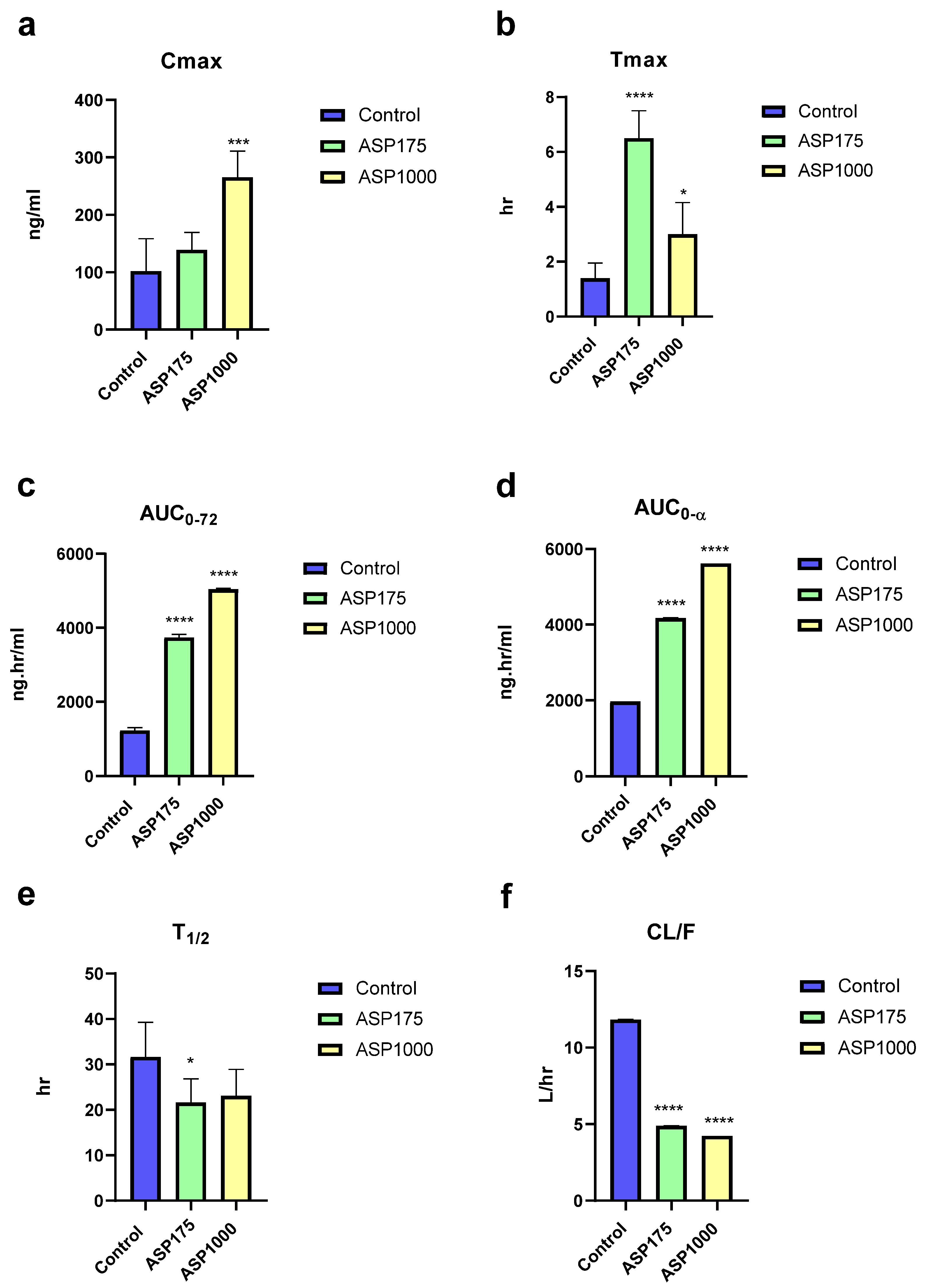
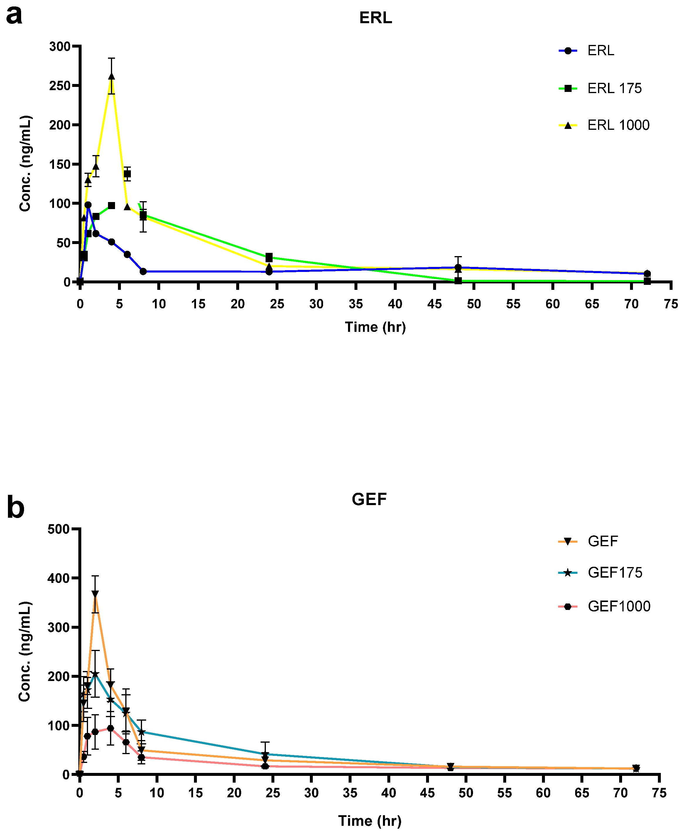
2.1. The Effect of ASP on ERL’s PKs
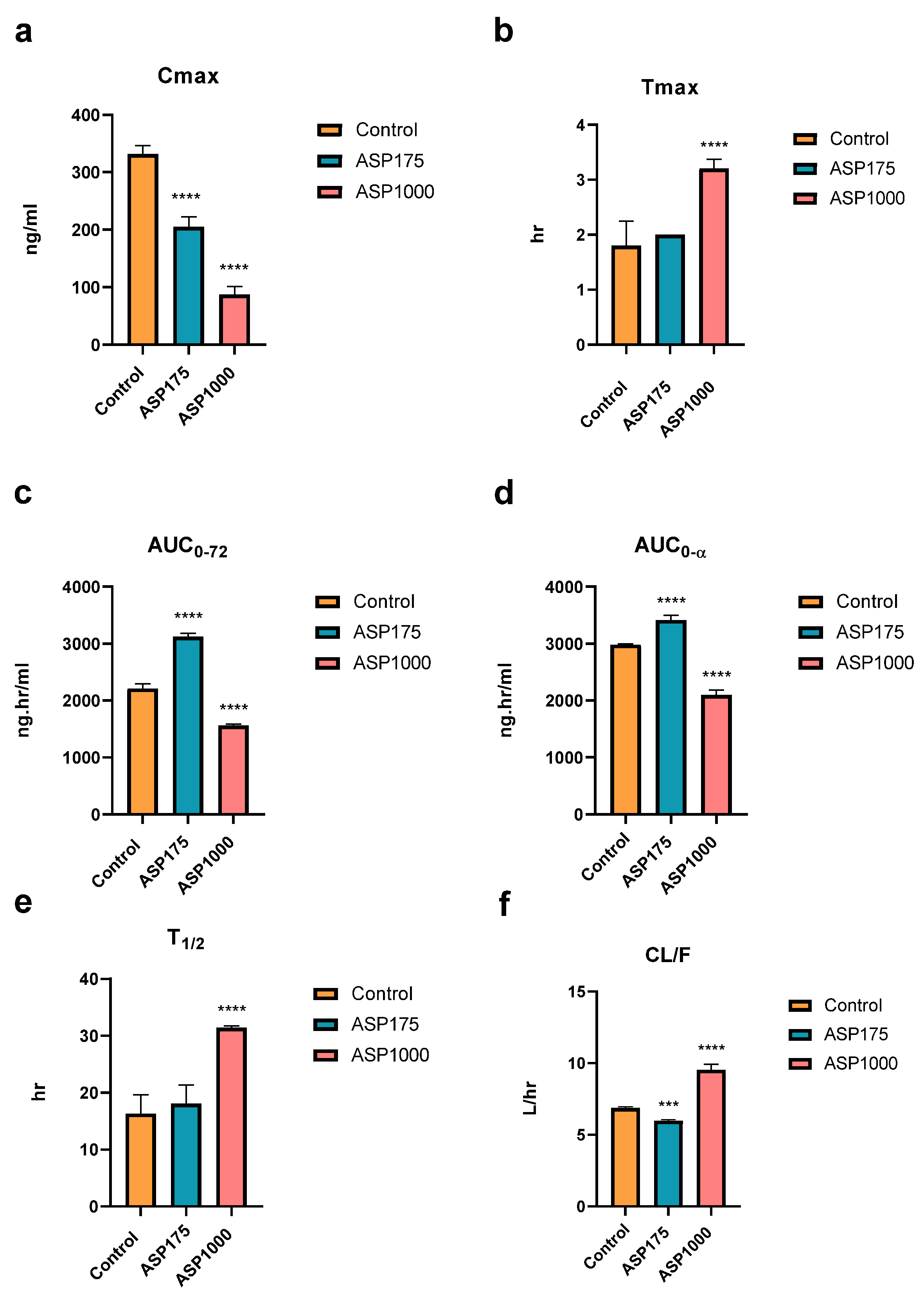
2.2. The Effect of ASP GEF’s PKs
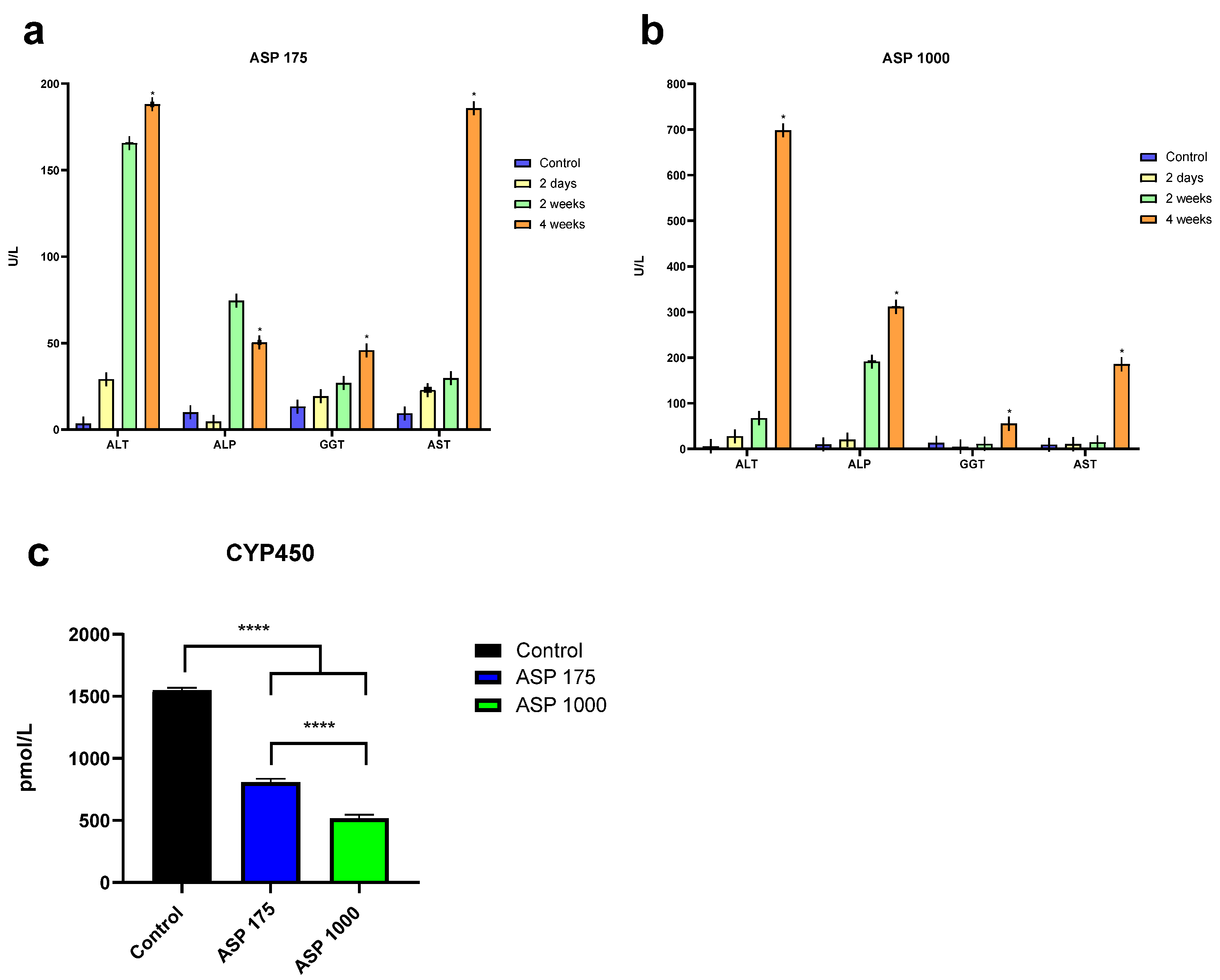
2.3. Liver Enzyme
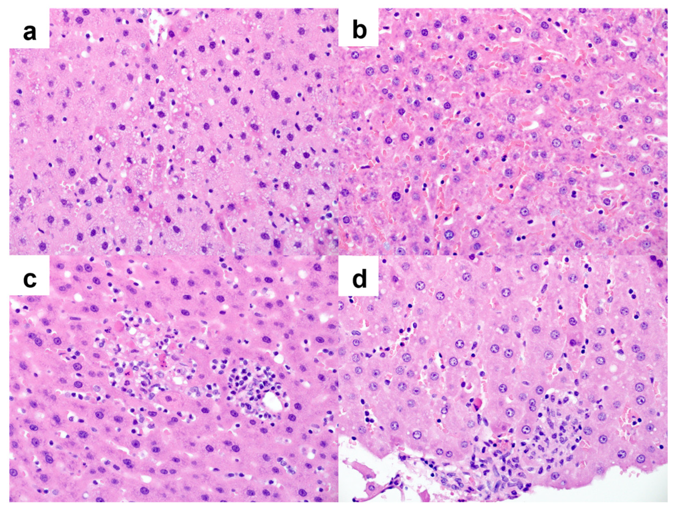
2.4. Histopathology
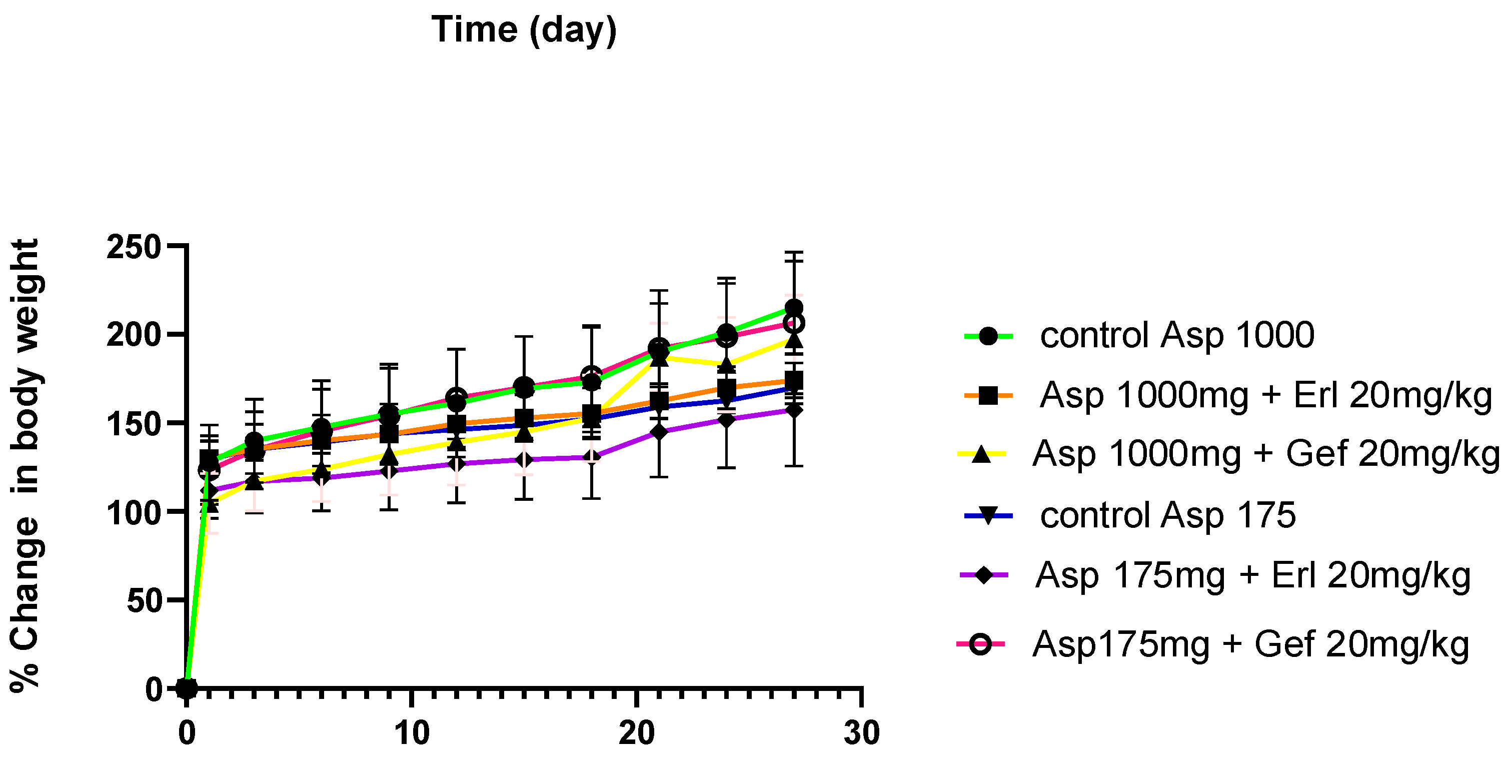
2.5. Rat Body Weight
3. Discussion
4. Materials and Methods
4.1. Materials
4.2. Experimental Animals
4.3. Animal Study
4.4. PK Analyses
4.5. Liver Enzyme Analysis
4.6. Liver Histology
4.7. Statistical Analysis
5. Conclusions
Supplementary Materials
Author Contributions
Funding
Institutional Review Board Statement
Informed Consent Statement
Data Availability Statement
Conflicts of Interest
References
- Lung Cancer—Non-Small Cell: Statistics|Cancer.Net. Available online: https://www.cancer.net/cancer-types/lung-cancer-non-small-cell/statistics (accessed on 15 January 2021).
- World Lung Cancer Day 2020 Fact Sheet—American College of Chest Physicians. Available online: https://www.chestnet.org/News/CHEST-News/2020/07/World-Lung-Cancer-Day-2020-Fact-Sheet (accessed on 15 January 2021).
- Masood, A.; Kancha, R.K.; Subramanian, J. Epidermal growth factor receptor (EGFR) tyrosine kinase inhibitors in non-small cell lung cancer harboring uncommon EGFR mutations: Focus on afatinib. Semi. Oncol. 2019, 46, 271–283. [Google Scholar] [CrossRef] [PubMed]
- Aisner, D.L.; Marshall, C.B. Molecular pathology of non-small cell lung cancer: A practical guide. Am. J. Clin. Pathol. 2012, 138, 332–346. Available online: https://academic.oup.com/ajcp/article/138/3/332/1766002 (accessed on 15 January 2021). [CrossRef] [Green Version]
- Xu, Z.Y.; Li, J.L. Comparative review of drug–drug interactions with epidermal growth factor receptor tyrosine kinase inhibitors for the treatment of non-small-cell lung cancer. Oncol. Targets Ther. 2019, 12, 5467–5484. [Google Scholar] [CrossRef] [Green Version]
- Choudhary, A.K.; Pretorius, E. Revisiting the safety of aspartame. Nutr. Rev. 2017, 75, 718–730. Available online: https://academic.oup.com/nutritionreviews/article/75/9/718/4101228 (accessed on 18 January 2021). [CrossRef] [PubMed] [Green Version]
- Magnuson, B.A.; Carakostas, M.C.; Moore, N.H.; Poulos, S.P.; Renwick, A.G. Biological fate of low-calorie sweeteners. Nutr. Rev. 2016, 74, 670–689. Available online: https://academic.oup.com/nutritionreviews/article-lookup/doi/10.1093/nutrit/nuw032 (accessed on 21 January 2021). [CrossRef] [PubMed] [Green Version]
- Fernstrom, J.D.; Munger, S.D.; Sclafani, A.; de Araujo, I.E.; Roberts, A.; Molinary, S. Mechanisms for sweetness. J. Nutr. 2012, 142, 1134S. [Google Scholar] [CrossRef] [PubMed] [Green Version]
- Almomen, A.; Maher, H.M.; Alzoman, N.Z.; Shehata, S.M.; Alsubaie, A. Flavoured water consumption alters pharmacokinetic parameters and increases exposure of erlotinib and gefitinib in a preclinical study using Wistar rats. PeerJ 2020, 8, e9881. Available online: https://pubmed.ncbi.nlm.nih.gov/33024629/ (accessed on 18 January 2021). [CrossRef] [PubMed]
- Ashok, I.; Sheeladevi, R.; Wankhar, D. Acute effect of aspartame-induced oxidative stress in Wistar albino rat brain. J. Biomed. Res. 2015, 29, 390–396. [Google Scholar] [PubMed]
- Abhilash, M.; Paul, M.V.S.; Varghese, M.V.; Nair, R.H. Effect of long term intake of aspartame on antioxidant defense status in liver. Food Chem. Toxicol. 2011, 49, 1203–1207. [Google Scholar] [CrossRef] [PubMed]
- Arakelyan, H. Elevated Liver Enzymes and Health; ResearchGate: Berlin, Germany, 2020. [Google Scholar]
- Mino, R.; Corina, L. Drug Metabolism: Current Concepts; Springer: Berlin/Heidelberg, Germany, 2005. [Google Scholar]
- Scheffler, M.; Di Gion, P.; Doroshyenko, O.; Wolf, J.; Fuhr, U. Clinical pharmacokinetics of tyrosine kinase inhibitors: Focus on 4-anilinoquinazolines. Clin. Pharmacokinet. 2011, 50, 371–403. Available online: https://link.springer.com/article/10.2165/11587020-000000000-00000 (accessed on 16 January 2021). [CrossRef] [PubMed]
- Khamise, N.; Tayel, D.; Helmy, M.; Aborhyem, S. Effect of aspartame and sucralose artificial sweeteners on weight and lipid profile of male albino rats. J. High Inst. Public Health 2020, 50, 87–100. [Google Scholar] [CrossRef]
- Haq, N.; Tafweez, R.; Saqib, S.; Bokhari, Z.H.; Ali, I.; Syami, A.F. Aspartame and sucralose-induced fatty changes in rat liver. J. Coll. Physicians Surg. Pak. 2019, 29, 848–851. [Google Scholar] [CrossRef]


Publisher’s Note: MDPI stays neutral with regard to jurisdictional claims in published maps and institutional affiliations. |
© 2022 by the authors. Licensee MDPI, Basel, Switzerland. This article is an open access article distributed under the terms and conditions of the Creative Commons Attribution (CC BY) license (https://creativecommons.org/licenses/by/4.0/).
Share and Cite
AlRasheed, H.; Almomen, A.; Aljohar, H.I.; Arafah, M.; Almotawa, R.Y.; Alossaimi, M.A.; Alzoman, N.Z. Aspartames Alter Pharmacokinetics Parameters of Erlotinib and Gefitinib and Elevate Liver Enzymes in Wistar Rats. Pharmaceuticals 2022, 15, 1400. https://doi.org/10.3390/ph15111400
AlRasheed H, Almomen A, Aljohar HI, Arafah M, Almotawa RY, Alossaimi MA, Alzoman NZ. Aspartames Alter Pharmacokinetics Parameters of Erlotinib and Gefitinib and Elevate Liver Enzymes in Wistar Rats. Pharmaceuticals. 2022; 15(11):1400. https://doi.org/10.3390/ph15111400
Chicago/Turabian StyleAlRasheed, Hajer, Aliyah Almomen, Haya I. Aljohar, Maria Arafah, Rana Y. Almotawa, Manal A. Alossaimi, and Nourah Z. Alzoman. 2022. "Aspartames Alter Pharmacokinetics Parameters of Erlotinib and Gefitinib and Elevate Liver Enzymes in Wistar Rats" Pharmaceuticals 15, no. 11: 1400. https://doi.org/10.3390/ph15111400