Article

Mangostanin, a Xanthone Derived from Garcinia mangostana Fruit, Exerts Protective and Reparative Effects on Oxidative Damage in Human Keratinocytes

1
Department of Medicine and Surgery, University of Salerno, 84081 Baronissi, Italy
2
Department of Molecular Medicine and Medical Biotechnology, University of Naples “Federico II”, 80131 Naples, Italy
3
Department of Pharmacy, University of Salerno, 84084 Fisciano, Italy
*
Authors to whom correspondence should be addressed.
These authors contributed equally to this work.
Pharmaceuticals 2022, 15(1), 84; https://doi.org/10.3390/ph15010084
Submission received: 25 October 2021 / Revised: 30 December 2021 / Accepted: 5 January 2022 / Published: 11 January 2022

Abstract

:
The fruit of Garcinia mangostana (mangosteen) is known in ancient traditional Asian medicine for its antioxidant, anti-inflammatory, immunomodulatory and anticancer activities. These effects are mainly due to the action of polyphenols known as xanthones, which are contained in the pericarp of the fruit. In recent years, there has been a growing interest from pharmaceutical companies in formulating new topicals based on mangosteen full extracts to prevent skin aging. However, the molecules responsible for these effects and the mechanisms involved have not been investigated so far. Here, the arils and shells of Garcinia mangostana were extracted with chloroform and methanol, and the extracts were further purified to yield 12 xanthone derivatives. Their effects were evaluated using in vitro cultures of human epidermal keratinocytes. After confirming the absence of cytotoxicity, we evaluated the antioxidant potential of these compounds, identifying mangostanin as capable of both protecting and restoring oxidative damage induced by H2O2. We showed how mangostanin, by reducing the generation of intracellular reactive oxygen species (ROS), prevents the activation of AKT (protein kinase B), ERK (extracellular signal-regulated kinase), p53, and other cellular pathways underlying cell damage and apoptosis activation. In conclusion, our study is the first to demonstrate that mangostanin is effective in protecting the skin from the action of free radicals, thus preventing skin aging, confirming a potential toward its development in the nutraceutical and cosmeceutical fields.

1. Introduction

Garcinia mangostana L. (mangosteen) is an evergreen tropical tree native to Southeast Asia belonging to the Clusiaceae or Guttiferae family [1]. It is renowned as the “queen of the fruits” due to the particularly delicious taste of the fruits as well as for its use in traditional medicine [2]. In fact, in Southeast Asia, the seeds and peels have been used not only as infusions and decoctions for the treatment of gastrointestinal and urinary tract infections but also as a laxative, anti-fever agent, and an insomnia treatment [2,3].
Various components have been found to be responsible for the pharmacological properties of mangosteen, such as saccharides, flavonoids, phenolic acids, and, most importantly, xanthones.
Plant-derived polysaccharides have been reported to possess a wide range of biological activities, such as anti-inflammatory and immunoregulatory activity. Furthermore, they can be easily used in the biomedical field as they are non-toxic and free from apparent side effects. Recently, a water-soluble arabinofuranan was purified from the rinds of Garcinia mangostana and was reported to have a strong stimulating effect on the immune system, particularly through the activation of macrophages [4].
Similarly, flavonoids are compounds often synthesized by plants, including mangosteen, as a defense against pathogens, such as Xanthomonas campestris and Fusarium species, nematodes, and phytophagous insects. Since their discovery, they have been described as having a vast number of biological benefits, such as anti-inflammatory, antiviral, antibacterial, antioxidant, anticancer, and neuroprotective properties. Baicalein, fisetin, quercetagetin, silymarin, curcumin, and nobiletin are just some examples of the numerous flavonoids that can be found in mangosteen; interestingly, various studies have reported that these flavonoids can inhibit the infection caused by chikungunya virus, a pathogen that can cause a fever characterized by severe myalgia [5,6].
Polyphenols, phytonutrients that have been shown to prevent cell degeneration and the proliferation of tumor cells, represent another important class of compounds that can be found in mangosteen. Several studies show how the implementation of diets rich in polyphenols of plant origin (such as mangosteen) can help avoid the development of tumors, especially in the stomach and surrounding areas. The group of flavonoids also includes catechins, moderate antioxidants that can help protect the cardiovascular system [7].
Despite the varied chemical composition of the mangosteen, many scientists are directing their research toward the isolation of the phytochemical compounds most represented in the mangosteen fruit, the xanthones, which may be responsible for its prodigious qualities. For the 68 xanthones that have been cataloged so far, multiple in vitro and in vivo experiments have been conducted with the aim of better understanding and identifying their multiple biological effects (antioxidant [8], antibacterial [9], anti-inflammatory [10,11], neuroprotective [12], hypoglycemic [13,14], anti-obesity [15], cytotoxic, anti-proliferative, and anti-cancer [16,17,18,19]). At the same time, several in vivo studies on mangosteen fruit extracts and its purified compounds, demonstrated the total absence of acute or chronic toxicity [20,21], confirming what has already been observed by the consumption of mangosteen for over 100 years.
For this reason, the use of bio-active extracts of mangosteen fruit is having great success in the nutraceutical and cosmetic fields. Recent studies show how mangosteen fruit extracts can be used as photoprotective agents from UVB damage [22], to improve skin elasticity, and to control inflammatory skin diseases [11,23]. However, although the extracts of mangosteen fruit are used for the preparation of cosmetics, the biological targets and the mechanism of action from which its dermatological benefits derive, are not yet clear.
Therefore, in this work we provided the first pharmacological evidence of how mangostanin, a xanthone present in the mangosteen extract, is able both to protect the skin from the deleterious action of free radicals preventing skin aging and to remedy to damage with a repairing effect on the skin after oxidative insult.

2. Results

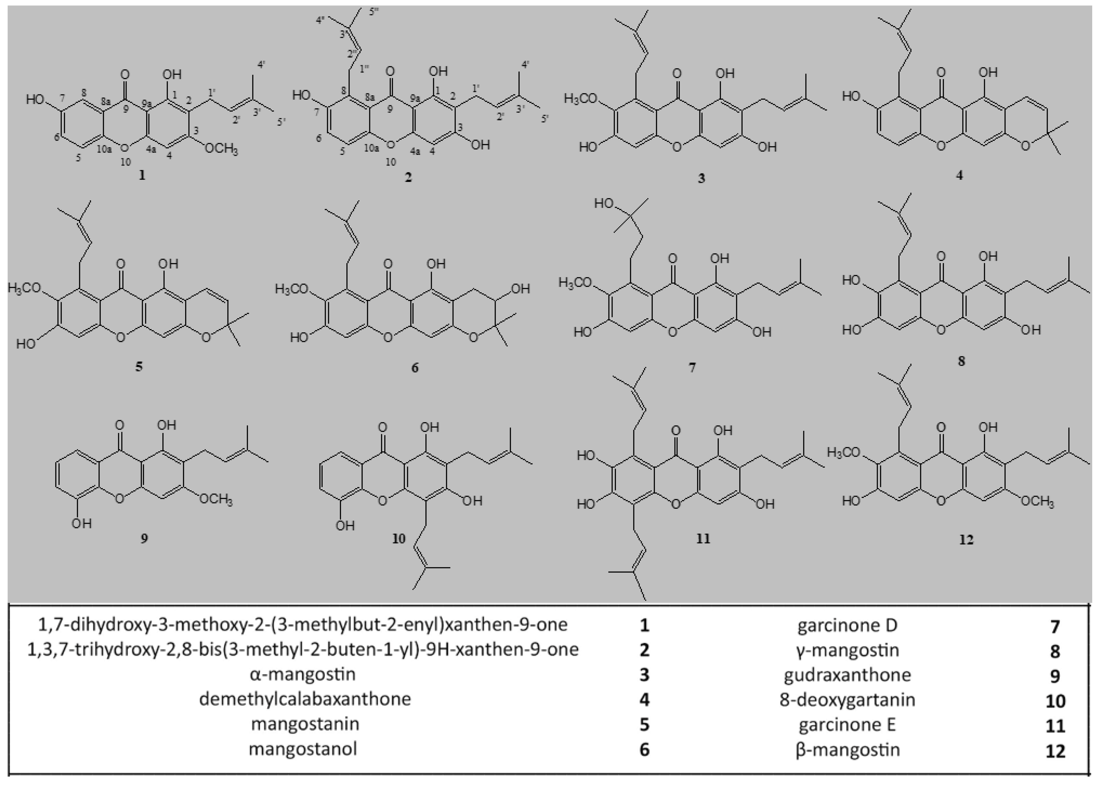
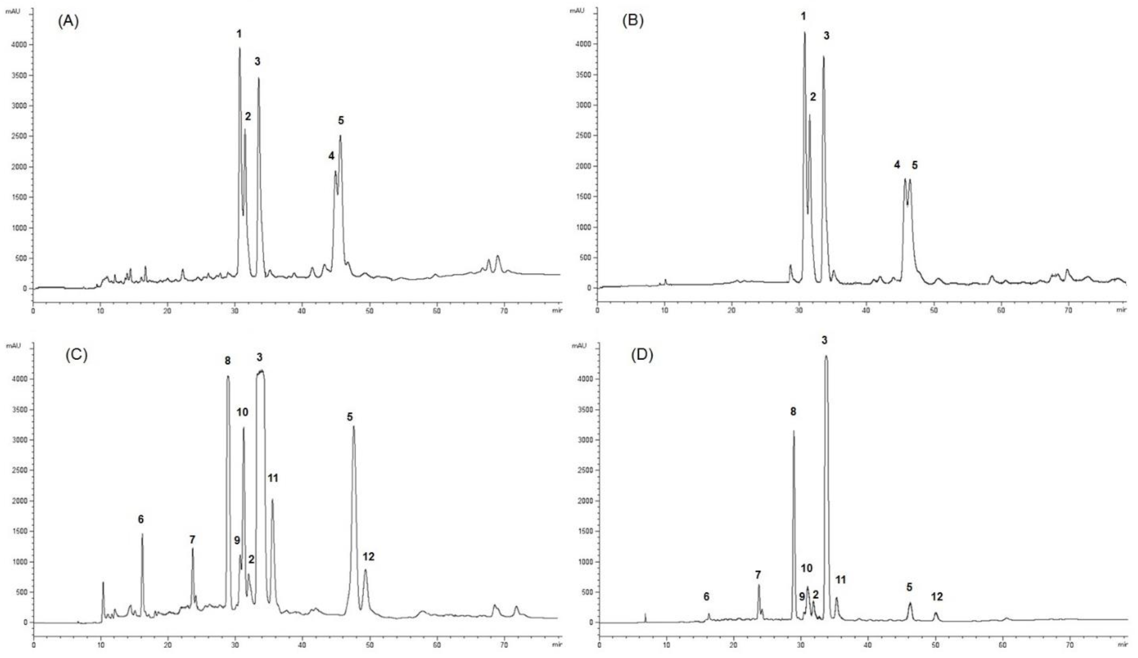
2.1. Extraction, Characterization, and Purification of Twelve Compounds from GARCINIA Mangostana Fruit

The CHCl3 and MeOH extracts of G. mangostana arils (1 Ext CHCL3 and 1 Ext MeOH) and shells (2 Ext CHCL3 and 2 Ext MeOH) were further purified by HPLC-UV to afford compounds 112 (Figure 1). The chloroform and methanol extracts of mangosteen arils submitted to purification by HPLC-UV showed a similar profile characterized by the occurrence of five main compounds, identified as 1,7-dihydroxy-3-methoxy-2-(3-methylbut-2-enyl)xanthen-9-one (1), 1,3,7-trihydroxy-2,8-bis(3-methyl-2-buten-1-yl)-9H-xanthen-9-one (2), α-mangostin (3), demethylcalabaxanthone (4), and mangostanin (5), also known as 6-hydroxycalabaxanthone or 9-hydroxycalabaxanthone [2,24,25]. The chloroform extract of mangosteen shells submitted to purification by HPLC-UV allowed the isolation of mangostanol (6), garcinone D (7), γ-mangostin (8), gudraxanthone (9), 8-deoxygartanin (10), garcinone E (11), β-mangostin (12), along with compounds 2, 3 and 5. In the methanol extract of mangosteen shells, compounds 3, 5, and 8 represented the main compounds, while all the other compounds occurring in the chloroform extract of shells were in smaller quantities (Figure 2). Compounds 112 were recognized by NMR analysis to be xanthone derivatives [26,27]. Their structures were established by 1D-(1H and 13C) and 2D-NMR (DQF-COSY, HSQC, and HMBC) experiments. The 13C NMR values of compounds 112 are reported in the Table 1.

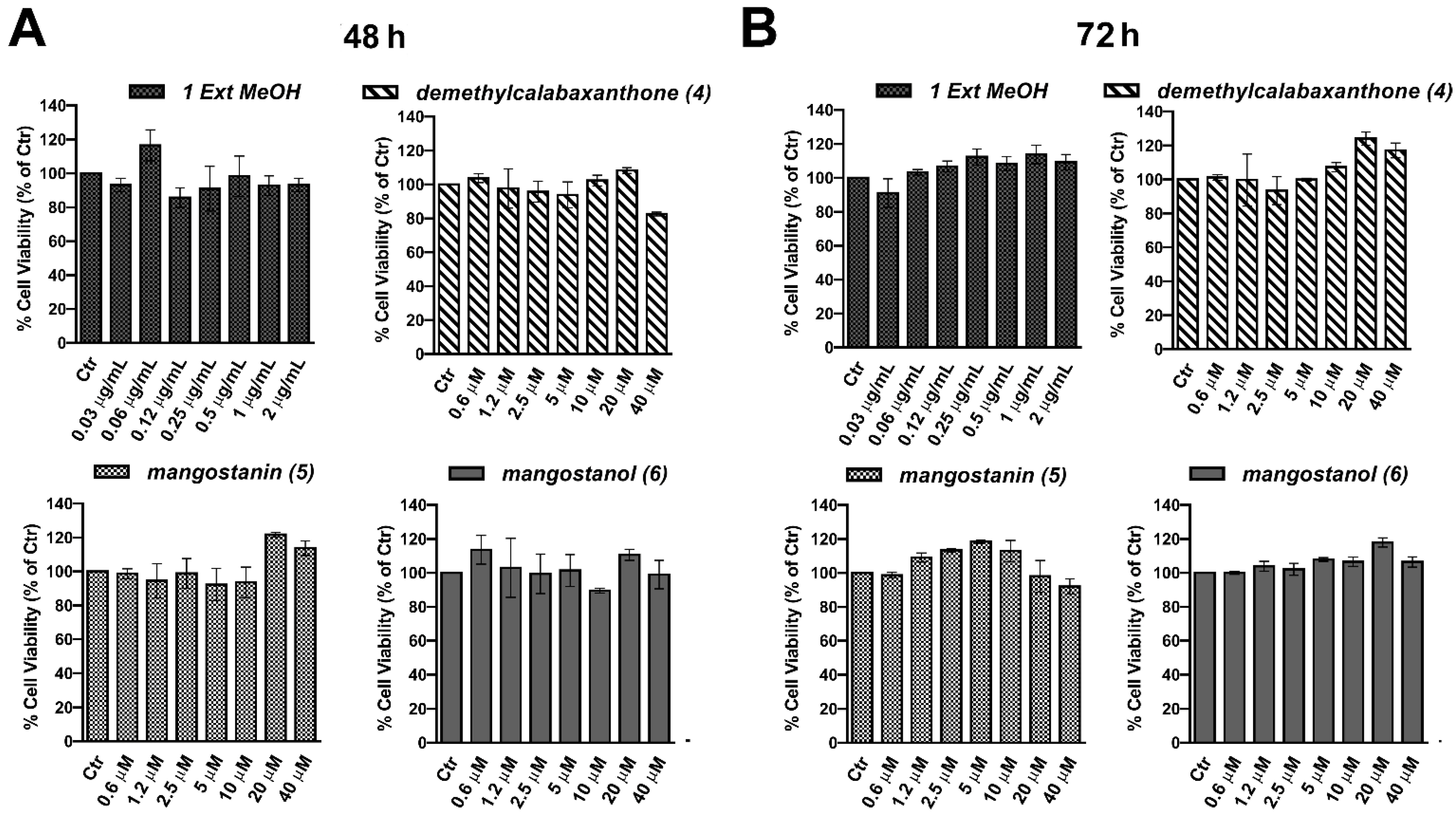
2.2. Evaluation of the Biological Effect of Garcinia mangostana Extracts and Isolated Compounds in Human Keratinocytes

First, we evaluated the effect of the four extracts and twelve compounds on human keratinocytes (HaCaT), a frequently employed skin model cell system. At first, we assessed the potential toxicity of both mangosteen extracts (0–2 μg/mL) and isolated compounds (0–40 μM). To this end, cells were grown with the substances for 48 h before evaluating cell viability through MTT assay (Supplementary Figure S1). In Figure 3A, we reported only the bar graphs relative to selected mangosteen methanol extract and the compounds demethylcalabaxanthone (4), mangostanin (5), and mangostanol (6). None of these showed an inhibitory effect on cell viability. Interestingly, we confirmed that even at 72 h, these compounds did not show any interference with cell viability, as shown in Figure 3B.

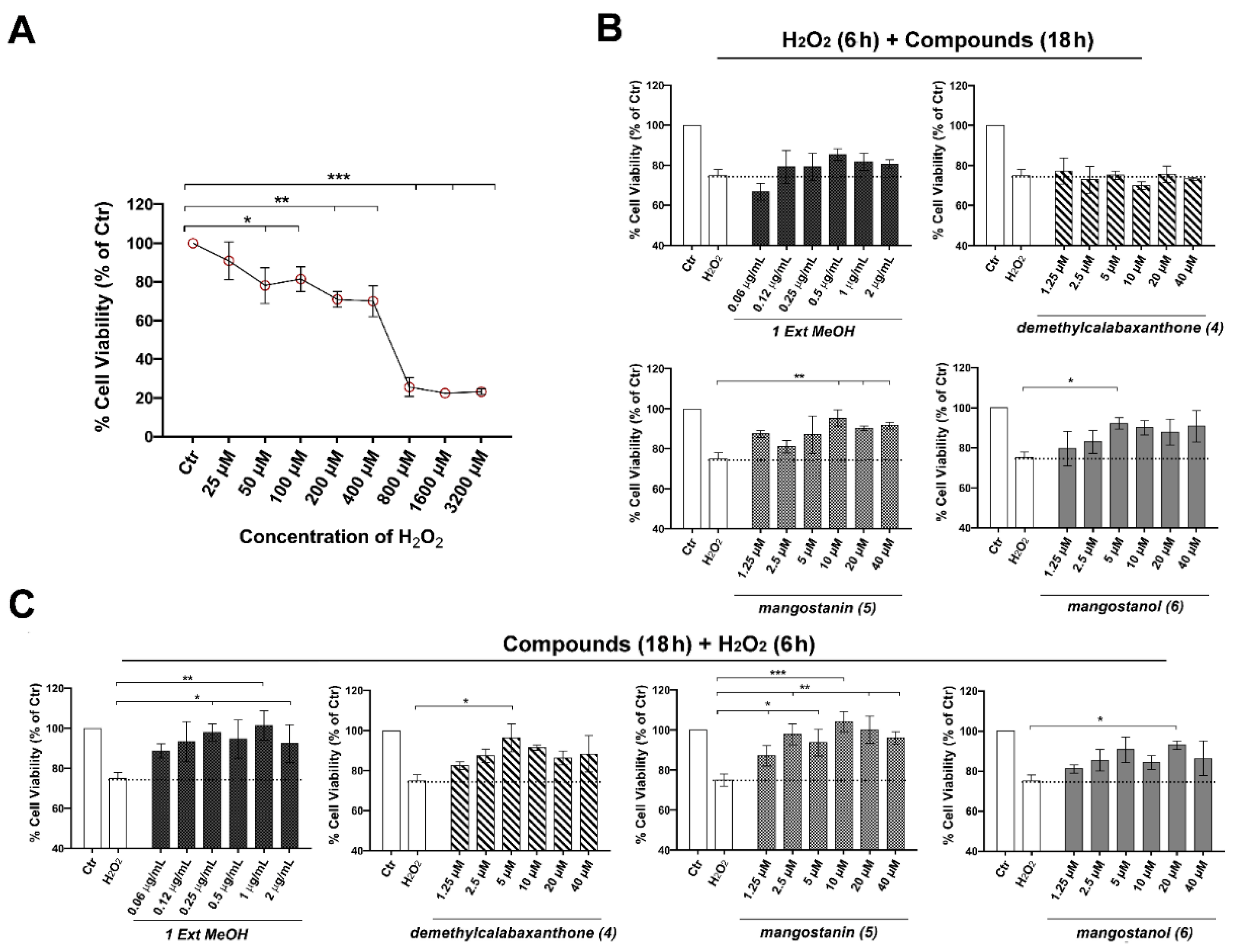
2.3. Mangostanin Reverts and Prevents Cytotoxicity Induced by H2O2 in HaCaT Cells

The next step was to evaluate both the potential reparative and preventive effect of the best mangosteen extract and selected compounds on the cell viability of HaCaT cells exposed to H2O2 (0–3200 μM) for 6 h as an inducer of cell damage. H2O2 induced cytotoxicity dose-dependently, starting at 50 μM, where the inhibition of cell viability was statistically significant, and reaching a reduction of 29.1% at 200 μM. Previous studies reported that lower concentrations of H2O2 (≤250 μM) induced apoptotic cell death of more cells, whereas higher H2O2 concentrations (1000 μM) resulted in an increase in overall cell death but a reduction in apoptosis [28,29]. Therefore, we decided to use 200 μM of H2O2 in the subsequent experiments. We observed that the pretreatment of keratinocytes with mangostanin in a concentration-dependent manner from 10 μM, and with mangostanol (5 μM), rescued the cell viability after H2O2-induced cytotoxicity. In detail, as shown in Figure 4B, survival rate, which dropped after exposure to H2O2 (200 μM for 6 h), increased to 95.45 ± 4.02%, 90.45 ± 1.14% and 91.81 ± 1.70% with mangostanin at 10, 20, and 40 μM, respectively. Furthermore, we evaluated the preventive effect of the same extract and compounds after 18-h pretreatment of HaCaT cells before H2O2-induced cell damage. We observed that the pretreatment with mangostanin from 2.5 to 40 μM was protective against H2O2-induced cytotoxicity. As shown in Figure 4C, cell viability declined after exposure to 200 μM of H2O2 for 6 h and increased to 97.90 ± 5.0%, 93.81 ± 6.66%, 104.18 ± 5.01%, 100.18 ± 6.84%, and 96.09 ± 3.0% upon mangostanin treatment at 1.25, 5, 10, 20, and 40 μM, respectively. Mangostanol (10 μM) and demethylcalabaxanthone (5 μM) showed a preventive effect that was statistically significant only at one of the concentrations tested. On the other hand, the methanol extract of G. mangostana arils was similarly effective in the protection from oxidative cell damage. For this reason, we selected the methanol extract and mangostanin for the subsequent experiments at the most effective concentrations of 1 μg/mL and 10 μM, respectively.

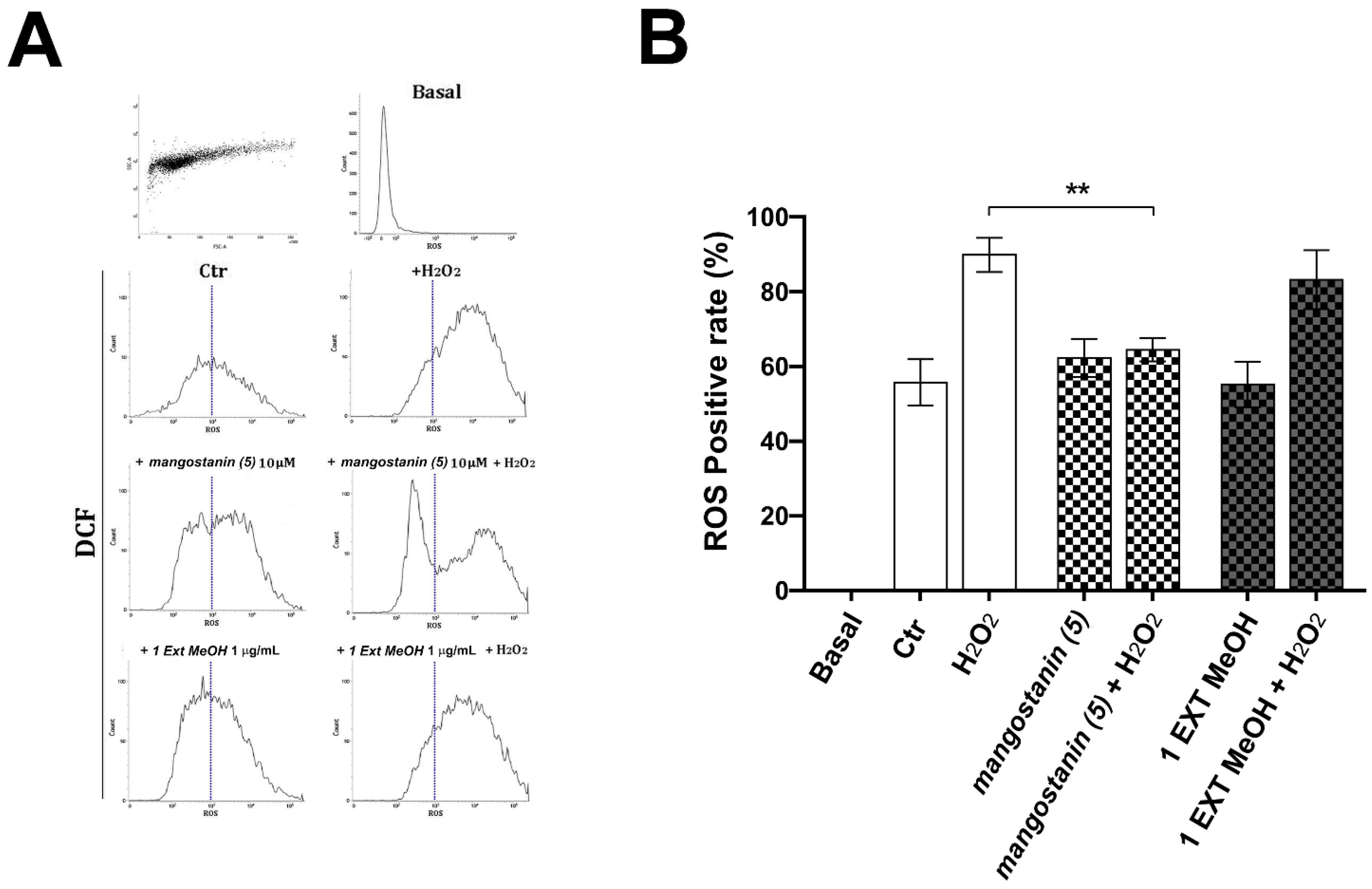
2.4. Mangostanin Prevents the Generation of Intracellular Reactive Oxygen Species (ROS)

Keratinocytes were pretreated with mangosteen methanol extract or mangostanin for 18 h before adding 200 μM of H2O2 for 6 h to verify the protective effect on intracellular ROS generation. Production of ROS was evaluated by 2′,7′-dichlorofluorescein (DCF) measurement and monitored using flow cytometry. As demonstrated by an incremented fluorescence (Figure 5A), the intracellular ROS levels were induced by H2O2 treatment for 6 h. The percentage of cells with increased ROS production was 89.88 ± 4.6% in the H2O2-treated group, which was decreased only by mangostanin 10 μM pretreatment down to 64.4 ± 3.11% as shown in the graph (Figure 5A,B). Compared to the treatment with the methanol extract of G. mangostana arils (1 μg/mL), results showed that only mangostanin pretreatment significantly decreased the production of intracellular ROS following H2O2 exposure.

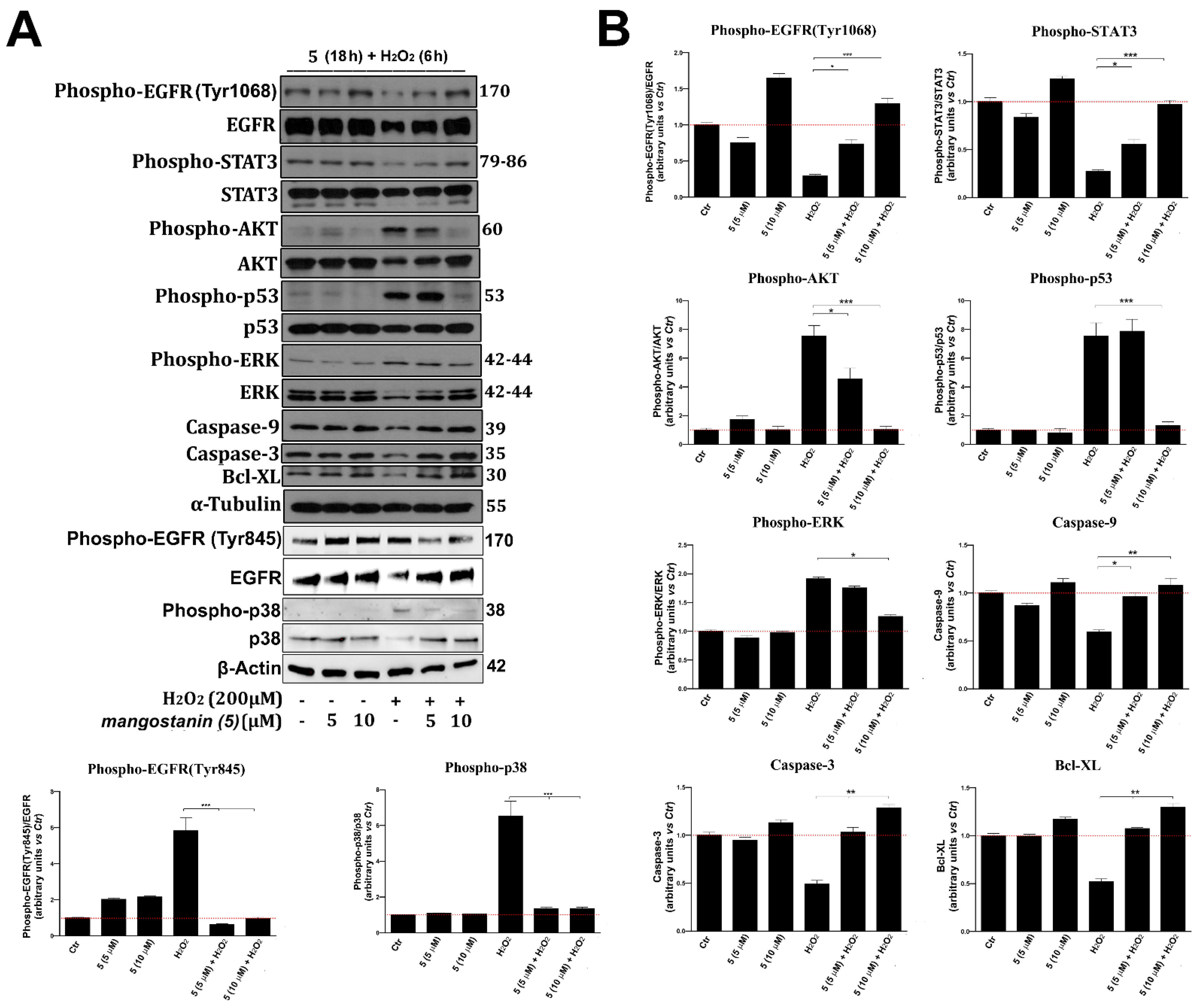
2.5. Mangostanin Prevents the Activation of Cell Death Molecular Pathways
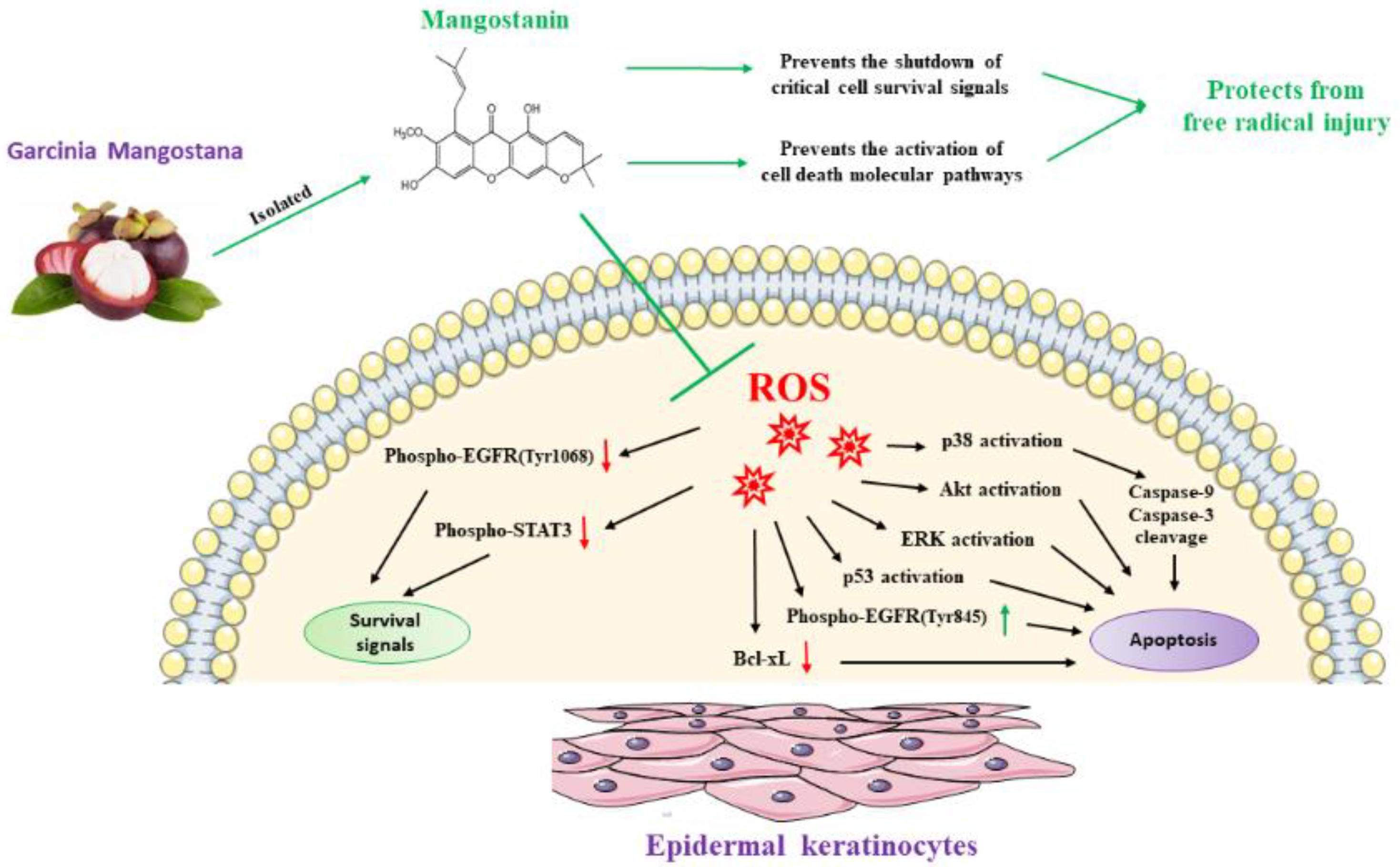
Mangostanin pretreatment significantly decreased the generation of intracellular ROS, and for this reason, we investigated the expression of proteins, such as epidermal growth factor receptor EGFR, protein kinase B (AKT), extracellular regulated protein kinase (ERK), p38 mitogen-activated protein kinases (p38 MAPK), p53, and signaling pathways which are implicated in the control of cell response to external damage induced by free radicals and in apoptosis activation [30,31,32,33]. Therefore, we determined by immunoblot the phosphorylation status of these proteins in cells pre-treated for 18 h with mangostanin (using both the more effective 10 μM concentration and the immediately lower one, 5 μM) and subsequently exposed to H2O2, as above described. We observed that the pre-treatment with mangostanin (10 μM) significantly prevented the phosphorylation and hence the activation of AKT, ERK, p53, p38 MAPK and EGFR (Tyr845), which are recognized markers of oxidative stress response. At the same time, the pre-treatment with mangostanin prevented the degradation of EGFR and the shutdown of its phosphorylation on Tyr1068, and hence of downstream signal transducer and activator of transcription-3 (STAT3) signaling, an index of cell survival and proliferation [34] (Figure 6A,B). Furthermore, mangostanin (10 μM) pre-treatment prevented the reduction of the total form of proteins, such as the caspases 3 and 9 and mitochondrial protein Bcl-XL involved in the induction of apoptosis [35], so preserving vitality.

3. Discussion

The biological relevance of the properties of the mangosteen fruit is widely known in natural medicine. It is reported as a natural regulator of biological functions, thanks to the antioxidants, anti-inflammatory and anticancer properties observed [8,9,10,11,12,13,14,15,17,18,19]. The unique nutritional composition of mangosteen could have a major impact in the design of novel food supplements [36,37]. At the moment, the greatest interest in this bioactive product is focused on its use as whole extract in many cosmeceutical products already available on the market. Despite its commercial success, its pharmacological efficacy in the field of cosmetic dermatology is yet not supported by in-depth scientific research. Furthermore, due to the high number of components, it is not yet clear which part of the phyto-complex or single molecule is the main responsible for the observed effects. Therefore, in our study, we evaluated the effect of different organic extracts of mangosteen and purified compounds on human keratinocytes, a well characterized cell line widely employed as an in vitro model of skin damage, for evaluating the dermatological properties of mangosteen.
Purified compounds were isolated from chloroform and methanol extracts of mangosteen arils and shells. Chloroform and methanol extracts of mangosteen arils showed a very similar HPLC profile, which led to the isolation of compounds 15. The remaining compounds (612) were isolated in the chloroform and methanol extracts of mangosteen shells which afforded also compounds 2, 3, and 5.
The lack of cytotoxicity from both the extracts and isolated compounds has been confirmed. Considering that numerous tests show how oxidative stress plays a key role in the pathogenesis of various diseases and skin aging, and that the regulation of ROS levels is fundamental for normal skin homeostasis [36,38], our experiments were aimed at demonstrating whether mangosteen was able to restore or prevent the oxidative damage induced by H2O2. We describe, for the first time, the biological effects of mangostanin in in vitro cultures of epidermal keratinocytes, comparing its activity with total extract and other xanthones. Indeed, we demonstrated that mangostanin was able to both rescue cells from H2O2-induced cytotoxicity and prevent cytotoxic damage caused by exposure to H2O2. Mangostanin, a xanthone purified from mangosteen extract, is therefore ideal for use in cosmeceutical formulations as it is more effective than whole mangosteen extract. The result obtained by the quantitative analysis of mangosteen arils highlighted a mangostanin (5) content of 106.4 mg per gram of chloroform extract.
As said before, mangosteen fruit extract has been shown to have elevated antioxidant effects as well as an anti-inflammatory potential, which can be beneficial to the vitality of various organs. For example, different compounds have been demonstrated to protect the hepatic tissue from the oxidative damage that may result from the drug detoxification process. Xanthones have been shown to prevent NfkB and MAPK activation in mice cell lines, attenuating cellular damage. In a similar manner, α-mangostin prevented the increase of pro-inflammatory cytokines, such as IL-6, interleukin-1β, and TNF-α after treatment with APAP and inhibited the increase of nitric oxide synthase (iNOS) expression, which further protects the liver tissue. Two other main compounds of mangosteen, isogarcinol and γ-mangostin, have been shown to exhibit a hepatoprotective effect by increasing anti-oxidative enzymes, such as SOD and glutathione, as well as inducing the expression of NRF2 (nuclear factor erythroid 2-related factor 2), a protein which regulates other anti-oxidative enzymes, such as heme oxygenase-1 and SOD2. Additionally, γ-mangostin can also upregulate SIRT1 (silent mating type information regulation 2 homologs 1), which in turn reduces ROS production from mitochondrial activity [39].
In our study, we showed that mangostanin has antioxidant potential and we supported these data at the molecular level by demonstrating that pretreatment with mangostanin not only decreased the generation of intracellular reactive oxygen species but prevented the activation of AKT, ERK, p53, cellular pathways underlying cell damage, and activation of apoptosis [29,30,31,32,33,34,35], as shown in Figure 7. Among the different markers shown here, Akt kinase, in particular, plays a central role in the cellular response against oxidative stress, specifically for H2O2-induced ROS. Increased Akt expression has been shown to be an antioxidant defense mechanism, and exogenous H2O2 has been reported to activate the PI3K/Akt pathway, which in turn controls apoptosis through the modulation of Bad/BcL-XL proteins [40]. Here, in fact, we showed how mangostanin can protect against oxidative stress by neutralizing the cell damage inflicted by ROS generated by H2O2 pre-treatment. The absence of ROS-induced damage allows cell survival and deactivation of the apoptosis pathway, a phenomenon that we also showed with the inhibition of ERK, p53 and the reduction of caspase-9 and caspase-3 cleavage.
We also investigated whether mangostanin could prevent p38 MAPK activation, induced by oxidative stress. As described in the literature, p38 acts as a nexus for signal transduction and has a central role in numerous biological processes [41]. It plays a vital role as signal transduction mediator for inflammation, cell cycle arrest, induction of cell death and senescence in several cell types. Therefore, it is recognized that the molecules that prevent the activation of p38 can have an important antioxidant role, also useful for minimizing the side effects of therapies in specific disease contexts [41,42]. In our experimental model, mangostanin at both tested concentrations, 5 μM and 10 μM, was able to significantly reduce (at 5 μM) and inhibit (at 10 μM) the activation of p38, thus preserving viability and proving to be a powerful antioxidant molecule.
The ability of mangostanin to inhibit p53 activation following oxidative stress was also evaluated in the present work. Indeed, p53 is a redox-active transcription factor that organizes and directs cellular responses in the face of a variety of stresses that lead to genomic instability, which acts by initiating DNA fragmentation to induce apoptosis, thus preventing aberrant cell proliferation [43].
ROS acts both as an upstream signal that triggers p53 activation and as a downstream factor that mediates apoptosis. In addition, p53 is also involved in an increasing number of diseases associated with aging [43,44]. We have shown that mangostanin, especially at a concentration of 10 μM, completely prevents the activation of p53 following oxidative stress. Therefore, a molecule such as mangostanin, by neutralizing the generation of ROS and preventing the activation of p53, can undoubtedly prevent aging and cell death.
The inhibition of p53 activation, thanks to mangostanin treatment, was also supported by a preservation of the total form of both Caspases 3 and 9, whose cleavage and activation is known to be a marker of apoptosis induced by oxidative stress or by various stressful conditions [29].
Finally, we evaluated EGFR expression and its phosphorylation on specific tyrosines. Indeed, it is known that not all EGFR phosphorylations mediate the same cellular response but, based on the phosphorylation positioning on the amino acid sequence, they can be both an indication of proliferation or viability or of response to oxidative stress. Recent studies show that EGFR phosphorylation on tyrosine 845 occurs following oxidative stress [45], while phosphorylation on tyrosine 1068 indicates cell proliferation and viability.
Therefore, in our work, we evaluated the expression of both phosphorylations (Tyr845 and Tyr1068) after treatment with H2O2 and in pretreatment with mangostanin. Treatment with mangostanin (5 and 10 μM) was able to inhibit EGFR phosphorylation on tyrosine 845, supporting previously obtained data, which once again demonstrates that mangostanin has antioxidant activity. At the same time, we have shown that mangostanin is able to prevent not only the degradation of EGFR, a protein necessary to preserve vitality and perform physiological functions, but also to preserve the phosphorylation of the receptor on tyrosine 1068, known to mediate signaling of cell proliferation and viability. Therefore, mangostanin proves to have a double activity following oxidative stress, capable of preventing the activation of cell stress and cell death pathways and at the same time preserving cellular vitality and proliferation.
Thanks to what has been carried out, our study provides evidence that mangostanin can protect the skin from the action of free radicals, thus preventing skin aging. These data, obtained in accordance with recent studies, show how mangosteen fruit extracts can be used as photoprotective agents against UVB damage [22,38], to improve skin elasticity and to control inflammatory skin diseases [23,46], configuring the mangosteen extract and, in our specific case, the mangostanin as a promising bio-active compound in the cosmetic sector. As evident in the HPLC-UV profiles, mangostanin represents not only one of the main compounds present in mangosteen aril extracts, but its presence in the shells highlights how the mangosteen by-products can also be a source of this biologically active compound.
Our study is in line with recent scientific works that aimed to evaluate not only the antioxidant activity of the total mangosteen extract, obtained with various extraction methods, but also to isolate its bioactive compounds.
Indeed, in recent years several studies have tried to implement the extraction methods to obtain mangosteen extracts with an increasingly greater antioxidant activity. The drying and extraction processes have been improved [47], fresh fruit has been used to try to avoid drying [8], different solvents and microwave-assisted extraction were used [48,49].
To date, the antioxidant activities of mangosteen extracts have been mainly associated to the prenylated xanthones α-, β-, and γ-mangosteens, which are also the main compounds represented in the fruit [50,51,52].
It has been shown that both mangosteen extract and α-mangosteen can decrease the activity of myeloperoxidase and at the same time increase the activity of catalase and superoxide dismutase inhibiting the production of reactive oxide species and malonaldehyde [53], configuring them as useful in all diseases related to aging and oxidative stress [54].
In addition to antioxidant and scavenging activities, mangosteen extract also has anti-elastase activity. A-mangosteen showed potent anti-collagenase activity, while γ-mangosteen has potent anti-hyaluronidase and anti-tyrosinase activities providing evidence of the possible use of mangosteen extract and its compounds as antioxidants and anti-aging treatments [55].
Mangosteen extract, also based on the extraction conditions, shows not only similar but also additional activities compared to the isolated molecules most studied for the proven antioxidant activities. For this reason, newly identified xanthones, even if less represented in the fruit, such as mangostanin, could show a higher antioxidant activity than other well-known molecules, explaining at least in part the superior effect of the total extract.
This study employing HaCaT immortalized keratinocytes that have known limits to fully resemble skin needs to be validated in the future in primary keratinocytes, in order to account for donor-to-donor variability and in more sophisticated three-dimensional skin models [56]. To evaluate the topical availability of mangostanin in cosmeceutical formulations, it will be necessary to understand its penetration through the skin barrier and the resistance of the stratum corneum. Therefore, it is undoubtedly necessary to investigate the stability, skin permeation, topical bioavailability, and efficacy of the molecule. Certainly, the potential of mangostanin as a topical ingredient will be confirmed if new studies are conducted as skin permeation studies using both in vitro skin models and bioactive compound delivery system techniques (going up to the use of nanotechnology) as much as the evaluation of stability of formulations. Finally, further in vivo and clinical studies will be required to develop and validate novel nutraceuticals, pharmacological formulations, and cosmeceuticals.

4. Materials and Methods

4.1. Chemicals and Materials

NMR spectroscopic data were acquired in MeOH-d4 (99.95%, Sigma-Aldrich, St. Louis, MO, USA) on a Bruker DRX-600 spectrometer (Bruker BioSpin GmBH, Rheinstetten, Germany) equipped with a Bruker 5 mm TCI CryoProbe at 300 K. Data processing was carried out with Topspin 3.2 software.
We solubilized the extracts and pure compounds in DMSO (dimethyl sulfoxide) (≤0.01% when added to cell cultures). H2O2 and 2′,7′-Dichlorofluorescein were purchased from Sigma-Aldrich, Inc. (St. Louis, MO, USA). For western blot analysis, we used: mouse monoclonal antihuman α-tubulin, rabbit monoclonal antihuman phospho-EGF receptor (p-EGFR; Tyr1068), rabbit monoclonal antihuman phospho-EGF receptor (p-EGFR; Tyr845), rabbit polyclonal antihuman phospho-STAT3 (p-STAT3; Tyr705), rabbit monoclonal anti-human Phospho-Akt (p-Akt; Ser473), rabbit polyclonal antihuman phospho-p38 MAPK, rabbit polyclonal antibody to phosphorylated p53, rabbit monoclonal anti-human phospho-p44/42 MAPK (p-Erk1/2; Thr202/Tyr204), rabbit monoclonal anti-human EGF receptor, rabbit monoclonal anti-human STAT3, rabbit monoclonal anti-human Akt, rabbit monoclonal anti-human p44/42 MAPK (Erk1/2), rabbit monoclonal anti-human p38 MAPK, rabbit polyclonal anti-human p53, rabbit polyclonal anti-human caspase-3, rabbit monoclonal anti-human caspase-9, rabbit monoclonal anti-human Bcl-xL, and secondary HRP-linked goat anti-mouse or goat anti-rabbit IgG were purchased from Cell Signaling Technology (Danvers, MA, USA). Rabbit polyclonal anti-human β-actin was purchased from Abcam (Cambridge, UK).

4.2. Plant Material

The fruits of Garcinia mangostana were purchased from an online market in March 2017 and classified by Prof. V. De Feo (Department of Pharmacy, University of Salerno, Italy).

4.3. Extraction and Isolation Procedures of G. mangostana Arils and Shells

Garcinia mangostana arils (200g) were stored in the freezer at a temperature of −5 °C. After some days, the fruits were submitted to lyophilization to obtain 42.32 g, and the dried arils deprived of seeds were extracted at room temperature using solvents of raising polarity, such as petroleum ether (300 mL for 3 days, three times), CHCl3 (300 mL for 3 days, three times), and MeOH (300 mL × 3 days × 3 times). We obtained 0.37 g and 16.20 g of crude extracts of CHCl3 and MeOH, respectively, by filtration and evaporation of the solvent to dryness in vacuo.
Garcinia mangostana shells (370 g) were dried and extracted at room temperature with petroleum ether (1500 mL for 3 days, three times), CHCl3 (1500 mL × 3 days × 3 times), and MeOH at room temperature (1500 mL × 3 days × 3 times). The filtrates were concentrated under reduced pressure until elimination of CHCl3 and MeOH to obtain 13.62 g and 55.89 g of crude extracts, respectively.
The CHCl3 and MeOH extracts of G. mangostana arils (1 Ext CHCL3 and 1 Ext MeOH) and shells (2 Ext CHCL3 and 2 Ext MeOH) were analyzed by an RP-HPLC-UV system on an Agilent 1260 Infinity system (Agilent Technologies, Palo Alto, CA, USA), equipped with a binary pump (G-1312C), and a UV detector (G-1314B) with a Phenomenex C18 Synergi-Hydro-RP (250 mm × 10 mm, 10 μm) column; the elution gradient was executed using water with 0.1% formic acid as eluent A and acetonitrile with 0.1% formic acid as B at a flow rate of 2.0 mL/min. In particular, the HPLC gradient started at 5% B, and after 5 min % B was at 50%, after 15 min at 80%, and after 10 min at 87%. % B remained at 87% for 20 min, and finally after 10 min, it reached 100% and so remained for 20 min. The selected wavelength was 254 nm.
The chloroform extract (twenty injection of 2.5 mg each) of mangosteen arils yielded 1,7-dihydroxy-3-methoxy-2-(3-methylbut-2-enyl)-xanthen-9-one (1) (2.3 mg, tR = 30.9 min), 1,3,7-trihydroxy-2,8-bis-(3-methyl-2-buten-1-yl)-9H-xanthen-9-one (2) (1.5 mg, tR = 33.3 min), α-mangostin (3) (2.9 mg, tR = 34.8 min), demethylcalabaxanthone (4) (1.1 mg, tR = 45.0 min), and mangostanin (5) (3.2 mg, tR = 46.1 min). The methanol extract submitted at HPLC purification in the same conditions afforded compounds 15.
The chloroform extract (twenty injection of 2.5 mg each) of mangosteen shells yielded mangostanol (6) (1.8 mg, tR = 16.2 min), garcinone D (7) (2.3 mg, tR = 24.0 min), γ-mangostin (8) (2.0 mg, tR = 29.5 min), gudraxanthone (9) (1.2 mg, tR = 31.0 min), 8-deoxygartanin (10) (2.2 mg, tR = 32.0 min), 1,3,7-trihydroxy-2,8-bis-(3-methyl-2-buten-1-yl)-9H-xanthen-9-one (2) (1.3 mg, tR = 33.3 min), α-mangostin (3) (5.2 mg, tR = 34.8 min), garcinone E (11) (2.1 mg, tR = 36.0 min), mangostanin (5) (2.1 mg, tR = 48.4 min), and β-mangostin (12) (0.2 mg, tR = 49.7 min). The methanol extract in the same conditions afforded compounds 3, 68 and 1012.
The purity of these compounds (>99%) was determined by HPLC analysis.

4.4. Quantitative Analysis of Mangostanin (5)

The quantitative determination of mangostanin (5) in the chloroform extract of mangosteen arils was performed by HPLC-UV. HPLC conditions were the same as used for qualitative analysis with slight modifications. Mangostanin (5) was quantified using the calibration curve of the corresponding standard. Standard stock solutions were prepared by stepwise dilution with 5 mg of mangostanin (5) in 5 mL of methanol, to reach the concentration range of 10 to 100 μg/mL. The calibration curve was obtained by plotting the UV peak area of external standard against the known concentration of the compound; each concentration of standard solution was analysed in triplicate. The amount of mangostanin (5) was expressed as milligrams per gram of extract as the mean of triplicate determinations.

4.5. Cells

We grew the human immortalized keratinocytes (HaCaT) in DMEM (Dulbecco’s modified Eagle’s medium) purchased from GIBCO (Grand Island, NY, USA) and supplemented as described in detail elsewhere, using 2 mM L-glutamine, 50 ng/mL, streptomycin, 50 units/mL of penicillin, and 10% heat-inactivated fetal bovine serum [28]. Cell cultures were maintained at 37 °C in a humidified 5% CO2 atmosphere.

4.6. Determination of Cell Viability—MTT Assay

HaCaT cells (6 × 10³/well) were grown for 24 h into 96 multi-well plates before the addition of each molecule at the indicated concentration and then further cultured for an additional 24, 48, or 72 h at 37 °C. Then, we employed the MTT (3-(4, 5-dimethylthiazolyl-2)-2, 5-diphenyltetrazolium bromide) tetrazolium salts assay to determine cell viability, as described in detail elsewhere [57]. Briefly, 10 μl of MTT solution was added (5 mg/mL, in water) to each well. After an incubation time of 4 h, the formazan crystals were dissolved in 100 μl of solubilization solution (10% Triton X-100 and 0.1 N HCL in isopropanol). A microplate reader (ThermoScientific, Basingstoke, UK) was used to detect the absorbance intensity at 595 nm. We have performed all experiments in triplicate and expressed the relative cell viability as % with respect to the untreated control cells.

4.7. Determination of Reactive Oxygen Species (ROS)—Flow Cytometric Assay

Production of reactive oxygen species (ROS) was evaluated by 2′7′-dichlorodihydrofluorescein diacetate dye staining, converted by cellular esterase to H2DCF and subsequently oxidized to highly fluorescent 2′,7′-dichlorofluorescein (DCF). At the end of the treatment with H2O2, methanol extract of Garcinia mangostana arils (1 μg/mL) and mangostanin (10 μM), cells were washed with ice-cold PBS 2% FBS and loaded with 20 μM of H2DCFDA for 30 min. The cells were washed again with PBS 2% FBS, acquired by flow cytometry where at least 10,000 events were collected and next analyzed by Cell-Quest Pro software (BD Biosciences, San Jose, CA, USA) [58].

4.8. Western Blot (WB) Assay

Cells were grown in p60 tissue culture plates at a density of 2 × 10⁴ cells/cm2 for 24 h. Cells were then treated with mangostanin, H2O2, or their combination, as indicated. Then, the cells were washed with PBS, collected, and lysed in cold RIPA (radioimmunoprecipitation assay) lysis buffer (50 mM Tris-HCl, 10 mg/mL aprotinin, 2 mM phenylmethylsulfonyl fluoride, 0.5% Triton X-100, 0.5% deoxycholic acid, 150 mM NaCl, and 10 mg/mL leupeptin) and then assayed for (WB) by the procedure described in detail elsewhere [59].

4.9. Statistical Analysis

We performed the statistical analysis by GraphPad Prism 6.0 software for Windows (GraphPad software, San Diego, CA, USA). Data obtained from multiple experiments expressed as mean ± (SD) of four independent experiments conducted in triplicate were analyzed for statistical significance through 2-tailed Student t-test for independent groups or 1-way ANOVA followed by Tukey post-hoc correction for multiple comparisons. p values < 0.05 were considered significant. * p < 0.05, ** p < 0.01, and *** p < 0.001).

5. Conclusions

We provided for the first time the scientific evidence that mangostanin, a xanthone identified in the fruit of Garcinia mangostana, remarkably protects epidermal keratinocytes from the action of free radicals, demonstrating a route for possible pharmaceutical applications. Taken together, our findings showed the protective effects of mangostanin on epidermal keratinocytes. Pre-treatment with the substance significantly reduced H2O2-induced cytotoxicity and ROS production, prevented activation of p53, p38 MAPK, ERK and AKT, cleavage of Caspase-9 and Caspase-3, and shutdown of critical signals of cell survival, such as EGFR and STAT-3, thereby increasing cell viability. Furthermore, the ability to prevent skin aging brings the molecule to the attention of the cosmeceutical sector. Undoubtedly, further in vivo and clinical studies are needed to develop and validate novel cosmeceutical, pharmacological or nutraceutical formulations, based on this interesting molecule.

Supplementary Materials

The following are available online at https://www.mdpi.com/article/10.3390/ph15010084/s1, Figure S1: Evaluation of 1 Ext MeOH, 1 Ext CHCl3, 2 Ext MeOH, 2 Ext MeOH, 1, 2, 3, 4, 5, 6, 7, 8, 9, 11, and 12 effects in HaCaT Cells.

Author Contributions

Conceptualization, M.A., S.P. (Simona Pisanti), S.P. (Sonia Piacente) and M.B.; methodology, M.A., M.M., S.P. (Simona Pisanti) and S.P. (Sonia Piacente); validation, M.A., M.M., S.P. (Simona Pisanti), S.P. (Sonia Piacente) and M.B.; formal analysis, M.A., M.M., M.C., S.P. (Simona Pisanti), S.P. (Sonia Piacente) and M.B.; investigation, M.A., C.P., M.M., M.C., S.P. (Simona Pisanti), S.P. (Sonia Piacente) and M.B.; writing—original draft preparation, M.A., C.P. and S.P. (Simona Pisanti); writing—review and editing, M.A., C.P., M.M., M.C., S.P. (Simona Pisanti), S.P. (Sonia Piacente) and M.B.; supervision, S.P. (Simona Pisanti), S.P. (Sonia Piacente) and M.B. All authors have read and agreed to the published version of the manuscript.

Funding

This research was funded by Associazione Italiana Ricerca sul Cancro (AIRC; IG 13312 and IG 18999 to M.B.); AIRC and Fondazione Cariplo TRIDEO 2015 (No. 17216 to S. Pisanti); Regione Campania POR Campania FESR 2014-2020 “Combattere la resistenza tumorale: piattaforma integrata multidisciplinare per un approccio tecnologico innovativo alle oncoterapie-Campania Oncoterapie (Project B61G18000470007 to S. Piacente).

Institutional Review Board Statement

Not applicable.

Informed Consent Statement

Not applicable.

Data Availability Statement

Data is contained within the article and supplementary material.

Conflicts of Interest

The authors declare no conflict of interest.

References

  1. Masullo, M.; Menegazzi, M.; Di Micco, S.; Beffy, P.; Bifulco, G.; Dal Bosco, M.; Novelli, M.; Pizza, C.; Masiello, P.; Piacente, S. Direct Interaction of Garcinol and Related Polyisoprenylated Benzophenones of Garcinia cambogia Fruits with the Transcription Factor STAT-1 as a Likely Mechanism of Their Inhibitory Effect on Cytokine Signaling Pathways. J. Nat. Prod. 2014, 77, 543–549. [Google Scholar] [CrossRef]
  2. Obolskiy, D.; Pischel, I.; Siriwatanametanon, N.; Heinrich, M. Garcinia mangostana L.: A phytochemical and pharmacological review. Phytother. Res. 2009, 23, 1047–1065. [Google Scholar] [CrossRef]
  3. Ovalle-Magallanes, B.; Eugenio-Pérez, D.; Pedraza-Chaverri, J. Medicinal properties of mangosteen (Garcinia mangostana L.): A comprehensive update. Food Chem. Toxicol. 2017, 109 Pt 1, 102–122. [Google Scholar] [CrossRef]
  4. Zhang, S.; Li, Z.; Wang, X.; An, L.; Bao, J.; Zhang, J.; Cui, J.; Li, Y.; Jin, D.Q.; Tuerhong, M.; et al. Isolation, structural elucidation, and immunoregulation properties of an arabinofuranan from the rinds of Garcinia mangostana. Carbohydr. Polym. 2020, 246, 116567. [Google Scholar] [CrossRef]
  5. Zakaryan, H.; Arabyan, E.; Oo, A.; Zandi, K. Flavonoids: Promising natural compounds against viral infections. Arch. Virol. 2017, 162, 2539–2551. [Google Scholar] [CrossRef] [PubMed]
  6. Tran, T.H.; Nguyen, V.T.; Le, H.T.; Nguyen, H.M.; Tran, T.H.; Do Thi, T.; Nguyen, X.C.; Ha, M.T. Garcinoxanthones SV, new xanthone derivatives from the pericarps of Garcinia mangostana together with their cytotoxic and antioxidant activities. Fitoterapia 2021, 151, 104880. [Google Scholar] [CrossRef]
  7. Panche, A.N.; Diwan, A.D.; Chandra, S.R. Flavonoids: An overview. J. Nutr. Sci. 2016, 5, e47. [Google Scholar] [CrossRef] [PubMed] [Green Version]
  8. Muzykiewicz, A.; Zielonka-Brzezicka, J.; Siemak, J.; Klimowicz, A. Antioxidant activity and polyphenol content in extracts from various parts of fresh and frozen mangosteen. Acta Sci. Pol. Technol. Aliment. 2020, 19, 261–270. [Google Scholar] [CrossRef]
  9. Meepagala, K.M.; Schrader, K.K. Antibacterial Activity of Constituents from Mangosteen Garcinia mangostana Fruit Pericarp against Several Channel Catfish Pathogens. J. Aquat. Anim. Health 2018, 30, 179–184. [Google Scholar] [CrossRef] [PubMed]
  10. Mohan, S.; Syam, S.; Abdelwahab, S.I.; Thangavel, N. An anti-inflammatory molecular mechanism of action of α-mangostin, the major xanthone from the pericarp of Garcinia mangostana: An in silico, in vitro and in vivo approach. Food Funct. 2018, 9, 3860–3871. [Google Scholar] [CrossRef] [PubMed]
  11. Tatiya-Aphiradee, N.; Chatuphonprasert, W.; Jarukamjorn, K. Anti-inflammatory effect of Garcinia mangostana Linn. pericarp extract in methicillin-resistant Staphylococcus aureus-induced superficial skin infection in mice. Biomed. Pharmacother. 2019, 111, 705–713. [Google Scholar] [CrossRef]
  12. Cho, J.; Chin, Y.W. Neuroprotective and Memory-Enhancing Effects of The Water Extract from Pericarp of Mangosteen Fruit. Clin. Ther. 2016, 38, e31. [Google Scholar] [CrossRef] [PubMed]
  13. Taher, M.; Zakaria, T.M.; Susanti, D.; Zakaria, Z.A. Hypoglycaemic activity of ethanolic extract of Garcinia mangostana Linn. in normoglycaemic and streptozotocin-induced diabetic rats. BMC Complement. Altern. Med. 2016, 16, 135. [Google Scholar] [CrossRef] [PubMed] [Green Version]
  14. Labban, R.S.M.; Alfawaz, H.A.; Almnaizel, A.T.; Al-Muammar, M.N.; Bhat, R.S.; El-Ansary, A. Garcinia mangostana extract and curcumin ameliorate oxidative stress, dyslipidemia, and hyperglycemia in high fat diet-induced obese Wistar albino rats. Sci. Rep. 2021, 11, 7278. [Google Scholar] [CrossRef]
  15. Liu, Q.Y.; Wang, Y.T.; Lin, L.G. New insights into the anti-obesity activity of xanthones from Garcinia mangostana. Food Funct. 2015, 6, 383–393. [Google Scholar] [CrossRef] [PubMed]
  16. Klein-Júnior, L.C.; Campos, A.; Niero, R.; Corrêa, R.; Vander Heyden, Y.; Filho, V.C. Xanthones and Cancer: From Natural Sources to Mechanisms of Action. Chem. Biodivers. 2020, 17, e1900499. [Google Scholar] [CrossRef]
  17. Kritsanawong, S.; Innajak, S.; Imoto, M.; Watanapokasin, R. Antiproliferative and apoptosis induction of α-mangostin in T47D breast cancer cells. Int. J. Oncol. 2016, 48, 2155–2165. [Google Scholar] [CrossRef] [PubMed] [Green Version]
  18. Ibrahim, S.R.M.; Abdallah, H.M.; El-Halawany, A.M.; Nafady, A.M.; Mohamed, G.A. Mangostanaxanthone VIII, a new xanthone from Garcinia mangostana and its cytotoxic activity. Nat. Prod. Res. 2019, 33, 258–265. [Google Scholar] [CrossRef]
  19. Li, K.; Wu, L.; Chen, Y.; Li, Y.; Wang, Q.; Li, M.; Hao, K.; Zhang, W.; Jiang, S.; Wang, Z. Cytotoxic and Antiproliferative Effects of β-Mangostin on Rat C6 Glioma Cells Depend on Oxidative Stress Induction via PI3K/AKT/mTOR Pathway Inhibition. Drug Des. Dev. Ther. 2020, 14, 5315–5324. [Google Scholar] [CrossRef]
  20. Benatrehina, P.A.; Pan, L.; Naman, C.B.; Li, J.; Kinghorn, A.D. Usage, biological activity, and safety of selected botanical dietary supplements consumed in the United States. J. Tradit. Complement. Med. 2018, 8, 267–277. [Google Scholar] [CrossRef] [PubMed]
  21. Choi, Y.H.; Han, S.Y.; Kim, Y.J.; Kim, Y.M.; Chin, Y.W. Absorption, tissue distribution, tissue metabolism and safety of α-mangostin in mangosteen extract using mouse models. Food Chem. Toxicol. 2014, 66, 140–146. [Google Scholar] [CrossRef] [PubMed]
  22. Im, A.R.; Kim, Y.M.; Chin, Y.W.; Chae, S. Protective effects of compounds from Garcinia mangostana L. (mangosteen) against UVB damage in HaCaT cells and hairless mice. Int. J. Mol. Med. 2017, 40, 1941–1949. [Google Scholar] [CrossRef] [Green Version]
  23. Ohno, R.; Moroishi, N.; Sugawa, H.; Maejima, K.; Saigusa, M.; Yamanaka, M.; Nagai, M.; Yoshimura, M.; Amakura, Y.; Nagai, R. Mangosteen pericarp extract inhibits the formation of pentosidine and ameliorates skin elasticity. J. Clin. Biochem. Nutr. 2015, 57, 27–32. [Google Scholar] [CrossRef] [PubMed] [Green Version]
  24. Wahyuni, S.F.; Shaari, K.; Stanslas, J.; Lajis, N.H.; Dachriyanus. Cytotoxic xanthones from the stem bark of Garcinia cowa Roxb. J. Chem. Pharm. Res. 2015, 7, 227–236. [Google Scholar]
  25. Sriyatep, S.; Siridechakorn, I.; Maneerat, W.; Pansanit, A.; Ritthiwigrom, T.; Andersen, R.J.; Laphookhieo, S. Bioactive Prenylated Xanthones from the Young Fruits and Flowers of Garcinia cowa. J. Nat. Prod. 2015, 78, 265–271. [Google Scholar] [CrossRef]
  26. Masullo, M.; Bassarello, C.; Bifulco, G.; Piacente, S. Polyisoprenylated benzophenone derivatives from the fruits of Garcinia cambogia and their absolute configuration by quantum chemical circular dichroism calculations. Tetrahedron 2010, 66, 139–145. [Google Scholar] [CrossRef]
  27. Masullo, M.; Bassarello, C.; Suzuki, H.; Pizza, C.; Piacente, S. Polyisoprenylated Benzophenones and an Unusual Polyisoprenylated Tetracyclic Xanthone from the Fruits of Garcinia cambogia. J. Agric. Food Chem. 2008, 56, 5205–5210. [Google Scholar] [CrossRef]
  28. Abate, M.; Pepe, G.; Randino, R.; Pisanti, S.; Basilicata, M.G.; Covelli, V.; Bifulco, M.; Cabri, W.; D’Ursi, A.M.; Campiglia, P.; et al. Ganoderma lucidum Ethanol Extracts Enhance Re-Epithelialization and Prevent Keratinocytes from Free-Radical Injury. Pharmaceuticals 2020, 13, 224. [Google Scholar] [CrossRef]
  29. Turner, N.A.; Xia, F.; Azhar, G.; Zhang, X.; Liu, L.; Wei, J.Y. Oxidative stress induces DNA fragmentation and caspase activation via the c-Jun NH2-terminal kinase pathway in H9c2 cardiac muscle cells. J. Mol. Cell Cardiol. 1998, 30, 1789–1801. [Google Scholar] [CrossRef]
  30. Li, L.; Wang, H.H.; Nie, X.T.; Jiang, W.R.; Zhang, Y.S. Sodium butyrate ameliorates lipopolysaccharide-induced cow mammary epithelial cells from oxidative stress damage and apoptosis. J. Cell Biochem. 2018, 120, 2370–2381. [Google Scholar] [CrossRef]
  31. Zhang, L.; Li, J.; Hu, J.; Li, D.; Wang, X.; Zhang, R.; Zhang, H.; Shi, M.; Chen, H. Cigarette smoke extract induces EGFR-TKI resistance via promoting EGFR signaling pathway and ROS generation in NSCLC cell lines. Lung Cancer 2017, 109, 109–116. [Google Scholar] [CrossRef]
  32. Wang, J.; Qi, C.; Liu, L.; Zhao, L.; Cui, W.; Tian, Y.; Liu, B.; Li, J. Taurine Protects Primary Neonatal Cardiomyocytes Against Apoptosis Induced by Hydrogen Peroxide. Int. Heart J. 2018, 59, 190–196. [Google Scholar] [CrossRef] [Green Version]
  33. Jin, G.F.; Hurst, J.S.; Godley, B.F. Hydrogen peroxide stimulates apoptosis in cultured human retinal pigment epithelial cells. Curr. Eye Res. 2001, 22, 165–173. [Google Scholar] [CrossRef]
  34. Zhang, Y.; Wang, D.; Xu, J.; Wang, Y.; Ma, F.; Li, Z.; Liu, N. Stat3 activation is critical for pluripotency maintenance. J. Cell Physiol. 2019, 234, 1044–1051. [Google Scholar] [CrossRef]
  35. Yue, J.; López, J.M. Understanding MAPK Signaling Pathways in Apoptosis. Int. J. Mol. Sci. 2020, 21, 2346. [Google Scholar] [CrossRef] [Green Version]
  36. Peerapattana, J.; Otsuka, K.; Otsuka, M. Application of NIR spectroscopy for the quality control of mangosteen pericarp powder: Quantitative analysis of alpha-mangostin in mangosteen pericarp powder and capsule. J. Nat. Med. 2013, 67, 452–459. [Google Scholar] [CrossRef] [PubMed]
  37. Stern, J.S.; Peerson, J.; Mishra, A.T.; Sadasiva Rao, M.V.; Rajeswari, K.P. Efficacy and tolerability of a novel herbal formulation for weight management. Obesity 2013, 21, 921–927. [Google Scholar] [CrossRef]
  38. Dunaway, S.; Odin, R.; Zhou, L.; Ji, L.; Zhang, Y.; Kadekaro, A.L. Natural Antioxidants: Multiple Mechanisms to Protect Skin from Solar Radiation. Front. Pharmacol. 2018, 9, 392. [Google Scholar] [CrossRef] [Green Version]
  39. Aizat, W.M.; Jamil, I.N.; Ahmad-Hashim, F.H.; Noor, N.M. Recent updates on metabolite composition and medicinal benefits of mangosteen plant. PeerJ 2019, 31, e6324. [Google Scholar] [CrossRef] [PubMed] [Green Version]
  40. Sadidi, M.; Lentz, S.I.; Feldman, E.L. Hydrogen peroxide-induced Akt phosphorylation regulates Bax activation. Biochimie 2009, 91, 577–585. [Google Scholar] [CrossRef] [PubMed] [Green Version]
  41. Zarubin, T.; Han, J. Activation and signaling of the p38 MAP kinase pathway. Cell. Res. 2005, 15, 11–18. [Google Scholar] [CrossRef] [PubMed] [Green Version]
  42. Coulthard, L.R.; White, D.E.; Jones, D.L.; McDermott, M.F.; Burchill, S.A. P38(MAPK): Stress responses from molecular mechanisms to therapeutics. Trends Mol. Med. 2009, 15, 369–379. [Google Scholar] [CrossRef] [Green Version]
  43. Liu, B.; Chen, Y.; Clair, D.K.S. ROS and p53: A versatile partnership. Free Radic. Biol. Med. 2008, 44, 1529–1535. [Google Scholar] [CrossRef] [PubMed] [Green Version]
  44. Vigneron, A.; Vousden, K.H. p53, ROS and senescence in the control of aging. Aging 2010, 2, 471–474. [Google Scholar] [CrossRef] [PubMed] [Green Version]
  45. Zhuang, S.; Schnellmann, R.G. H2O2-induced transactivation of EGF receptor requires Src and mediates ERK1/2, but not Akt, activation in renal cells. Am. J. Physiol. Renal Physiol. 2004, 286, F858–F865. [Google Scholar]
  46. Tatiya-Aphiradee, N.; Chatuphonprasert, W.; Jarukamjorn, K. In vivo antibacterial activity of Garcinia mangostana pericarp extract against methicillin-resistant Staphylococcus aureus in a mouse superficial skin infection model. Pharm. Biol. 2016, 54, 2606–2615. [Google Scholar] [CrossRef] [Green Version]
  47. García, M.L.; Carrión, M.H.; Escobar, S.; Rodríguez, A.; Cortina, J.R. Optimization of the antioxidant capacity of mangosteen peels (Garcinia mangostana L.) extracts: Management of the drying extraction processes. Food Sci. Technol. Int. 2021, 27, 404–412. [Google Scholar] [CrossRef]
  48. Ngawhirunpat, T.; Opanasopi, P.; Sukma, M.; Sittisombut, C.; Kat, A.; Adachi, I. Antioxidant, free radical-scavenging activity and cytotoxicity of different solvent extracts and their phenolic constituents from the fruit hull of mangosteen (Garcinia mangostana). Pharm. Biol. 2010, 48, 55–62. [Google Scholar] [CrossRef]
  49. Mohammad, N.A.; Abang Zaidel, D.N.; Muhamad, I.I.; Abdul Hamid, M.; Yaakob, H.; Mohd Jusoh, Y.M. Optimization of the antioxidant-rich xanthone extract from mangosteen (Garcinia mangostana L.) pericarp via microwave-assisted extraction. Heliyon 2019, 5, e02571. [Google Scholar] [CrossRef] [Green Version]
  50. Pedraza-Chaverri, J.; Cárdenas-Rodríguez, N.; Orozco-Ibarra, M.; Pérez-Rojas, J.M. Medicinal properties of mangosteen (Garcinia mangostana). Food Chem. Toxicol. 2008, 46, 3227–3239. [Google Scholar] [CrossRef]
  51. Cui, J.; Hu, W.; Cai, Z.; Liu, Y.; Li, S.; Tao, W.; Xiang, H. New medicinal properties of mangostins: Analgesic activity and pharmacological characterization of active ingredients from the fruit hull of Garcinia mangostana L. Pharmacol. Biochem. Behav. 2010, 95, 166–172. [Google Scholar] [CrossRef]
  52. Sukatta, U.; Takenaka, M.; Ono, H.; Okadome, H.; Sotome, I.; Nanayama, K.; Thanapase, W.; Isobe, S. Distribution of major xanthones in the pericarp, aril, and yellow gum of mangosteen (Garcinia mangostana linn.) fruit and their contribution to antioxidative activity. Biosci. Biotechnol. Biochem. 2013, 77, 984–987. [Google Scholar] [CrossRef] [PubMed] [Green Version]
  53. Tatiya-Aphiradee, N.; Chatuphonprasert, W.; Jarukamjorn, K. Ethanolic Garcinia mangostana extract and α-mangostin improve dextran sulfate sodium-induced ulcerative colitis via the suppression of inflammatory and oxidative responses in ICR mice. J. Ethnopharmacol. 2021, 265, 113384. [Google Scholar] [CrossRef] [PubMed]
  54. Do, H.T.T.; Cho, J. Mangosteen Pericarp and Its Bioactive Xanthones: Potential Therapeutic Value in Alzheimer’s Disease, Parkinson’s Disease, and Depression with Pharmacokinetic and Safety Profiles. Int. J. Mol. Sci. 2020, 21, 6211. [Google Scholar] [CrossRef] [PubMed]
  55. Widowati, W.; Ginting, C.N.; Lister, I.N.E.; Girsang, E.; Amalia, A.; Wibowo, S.H.B.; Kusuma, H.S.W.; Rizal. Anti-aging Effects of Mangosteen Peel Extract and Its Phytochemical Compounds: Antioxidant Activity, Enzyme Inhibition and Molecular Docking Simulation. Trop. Life Sci. Res. 2020, 31, 127. [Google Scholar] [CrossRef] [PubMed]
  56. Moran, M.C.; Pandya, R.P.; Leffler, K.A.; Yoshida, T.; Beck, L.A.; Brewer, M.G. Characterization of Human Keratinocyte Cell Lines for Barrier Studies. JID Innov. 2021, 1, 100018. [Google Scholar] [CrossRef] [PubMed]
  57. Abate, M.; Pisanti, S.; Caputo, M.; Citro, M.; Vecchione, C.; Martinelli, R. 3-Hydroxytyrosol Promotes Angiogenesis In Vitro by Stimulating Endothelial Cell Migration. Int. J. Mol. Sci. 2020, 21, 3657. [Google Scholar] [CrossRef] [PubMed]
  58. Abate, M.; Citro, M.; Pisanti, S.; Caputo, M.; Martinelli, R. Keratinocytes Migration Promotion, Proliferation Induction, and Free Radical Injury Prevention by 3-Hydroxytirosol. Int. J. Mol. Sci. 2021, 22, 2438. [Google Scholar] [CrossRef]
  59. Ciaglia, E.; Grimaldi, M.; Abate, M.; Scrima, M.; Rodriquez, M.; Laezza, C.; Ranieri, R.; Pisanti, S.; Ciuffreda, P.; Manera, C.; et al. The isoprenoid derivative N6 -benzyladenosine CM223 exerts antitumor effects in glioma patient-derived primary cells through the mevalonate pathway. Br. J. Pharmacol. 2017, 174, 2287–2301. [Google Scholar] [CrossRef] [Green Version]
Figure 1. Chemical structures of xanthone derivatives (112) isolated from Garcinia mangostana.
Pharmaceuticals 15 00084 g001
Figure 2. HPLC-UV chromatograms of G. mangostana extracts. (A) chloroform extract of arils, (B) methanol extract of arils, (C) chloroform extract of shells, and (D) methanol extract of shells.
Pharmaceuticals 15 00084 g002
Figure 3. Evaluation of the methanol extract of G. mangostana arils and compounds 4–6 effects in HaCaT cells. Human keratinocytes were grown for 48 h (A) or 72 h (B) with individual compounds (0–40 μM) or extract (0–2 μg/mL) before MTT assay. Independent experiments were performed in triplicate, and the results are expressed as means ± SD and reported as % vs. the untreated cell (Ctr) (ANOVA).
Pharmaceuticals 15 00084 g003
Figure 4. Mangostanin rescued and prevented H2O2-induced cytotoxicity in HaCaT cells. (A) Cells exposed to H2O2 (0–3200 μM) for 6 h before MTT assay. Results of three independent experiments performed in triplicate are expressed as means ± SD and reported as % vs. the untreated control (ANOVA, * p <0.05, ** p < 0.01, *** p < 0.001 vs. control). (B) Cells were grown in the presence of H2O2 for 6 h, before treatment with methanol extract of G. mangostana arils (0–2 μg/mL) or compounds demethylcalabaxanthone (4), mangostanin (5), mangostanol (6) at the indicated concentrations (0–40 μM) for 18 h. Results of independent experiments performed in triplicate are expressed as means ± SD and reported as % vs. the untreated control (ANOVA, * p < 0.05, ** p < 0.01 vs. control). (C) Keratinocytes were cultured in the presence of methanol extract of G. mangostana arils (0–2 μg/mL) or compounds 4–6 (0–40 μM) for 18 h, before treatment with H2O2 for 6 h. Results of independent experiments performed in triplicate are expressed as means ± SD and reported as % vs. the untreated control (ANOVA, * p <0.05, ** p < 0.01, *** p < 0.001 vs. control).
Pharmaceuticals 15 00084 g004
Figure 5. Mangostanin prevents the intracellular generation of ROS. HaCaT cells were treated with 200 μM of H2O2 with or without methanol extract of G. mangostana arils (1 μg/mL) or mangostanin (10 μM) for 6 h. Cells were incubated with the fluorescent dye for 30 min, and the mean fluorescence intensity was analyzed by flow cytometry. Representative cytofluorimetric histogram profiles are shown in (A), while the bar graph (B) reports the % of positive cells (ANOVA, ** p < 0.01 vs. control).
Pharmaceuticals 15 00084 g005
Figure 6. (A) WB analysis of EGFR, STAT3, AKT, p53, p38, ERK (total and phosphorylated), Caspase-9, Caspase-3, and Bcl-XL in whole cell lysates from keratinocytes cultured for 18 h with or without mangostanin (5 and 10 μM) and H2O2 (200 μM) for 6 h. As protein loading housekeeping, α-tubulin or β-actin were used. A representative WB of three different experiments performed with similar results is showed. (B) Histograms represent means ± SD in densitometry units of scanned immunoblots from the 3 different experiments (ANOVA, * p < 0.05, ** p < 0.01, *** p < 0.001, vs. control).
Pharmaceuticals 15 00084 g006
Figure 7. The xanthone mangostanin, isolated from Garcinia mangostana fruit, showed different protective effects against the cytotoxicity exerted from H2O2. Briefly, we showed that mangostanin prevents the shutdown of critical cell survival signals as the phosphorylation of EGFR on Tyr1068, STAT3, prevents the activation of p38, p53, AKT, ERK, the phosphorylation of EGFR on Tyr845, and the cleavage of caspase-9 and caspase-3, thus increasing cell vitality.
Pharmaceuticals 15 00084 g007
Table 1. 13C NMR Data (150 MHz MeOH-d4) for Compounds 112.
123456789101112
1159.9161.0161.8153.4150.4156.4161.0161.5157.0157.0161.8161.4
2112.3110.8111.4104.7105.3105.0111.3110.8112.3112.3110.6111.0
3165.8163.7163.8160.9160.3162.4163.8163.0165.5165.5163.8163.8
490.492.592.994.494.594.492.892.490.890.892.892.9
5119.4116.4102.5123.4102.7102.1102.3100.6147.8147.8114.3102.5
5a157.7156.0156.2157.7157.7158.6156.1155.9157.7157.7155.7156.1
6124.8123.4156.6116.5157.0155.9156.4154.2121.2121.2148.6156.4
7155.3151.9144.8152.5144.2144.4144.8141.8124.6124.6141.4144.8
8 108.9129.6138.6119.0138.6137.7139.0129.4115.3115.3128.6138.8
8a121.8119.6112.0129.0111.3115.1111.5111.8122.3122.3114.0112.0
9a104.5103.4103.8104.5104.5107.0104.0103.5104.3104.3102.8103.8
9183.8184.4183.6184.4183.6179.0180.9182.7181.9182.5183.0183.5
10a150.0151.8158.3152.7157.7156.8157.2154.0146.5146.5152.2157.9
1′21.722.122.1115.8116.027.321.921.621.921.922.022.1
2′123.5123.4123.8128.1128.369.3123.6123.6123.0123.0124.0123.9
3′132.1131.1131.878.878.679.4131.6131.3131.8131.8130.9131.7
4′17.517.217.628.127.919.917.717.417.517.517.417.8
5′25.826.125.928.127.925.325.825.826.026.025.825.9
1″ 26.526.926.526.326.923.026.0 26.726.5
2″ 123.8124.8124.4124.6125.445.7124.5 124.0124.4
3″ 131.0132.0131.4131.5131.871.6131.2 131.2132.0
4″ 18.518.118.318.518.629.117.9 18.118.1
5″ 26.125.926.225.926.329.125.5 25.825.9
1‴ 23.3
2‴ 124.0
3‴ 131.8
4‴ 18.3
5‴ 28.0
OCH356.1 56.0 56.0
OCH3 61.1 60.961.061.3 61.1
Publisher’s Note: MDPI stays neutral with regard to jurisdictional claims in published maps and institutional affiliations.

