
The Antibacterial and Remineralization Effect of Silver-Containing Mesoporous Bioactive Glass Sealing and Er-YAG Laser on Dentinal Tubules Treated in a Streptococcus mutans Cultivated Environment


 and
and
Abstract
:
1. Introduction
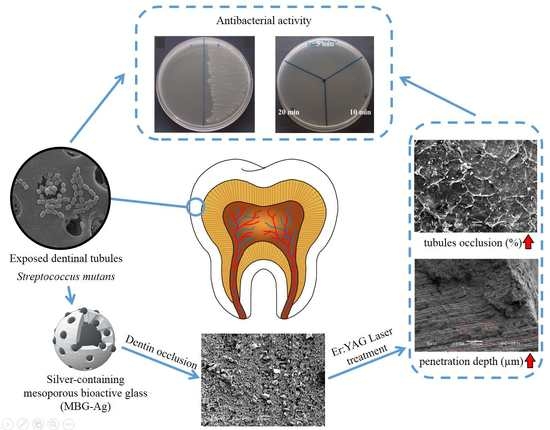
2. Results
2.1. This X-ray Diffractometer (XRD) Analysis
2.2. Fourier Transform Infrared (FTIR) Spectrometer Analysis
2.3. Scanning Electron Microscope (SEM) and Energy Dispersive X-ray (EDX) Spectrometer Analysis
2.4. Antibacterial Activity Analysis
3. Discussion
4. Materials and Methods
4.1. Preparation of Materials
4.2. Preparation of Dentin Specimens
4.3. Laser Treatment
4.4. Characteristics
4.5. Analysis of the Antibacterial Activity
4.6. Statistical Analysis
5. Conclusions
Author Contributions
Funding
Institutional Review Board Statement
Informed Consent Statement
Data Availability Statement
Acknowledgments
Conflicts of Interest
References
- West, N.X.; Lussi, A.; Seong, J.; Hellwig, E. Dentin hypersensitivity: Pain mechanisms and aetiology of exposed cervical dentin. Clin. Oral Investig. 2013, 17, 9–19. [Google Scholar] [CrossRef] [PubMed] [Green Version]
- Bartold, P.M. Dentinal hypersensitivity: A review. Aust. Dent. J. 2006, 51, 212–218. [Google Scholar] [CrossRef] [PubMed] [Green Version]
- Demi, M.; Delmé, K.I.; De Moor, R.J. Hypersensitive Teeth: Conventional vs Laser Treatment. Part II: Laser Treatment of Dentin Hypersensitivity. J. Oral Laser Appl. 2009, 9, 75–92. [Google Scholar]
- Cheng, L.; Zhang, K.; Zhang, N.; Melo, M.A.S.; Weir, M.D.; Zhou, X.D.; Xu, H.H.K. Developing a new generation of antimicrobial and bioactive dental resins. J. Dent. Res. 2017, 96, 855–863. [Google Scholar] [CrossRef] [PubMed]
- Naidu, G.M.; Ram, K.C.; Sirisha, N.R.; Sree, Y.S.; Kopuri, R.K.; Satti, N.R.; Thatimatla, C. Prevalence of dentin hypersensitivity and related factors among adult patients visiting a dental school in andhra pradesh, southern India. J. Clin. Diagn. Res. 2014, 8, ZC48–ZC51. [Google Scholar] [CrossRef]
- Lussi, A.; Schlüter, N.; Rakhmatullina, E.; Ganss, C. Dental erosion–an overview with emphasis on chemical and histopathological aspects. Caries Res. 2011, 45, 2–12. [Google Scholar] [CrossRef] [PubMed]
- Cersosimo, M.C.P.; Matos, A.B.; Couto, R.S.D.A.; Marques, M.M.; de Freitas, P.M. Short-pulse Er: YAG laser increases bond strength of composite resin to sound and eroded dentin. J. Biomed. Opt. 2016, 21, 048001. [Google Scholar] [CrossRef] [PubMed]
- Mei, M.L.; Chu, C.H.; Low, K.H.; Che, C.M.; Lo, E.C. Caries arresting effect of silver diamine fluoride on dentine carious lesion with S. mutans and L. acidophilus dual-species cariogenic biofilm. Med. Oral Patol. Oral Y Cir. Bucal 2013, 18, e824–e831. [Google Scholar] [CrossRef] [PubMed]
- He, X.S.; Shi, W.Y. Oral microbiology: Past, present and future. Int. J. Oral Sci. 2009, 1, 47–58. [Google Scholar] [CrossRef] [PubMed]
- Teughels, W.; Van Assche, N.; Sliepen, I.; Quirynen, M. Effect of material characteristics and/or surface topography on biofilm development. Clin. Oral Implant. Res. 2006, 17, 68–81. [Google Scholar] [CrossRef] [PubMed]
- Gotouda, H.; Shinozaki-Kuwahara, N.; Taguchi, C.; Shimosaka, M.; Ohta, M. Evaluation of the proportion of cariogenic bacteria associated with dental caries. Epidemiology 2017, 7, 1000327. [Google Scholar] [CrossRef] [Green Version]
- Gerschman, J.; Ruben, J.; Gebart-Eaglemont, J. Low level laser therapy for dentinal tooth hypersensitivity. Aust. Dent. J. 1994, 39, 353–357. [Google Scholar] [CrossRef]
- Wang, X.; Cheng, X.; Liu, X.; Wang, Z.; Wang, J.; Guo, C.; Zhang, Y.; He, W. Bactericidal Effect of Various Laser Irradiation Systems on Enterococcus faecalis Biofilms in Dentinal Tubules: A Confocal Laser Scanning Microscopy Study. Photomed. Laser Surg. 2018, 36, 472–479. [Google Scholar] [CrossRef] [PubMed]
- Esteves-Oliveira, M.; El-Sayed, K.F.; Dörfer, C.; Schwendicke, F. Impact of combined CO2 laser irradiation and fluoride on enamel and dentin biofilm-induced mineral loss. Clin. Oral Investig. 2017, 21, 1243–1250. [Google Scholar] [CrossRef] [PubMed]
- Hu, X.L.; Ho, B.; Lim, C.T.; Hsu, C.S. Thermal treatments modulate bacterial adhesion to dental enamel. J. Dent. Res. 2011, 90, 1451–1456. [Google Scholar] [CrossRef]
- Abdulsamee, N. All Tissues Dental Laser Er:YAG laser—Review Article. Biomed. J. Sci. Tech. Res. 2017, 1, 9–17. [Google Scholar] [CrossRef]
- Armengol, V.; Jean, A.; Marion, D. Temperature rise during Er:YAG and Nd:YAP laser ablation of dentin. J. Endod. 2000, 26, 138–141. [Google Scholar] [CrossRef] [PubMed]
- Jung, J.H.; Kim, D.H.; Yoo, K.H.; Yoon, S.Y.; Kim, Y.; Bae, M.K.; Chung, J.; Ko, C.C.; Kwon, Y.H.; Kim, Y.I. Dentin sealing and antibacterial effects of silver-doped bioactive glass/mesoporous silica nanocomposite: An in vitro study. Clin. Oral Investig. 2019, 23, 253–266. [Google Scholar] [CrossRef] [PubMed]
- Noronha, V.T.; Paula, A.J.; Duran, G.; Galembeck, A.; Cogo-Mueller, K.; Franz-Montan, M.; Duran, N. Silver nanoparticles in dentistry. Dent. Mater. 2017, 33, 1110–1126. [Google Scholar] [CrossRef] [PubMed]
- Fernandez, C.C.; Sokolonski, A.R.; Fonseca, M.S.; Stanisic, D.; Araújo, D.B.; Azevedo, V.; Portela, R.D.; Tasic, L. Applications of Silver Nanoparticles in Dentistry: Advances and Technological Innovation. Int. J. Mol. Sci. 2021, 22, 2485. [Google Scholar] [CrossRef] [PubMed]
- Mei, M.L.; Ito, L.; Cao, Y.; Li, Q.L.; Lo, E.C.; Chu, C.H. Inhibitory effect of silver diamine fluoride on dentine demineralisation and collagen degradation. J. Dent. 2013, 41, 809–817. [Google Scholar] [CrossRef]
- Osorio, R.; Osorio, E.; Aguilera, F.S.; Medina-Castillo, A.L.; Toledano, M.; Toledano-Osorio, M. Silver improves collagen structure and stability at demineralized dentin: A dynamic-mechanical and Raman analysis. J. Dent. 2018, 79, 61–67. [Google Scholar] [CrossRef]
- Kwan, K.H.; Liu, X.; To, M.K.; Yeung, K.W.; Ho, C.M.; Wong, K.K. Modulation of collagen alignment by silver nanoparticles results in better mechanical properties in wound healing. Nanomed. Nanotechnol. Biol. Med. 2011, 7, 497–504. [Google Scholar] [CrossRef] [PubMed]
- Lopes, C.D.C.A.; Limirio, P.H.J.O.; Novais, V.R.; Dechichi, P. Fourier transform infrared spectroscopy (FTIR) application chemical characterization of enamel, dentin and bone. Appl. Spectrosc. Rev. 2018, 53, 747–769. [Google Scholar] [CrossRef]
- Lee, E.M.; Borges, R.; Marchi, J.; de Paula Eduardo, C.; Marques, M.M. Bioactive glass and high-intensity lasers as a promising treatment for dentin hypersensitivity: An in vitro study. J. Biomed. Mater. Res. Part B Appl. Biomater. 2020, 108, 939–947. [Google Scholar] [CrossRef] [PubMed]
- Chiang, Y.C.; Chen, H.J.; Liu, H.C.; Kang, S.H.; Lee, B.S.; Lin, F.H.; Lin, H.P.; Lin, C.P. A novel mesoporous biomaterial for treating dentin hypersensitivity. J. Dent. Res. 2010, 89, 236–240. [Google Scholar] [CrossRef] [PubMed]
- Wong, K.W.; Chien, C.S.; Hsiao, Y.C.; Shih, C.J. Re-crystallization of bioactive glass mixed with various hardening agents. Ceram. Int. 2017, 43, 7026–7032. [Google Scholar] [CrossRef]
- Kung, J.C.; Chen, Y.J.; Chiang, Y.C.; Lee, C.L.; Yang-Wang, Y.T.; Hung, C.C.; Shih, C.J. Antibacterial activity of silver nanoparticle (AgNP) confined mesoporous structured bioactive powder against Enterococcus faecalis infecting root canal systems. J. Non-Cryst. Solids 2018, 502, 62–70. [Google Scholar] [CrossRef]
- Rai, M.; Yadav, A.; Gade, A. Silver nanoparticles as a new generation of antimicrobials. Biotechnol. Adv. 2009, 27, 76–83. [Google Scholar] [CrossRef] [PubMed]
- Ciang, Y.C.; Wang, Y.C.; Kung, J.C.; Shih, C.J. Antibacterial silver containing composite bioglass as dentin remineralization agent in microorganism challenge environment. J. Dent. 2021, 106, 103563. [Google Scholar] [CrossRef]
- Bakry, A.S.; Takahashi, H.; Otsuki, M.; Sadr, A.; Yamashita, K.; Tagami, J. CO2 laser improves 45S5 bioglass interaction with dentin. J. Dent. Res. 2011, 90, 246–250. [Google Scholar] [CrossRef] [PubMed]
- Mei, M.L.; Lo, E.C.M.; Chu, C.H. Arresting Dentine Caries with Silver Diamine Fluoride: What’s Behind It? J. Dent. Res. 2018, 97, 751–758. [Google Scholar] [CrossRef] [PubMed]
- Prado, M.; Gusman, H.; Gomes, B.P.; Simão, R.A. Scanning electron microscopic investigation of the effectiveness of phosphoric acid in smear layer removal when compared with EDTA and citric acid. J. Endod. 2011, 37, 255–258. [Google Scholar] [CrossRef] [PubMed] [Green Version]
- Lin, W.-T.; Chen, J.-C.; Hsiao, Y.-C.; Shih, C.-J. Re-crystallization of silica-based calcium phosphate glass prepared by sol–gel technique. Ceram. Int. 2017, 43, 13388–13393. [Google Scholar] [CrossRef]


| Dentin Specimens | Percentage of Dentinal Tubule Occlusion (%) | Maximum Penetration Depth (μm) |
|---|---|---|
| Group a | -- | -- |
| Group b | 63 ± 30 | 4 |
| Group c | 94 ± 6 | 68 |
| Group d | 94 ± 6 | >300 |
Publisher’s Note: MDPI stays neutral with regard to jurisdictional claims in published maps and institutional affiliations. |
© 2021 by the authors. Licensee MDPI, Basel, Switzerland. This article is an open access article distributed under the terms and conditions of the Creative Commons Attribution (CC BY) license (https://creativecommons.org/licenses/by/4.0/).
Share and Cite
Kung, J.-C.; Wang, W.-H.; Chiang, Y.-C.; Yang-Wang, Y.-T.; Wang, Y.-C.; Chen, W.-C.; Shih, C.-J. The Antibacterial and Remineralization Effect of Silver-Containing Mesoporous Bioactive Glass Sealing and Er-YAG Laser on Dentinal Tubules Treated in a Streptococcus mutans Cultivated Environment. Pharmaceuticals 2021, 14, 1124. https://doi.org/10.3390/ph14111124
Kung J-C, Wang W-H, Chiang Y-C, Yang-Wang Y-T, Wang Y-C, Chen W-C, Shih C-J. The Antibacterial and Remineralization Effect of Silver-Containing Mesoporous Bioactive Glass Sealing and Er-YAG Laser on Dentinal Tubules Treated in a Streptococcus mutans Cultivated Environment. Pharmaceuticals. 2021; 14(11):1124. https://doi.org/10.3390/ph14111124
Chicago/Turabian StyleKung, Jung-Chang, Wei-Hsun Wang, Yu-Ching Chiang, Yuan-Ting Yang-Wang, Yueh-Ching Wang, Wen-Cheng Chen, and Chi-Jen Shih. 2021. "The Antibacterial and Remineralization Effect of Silver-Containing Mesoporous Bioactive Glass Sealing and Er-YAG Laser on Dentinal Tubules Treated in a Streptococcus mutans Cultivated Environment" Pharmaceuticals 14, no. 11: 1124. https://doi.org/10.3390/ph14111124