
High Spectral Sensitivity of Strongly Coupled Hybrid Tamm-Plasmonic Resonances for Biosensing Application
 and
and 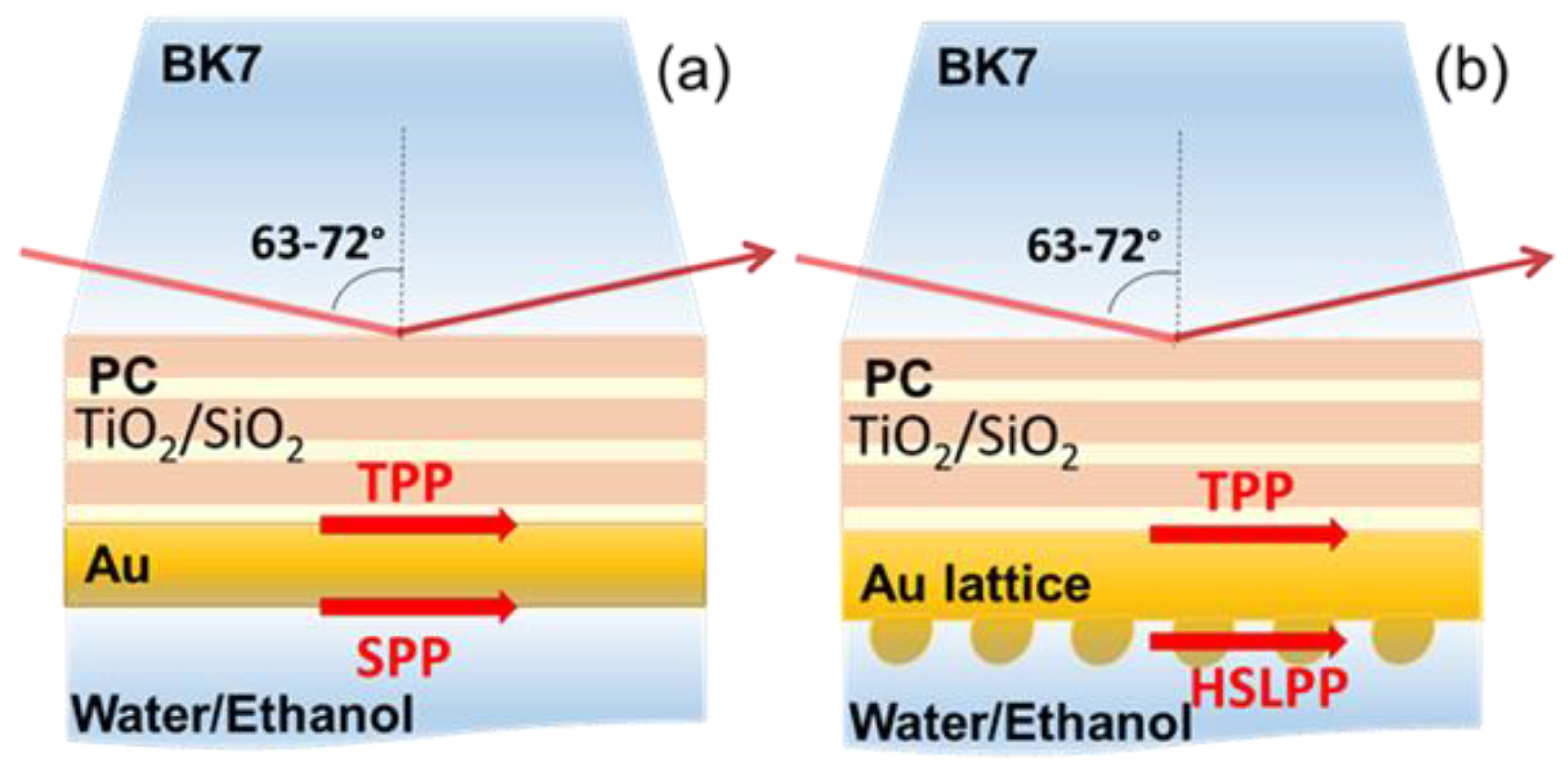
Abstract
:1. Introduction
2. Materials and Methods
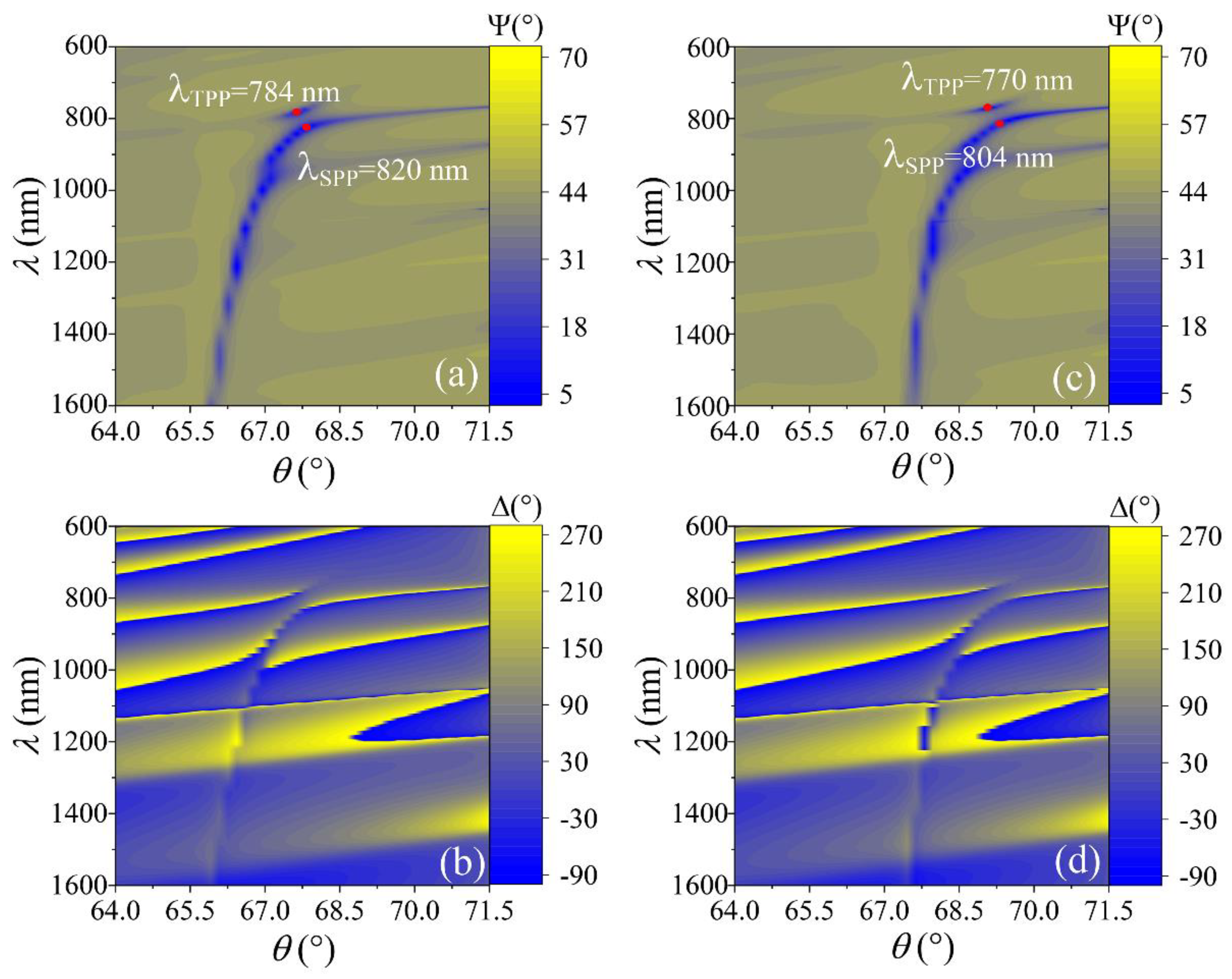
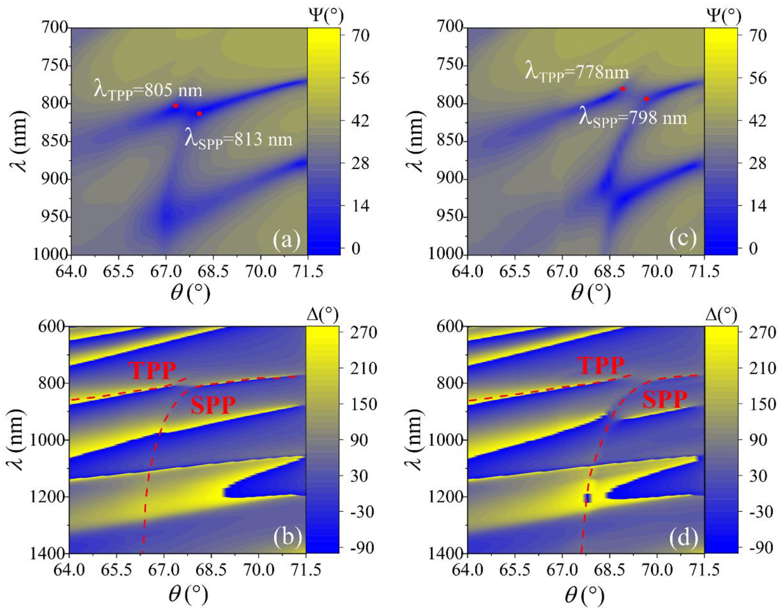
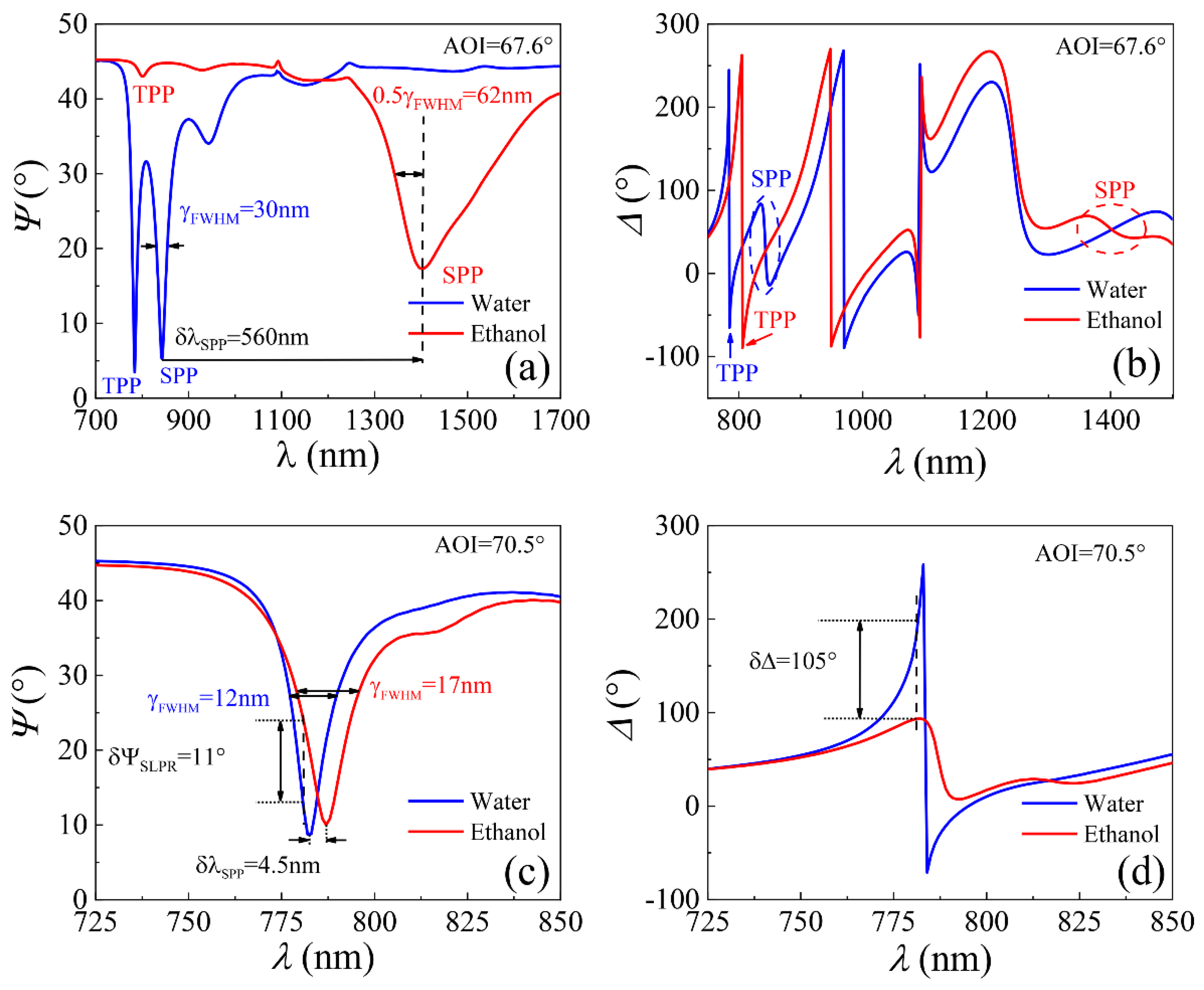
3. Results
4. Conclusions
Author Contributions
Funding
Institutional Review Board Statement
Informed Consent Statement
Data Availability Statement
Conflicts of Interest
References
- Barnes, W.L. Surface Plasmon–Polariton Length Scales: A Route to Sub-Wavelength Optics. J. Opt. A Pure Appl. Opt. 2006, 8, S87–S93. [Google Scholar] [CrossRef]
- Liedberg, B.; Nylander, C.; Lundström, I. Biosensing with Surface Plasmon Resonance—How It All Started. Biosens. Bioelectron. 1995, 10, i–ix. [Google Scholar] [CrossRef]
- Kravets, V.G.; Schedin, F.; Jalil, R.; Britnell, L.; Gorbachev, R.V.; Ansell, D.; Thackray, B.; Novoselov, K.S.; Geim, A.K.; Kabashin, A.V.; et al. Singular Phase Nano-Optics in Plasmonic Metamaterials for Label-Free Single-Molecule Detection. Nat. Mater. 2013, 12, 304–309. [Google Scholar] [CrossRef] [PubMed]
- Tsurimaki, Y.; Tong, J.K.; Boriskin, V.N.; Semenov, A.; Ayzatsky, M.I.; Machekhin, Y.P.; Chen, G.; Boriskina, S.V. Topological Engineering of Interfacial Optical Tamm States for Highly Sensitive Near-Singular-Phase Optical Detection. ACS Photonics 2018, 5, 929–938. [Google Scholar] [CrossRef] [Green Version]
- Nishijima, Y.; To, N.; Balčytis, A.; Juodkazis, S. Absorption and Scattering in Perfect Thermal Radiation Absorber-Emitter Metasurfaces. Opt. Express 2022, 30, 4058. [Google Scholar] [CrossRef]
- Koch, U.; Uhl, C.; Hettrich, H.; Fedoryshyn, Y.; Moor, D.; Baumann, M.; Hoessbacher, C.; Heni, W.; Baeuerle, B.; Bitachon, B.I.; et al. Plasmonics—High-Speed Photonics for Co-Integration with Electronics. Jpn. J. Appl. Phys. 2021, 60, SB0806. [Google Scholar] [CrossRef]
- Ma, R.-M.; Oulton, R.F.; Sorger, V.J.; Bartal, G.; Zhang, X. Room-Temperature Sub-Diffraction-Limited Plasmon Laser by Total Internal Reflection. Nat. Mater. 2011, 10, 110–113. [Google Scholar] [CrossRef] [Green Version]
- Kreibig, U.; Vollmer, M. Optical Properties of Metal Clusters; Springer Series in Materials Science; Springer: Berlin/Heidelberg, Germany, 1995; Volume 25, ISBN 978-3-642-08191-0. [Google Scholar]
- Homola, J.; Yee, S.S.; Gauglitz, G. Surface Plasmon Resonance Sensors: Review. Sens. Actuators B Chem. 1999, 54, 3–15. [Google Scholar] [CrossRef]
- Kravets, V.G.; Kabashin, A.V.; Barnes, W.L.; Grigorenko, A.N. Plasmonic Surface Lattice Resonances: A Review of Properties and Applications. Chem. Rev. 2018, 118, 5912–5951. [Google Scholar] [CrossRef]
- Reshetnyak, V.; Pinkevych, I.; Bunning, T.; Evans, D. Influence of Rugate Filters on the Spectral Manifestation of Tamm Plasmon Polaritons. Materials 2021, 14, 1282. [Google Scholar] [CrossRef]
- Plikusienė, I.; Bužavaitė-Vertelienė, E.; Mačiulis, V.; Valavičius, A.; Ramanavičienė, A.; Balevičius, Z. Application of Tamm Plasmon Polaritons and Cavity Modes for Biosensing in the Combined Spectroscopic Ellipsometry and Quartz Crystal Microbalance Method. Biosensors 2021, 11, 501. [Google Scholar] [CrossRef] [PubMed]
- Gupta, N.K.; Tiwari, A.K.; Wanare, H.; Ramakrishna, S.A. Near Singular-Phase Optical Biosensing with Strongly Coupled Modes of a Plasmonic–Photonic Trimer. J. Opt. 2021, 23, 065003. [Google Scholar] [CrossRef]
- Sasin, M.E.; Seisyan, R.P.; Kalitteevski, M.A.; Brand, S.; Abram, R.A.; Chamberlain, J.M.; Egorov, A.Y.; Vasil’ev, A.P.; Mikhrin, V.S.; Kavokin, A.V. Tamm Plasmon Polaritons: Slow and Spatially Compact Light. Appl. Phys. Lett. 2008, 92, 251112. [Google Scholar] [CrossRef] [Green Version]
- Balevicius, Z.; Baskys, A. Optical Dispersions of Bloch Surface Waves and Surface Plasmon Polaritons: Towards Advanced Biosensors. Materials 2019, 12, 3147. [Google Scholar] [CrossRef] [Green Version]
- Sinibaldi, A.; Danz, N.; Descrovi, E.; Munzert, P.; Schulz, U.; Sonntag, F.; Dominici, L.; Michelotti, F. Direct Comparison of the Performance of Bloch Surface Wave and Surface Plasmon Polariton Sensors. Sens. Actuators B Chem. 2012, 174, 292–298. [Google Scholar] [CrossRef]
- Bužavaitė-Vertelienė, E.; Maciulis, V.; Anulytė, J.; Tolenis, T.; Baskys, A.; Plikusiene, I.; Balevičius, Z. Total Internal Reflection Ellipsometry Approach for Bloch Surface Waves Biosensing Applications. Biosensors 2022, 12, 584. [Google Scholar] [CrossRef]
- Amra, C.; Zerrad, M.; Lemarchand, F.; Lereu, A.; Passian, A.; Zapien, J.A.; Lequime, M. Energy Density Engineering via Zero-Admittance Domains in All-Dielectric Stratified Materials. Phys. Rev. A 2018, 97, 023819. [Google Scholar] [CrossRef]
- Niu, D.; Zerrad, M.; Lereu, A.; Moreau, A.; Lumeau, J.; Zapien, J.A.; Passian, A.; Aubry, V.; Amra, C. Excitation of Bloch Surface Waves in Zero-Admittance Multilayers for High-Sensitivity Sensor Applications. Phys. Rev. Appl. 2020, 13, 054064. [Google Scholar] [CrossRef]
- Kravets, V.G.; Schedin, F.; Kabashin, A.V.; Grigorenko, A.N. Sensitivity of Collective Plasmon Modes of Gold Nanoresonators to Local Environment. Opt. Lett. 2010, 35, 956. [Google Scholar] [CrossRef] [Green Version]
- Anulytė, J.; Bužavaitė-Vertelienė, E.; Vertelis, V.; Stankevičius, E.; Vilkevičius, K.; Balevičius, Z. Influence of a Gold Nano-Bumps Surface Lattice Array on the Propagation Length of Strongly Coupled Tamm and Surface Plasmon Polaritons. J. Mater. Chem. C 2022, 10, 13234–13241. [Google Scholar] [CrossRef]
- Bužavaitė-Vertelienė, E.; Vertelis, V.; Balevičius, Z. The Experimental Evidence of a Strong Coupling Regime in the Hybrid Tamm Plasmon-Surface Plasmon Polariton Mode. Nanophotonics 2021, 10, 1565–1571. [Google Scholar] [CrossRef]
- Afinogenov, B.I.; Bessonov, V.O.; Nikulin, A.A.; Fedyanin, A.A. Observation of Hybrid State of Tamm and Surface Plasmon-Polaritons in One-Dimensional Photonic Crystals. Appl. Phys. Lett. 2013, 103, 061112. [Google Scholar] [CrossRef] [Green Version]
- Törmä, P.; Barnes, W.L. Strong Coupling between Surface Plasmon Polaritons and Emitters: A Review. Rep. Prog. Phys. 2015, 78, 013901. [Google Scholar] [CrossRef] [PubMed]
- Pelton, M.; Storm, S.D.; Leng, H. Strong Coupling of Emitters to Single Plasmonic Nanoparticles: Exciton-Induced Transparency and Rabi Splitting. Nanoscale 2019, 11, 14540–14552. [Google Scholar] [CrossRef] [PubMed]
- Kravets, V.G.; Schedin, F.; Grigorenko, A.N. Extremely Narrow Plasmon Resonances Based on Diffraction Coupling of Localized Plasmons in Arrays of Metallic Nanoparticles. Phys. Rev. Lett. 2008, 101, 087403. [Google Scholar] [CrossRef] [PubMed]
- Stankevičius, E.; Vilkevičius, K.; Gedvilas, M.; Bužavaitė-Vertelienė, E.; Selskis, A.; Balevičius, Z. Direct Laser Writing for the Formation of Large-Scale Gold Microbumps Arrays Generating Hybrid Lattice Plasmon Polaritons in Vis–NIR Range. Adv. Opt. Mater. 2021, 9, 2100027. [Google Scholar] [CrossRef]
- Sarkar, M.; Besbes, M.; Moreau, J.; Bryche, J.-F.; Olivéro, A.; Barbillon, G.; Coutrot, A.-L.; Bartenlian, B.; Canva, M. Hybrid Plasmonic Mode by Resonant Coupling of Localized Plasmons to Propagating Plasmons in a Kretschmann Configuration. ACS Photonics 2015, 2, 237–245. [Google Scholar] [CrossRef]
- Hoang, C.V.; Hayashi, K.; Ito, Y.; Gorai, N.; Allison, G.; Shi, X.; Sun, Q.; Cheng, Z.; Ueno, K.; Goda, K.; et al. Interplay of Hot Electrons from Localized and Propagating Plasmons. Nat. Commun. 2017, 8, 771. [Google Scholar] [CrossRef] [Green Version]
- Buzavaite-Verteliene, E.; Plikusiene, I.; Tolenis, T.; Valavicius, A.; Anulyte, J.; Ramanavicius, A.; Balevicius, Z. Hybrid Tamm-Surface Plasmon Polariton Mode for Highly Sensitive Detection of Protein Interactions. Opt. Express 2020, 28, 29033–29043. [Google Scholar] [CrossRef]
- Balevičius, Z. Strong Coupling between Tamm and Surface Plasmons for Advanced Optical Bio-Sensing. Coatings 2020, 10, 1187. [Google Scholar] [CrossRef]
- Arwin, H.; Poksinski, M.; Johansen, K. Total Internal Reflection Ellipsometry: Principles and Applications. Appl. Opt. 2004, 43, 3028. [Google Scholar] [CrossRef] [PubMed]
- Arwin, H. TIRE and SPR-Enhanced SE for Adsorption Processes. In Ellipsometry of Functional Organic Surfaces and Films; Hinrichs, K., Eichhorn, K.-J., Eds.; Springer: Berlin/Heidelberg, Germany, 2014; Volume 52, pp. 249–264. ISBN 978-3-642-40127-5. [Google Scholar]
- Balevicius, Z.; Makaraviciute, A.; Babonas, G.-J.; Tumenas, S.; Bukauskas, V.; Ramanaviciene, A.; Ramanavicius, A. Study of Optical Anisotropy in Thin Molecular Layers by Total Internal Reflection Ellipsometry. Sens. Actuators B Chem. 2013, 181, 119–124. [Google Scholar] [CrossRef]
- Balevicius, Z.; Baleviciute, I.; Tumenas, S.; Tamosaitis, L.; Stirke, A.; Makaraviciute, A.; Ramanaviciene, A.; Ramanavicius, A. In Situ Study of Ligand–Receptor Interaction by Total Internal Reflection Ellipsometry. Thin Solid Films 2014, 571, 744–748. [Google Scholar] [CrossRef]
- Moening, J.P.; Thanawala, S.S.; Georgiev, D.G. Formation of High-Aspect-Ratio Protrusions on Gold Films by Localized Pulsed Laser Irradiation. Appl. Phys. A 2009, 95, 635–638. [Google Scholar] [CrossRef]
- Itapu, S.; Borra, V.; Georgiev, D.G. Laser-Based Fabrication of Microstructures on Nickel Thin Films and Its Applications in On-Chip Thin Film Inductors. IEEE Trans. Nanotechnol. 2020, 19, 455–460. [Google Scholar] [CrossRef]
- J. A. Woollam Company. CompleteEase; v6.57; J. A. Woollam Company: Lincoln, NE, USA, 2020. [Google Scholar]
- Rhodes, C.; Franzen, S.; Maria, J.-P.; Losego, M.; Leonard, D.N.; Laughlin, B.; Duscher, G.; Weibel, S. Surface Plasmon Resonance in Conducting Metal Oxides. J. Appl. Phys. 2006, 100, 054905. [Google Scholar] [CrossRef]


Publisher’s Note: MDPI stays neutral with regard to jurisdictional claims in published maps and institutional affiliations. |
© 2022 by the authors. Licensee MDPI, Basel, Switzerland. This article is an open access article distributed under the terms and conditions of the Creative Commons Attribution (CC BY) license (https://creativecommons.org/licenses/by/4.0/).
Share and Cite
Anulytė, J.; Bužavaitė-Vertelienė, E.; Stankevičius, E.; Vilkevičius, K.; Balevičius, Z. High Spectral Sensitivity of Strongly Coupled Hybrid Tamm-Plasmonic Resonances for Biosensing Application. Sensors 2022, 22, 9453. https://doi.org/10.3390/s22239453
Anulytė J, Bužavaitė-Vertelienė E, Stankevičius E, Vilkevičius K, Balevičius Z. High Spectral Sensitivity of Strongly Coupled Hybrid Tamm-Plasmonic Resonances for Biosensing Application. Sensors. 2022; 22(23):9453. https://doi.org/10.3390/s22239453
Chicago/Turabian StyleAnulytė, Justina, Ernesta Bužavaitė-Vertelienė, Evaldas Stankevičius, Kernius Vilkevičius, and Zigmas Balevičius. 2022. "High Spectral Sensitivity of Strongly Coupled Hybrid Tamm-Plasmonic Resonances for Biosensing Application" Sensors 22, no. 23: 9453. https://doi.org/10.3390/s22239453