
An Overview of Optical and Electrochemical Sensors and Biosensors for Analysis of Antioxidants in Food during the Last 5 Years

Abstract
:1. Introduction
2. Optical Sensors and Assays
2.1. Colorimetric Sensors
2.1.1. AuNPs-Based Colorimetric Assays
2.1.2. AgNPs-Based Colorimetric Assays
2.1.3. CeONPs-Based Colorimetric Assays
2.1.4. Other Nanomaterial-Based Colorimetric Assays
2.1.5. Advantages, Limitations, and Potential for Practical Applications
2.2. Fluorescence Assays
2.2.1. Quantum Dots (QDs)-Based Fluorescence Assays
2.2.2. Advantages, Limitations, and the Potential for Practical Applications
3. Electrochemical Sensors and Biosensors
3.1. Enzyme-Based Electrochemical Biosensors
3.1.1. Peroxidase-Based Electrochemical Biosensors
3.1.2. Laccase-Based Electrochemical Biosensors
3.1.3. Tyrosinase-Based Electrochemical Biosensors
3.1.4. Advantages, Limitations, and the Potential for Practical Applications of Enzyme-Based Biosensors
3.2. Cell-Based Electrochemical Biosensors
Advantages, Limitations, and the Potential for Practical Applications of Cell-Based Biosensors
3.3. DNA-Based Electrochemical Biosensors
Advantages, Limitations, and the Potential for Practical Applications of DNA-Based Biosensors
3.4. Other Electrochemical Sensors and Biosensors for Antioxidant Analysis
4. Conclusions
Author Contributions
Funding
Institutional Review Board Statement
Informed Consent Statement
Data Availability Statement
Conflicts of Interest
References
- Mello, L.D.; Kubota, L.T. Biosensors as a tool for the antioxidant status evaluation. Talanta 2007, 72, 335–348. [Google Scholar] [CrossRef]
- Ye, Y.; Ji, J.; Sun, Z.; Shen, P.; Sun, X. Recent advances in electrochemical biosensors for antioxidant analysis in foodstuff. TrAC Trends Anal. Chem. 2020, 122, 115718. [Google Scholar] [CrossRef]
- Shahidi, F. Antioxidants in food and food antioxidants. Food Nahr. 2000, 44, 158–163. [Google Scholar] [CrossRef]
- Wojcik, M.; Burzynska-Pedziwiatr, I.; Wozniak, L. A review of natural and synthetic antioxidants important for health and longevity. Curr. Med. Chem. 2010, 17, 3262–3288. [Google Scholar] [CrossRef] [PubMed]
- Della Pelle, F.; Scroccarello, A.; Sergi, M.; Mascini, M.; Del Carlo, M.; Compagnone, D. Simple and rapid silver nanoparticles based antioxidant capacity assays: Reactivity study for phenolic compounds. Food Chem. 2018, 256, 342–349. [Google Scholar] [CrossRef] [PubMed]
- Zhang, X.; Liu, Q.; Chen, Z.; Zuo, X. Colorimetric sensor array for accurate detection and identification of antioxidants based on metal ions as sensor receptors. Talanta 2020, 120935. [Google Scholar] [CrossRef]
- Pisoschi, A.M.; Negulescu, G.P. Methods for total antioxidant activity determination: A review. Anal. Biochem. 2011, 1, 106. [Google Scholar] [CrossRef] [Green Version]
- Sindhi, V.; Gupta, V.; Sharma, K.; Bhatnagar, S.; Kumari, R.; Dhaka, N. Potential applications of Antioxidants—A review. J. Pharm. Res. 2013, 7, 828–835. [Google Scholar] [CrossRef]
- Sharpe, E.; Frasco, T.; Andreescu, D.; Andreescu, S. Portable ceria nanoparticle-based assay for rapid detection of food antioxidants (NanoCerac). Analyst 2013, 138, 249–262. [Google Scholar] [CrossRef] [Green Version]
- Bener, M.; Apak, R. Ferric-O-Phenanthroline adsorbed on a nafion membrane: A novel optical sensor for antioxidant capacity measurement of food extracts. Sens. Actuators B Chem. 2017, 247, 155–162. [Google Scholar] [CrossRef]
- Piyanan, T.; Athipornchai, A.; Henry, C.S.; Sameenoi, Y. An instrument-free detection of antioxidant activity using paper-based analytical devices coated with nanoceria. Anal. Sci. 2018, 34, 97–102. [Google Scholar] [CrossRef] [Green Version]
- Apak, R.; Demirci Çekiç, S.; Üzer, A.; Çelik, S.E.; Bener, M.; Bekdeşer, B.; Can, Z.; Sağlam, Ş.; Önem, A.N.; Erçağ, E. Novel spectroscopic and electrochemical sensors and nanoprobes for the characterization of food and biological antioxidants. J. Sens. 2018, 18, 186. [Google Scholar] [CrossRef] [Green Version]
- Zhang, H.; Tsao, R. Dietary polyphenols, oxidative stress and antioxidant and anti-inflammatory effects. Curr. Opin. Food Sci. 2016, 8, 33–42. [Google Scholar] [CrossRef]
- Rauhut, R.T.; Bülbül, G.; Andreescu, S. Nanotechnology-Enabled approaches for the detection of antioxidants by spectroscopic and electrochemical methods. In Measurement of Antioxidant Activity & Capacity; Apak, R., Capanoglu, E., Shahidi, F., Eds.; Wiley: Hoboken, NJ, USA, 2018; pp. 187–207. [Google Scholar]
- Puangbanlang, C.; Sirivibulkovit, K.; Nacapricha, D.; Sameenoi, Y. A paper-based device for simultaneous determination of antioxidant activity and total phenolic content in food samples. Talanta 2019, 198, 542–549. [Google Scholar] [CrossRef]
- Apak, R.; Güçlü, K.; Özyürek, M.; Karademir, S.E. Novel total antioxidant capacity index for dietary polyphenols and vitamins C and E, using their cupric ion reducing capability in the presence of neocuproine: CUPRAC method. J. Agric. Food Chem. 2004, 52, 7970–7981. [Google Scholar] [CrossRef]
- Olgun, F.A.O.; Üzer, A.; Ozturk, B.D.; Apak, R. A novel cerium oxide nanoparticles-based colorimetric sensor using tetramethyl benzidine reagent for antioxidant activity assay. Talanta 2018, 182, 55–61. [Google Scholar] [CrossRef]
- Della Pelle, F.; Compagnone, D. Nanomaterial-Based sensing and biosensing of phenolic compounds and related antioxidant capacity in food. J. Sens. 2018, 18, 462. [Google Scholar] [CrossRef] [Green Version]
- Azeem, S.M.A.; Al Mohesen, I.A.; Ibrahim, A.M. Analysis of total phenolic compounds in tea and fruits using diazotized aminobenzenes colorimetric spots. Food Chem. 2020, 332, 127392. [Google Scholar] [CrossRef] [PubMed]
- David, M.; Florescu, M.; Bala, C. Biosensors for antioxidants detection: Trends and perspectives. J. Biosens. 2020, 10, 112. [Google Scholar] [CrossRef] [PubMed]
- Busa, L.S.A.; Mohammadi, S.; Maeki, M.; Ishida, A.; Tani, H.; Tokeshi, M. Advances in microfluidic paper-based analytical devices for food and water analysis. Micromachines 2016, 7, 86. [Google Scholar] [CrossRef] [PubMed]
- Vilela, D.; González, M.C.; Escarpa, A. Nanoparticles as analytical tools for in-vitro antioxidant-capacity assessment and beyond. TrAC Trends Anal. Chem. 2015, 64, 1–16. [Google Scholar] [CrossRef]
- Chang, C.C.; Chen, C.P.; Wu, T.H.; Yang, C.H.; Lin, C.W.; Chen, C.Y. Gold nanoparticle-based colorimetric strategies for chemical and biological sensing applications. J. Nanomater. 2019, 9, 861. [Google Scholar] [CrossRef] [PubMed] [Green Version]
- Chen, Y.; Xianyu, Y.; Jiang, X. Surface modification of gold nanoparticles with small molecules for biochemical analysis. Acc. Chem. Res. 2017, 50, 310–319. [Google Scholar] [CrossRef] [PubMed]
- Choleva, T.G.; Kappi, F.A.; Giokas, D.L.; Vlessidis, A.G. Paper-Based assay of antioxidant activity using analyte-mediated on-paper nucleation of gold nanoparticles as colorimetric probes. Anal. Chim. Acta 2015, 860, 61–69. [Google Scholar] [CrossRef]
- Della Pelle, F.; Vilela, D.; González, M.C.; Sterzo, C.L.; Compagnone, D.; Del Carlo, M.; Escarpa, A. Antioxidant capacity index based on gold nanoparticles formation. Application to extra virgin olive oil samples. Food Chem. 2015, 178, 70–75. [Google Scholar] [CrossRef]
- Della Pelle, F.; González, M.C.; Sergi, M.; Del Carlo, M.; Compagnone, D.; Escarpa, A. Gold nanoparticles-based extraction-free colorimetric assay in organic media: An optical index for determination of total polyphenols in fat-rich samples. Anal. Chem. 2015, 87, 6905–6911. [Google Scholar] [CrossRef]
- Tułodziecka, A.; Szydłowska-Czerniak, A. Development of a novel gold nanoparticle-based method to determine antioxidant capacity of Brassica oilseeds, white flakes and meal. Food Chem. 2016, 208, 142–149. [Google Scholar] [CrossRef]
- Chou, J.; Li, X.; Yin, Y.; Indrisek, N. Determination of antioxidant activities in fruit juices based on rapid colorimetric measurement and characterisation of gold nanoparticles. Int. J. Environ. Anal. Chem. 2015, 95, 531–541. [Google Scholar] [CrossRef]
- Tonello, N.V.; D’Eramo, F.; Marioli, J.M.; Crevillen, A.G.; Escarpa, A. Extraction-Free colorimetric determination of thymol and carvacrol isomers in essential oils by pH-dependent formation of gold nanoparticles. Microchim. Acta 2018, 185, 352. [Google Scholar] [CrossRef]
- Scroccarello, A.; Della Pelle, F.; Neri, L.; Pittia, P.; Compagnone, D. Silver and gold nanoparticles based colorimetric assays for the determination of sugars and polyphenols in apples. Food Res. Int. 2019, 119, 359–368. [Google Scholar] [CrossRef]
- Bener, M.; Şen, F.B.; Apak, R. Heparin-Stabilized gold nanoparticles-based CUPRAC colorimetric sensor for antioxidant capacity measurement. Talanta 2018, 187, 148–155. [Google Scholar] [CrossRef]
- Bordbar, M.M.; Hemmateenejad, B.; Tashkhourian, J.; Nami-Ana, S. An optoelectronic tongue based on an array of gold and silver nanoparticles for analysis of natural, synthetic and biological antioxidants. Microchim. Acta 2018, 185, 493. [Google Scholar] [CrossRef]
- Li, L.; Zhang, P.; Fu, W.; Yang, M.; Wang, Y. Use of seed-mediated growth of bimetallic nanorods as a knob for antioxidant assay. Sens. Actuators B Chem. 2018, 276, 158–165. [Google Scholar] [CrossRef]
- Prosposito, P.; Burratti, L.; Venditti, I. Silver nanoparticles as colorimetric sensors for water pollutants. Chemosensors 2020, 8, 26. [Google Scholar] [CrossRef] [Green Version]
- Sabela, M.; Balme, S.; Bechelany, M.; Janot, J.M.; Bisetty, K. A review of gold and silver nanoparticle-based colorimetric sensing assays. Adv. Eng. Mater. 2017, 19, 1700270. [Google Scholar] [CrossRef]
- Teerasong, S.; Jinnarak, A.; Chaneam, S.; Wilairat, P.; Nacapricha, D. Poly (vinyl alcohol) capped silver nanoparticles for antioxidant assay based on seed-mediated nanoparticle growth. Talanta 2017, 170, 193–198. [Google Scholar] [CrossRef] [PubMed]
- Selvan, D.A.; Mahendiran, D.; Kumar, R.S.; Rahiman, A.K. Garlic, green tea and turmeric extracts-mediated green synthesis of silver nanoparticles: Phytochemical, antioxidant and in vitro cytotoxicity studies. J. Photochem. Photobiol. B 2018, 180, 243–252. [Google Scholar] [CrossRef]
- Charbgoo, F.; Ramezani, M.; Darroudi, M. Bio-Sensing applications of cerium oxide nanoparticles: Advantages and disadvantages. Biosens. Bioelectron. 2017, 96, 33–43. [Google Scholar] [CrossRef] [PubMed]
- Karakoti, A.; Monteiro-Riviere, N.; Aggarwal, R.; Davis, J.; Narayan, R.; Self, W.; McGinnis, J.; Seal, S. Nanoceria as antioxidant: Synthesis and biomedical applications. JOM 2008, 60, 33–37. [Google Scholar] [CrossRef] [Green Version]
- Andreescu, D.; Bulbul, G.; Özel, R.E.; Hayat, A.; Sardesai, N.; Andreescu, S. Applications and implications of nanoceria reactivity: Measurement tools and environmental impact. Environ. Sci. Nano 2014, 1, 445–458. [Google Scholar] [CrossRef]
- Tułodziecka, A.; Szydłowska-Czerniak, A. Determination of total antioxidant capacity of rapeseed and its by-products by a novel cerium oxide nanoparticle-based spectrophotometric method. Food Anal. Methods 2016, 9, 3053–3062. [Google Scholar] [CrossRef] [Green Version]
- Sachdev, A.; Samanta, P.; Kumar, V.; Kandhal, K.; Matai, I. PMAA-CeO2 nanoparticle-based paper microfluidic device with customized image processing software for antioxidant assay. Anal. Bioanal. Chem. 2020, 412, 8197–8209. [Google Scholar] [CrossRef]
- Szydłowska-Czerniak, A.; Łaszewska, A.; Tułodziecka, A. A novel iron oxide nanoparticle-based method for the determination of the antioxidant capacity of rapeseed oils at various stages of the refining process. Anal. Methods 2015, 7, 4650–4660. [Google Scholar] [CrossRef]
- Wu, Z.; Xu, E.; Li, J.; Long, J.; Jiao, A.; Jin, Z.; Xu, X. Determination of antioxidant capacity of Chinese rice wine and Zhuyeqing liquor using nanoparticle-based colorimetric methods. Food Anal. Methods 2017, 10, 788–798. [Google Scholar] [CrossRef]
- Jaberie, H.; Momeni, S.; Nabipour, I. Total antioxidant capacity assessment by a development of an antioxidant assay based on green synthesized MnO2 nanosheets. Microchem. J. 2020, 157, 104908. [Google Scholar] [CrossRef]
- Li, X.; Wang, J.; Yi, C.; Jiang, L.; Wu, J.; Chen, X.; Shen, X.; Sun, Y.; Lei, H. A smartphone-based quantitative detection device integrated with latex microsphere immunochromatography for on-site detection of zearalenone in cereals and feed. Sens. Actuators B Chem. 2019, 290, 170–179. [Google Scholar] [CrossRef]
- Huang, W.; Deng, Y.; He, Y. Visual colorimetric sensor array for discrimination of antioxidants in serum using MnO2 nanosheets triggered multicolor chromogenic system. Biosens. Bioelectron. 2017, 91, 89–94. [Google Scholar] [CrossRef] [PubMed]
- Can, K.; Üzer, A.; Apak, R. A manganese oxide (MnOx)-Based colorimetric nanosensor for indirect measurement of lipophilic and hydrophilic antioxidant capacity. Anal. Methods 2020, 12, 448–455. [Google Scholar] [CrossRef]
- Romero, M.P.R.; Brito, R.E.; Mellado, J.M.R.; González-Rodríguez, J.; Montoya, M.R.; Rodríguez-Amaro, R. Exploring the relation between composition of extracts of healthy foods and their antioxidant capacities determined by electrochemical and spectrophotometrical methods. LWT 2018, 95, 157–166. [Google Scholar] [CrossRef]
- Zhang, C.; Li, X.; Wei, W.; Chen, Z. Lanthanide ions as sensor elements based sensor array for colorimetric identification of antioxidants. Sens. Actuators B Chem. 2020, 305, 127532. [Google Scholar] [CrossRef]
- Krylova, E.; Gavrilenko, N.; Saranchina, N.; Gavrilenko, M. Novel colorimetric sensor for cupric reducing antioxidant capacity (CUPRAC) measurement. Procedia Eng. 2016, 168, 355–358. [Google Scholar] [CrossRef]
- Popa, C.V.; Vasilescu, A.; Litescu, S.C.; Albu, C.; Danet, A.F. Metal nano-oxide based colorimetric sensor array for the determination of plant polyphenols with antioxidant properties. Anal. Lett. 2020, 53, 627–645. [Google Scholar] [CrossRef]
- Gatselou, V.; Christodouleas, D.C.; Kouloumpis, A.; Gournis, D.; Giokas, D.L. Determination of phenolic compounds using spectral and color transitions of rhodium nanoparticles. Anal. Chim. Acta 2016, 932, 80–87. [Google Scholar] [CrossRef]
- Aid, T.; Kaljurand, M.; Vaher, M. Colorimetric determination of total phenolic contents in ionic liquid extracts by paper microzones and digital camera. Anal. Methods 2015, 7, 3193–3199. [Google Scholar] [CrossRef]
- Hidayat, M.A.; Puspitaningtyas, N.; Gani, A.A.; Kuswandi, B. Rapid test for the determination of total phenolic content in brewed-filtered coffee using colorimetric paper. J. Food Sci. Technol. 2017, 54, 3384–3390. [Google Scholar] [CrossRef]
- Pérez-López, B.; Merkoçi, A. Nanomaterials based biosensors for food analysis applications. Trends Food Sci. Technol. 2011, 22, 625–639. [Google Scholar] [CrossRef]
- Liu, G.; Lu, M.; Huang, X.; Li, T.; Xu, D. Application of gold-nanoparticle colorimetric sensing to rapid food safety screening. J. Sens. 2018, 18, 4166. [Google Scholar] [CrossRef] [Green Version]
- Özyürek, M.; Güngör, N.; Baki, S.; Güçlü, K.; Apak, R. Development of a silver nanoparticle-based method for the antioxidant capacity measurement of polyphenols. Anal. Chem. 2012, 84, 8052–8059. [Google Scholar] [CrossRef] [PubMed]
- Nezhad, M.R.H.; Alimohammadi, M.; Tashkhourian, J.; Razavian, S.M. Optical detection of phenolic compounds based on the surface plasmon resonance band of Au nanoparticles. Spectrochim. Acta A Mol. Biomol. Spectrosc. 2008, 71, 199–203. [Google Scholar] [CrossRef] [PubMed]
- Vilela, D.; Castañeda, R.; González, M.C.; Mendoza, S.; Escarpa, A. Fast and reliable determination of antioxidant capacity based on the formation of gold nanoparticles. Microchim. Acta 2015, 182, 105–111. [Google Scholar] [CrossRef]
- Piroozmand, F.; Mohammadipanah, F.; Faridbod, F. Emerging biosensors in detection of natural products. Synth. Syst. Biotechnol. 2020, 5, 293–303. [Google Scholar] [CrossRef]
- Ma, B.; Zeng, F.; Zheng, F.; Wu, S. A fluorescence turn-on sensor for iodide based on a Thymine-HgII-Thymine complex. Chem. Eur. J. 2011, 17, 14844–14850. [Google Scholar] [CrossRef]
- Shen, Y.; Xu, L.; Li, Y. Biosensors for rapid detection of Salmonella in food: A review. Compr. Rev. Food Sci. Food Saf. 2020, 20. [Google Scholar] [CrossRef] [PubMed]
- Ma, J.; Chen, J.Y.; Zhang, Y.; Wang, P.N.; Guo, J.; Yang, W.L.; Wang, C.C. Photochemical instability of thiol-capped CdTe quantum dots in aqueous solution and living cells: Process and mechanism. J. Phys. Chem. B 2007, 111, 12012–12016. [Google Scholar] [CrossRef]
- Petryayeva, E.; Algar, W.R.; Medintz, I.L. Quantum dots in bioanalysis: A review of applications across various platforms for fluorescence spectroscopy and imaging. Appl. Spectrosc. 2013, 67, 215–252. [Google Scholar] [CrossRef] [PubMed] [Green Version]
- Dwiecki, K.; Nogala-Kałucka, M.; Polewski, K. Determination of total phenolic compounds in common beverages using CdTe quantum dots. J. Food Process. Preserv. 2017, 41, e12863. [Google Scholar] [CrossRef]
- Li, Y.; Li, W.; Zhou, H.; Wang, F.; Chen, Y.; Wang, Y.; Yu, C. A facile method for the sensing of antioxidants based on the redox transformation of polyaniline. Sens. Actuators B Chem. 2015, 208, 30–35. [Google Scholar] [CrossRef]
- Rodrigues, D.M.; Ribeiro, D.S.; Frigerio, C.; Rodrigues, S.S.M.; Santos, J.L.; Prior, J.A. Antioxidant capacity automatic assay based on inline photogenerated radical species from L-glutathione-capped CdTe quantum dots. Talanta 2015, 141, 220–229. [Google Scholar] [CrossRef] [Green Version]
- Liu, H.; Fang, G.; Deng, Q.; Wang, S. A triple-dimensional sensing chip for discrimination of eight antioxidants based on quantum dots and graphene. Biosens. Bioelectron. 2015, 74, 313–317. [Google Scholar] [CrossRef]
- Álvarez-Diduk, R.; Orozco, J.; Merkoçi, A. Paper strip-embedded graphene quantum dots: A screening device with a smartphone readout. Sci. Rep. 2017, 7, 1–9. [Google Scholar]
- Liu, J.J.; Chen, Z.T.; Tang, D.S.; Wang, Y.B.; Kang, L.T.; Yao, J.N. Graphene quantum dots-based fluorescent probe for turn-on sensing of ascorbic acid. Sens. Actuators B Chem. 2015, 212, 214–219. [Google Scholar] [CrossRef]
- Gao, Y.; Yan, X.; Li, M.; Gao, H.; Sun, J.; Zhu, S.; Han, S.; Jia, L.-N.; Zhao, X.-E.; Wang, H. A “turn-on” fluorescence sensor for ascorbic acid based on graphene quantum dots via fluorescence resonance energy transfer. Anal. Methods 2018, 10, 611–616. [Google Scholar] [CrossRef]
- Liu, H.; Na, W.; Liu, Z.; Chen, X.; Su, X. A novel turn-on fluorescent strategy for sensing ascorbic acid using graphene quantum dots as fluorescent probe. Biosens. Bioelectron. 2017, 92, 229–233. [Google Scholar] [CrossRef] [PubMed]
- Wang, M.; Chen, J.; Liu, C.; Qiu, J.; Wang, X.; Chen, P.; Xu, C. A graphene quantum dots–hypochlorite hybrid system for the quantitative fluorescent determination of total antioxidant capacity. Small 2017, 13, 1700709. [Google Scholar] [CrossRef] [PubMed]
- Huang, S.; Qiu, H.; Zhu, F.; Lu, S.; Xiao, Q. Graphene quantum dots as on-off-on fluorescent probes for chromium (VI) and ascorbic acid. Microchim. Acta 2015, 182, 1723–1731. [Google Scholar] [CrossRef]
- Xu, Y.; Niu, X.; Zhang, H.; Xu, L.; Zhao, S.; Chen, H.; Chen, X. Switch-On fluorescence sensing of glutathione in food samples based on a graphitic carbon nitride quantum dot (g-CNQD)-Hg2+ chemosensor. J. Agric. Food Chem. 2015, 63, 1747–1755. [Google Scholar] [CrossRef] [PubMed]
- Pavun, L.; Uskoković-Marković, S.; Jelikić-Stankov, M.; Đikanović, D.; Đurđević, P. Determination of flavonoids and total polyphenol contents in commercial apple juices. Czech J. Food Sci. 2018, 36, 233–238. [Google Scholar] [CrossRef] [Green Version]
- Prior, R.L.; Wu, X.; Schaich, K. Standardized methods for the determination of antioxidant capacity and phenolics in foods and dietary supplements. J. Agric. Food Chem. 2005, 53, 4290–4302. [Google Scholar] [CrossRef] [PubMed]
- Xie, R.; Wang, Z.; Zhou, W.; Liu, Y.; Fan, L.; Li, Y.; Li, X. Graphene quantum dots as smart probes for biosensing. Anal. Methods 2016, 8, 4001–4016. [Google Scholar] [CrossRef]
- Zor, E.; Morales-Narváez, E.; Zamora-Gálvez, A.; Bingol, H.; Ersoz, M.; Merkoçi, A. Graphene quantum dots-based photoluminescent sensor: A multifunctional composite for pesticide detection. ACS Appl. Mater. Interfaces 2015, 7, 20272–20279. [Google Scholar] [CrossRef]
- Serra, B.; Reviejo, A.J.; Pingarrón, J.M. Application of electrochemical enzyme biosensors for food quality control. Compr. Anal. Chem. 2007, 49, 255–298. [Google Scholar]
- Mishra, S.K.; Chiang, K.S. Phenolic-Compounds sensor based on immobilization of tyrosinase in polyacrylamide gel on long-period fiber grating. Opt. Laser Technol. 2020, 131, 106464. [Google Scholar] [CrossRef]
- Wu, L.; Yin, W.; Tang, K.; Li, D.; Shao, K.; Zuo, Y.; Ma, J.; Liu, J.; Han, H. Enzymatic biosensor of horseradish peroxidase immobilized on Au-Pt Nanotube/Au-Graphene for the simultaneous determination of antioxidants. Anal. Chim. Acta 2016, 933, 89–96. [Google Scholar] [CrossRef] [PubMed]
- de Oliveira Neto, J.R.; Rezende, S.G.; Lobón, G.S.; Garcia, T.A.; Macedo, I.Y.L.; Garcia, L.F.; Alves, V.F.; Torres, I.M.S.; Santiago, M.F.; Schmidt, F. Electroanalysis and laccase-based biosensor on the determination of phenolic content and antioxidant power of honey samples. Food Chem. 2017, 237, 1118–1123. [Google Scholar] [CrossRef]
- Zhang, Z.; Liu, J.; Fan, J.; Wang, Z.; Li, L. Detection of catechol using an electrochemical biosensor based on engineered Escherichia coli cells that surface-display laccase. Anal. Chim. Acta 2018, 1009, 65–72. [Google Scholar] [CrossRef]
- Cerrato-Alvarez, M.; Bernalte, E.; Bernalte-García, M.J.; Pinilla-Gil, E. Fast and direct amperometric analysis of polyphenols in beers using tyrosinase-modified screen-printed gold nanoparticles biosensors. Talanta 2019, 193, 93–99. [Google Scholar] [CrossRef]
- Raymundo-Pereira, P.A.; Silva, T.A.; Caetano, F.R.; Riboviski, L.; Zapp, E.; Brondani, D.; Bergamini, M.F.; Junior, L.H.M.; Banks, C.E.; Oliveira, O.N., Jr. Polyphenol oxidase-based electrochemical biosensors: A review. Anal. Chim. Acta 2020, 1139, 198–221. [Google Scholar] [CrossRef]
- Zrinski, I.; Pungjunun, K.; Martinez, S.; Zavašnik, J.; Stanković, D.; Kalcher, K.; Mehmeti, E. Evaluation of phenolic antioxidant capacity in beverages based on laccase immobilized on screen-printed carbon electrode modified with graphene nanoplatelets and gold nanoparticles. Microchem. J. 2020, 152, 104282. [Google Scholar] [CrossRef]
- Mohtar, L.G.; Aranda, P.; Messina, G.A.; Nazareno, M.A.; Pereira, S.V.; Raba, J.; Bertolino, F.A. Amperometric biosensor based on laccase immobilized onto a nanostructured screen-printed electrode for determination of polyphenols in propolis. Microchem. J. 2019, 144, 13–18. [Google Scholar] [CrossRef]
- Gul, I.; Ahmad, M.S.; Naqvi, S.S.; Hussain, A.; Wali, R.; Farooqi, A.A.; Ahmed, I. Polyphenol oxidase (PPO) based biosensors for detection of phenolic compounds: A review. J. Appl. Biol. Biotechnol. 2017, 5, 72–85. [Google Scholar]
- Vlamidis, Y.; Gualandi, I.; Tonelli, D. Amperometric biosensors based on reduced GO and MWCNTs composite for polyphenols detection in fruit juices. J. Electroanal. Chem. 2017, 799, 285–292. [Google Scholar] [CrossRef]
- Wang, Y.; Zhai, F.; Hasebe, Y.; Jia, H.; Zhang, Z. A highly sensitive electrochemical biosensor for phenol derivatives using a graphene oxide-modified tyrosinase electrode. Bioelectrochemistry 2018, 122, 174–182. [Google Scholar] [CrossRef] [PubMed]
- Draghi, P.F.; Fernandes, J.C.B. Label-Free potentiometric biosensor based on solid-contact for determination of total phenols in honey and propolis. Talanta 2017, 164, 413–417. [Google Scholar] [CrossRef]
- Kucherenko, I.; Soldatkin, O.; Kucherenko, D.Y.; Soldatkina, O.; Dzyadevych, S.V. Advances in nanomaterial application in enzyme-based electrochemical biosensors: A review. Nanoscale Adv. 2019, 1, 4560–4577. [Google Scholar] [CrossRef] [Green Version]
- Kurbanoglu, S.; Ozkan, S.A.; Merkoci, A. Nanomaterials-Based enzyme electrochemical biosensors operating through inhibition for biosensing applications. Biosens. Bioelectron. 2017, 89, 886–898. [Google Scholar] [CrossRef]
- Liu, Q.; Wu, C.; Cai, H.; Hu, N.; Zhou, J.; Wang, P. Cell-Based biosensors and their application in biomedicine. Chem. Rev. 2014, 114, 6423–6461. [Google Scholar] [CrossRef] [PubMed]
- Ye, Y.; Guo, H.; Sun, X. Recent progress on cell-based biosensors for analysis of food safety and quality control. Biosens. Bioelectron. 2019, 126, 389–404. [Google Scholar] [CrossRef] [PubMed]
- Lim, J.W.; Ha, D.; Lee, J.; Lee, S.K.; Kim, T. Review of micro/nanotechnologies for microbial biosensors. Front. Bioeng. Biotechnol. 2015, 3, 61. [Google Scholar] [CrossRef] [PubMed] [Green Version]
- Ge, Q.; Ge, P.; Jiang, D.; Du, N.; Chen, J.; Yuan, L.; Yu, H.; Xu, X.; Wu, M.; Zhang, W. A novel and simple cell-based electrochemical biosensor for evaluating the antioxidant capacity of Lactobacillus plantarum strains isolated from Chinese dry-cured ham. Biosens. Bioelectron. 2018, 99, 555–563. [Google Scholar] [CrossRef]
- Xing, L.; Ge, Q.; Jiang, D.; Gao, X.; Liu, R.; Cao, S.; Zhuang, X.; Zhou, G.; Zhang, W. Caco-2 cell-based electrochemical biosensor for evaluating the antioxidant capacity of Asp-Leu-Glu-Glu isolated from dry-cured Xuanwei ham. Biosens. Bioelectron. 2018, 105, 81–89. [Google Scholar] [CrossRef] [PubMed]
- Ye, Y.; Ji, J.; Pi, F.; Yang, H.; Liu, J.; Zhang, Y.; Xia, S.; Wang, J.; Xu, D.; Sun, X. A novel electrochemical biosensor for antioxidant evaluation of phloretin based on cell-alginate/ʟ-cysteine/gold nanoparticle-modified glassy carbon electrode. Biosens. Bioelectron. 2018, 119, 119–125. [Google Scholar] [CrossRef] [PubMed]
- Zhang, Z.; Zhang, Z.; Hu, Y.; Liu, J.; Ni, H.; Li, L. Phenol biosensor based on glassy carbon electrode directly absorbed Escherichia coli cells with surface-displayed bacterial laccase. Proc. Technol. 2017, 27, 137–138. [Google Scholar] [CrossRef]
- Gupta, N.; Renugopalakrishnan, V.; Liepmann, D.; Paulmurugan, R.; Malhotra, B.D. Cell-Based biosensors: Recent trends, challenges and future perspectives. Biosens. Bioelectron. 2019, 141, 111435. [Google Scholar] [CrossRef] [PubMed]
- Bora, U.; Sett, A.; Singh, D. Nucleic acid based biosensors for clinical applications. Biosens. J. 2013, 2, 1–8. [Google Scholar] [CrossRef]
- Yang, T.; Luo, Z.; Tian, Y.; Qian, C.; Duan, Y. Design strategies of AuNPs-based nucleic acid colorimetric biosensors. TrAC Trends Anal. Chem. 2020, 124, 115795. [Google Scholar] [CrossRef]
- Khoshbin, Z.; Housaindokht, M.R.; Verdian, A.; Bozorgmehr, M.R. Simultaneous detection and determination of mercury (II) and lead (II) ions through the achievement of novel functional nucleic acid-based biosensors. Biosens. Bioelectron. 2018, 116, 130–147. [Google Scholar] [CrossRef]
- Huo, B.; Hu, Y.; Gao, Z.; Li, G. Recent advances on functional nucleic acid-based biosensors for detection of food contaminants. Talanta 2021, 222, 121565. [Google Scholar] [CrossRef]
- Chen, Y.; Qian, C.; Liu, C.; Shen, H.; Wang, Z.; Ping, J.; Wu, J.; Chen, H. Nucleic acid amplification free biosensors for pathogen detection. Biosens. Bioelectron. 2020, 153, 112049. [Google Scholar] [CrossRef]
- Yue, Y.; Zhihong, B.; Sanming, L.; Kun, Z. Electrochemical evaluation of antioxidant capacity in pharmaceutical antioxidant excipient of drugs on guanine-based modified electrode. J. Electroanal. Chem. 2016, 772, 58–65. [Google Scholar] [CrossRef]
- Barroso, M.F.; De-Los-Santos-Álvarez, N.; Delerue-Matos, C.; Oliveira, M.B.P.P. Towards a reliable technology for antioxidant capacity and oxidative damage evaluation: Electrochemical (bio) sensors. Biosens. Bioelectron. 2011, 30, 1–12. [Google Scholar] [CrossRef] [Green Version]
- Tomac, I.; Šeruga, M.; Labuda, J. Evaluation of antioxidant activity of chlorogenic acids and coffee extracts by an electrochemical DNA-based biosensor. Food Chem. 2020, 325, 126787. [Google Scholar] [CrossRef]
- Fojta, M.; Daňhel, A.; Havran, L.; Vyskočil, V. Recent progress in electrochemical sensors and assays for DNA damage and repair. TrAC Trends Anal. Chem. 2016, 79, 160–167. [Google Scholar] [CrossRef]
- Mousavisani, S.Z.; Raoof, J.B.; Ojani, R.; Bagheryan, Z. An impedimetric biosensor for DNA damage detection and study of the protective effect of deferoxamine against DNA damage. Bioelectrochemistry 2018, 122, 142–148. [Google Scholar] [CrossRef]
- Hashkavayi, A.B.; Hashemnia, S.; Osfouri, S. Investigations of antioxidant potential and protective effect of Acanthophora algae on DNA damage: An electrochemical approach. Microchem. J. 2020, 159, 105455. [Google Scholar] [CrossRef]
- Barroso, M.F.; Ramalhosa, M.J.; Alves, R.C.; Dias, A.; Soares, C.M.; Oliva-Teles, M.T.; Delerue-Matos, C. Total antioxidant capacity of plant infusions: Assessment using electrochemical DNA-based biosensor and spectrophotometric methods. Food Control 2016, 68, 153–161. [Google Scholar] [CrossRef]
- Mohamadi, M.; Mostafavi, A.; Torkzadeh-Mahani, M. Electrochemical determination of biophenol oleuropein using a simple label-free DNA biosensor. Bioelectrochemistry 2015, 101, 52–57. [Google Scholar] [CrossRef]
- Becker, M.M.; Ribeiro, E.B.; de Oliveira Marques, P.R.B.; Marty, J.-L.; Nunes, G.S.; Catanante, G. Development of a highly sensitive xanthine oxidase-based biosensor for the determination of antioxidant capacity in Amazonian fruit samples. Talanta 2019, 204, 626–632. [Google Scholar] [CrossRef] [PubMed]
- Zouaoui, F.; Bourouina-Bacha, S.; Bourouina, M.; Jaffrezic-Renault, N.; Zine, N.; Errachid, A. Electrochemical sensors based on molecularly imprinted chitosan: A review. TrAC Trends Anal. Chem. 2020, 130, 115982. [Google Scholar] [CrossRef]
- Shojaei, S.; Nasirizadeh, N.; Entezam, M.; Koosha, M.; Azimzadeh, M. An electrochemical nanosensor based on molecularly imprinted polymer (MIP) for detection of gallic acid in fruit juices. Food Anal. Methods 2016, 9, 2721–2731. [Google Scholar] [CrossRef]
- Singh, S. Nanomaterials exhibiting enzyme-like properties (Nanozymes): Current advances and future perspectives. Front. Chem. 2019, 7, 46. [Google Scholar] [CrossRef]
- Andrei, V.; Sharpe, E.; Vasilescu, A.; Andreescu, S. A single use electrochemical sensor based on biomimetic nanoceria for the detection of wine antioxidants. Talanta 2016, 156, 112–118. [Google Scholar] [CrossRef] [Green Version]
- David, M.; Serban, A.; Radulescu, C.; Danet, A.F.; Florescu, M. Bioelectrochemical evaluation of plant extracts and gold nanozyme-based sensors for total antioxidant capacity determination. Bioelectrochemistry 2019, 129, 124–134. [Google Scholar] [CrossRef]
- Wang, P.L.; Xie, L.H.; Joseph, E.A.; Li, J.R.; Su, X.O.; Zhou, H.C. Metal-Organic frameworks for food safety. Chem. Rev. 2019, 119, 10638–10690. [Google Scholar] [CrossRef] [PubMed]
- Nong, W.; Liu, X.; Wang, Q.; Wu, J.; Guan, Y. Metal-Organic framework-based materials: Synthesis, stability and applications in food safety and preservation. ES Food Agrofor. 2020, 1, 11–40. [Google Scholar] [CrossRef]
- Li, Y.; Ye, W.; Cui, Y.; Li, B.; Yang, Y.; Qian, G. A metal-organic frameworks@ carbon nanotubes based electrochemical sensor for highly sensitive and selective determination of ascorbic acid. J. Mol. Struct. 2020, 1209, 127986. [Google Scholar] [CrossRef]
- Mollarasouli, F.; Kurbanoglu, S.; Asadpour-Zeynali, K.; Ozkan, S.A. Preparation of porous Cu metal organic framework/ZnTe nanorods/Au nanoparticles hybrid platform for nonenzymatic determination of catechol. J. Electroanal. Chem. 2020, 856, 113672. [Google Scholar] [CrossRef]
- Jiang, X.; Liu, X.; Wu, T.; Li, L.; Zhang, R.; Lu, X. Metal-Organic framework derived carbon-based sensor for monitoring of the oxidative stress of living cell and assessment of antioxidant activity of food extracts. Talanta 2019, 194, 591–597. [Google Scholar] [CrossRef]
- Maleki, N.; Kashanian, S.; Maleki, E.; Nazari, M. A novel enzyme based biosensor for catechol detection in water samples using artificial neural network. Biochem. Eng. J. 2017, 128, 1–11. [Google Scholar] [CrossRef]
- de Macêdo, I.Y.L.; Garcia, L.F.; Neto, J.R.O.; de Siqueira Leite, K.C.; Ferreira, V.S.; Ghedini, P.C.; de Souza Gil, E. Electroanalytical tools for antioxidant evaluation of red fruits dry extracts. Food Chem. 2017, 217, 326–331. [Google Scholar] [CrossRef] [PubMed]
- Almeida, L.; Correia, R.; Squillaci, G.; Morana, A.; La Cara, F.; Correia, J.; Viana, A. Electrochemical deposition of bio-inspired laccase-polydopamine films for phenolic sensors. Electrochim. Acta 2019, 319, 462–471. [Google Scholar] [CrossRef]
- Patel, S.K.; Anwar, M.Z.; Kumar, A.; Otari, S.V.; Pagolu, R.T.; Kim, S.Y.; Kim, I.W.; Lee, J.K. Fe2O3 yolk-shell particle-based laccase biosensor for efficient detection of 2,6-dimethoxyphenol. Biochem. Eng. J. 2018, 132, 1–8. [Google Scholar] [CrossRef]
- Sýs, M.; Metelka, R.; Vytřas, K. Comparison of tyrosinase biosensor based on carbon nanotubes with DPPH spectrophotometric assay in determination of TEAC in selected Moravian wines. Monatshefte Chem. 2015, 146, 813–817. [Google Scholar] [CrossRef]
- Frangu, A.; Ashrafi, A.M.; Sýs, M.; Arbneshi, T.; Metelka, R.; Adam, V.; Vlček, M.; Richtera, L. Determination of trolox equivalent antioxidant capacity in berries using amperometric tyrosinase biosensor based on multi-walled carbon nanotubes. Appl. Sci. 2020, 10, 2497. [Google Scholar] [CrossRef] [Green Version]
- Oriero, D.A.; Gyan, I.O.; Bolshaw, B.W.; Cheng, I.F.; Aston, D.E. Electrospun biocatalytic hybrid Silica-PVA-tyrosinase fiber mats for electrochemical detection of phenols. Microchem. J. 2015, 118, 166–175. [Google Scholar] [CrossRef]
- de Matos Morawski, F.; Deon, M.; Nicolodi, S.; de Menezes, E.W.; Costa, T.M.H.; Dias, S.L.P.; Benvenutti, E.V.; Arenas, L.T. Magnetic silica/titania xerogel applied as electrochemical biosensor for catechol and catecholamines. Electrochim. Acta 2018, 264, 319–328. [Google Scholar] [CrossRef]
- Lete, C.; Lupu, S.; Lakard, B.; Hihn, J.-Y.; del Campo, F.J. Multi-Analyte determination of dopamine and catechol at single-walled carbon nanotubes-conducting polymer-tyrosinase based electrochemical biosensors. J. Electroanal. Chem. 2015, 744, 53–61. [Google Scholar] [CrossRef]
- Apetrei, R.-M.; Cârâc, G.; Bahrim, G.; Camurlu, P. Utilization of enzyme extract self-encapsulated within polypyrrole in sensitive detection of catechol. Enzym. Microb. Technol. 2019, 128, 34–39. [Google Scholar] [CrossRef]


| Strategy | Nanomaterial | Antioxidants | LOD | Linear Range | Food Matrix | Advantages/Disadvantages | Ref. |
|---|---|---|---|---|---|---|---|
| Paper based sensor realization (Au reduction on paper) | AuNPs | Catechin, gallic acid, caffeic acid, ascorbic acid, coumaric acid, vanillic acid, ferullic acid, cinammic acid | ˂1.0 µM | 10–1000 µM | Tea, Wine | Portable, easy to use, without specialized equipment, simple, fast, low cost, sensitive, high stability, robust, reproducible, short incubation time (60 min), without sample pretreatment and instrumental detectors | [25] |
| Water-based mild chemical route, (Au reduction, mild condition) | AuNPs | Gallic acid | Not reported | Not reported | Olive oil | Good repeatability, does not require the use of radical compounds and organic solvents, easily adaptable to other detection strategies/Failure to report LOD and linear range, required developed method | [26] |
| Au reduction in fat matrix, DMSO strategic solvent | AuNPs | Gallic acid | 206 µM | 206–1323 µM | Olive oil, Chocolate | Rapid, simple (does not require expensive/complex equipment), no sample extraction requirement, cheap, good selectivity, require a limited amount of sample (30 µL), short incubation time (15 min) and a significant lower solvent consumption/No evaluation of stability, reproducibility | [27] |
| Au reduction at pH 4.6 | AuNPs | Sinapic acid, gallic acid, caffeic acid, ferulic acid, quercetin | 0.012, 0.006, 0.020, 0.091, 0.003 mM | 0.01–0.40 mM | Rapeseed | High sensitivity, good repeatability, short incubation time (20 min)/Failure to report stability | [28] |
| Au (III) reduction (formation of citrate-capped AuNPs) | AuNPs | Gallic acid, caffeic acid, ferulic acid, protocatechuic acid, vanillic acid | 3µM | 10–125 µM | Tea solution and orange juice | Green synthesis, medical applications, simple, effective, reliable and short incubation time (10 min)/Failure to report stability, reproducibility | [29] |
| AuNPs formation at different pH values | AuNPs | Thymol, carvacrol | 0.09 μM (pH 9), 0.02 μM (pH 12) | 100–1000 μM (pH 9), 50–200 μM (pH 12) | Essential oil | Simple, fast, reliable, no need of any extraction procedure before analysis, good reproducibility/Failure to report stability, incubation time | [30] |
| AuNPs formation based on the analyte structure and concentration (Au3+ reduction) | AuNPs | Chlorogenic acid, epicatechin, gallic acid | ≤3.3 μM | 1–25 µM | Apples | Rapid, easy to use, good reproducibility, sensitivity, recoveries/Low stability (6 h) | [31] |
| (AuNPs)-based cupric reducing antioxidant capacity | AuNPs | Gallic acid, rutin, caffeic acid | 0.2 µM | 3.1–90.5 µM | Tea | Reduced reagent consumption, simple, reliable, robust, good stability (1 month), short incubation time (30 min), antioxidant capacity measurement in human serum samples preserved with heparin | [32] |
| Interaction of the antioxidants with the nanoparticles which causes aggregation or morphological changes | AuNPs, AgNPs | Gallic acid, | 4.2, | 6.0 × 10⁻⁹–2.0 × 10⁻⁵ M, | Tea and lemon juice | Determination of individual and combined antioxidants, reliable, sensitive, selective and short incubation time (4 min)/Not reversible, failure to report stability and reproducibility | [33] |
| caffeic acid, | 13, | 2.0 × 10⁻⁸–4.0 × 10⁻⁵ M, | |||||
| catechin, | 53, | 6.0 × 10⁻⁸–1.0 × 10⁻⁵ M, | |||||
| dopamine, | 6.9, | 8.0 × 10⁻⁹–1.0 × 10⁻⁵ M, | |||||
| citric acid, | 47, | 6.0 × 10⁻⁸–6.0 × 10⁻⁵ M, | |||||
| butylated hydroxytoluene, | 3.5, | 4.0 × 10⁻⁹–2.0 × 10⁻⁵ M, | |||||
| ascorbic acid | 43 nM | 6.0 × 10⁻⁸–6.0 × 10⁻⁶ M | |||||
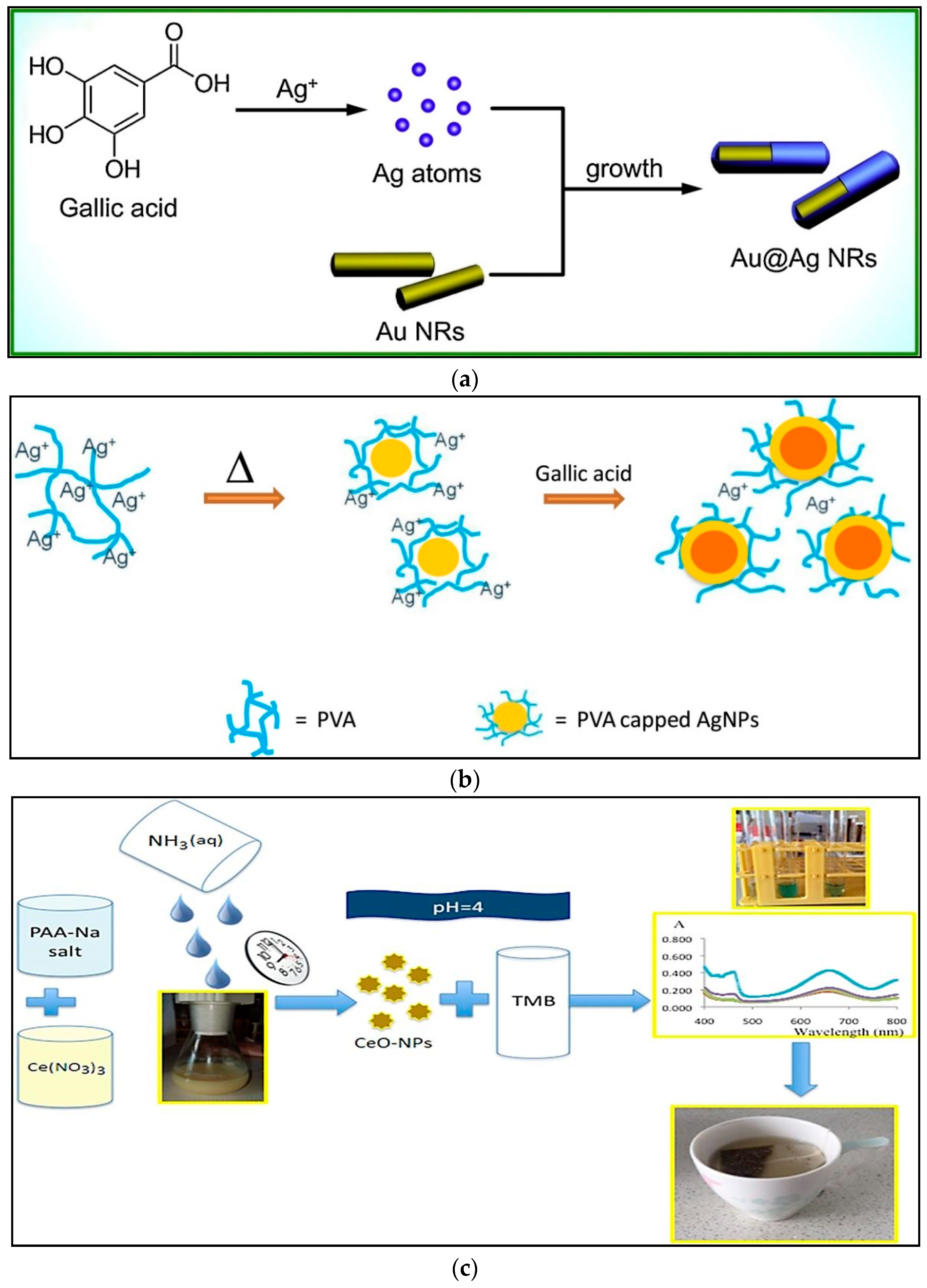
| Absorption, Formation of Au@Ag NRs by seed-mediated growth | Au nanorods | Gallic acid | 0.0064 µM | 0.01–30 μM | Tea | Simple, reliable, highly sensitive, selective, stable/Failure to report reproducibility, incubation time | [34] |
| AgNPs seed growth (PVA-AgNPs), reduction Ag + to Ag | AgNPs | Gallic acid | 22.1 µM | 25–200 µM | Ginger tea powder | Simple, fast, greener method, stable, easier to use, sensitive, precise, short incubation time (10 min)/Failure to report reproducibility | [37] |
| Ag+1 reduction at room temperature) | AgNPs | Caffeic acid, catechin, catechol, chlorogenic acid, epicatechin, epigallocatechin, ferulic acid, gallic acid, kaempferol, myricetin, quercetin, rutin, Trolox | AgNPs-RT (0.4 µM) and AgNPs-HT (58 μM) | AgNPs-RT (0.25–125 µM) and AgNPs-HT (20–600 μM) | Tea | Good reproducibility, simple, sensitive, cost effective, short incubation time (10 min)/Lack of stability | [5] |
| Ag+ reduction | AgNPs | Ascorbic acid, rutin | Not reported | Not reported | Garlic, green tea and turmeric extracts | Good stability/Failure to report LOD, linear range, reproducibility, sensitivity | [38] |
| Ce (IV) reduction, mild condition 60 min) | CeO-NPs | Sinapic acid | 2.75 × 10⁻³ μM | 1.2–1.7 mM | Rapeseed and its by products | Simple, rapid, low-cost, precise, accurate, sensitive, applied by oil industry laboratories, low incubation time (120 min)/Failure to report stability and reproducibility | [42] |
| Interaction between the polyphenolic analyte and nanoceria in acidic medium | CeO-NPs | Quercetin, | 8.25 × 10⁻⁹, | 1.00 × 10⁻⁴–7.81 × 10⁻⁶ M | Not reported | Sensitive, selective, low-cost, robust, stable, reproducible, and can be combined with other conventional laboratory equipment/ No evaluation food matrix, failure to report incubation time | [17] |
| ascorbic acid, | 6.87 × 10⁻⁹, | ||||||
| rutin, | 3.77 × 10⁻⁹, | ||||||
| caffeic acid, | 5.21 × 10⁻⁹, | ||||||
| naringenin, | 1.20 × 10⁻⁸, | ||||||
| gallic acid, | 6.81 × 10⁻⁹, | ||||||
| BHT, | 5.54 × 10⁻⁸, | ||||||
| ferulic acid, | 5.47 × 10⁻⁹, | ||||||
| vitamin E, | 6.68 × 10⁻⁹, | ||||||
| catechin, | 5.23 × 10⁻⁹, | ||||||
| Trolox | 5.94 × 10⁻⁹ M | ||||||
| Paper based, reduction of cerium ion | CeNPs | Epigallocatechin, gallate equivalent, gallic acid, caffeic acid, quercetin, ascorbic acid, vanillic acid | 4.0, 5.0, 6.0, 6.0, 8.0, 8.0 µM | 0.02–0.10, 0.08–1.00, 0.04–1.00, 0.40–10.00, 0.10–4.00, 0.01–0.08 mM | Tea | Fast, simple, instrument-free, cheap, portable, good stability (50 days), good reproducibility and high recovery/Failure to report incubation time | [11] |
| Microfluidic paper-based analytical devices, PMAA-coated ceria nanoparticles | CeO2NPs | ascorbic acid, quercetin, riboflavin, gallic acid, catechin, caffeic acid, PMAA-coated ceria nanoparticles comparison gallic acid | 0.27, 0.35, 0.27, 0.10, 0.28, 0.20, (μg mL−1), 0.6 μM (0.10 μg mL−1) | 30–150 μM (~5–25 μg mL−1) | Tea | Low-cost, convenient, portable, good stability, sensitive, reproducibility, and reliable method/Failure to report incubation time | [43] |
| Fe(III) reduction, mild condition 50 min | Iron oxide NPs | Sinapic acid, | 0.019, | 0.06–4.80 µM | Rapeseed oil | Simple, low cost, precise, convenient, not require specialized equipment, short incubation time (5–60 min), good stability and special Reagents/Less sensitive, failure to report reproducibility | [44] |
| caffeic acid, | 0.016, | ||||||
| gallic acid, | 0.024, | ||||||
| ferulic acid, | 0.012, | ||||||
| vanillic acid, | 0.071, | ||||||
| Trolox | 0.047 µM | ||||||
| Immobilizing a chromogenic onto a Nafion cation-exchange membrane oxidant | Fe(III) | Trolox, caffeic acid, ferulic acid, catechin, gallic acid, quercetin, rutin, rosmarinic acid, ascorbic acid, uric acid, α-tocopherol, bilirubin, glutathione, cysteine, homocysteine | 0.26 µM | 2.45–47.39 µM, 0.46–104.8 µM | Fruit juices | Sensitive, small, cheap, rapid, selective, stable, easily convertible to kit format, without sample pretreatment, reliable, robust, precise, without incubation/Failure to report reproducibility | [10] |
| Ferric reducing antioxidant power | AuNPs, AgNPs, Iron oxide NPs | Catechin, protocatechuic acid, gallic acid, vanillic acid, caffeic acid, syringic acid, hydroxybenzoic acid, chlorogenic acid, ferulic acid, quercetin, rutin | Not reported | Not reported | Rice wine, zhuyeqing liquor | Not require the use of expensive radical compounds and organic solvents, stable, good reproducibility, short incubation time (60 min)/Lower precision, Failure to report LOD and linear range | [45] |
| Transform MnO2 nanosheets to Mn2+) | MnO2 nanosheets (UV–vis) MnO2 nanosheets | Gallic acid | 0.01 µM, 0.3 µM | 0.1–12 µM, 3–15 µM | Red wine | Easy operation, low cost, rapid detection, high sensitivity, a portable and user-friendly method/Failure to report incubation time and reproducibility | [47] |
| Reaction between MnO2 nanosheets and Tetramethylbenzidine | MnO2 nanosheets | Uric acid, glutathione, ascorbic acid, cysteine, melatonin | 20 µM | Not reported | Fetal bovine serum | Simple, rapid, economical, short incubation time (15 min)/Not determined ultralow antioxidant concentration (such as nM level), Failure to report linear range, sensitivity, stability, reproducibility, no evaluation food matrix | [48] |
| Synthesized MnO2 nanosheets | MnO2 nanosheets | Gallic acid, | 0.1 µM | 0.1–35, | Human serum, plant extracts, fruit juice, and liver tissue extracts | Reliable, accurate, sensitive, selective, robust, cost effective/Failure to report incubation time, stability and reproducibility | [46] |
| Trolox, | 1–180, | ||||||
| quercetin, | 0.1–35, | ||||||
| caffeic acid, | 0.5–100, | ||||||
| hesperidin, | 0.1–50, | ||||||
| α-tocopherol, | 1.0–160, | ||||||
| resveratrol, | 0.5–100, | ||||||
| gluthathione, | 1.0–140, | ||||||
| cysteine, | 1.5–200, | ||||||
| ascorbic acid, | 0.5–160, | ||||||
| uric acid | 0.5–160 µM | ||||||
| Redox reaction between MnOx NPs and the TMB chromophore | MnO2 NPs | Catechin, | 8.16 × 10⁻⁹, | 3.3 × 10⁻⁴–6.67 × 10⁻⁶ M | Tea and orange juice | Low cost, easy use, rapid response, high precision, repeatability, stable (1 month), high sensitivity, reproducible, short incubation time | [49] |
| quercetin, | 1.23 × 10⁻⁹, | ||||||
| ascorbic acid, | 1.60 × 10⁻⁹, | ||||||
| caffeic acid, | 5.96 × 10⁻⁹, | ||||||
| gallic acid, | 2.49 × 10⁻⁹, | ||||||
| rutin, | 5.38 × 10⁻⁸, | ||||||
| p-coumaric acid, | 2.61 × 10⁻⁸, | ||||||
| chlorogenic acid, | 1.67 × 10⁻⁹, | ||||||
| vanilic acid, | 1.71 × 10⁻⁷, | ||||||
| ferulic acid, | 2.75 × 10⁻⁹, | ||||||
| kaempferol, | 9.94 × 10⁻⁹, | ||||||
| α-tocopherol, | 1.56 × 10⁻⁸, | ||||||
| glutathione, | 3.63 × 10⁻⁹, | ||||||
| L-cysteine | 1.14 × 10⁻⁹ M | ||||||
| Solution based | PtNPs | Trolox gallic acid, vanillin, caffeine, theobromine | 17.2 µg g−1 (in methanol), 763.3 µg g−1 (in water) | Not reported | Tea, herbal infusions | Failure to report stability, sensitivity, selectivity, incubation time | [50] |
| Colorimetric Spots—digital image-based (DIB) based on reacting diazotized amino benzene with phenolic compounds | Sulfanilic acid, sulfanilamide, aniline | 6.5, 5.5, 5.1 mg (gallic acid equivalent) L−1 | 25–400, 20–400, 18–200 mg GAE L−1 | Tea, Fruits | Fast, low cost, versatile, robust, portable, used for in situ-analysis, good reproducibility, stable, short incubation time (60 min), sensitive, selective | [19] | |
| Paper based | Gallic acid | 1 mM | 3–13 mM | Tea, Wine, Fruit juices | Portability, low reagent and sample consumption, inexpensive, simple, rapid/Failure to report stability, selectivity, reproducibility, incubation time | [15] | |
| Bind EBT and lanthanide ions | Lanthanide ions (Eu3+, La3+, and Sm3+) | quinolinic acid, 2,3-pyridine-dicarboxylic acid, tannic acid, tartaric acid, and gallic acid | Not reported | Not reported | Not reported | Facile, robust, sensitive/Failure to report stability, reproducibility, selectivity, LOD, linear range, food matrix | [51] |
| Cupric-neocuproine immobilized into a polymethacrylate matrix | Cu(II)−Nc | Gallic acid, | Not reported | 0.5–4.0, | Tea | Small, cheap, suitable to fit in a portable instrument for in situ antioxidant analysis, without sample pretreatment, sensitive, selective, short incubation time (45 min)/Failure to report stability, reproducibility, LOD | [52] |
| quercetin, | 0.05–0.8, | ||||||
| ascorbic acid, | 0.4–3.0, | ||||||
| catechin, | 0.3–11.0, | ||||||
| dihydroquercetin, | 0.05–0.8, | ||||||
| tannin, | 0.1–0.8, | ||||||
| luteolin, | 0.05–0.8, | ||||||
| rutin, | 1.0–15.0, | ||||||
| cysteine | 0.5–11.0 mg L−1 | ||||||
| Paper based | Nano-oxides of Al2O3, ZnO, MgO, CeO2, TiO2 and MoO3 | Caffeic acid, | Not reported | 8.0 × 10⁻⁴–1.0 × 10⁻² | Tea | Simplicity, low cost, portable, sensitive, good reproducibility/Failure to report stability, incubation time, LOD | [53] |
| rosmarinic acid, | 4.0 × 10⁻⁴–1.0 × 10⁻² | ||||||
| gallic acid, | 8.0 × 10⁻⁴–1.0 × 10⁻² | ||||||
| ellagic acid, | 8.0 × 10⁻⁴–1.0 × 10⁻² | ||||||
| and quercitrin | 8.0 × 10⁻⁴–1.0 × 10⁻² (moles L−1) | ||||||
| RhNPS LSPR shifting | Rhodium NPs | Catechins, gallates, cinnamates, dihydroxybenzoic acids | 29 µM | 50–500 µM | Teas | High stability, short incubation time, without sample pretreatment, good reproducibility/Not designed for in-field assays | [54] |
| Paper microzone assay | Polyoxometalate-imidazol | Gallic acid catechin | 0.08, 0.15 mM | 0.25–2 mM | Lime fruit | Simple, fast, not require instrument, reliable, robust, reproducible/Low stability (72 h), failure to report incubation time | [55] |
| Immobilization of NaIO4 and MBTH in paper | Not reported | Chlorogenic acids | 0.002 mM | 0.07–0.71 mM | Coffee | Reproducible, good recovery, selective, rapid, easy to operate, low-cost, reliable, good stability (28 days)/Failure to report incubation time | [56] |
| Reactions between 3,3′,5,5′-tetramethylbenzidine (TMB) and metal ions | Ag+, Au3+, and Cr6+ | Lipoic acid, | 4.3, | 0–1000 nM | Serum samples | High sensitivity, high selectivity, high reproducibility, stable, short incubation time (30 min) | [6] |
| cysteine, | 4.74, | ||||||
| tannin, | 4.88, | ||||||
| ascorbic acid, | 4.23, | ||||||
| glutathione, | 2.44, | ||||||
| uric acid, | 7.48, | ||||||
| glycine, | 3.07, | ||||||
| dopamine | 1.97 nM |
| Strategy | Nanomaterial | Antioxidants | LOD | Linear Range | Food Matrix | Advantages/Disadvantages | Ref. |
|---|---|---|---|---|---|---|---|
| CdTe QDs fluorescence quenching inhibition | CdTe sodium periodate | Catechin, quercetin, rutin, chlorogenic acid, gallic acid, caffeic acid | 0.63 nM | 0–4.24 µM | Tea, lemon balm, peppermint, lime, chamomile, and coffee infusions | More sensitive, selective/Failure to report stability, reproducibility, incubation time | [67] |
| Polyaniline quenched CdTe QDs fluorescence | CdTe QDs | Glutathione, ascorbic acid | 50, 100 nM | 2–40 µM | Not reported | Simple, sensitive, label-free, stable, selective/Failure to report reproducibility, incubation time, food matrix | [68] |
| Glutathione-capped CdTe quantum dots | CdTe QDs | Glutathione | 0.00005 mM | 0.0001–0.005 mM | Juice beverages | Very low consumption of reagents, reproducible/Failure to report incubation time, stability | [69] |
| Fluorescence quenching of the QDs | CdSe/ZnS QDs and graphene | tert-butylhydroxy-anisole, tert-butylhydroxy-toluene, tert-butylhydroquinone, Propyl gallate, sesamol, ferulic acid, daidzein, carnosol | 0.7 µM | 2–27 µM | Not reported | High sensitivity, accuracy, rapid detection/Failure to report stability, reproducibility, incubation time | [70] |
| Graphene QDs quenching on paper | Graphene QDs | Morin, myricetin, quercetin, kaempherol | 6.67 × 10⁻⁵ M (UV), 2.35 × 10⁻⁵ M (UV LED) | 1.66 × 10⁻⁵ to 1.33 × 10⁻⁴ M (UV), 1.66 × 10⁻⁵ to 2.50 × 10⁻⁴ M (UV LED) | Wine samples | Simple, inexpensive, rapid sensing systems, sensitive, reproducible, stable/Failure to report incubation time | [71] |
| Reduction of Cu2+ to Cu+ | Graphene QDs | Ascorbic acid | 0.094 µM | 0.3–10 µM | Not reported | Rapid, sensitive, low-cost, simple, highly efficient, selective, stable/Failure to report reproducibility, short incubation time (1 min) | [72] |
| Reaction between (GQDs) and squaric acid (SQA)-iron(III) | Graphene QDs | Ascorbic acid | 0.2 µM | 1–95 µM | Fruit juices | Simple, sensitive, selective, rapid, label free, versatile, not require high toxic metal (e.g., Cr(VI)) and high-cost enzyme, satble, short incubation time (1 min)/ Failure to report reproducibility | [73] |
| Benzoquinone in the presence of hydroxyl radicals caused quenching of GQDs) | Graphene QDs | Ascorbic acid | 0.32 µM | 1.11–300 µM | Human serum | Simple, low cost, higher sensitivity, selectivity, rapid, stable/Failure to report incubation time, reproducibility, without evaluation food matrix | [74] |
| Graphene QDs Hypochlorite hybrid system | Graphene QDs | Ascorbic acid | 1.4 µM | 8–60 µM | Commercial drinks (Orange juice, Apple juice, Tea) | High recovery, stability, short incubation time (30 min), robust, sensitive/Failure to report reproducibility | [75] |
| Quenching graphene QDs by Cr(VI) | Graphene QDs | Ascorbic acid | 0.0037 µM | 0.05–500 µM | Water samples | Simple, rapid, sensitive, selective, short incubation time (5 min)/Failure to report stability, reproducibility | [76] |
| Glutathione binding Hg2+ | Graphitic carbon nitride quantum dots (g-CN QD) | Glutathione | 37 nM | 0.16–16 µM | Not reported | High selectivity, sensitivity, cost-effectivity, rapidity, stable, short incubation time (5 min)/Failure to report reproducibility, not evaluation in food matrix | [77] |
| Aluminium(III)-quercetin complex | Not reported | Quercetin | 0.3 mg L−1 | 1.5–60.5 mg L−1 | Apple juices | Sensitive, selective, accurate, short incubation time (30 min)/Failure to report stability, reproducibility | [78] |
| Detection Method | Strategy | Receptor | Target Molecule | LOD | Linear Range | Specificity | Food Matrix | Advantages/Disadvantages | Ref. |
|---|---|---|---|---|---|---|---|---|---|
| Linear-sweep voltammetry (LSV) | HRP immobilization on the surface of Au-Pt nanotube/Au graphene-modifed carbon electrode | HRP | BHA | 0.046 mg L−1 | 0.3–50 mg L−1 | Excellent | Peanut oil, potato chips and cookies | Good sensitivity, stability and reproducibility, high selectivity, long-term stability (2 months)/failure to report detection time | [84] |
| PG | 0.024 mg L−1 | 0.1–100 mg L−1 | |||||||
| Differential pulse voltammetry (DPV) | Carbon paste electrode modified with laccase | Laccase | Phenolic compounds | Not reported | Not reported | Not reported | Honey | High simplicity and accuracy, good correlation with the spectrophotometric method/failure to report sensitivity, stability and reproducibility, semi-quantitative | [85] |
| Amperometry | Laccase immobilization onto AuNPs/graphene nanoplatelets-modified screen-printed carbon electrode (AuNPs/GNPI-SPCE) | Laccase | Hydroquinone | 1.5 µM | 4–130 µM | High | Blueberry syrup, wine | High sensitivity and selectivity, high repeatability, cost-effective /short-term stability (5 days), failure to report detection time | [89] |
| Amperometry | Laccase immobilization onto SPCE modified with AuNPs and polypyrrole | Laccase | Polyphenolic compounds | 0.83 µM | 1–250 µM | Excellent | Propolis | High sensitivity and selectivity, portable, low-cost, rapid (15 min), long-term stability (1 month), high accuracy and reproducibility/- | [90] |
| Amperometry | Entrapment of laccase within poly (3,4-ethylenedioxy-thiophene) and graphene oxide nano-sheets composite and electrode modification | Laccase | Catechol | 0.032 µM | 0.036–0.35, 0.35–2.5 µM | Excellent | Water | High sensitivity and selectivity, good reproducibility/no evaluation of stability, failure to report detection time | [129] |
| Amperometry | Immobilization of bacterial laccase on the surface of Escherichia coli cells and adsorption of modified live cells onto a GCE | Laccase | Catechol | 0.1 µM | 0.5–300.0 µM | Good | Wine, tea | High sensitivity, good selectivity, long-term stability (one month at room temperature), high reproducibility/failure to report detection time | [86] |
| Amperometry | Mixing of laccase crude extract with graphite and filling the cavity of electrode support | Laccase | Total phenol content | Red fruits | Simple, low-cost/no evaluation of sensitivity, selectivity, reproducibility and stability, failure to report detection time | [130] | |||
| Amperometry | Laccase immobilization during the potentiostatic deposition of a thin polydopamine film on carbon surfaces | Laccase | Gallic acid, caffeic acid, rosmarinic acid | 0.29 µM | 1–150 µM | Chestnut shell extract | High sensitivity, simple, high reproducibility/no evaluation of, selectivity and stability, failure to report detection time | [131] | |
| Amperometry | Laccase immobilization on the surface of GCE modified with Fe2O3 yolk-shell particles | Laccase | 2,6-dimethoxy-phenol | 0.010 µM | 0.025–750 µM | High | High sensitivity and selectivity, good reproducibility, long-term stability (2 months at 4 °C)/failure to report detection time | [132] | |
| Amperometry | Tyrosinase or laccase immobilization on the surface of GCE modified with rGO-MWCNTs hybrid | Tyrosinase Laccase | Catechol, gallic acid, pyrogallol, 1,2-dihydroxy-benzoic acid, dopamine, epicatechin, rutin, caffeic acid, chlorogenic acid | Tyrosinase (0.5, -, 2, -, 1.7, 1.8, 7, -, 2, 16 µM) Laccase (0.3, 7.5, 2, 0.6, 1.2, 0.5, 1, 2, 0.5, 0.5 µM) | 1–340 µM | Not reported | Fruit juice | High sensitivity, low-cost, long-term stability (laccase-based), excellent repeatability/Short term stability (tyrosinase-based), failure to evaluate selectivity, failure to report detection time | [92] |
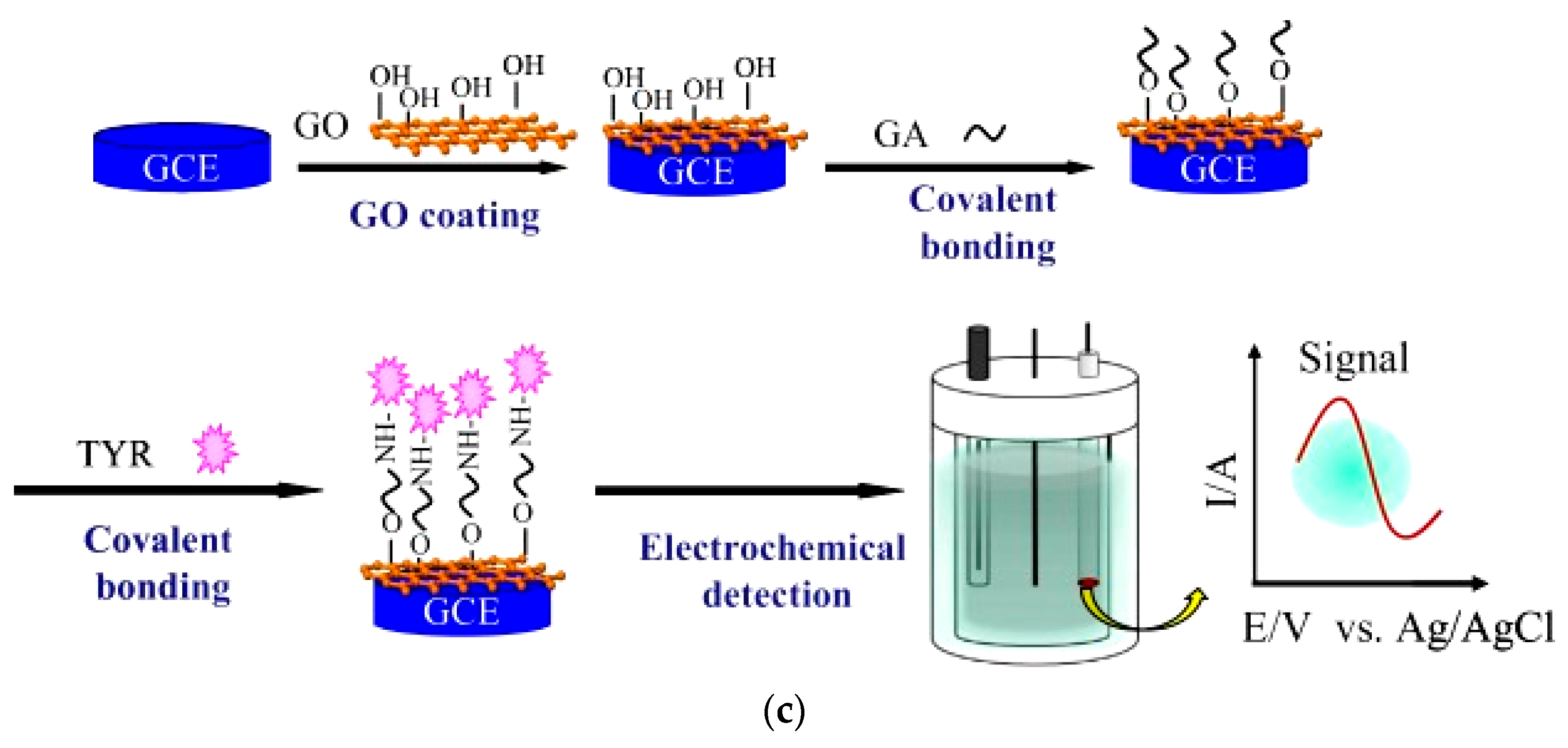
| Amperometry | Tyrosinase immobilization on the surface of graphene oxide-modified GCE | Tyrosinase | Catechol | 0.03 µM | 0.05–50 µM | High | High sensitivity and selectivity, good reproducibility and repeatability, long-term stability (1 month), short assay time/no evaluation of food matrix | [93] | |
| Potentiometry | Tyrosinase immobilization on the solid-contact transducer | Tyrosinase | Catechol | 0.73 µM | 0.93–8.3 × 10⁴ µM | High | Honey, propolis | High sensitivity and selectivity, long-term stability (3 month), re-usability, high mechanical resistance/failure to report detection time | [94] |
| Amperometry | Carbon paste electrode coated with MWCNTs and Nafion film containing the tyrosinase | Tyrosinase | Trolox equivalent antioxidant capacity | Moravian wines | Simple, rapid/no evaluation of sensitivity, selectivity, reproducibility and stability | [133] | |||
| Amperometry | Carbon paste electrode coated with MWCNTs and Nafion film containing the tyrosinase | Tyrosinase | Trolox equivalent antioxidant capacity | Blackberries, blueberries, cranberries, raspberries and strawberries | Low-cost, simple, portable/short-term stability (one week at 5 °C), no evaluation of sensitivity, selectivity and reproducibility | [134] | |||
| Amperometry | Immobilization of fibrous electrode material of silica–PVA with immobilized tyrosinase onto an indium–tin oxide-coated glass substrate | Tyrosinase | Catechol | 10 µM | 10–200 | Low-cost, simple/short-term stability (4 days at 5 °C), no evaluation of selectivity and reproducibility, failure to report detection time | [135] | ||
| Phenol | 10–150 | ||||||||
| p-cresol | 10–100 µM | ||||||||
| Amperometry | Tyrosinase immobilization onto SPCE modified with AuNPs | Tyrosinase | Catechol | 1.2 | 2.5–20 | Not reported | Beers | High sensitivity, low-cost, acceptable repeatability and reproducibility/failure to report detection time, no evaluation of stability and selectivity | [87] |
| phenol | 1.2 | 2.5–20 | |||||||
| caffeic acid | 2.3 | 2.5–12.5 | |||||||
| tyrosol | 1.7 µM | 2.5–40 µM | |||||||
| DPV | Magnetic silica/titania xerogel as support for direct immobilization of tyrosinase | Tyrosinase | Catechol | 0.23 | 40–530 | High | High sensitivity and selectivity, good reproducibility, ability of simultaneous detection of catechol and catecholamines/no evaluation of stability, failure to report detection time | [136] | |
| Dopamine | 0.72 | 40–400 | |||||||
| Epinephrine. | 2.94 µM | 40–530 µM | |||||||
| Cyclic voltammetry (CV) | Gold disk microelectrode arrays and interdigitated microband electrode arrays modified with SWCNTs, poly(3,4-ethylenedioxythiophene), and tyrosinase | Tyrosinase | Catechol dopamine | 2.4 µM | 100–500 µM | Not reported | High sensitivity, good repeatability, simultaneous determination of catechol and dopamine/short-term stability (3 days at 4 °C), failure to report detection time, no evaluation of selectivity | [137] | |
| Amperometry | GCE modification with enzyme extract encapsulated within polypyrrole | Polyphenol oxidases (laccase, tyrosinase) | Catechol | 1.8–5.0 µM | 1–60/70 µM | High | Fruit wines | High sensitivity and selectivity, low-cost/no evaluation of stability and reproducibility, failure to report detection time | [138] |
| DPV | RAW264.7 cells immobilization onto acidified manganese dioxide (a-MnO2)-modified gold electrode | RAW264.7 macrophage cells | Antioxidant capacity | 0.02 µM | 0.05–0.85 µM | Not reported | Cell-free extracts of L. plantarum | High sensitivity, low cost, simple, good reproducibility/short-term stability (10 days), failure to report detection time | [100] |
| DPV | Caco-2 cells immobilization onto gold electrode modified with PtNPs and silver nanowires | Caco-2 cells | Antioxidant capacity | 0.12 µM | 0.2–2 µM | Not reported | Asp-Leu-Glu-Glu peptide | High sensitivity, low cost, simple, acceptable reproducibility, good stability (15 days at room temperature)/long incubation time (14 h) | [101] |
| Electrochemical impedance spectroscopy (EIS) | A549 cells immobilization on the self-assembled ʟ-cysteine/AuNPs-modified GCE surface | A549 cells | Phloretin | 1.96 µM | 20–100 µM | Not reported | High sensitivity, high accuracy, good reproducibility/short-term stability (10 days at 80 °C), failure to evaluate selectivity, failure to report detection time | [102] | |
| DPV | Adsorption of E. coli cells with surface-displayed bacterial laccase on the GCE | E. coli MB275 cells | Catechol, caffeic acid, dopamine, gallic acid, 2-amino- phenol | 1.0–5.0 µM | 5.0–500.0 µM | Not reported | High sensitivity, high accuracy, good reproducibility and stability/failure to report detection time and selectivity | [103] | |
| DPV | Immobilization of dsDNA on the SPCE modified with SWCNTs | dsDNA | Chlorogenic acids | Not reported | Coffee | High accuracy, rapid (15 min), simple/failure to report sensitivity, stability and reproducibility | [112] | ||
| EIS | Human interleukin-2 (IL-2) gene probe immobilization on the surface of AuNPs-modified SPE and Fenton reaction | ssDNA | Phenolic compounds | Not reported | Acanthophora algea | Simple, low-cost, short incubation time (1 h), high reproducibility, long-term stability (40 days at 4 °C)/- | [115] | ||
| Square wave voltammetry (SWV) | dA20 oligonucleotide immobilization onto CPE and Fenton reaction | dA20 oligonucleotide | Phenolic compounds and TAC | Not reported | Green tea, black tea, peppermint, balm, senna, fennel, dandelion, a mixture of orange blossom, fennel, corn silk, bladderwrack, senna, and a mixture of horsetail, artichoke, green nettle, whitethorn | Simple, low-cost, short reaction time (30 s)/no evaluation of stability and reproducibility | [116] | ||
| DPV | dsDNA immobilization on the surface of chitosan-coated CPE | dsDNA | Oleuropein | 0.090 µM | 0.30–12 µM | Low | Olive leaf extract | High sensitivity, short reaction time (10 min), simple operation, good repeatability/no evaluation of stability and reproducibility, low specificity | [117] |
| Amperometry | SPE modified with Prussian Blue and xanthine oxidase | Xanthine oxidase | Gallic acid | 2.17 µM | 1.0–75 µM | High | Amazonian fruits samples | High sensitivity and selectivity, short detection time, low-cost, portable, signal stability, good reproducibility/short-term stability (2 days at 4 °C) | [118] |
| DPV | CPE modified with MIP/MWCNTs | MIP | Gallic acid | 47.0 nM | 0.12–380 µM | High | Fruit juices | High sensitivity and selectivity, short detection time (14.5 min), simple, low-cost, reproducible/short-term stability (7 days at 4 °C) | [120] |
| Amperometry | SPCE modified with nanoceria | CeO2NPs | Gallic acid | 1.5 | 2–20 | High | Wine | High sensitivity and selectivity, short detection time (40 s), simple, low-cost, reproducible, high stability/- | [122] |
| caffeic acid | 15.3 | 50–200 | |||||||
| quercetin | 8.6 | 50–200 | |||||||
| ascorbic acid | 0.4 µM | 0.5–20 | |||||||
| Amperometry | Scavenging capacity of AOx present in the plant extracts towards H2O2 using SPE modified with AuNPs | AuNPs | Lavender and sea buckthorn extracts | Simple, low-cost/no evaluation of stability, sensitivity and reproducibility, non-selective | [123] | ||||
| DPV | GCE modified with ZIF-65 MOFs @CNTs | ZIF-65 MOFs @CNTs | Ascorbic acid | 1.03 µM | 200–2267 µM | High | High sensitivity and selectivity, anti-interference and high reproducibility, time stability of DPV responses in 9 h/no evaluation of real samples, no evaluation of stability during storage | [126] | |
| DPV | GCE modified with Cu-MOF/ZnTe NRs/AuNPs nanocomposite | MOF/ZnTe NRs/AuNPs | Catechol | 16 nM | 0.25–300 µM | High | Water and tea samples | Excellent sensitivity, high selectivity, enhanced catalytic properties, anti-interference ability, excellent reproducibility, high accuracy and feasibility/short-term stability (15 days at room temperature) | [127] |
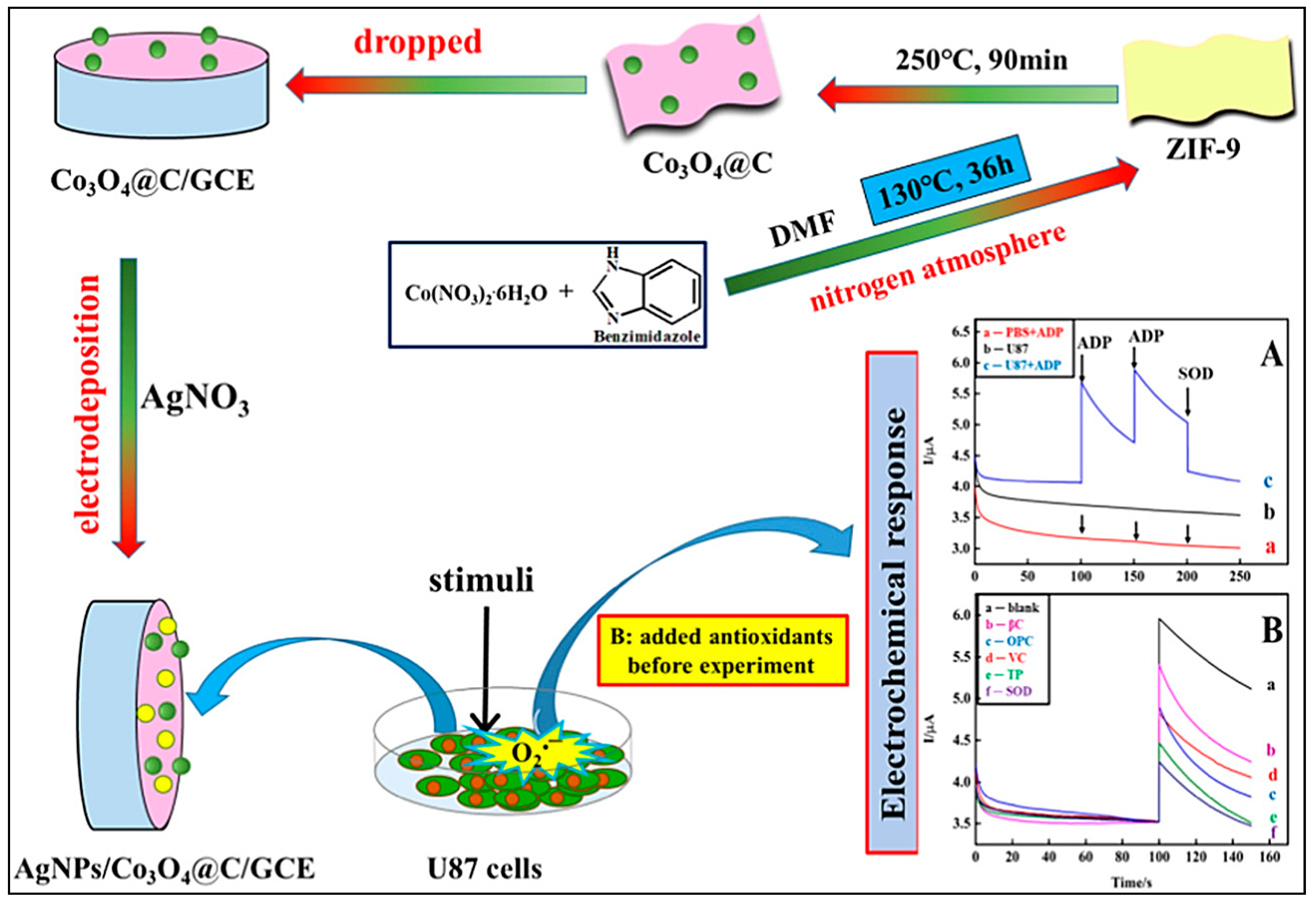
| Chronoamperometry | GCE modified with ZIF-9-derived cobalt oxide porous carbon material and decorated by AgNPs | AgNPs/Co3O4@C | O2•− | 0.0564 pM | 1.69 × 10⁻⁷–1.69 × 10⁻¹ µM | High | Food antioxidants | Excellent sensitivity, high selectivity, good reproducibility/no evaluation of stability during storage | [128] |
Publisher’s Note: MDPI stays neutral with regard to jurisdictional claims in published maps and institutional affiliations. |
© 2021 by the authors. Licensee MDPI, Basel, Switzerland. This article is an open access article distributed under the terms and conditions of the Creative Commons Attribution (CC BY) license (http://creativecommons.org/licenses/by/4.0/).
Share and Cite
Nejadmansouri, M.; Majdinasab, M.; Nunes, G.S.; Marty, J.L. An Overview of Optical and Electrochemical Sensors and Biosensors for Analysis of Antioxidants in Food during the Last 5 Years. Sensors 2021, 21, 1176. https://doi.org/10.3390/s21041176
Nejadmansouri M, Majdinasab M, Nunes GS, Marty JL. An Overview of Optical and Electrochemical Sensors and Biosensors for Analysis of Antioxidants in Food during the Last 5 Years. Sensors. 2021; 21(4):1176. https://doi.org/10.3390/s21041176
Chicago/Turabian StyleNejadmansouri, Maryam, Marjan Majdinasab, Gilvanda S. Nunes, and Jean Louis Marty. 2021. "An Overview of Optical and Electrochemical Sensors and Biosensors for Analysis of Antioxidants in Food during the Last 5 Years" Sensors 21, no. 4: 1176. https://doi.org/10.3390/s21041176