Article

Molecular Assisted Identification Reveals Hidden Red Algae Diversity from the Burica Peninsula, Pacific Panama

1
Center for Marine Science, University of North Carolina at Wilmington, Wilmington, NC 28409, USA
2
Centro de Investigación en Ciencias del Mar y Limnlogía y Escuela de Biología, Universidad de Costa Rica, San José 2060, Costa Rica
3
Departmento de Botánica, Universidad de Panamá, Panamá 4, Panama
4
NCU Herbarium, University of North Carolina at Chapel Hill, Chapel Hill, NC 27599-3280, USA
5
Department of Biology, Marine Biology & Environmental Science, Roger Williams University, Bristol, RI 02809, USA
*
Author to whom correspondence should be addressed.
Diversity 2017, 9(2), 19; https://doi.org/10.3390/d9020019
Submission received: 31 January 2017 / Revised: 23 March 2017 / Accepted: 8 April 2017 / Published: 14 April 2017
(This article belongs to the Special Issue Tropical Marine Biodiversity)

Abstract

:
The marine flora of Panama harbors a rich diversity of green, red and brown algae, and despite chronic understudy, it is reported as the second most diverse marine flora along the Pacific Central American coast, with 174 macroalgal species. Extensive new collections and molecular assisted identification (MAI) by an international team of researchers has revealed an even greater diversity for this country. Here, the intertidal and shallow subtidal marine flora of the remote Burica Peninsula is introduced. This area is characterized by an uplifted extensive intertidal flat composed of firm, sedimentary benthos known as mudrock, on which abundant algal communities thrive, even during extended periods of exposure. A collection of nearly 200 brown, green and red macroalgae specimens representing the first marine floristic inventory of this region was made in January 2011, and results of analyses of 45 foliose red algae specimens are presented. DNA sequence data for several loci (rbcL-3P; COI-5P; UPA) have been generated for molecular assisted identification and to guide morphological assessments. Twenty-six species were identified among the specimens including 21 new Pacific Panama records, as well as previously unrealized transisthmian distributions, and two new species, Neorubra parvolacertoides sp. nov. and Grateloupia irregularis sp. nov.

1. Introduction

…our surroundings seemed ideal for successful work with the marine flora—except for the one unhappy fact, which gradually became apparent, that a marine flora, in the ordinary sense, was in that region almost non-existent.
—Marshall Avery Howe [1]
The study of marine algae from the Pacific coast of Panama had an inauspicious start. An extreme tidal amplitude and often hot and dry conditions make the intertidal environment of Pacific Panama inhospitable for most marine algae and the subtidal region has only rarely been explored. This is especially true for the Gulf of Chiriqui. Taylor [2] made limited, collections of marine algae from the middle and eastern regions of the Gulf during the 1934 and 1939 Allan Hancock expeditions to the Galapagos Islands. Only a few subsequent efforts followed in this region and substantial marine algal collections had only been made around the Coiba Island world heritage site [3,4].
The Burica Peninsula is a phycologically unexplored region in the western Gulf of Chiriqui, and Punta Burica, the southernmost point of the peninsula, is a relatively remote area accessible by motorized vehicle only at low tide (Figure 1). There are no known collecting sites or reports of marine macroalgae from this area [5]. In contrast, multiple collections had been made along the immediately adjacent southeastern coast of Costa Rica [2,6], however, these have all been from sites within Golfo Dulce and not along the western coast of the Burica Peninsula.
The Punta Burica intertidal zone is distinctive from other sites throughout the region, in being composed of compacted, fine-grained, layered sedimentary rock known as mudstone. This layered substratum has a block-in-matrix texture at various scales produced by sedimentary processes, and has been thrust at an angle to the ground (Figure 1B,C) owing to tectonic activity dating back 30–60 Ka [7,8]. It is the product of thrust sheets of the NW margin of the Caribbean plate being forced over the subducting Nazca plate, along the Cocos Ridge. The differential integrity of these tilted marine sediments, thought to be of Charcoal Azul or Armuelles formations [7], results in layers of intact benthos, separated by eroded channels that form labyrinths of interconnected channels and pools on the ebb tide (Figure 1C). The compacted sediments can be easily broken by a light hammer, but are also not compromised under the weight of a small truck. These traits appear to make the Burica intertidal an adequate habitat for the colonization of marine algae, characterized by turfs and biofilms of microfilamentous and encrusting algae that occupy exposed substrates at low tide (Figure 1D). Colonization of this habitat may be facilitated by the ability of turf algae to penetrate a porous and laminated surface that also retains moisture. In addition, the entrainment of water in small intertidal pools supports the development of small, foliose algal communities (Figure 1E).
Molecular Assisted Identification (MAI) uses relatively short DNA sequences to cluster specimens into species for further phylogenetic and morphological analyses [9]. Where appropriate baseline data is available, DNA sequences may be used as DNA barcodes to provide objective species identifications of specimens [10]. Saunders [11] and Robba et al. [12] introduced the five prime end of the mitochondrial marker cytochrome c oxidase subunit 1 (COI-5P) for DNA barcoding in red algae and many subsequent studies have demonstrated its utility for species delimitation and identification e.g., [13,14,15]. Domain V of the plastid-encoded large ribosomal subunit gene, which is also referred to as the Universal Plastid Amplicon (UPA) and the RuBisCO large subunit gene (rbcL), specifically the 3′ end of this locus (rbcL-3P) are other DNA sequences that have been used to delimit and identify red macroalgae e.g., [16,17,18].
Here MAI results based on three DNA loci (rbcL-3P, COI-5P and UPA) and morphological characters for foliose marine red macroalgae collected from Punta Burica, Panama are presented, and when suitable background data is available, their phylogenetic relationships explored. An unrealized diversity of species including many new records for Pacific Panama and the eastern tropical Pacific are revealed. Two new species are described and others identified that may represent new species, but require additional study. That 26 species are documented among 45 specimens, from a preliminary investigation spanning only 3 days of field work, foreshadows a likely productivity of further field investigations coupled with MAI methods, and contradicts the apparent “non-existent” diversity mentioned by Howe [1].

2. Materials and Methods

Marine macroalgae were collected at four sites around Punta Burica, Panama during 8–10 January 2011 (Figure 1A). Specimens were sorted in the field, a herbarium specimen made and a portion of the thallus dried with silica gel desiccant [19]. When specimens were small and possibly included a mixture of species, the entire collection was silica gel dried, brought back to the lab, and sorted after rewetting. Vouchers (herbarium specimens and/or permanent slides) for all collections were accessioned in the David J. Seiren Herbarium at the University of North Carolina Wilmington (WNC), and duplicates deposited in the herbaria of the University of Panama (UPAN), the University of Costa Rica (USJ), and Roger Williams University. Specimen collection data is publically available at doi: www.dx.doi.org/10.5883/DS-RAPBPAN.
Silica gel dried specimens were rewet with seawater and sorted to insure monospecificity of samples. Total genomic DNA was extracted from these samples following the protocol of Hughey et al. [20] or using the illustra Nucleon Phytopure Genomic DNA Extraction Kit (GE Healthcare, Pittsburgh, PA, USA). Three different loci UPA, COI-5P and rbcL-3P were targeted for amplification and sequencing in this study. PCR amplifications were set up and run as described in Freshwater et al. [21] and Mamoozadeh and Freshwater [9]. Successful amplifications were cleaned using a StrataPrep PCR Purification Kit (Stratagene, La Jolla, CA, USA) and used as templates in BigDye Terminator v.3 (ThermoFisher Scientific, Foster City, CA, USA) sequencing reactions. Sequence reactions were cleaned with a Zymo DNA Sequencing Clean up Kit (Zymo Research, Irvine, CA, USA) and run on an ABI 3130xl Genetic Analyzer (ThermoFisher Scientific). Primers used in amplification and sequencing reactions are shown in Table 1. Amplifications with the rbcL-3P primer pair also spanned the rbcL-rbcS spacer region, but this locus was specifically analyzed for only the collected Asparagopsis specimens.
Individual sequence reactions were compiled and edited using Sequencher (Gene Codes Corporation, Ann Arbor, MI, USA) and final sequences accessioned in the BOLD and GenBank databases. Accession numbers and sequences are available at doi: dx.doi.org/10.5883/DS-RAPBPAN. Alignments of the different loci were constructed using MUSCLE [26] as implemented in the Geneious software package (Biomatters Limited, Aukland, New Zealand) and adjusted by eye. UPGMA cluster diagrams were generated from alignments of the three loci with the Geneious tree builder option and Jukes-Cantor distances. These analyses were done exclusively to cluster sequences by similarity for genetic species definition, and UPGMA was used explicitly to indicate that phylogenetic relationships were not inferred. Closest taxonomic affinities of the cluster-analyses defined genetic species were determined by BLAST [27] queries. DNA sequence data sets to examine the phylogenetic relationships of individual species were compiled from GenBank, aligned using MUSCLE and analyzed with RAxML [28] and MrBayes [29] as implemented in Geneious. Data matrix dimensions and specific methodologies for these analyses are included in Supplementary Material Table S1.

3. Results

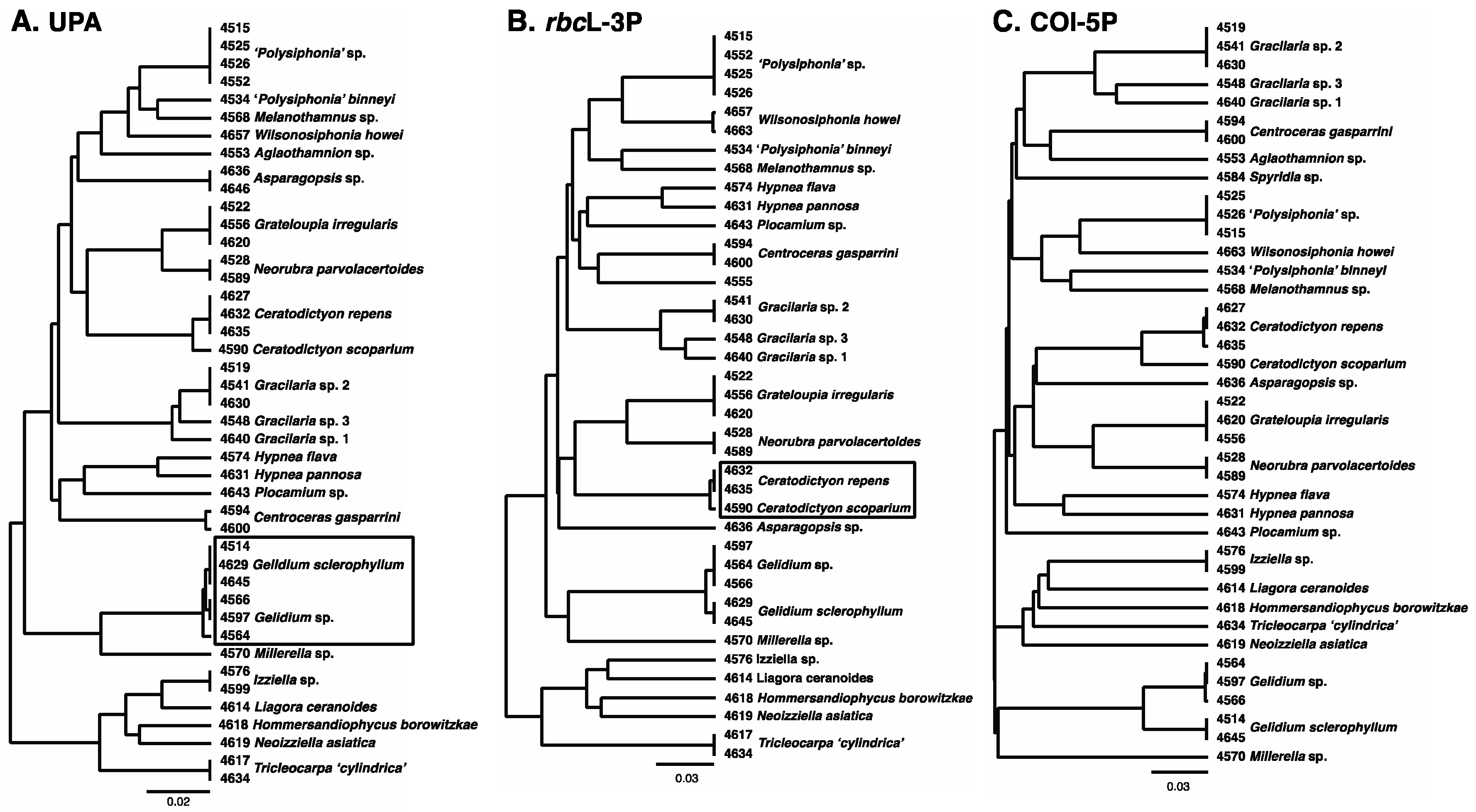
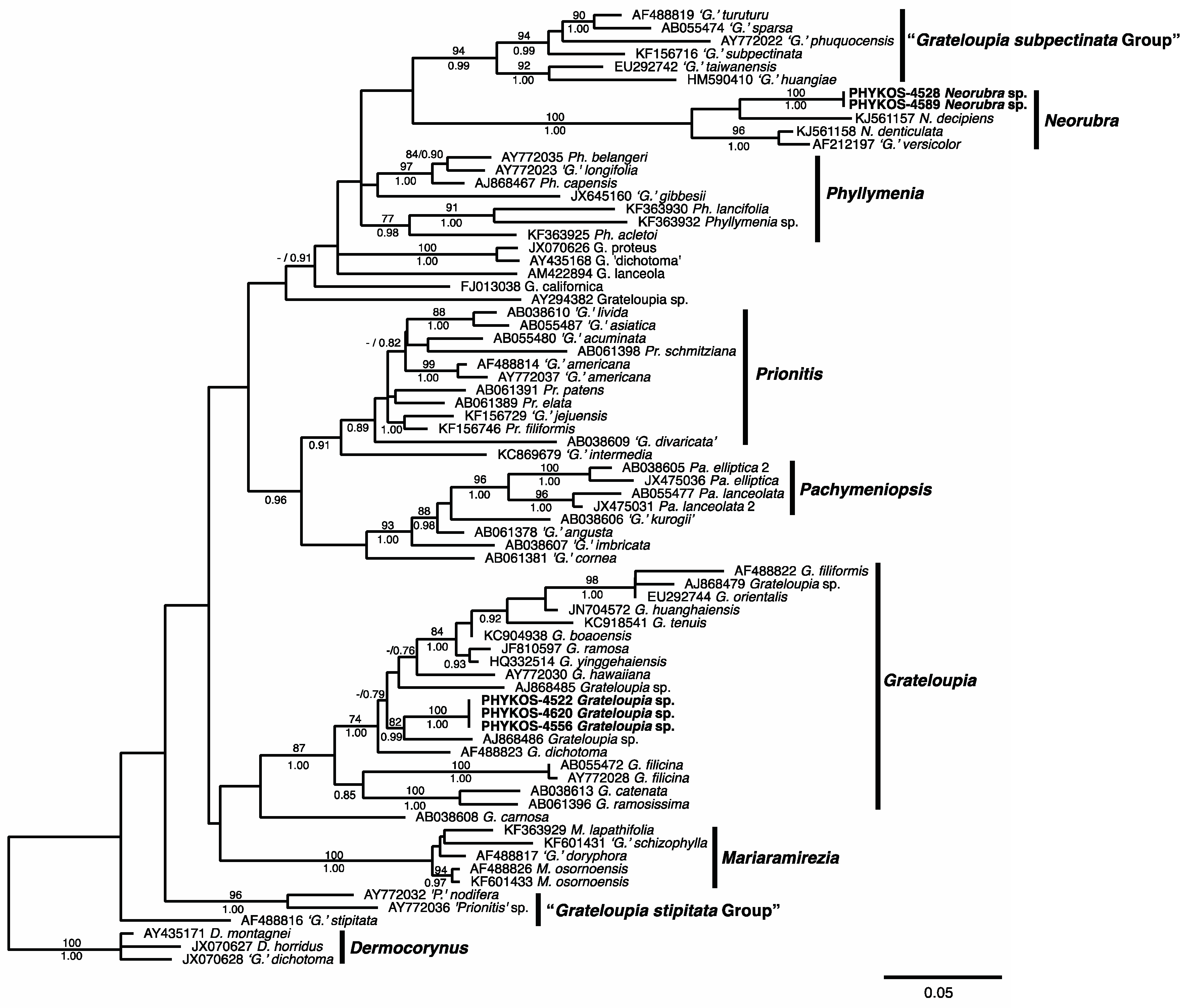
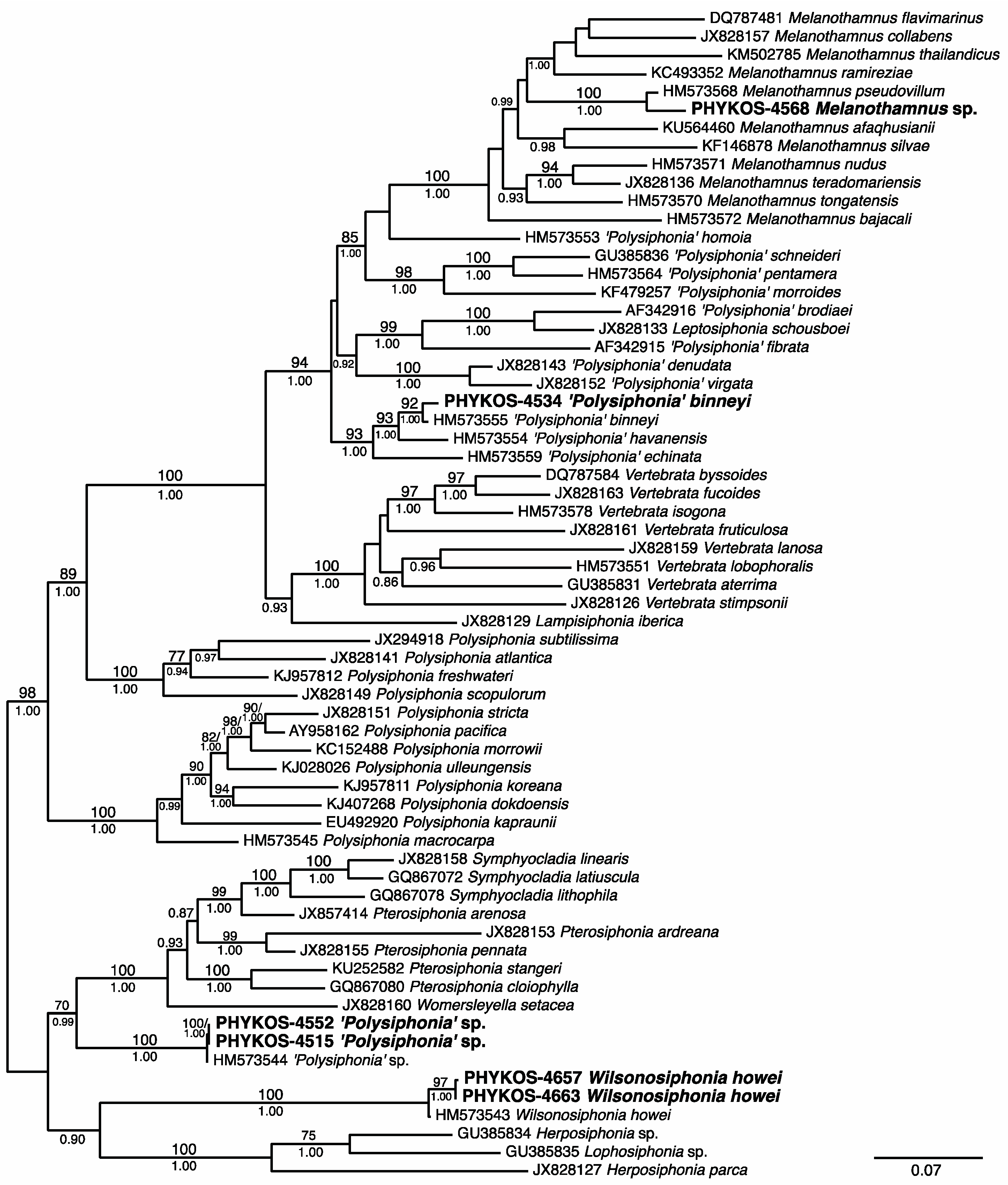
3.1. Overall “Barcoding” Cluster Analyses

Forty-five samples of foliose red algae were included in this study, and sequence data for at least two of the three targeted loci were generated for all but two of the samples (Table 2). Cluster analyses of the UPA data resulted in 24 clusters or individual sequences at least 1.1% different from the next closest cluster/sequence (Figure 2A). These clusters and individual sequences were considered to represent sequence-based species. Sequence variation within clusters of multiple specimens ranged from 0–0.5%. The maximum intra-cluster value was for the Gelidium specimens that rbcL-3P and COI-5P analyses indicated were different species and the next highest intra-cluster sequence variation was 0.3%. Cluster analysis of rbcL-3P also revealed 24 sequenced-based species (Figure 2B). Intra-cluster sequence variation ranged from 0–0.5%, and clusters or individual sequences were at least 0.9% different. As with the UPA data, the maximum intra-cluster variation value was between specimens of Ceratodictyon that formed distinct clusters in UPA and COI-5P analyses. The next highest rbcL-3P intra-cluster variation value was only 0.1%. The species identified by cluster analyses of these two data sets were identical except that the UPA cluster diagram did not clearly differentiate the two Gelidium species collected, and the rbcL-3P cluster diagram did not clearly differentiate the two Ceratodoctyon species collected. The analysis of the COI-5P data clearly differentiated 26 sequenced-based species (Figure 2C) including all those in the UPA and rbcL-3P data. Intra-cluster variation ranged from 0–0.3%, and clusters or individual sequences were at least 6.5% different.
Initial assessments by BLAST searches provided identifications at the genus level for all sequenced specimens (Table 2), but in some cases investigation of the current generic nomenclature was required because of changes that had occured since the submission of sequences into GenBank. Examples of this were the specimens of Hommersandiophycus and Ceratodictyon. Positive species identifications based on BLAST results were also possible for some specimens. The study of Centroceras by Won et al. [30] populated GenBank with rbcL sequence data for multiple species and provided the contex by which positive identifications of PHYKOS-4594 and PHYKOS-4600 as C. gasparrini were made. Similarly, Grusz and Freshwater [31] and Boo et al. [32] deposited rbcL and COI sequences for Gelidium sclerophyllum type specimens in GenBank and this allowed the positive identification of PHYKOS-4514, 4629, and 4645 as this species.

3.2. Species Treatments

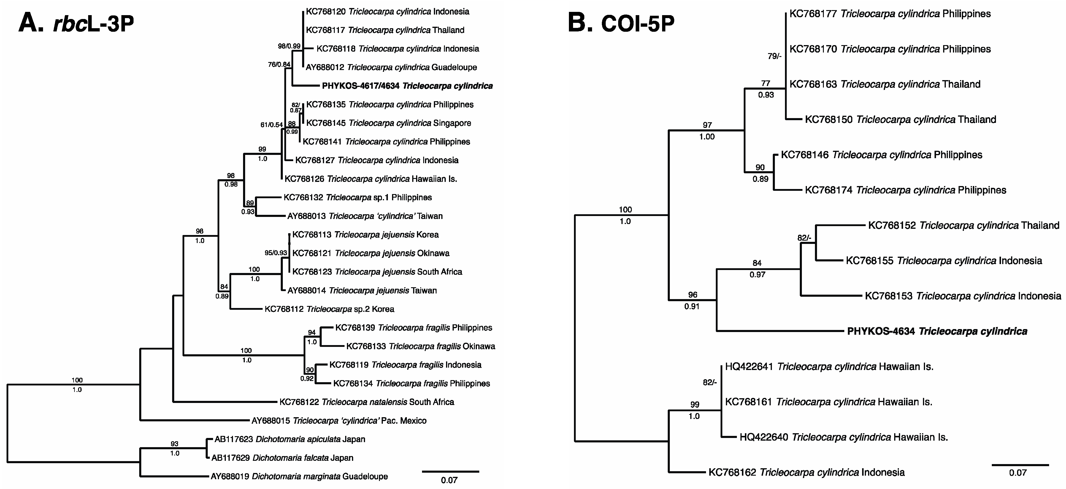
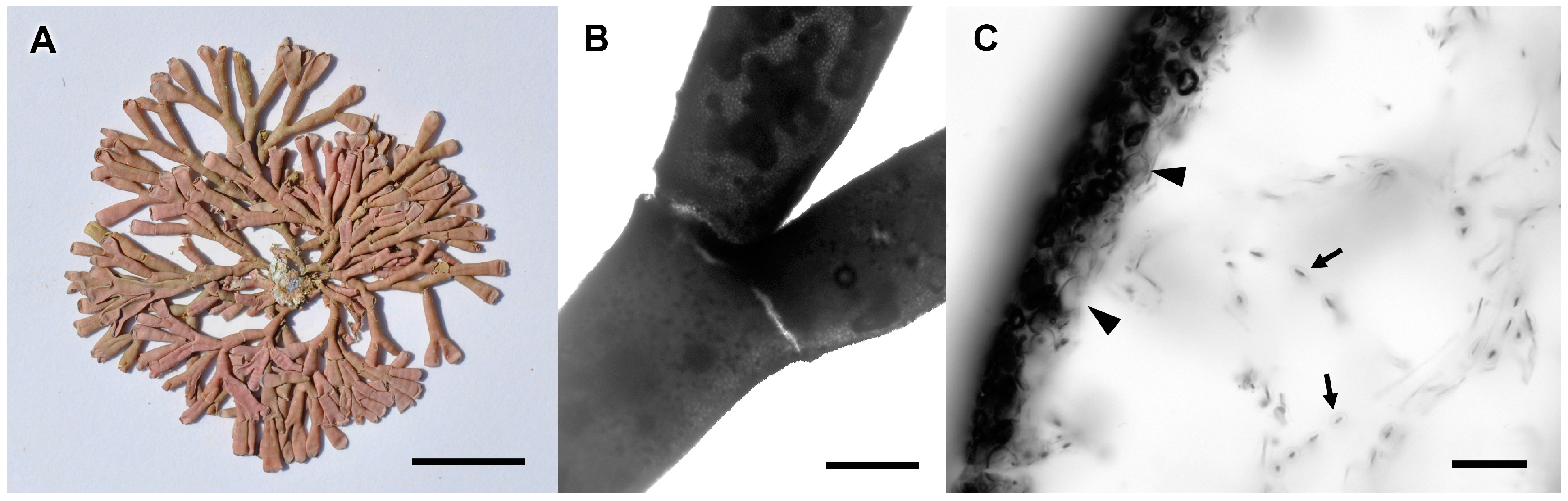
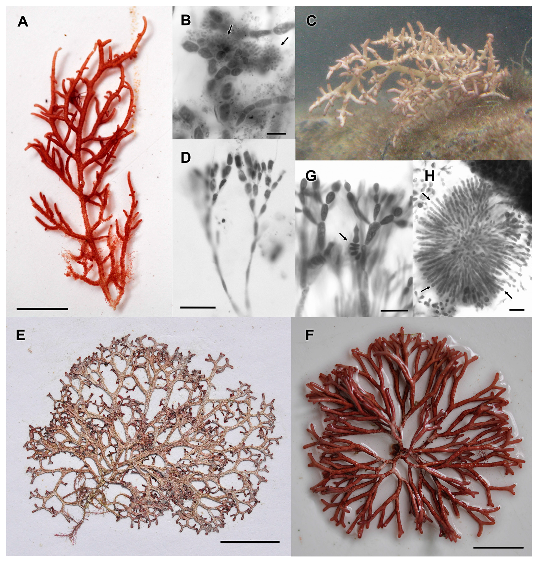
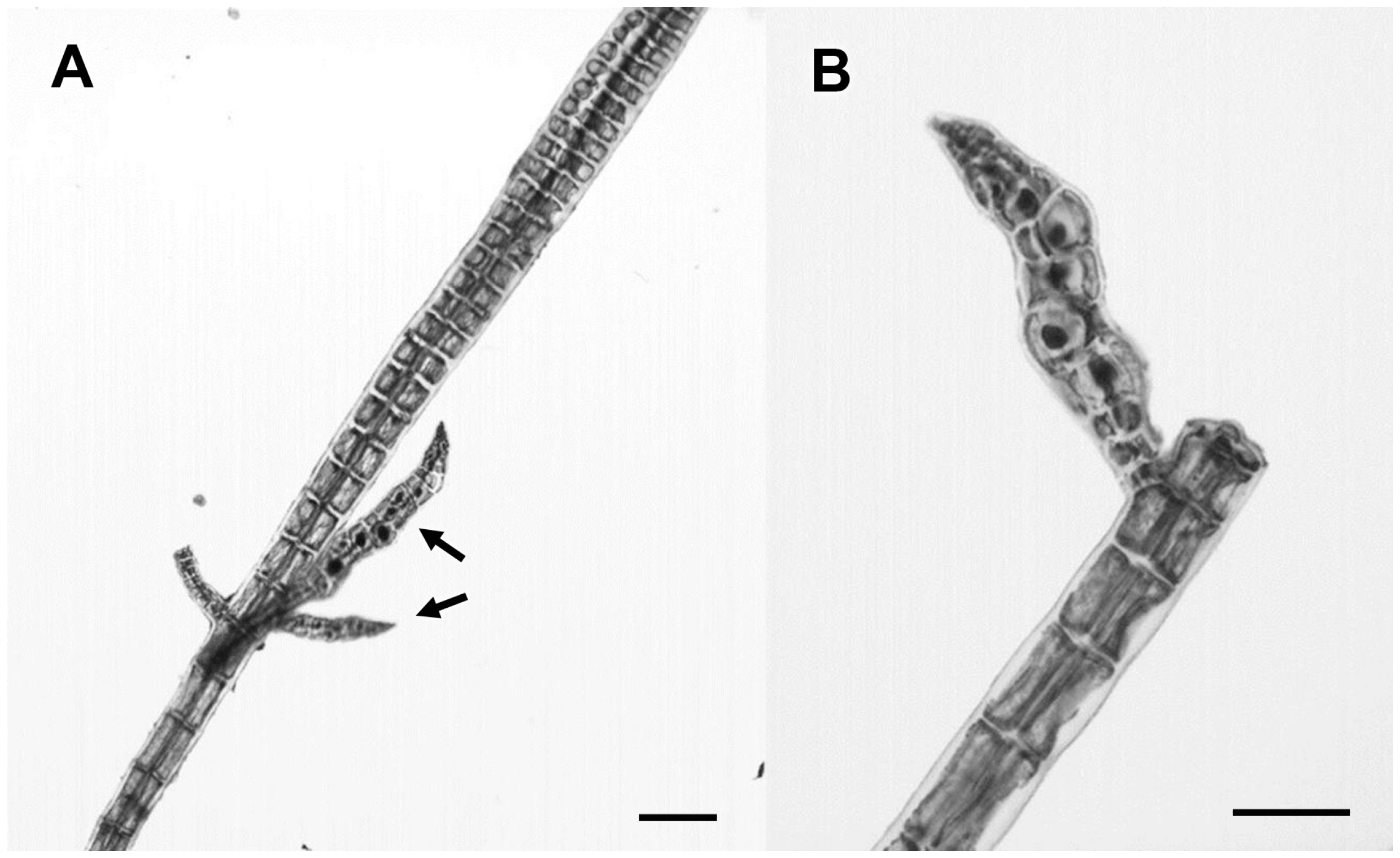
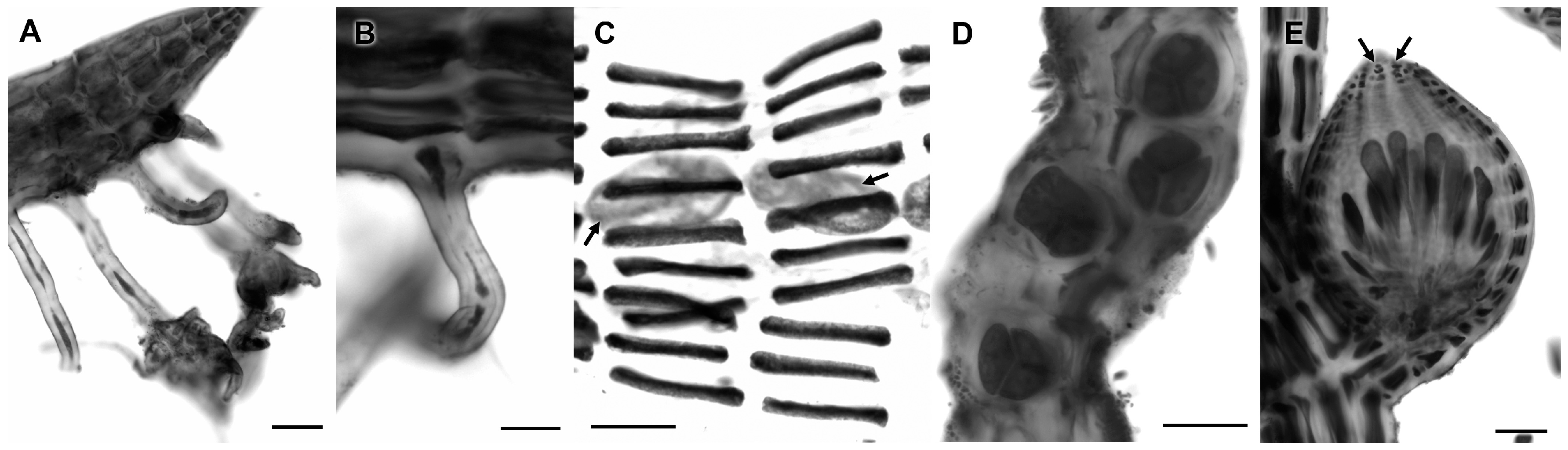
3.2.1. Tricleocarpa cylindrica (J. Ellis and Solander) Huisman and Borowitska

Sequences from PHYKOS-4617 and PHYKOS-4634 were found to be closest matches to Tricleocarpa species, with a closest homology to rbcL-3P and COI-5P sequences of specimens identified as Tricleocarpa cylindrica (Table 2). Phylogenetic analyses of Tricleocarpa species rbcL-3P sequences resolved the Punta Burica specimens within a T. cylindrica clade that includes specimens from the Indo-Pacific, Hawaiian Islands and Caribbean (Figure 3A). The Punta Burica specimens were weakly grouped (maximum likelihood bootstrap [ML] = 76%; Bayesian posterior probabilities [PP] = 0.84) with specimens from Thailand, Indonesia and Guadeloupe within this clade. COI-5P data is available for most of these same specimens and analyses of this locus more strongly supports (ML = 96; PP = 0.91) this relationship (Figure 3B). Percent divergences among T. cylindrica rbcL-3P and COI-5P sequences ranged from 0.0–2.0% and 0.0–7.3%, respectively. Pairwise divergences between the Punta Burica specimens and the most closely allied specimens in the phylogenetic analyses were 1.5–2.0% for rbcL-3P and 1.6–4.7% for COI-5P.
The specimens were collected from two different habitats. PHYKOS-4617 was growing on mudstone substrate in tide pools along the Punta Burica shoreline, while PHYKOS-4634 was collected from subtidal boulders at 3–5 m depth off the southwest point of Isla Burica (Figure 1). The specimens were heavily calcified except at the non-calcified joints, and had dichotomously branched cylindrical axes that were constricted at branch points (Figure 4A,B). They had relatively few, longitudinally oriented medullary cell filaments; inflated subsurface cells, and fused, polygonal surface cells (Figure 4C).
Remarks: Previous studies [33,34] found that specimens of Tricleocarpa from a wide geographic range matched the morphology described for T. cylindrica [35], but varied greatly in their rbcL and COI sequences, suggesting the presence of cryptic species. Both PHYKOS-4617 and PHYKOS-4634 matched the morphological concept of T. cylindrica, and they are resolved with varying levels of support in a clade that includes specimens from the Caribbean where the type was collected. However, the level of sequence variation between the Punta Burica specimens and other specimens in this clade are greater than the intraspecific values that have been found in many studies i.e., [36,37], and although T. cylindrica is assigned to the Punta Burica specimens here, a world-wide reassessment of the species is needed to determine its true distribution. Tricleocarpa cylindrica has been recorded previously from both the Caribbean and Pacific coasts of Central America e.g., [5,38,39]. This is the first report of the species from Pacific Panama.

3.2.2. Hommersandiophycus borowitzkae (Huisman) S.-M. Lin and Huisman

BLAST searches of the rbcL-3P sequence for PHYKOS-4618 revealed a 99% similarity with specimens of Hommersandiophycus borowitzkae. The UPA sequence BLAST showed a 99% similarity with specimens of both H. borowitzkae and Ganonema yoshizaki Huisman, I.A. Abbott and A.R. Sherwood, and the COI-5P BLAST showed no close homology (Table 2). Hommersandiophycus species were resolved as a strongly to fully supported clade (maximum likelihood bootstrap [ML] = 99%; Bayesian posterior probabilities [PP] = 1.00) in phylogenetic analyses of rbcL-3P sequences for Liagoraceae species (Figure 5). PHYKOS-4618 is positioned in a clade with H. borowitzkae specimens from Taiwan and The Philippines (ML = 87%; PP = 1.00) that is sister to H. samaensis (C.K. Tseng) Showe M. Lin and Huisman. The Western Pacific H. borowitzkae specimens shared identical rbcL sequences that were only 0.2% different from that of the Panama specimen. Divergences between H. borowitzkae specimens and sequences available from other Hommersandiophycus species ranged from 3.9–9.1%.
PHYKOS-4618 was collected from mudstone substrate in a tide pool along the Punta Burica shoreline (Figure 1).
Remarks: Hommersandiophycus borowitzkae was originally described as Ganonema borowitzkae Huisman based on Western Australia specimens [40]. Lin et al. [41] established Hommersandiophycus for G. borowitzkae and two other Ganonema species after molecular and morphological analyses resolved these species in a distinct clade with a number of synapomorphic characters states for carpogonial branches and carposporophytes. The examined PHYKOS-4618 specimen was male and matched the description of spermatangia initials being cut off from subapical cells and developing in clusters [41] (Figure 6A,B). The species was known only from East Asia (Taiwan, The Philippines) and Western Australia, and this is the first report from the Eastern Pacific.

3.2.3. Izziella sp.

Sequences of PHYKOS-4576 and PHYKOS-4599 shared identical COI-5P and UPA sequences, but an rbcL-3P sequence was only generated from PHYKOS-4576. These sequences were found to have closest homology to sequences from Izziella formosana (Yamada) Showe M.Lin, S.-Y.Yang and Huisman (rbcL-3P, UPA) and I. orientalis (J. Agardh) Huisman and Schils (COI-5P) in BLAST searches (Table 2). An Izziella clade received weak (ML = 73%) to strong (PP = 0.99) support in the Liagoraceae rbcL-3P analyses (Figure 5). PHYKOS-4576 was fully supported as the sister taxa to I. formosana and rbcL-3P sequence divergences between the sequenced specimens were only 2.4%. The only COI-5P sequence data available in GenBank is for three specimens from the Hawaiian Islands identified as I. orientalis that share identical sequences. The COI-5P sequence divergence between specimens from Panama and Hawai’i is 5%.
The PHYKOS-4576 and PHYKOS-4599 specimens were collected from intertidal pools along the Punta Burica shoreline (Figure 1). They were 8–9 cm in height and irregularly branched with many short branchlets (Figure 6C). Assimilatory filaments had cylindrical lower cells and ellipsoidal to obovoid upper cells (Figure 6D).
Remarks: Four Izziella species are currently recognized and distinguished by size, branching and specifics of carpogonial branches and involucral filaments [42,43]. Two of these species, are known only from Taiwan. Izziella orientalis has been recorded in both the Indian and Pacific Oceans, while I. formosana, originally described from Taiwan, was also recently reported from Pacific Costa Rica [39]. This is the first report of an Izziella species from Pacific Panama. Few Liagoraceae are reported along the Pacific coast of Central America [5], and the family is little studied in the region. The Punta Burica specimens likely represent a new Izziella species, but a better understanding of Eastern Pacific Liagoraceae is needed to make this determination.

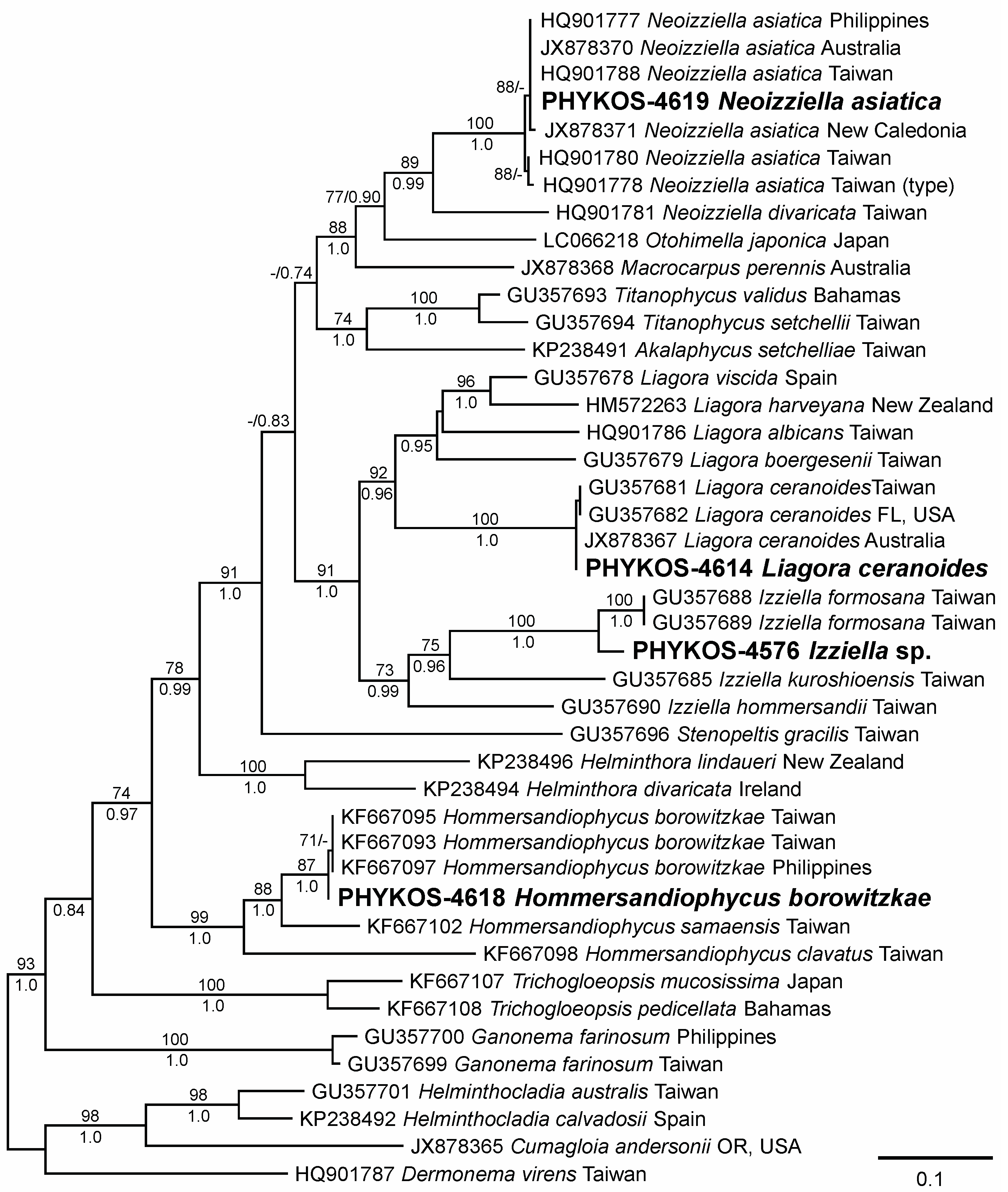
3.2.4. Liagora ceranoides J.V. Lamouroux

BLAST searches with the PHYKOS-4614 sequences revealed a closest affinity with specimens identified as Liagora ceranoides (rbcL-3P), Liagora sp. (COI-5P), and L. ceranoides (UPA) (Table 2). Liagora species formed a well supported monophyletic clade (ML = 92%; PP = 0.96) in the Liagoraceae rbcL-3P analyses (Figure 5). PHYKOS-4614 was resolved within a clade of L. ceranoides specimens collected from multiple geographic locations. The rbcL-3P sequence divergences within this clade were only 0.0–0.3% despite the wide geographic separation of the analyzed specimens.
PHYKOS-4614 was collected from mudstone stubstrate in tide pool along the Punta Burica shoreline (Figure 1C and Figure 6C). It was moderately calcified and relatively small (3 cm high), with sparse, irregular branching proximally and prolific, widely dichotomous branching distally (Figure 6E). The ultimate branches were generally short, 1–2 mm diameter tapering to 0.5 mm diameter apices.
Remarks: Morphologically PHYKOS-4614 closely matched descriptions of L. ceranoides [44,45]. Liagora ceranoides was originally described from the Virgin Islands in the Western Atlantic, and it has been reported in tropical waters around the globe. Although Lin et al. [45] demonstrated that some specimens identified as L. ceranoides in the Indo-Pacific represented a different genus and species, they also showed that L. ceranoides was distributed in Indo-Pacific as well as Western Atlantic. This species has been reported from the Pacific coast of Costa Rica [5]. Taylor [2] stated that it was common in Golfo Dulce, Costa Rica, which opens to the Pacific on the western side of the Burica penninsula, but it was not found during recent sampling.

3.2.5. Neoizziella asiatica Showe M. Lin, S.-Y. Yang and Huisman

The rbcL and UPA sequences of PHYKOS-4619 were found by BLAST searches to match sequences of Neoizziella asiatica specimens. BLAST searches of the PHYKOS-4619 COI-5P sequences returned a 99% sequence similarity to an Hawaiian specimen identified as N. divaricata (C.K. Tseng) Showe M. Lin, S.-Y. Yang and Huisman. This is the only Neoizziella COI-5P sequence available in GenBank and may represent a mis-identified specimen of N. asiatica. Sequences of rbcL-3P for PHYKOS-4619 and N. asiatica specimens from geographically diverse locations in the western Pacific were resolved in a fully supported clade that was well supported (ML = 89%; PP = 0.99) as the sister species to N. divaricata in the Liagoraceae rbcL-3P phylogeny (Figure 5). Pairwise sequence divergences among PHYKOS-4619 and the western Pacific N. asiatica specimens ranged from 0.0–0.6%, and these sequences were 5.9–6.2% divergent from the two available Taiwan N. divaricata rbcL sequences.
The specimen was collected from a tide pool along the Punta Burica shoreline (Figure 1). It was attached to the substrate by a discoid holdfast, and axes had 6–7 orders of dichotomous to subdichotomous branching and untapered ultimate branches with obtuse apicies (Figure 6F). The specimen was a female gametophyte with many carpogonial branches of 5–6 cells and mature carposporophytes (Figure 6G,H).
Remarks: PHYKOS-4619 matches the morphological descriptions of N. asiatica [46] except in its stature, which was less than 3 cm, and number of carpogonial branch cells, which was 5–6 versus the 4–5 reported by Lin et al. [46]. Neoizziella currently includes only two species, N. asiatica and N. divaricata [42,46]. A recent Costa Rica record of Neoizziella asiatica was the first from outside of the Indo-Pacific region [39]. This is the first report of the species from Pacific Panama.

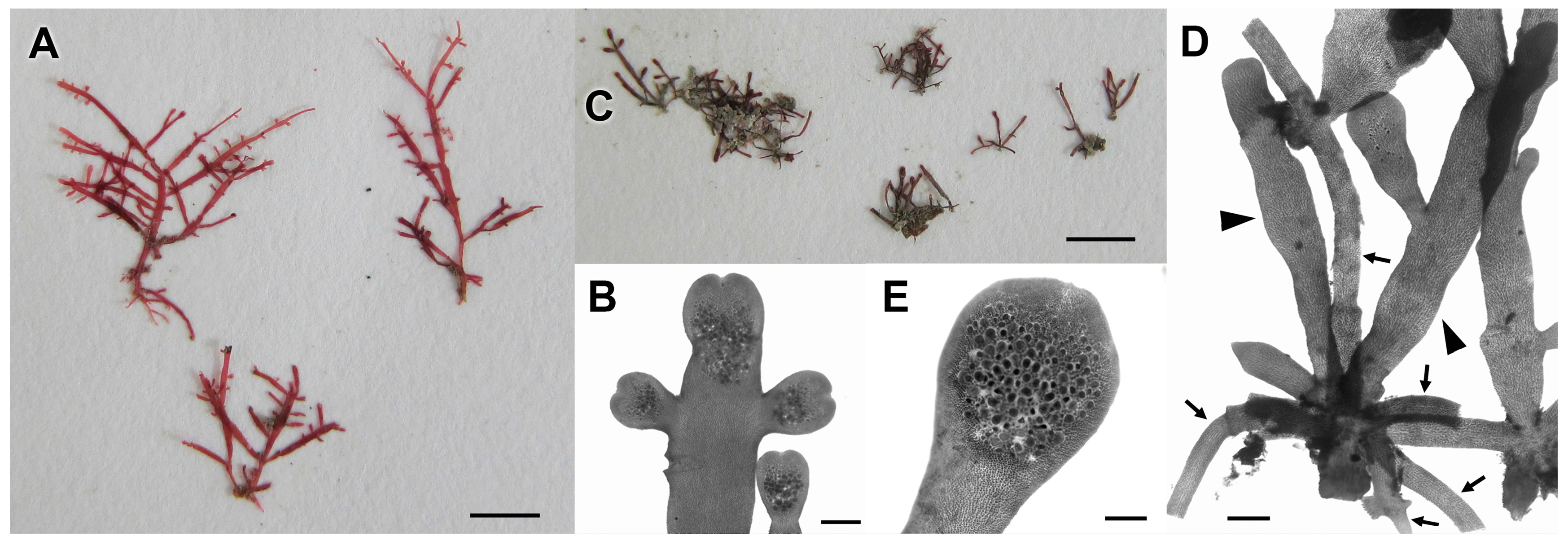
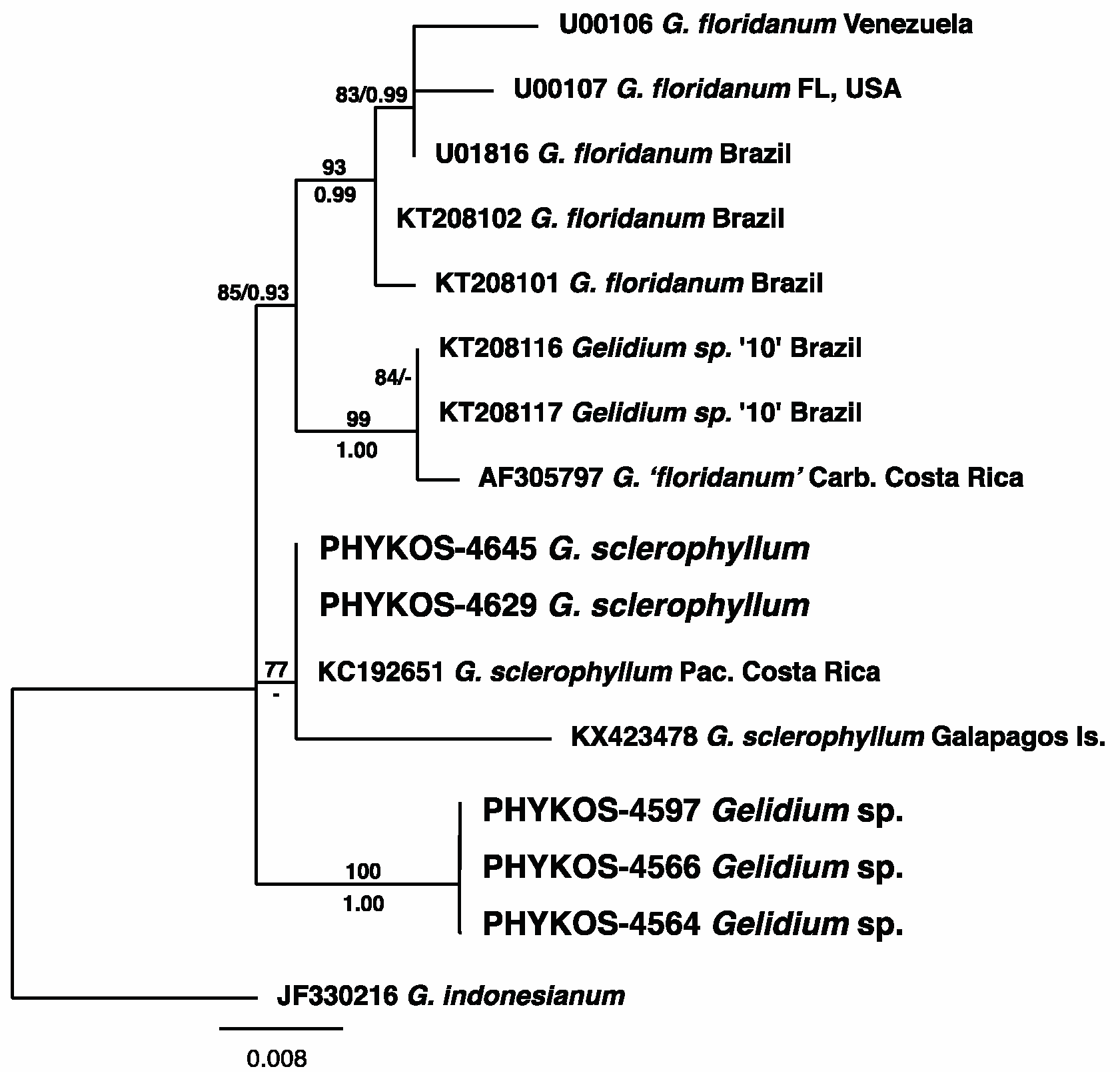
3.2.6. Gelidium sclerophyllum W.R. Taylor

PHYKOS-4514, PHYKOS-4629, and PHYKOS-4645 shared identical COI-5P and UPA sequences. An rbcL-3P sequence was not generated for PHYKOS-4514, but PHYKOS-4629 and 4645 also shared identical sequences at this locus. BLAST searches indicated exact matches and 98% identity with COI sequences from Pacific Costa Rica specimens of Gelidium sclerophyllum (Table 2).
All three collections were made from subtidal hard substrates. PHYKOS-4514 was collected from the mixed sediment-mudstone bottom just beyond the surf zone off the Punta Burica shoreline. PHYKOS-4629 and PHYKOS-4645 were collected from subtidal boulders at 3–5 m depth off the southwest point of Isla Burica (Figure 1).
Remarks: The rbcL-3P and COI-5P sequences of the Punta Burica specimens and Costa Rica GenBank records have been tied to a partial rbcL [31] and full rbcL and COI [32] sequences from type specimens providing positive identifications. Gelidium sclerophyllum and G. floridanum W.R. Taylor have been consistently resolved together in a strongly supported clade that increased sampling is revealing to represent a closely related species complex [31,32,47]. Gelidium sclerophyllum has been reported from Mexico south to Ecuador, but this is the first report from Panama. A detailed description of the species was recently provided by Grusz and Freshwater [31], and the Punta Burica specimens match this description including the characteristic wide sterile margin around tetrasporangial sori and axes frequently with emarginate apices (Figure 7A,B).

3.2.7. Gelidium sp.

PHYKOS-4564, PHYKOS-4566, and PHYKOS-4597, shared identical sequences for the three tested loci except for PHYKOS-4566 where the COI sequence was one base pair different from the other two sequences. BLAST searches revealed a close affinity with Gelidium floridanum and G. sclerophyllum (Table 2), and this Punta Burica Gelidium species is believed to be part of a complex of closely related species.
All three collections were made from the intertidal mudstone sustrate along the Punta Burica shoreline (Figure 1) where they grew as a short turf. The thalli were composed of an extensive system of tangled stoloniferous axes that gave rise, sometimes in clusters, to generally short (<5 mm), simple to irregularly branched erect axes (Figure 7C,D), although some taller axes with more regular distichous branching were observed. The latter were similar in outline to small specimens of G. sclerophyllum but lacked emarginate tips and the sometimes swollen sterile margins around tetrasporangial sori (Figure 7E).
Remarks: Phylogenetic analyses of rbcL-3P revealed four separate groupings of specimens within this clade (Figure 8). These include a well supported G. floridanum, a strongly supported clade of specimens from Brazil and one from Caribbean Costa Rica that was originally identified as G. floridanum, a weakly supported clade of G. sclerophyllum specimens from Punta Burica, Pacific Costa Rica and Galapagos Islands, and the clade of Gelidium sp. specimens collected from Punta Burica. Thomas and Freshwater [48] suggested based on rbcL sequence divergences and slight morphological differences, that there was a genetic dicontinuity between specimens they identified as G. floridanum from Caribbean Costa Rica and G. floridanum in the Western Atlantic. Recent species delimination analyses with both UPA and COI-5P data distinguished G. floridanum from a closely related sister species that included the Caribbean Costa Rica G.floridanum’ [18], and this separation was also present in the analyses here (Figure 8). Gelidium sclerophyllum and G. floridanum are considered to represent geminate sister species with a transisthmian distribution [31]. Our current analyses suggest that these three species are part of a complex of closely related taxa that also includes the second unidentified Punta Burica Gelidium species. Analyses of additional specimens and the inclusion of more variable COI data are required to determine the exact number and relationships of the species involved in this complex.
Gelidium pusillum (Stackhouse) Le Jolis was the only Gelidium species recorded from Pacific Panama by Taylor [2], and Littler and Littler [49] included both G. pusillum and G. pusillum var. pacificum W.R. Taylor in their web database. The Punta Burica Gelidium species has the general morphological characteristics that were associated with the previous concept of a widely distributed G. pusillum, however, the current concept of G. pusillum restricts its distribution to the North Atlantic [50]. A number of small Gelidium and Pterocladiella species are reported from the eastern tropical Pacific [5], but they are generally poorly known. Additional morphological and molecular analyses are required to determine the relationship of this species to others reported in the region and if it represents a new species.

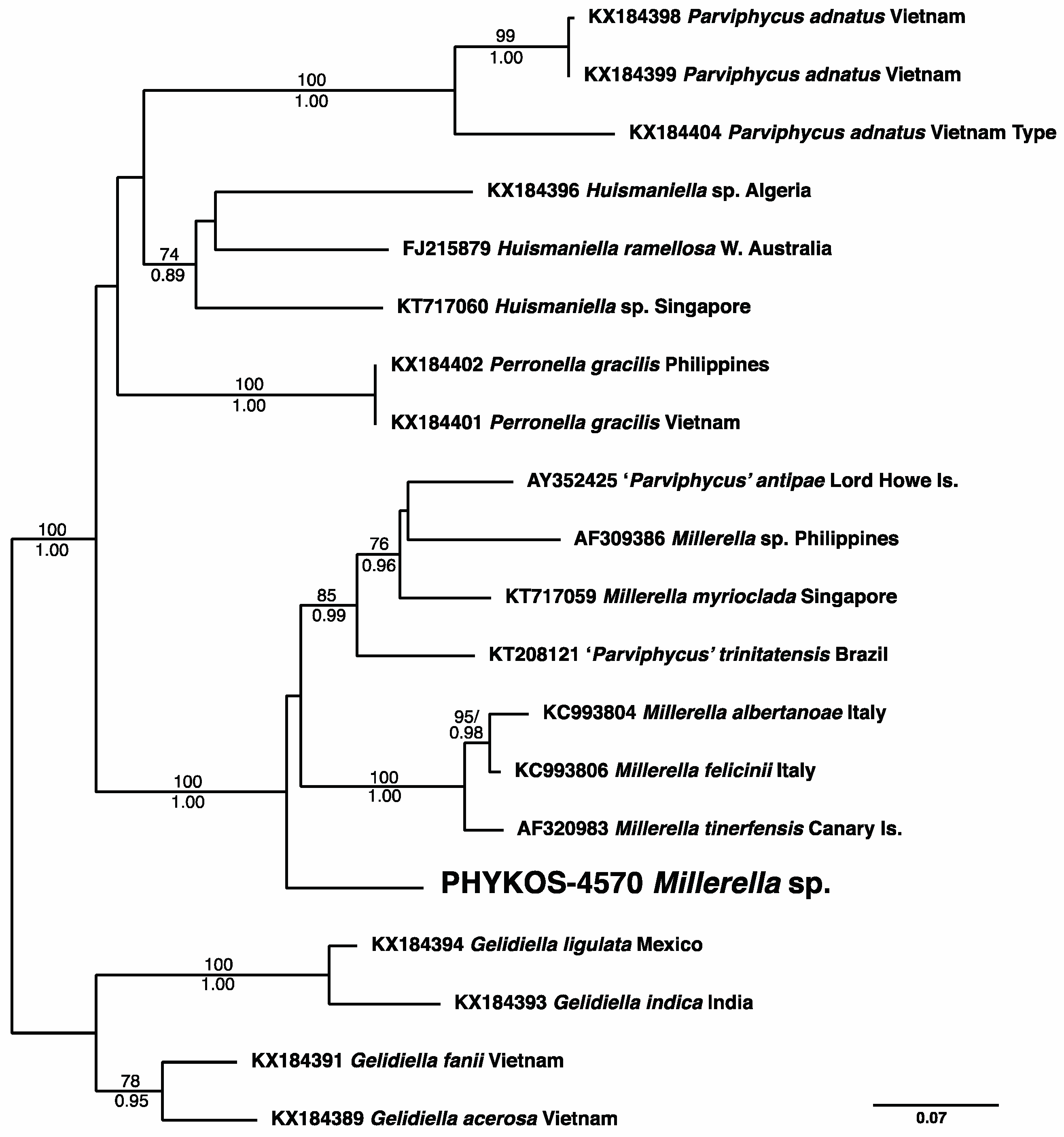
3.2.8. Millerella sp.

BLAST searches with sequences of PHYKOS-4570 returned a closest affinity with Millerella spp. (Table 2). The largest amount of available Gelidiellacaeae sequence data is for the rbcL locus, and trees generated with these data resolve PHYKOS-4570 within a strongly supported Millerella clade but with no close relationship to any of the other sequenced species (Figure 9).
The specimens were growing as a short turf on the intertidal mudstone substrate along the Punta Burica shoreline (Figure 1). PHYKOS-4570 matched the states of all important morphological characters described for Millerella species. Attachment rhizoids were in irregularly arranged clumps on stoloniferous axes and developed from outer cortical cells (Figure 10A,B). Second-order cell filaments developed from the axial cell filament in a distichous pattern during vegetative growth (Figure 10C,D). Tetrasporangial sori formed at the tips of erect axes and branches, and had acropetally developing tetrasporangia that were regularly arranged following the pattern of the second-order cell filaments, with six tetrasporangia in a row on both sides of a sorus (Figure 10E).
Remarks: Santelices [51] described Parviphycus to accomodate Gelidiellaceae species with a distichous arrangement of second-order cell filaments and the development of sporangia in regularly arranged rows. Further morphological and DNA sequence analyses lead Boo et al. [52] to describe Millerella and Perronella for some of the species classified within Parviphycus. Dawson [6,53,54] described a number of small Gelidiella species from the tropical East Pacific, and other small Gelidiellaceae such as Parviphycus tenuissimus (Feldmann et Hamel) B. Santelices and P. trinitatensis (W.R. Taylor) M.J. Wynne have also been reported from Pacific Central America e.g., [5,55]. The status of the Punta Burica Millerella species as new or representative of one of these reported species will require a reassessment of the Gelidiellaceae in the East Pacific.

3.2.9. Asparagopsis sp.

BLAST searches of PHYKOS-4636 sequences returned identical matches (COI-5P, UPA) or close homology (rbcL-3P) to sequences of specimens identified as Asparagopsis taxiformis (Delile) Trevisan. Only a UPA sequence was generated for PHYKOS-4646, and it was identical to that of PHYKOS-4636. GenBank specimens with identical COI-5P and UPA sequences were from the Pacific.
Both PHYKOS-4636 and PHYKOS-4646 were collected from subtidal boulders at 4–5 m depth off the southwest point of Isla Burica (Figure 1A and Figure 11A). The specimens were all in the Falkenbergia-stage and some had tetrasporangia (Figure 11B,C).
Remarks: Currently there are two recognized species of Asparagopsis, A. armata Harvey, and A. taxiformis. The most recent DNA sequence analyses of the genus utilized nuclear-encoded LSU, chloroplast-encoded RuBisCO spacer, and mitochondria-encoded cox2-3 spacer sequences to identify two major lineages within A. armata and five within A. taxiformis [56]. Dijoux et al. [56] designated lineages based on cox2-3 spacer haplotypes, which were not generated here, but RuBisCO spacer sequences for PHYKOS-4636 and PHYKOS-4646 matched the haplotypes of A.taxiformis’ specimens found in their Lineage 4. This lineage contained specimens distributed widely within the Pacific and Indian Oceans including specimens from the Gulf of Panama and Costa Rica [56]. The taxonomic status of this lineage requires further study, but its genetic distiction [56,57] and recent morphological differentiation from other lineages [58] suggests that it represents a biological species distinct from A. taxiformis.
Asparagopsis (as A. taxiformis) was first reported from Panama by Andreakis et al. [57], who included tetrasporophyte (Falkenbergia-stage) specimens collected from the Pacific terminus of the Panama Canal, Gulf of Panama in their biogeography study. Zanolla et al. [58] reported that Falkenbergia-stage specimens of A. taxiformis lineages could be morphologically distinguished by the length and width of axial cells, width of apical cells and thickness of cell walls. While measurements of these characters from Punta Burica specimens overlap with those cited for Lineage 4 by Zanolla et al. [58], a greater level of variation was seen.

3.2.10. Plocamium sp.

PHYKOS-4643 sequences were found to have closest homology to GenBank sequences from specimens identified as Plocamium pacificum Kylin (rbcL-3P 99%; COI-5P 95%), or P.cartilagineum’ (Linnaeus) P.S. Dixon and P. telfairiae (W.J. Hooker and Harvey) Harvey ex Kützing (UPA 99%). However, phylogenetic analyses of available rbcL-3P sequences and cluster analyses of available COI-5P sequences did not strongly associate PHYKOS-4643 with P. pacificum or any other Plocamium species.
PHYKOS-4643 was collected from subtidal boulders at 3–5 m depth off the southwest point of Isla Burica.
Remarks: The only reports of Plocamium species from the Pacific coast of Central America are found in the Littler and Littler [49] web database of Pacific Panama marine algae. They list two species, P. cartilagineum subsp. pacificum (Kylin) P.C. Silva [=P. pacificum Kylin] and P. violaceum Farlow. The included images of the former show recurved pinnae and these are also part of the description and figure of this species in Abbott and Hollenberg [59]. Recurved branches were not present in PHYKOS-4643, which more closely matches the images of P. violaceum in the Littler and Littler [49] list. Plocamium violaceum was described by Farlow [60] based on California specimens, and he noted that ramuli were arranged in alternate groups of 3 or 4, with the lower most ramulus in a group larger than the others and slightly recurved. Lower ramuli in the Punta Burica specimen were not noticably larger or recurved (Figure 11D). COI-5P divergences between the Punta Burica and California P. violaceum specimens (5.6–5.9%) were greater than that between Punta Burica and specimens identified as P. pacificum from California, P. oregonum Doty from Oregon, and P. nanum G.W. Saunder and K.V. Lehmkuhl from France. Both the morphological and molecular discripancies indicate that the PHYKOS-4643 Plocamium specimen is not P. violaceum, and likely represents a new species.

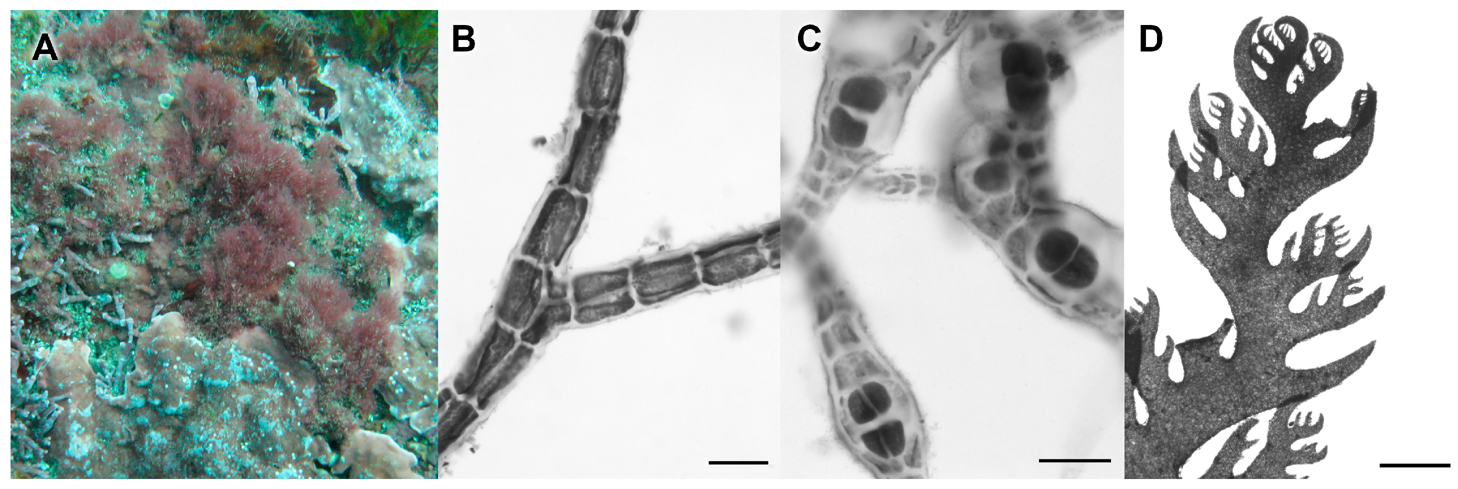
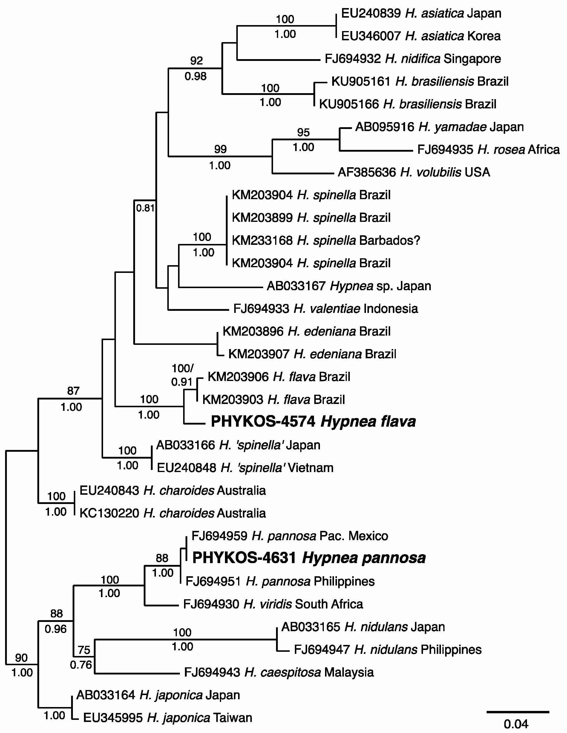
3.2.11. Hypnea flava Nauer, Cassano and M.C. Oliveira

BLAST searches with PHYKOS-4574 sequences returned records from specimens identified as either Hypnea flava from Brazil (rbcL-3P 99%) or H. ‘spinella’ from Hawai’i (COI-5P 96%, UPA 99%). Phylogenetic analyses of rbcL-3P sequences from Hypnea species resolved the Punta Burica specimen within a fully-supported H. flava clade (Figure 12). The rbcL-3P divergence between the Pacific specimen from Punta Burica and the Brazilian specimens was only 0.9–1.0%.
PHYKOS-4574 specimens were collected from tide pools in the mudstone substrate along the Punta Burica shoreline (Figure 1) and included tetrasporophytes, which are described here for the first time. Tetrasporangial sori surround the median, swollen portions of generally short branchlets, 200–300 µm in diameter and may cover a majority of the branchlet surface, especially during early stages of development (Figure 13A). Sori sometimes also develop on the apical portions of longer branchlets or as patches on the surface of predominately vegetative axes. Tetrasporangia mother cells are cut off laterally from inner cortical cells and develop into zonately divided tetrasporangia (Figure 13B).
Remarks: The Punta Burica specimens closely matched the vegetative morphology of Brazilian H. flava, but reproductive specimens were not available when the species was orginally described by Nauer et al. [61], and tetrasporophytic thalli are described here for the first time. This is the first report of the species outside of Brazil.

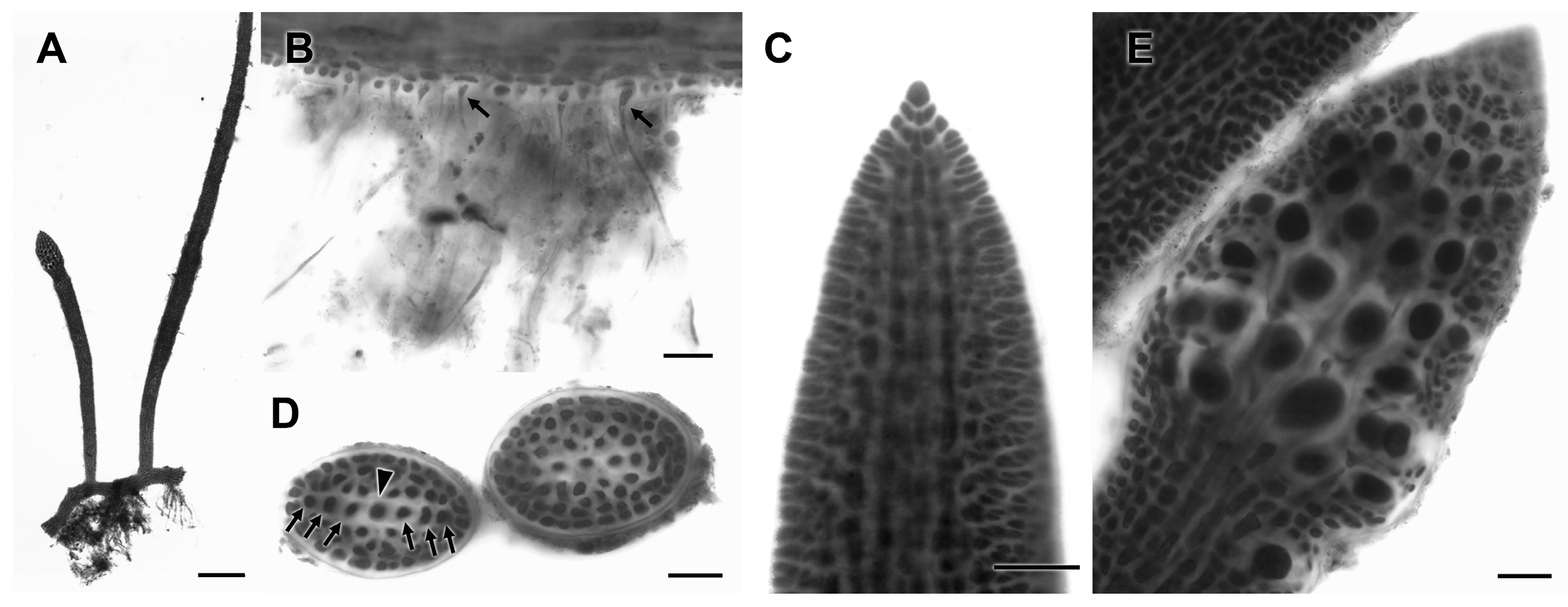
3.2.12. Hypnea pannosa J. Agardh

PHYKOS-4631 rbcL-3P and COI-5P sequences were found to be identical, or nearly identical with GenBank sequences assigned to Hypnea pannosa. BLAST searches of the PHYKOS-4631 UPA sequence returned sequences of five species all at 99% similarity. Sequences of Eastern- and Western Pacific H. pannosa specimens formed a well- to fully-supported clade in the Hypnea rbcL-3P tree (Figure 12). Hypnea pannosa was originally described based on specimens from the Pacific coast of Mexico, and the Punta Burica rbcL-3P sequence was a match for that of a Pacific Mexico specimen.
The PHYKOS-4643 collection was made from subtidal boulders at 4–5 m depth off the southwest point of Isla Burica (Figure 1). Specimens had compressed to subterete, entangled, irregularly branched axes with little difference between the diameter of axes and branches. Axes also anastomosed and had multiple attachments to the substrate. The Punta Burica specimens had generally acute to widely acute branch tips and tetrasporangial sori developed on both small branchlets and major axes (Figure 13C,D).
Remarks: Hypnea pannosa is a widely recorded species from tropical waters and also Pacific Central America [5]. Taylor [2] collected specimens from high tide pools at Isla Secas in the central Gulf of Chiriqui, Panama. The Punta Burica specimens matched the morphological descriptions of this species from Pacific Mexico and Hawai’i [44,62,63]. Some specimens identified as H. pannosa in the western Pacific are described as being mostly terete [64,65,66] and probably represent a different species; however, no DNA sequence data is currently available for these specimens.

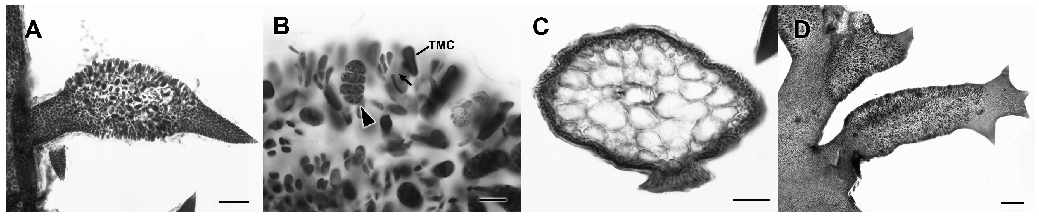
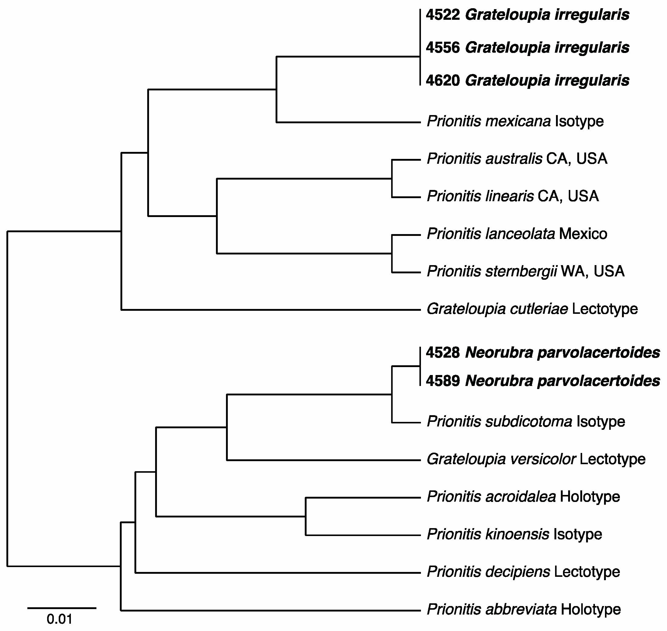
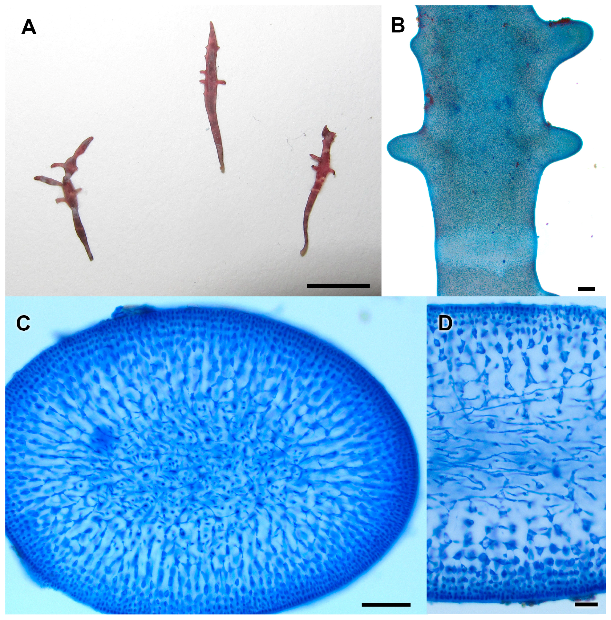
3.2.13. Neorubra parvolacertoides Freshwater and P.W. Gabrielson sp. nov.

BLAST searches with PHYKOS-4528, PHYKOS-4589 rbcL-3P sequences revealed a closest affinity with sequences of Neorubra decipiens (Montagne) M.S. Calderon, G.H. Boo and S.M. Boo (Table 2). COI-5P and UPA sequence BLAST searches provided little context for the identification of this species. Phylogenetic analyses with available rbcL-3P sequences from species of Grateloupia sensu lato resolved PHYKOS-4528 and PHYKOS-4589 within a fully supported Neorubra clade (Figure 14). Cluster analyses of a 137 bp section of the rbcL-3P locus from Eastern Pacific types and contemporary topotype specimens further indicated that the Punta Burica Neorubra species is unique (Figure 15).
Remarks: Gargiulo et al. [67] recently described morphological character states of the female reproductive system and post-fertilization development for a number of newly distinguished genera previously synonymized under Grateloupia that also were resolved as distinct clades within rbcL phylogenies of Grateloupia sensu lato. Continued molecular and morphological investigation has led to descriptions of additional genera [68,69,70], including Neorubra, which contains two species, N. decipiens and N. denticulata (Montagne) M.S. Calderon, G.H. Boo and S.M. Boo. Considering the position of PHYKOS-4528 and PHYKOS-4589 specimens within a fully supported Neorubra clade and their uniqueness among known eastern Pacific taxa we regard the Punta Burica specimens as a new Neorubra species that we describe here.
Neorubra parvolacertoides Freshwater and P.W. Gabrielson sp. nov.
Figure 16A–D.
Description: Thalli dark red with horizontal bands, sometimes wavy, of lighter color; small and erect, 9–12 mm high, laceolate to clavate with extended proximal tapering (Figure 16A,B); bases terete to subterete, becoming compressed to flattened distally, 0.5–1.5 mm wide, 150–400 µm thick; with one order of short, irregular distichous branching; axes multiaxial; tips obtuse to widely acute; medulla of longitudinal filaments of elongated cells; cortex of anticlinal filaments; medulla-cortex transition gradual; outer cortex of 5–7 layers of small, globose to elliptical, pigmented cells, 1–5 µm × 2–10 µm; inner cortex of 3–4 layers of elliptical to stellate cells, 4–7 µm × 10–18 µm (Figure 16C,D); COI-5P sequence = GenBank accession KY656538, rbcL-3P sequence = GenBank accession KY573975, UPA sequence = GenBank accession KY573934.
Holotype: WNC 34262, herbarium specimen and two slides (WNC 2012-s073, WNC 2012-s074) in packet on sheet, collection # PHYKOS-4528, attached to rock on mixed sediment-rock bottom just beyond breakers, 3 m depth, near Mono Feliz, Punta Burica, Panama, 8 January 2011, leg. B. Wysor and D.W. Freshwater.
Paratype: slide # WNC 2011-s072, collection PHYKOS-4589, growing with brown algal turf, shallow subtidal, <1 m depth, near Mono Feliz, Punta Burica, Panama, 9 January 2011, leg. B. Wysor and D.W. Freshwater.
Type Locality: Punta Burica, Panama, 08.03042° N; 082.87574° W
Etymology: The epithet refers to the field name given to this species by its collectors who thought that it resembled small lizards of the region.
Comment: The diminutive size, <15 mm tall, and having only 1 order of branches distinguishes N. parvolacertoides from all other Halymeniales in the eastern tropical Pacific. It is an order of magnitude smaller than both described species of Neorubra, N. decipiens and N. denticulata (Montagne) M.S. Calderon, G.H. Boo and S.M. Boo and, being tropical, does not overlap the distributions of the aforementioned species.

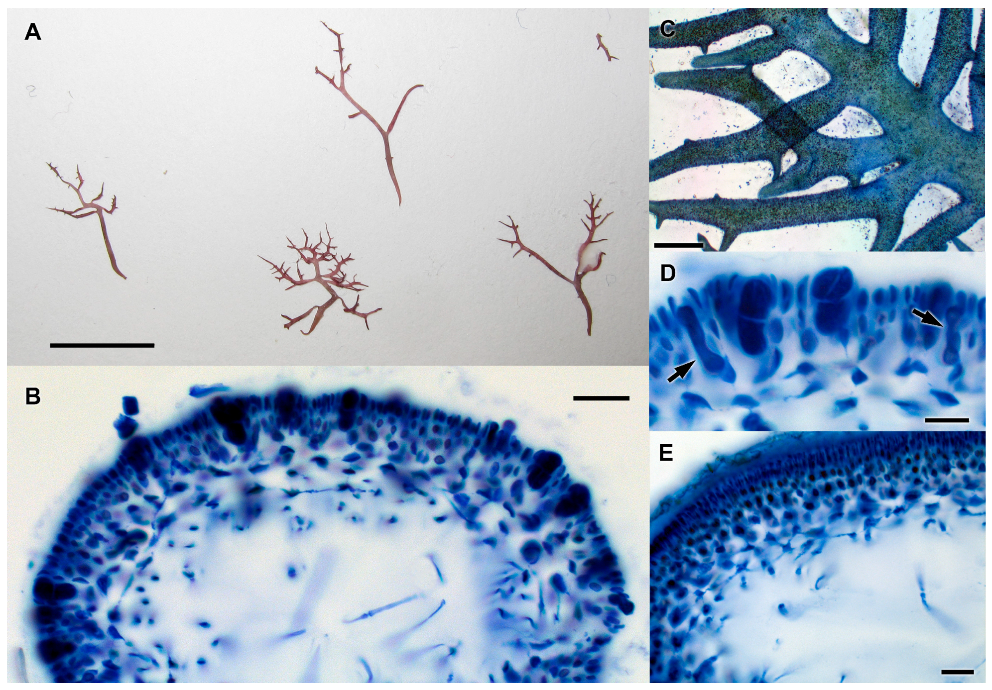
3.2.14. Grateloupia irregularis P.W. Gabrielson and Freshwater sp. nov

PHYKOS-4522, PHYKOS-4556, and PHYKOS-4620 all shared identical sequences for the three examined loci. BLAST searches returned a variety of Grateloupia sensu lato species, but none at a level of homology indicating a potential species match. Cluster analyses with a short segment of the rbcL-3P locus showed that this species was distinct from all the included eastern Pacific Grateloupia sensu lato types and contemporary topotype specimens (Figure 15). Phylogenetic analyses of the rbcL-3P locus resolved this specimen within a well supported Grateloupia sensu stricto clade (Figure 14). Similar to Neorubra parvolacertoides, the uniqueness among known eastern Pacific taxa and position within the well resolved Grateloupia clade, indicates that these specimens represent a previously undescribed Grateloupia species.
Grateloupia irregularis P.W. Gabrielson and Freshwater sp. nov.
Figure 17A–E.
Description: Thalli various shades of red; small and erect, 10–20 mm high, lanceolate to linear, fractiflexus in part (Figure 17A), attached by discoid holdfast, narrow terete to subterete bases, quickly becoming compressed to flattened except at tips and young ultimate branches where terete, 0.25–1.5 mm wide; 200–350 µm thick; with up to four orders of irregular, disticus branching and sometimes sparsly scattered small denticulate proliferations; axes multiaxial; tips acute; medulla of widely spaced, longitudinal filaments of elongated cells; cortex of anticlinal filaments; medulla-cortex transition sharp; outer cortex of 5–6 layers of small, mostly elliptical cells, 2–5 µm × 3–10 µm; inner cortex of 2–3 layers of globose to elliptical or stellate cells, 5–10 µm × 10–20 µm (Figure 17B); tetrasporangia across entire thallus surface of main axes and branches (Figure 17C); tetrasporangial mother cells cut off from inner cells of the outer cortex, becoming somewhat elongated and irregular in shape during development; tetrasporangia cruciate to decussately cruciate divided(Figure 17D); spermatiangial sori covered by thick sheath, outer cortical cells develop into elongated spermatangial mother cells (Figure 17E); COI-5P sequence = GenBank accession KY656551, rbcL-3P sequence = GenBank accession KY573991, UPA sequence = GenBank accession KY573951.
Holotype: WNC 34263, herbarium specimen and two slides (WNC 2012-s067, WNC 2012-s068) in packet on sheet, collection # PHYKOS-4556, attached to rock on mixed sediment-rock bottom just beyond breakers, 3 m depth, near Mono Feliz, Punta Burica, Panama, 8 January 2011, leg. B. Wysor and D.W. Freshwater
Paratypes: WNC 34264, herbarium specimen and two slides (WNC 2012-s071, WNC 2012-s072) in packet on sheet, collection # PHYKOS-4522, attached to rock on mixed sediment-rock bottom just beyond breakers, 3m depth, near Mono Feliz, Punta Burica, Panama, 8 January 2011, leg. B. Wysor and D.W. Freshwater; Slides WNC 2012-s069 and WNC 2012-s070, collecction # PHYKOS-4620, growing in mixed algal turf, shallow subtidal, <1 m depth, near Mono Feliz, Punta Burica, Panama, 9 January 2011, leg. B. Wysor and D.W. Freshwater
Type locality: Punta Burica, Panama, 08.03042° N; 082.87574° W
Etymology: The epithet reflects the irregular branching pattern exhibited by the species.
Comment: Similar to N. parvolacertoides, the diminutive size of G. irregularis, <15 mm tall, distinguishes it from other previously described species of eastern tropical Pacific Halymeniales. Grateloupia irregularis has more orders of branches (Figure 17A) and its medulla is comprised of loosely arranged filaments (Figure 17B) compared to N. parvolacertoides (Figure 16 A,C respectively).

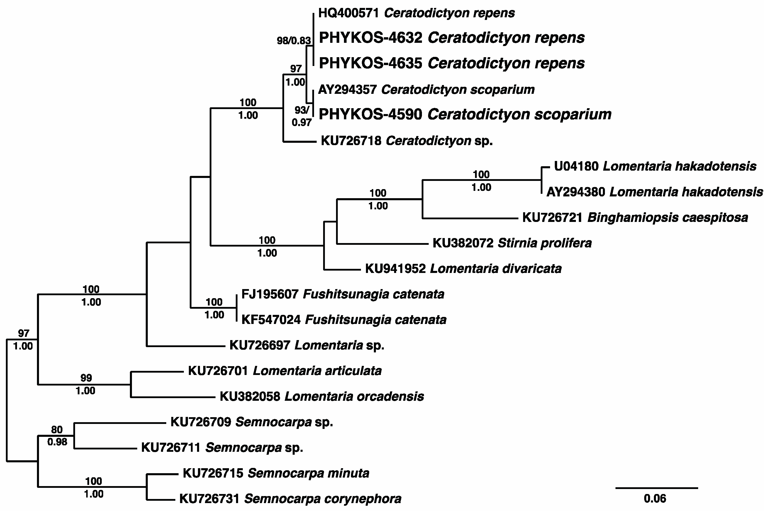
3.2.15. Ceratodictyon repens (Kützing) R.E. Norris

BLAST searches of COI-5P and UPA sequences for PHYKOS-4627, PHYKOS-4632 and PHYKOS-4635 returned closest matches (94% and 99% respectively) with sequences of Ceratodictyon scoparium (Table 2). An rbcL-3P sequences was not generated for PHYKOS-4627, but searches with this locus for the other two samples showed them to be matches for the sequence from a South African specimen of C. repens. Phylogenetic analyses of rbcL-3P data for members of the Lomentariaceae resolve these C. repens specimens within a strongly supported Cearatodictyon clade (Figure 18).
All Punta Burica specimens were growing in tufts on subtidal rocks at 4–5 m depth off the southwest point of Isla Burica (Figure 1). The thalli consisted of prostrate axes giving rise to erect axes up to 2–3 cm in height (Figure 19A). Erect axes were proximally terete and became distally compressed.
Remarks: Saunders et al. [71] established that Gelidiopsis was a genus within the Rhodymeniales family Lomentariaceae. Ceratodictyon was originally described for an algal species living symbiotically with a sponge [72]. Norris [73] proposed the merger of Ceratodictyon and Gelidiopsis, but this synonymy was generally not followed until DNA sequence analyses supported their congeneric status [74,75]. Ceratodictyon repens was originally described from New Caledonia and is reported from many Indo-Pacific localities e.g., [65,76]. The species is known from El Salvador in the tropical East Pacific (as Gelidiopsis repens (Kützing) Weber-van Bosse [55,77]), but this is the first report from Panama.

3.2.16. Ceratodictyon scoparium (Montagne and Millardet) R.E. Norris

BLAST results with sequences of PHYKOS-4590 showed that they were identical to the rbcL-3P sequence of an unidentified Ceratodictyon sp. from Pacific Mexico, and identical (UPA) and 99% (COI-5P) similar to sequences from Hawaiian specimens of C. scoparium. PHYKOS-4590 was resolved sister to the Punta Burica C. repens specimens in the rbcL-3P phylogeny (Figure 18).
The PHYKOS-4590 specimens were growing as a turf on the exposed intertidal mudstone substrate and in tide pools along the Punta Burica shoreline (Figure 1).
Remarks: Ceratodictyon scoparium was originally described from Réunion Island in the western Indian Ocean and similar to C. repens is reported throughout much of the Indo-Pacific [35]. Morphologically the two species are also similar, with both demonstrating the condensed branching that results in a pseudopalmate appearance for some axes [44,76]. Punta Burica C. scoparium specimens were smaller in stature than both the collected C. repens specimens (Figure 19B) and Hawaiian specimens of C. scoparium described by Abbott [44] although they otherwise closely match Abbott’s description. This small stature may have an environmental basis as the Punta Burica C. scoparium was growing as a turf in the intertidal mudstone and tide pool habitat. This is the first record of C. scoparium from the eastern Pacific.

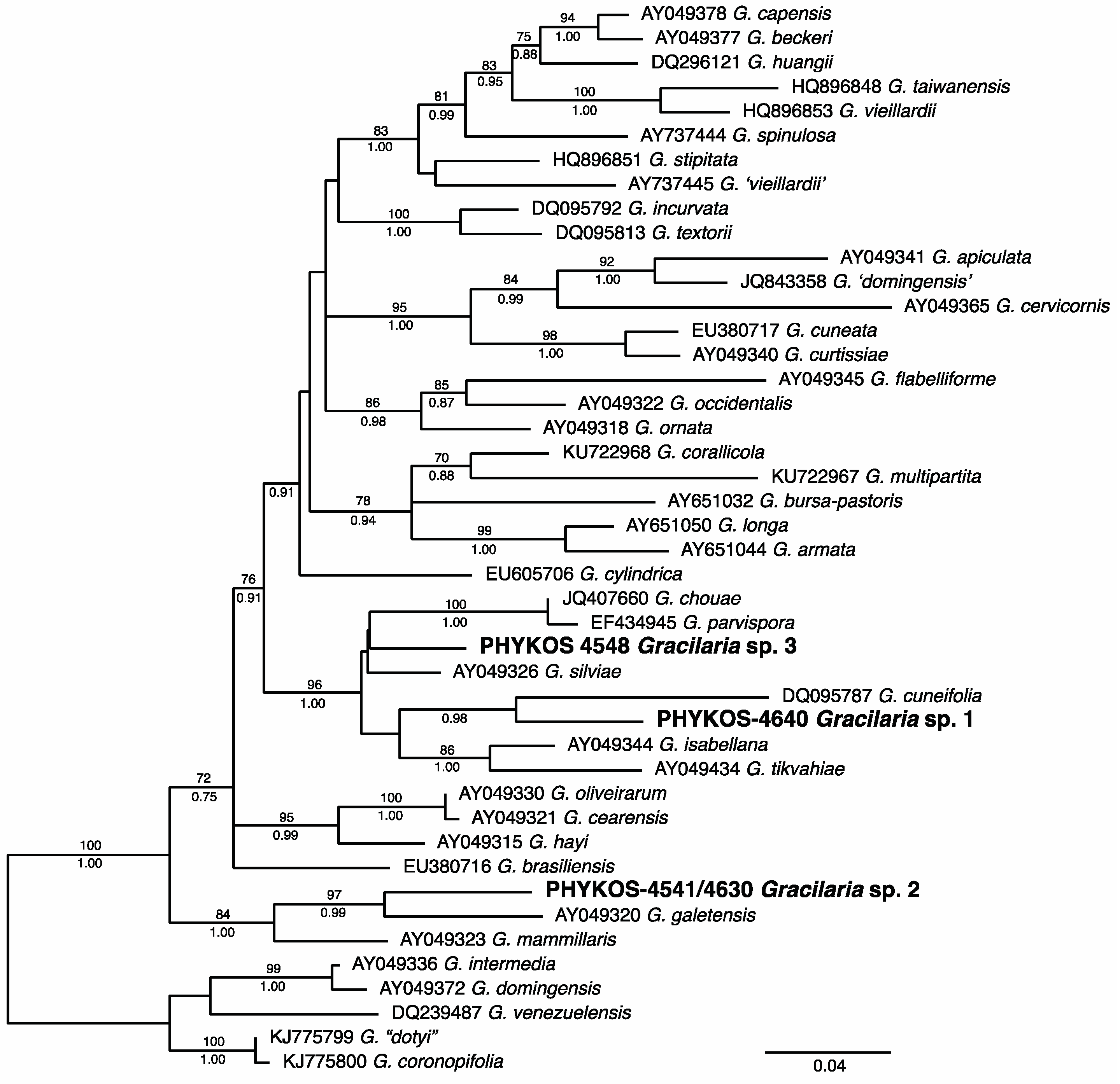
3.2.17. Gracilaria Species

Sequences for the three analyzed loci from five of the Punta Burica specimens (PHYKOS-4519, 4541, 4548, 4630, and 4640) separated out as three related species: (1) PHYKOS-4640; (2) PHYKOS-4519, 4541 and 4630; (3) PHYKOS-4548 (Figure 2). BLAST searches with these sequences returned Gracilaria specimen hits, but none with homology at a level to suggest species identifications (Table 2). Phylogenetic analyses of publically available rbcL-3P sequence data for Gracilaria species resolved all three of the Punta Burica species within Gracilaria sensu stricto (Figure 20). Gracilaria sp. 2 was strongly supported in a sister position to G. galetensis Gurgel, Fredericq and J.N. Norris, a species of the tropical Western Atlantic that was originally described from Caribbean Panama. Thus, these two species potentially represent a transisthmian geminate species pair. Gracilaria sp. 1 and Gracilaria sp. 3 were resolved within a strongly supported clade of Gracilaria species from both the Western Atlantic and Pacific (Figure 20).
Gracilaria sp. 1 was collected from subtidal rock at 4–5 m depth off the southwest point of Isla Burica (Figure 1). Specimens of Gracilaria sp. 2 were collected subtidally from both the mixed sediment and mudstone bottom just beyond the surf zone along Punta Burica (PHYKOS-4519; PHYKOS-4541), and rocks at 4–5 m depth off the southwest point of Isla Burica. Gracilaria sp. 3 was also collected from the mixed sediment and mudstone bottom just beyond the Punta Burica surf zone. Observed specimens of the three species were small and mostly less than 2 cm high (Figure 21). Gracilaria sp. 3 differed from the others in having dissected, acute branch apices (Figure 21C).
Remarks: Eight species of Gracilaria are reported from Pacific Central America. Most of these are relatively large compared to the Punta Burica species. Gracilaria brevis W.R. Taylor was originally described as reaching only 5 cm in height [2], and it was included in the Littler and Littler [49] web database of Pacific Panama species. The Punta Burica Gracilaria sp.3 is similar to G. crispata Setchell and Gardner and G. spinigera E.Y. Dawson in having acute, dentate-like apices, but the collected Punta Burica specimens were much smaller than the reported sizes for these species [62]. A comprehensive examination of tropical eastern Pacific Gracilaria species will be needed to determine if the Punta Burica species are simply juvenile growth forms or undescribed species. At least two, if not all three of these species are unreported for Panama.

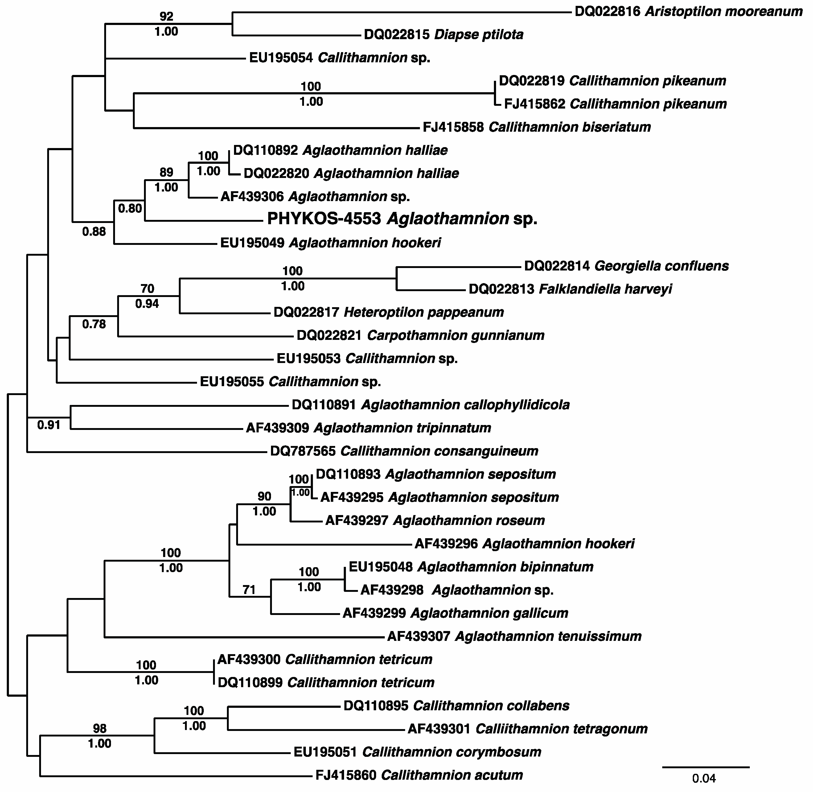
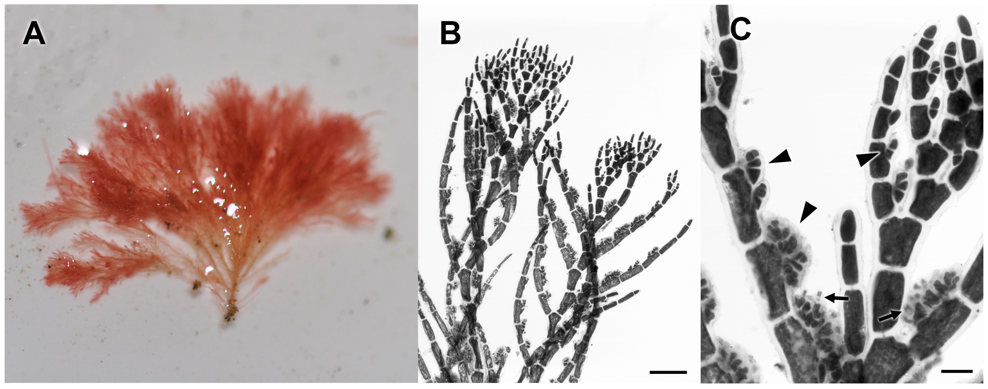
3.2.18. Aglaothamnion sp.

The PHYKOS-4553 rbcL-3P BLAST search returned sequences of Aglaothamnion halliae (Collins) N.E. Aponte, D.L. Ballantine and J.N. Norris and A. hookeri (Dillwyn) Maggs and Hommersand at 96% similarity, and a variety of Aglaothamnion and Callithamnion species without close homology in BLAST searches with COI-5P and UPA. Analyses of the rbcL-3P locus with other available Callithamnieae sequences resolved the Punta Burica species within a clade that also included A. halliae, A. hookeri and another unidentified Aglaothamnion species (Figure 22).
PHYKOS-4553 was growing attached to mudstone substrate on the mixed sediment and mudstone bottom just beyond the surf zone at Punta Burica (Figure 1).
Remarks: There currently is little available sequence data for species of Aglaothamnion or Callithamnion, and their relationships within the tribe Callithamnieae has been best characterized by concatenated data sets of multiple loci, albeit with limited taxon sampling [78,79]. Aglaothamnion and Callithamnion species are similar in general habit with both genera having distichous or radial, alternate branching from a uniseriate main axis. The genera are separated by the presence of multinucleate cells in Callithamnion, in contrast to Aglaothamnion having only uninucleate cells [80]. Aglaothamnion has been relatively well characterized in the tropical Western Atlantic [81], but both Aglaothamnion and Callithamnion are poorly known in the tropical East Pacific [5]. Taylor [2] included five species in his list, four of which were newly described by him. However, two were from Pacific Mexico and the remaining three were only collected in the Galapagos Islands. Callithamnion marshallense E.Y. Dawson is the only species of either genus known from Pacific Central America with reports from Costa Rica [5], and it is also included in the Littler and Littler [49] web database of Pacific Panama species. The Punta Burica species had lateral branchlets that develop from nearly all main axis cells in an alternate distichous pattern (Figure 23A,B), in contrast to C. marshallense that has branchlets develop in an alternate radial pattern and not from every main axis cell [82]. The Punta Burica specimen was male, and spermatangia development was similar to that described for other Aglaothamnion species [44,81]. Spermatangial initials were cut off from the adaxial side of lateral branchlet cells towards their distal ends and divided to form “antheridial stands” [83] that cut off spermatia and may overlap with the adjacent branchlet cell (Figure 23B,C). PHYKOS-4553 was similar to Taylor’s [2] description of C. pacificum W.R. Taylor, which is only known from the type collection, (epiphytic on larger algae dredged from 41–84 m depth near Isla Soccoro, Mexico) an environment that is likely quite different from the turbulent mixed sediment and mudstone bottom where the Punta Burica specimen was collected. Callithamnion pacificum, may be further distinguished by its more irregular branching, though clearly, additional study of both is needed to determine their relationship. Regardless, this species has not be reported previously from Pacific Panama or Pacific Central America and may represent a new species.

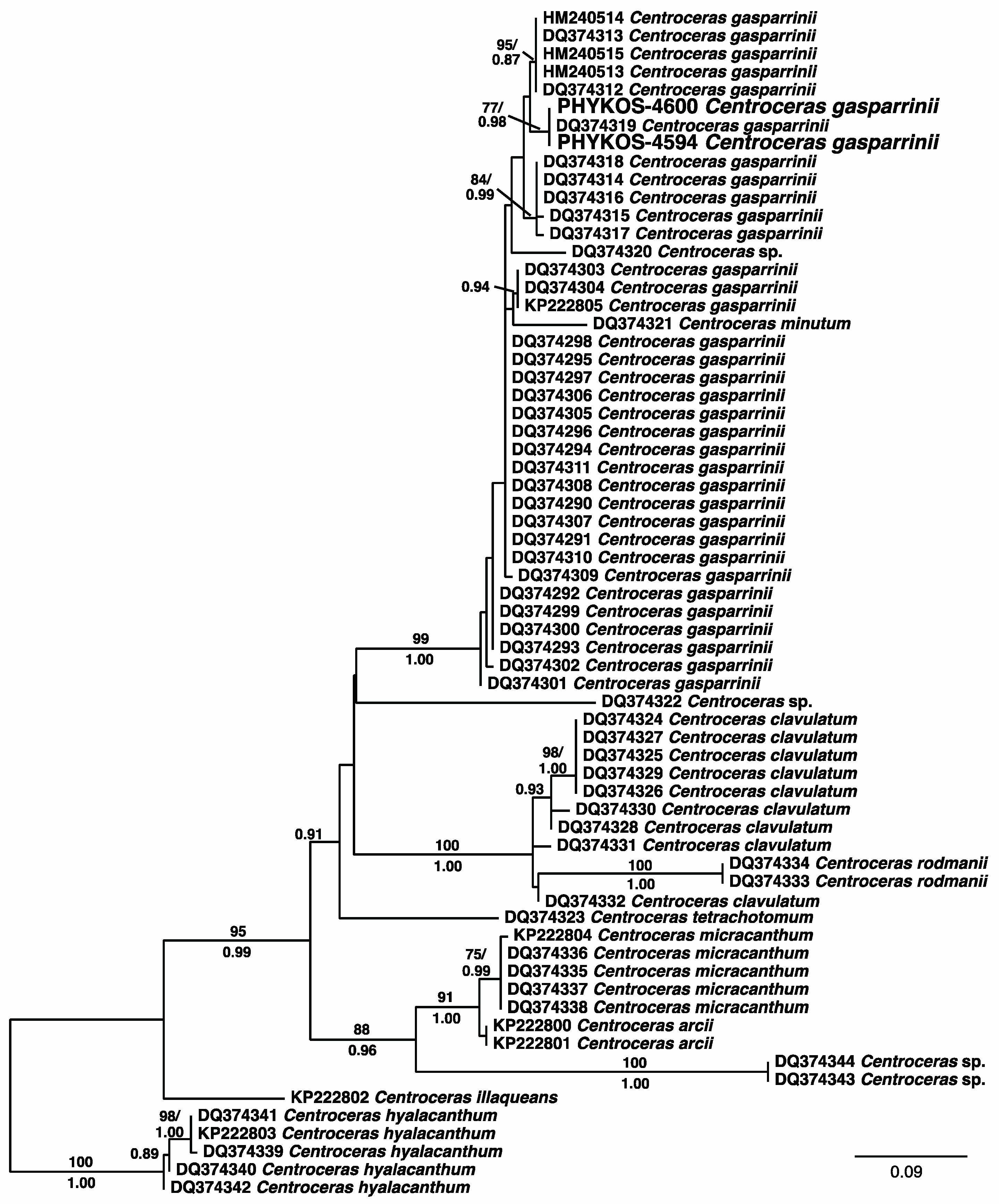
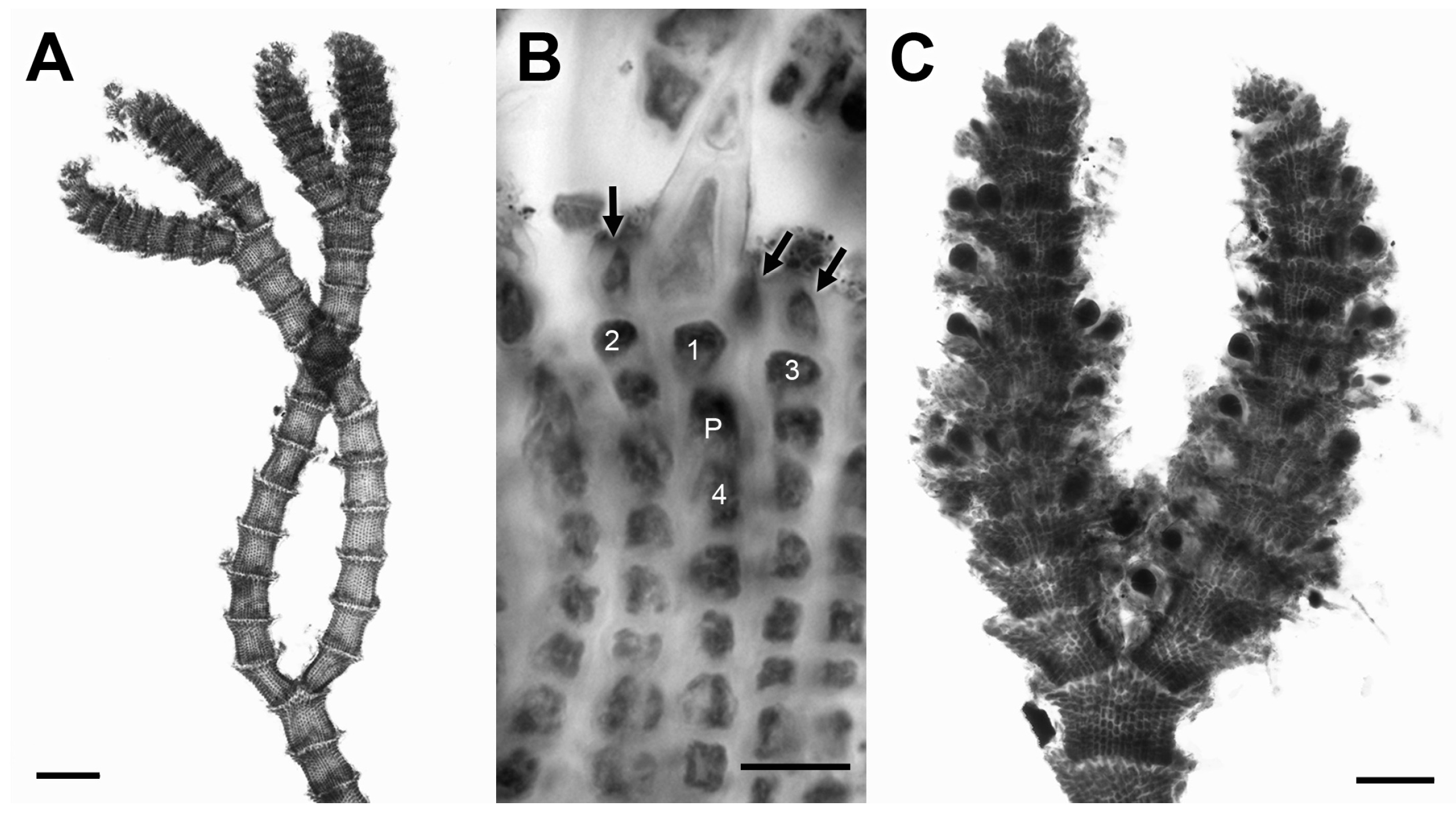
3.2.19. Centroceras gasparrinii (Meneghini) Kützing

PHYKOS-4594 and PHYKOS-4600 shared identical sequences for the three analyzed loci. BLAST searches with COI-5P and UPA returned specimens identified as Centroceras clavulatum (C. Agardh) Montagne (94%) and Centroceras sp. (99%) respectively. The Punta Burica specimens rbcL-3P sequences were found to be exact matches with a specimen identified as C. gasparrinii that had been collected from near the Pacific terminus of the Panama canal. Phylogenetic analyses of rbcL-3P sequence data available from GenBank resolved Pacific Panama specimens in a well supported C. gasparrinii clade (Figure 24).
Both specimen collections were made from intertidal turfs growing on the mudstone substrate along the Punta Burica shoreline (Figure 1).
Remarks: Centroceras gasparrinii was long considered a synonym of C. clavulatum until the detailed morphological and molecular analyses of Won et al. [30] established that the shape of acropetal cortical cells, spines and gland cells were vegetative characters that could be used to distinguish most Centroceras species and these have been used in subsequent studies describing new species [84,85]. Erect axes of Punta Burica specimens had forcipate, slightly inrolled apices, di-trichotomous branching and nodal cortication characteristics as descibed for C. gasparrini [30] (Figure 25A,B). Tetrasporangia were produced in a whorl around the axes at upper nodes (Figure 25C).
Dawson [86] reported C. minutum Yamada from Pacific Panama, and this was the only record of Centroceras from Pacific Central America until the study of Won et al. [30] that included C. gasparrinii. The two species are easily distinguished by branching pattern, which is alternate in C. minutum and di-trichotomous in C. gasparrinii. Centroceras gasparrinii is reported from both Caribbean and Pacific Panama, and notably at both termini of the Panama Canal. Whether these populations are more closely related than their intra-oceanic conspecifics remains to be studied.

3.2.20. Spyridia sp.

Only a COI-5P sequence was generated from PHYKOS-4584, and BLAST searches revealed a closest match (95%) with a specimen identified as Spyridia filamentosa (Wulfen) Harvey from Hawaii. The next closest matches (88–89%) were to other Hawaiian specimens also identified as S. filamentosa.
The PHYKOS-4584 collection was made from a tide pool along the Punta Burica shoreline (Figure 1). The specimen displays the general morphological characteristics of Spyridia including fully corticated indeterminate axes with a determinate branchlet produced by each indeterminate axis cell; inderminate axis cortication alternating bands of shorter, broader nodal cells and longer, narrower internodal cells, and determinate branchlets corticated only at nodes and with some tips terminated by acuminate spines (Figure 26).
Remarks: Spyridia filamentosa is widely reported around the globe [42], but previous studies have revealed that it is a complex of cryptic species [87,88,89]. Conklin and Sherwood [89] identified two separate S. filamentosa lineages within the Hawaiian Islands based on nuclear-encoded LSU sequences and determinate branchlet cell dimensions. The Hawaiian specimen with a 95% COI-5P similarity to the Punta Burica species was part of Conklin and Sherwood’s LSU lineage-1, and the specimens for the next closest BLAST hits were part of their LSU lineage-2 [89]. Although the dimensions of determinate branchlet cells were found by Conklin and Sherwood [89] to be consistent character states separating the Hawai’i S. filamentosa LSU lineages, other morphological characters used to distinguish Spyridia species have been found to be variable within species and even the same thallus [90,91]. Taylor [2] reported S. filamentosa from Isla Taboga in the Bay of Panama and Littler and Littler [49] included it in their web database. Based on its Adriatic Sea type locality and the established diversity of cryptic species that are included under this name, the Punta Burica species and other reports of S. filamentosa from Pacific Central America most likely do not belong in this species and should be considered as unidentified Spyridia species.

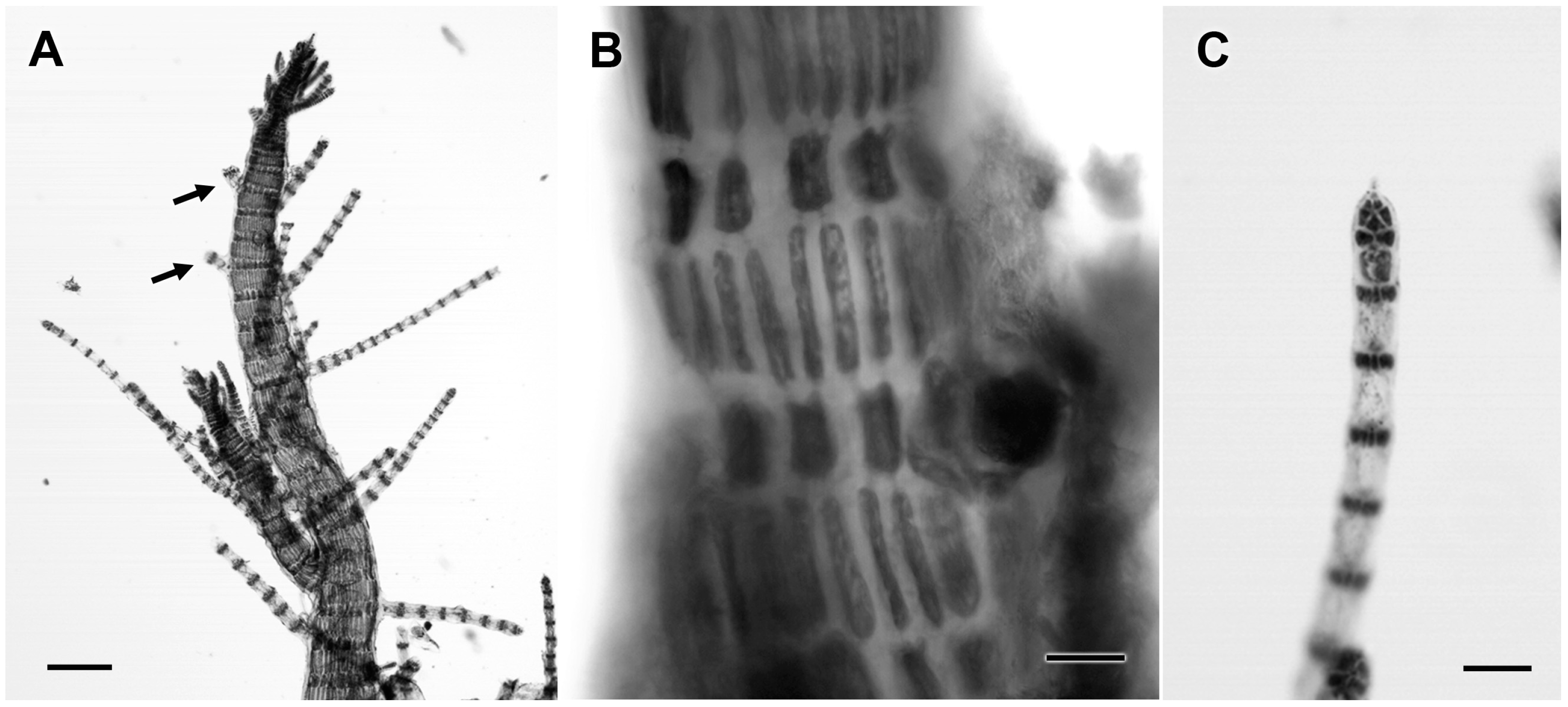
3.2.21. Melanothamnus sp.

BLAST searches with rbcL-3P and COI-5P sequences for PHYKOS-4568 showed a closest match with sequences from a specimen identified as Melanothamnus pseudovillum (Hollenberg) Díaz-Tapia and Maggs (as Polysiphonia pseudovillum Hollenberg) from Caribbean Panama (Table 2). The Caribbean Panama M. pseudovillum and PHYKOS-4568 Melanothamnus sp. were also fully supported as sister species in phylogenetic analyses of rbcL sequences of Polysiphonia sensu lato species (Figure 27).
The Punta Burica Melanothamnus species was found growing as patches of near monospecific turf within tide pools (Figure 1A and Figure 28A). Axes were ecorticate with four pericentral cells displaying plastids restricted to their radial walls, and had rhizoids that were cut off from pericentral cells (Figure 28B–D). Tetrasporangia developed in short straight series that shifted to offset positions around the axis (Figure 28E).
Remarks: Melanothamnus was a poorly known genus of two relatively robust rhodomelacean species restricted in distribution to northeast Africa and southwest Asia [92,93]. Recent DNA sequence analyses of Polysiphonia sensu lato species did not resolve Melanothamnus, Fernandosiphonia, and Neosiphonia as independent monophyletic lineages [94]. This combined with a re-evaluation of morphological characters lead to the synonymy of Fernandosiphonia and Neosiphonia under Melanothamnus. The restriction of plastids to radial walls and their absence from the outer walls of pericentral cells along with 3-celled carpogonial branches are synapomorphic character states for Melanothamnus species [94]. Rhizoids that are cut off from pericentral cells is another character state shared by Melanothamnus species, but this is a state that is found in many Rhodomelaceae genera.
Many of the character states observed in the Punta Burica Melanothamnus species match those described for Caribbean Panama M. pseudovillum [95], but the two species differ in the development of tetrasporangia, which is considered to be a consistent character within Polysiphonia sensu lato species [25]. Caribbean Panama M. pseudovillum had tetrasporangia that developed in a long spiral series, in contrast to the offset short straight series of tetrasporangia observed in the Punta Burica species.
The rbcL-3P divergence between the Caribbean Panama M. pseudovillum and Punta Burica Melanothamnus sp. was 2.0%, which is generally considered to represent the low end of interspecific divergence values for Polysiphonia sensu lato species e.g., [96,97,98]. COI-5P divergence values in red algae are generally greater than those for rbcL in complementary comparisons e.g., [99,100]. A recent study of the closely related species Melanothamnus japonica (Harvey) Díaz-Tapia and Maggs, M. harveyi (Bailey) Díaz-Tapia and Maggs, and Polysiphonia akkeshiensis Segi found interspecific COI-5P divergences ranging from 1.7–3.2% [101]. The COI-5P divergence of 2.8% between the Caribbean Panama M. pseudovillum and Punta Burica Melanothamnus sp. falls within this range of interspecific values for closely related species, and they represent a transisthmian geminate species pair.

3.2.22. ‘Polysiphoniabinneyi Harvey

PHYKOS-4534 COI-5P and UPA sequences were found by BLAST searches to be closest matches with specimens of ‘Polysiphoniaechinata Harvey and a ‘Polysiphonia’ sp. respectively. Searches with the PHYKOS-4534 rbcL-3P sequence revealed a close homology (99%) with the sequence from a Caribbean Panama ‘P.binneyi specimen, and they were resolved together with strong support in the Polysiphonia sensu lato rbcL phylogeny (Figure 27).
The PHYKOS-4534 sample was collected with a mixture of other algae from mixed sediment-mudstone bottom just beyond the Punta Burica surf zone (Figure 1), and a limited amount of material was available for study. The specimens shared character states described for Caribbean Panama ‘P.binneyi including four pericentral cells; basally attenuated lateral branches; segments that were mostly shorter than broad with this being especially prominent near axes’ tips; trichoblasts or scar cells at each segment, and tetrasporangia in spiral series (Figure 29).
Remarks: Mamoozadeh and Freshwater [95] generated rbcL sequences from four Caribbean Panama specimens identified as ‘P.binneyi. Three shared identical sequences and these were 1.1% different from the fourth specimen over the rbcL-3P region. Direct comparisons of the Punta Burica and Caribbean ‘P.binneyi rbcL-3P sequences showed that they differed by 0.8–1.6%. This is within what has been generally considered an intraspecific level of variation e.g., [95,96]. Comparisons of COI-5P sequences among these specimens are currently not possible because Mamoozadeh and Freshwater [95] were unable to amplify and sequence this locus from the Caribbean ‘P.binneyi specimens. ‘Polysiphoniabinneyi is resolved in a strongly supported clade that is part of a series of clades only distantly related to Polysiphonia sensu stricto that need reclassification at the genus level see [94]. Originally described from Key West, FL, USA, this species was previously known only from the tropical Western Atlantic and Caribbean [42].

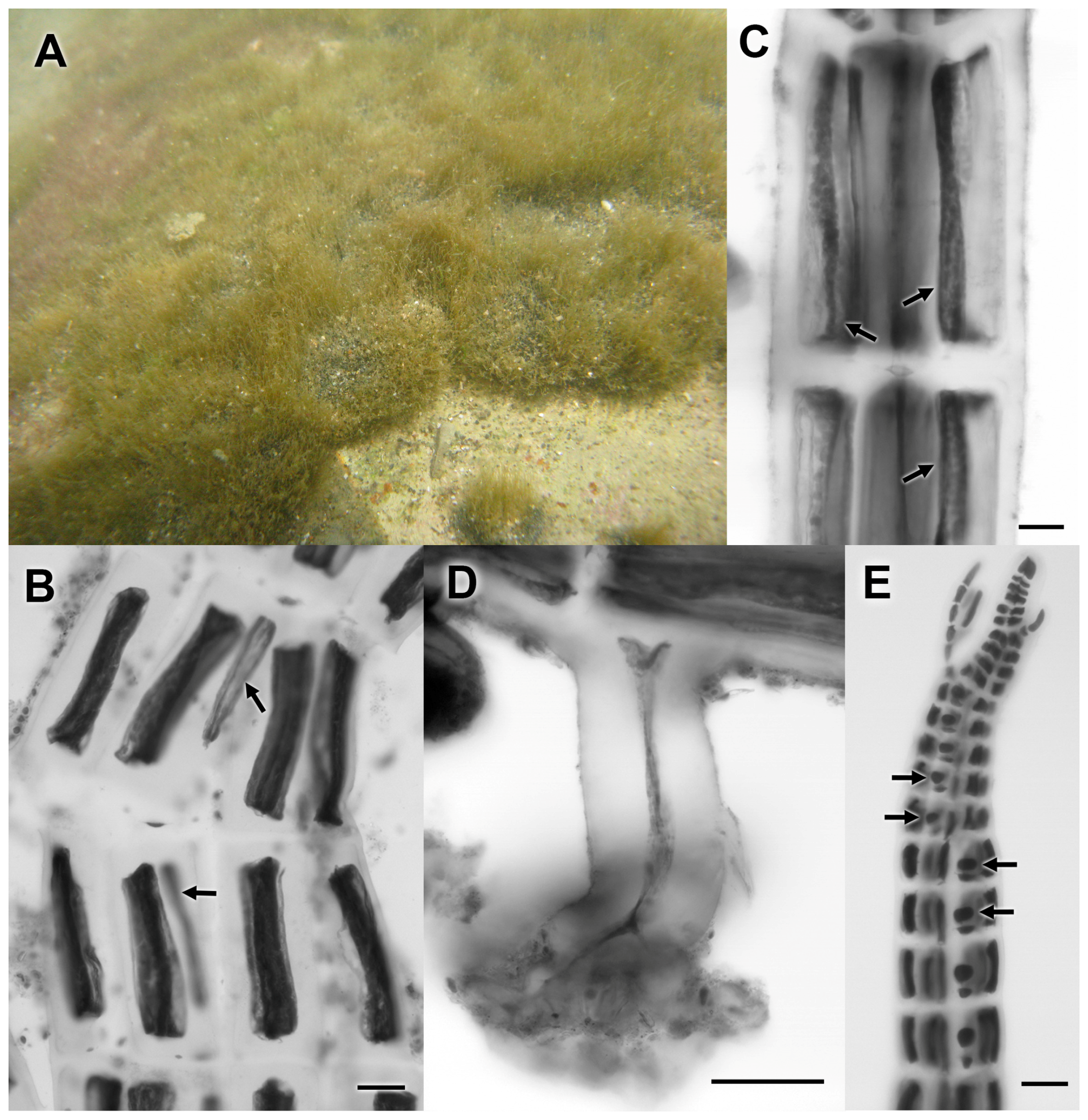
3.2.23. ‘Polysiphonia’ sp.

BLAST searches with sequences of PHYKOS-4515, PHYKOS-4525, PHYKOS-4526 and PHYKOS-4552 resulted in hits with sequences from Caribbean Panama specimens identified as ‘Polysiphonia’ sp. for rbcL-3P and COI-5P, and species of Herposiphonia and Symphyocladia for UPA. Analyses of rbcL sequences in this study resolve the Caribbean and Pacific Panama specimens within a fully supported clade that terminates an independent lineage among representatives of Herposiphonia, Lophosiphonia, Pterosiphonia, Symphyocladia, and Wilsonosiphonia (Figure 27).
All four specimen collections were made from the mixed sediment-mudstone bottom just beyond the Punta Burica surf zone. Axes were ecorticate and had four pericentral cells with relatively large central axial cells; rhizoids were cut off from pericentral cells, and trichoblasts and scar cells were variable in pattern. Mid-axis segments were approximately 1× as long as wide, but this abruptly shifted to 0.5× as long as wide in upper portions of axes giving them an elongated clavate shape (Figure 30A).
Remarks: The Caribbean Panama ‘Polysiphonia’ sp. rbcL and COI-5P sequences were generated from four specimens that shared identical sequences at both loci [102], and direct comparisons with the Punta Burica sequences revealed only 0.1% (rbcL) and 0.3% (COI-5P) divergence. The Punta Burica and Caribbean Panama ‘Polysiphonia’ sp. specimens shared states for all observed key morphological characters. Reproductive structures were not observed in Caribbean specimens [102], but tetrasporangia in spiral series were seen developing in short lateral branches of the Punta Burica specimens (Figure 30A,B). Previous phylogenetic analyses have resolved this species as an independent lineage with no close affinity to any Polysiphonia sensu lato genera [95].

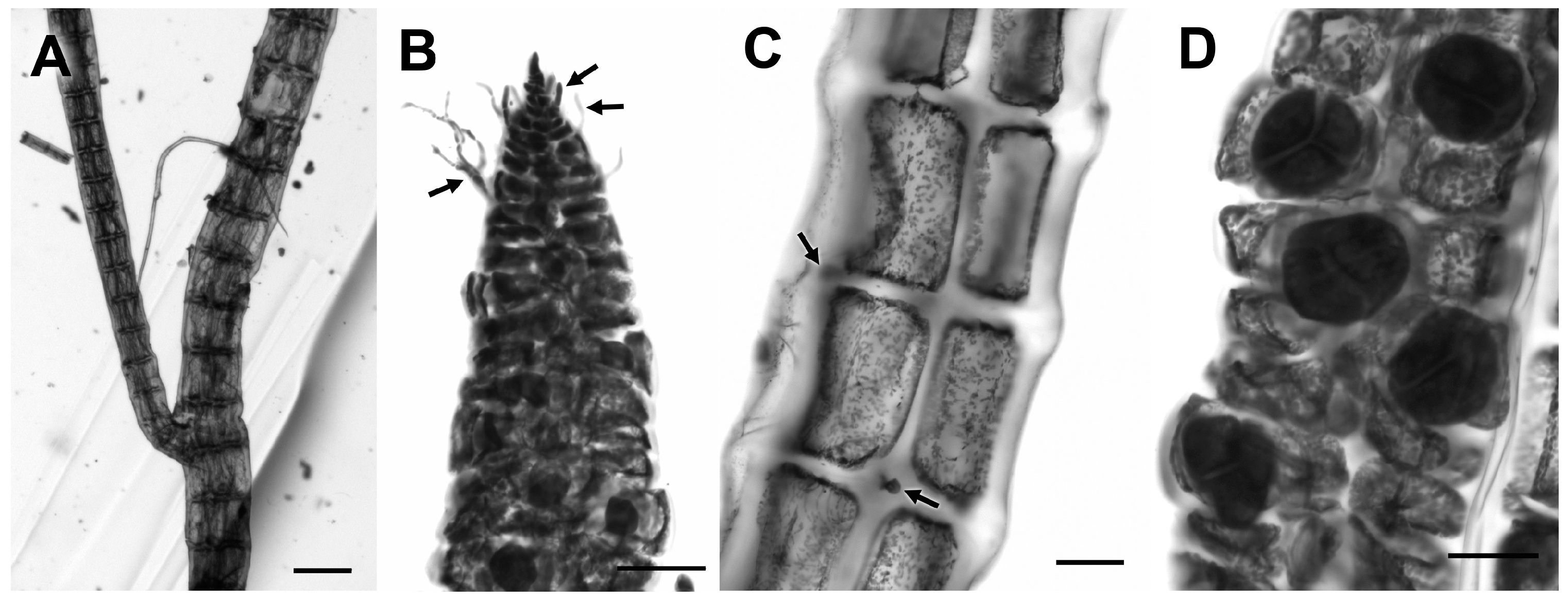
3.2.24. Wilsonosiphonia howei (Hollenberg) D. Bustamante, Won and T.O. Cho

Sequences for the three analyzed loci from PHYKOS-4657 and PHYKOS-4663 returned BLAST hits for Caribbean Panama specimens of Wilsonosiphonia howei (Table 2). Variation between the Pacific and Caribbean specimens in nearly complete rbcL sequences was 1.1–1.2% and in COI-5P sequences 2.7–2.9%. Phylogenetic analyses of rbcL sequences for Polysiphonia sensu lato species and closely related genera resolve the Pacific and Caribbean Wilsonosiphonia howei together in a fully supported clade (Figure 27).
Both PHYKOS-4657 and PHYKOS-4663 were collected from short turf patches growing on high intertidal rocks along the eastern shoreline of the Burica Peninusla (Figure 1). These specimens displayed the key morphological characters states attributed to Wilsonosiphonia species including rhizoids cut off from the distal ends of pericentral cells and “taproot shaped”, multicellular rhizoid tips (Figure 31A,B). The specimens were also ecorticate; had 10–14 pericentral cells around relatively wide central axial cells (Figure 31C); displayed a pericentral cell shift to offset positons across segments; tetrasporangia that develop in a spiral series (Figure 31D), and cystocarps that were ovate and without enlarged pericarp cells around the ostiole (Figure 31E).
Remarks: Wilsonosiphonia howei was originally described as Polysiphonia howei Hollenberg based on specimens from the Bahamas [2]. Taylor [2] also refered Pacific Panama specimens collected from Isla Taboga in the Bay of Panama to this species, and it has been widely reported in warm-temperate and tropical waters since then e.g., [90,103,104,105]. Mamoozadeh and Freshwater [95] suggested that the concept of W. howei (as Polysiphonia howei) encompassed multiple species based on reported variation in spermatiagial branch development e.g., [104,106,107], pericentral cell numbers e.g., [44,90], and divergence between available nuclear-encoded 18S rRNA gene sequences. Bustamante et al. [103] included two new species in Wilsonosiphonia when they described the genus, W. fujiae D. Bustamante, Won and T.O. Cho, the generitype, and W. indica D. Bustamante, Won and T.O. Cho. They also considered the previous concept of W. howei to included multiple species, and suggested that its distribution was limited to the western Atlantic. Two recent barcoding analyses of Polysiphonieae [101] and Pterosiponieae [108] species found maximum intraspecific divergences in rbcL and COI-5P sequences to be 0.7% and 0.9% respectively. The rbcL (1.1–1.2%) and COI-5P (2.7–2.9%) divergences between the Pacific Panama and Caribbean Panama W. howei specimens analyzed here suggest that they are separate species and represent a transisthmian geminate pair. An examination of W. howei from throughout its current distribution in needed to determine how many species may be included under this name.

4. Discussion

Molecular assisted identification proved to be a useful tool in this study of the previously unexplored Punta Burica marine algal flora. The three loci employed in the initial DNA barcoding approach showed different levels of variation with COI-5P having the highest, rbcL-3P the next highest and UPA the lowest. Despite these differences, cluster analyses with the three loci grouped the specimens into nearly identical genetic species, and analyses of the more variable COI-5P sequences resolved only one more species than analyses of complementary rbcL-3P and UPA sequences. Searches of publicly available databases with these sequences suggested at minimum possible genus-level classifications, and guided the assessment of morphological characters for the verification of species-level classifications. However, careful evaluation of search results is required because many database entries have not been updated to the most recent classification and others represent misidentifications.
The recent compilation of marine algae reported in the Central American Pacific by Fernández-García et al. [5] recorded 379 species in the region. Pacific Panama had the second highest number with 174, yet there were no records of marine algae from the western Gulf of Chiriqui mainland coast, and fewer than 20 species had been reported from islands in this area [2,3,4,109]. As pointed out in Fernández-García et al. [5], much of the discrepancy in the number of reported species along Pacific Central America is a product of exploration effort, as the region’s high heterogeneity in habitats and physical environmental factors should be conducive to generally high marine algal diversity. In contrast to the small number of marine algal records in the western Gulf of Chiriqui, the adjacent coastal area of southeastern Costa Rica to the west has a high number of reported species, but it is also one of the better-explored areas in Pacific Central America [5].
Despite our Punta Burica collecting being limited to only four short events, numerous species of red, green and brown algae were found. The 26 species treated here represent only the collections of foliose red algae and are the first reports of marine algae from the mainland coast of the western Gulf of Chiriqui. Twenty-one of these are new reports for Panama, and nine are new reports for Pacific Central America. Four records appear to be new to the Pacific entirely, including the two newly described species, Neorubra parvolacertoides and Grateloupia irregularis, as well as ‘Polysiphoniabinneyi and Hypnea flava. It is perhaps not surprising, given the paucity of historical study for this remote region of Panama that 12 of the 26 species reported here likely represent novel species, requiring further morphological and molecular examination.
The mudstone substrate in the Punta Burica intertidal zone provides an environment more conducive to the attachment and growth of marine algae than many other hard substrates along the Pacific Panama coast. Perhaps this distinctive environment has driven evolutionary adaptation resulting in a local biodiversity hotspot. Alternatively, it may simply be, as others have asserted [56], that the more we look, the more we find and that as our geographic coverage expands within Panama or throughout Central America, redundancy in species richness assessment will be revealed.
Another important aspect of the work presented here is the recognition of transisthmian geminate species pairs. Evolutionary divergence between geminate species may facilitate the calibration of important DNA barcode markers, at least among closely related species. The greater the availability of geminate species pairs for marine algae lacking a meaningful fossil record, the more accurately can molecular calibrations be estimated, while recognizing the inherent challenge e.g., [110] to isthmian-derived evolutionary rate calculations. Here, the value of DNA barcoding as a floristics tools is remarkable; DNA barcoding in our floristic study has provided a more accurate assessment of marine algal biodiversity, even when the amount of material or specimens available for study was small. Continued DNA barcoding studies of the Burica green, brown and red algal floras, as well as continuing studies of the Panamanian marine flora more generally, will undoubtedly shed new light on the novelty of the mudrock environment as a potential refuge for algal diversity and continue to illuminate new subjects for studying the evolutionary biology of marine macroalgae.

Supplementary Materials

The following are available online at https://www.mdpi.com/1424-2818/9/2/19/s1, Table S1: Data matrix dimensions and analysis parameters for phylogenetic analyses carried out in this study.

Acknowledgments

This study was funded by US National Science Foundation Biotic Surveys and Inventories grant 0743334, the Center for Marine Science DNA-Algal Trust, and the Red Pond Trust. The authors wish to thank Melissa Smith for help with graphics and Melissa LaCroce for technical help with putting the manuscript together.

Author Contributions

B.W. organized and planned the collecting expedition; B.W., D.W.F., C.F.-G. and N.L. collected the studied specimens, S.L.P., P.W.G. and D.W.F. generated sequence data; J.N.I., P.W.G. and D.W.F. carried out DNA sequence and morphological analyses; D.W.F., P.W.G., C.F.-G. and B.W. wrote the paper.

Conflicts of Interest

The authors declare no conflict of interest.

References

  1. Howe, M.A. Report on a botanical visit to the Isthmus of Panama. J. N. Y. Bot. Gard. 1910, 11, 30–44. [Google Scholar]
  2. Taylor, W.R. Pacific Marine Algae of the Allan Hancock Expeditions to the Galapagos Islands; The University of Southern California Press: Los Angeles, CA, USA, 1945; p. 528. [Google Scholar]
  3. Wysor, B. An annotated list of marine Chlorophyta from the Pacific coast of the republic of Panama with a comparison to Caribbean Panama species. Nova Hedwigi. 2004, 78, 209–241. [Google Scholar] [CrossRef]
  4. Littler, M.M.; Littler, D.S. Coralline algal rhodoliths form extensive benthic communities in the Gulf of Chiriqui, Pacific Panama. Coral Reefs 2008, 27, 553. [Google Scholar] [CrossRef]
  5. Fernández-García, C.; Riosmena-Rodríguez, R.; Wysor, B.; Tejada, O.L.; Cortés, J. Checklist of the Pacific marine macroalgae of Central America. Bot. Mar. 2011, 54, 53–73. [Google Scholar] [CrossRef]
  6. Dawson, E.Y. Marine algae from the Pacific Costa Rican gulfs. Contrib. Sci. 1957, 15, 1–28. [Google Scholar]
  7. Morell, K.D.; Fisher, D.M.; Gardner, T.W.; Femina, P.L.; Davidson, D.; Teletzke, A. Quaternary outer fore-arc deformation and uplift inboard of the Panama Triple Junction, Burica Peninsula. J. Geophys. Res. 2011, 116, B05402. [Google Scholar] [CrossRef]
  8. Buchs, D.M.; Baumgartner, P.O.; Baumgartner-Mora, C.; Bandini, A.N.; Jackett, S.-J.; Diserens, M.-O.; Stucki, J. Late Cretaceous to Miocene seamount accretion and mélange formation in the Osa and Burica Peninsulas (Southern Costa Rica): Episodic growth of a convergent margin. In The Origin and Evolution of the Caribbean Plate; Lorente, J.K., Pindell, M.A., Eds.; Geological Society: London, UK, 2007; Volume 28, pp. 411–456. [Google Scholar]
  9. Mamoozadeh, N.R.; Freshwater, D.W. Taxonomic notes on Caribbean Neosiphonia and Polysiphonia (Ceramiales, Flordeophyceae): Five species from Florida, USA and Mexico. Bot. Mar. 2011, 54, 269–292. [Google Scholar] [CrossRef]
  10. Hebert, P.D.N.; Cywinska, A.; Ball, S.L.; de Waard, J.R. Biological identifications through DNA barcodes. Philos. Trans. R. Soc. B 2003, 270, 313–321. [Google Scholar] [CrossRef] [PubMed]
  11. Saunders, G.W. Applying DNA barcoding to red macroalgae: A preliminary appraisal holds promise for future applications. Phil. Trans. R. Soc. B 2005, 360, 1879–1888. [Google Scholar] [CrossRef] [PubMed]
  12. Robba, L.; Russell, S.J.; Barker, G.L.; Brodie, J. Assessing the use of the mitochondrial cox1 marker for use in DNA barcoding of red algae (Rhodophyta). Am. J. Bot. 2006, 93, 1101–1108. [Google Scholar] [CrossRef] [PubMed]
  13. Le Gall, L.; Saunders, G.W. DNA barcoding is a powerful tool to uncover algal diversity: A case study of the Phyllophoraceae (Gigartinales, Rhodophyta) in the Canadian flora. J. Phycol. 2010, 46, 374–389. [Google Scholar] [CrossRef]
  14. Milstein, D.; Saunders, G.W. DNA barcoding of Canadian Ahnfeltiales (Rhodophyta) reveals a new species—Ahnfeltia borealis sp. nov. Phycologia 2012, 51, 247–259. [Google Scholar] [CrossRef]
  15. Lyra, G.D.M.; Gurgel, C.F.D.; Costa, E.D.S.; de Jesus, P.B.; Oliveira, M.C.; Oliveira, E.C.; Davis, C.C.; Nunes, J.M.D.C. Delimitating cryptic species in the Gracilaria domingensis complex (Gracilariaceae, Rhodophyta) using molecular and morphological data. J. Phycol. 2016, 52, 997–1017. [Google Scholar] [CrossRef] [PubMed]
  16. Sherwood, A.R.; Presting, G.G. Universal primers amplify a 23S rDNA plastid marker in eukaryotic algae and cyanobacteria. J. Phycol. 2007, 43, 605–608. [Google Scholar] [CrossRef]
  17. Sherwood, A.R.; Sauvage, T.; Kurihara, A.; Conklin, K.T.; Presting, G.G. A comparative analysis of COI, LSU and UPA marker data for the Hawaiian florideophyte Rhodophyta: Implications for DNA barcoding of red algae. Cryptogam. Algologie 2010, 31, 451–465. [Google Scholar]
  18. Iha, C.; Milstein, D.; Guimarães, S.M.P.B.; Freshwater, D.W.; Oliveira, M.C. DNA barcoding reveals high diversity in the Gelidiales of the Brazilian southeast coast. Bot. Mar. 2015, 58, 295–305. [Google Scholar] [CrossRef]
  19. Chase, M.W.; Hills, H.H. Silica gel: An ideal material for field preservation of leaf samples for DNA studies. Taxon 1991, 40, 215–220. [Google Scholar] [CrossRef]
  20. Hughey, J.R.; Silva, P.C.; Hommersand, M.H. Solving taxonomic and nomenclatural problems in Pacific Gigartinaceae (Rhodophyta) using DNA from type material. J. Phycol. 2001, 37, 1091–1109. [Google Scholar] [CrossRef]
  21. Freshwater, D.W.; Braly, S.K.; Stuercke, B.; Hamner, R.M.; York, R.A. Phylogenetic analyses of North Carolina Rhodymeniales. I. The genus Asteromenia. J. N. C. Acad. Sci. 2005, 121, 49–55. [Google Scholar]
  22. Saunders, G.W. A DNA barcode examination of the red algal family Dumontiaceae in Canadian waters reveals substantial cryptic species diversity. 1. The foliose Disea-Neodilsea complex and Weeksia. Botany 2008, 86, 773–789. [Google Scholar] [CrossRef]
  23. Saunders, G.W.; McDevit, D.C. Methods for DNA barcoding photosynthetic protists emphasizing the macroalgae and diatoms. Methods Mol. Biol. 2012, 858, 207–222. [Google Scholar] [PubMed]
  24. Freshwater, D.W.; Rueness, J. Phylogenetic relationships of some European Gelidium (Gelidiales, Rhodophyta) species, based on rbcL nucleotide sequence analysis. Phycologia 1994, 33, 187–194. [Google Scholar] [CrossRef]
  25. Stuercke, B.; Freshwater, D.W. Consistency of morphological characters used to delimit Polysiphonia sensu lato species (Ceramiales, Florideophyceae): Analyses of North Carolina, USA specimens. Phycologia 2008, 47, 541–559. [Google Scholar] [CrossRef]
  26. Edgar, R.C. MUSCLE: Multiple sequence alignment with high accuracy and high throughput. Nucleic Acids Res. 2004, 32, 1792–1797. [Google Scholar] [CrossRef] [PubMed]
  27. Altschul, S.F.; Gish, W.; Miller, W.; Myers, E.W.; Lipman, D.J. Basic local alignment search tool. J. Mol. Biol. 1990, 215, 403–410. [Google Scholar] [CrossRef]
  28. Stamatakis, A. RAxML-VI-HPC: Maximum likelihood-based phylogenetic analyses with thousands of taxa and mixed models. Bioinformatics 2006, 22, 2688–2690. [Google Scholar] [CrossRef] [PubMed]
  29. Huelsenbeck, J.P.; Ronquist, F. MRBAYES: Bayesian inference of phylogenetic trees. Bioinformatics 2001, 17, 754–755. [Google Scholar] [CrossRef] [PubMed]
  30. Won, B.Y.; Cho, T.O.; Fredericq, S. Morphological and molecular characterization of species of the genus Centroceras (Ceramiaceae, Ceramiales), including two new species. J. Phycol. 2009, 45, 227–250. [Google Scholar] [CrossRef] [PubMed]
  31. Grusz, A.L.; Freshwater, D.W. Studies of Costa Rican Gelidiales (Florideophyceae). II. Two Pacific taxa including Gelidium microglossum, n. sp. Pac. Sci. 2014, 68, 97–110. [Google Scholar] [CrossRef]
  32. Boo, G.H.; Hughey, J.R.; Miller, K.A.; Boo, S.M. Mitogenomes from type specimens, a genotyping tool for morphologically simple species: Ten genomes of agar-producing red algae. Sci. Rep. 2016, 6, 35337. [Google Scholar] [CrossRef] [PubMed]
  33. Wang, W.-L.; Liu, S.-L.; Lin, S.-M. Systematics of the calcified genera of the Galaxauraceae (Nemaliales, Rhodophyta) with an emphasis on Taiwan species. J. Phycol. 2005, 41, 685–703. [Google Scholar] [CrossRef]
  34. Wiriyadamrikul, J.; Geraldino, P.J.L.; Huisman, J.M.; Lewmanomont, K.; Boo, S.M. Molecular diversity of the calcified red algal genus Tricleocarpa (Galaxauraceae, Nemaliales) with the description of T. jejuensis and T. natalensis. Phycologia 2013, 52, 338–351. [Google Scholar] [CrossRef]
  35. Huisman, J.M.; Borowitzka, M.A. A revision of the Australian species of Galaxaura (Rhodophyta, Galaxauraceae), with a description of Tricleocarpa gen. nov. Phycologia 1990, 29, 150–172. [Google Scholar] [CrossRef]
  36. Le Gall, L.; Saunders, G.W. Establishment of a DNA-barcode library for the Nemaliales (Rhodophyta) from Canada and France uncovers overlooked diversity in the species Nemalion helminthoides (Velley) Batters. Cryptogam. Algologie 2010, 31, 403–421. [Google Scholar]
  37. Machín-Sánchez, M.; Rousseau, F.; Le Gall, L.; Cassano, V.; Neto, A.I.; Sentíes, A.; Fuji, M.T.; Gil-Rodríguez, M.C. Species diversity of the genus Osmundea (Ceramiales, Rhodophyta) in the Macaronesian region. J. Phycol. 2016, 52, 664–681. [Google Scholar] [CrossRef] [PubMed]
  38. Taylor, W.R. Marine Algae of the Eastern Tropical and Subtropical Coasts of the Americas; The University of Michigan Press: Ann Arbor, MI, USA, 1960; p. 870. [Google Scholar]
  39. Costa, J.; Lin, S.-M.; Macaya, E.; Fernández-Garcia, C.; Verbruggen, H. Chloroplast genomes as a tool to resolve red algal phylogenies: A case study in the Nemaliales. BMC Evol. Biol. 2016, 16, 205–218. [Google Scholar] [CrossRef] [PubMed]
  40. Huisman, J.M. The type and Australian species of the red algal genera Liagora and Ganonema (Liagoraceae, Nemaliales). Aust. Syst. Bot. 2002, 15, 773–838. [Google Scholar] [CrossRef]
  41. Lin, S.-M.; Huisman, J.M.; Ballantine, D.L. Revisiting the systematics of Ganonema (Liagoraceae, Rhodophyta) with emphasis on species from the northwest Pacific Ocean. Phycologia 2014, 53, 37–51. [Google Scholar] [CrossRef]
  42. Guiry, M.D.; Guiry, G.M. AlgaeBase. World-Wide Electronic Publication, National University of Ireland, Galway, 2017. Available online: www.algaebase.org (accessed on 30 January 2017).
  43. Lin, S.-M.; Yang, S.-Y.; Huisman, J.M. Systematic revision of the genera Liagora and Izziella (Liagoraceae, Rhodophyta) from Taiwan based on molecular analyses an carposporophyte development, with the description of two new species. J. Phycol. 2011, 47, 352–365. [Google Scholar] [CrossRef] [PubMed]
  44. Abbott, I.A. Marine Red Algae of the Hawaiian Islands; Bishop Museum Press: Honolulu, HI, USA, 1999; p. 477. [Google Scholar]
  45. Lin, S.-M.; Huisman, J.M.; Payri, C. Characterization of Liagora ceranoides (Liagoraceae, Rhodopyta) on the basis of rbcL sequence analyses and carposporophyte development, including Yoshizakia indopacifica gen. et sp. nov. from the Indo-Pacific region. Phycologia 2013, 52, 161–170. [Google Scholar] [CrossRef]
  46. Lin, S.-M.; Yang, S.-Y.; Huisman, J.M. Systematics of Liagora with diffuse gonimoblasts based on rbcL sequences and carposporophyte development, including the description of the genera Neoizziella and Macrocarpus (Liagoraceae, Rhodophyta). Eur. J. Phycol. 2011, 46, 249–262. [Google Scholar] [CrossRef]
  47. Boo, G.H.; Kim, K.M.; Nelson, W.A.; Riosmena-Rodríguez, R.; Yoon, K.J.; Boo, S.M. Taxonomy and distribution of selected species of the agarophyte genus Gelidium (Gelidiales, Rhodophyta). J. Appl. Phycol. 2014, 26, 1243–1251. [Google Scholar] [CrossRef]
  48. Thomas, D.T.; Freshwater, D.W. Studies of Costa Rican Gelidiales (Rhodophyta): Four Caribbean taxa including Pterocladiella beachii sp. nov. Phycologia 2001, 40, 340–350. [Google Scholar] [CrossRef]
  49. Littler, D.S.; Littler, M.M. Marine Plants of Pacific Panama. 2009. Available online: http://biogeodb.stri.si.edu/pacificalgae/list (accessed on 30 January 2017).
  50. Kim, K.M.; Boo, S.M. Phylogenetic relationships and distribution of Gelidium crinale and G. pusillum (Gelidiales, Rhodophyta) using cox1 and rbcL sequences. Algae 2012, 27, 83–94. [Google Scholar] [CrossRef]
  51. Santelices, B. Parviphycus, a new genus in the Gelidiellaceae (Gelidiales, Rhodophyta). Cryptogam. Algologie 2004, 25, 313–326. [Google Scholar]
  52. Boo, G.H.; Nguyen, T.V.; Kim, J.Y.; Le Gall, L.; Rico, J.M.; Bottalico, A.; Boo, S.M. A revised classification of the Gelidiellaceae (Rhodophyta) with descriptions of three new genera: Huismaniella, Millerella and Perronella. Taxon 2016, 65, 965–979. [Google Scholar] [CrossRef]
  53. Dawson, E.Y. The marine algae of the Gulf of California. Allan Hancock Pac. Exped. 1944, 3, 189–454. [Google Scholar]
  54. Dawson, E.Y. Marine red algae of Pacific Mexico. Part I. Bangiales to Corallinaceae subf. Corallionoidea. Allan Hancock Pac. Exped. 1953, 17, 1–239. [Google Scholar]
  55. Dawson, E.Y. Plantas Marinas de la zona de las mareas de El Salvador. Pac. Nat. 1961, 2, 389–461. [Google Scholar]
  56. Dijoux, L.; Viard, F.; Payri, C. The more we search, the more we find: Discovery of a new lineage and new species complex in the genus Asparagopsis. PLoS ONE 2014, 9, e103826. [Google Scholar] [CrossRef] [PubMed]
  57. Andreakis, N.; Procaccini, G.; Maggs, C.; Kooistra, W.H.C.F. Phylogeography of the invasive seaweed Asparagopsis (Bonnemaisoniales, Rhodophyta) reveals cryptic diversity. Mol. Ecol. 2007, 16, 2285–2299. [Google Scholar] [CrossRef] [PubMed]
  58. Zanolla, M.; Carmona, R.; De La Rosa, J.; Salvador, N.; Sherwood, A.R.; Andreakis, N.; Altamirano, M. Morphological differentiation of cryptic lineages within the invasive genus Asparagopsis (Bonnemaisoniales, Rhodophyta). Phycologia 2014, 53, 233–242. [Google Scholar] [CrossRef]
  59. Abbott, I.A.; Hollenberg, G.J. Marine Algae of California; Stanford University Press: Stanford, CA, USA, 1976; p. 827. [Google Scholar]
  60. Farlow, W.G. On some algae new to the United States. Proc. Am. Acad. Arts Sci. 1877, 4, 235–245. [Google Scholar] [CrossRef]
  61. Nauer, F.; Guimarães, N.R.; Cassano, V.; Yokoya, N.S.; Oliveira, M.C. Hypnea species (Gigartinales, Rhodophyta) from the southeastern coast of Brazil based on molecular studies complemented with morphological analyses, including descriptions of Hypnea edeniana sp. nov. and H. flava sp. nov. Eur. J. Phycol. 2014, 49, 550–575. [Google Scholar] [CrossRef]
  62. Dawson, E.Y. Marine red algae of Pacific Mexico, part 4. Gigartinales. Pac. Nat. 1961, 2, 191–343. [Google Scholar]
  63. Norris, J.N. Marine Algae of the Northern Gulf of California II: Rhodophyta; Smithsonian Institution Scholarly Press: Washington, DC, USA, 2014; Volume 96, pp. 1–555. [Google Scholar]
  64. Payri, C.; De Ramon N’Yeurt, A.; Orempuller, J. Algae of French Polynesia; Au Vent Des Iles: Tahiti, France, 2000; p. 320. [Google Scholar]
  65. De Ramon N’Yeurt, A. Marine algae from the Suva Lagoon and reef, Fiji. Aust. Syst. Bot. 2001, 14, 689–869. [Google Scholar]
  66. Littler, D.S.; Littler, M.M. South Pacific Reef Plants, A Diver’s Guide to the Plant Life of South Pacific Coral Reefs; Offshore Graphics, Inc.: Washington, DC, USA, 2003; p. 331. [Google Scholar]
  67. Gargiulo, G.M.; Morabito, M.; Manghisi, A. A re-assessment of reproductive anatomy and postfertilization development in the systematics of Grateloupia (Halymeniales, Rhodophyta). Cryptogam. Algologie 2013, 34, 3–35. [Google Scholar] [CrossRef]
  68. Calderon, M.S.; Boo, G.H.; Boo, S.M. Neorubra decipiens gen. & comb. nov. and Phyllymenia lancifolia comb. nov. (Halymeniales, Rhodophyta) from South America. Phycologia 2014, 53, 409–422. [Google Scholar]
  69. Calderon, M.S.; Boo, G.H.; Boo, S.M. Morphology and phylogeny of Ramirezia osornoensis gen. & sp. nov. and Phyllymenia acletoi sp. nov. (Halymeniales, Rhodophyta) from South America. Phycologia 2014, 53, 23–36. [Google Scholar]
  70. Calderon, M.S.; Boo, G.H.; Boo, S.M. Corrigendum of Morphology and phylogeny of Ramirezia osornoensis gen. & sp. nov. and Phyllymenia acletoi sp. nov. (Halymeniales, Rhodophyta) from South America. Phycologia 2016, 55, 610. [Google Scholar]
  71. Saunders, G.W.; Strachan, I.M.; Kraft, G.T. The families of the order Rhodymeniales (Rhodophyta): A molecular-systematic investigation with a description of Faucheaceae fam. nov. Phycologia 1999, 38, 23–40. [Google Scholar] [CrossRef]
  72. Zanardini, G. Phyceae papuanae novae vel minus cognitae a Cl. O. Beccari in itinere ad Novam Guineam annis 1872-75 collectae. Nuovo Giorn. Bot. Ital. 1878, 10, 34–40. [Google Scholar]
  73. Norris, R.E. The systematic position of Gelidiopsis and Ceratodictyon (Gigartinales, Rhodophyceae), genera new to South Africa. S. Afr. J. Bot. 1987, 53, 239–246. [Google Scholar] [CrossRef]
  74. Le Gall, L.; Dalen, J.L.; Saunders, G.W. Phylogenetic analyses of the red algal order Rhodymeniales supports recognition of the Hymenocldiaceae fam. nov., Fryeellaceae fam. nov., and Neogastroclonium gen. nov. J. Phycol. 2008, 44, 1556–1571. [Google Scholar] [CrossRef] [PubMed]
  75. Filloramo, G.V.; Saunders, G.W. Application of multigene phylogenetics and site-stripping to resolve intraordinal relationships in the Rhodymeniales (Rhodophyta). J. Phycol. 2016, 52, 339–355. [Google Scholar] [CrossRef] [PubMed]
  76. De Clerck, O.; Tronchin, E.M.; Schils, T. Red Algae, Rhodophyceae. In Guide to the Seaweeds of Kwazulu-Natal; De Clerk, O., Bolton, J.J., Anderson, R.J., Coppejans, E., Eds.; Scripta Botanica Belgica 33, National Botanic Garden of Belgium: Meise, Belgium, 2005; pp. 132–267. [Google Scholar]
  77. Tejada, O.L. Listado de macroalgas en el litoral de El Salvador, basado en registros entre 1961 al 2001. In Diagnóstico de la Diversidad Biológica de El Salvador; Flores, V.O., Handal, A., Eds.; Red Mesoamericana de Recursos Bióticos: Mexico City, Mexico, 2003; pp. 1–171. [Google Scholar]
  78. Hommersand, M.H.; Freshwater, D.W.; Lopez-Bautista, J.M.; Fredericq, S. Proposal of the Euptiloteae Hommersand et Fredericq trib. nov. and transfer of some southern hemisphere Ptiloteae to the Callithamnieae (Ceramiaceae, Rhodophyta). J. Phycol. 2005, 42, 203–225. [Google Scholar] [CrossRef]
  79. Rodríguez-Prieto, C.; Freshwater, D.W.; Hommersand, M.H. Vegetative and reproductive development of Mediterranean Gulsonia nodulosa (Ceramiales, Rhodophyta) and its genetic affinities. Phycologia 2013, 52, 357–367. [Google Scholar] [CrossRef]
  80. McIvor, L.; Maggs, C.A.; Stanhope, M.J. RbcL sequences indicate a single evolutionary origin of multinucleate cells in the red algal tribe Callithamnieae. Mol. Phylogenet. Evol. 2002, 23, 433–446. [Google Scholar] [CrossRef]
  81. Aponte, N.E.; Ballantine, D.L.; Norris, J.N. Aglaothamnion halliae comb. nov. and A. collinsii sp. nov. (Ceramiales, Rhodophyta): Resolution of nomenclatural and taxonomic confusion. J. Phycol. 1997, 33, 81–87. [Google Scholar] [CrossRef]
  82. Dawson, E.Y. An annotated list of marine algae from Eniwetok Atoll, Marshall Islands. Pac. Sci. 1957, 11, 92–132. [Google Scholar]
  83. Børgesen, F. The marine algae of the Danish West Indies. Part 3. Rhodophyceae (3). Dansk Bot. Arkiv. 1917, 3, 145–240. [Google Scholar]
  84. Won, B.Y.; Fredericq, S.; Cho, T.O. Two new species of Centroceras (Ceramiales, Rhodophyta) from KwaZulu-Natal, South Africa. Eur. J. Phycol. 2010, 45, 240–246. [Google Scholar] [CrossRef]
  85. Schneider, C.W.; Cianciola, E.N.; Popolizio, T.R.; Spagnuolo, D.S.; Lane, C.E. A molecular-assisted alpha taxonomic study of the genus Centroceras (Ceramiaceae, Rhodophyta) in Bermuda reveals two novel species. Algae 2015, 30, 15–33. [Google Scholar] [CrossRef]
  86. Dawson, E.Y. New records of marine algae from Pacific Mexico and Central America. Pac. Nat. 1960, 1, 31–52. [Google Scholar]
  87. Zuccarello, G.C.; Sandercock, B.; West, J.A. Diversity within red algal species: Variations in world-wide samples of Spyridia filamentosa (Ceramiaceae) and Murrayella periclados (Rhodomelaceae) using DNA markers and breeding studies. Eur. J. Phycol. 2002, 37, 403–417. [Google Scholar] [CrossRef]
  88. Zuccarello, G.C.; Purd’homme van Reine, W.F.; Stegenga, H. Recognition of Spyridia griffithsiana comb. nov. (Ceramiales, Rhodophyta): A taxon previously misidentified as Spyridia filamentosa from Europe. Bot. Mar. 2004, 47, 481–489. [Google Scholar] [CrossRef]
  89. Conklin, K.Y.; Sherwood, A.R. Molecular and morphological variation of the red alga Spyridia filamentosa (Ceramiales, Rhodophyta) in the Hawaiian Archipelago. Phycologia 2012, 51, 347–357. [Google Scholar] [CrossRef]
  90. Kapraun, D.F. An Illustrated Guide to the Benthic Marine Algae of Coastal North Carolina I. Rhodophyta; The University of North Carolina Press: Chapel Hill, NC, USA, 1980; p. 206. [Google Scholar]
  91. Schneider, C.W.; Searles, R.B. Seaweeds of the Southeastern United States, Cape Hatteras to Cape Canaveral; Duke University Press: Durham, NC, USA, 1991; p. 553. [Google Scholar]
  92. Wynne, M.J.; Banaimoon, S.A. The occurrence of Jolyna laminarioides (Phaeophyta) in the Arabian Sea and the Indian Ocean, and a new report of Melanothamnus somalensis (Rhodophyta). Bot. Mar. 1990, 33, 213–218. [Google Scholar] [CrossRef]
  93. Shameel, M. Melanothamnus afaqhusainii, a new red alga from the coast of Karachi. Pak. J. Bot. 1999, 31, 211–214. [Google Scholar]
  94. Díaz-Tapia, P.; McIvor, L.; Freshwater, D.W.; Verbruggen, H.; Wynne, M.J.; Maggs, C.A. The genera Melanothamnus Bornet & Falkenberg and Vertebrata S.F. Gray constitute well-defined clades of the red algal tribe Polysiphonieae (Rhodomelaceae, Ceramiales). Eur. J. Phycol. 2017, 52, 1–30. [Google Scholar]
  95. Mamoozadeh, N.R.; Freshwater, D.W. Polysiphonia sensu lato (Ceramiales, Florideophyceae) species of Caribbean Panama including Polysiphonia lobophoralis sp. nov. and Polysiphonia nuda sp. nov. Bot. Mar. 2012, 55, 317–347. [Google Scholar] [CrossRef]
  96. McIvor, L.; Maggs, C.A.; Provan, J.; Stanhope, M.J. rbcL sequences reveal multiple cryptic introductions of the Japanese red alga Polysiphonia harveyi. Mol. Ecol. 2001, 10, 911–919. [Google Scholar] [CrossRef] [PubMed]
  97. Kim, M.S.; Yang, E.C. Taxonomic note of Polysiphonia pacifica (Ceramiales, Rhodophyta) complex with focus on Pacific isolates. Algae 2005, 20, 15–23. [Google Scholar] [CrossRef]
  98. Bustamante, D.E.; Won, B.Y.; Cho, T.O. Polysiphonia ulleungensis sp. nov. (Rhodomelaceae, Rhodophyta): A new diminutive species from Korea belonging to Polysiphonia sensu stricto. Algae 2014, 29, 111–120. [Google Scholar] [CrossRef]
  99. Freshwater, D.W.; Tudor, K.; O’Shaughnessy, K.; Wysor, B. DNA barcoding in the red algal order Gelidiales: Comparison of COI with rbcL and verification of the “barcoding gap”. Cryptogam. Algologie 2010, 31, 435–449. [Google Scholar]
  100. Tan, J.; Lim, P.-E.; Phang, S.-M.; Hong, D.D.; Sunarpi, H.; Hurtado, A.Q. Assessment of four molecular markers as potential DNA barcodes for red algae Kappaphycus Doty and Eucheuma J. Agardh (Solieriaceae, Rhodophyta). PLoS ONE 2012, 7, e52905. [Google Scholar] [CrossRef] [PubMed]
  101. Savoie, A.M.; Saunders, G.W. Evidence for the introduction of the Asian red alga Neosiphonia japonica and its introgression with Neosiphonia harveyi (Ceramiales, Rhodophyta) in the Northwest Atlantic. Mol. Ecol. 2015, 24, 5927–5937. [Google Scholar] [CrossRef] [PubMed]
  102. Mamoozadeh, N.R. Morphological and Molecular Analyses of Polysiphonia sensu lato in Southern Central America and the Caribbean. Master’s Thesis, University of North Carolina Wilmington, Wilmington, NC, USA, 2010. [Google Scholar]
  103. Bustamante, D.E.; Won, B.Y.; Miller, K.A.; Cho, T.O. Wilsonosiphonia gen. nov. (Rhodomelaceae, Rhodophyta) based on molecular and morpho-anatomical characters. J. Phycol. 2017, 53. [Google Scholar] [CrossRef] [PubMed]
  104. Kapraun, D.F.; Lemus, A.J.; Bula Meyer, G. Genus Polysiphonia (Rhodophyta, Ceramiales) in the tropical western Atlantic. I. Colombia and Venezuela. Bull. Mar. Sci. 1983, 33, 881–898. [Google Scholar]
  105. Skelton, P.A.; South, G.R. The benthic marine algae of the Samoan Archipelago, South Pacific, with emphasis on the Apia District. Beih. Nova Hedwig. 2007, 132, 1–350. [Google Scholar]
  106. Hollenberg, G.J. An account of the species of the red alga Polysiphonia of the central and western tropical Pacific Ocean. II. Polysiphonia. Pac. Sci. 1968, 22, 198–207. [Google Scholar]
  107. Dawes, C.J.; Mathieson, A.C. The seaweeds of Florida; University Press of Florida: Gainesville, FL, USA, 2008; p. 591. [Google Scholar]
  108. Savoie, A.M.; Saunders, G.W. A molecular phylogenetic and DNA barcode assessment of the tribe Pterosiphonieae (Ceramiales, Rhodophyta) emphasizing the Northeast Pacific. Botany 2016, 94, 917–939. [Google Scholar] [CrossRef]
  109. Lemoine, P. Les Corallinacees de l’Archipel des Galapagos et du Golfo de Panama. Arch. Mus. Hist. Nat. 1929, 6, 47–88. [Google Scholar]
  110. Knowlton, N.; Weigt, L.A. New dates and new rates for divergence across the Isthmus of Panama. Proc. Biol. Sci. 1998, 265, 2257–2263. [Google Scholar] [CrossRef]
Figure 1. (A) Map showing the location of Punta Burica along the Pacific coast of Panama. Collection sites are shown as dots on inset: (1) intertidal mudstone substrate and tide pools, near Mono Feliz, Punta Burica; (2) shallow subtidal mixed sediment-rock substrate just beyond the surf zone, near Mono Feliz, Punta Burica; (3) subtidal rock and bolder substrate southwest of Isla Burica; (4) high intertidal rock, east side of Burica peninsula; (B) Vehicle on the road to Punta Burica with extensive intertidal mudstone layers exposed at low tide; (C) Punta Burica intertidal and shallow subtidal habitat: long, shallow tide pools formed by the erosion of intertidal mudstone layers thrust on angles and deeper channels to seaward; (D) Turf algal community growing on intertidal mudstone substrate; (E) Tide pool algal community.
Diversity 09 00019 g001
Figure 2. UPGMA cluster diagrams for three sequenced loci: (A) UPA, (B) rbcL-3P, (C) COI-5P. Boxed species were not clearly separated by the analysis.
Diversity 09 00019 g002
Figure 3. Maximum likelihood trees for the (A) rbcL-3P alignment of Tricleocarpa species and (B) T. cylindrica specimen COI-5P sequences. Bootstrap support and Bayesian posterior probability values are given for branches when >70% and >0.75, respectively.
Diversity 09 00019 g003
Figure 4. Tricleocarpa cylindrica. (A) Herbarium specimen, scale = 1 cm; (B) Surface view of whole mount showing non-calcified joints at branching point, scale = 500 µm; (C) Transverse section with widely spaced medullary filaments (arrows) and inflated subsurface cells (arrowheads), scale = 50 µm.
Diversity 09 00019 g004
Figure 5. Maximum likelihood tree for Liagoraceae species rbcL-3P sequences with Punta Burica species shown in bold font. Bootstrap support and Bayesian posterior probability values are given for branches when >70% and >0.75, respectively.
Diversity 09 00019 g005
Figure 6. Liagoraceae species of Punta Burica. (A) Habit of Hommersandiophycus borowitzkae, scale = 1 cm; (B) Clusters of spermatangia (arrows) developing on H. borowitzkae assimilatory filaments, scale = 20 µm; (C) Izziella sp. in situ; (D) Izziella sp. assimilatory filaments, scale = 50 µm; (E) Liagora ceranoides habit, scale = 1 cm; (F) Neoizziella asiatica habit, scale = 1 cm; (G) N. asiatica assimilatory filament with 5-celled carpogonial branch (arrow), scale = 20 µm; (H) N. asiatica carposporophyte with terminal chains of carposporangia (arrows), scale = 20 µm.
Diversity 09 00019 g006
Figure 7. Gelidium sclerophyllum and Gelidium sp. (A) G. sclerophyllum habit, scale = 500 µm; (B) G. sclerophyllum tetrasporangial sori on erect axis and branchlets showing emarginate apices, and wide sterile margins surrounding sori, scale = 200 µm; (C) Gelidium sp. habit, scale = 500 µm; (D) Gelidium sp. branching point of stoloniferous branch where multiple prostrate (arrows) and erect axes originate, scale = 200 µm; (E) Gelidium sp. tetrasporangial sorus at the tip of an erect axis, scale = 100 µm.
Diversity 09 00019 g007
Figure 8. Maximum likelihood tree of rbcL-3P sequences for Gelidium specimens in the G. floridanum–G. sclerophyllum species complex. Bootstrap support and Bayesian posterior probability values are shown on branches when >70% and >0.75 respectively.
Diversity 09 00019 g008
Figure 9. Maximum likelihood tree of rbcL-3P sequences for Gelidiellaceae species. Bootstrap support and Bayesian posterior probability values are shown on branches when >70% and >0.75 respectively.
Diversity 09 00019 g009
Figure 10. Millerella sp. morphological characteristics. (A) Habit of simple erect axes growing from a stoloniferous axis with irregular clumps of attachment rhizoids; left erect axis with a tetrasporangial sorus at the apical tip, scale = 200 µm; (B) Optical longitudinal section of stoloniferous axis with rhizoids developing from outer cortical cells (arrow), scale = 20 µm; (C) Tip of erect axis with a distinct apical cell and second-order cell filaments arising from the central axial cell filament in a distichous pattern, scale = 20 µm; (D) Transverse sections through erect axes at earlier (left) and later (right) stages of vegetative development; the central axial cell (arrowhead) and cells of the second-order cell filaments (arrows) are clearly visible, scale = 20 µm; (E) Tetrasporangial sorus with acropetally developing tetrasporangia in a regular arrangement, scale = 20 µm.
Diversity 09 00019 g010
Figure 11. Asparagopsis sp. and Plocamium sp. from Punta Burica. (A) Asparagopsis sp. “Falkenbergia” stage growing on subtidal rock at 5 m depth, southwest of Isla Burica; (B) Asparagopsis sp. “Falkenbergia” stage vegetative filament with characteristic three pericentral cells, sclae = 50 µm; (C) Asparagopsis sp. “Falkenbergia” stage filaments producing tetrasporangia, scale = 50 µm; (D) Plocamium sp. branching pattern with lower most ramuli in a group that are not larger or recurved, scale = 500 µm.
Diversity 09 00019 g011
Figure 12. Maximum likelihood tree of rbcL-3P sequences for Hypnea species of the sections Pulvinatae and Spinuligerae. Bootstrap support and Bayesian posterior probability values are shown on branches when >70% and >0.75 respectively.
Diversity 09 00019 g012
Figure 13. Hypnea flava and H. pannosa. (A) H. flava tetrasporangial sorus surrounding the median portion of branchlet, scale = 100 µm; (B) H. flava tetrasporangia mother cell (TMC) cut off laterally from cortical cell (pit connection shown with an arrow), and mature zonately divided tetrasporangium (arrowhead), scale = 20 µm; (C) H. pannosa transverse section through compressed prostrate branch with holdfast structure, scale = 100 µm; (D) H. pannosa branches bearing tetrasporangial sori, scale = 200 µm.
Diversity 09 00019 g013
Figure 14. Maximum likelihood tree of rbcL-3P alignment for 74 ‘Grateloupia’ sensu lato taxa. Bootstrap support values and Bayesian posterior probabilities are given for branches when >70% and >0.75 respectively. Punta Burica Neorubra and Grateloupia specimens are shown in bold type.
Diversity 09 00019 g014
Figure 15. UPGMA cluster diagram of a 137 bp section of the rbcL-3P locus from eight historical type specimens, four contemporary specimens from or near species type localities and specimens of the Punta Burica Neorubra and Grateloupia species.
Diversity 09 00019 g015
Figure 16. Neorubra parvolaertoides. (A) Holotype specimen WNC34262, scale = 5 mm; (B) Surface view of wholemount specimen with short distichous branches and horizontal band of lightly pigmented cortical cells, scale = 200 µm; (C) Transverse section of compressed axis with medulla of longitudinal cell filaments and cortex of anticlinal cell filaments, scale = 50 µm; (D) Tangential section of axis showing elongated medullary cells and anticlinal cortical cell filaments, scale = 20 µm.
Diversity 09 00019 g016
Figure 17. Grateloupia irregularis. (A) Holotype specimen WNC34263, scale = 1 cm; (B) Transverse section through tetrasporophytic axis with loosely arranged, widely spaced longitudinal filaments of elongated medullary cells and anticlinal cortical cell filaments, scale = 30 µm; (C) Whole mount of tetrasporphytic specimen, scale = 500 µm.; (D) Transverse section of tetrasporophytic axis showing elongated and irregularly shaped tetraspore mother cells (arrows) and decussately cruciate tetrasporangia, scale = 15 µm; (E) Transverse section of fertile male axis with widely spaced medullary filaments, anticlinal cortical filaments, and elongated spermatangial mother cells covered by a thick outer sheath, scale = 20 µm.
Diversity 09 00019 g017
Figure 18. Maximum likelihood tree of Lomentariaceae rbcL-3P sequences available in GenBank. Bootstrap support values and Bayesian posterior probabilities are given for branches when >70% and >0.75 respectively. Punta Burica Ceratodictyon specimens are shown in larger type.
Diversity 09 00019 g018
Figure 19. Habits of Punta Burica Ceratodictyon species. (A) Ceratodictyon repens, scale = 1 cm; (B) C. scoparium, scale = 1 cm.
Diversity 09 00019 g019
Figure 20. Maximum likelihood tree of Gracilaria rbcL-3P sequences from GenBank. Bootstrap support values and Bayesian posterior probabilities are given for branches when >70% and >0.75 respectively. Punta Burica Gracilaria specimens are shown in larger type.
Diversity 09 00019 g020
Figure 21. Habits of Punta Burica Gracilaria species. (A) Gracilaria sp. 1 (aniline blue stained, whole mount slide specimen), scale = 5 mm; (B) Gracilaria sp. 2, scale = 5 mm; (C) Gracilaria sp. 3, scale = 5 mm.
Diversity 09 00019 g021
Figure 22. Maximum likelihood tree of Callithamnieae rbcL-3P sequences from GenBank. Bootstrap support values and Bayesian posterior probabilities are given for branches when >70% and >0.75 respectively. The Punta Burica Aglaothamnion species is shown in larger type.
Diversity 09 00019 g022
Figure 23. Aglaothamnion sp. (A) Habit of Punta Burica specimen; (B) Apical portions of two axes with alternate distichous pattern of branching and antheridial stands on the adaxial side of branchlets, scale = 100 µm; (C) Fertile male axis with antheridial stands at various stages of development (arrowheads) and spermatia being cut off (arrows), scale = 20 µm.
Diversity 09 00019 g023
Figure 24. Maximum likelihood tree of Centroceras species rbcL-3P sequences from GenBank. Bootstrap support values and Bayesian posterior probabilities are given for branches when >70% and >0.75 respectively. The Punta Burica C. gasparrinii specimens are shown in larger type.
Diversity 09 00019 g024
Figure 25. Centroceras gasparrinii. (A) Axis with dichotomous branching and forcipate, slightly enrolled tips, scale = 200 µm; (B) Cortical unit with four cortical initials (numbered cells) derived from a pericentral cell (P) with the acropetal cortical cells cut off from initials 1–3 indicated (arrows), scale = 20 µm; (C) Fertile tetrasporophyte axis with tetrasporangia produced in whorls at the nodes, scale = 100 µm.
Diversity 09 00019 g025
Figure 26. Spyridia sp. from Punta Burica. (A) Apical region of interminate axis with one determinate branchlet produced by each axial cell in a radial pattern. Some of the determinate branchlets have been broken off during specimen preservation (arrows), scale = 200 µm; (B) Inderminate axis cortication pattern of alternating bands of shorter, broader and narrower, longer cells, scale = 20 µm; (C) Determinate branchlet with nodal bands of corticating cells and an acuminate tip, scale = 50 µm.
Diversity 09 00019 g026
Figure 27. Maximum likelihood rbcL tree of Polysiphonia sensu lato and other species of closely related genera. Bootstrap support values and Bayesian posterior probabilities are given for branches when >70% and >0.75 respectively. The Punta Burica specimens are shown in larger type.
Diversity 09 00019 g027
Figure 28. Melanothamnus sp. from Punta Burica. (A) In situ image of specimens growing as a turf within tide pools; (B) Smash preparation showing one central axial (arrows) and four pericentral cells per segment, scale = 20 µm; (C) Pericentral cells displaying plastids (arrows) restricted to radial walls, scale = 20 µm; (D) Rhizoid cut off from pericentral cell, scale = 50 µm; (E) Tip of fertile axis producing tetrasporangia mother cells (arrows) in offset, short straight series, scale = 20 µm.
Diversity 09 00019 g028
Figure 29.Polysiphoniabinneyi from Punta Burica. (A) Axes with segments shorter than broad, scale = 200 µm; (B) Tip of axis with segments much shorter than broad and trichoblasts (arrows) produced on every segment, scale = 50 µm; (C) Pericentral cells with plastids present on outer walls and two visible scar cells (arrows), scale = 50 µm; (D) Tetrasporangia developing in a spiral series, scale = 50 µm.
Diversity 09 00019 g029
Figure 30.Polysiphonia’ sp. from Punta Burica. (A) Erect axis with a short lateral branches bearing tetrasporangia (arrows) and segments that become shorter than broad distally, scale = 200 µm; (B) Short lateral branch with tetrasporangia in a spiral series, scale = 100 µm.
Diversity 09 00019 g030
Figure 31. Wilsonosiphonia howei morphological characters. (A) Rhizoids with multicellular “taproot” tips near the apical tip of a prostrate axis, scale = 50 µm; (B) Base of a rhizoid showing that it is cut off from the distal end of the pericentral cell, scale = 25 µm; (C) Squash preparation of axis with 10 elogated pericentral cells and a relatively wide central axial (arrows) per segment, scale = 50 µm; (D) Tetrasporangia developing in a spiral series, scale = 50 µm; (E) Optical section through ovate cystocarp with developing narrowly obovate carpospores and non-enlarged pericarp cells surrounding the ostiole (arrows), scale = 50 µm.
Diversity 09 00019 g031
Table 1. Oligonucleotide primers used in the amplification and sequencing of three loci from marine red algae of Punta Burica, Panama.
LocusPrimerSequenceCitation
COI-5PGHalFTCAACAAATCATAAAGATATYGG[22]
GazR1ACTTCTGGATGTCCAAAAAAYCA[11]
GWSFnTCAACAAAYCAYAAAGATATYGG[13]
GWSRxACTTCTGGRTGICCRAARAAYCA[23]
rbcL-3PF753GGAAGATATGTATGAAAGAGC[24]
RrbcSstartGTTCCTTGTGTTAATCTCACModified from [24]
5P-rbcL 1F57GTAATTCCATATGCTAAAATGGG[24]
R893GAATAAGTTGARTTWCCIGCAC[25]
UPAp23SrV-f1GGACAGAAAGACCCTATGAA[16]
p23SrV-r1TCAGCCTGTTATCCCTAGAG[16]
1 Used to generate the larger rbcL segments for Polysiphonia sensu lato species.
Table 2. MAI Identification, PHYKOS database collection number and closest BLAST result for 45 red algal collections from Punta Burica, Panama. Values in parentheses represent BLAST maximum identity scores. 1 First report from Pacific Panama; 2 First report from the tropical East Pacific. " = BLAST result is identical to that of the previous PHYKOS number.
PHYKOSrbcL-3PCOI-5PUPA
MAI IDColl. No.BLASTBLASTBLAST
1 Tricleocarpa cylindrica (J. Ellis and Solander) Huisman and Borowitzka 4617Tricleocarpa cylindrica (98%)-Dichotomaria marginata and Galaxaura rugosa (97%)
4634Tricleocarpa cylindrica (96%)
1,2 Hommersandiophycus borowitzkae (Huisman) S.-M. Lin and Huisman4618Hommersandiophycus borowitzkae (99%)Ganonema yoshizakii (91%)Ganonema yoshizaki and Hommersandiophycus borowitzkae (99%)
1 Izziella sp.4576Izziella formosana (98%)Izziella orientalis (95%)Izziella formosana (100%)
4599-
1 Liagora ceranoides J.V. Lamouroux4614Liagora ceranoides (100%)Liagora sp. (96%)Liagora ceranoides (99%)
1 Neoizziella asiatica S.-M. Lin, S.-Y. Yang and Huisman4619Neoizziella asiatica (100%)Neoizziella divaricata (99%)Neoizziella asiatica (100%)
1 Gelidium sclerophyllum W.R. Taylor4514-Gelidium sclerophyllum (98%)Gelidium sclerophyllum (100%)
4629Gelidium sclerophyllum (100%)-
4645
1 Gelidium sp.4564Gelidium floridanum and G. sclerophyllum (99%)Gelidium floridanum and Gelidium sp. (94%)Gelidium sclerophyllum (99%)
4566
4597
Millerella sp.4570Millerella myriocladus, M. tinerfensis and M. felicinii (93%)Milerella myriocladus (86%)Millerella sp. (97%)
Asparagopsis sp.4636Asparagopsis taxiformis (97%)Asparagopsis taxiformis (100%)Asparagopsis taxiformis (100%)
4646--
1 Plocamium sp.4643Plocamium pacificum [as P. cartilagineum] (99%)Plocamium pacificum (95%)Plocamium cartilagineum and P. telfairiae (99%)
1,2 Hypnea flava Nauer, Cassano and M.C. Oliveira4574Hypnea flava (99%)Hypneaspinella’ (96%)Hypneaspinella’ (99%)
Hypnea pannosa J. Agardh4631Hypnea pannosa (100%)Hypnea panosa (99%)Hypnea nudifica + 4 others (99%)
1,2 Neorubra parvolacertoides sp. nov.4528Neorubra decipiens (96%)Grateloupia angusta, G. taiwanensis (91%)Grateloupia phuquocensis + 6 others (98%)
4589
1,2 Grateloupia irregularis sp. nov. 4522Grateloupia dichotoma and G. filicina (97%)Prionitis filiformis (92%) Grateloupia phuquocensis (99%)
4556
4620
1 Ceratodictyon repens (Kützing) R.E. Norris4627-Ceratodictyon scoparia (94%)Ceratodictyon scoparia (99%)
4632Ceratodictyon repens (100%)
4635
1,2 Ceratodictyon scoparium (Montagne and Millardet) R.E. Norris4590Ceratodictyon sp. ’Calerita’ (100%)Ceratodictyon scoparium (99%)Ceratodictyon scoparium (100%)
1 Gracilaria sp.14640Gracilaria tikvahiae, G. cuneifolia and G. isabellana (97%)Gracilaria incurvata (94%)Gracilaria parvispora + 6 others (99%)
1 Gracilaria sp.24519-Gracilaria galetensis (92%)Gracilaria galetensis (98%)
4541Gracilaria galetensis (97%)
4630
1 Gracilaria sp.34548Gracilaria damaecornis, G. isabellana, G. chouae and G. parvispora (98%)Gracilaria isabellana, G. caudata (94%)Gracilaria parvispora + 7 others (99%)
1,2 Aglaothamnion sp.4553Aglaothamnion halliae and A. hookeri (96%)Callithamnion tetragonum and Aglaothamnion sp. (91%)Callithamnion corymbosum and Aglaothamnion spp. (96%)
Centroceras gasparrinii (Meneghini) Kützing4594C. gasparrini (100%)Centroceras clavulatum (94%)Centroceras sp. (99%)
4600
1,2 Spyridia sp.4584-Spyridia filamentosa (95%)-
1 Melanothamnus sp. 4568Melanothamnus pseudovillum (98%)Melanothamnus pseudovillum (97%)Melanothamnus spp. and ’Polysiphonia’ spp. (98%)
1,2Polysiphoniabinneyi Harvey4534’Polysiphonia’ binneyi (99%)’Polysiphonia’ echinata (93%)’Polysiphonia’ sp. (97%)
1,2 ’Polysiphonia’ sp.4515Polysiphonia’ sp. (99%)Polysiphonia’ sp. (99%)Herposiphonia sp., Symphyocladia latiuscula (97%)
4525
4526
4552-
Wilsonosiphonia howei (Hollenberg) D. Bustamante, Won and T.O. Cho4657Wilsonosiphonia howei (99%)-Wilsonosiphonia howei (99%)
4663Wilsonosiphonia howei (97%)-

