Article

Community Richness and Diversity of Endophytic Fungi Associated with the Orchid Guarianthe skinneri Infested with “Black Blotch” in the Soconusco Region, Chiapas, Mexico

by
Fabiola Hernández-Ramírez
1,
Anne Damon
1,*,
Sylvia Patricia Fernández Pavía
2,
Karina Guillén-Navarro
1,
Leobardo Iracheta-Donjuan
3,
Eugenia Zarza
1,4 and
Ricardo Alberto Castro-Chan
1
1
El Colegio de la Frontera Sur., Tapachula 30700, Mexico
2
Instituto de Investigaciones Agropecuarias y Forestales, Universidad Michoacana de San Nicolás de Hidalgo, Tarímbaro 58880, Mexico
3
Experimental Rosario Izapa del Instituto Nacional de Investigaciones Forestales, Agrícolas y Pecuarias (CERI-INIFAP), Tuxtla Chico 30870, Mexico
4
Investigadora-CONAHCYT, Consejo Nacional de Humanidades, Ciencias y Tecnologías, Ciudad de México 03940, Mexico
*
Author to whom correspondence should be addressed.
Diversity 2023, 15(7), 807; https://doi.org/10.3390/d15070807
Submission received: 21 February 2023 / Revised: 18 June 2023 / Accepted: 23 June 2023 / Published: 26 June 2023
(This article belongs to the Special Issue Orchid Conservation and Associated Fungal Diversity)

Abstract

:
Orchids coexist with a diversity of endophytic fungi within their roots and other parts of the plant. These are presumed to contribute to nutrition, and may protect the plants against pathogens and herbivores; however, some may be latent pathogens and/or bring no benefit to the plant. Guarianthe skinneri is an epiphytic Central American threatened orchid used as an ornamental plant and in the rituals and celebrations of many communities. However, in the Soconusco region (Chiapas, Mexico), the pseudobulbs of mature plants are affected by the Lasiodiplodia theobromae fungus, causing the disease “black blotch”. We evaluated and compared the diversity of the endophytic fungal community within the leaves, pseudobulbs and roots of mature plants in two conditions, asymptomatic and symptomatic. Thirty samples from each condition and tissue were amplified with ITS and sequenced by Illumina MiSeq. Sequences were obtained and analyzed to determine taxonomic assignment and functionality with FUNGuild, obtaining 1857 amplicon sequence variants (ASVs). Alpha diversity was similar between plant conditions. In symptomatic plants, significant differences were found between the three types of tissue. According to the FUNGuild functionality analysis, 368 ASVs were determined to be endophytic fungi. The tissues of G. skinneri plants are reservoirs of fungal endophytes that should be considered for further exploration for research and conservation purposes.

1. Introduction

The fungal community performs a fundamental role in ecosystems [1] and effectively interacts with all organisms. Fungi have evolved a variety of mechanisms through which they interact with plants, which includes deriving nutritional benefits from them. Interactions may be divided into three main categories: pathotrophic fungi receive nutrients at the expense of host cells and cause disease, saprotrophic fungi obtain nutrients by breaking down dead host cells, and symbiotrophs obtain nutrients, and possibly other benefits, through resource exchange with host cells [2]. Such is the case of associations between orchids and mycorrhizal-forming endophytic fungi (MFEF). Studies offer evidence that certain fungi promote or provide direct mechanisms for phytohormone production, nitrogen uptake, phosphate solubilization, siderophore production, and antimicrobial metabolite production, or indirect mechanisms through abiotic and biotic resistance, involving physiological responses, competition for resources, parasitism and changes in microbial diversity [3,4,5].
Metagenomic studies and bioinformatic analyses have provided insight into and recognition of fungal communities by confirming a high diversity of taxa with different trophic modes [6,7,8,9,10,11,12,13]. Interpretation of the biological implications of patterns of diversity revealed by next-generation sequencing (NGS) studies requires knowledge of key functional attributes of different fungal species that have been described by comparing the information in international databases such as FUNGuild [2]. The composition and structure of the endophyte community may vary with the health of the plant. The presence of a pathogen can lead to a change in the composition of the community of microorganisms associated with plants, often with a reduction in diversity. This phenomenon has been explored in crops of agricultural importance [14], usually for economic reasons, focusing on plants used for medicine [15] and food [16], but has been little studied in orchids. In the case of epiphytic orchids, leaves, pseudobulbs, and particularly roots are considered complex habitats, where interactions between the fungal community of the orchid and the phorophyte (tree where the epiphyte grows) lead to the development of complex communities of endophytic fungi, possibly involving elements such as the attraction of fungi via the proliferation of exudates on the surface of the plant, and the development of mechanisms by which fungi may enter the tissues of living plants without causing them harm. The space within living plants becomes an important habitat for endophytes, providing them with shelter from adverse conditions, and in the case of the endophytes of epiphytic plants, supporting their ability to regulate and manage water resources, and avoid competition, predation and parasitism. This interaction is not specific to epiphytic orchids, but applies to terrestrial orchids too [17,18,19].
Metagenomic studies of vulnerable plant species are useful to determine the potential pathotrophs and symbiotrophs associated with healthy and diseased plants, which is essential information for the development of conservation programs and strategies for sustainable exploitation.
In Mexico, environmental management units (UMAs) are designed to ensure the use of sustainable cultivation strategies for the lucrative exploitation of species listed in the Mexican legislation NOM-ECOL-059-SEMARNAT. The community Ejido Santa Rita Las Flores (municipality Mapastepec, in the state of Chiapas), located in the buffer zone of the El Triunfo Biosphere Reserve, has an authorized UMA for the cultivation of orchids, applying a rustic, organic model. The area dedicated to the UMA is surrounded by a diversity of fruit and timber tree species in a subsistence farming context. Following this model, orchids are rescued after falling to the ground due to strong winds, rain, the breaking of branches, and tree pruning or felling. These plants are taken to the quarantine area, and if found to be healthy, are cleaned, divided, and attached to pieces of Cedrela odorata L. (Meliaceae) bark for monitoring, study, display, and vegetative division, and later, establishment in restoration areas. One of the species managed in this UMA is Guarianthe skinneri (Bateman) Dressler and W. E. Higgins. Unfortunately, extensive damage caused by Lasiodiplodia theobromae has been observed in the G. skinneri plants maintained in the UMA.
Guarianthe skinneri is a native epiphytic orchid with sympodial growth and pseudobulbs, each with two leathery leaves. Originally abundant, it has persisted on mature tree phorophytes in agroecosystems such as coffee, and remnant forest fragments, where it is characteristic of the crowns of these forest systems and provides a niche for various coexisting species. This orchid has ornamental, cultural, and ecological value within its area of distribution, ranging from southeastern Mexico to Panama [20]. Currently, G. skinneri is extremely scarce because its wild populations have been decimated by the extraction of plants during the flowering period (December to March), combined with habitat reduction, which implies the reduced availability of pollinators and possibly also a scarcity of the mycorrhizal-forming endophytic fungi (MFEF) necessary for orchid survival. Furthermore, efforts to conserve this species within the UMA in Ejido Santa Rita las Flores have been undermined because the pseudobulbs are affected by the “black blotch” disease caused by pathogenic strains of the fungus Lasiodiplodia theobromae (Pat.) Griffon and Maubl. [21]. The endophytic fungal community associated with asymptomatic G. skinneri plants, and whether L. theobromae is part of that community, is unknown. It has been documented that L. theobromae can be found as an endophyte in shoots of Mangifera indica L. (Anacardiaceae) [22], a commonly cultivated fruit tree in the area. Furthermore, there is no information regarding how L. theobromae infestation affects the established endophytic microbial community of individuals of G. skinneri.
For this reason, the objective of this study was to evaluate the diversity of endophytic fungi associated with the leaves, pseudobulbs and roots of G. skinneri plants exposed to “black blotch” disease, under the conditions included in the management of UMAs (where efforts are being made to maintain and propagate plants for reintroduction), compared to asymptomatic plants grown under culture conditions that are “ideal” or more propitious for those orchids. This information could eventually serve to help this and other UMAs to produce healthy plants.

2. Materials and Methods

2.1. Sampling Sites

The collection sites were located in southeast Mexico, within the Soconusco region of Chiapas state. In June 2021, five asymptomatic (AP) mature G. skinneri plants (1–5) were collected from the collection of orchids cultivated in the Jardín Etnobiológico de las Selvas del Soconusco (JESS), of the Colegio de la Frontera Sur (ECOSUR), located in the municipality of Tuzantán at 80 masl (coordinates: 15°07′02.3″ N, 92°24′48.7″ W). The JESS borders the limits of the Sierra Madre on one side and the Pacific coastal plain on the other, and has a warm humid climate Am (w), abundant rain in summer, a minimum temperature of 22 °C, a maximum of 37 °C and annual rainfall of 2500 mm. Vegetation is representative of the different forest ecosystems of the region (high evergreen, medium subdeciduous and low deciduous) [23,24,25]. In the JESS, the tree species present are Chrysophyllum cainito L., Theobroma cacao L., Mangifera indica L, Calophyllum brasiliense L. Cambess, Cedrela odorata and Vatairea lundellii (Standl) R., among others. Under these conditions, no symptoms of disease have been observed. Additionally, five mature plants (6–10) with symptoms of “black blotch” (SP), as reported by Hernández-Ramírez et al. (2022) [21], were collected in the UMA of Ejido Santa Rita las Flores (SRLF), Mapastepec, at 520 masl (coordinates: 15°34′02.7″ N, 92°50′16.4″ W). The symptoms of the disease so far have only occurred under the management conditions of the UMA, and have not been observed in other cultivation sites. Furthermore, the UMA does not have asymptomatic plants. The UMA is immersed in the buffer zone of the El Triunfo Biosphere Reserve, and the climate is warm, humid Am (w), with abundant summer rain a minimum temperature of 20.6 °C and a maximum of 34.5 °C, and annual precipitation of 2790.6 mm. Thus, its vegetation is representative of cloud forest, medium and high evergreen forest, and secondary vegetation associated with coffee agroecosystems [25,26,27]. Species such as Roseodendron donnell-smithii Rose, Terminalia oblonga (Ruiz and Pav.) Steud, Astronium graveolens Jacq, Enterolobium cyclocarpum (Jacq.) Griseb., Citrus sp., Spondias sp., Erythrina americana Mill, Guazuma ulmifolia Lam, and Coffea arabica, among others, form part of the vegetation. The two sites are separated by 64.14 km. The sites were selected for the availability of the plants cultivated with regulatory permits. The individual samples were deposited in labeled bags and stored in a cooler for transfer to the Laboratory of Sustainable Management and Biotic Interactions of Epiphytes of ECOSUR, Tapachula, Chiapas, Mexico.

2.2. Disinfestation and Preservation of Biological Material for Mass Sequencing

The samples were superficially disinfected with a cotton swab moistened with Salvo® liquid detergent, and then rinsed with tap water. Cuts were made to separate the leaves (L), pseudobulbs (P) and roots (without velamen) (R). Although the velamen is part of the root, the decision was made not to include it, because in many cases, when the roots were separated from the bark, part of the velamen came off. Therefore, to assure homogeneity, it was removed from all the samples. The velamen, being the outer covering of the roots, is invariably contaminated with organic matter, particularly bark, which is not part of the orchid and may host microorganisms irrelevant to this study. Inside the laminar flow hood, each part of the plant was submerged separately in 70% ethyl alcohol for 1 min, followed by 3% sodium hypochlorite solution for 30 s, and finally rinsed three times with distilled sterilized water. From each part of the plant, a sample of approximately 180 mg was obtained and deposited in a 2 mL cryotube, immediately frozen in liquid nitrogen, and stored at −80 °C for subsequent extraction of metagenomic DNA.

2.3. DNA Extraction and PCR

The tissues were ground separately in liquid nitrogen. DNA extraction was performed with the Quick-DNA Plant/Seed Miniprep Kit (Zymo-Research, Irvine, CA, USA) according to the manufacturer’s instructions. A PCR was carried out to ensure the presence of DNA after the disinfestation process, that it did not present PCR inhibitors, and that the DNA was adequate for the amplicon libraries; for this, the ITS regions were amplified with the primers ITS5 (5′ GGAAGTAAAAGTCGTAACAAGG 3′) and ITS4 (5′ TCCTCCGCTTATTGATATGC 3′) (Schoch et al. 2012) [28], which align to the regions encoding the nuclear ribosomal genes 18S, 5.8S and 28S, which themselves remain well conserved internally linked by the ITS1 and ITS2 regions [29]. For each sample, the reaction mixture was as follows: 10X Buffer, 10 pM ITS4, 10 pM ITS5, 10 mM dNTP, 25 mM MgCl2, 1 U Taq polymerase, 1 µL of nucleic acids. The PCR product was verified with a 1% agarose gel electrophoresis, using a mixture of 0.5 μL of SYBR Green® (10X), 0.5 μL of 6X gel loading buffer (FERMENTAS®) and 5 μL of the sample. The gel was visualized on a Kodak® Gel Logic 200 Imaging System transilluminator.
Samples were sent to Molecular Research Mr. DNA (USA) for sequencing on the Illumina platform to obtain 300 bp paired-end reads, following the manufacturer’s protocol. For the endophytic fungal microbiome, the primers for the ITS1F (5-CTTGGTCATTTAGAGGAAGTAA-3) and ITS2R (5-GCTGCGTTCTTCATCGATGC-3) regions were used. Raw read files were deposited in the NCBI Sequence Read Archive (BioProject PRJNA950102).

2.4. Statistical Analysis and Bioinformatics

The ITS rDNA sequences corresponding to the forward and reverse primers and barcodes were removed from the demultiplexed crude reads using the FASTqProcessor software (Version 1.1.8125.24140). Upon obtaining the libraries, QC was performed with FastQC (version 0.11.9) and MultiQC (version v1.12). The sequences were imported for analysis with the QIIME2 software (Version 2022.2). The ITSxpress plugin was used to remove conserved regions flanking ITS1 and improve taxonomic assignment accuracy, followed by DADA2, which was used to remove sequence noise, correct errors in fringe sequences, join paired-end reads, and remove chimeras and singletons. Later, UCHIME was used to further remove chimeras and borderline chimeras. The UCHIME plugin produced a feature table and a file with representative sequences (ASVs amplicon sequence variants). Results and statistics were evaluated visually with QIIME2-view. Taxonomic assignment was carried out using the UNITE database (Version 9.0) for QIIME2, previously trained with the “fit-classifier-naïve-Bayes” method.
Alpha and beta diversity analyses were calculated using the q2 diversity complement, associating plant condition and tissue type. Feature tables and fungal taxonomic matrices were imported into R to perform further diversity analysis and create visualizations. The ampvis2 library and amplicon were used to filter samples with a minimum of 10,000 reads and rarefaction at two different depths (8844 and 20,000 reads) to decide the optimal depth for enabling the retention of the largest number of samples. Species richness (observed richness and Shannon index) in relation to plant tissue (leaf, pseudobulb, and root) and conditions (symptomatic and asymptomatic) were tested using Kruskal–Wallis and Wilcoxon nonparametric rank tests. To test whether there were significant differences between the alpha diversity of fungi related to plant condition and tissue type, a Tukey’s rank test was performed. Additionally, to visualize the differences in beta diversity between the groups (same as above), a principal component analysis was performed. The effect of tissue and condition on the composition of the fungal community was tested with PERMANOVA, applying the Adonis function implemented in Vegan and included in the amplicon package.
ASVs classified with QIIME2 were assigned to an ecological role using the FUNGuild database.

3. Results

3.1. Fungal Diversity Overview

DNA was successfully purified from all 30 samples. Between 151,471 and 762,220 sequences were obtained. After fungal-only ITSxpress trimming, filtering, and DADA2 denoising, between 10,463 and 237,646 sequences were conserved. At the end of chimera removal with UCHIME, between 9346 and 236,501 sequences remained (Table S1). A total of 1857 amplicon sequence variants (ASVs) were retrieved.
Asymptomatic plants (JESS) had 880 ASVs, while symptomatic plants (SRLF) had 1118 ASVs. Of the total of the ASVs, 5 phyla, 28 classes, 70 orders, 156 families, 234 genera, and 164 species were obtained (Table S2).
As shown in Figure 1a, the rarefaction curve for each of the analyzed samples showed that they reached the asymptote, indicating that the number of sequences was sufficient for this analysis.
The results of the alpha diversity indices determined by the Shannon Index are shown in Figure 1b for condition and Figure 1c for tissue type. Group significance tests suggested that there was not a strong association between community richness and plant condition (AP and SP) (p = 0.79), or between community richness and tissue type, L vs. P (p = 0.19), L vs. R (p = 0.22) and P vs. R (p = 0.87).

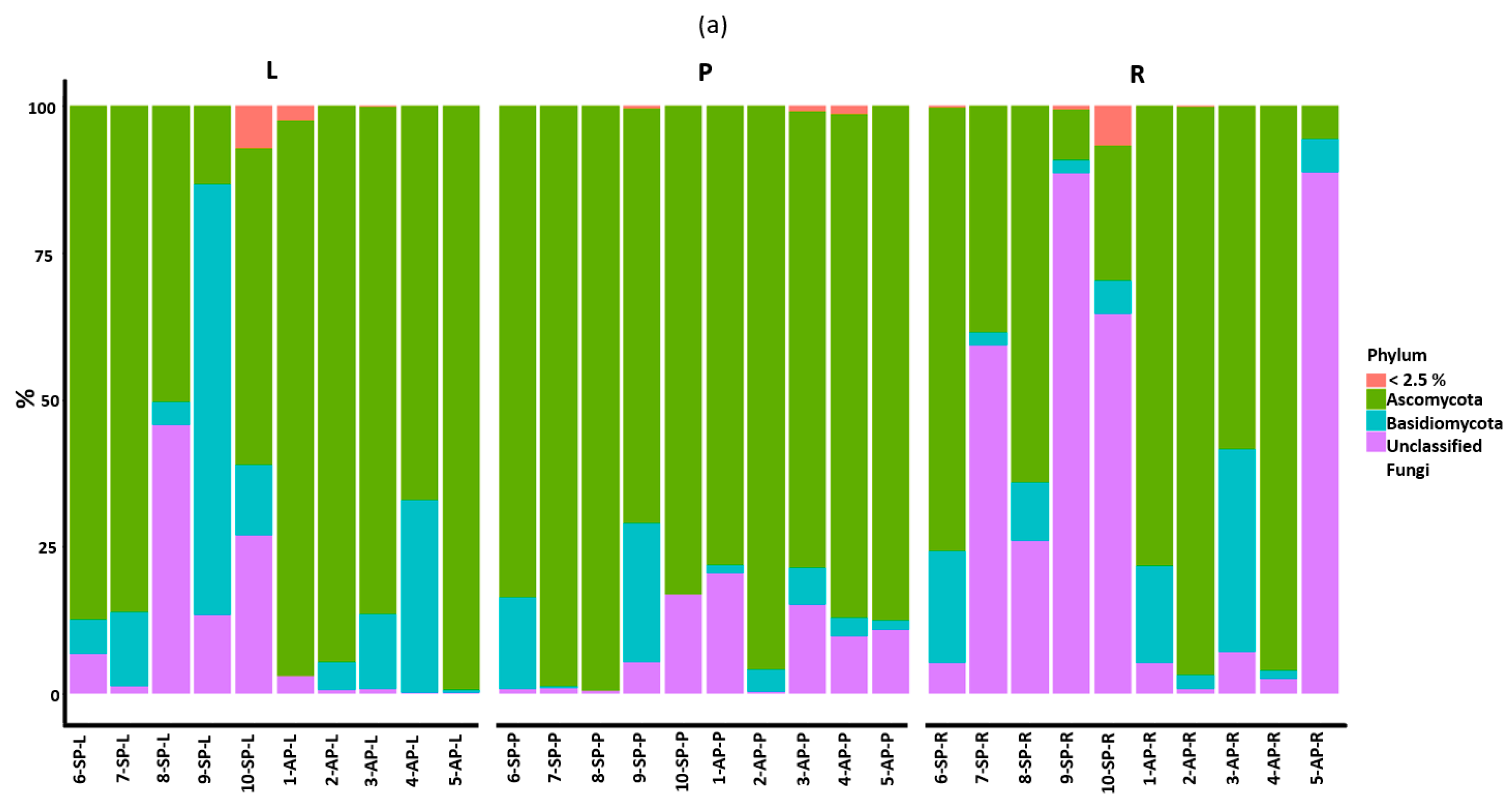
3.2. Taxonomic Assignment

In general, of the 1857 resulting ASVs, according to the relative abundances obtained in the samples, the main phyla were Ascomycota, which made up between 5 and 95% of the ASVs per sample, and Basidiomycota, which made up between 0.05 and 75%; those that were only assigned to fungi in general were present at 0.1 to 88.6%, in addition to some other groups, each with a small percentage (<2.5%), that only occurred in three samples of symptomatic plants. At the class level, Sordariomycetes occurred in the majority of the samples (0.7 to 71.9%), followed by Dothideomycetes (0.3 to 89%), Eurotiomycetes, and Agaricomycetes (0.2 to 72%); Saccharomycetes occurred in both conditions in <27% of the samples, although with 95% relative abundance in the 5-AP-L sample. There were 22 fungal classes that were present in fewer than 15% of samples (Figure S1a). There were seven main orders present among the samples; Chaetothyriales made up 0.17 to 53%, Pleosporales 0.2 to 80%, Agaricales 0.4 to 73%, Xylariales, Saccharomycetales and Hypocreales from 0.09 to 44%, Glomerellales from 0.12 to 32%, and Botryosphaeriales from 0.11 to 48% of ASVs (the order to which L. theobromae belongs); this order could be observed in most of the samples, although with relatively lower percentages and only standing out in the samples that corresponded to asymptomatic leaves. The rest of the orders were in less than 32% of the ASVs per sample. The dominant families were Pleurotaceae, with a relative abundance of 0.15 to 72%, Herpotrichiellaceae, at 0.06 to 50%, Glomerellaceae, at 0.2 to 32%, and Nectriaceae and Parapyrenochaetaceae, at 0.1 to 80%; the rest of the families were rare. Among the most predominant genera were Pleurotus, Exophiala, Colletrotrichum, Phyllosticta and Cyberlindnera, making up 0.03 to 72% of the ASVs of the samples, and a small proportion of undetermined species. The asymptomatic plants presented a higher frequency of Ascomycota, while the symptomatic plants had a higher frequency of fungi that could not be classified. At the class level, asymptomatic plants had a higher relative abundance of Dothideomycetes, followed by Sordariomycetes, in comparison with symptomatic plants (Figure S1b).
Regarding the composition at the phylum level by type of tissue, leaves and pseudobulbs presented a higher relative abundance of Ascomycetes, while the roots presented a higher relative abundance of unclassified fungi (Figure 2a). At class level, Sordariomycetes were more abundant in both conditions (and slightly higher in symptomatic plants), with leaves highlighted as the tissue with the highest relative abundance of this group. Eurotiomycetes presented greater abundance in symptomatic pseudobulbs, and Dothideomycetes in asymptomatic pseudobulbs. In roots, the abundance of unclassified fungi stands out in symptomatic plants, while Dothideomycetes and Sordariomycetes were relatively more abundant in asymptomatic plants (Figure 2b). On the other hand, the pseudobulbs with symptoms presented a greater relative abundance of Sordariomycetes and Eurotiomycetes, and a reduced number of Dothideomycetes, all of which are Ascomycetes.
In leaves, we observed representation of Xylariales, Glomerales, and in a relatively lower abundance, Botryosphaeriales (includes the genus Lasiodiplodia). Among the three tissues, pseudobulbs presented a slightly higher relative abundance of Chaetothyriales, followed by the order Botryosphaeriales; in roots, there was a greater relative abundance of fungi that could not be classified, along with lower numbers of Glomerales and Pleosporales. The genus Lasiodiplodia was recovered in the metabarcode data with lower relative abundance, in a sample of symptomatic pseudobulb, and in asymptomatic plants in leaf, pseudobulb and root tissue.
The order Sebacinales was recovered from the roots of symptomatic plants, and from the pseudobulbs and roots of asymptomatic plants, while the family Ceratobasidiaceae was recovered from the roots of symptomatic and asymptomatic plants. The genus Trichoderma was observed in the roots of symptomatic plants.
The most abundant genera in leaves were Colletotrichum followed by Phyllosticta, while in the pseudobulbs of symptomatic plants, Exophiala was relatively more abundant. In the pseudobulbs of asymptomatic plants, fungi that represented less than 1% of total diversity were more abundant, compared to pseudobulbs of diseased plants. The roots also presented a greater relative abundance of fungi within the group that overall had a relative abundance of <1% within the asymptomatic plants, compared to the roots of symptomatic plants. The genus Cyberlindnera occurred only in symptomatic plants, while Curvularia occurred only in asymptomatic plants.

3.3. Beta Diversity of Tissue-Hosted Fungal Communities

To detect whether the diversity of the fungal communities corresponding to the different tissues was similar to each other (beta diversity), pairwise tests were carried out with PERMANOVA. To determine which specific pairs differed from each other, the Bray–Curtis distance obtained with QIIME2 was used, which indicated significant differences when comparing the type of organ L vs. P (p = 0.001) and L vs. R (p = 0.001), while there were no differences between P vs. R (p = 0.079) (Table S3). Visualizations of beta diversity associations can be seen in Figure S2. These results were verified with a principal component analysis in R; for the case of condition, 12 of the AP samples overlap with 5 of the SP samples, the rest of the samples were scattered, and there was no observed tendency for tissue samples to be separated by collection site (Figure 3a,b).

3.4. Beta Diversity of Associated Fungal Communities by Asymptomatic and Symptomatic Plant Tissues

For asymptomatic plants, there was a significant difference between the beta diversity of the fungal communities found in leaves and pseudobulbs L vs. P (p = 0.005), while there were no differences between L vs. R (p = 0.097) and P vs. R (p = 0.105) (Table S4).
The beta diversity of the fungal communities of the symptomatic plants presented significant differences for all tissue types L vs. P (p = 0.02), L vs. R (p = 0.01), and P vs. R (p = 0.02) (Table S5).
A class-level heatmap was created to assess possible trends in the relative abundance of endophytic fungal taxa by sample, plant condition, or tissue (Figure 4); no undetermined fungi were included in this graph. Sordariomycetes was, relatively, the most abundant class in most of the samples, followed by Dothideomycetes, Eurotiomycetes, Agaricomycetes, and Saccharomycetes; the rest of the classes had a <2% relative abundance. It can be seen that the root samples contain a minimal amount of Eurotiomycetes.

3.5. Analysis of Functional Groups

A total of 942 ASVs (50.7%) were assigned to seven trophic modes with the FUNGuild tool (Figure 5), in which Saprotroph was the most diverse group, and it had higher relative abundance (Table S6). Seven families were found that were assigned to symbiotrophs, associated with the leaves, pseudobulbs, and roots of asymptomatic and symptomatic plants. In the leaf tissues of symptomatic plants, there was a higher relative abundance of Pathotroph-Saprotroph, while in leaves of asymptomatic plants, the Pathotroph-Saprotroph-Symbiotroph trophic mode was more abundant. The pseudobulbs in both conditions were dominated by the Pathotroph-Saprotroph trophic mode, with greater abundance in the pseudobulbs of symptomatic plants. The root tissue of symptomatic plants presented a greater relative abundance of the Saprotroph mode compared to the roots of asymptomatic plants (Figure 6). The Pathotroph mode was not dominant, but when compared, we observed that it was more abundant in asymptomatic than symptomatic plants, mainly in pseudobulbs and roots; 5 ASVs (0.26%) detected from L. acacia (possibly L. theobromae), assigned to the Plant Pathogen guild, are included in this trophic mode. The modes that contain the term “endophyte” were grouped, and a total of 364 ASVs (19.6%) were obtained.

4. Discussion

In nature, epiphytic orchids live with a limited supply of directly accessible resources. Interactions with microorganisms, particularly endophytic fungi and MFEF, among other possible functions, serve to provide essential nutritional requirements through pathways not directly available to orchids. This supplementary source of nutrients enables epiphytic orchids to complete their development and activate defense mechanisms against diseases and insect pest attacks. This study contributes significantly to the knowledge of the composition of the endophytic fungal community of mature plants of Guarianthe skinneri, divided into two conditions, asymptomatic and symptomatic (infested with “black blotch” fungal disease), in the region of Soconusco, Chiapas, Mexico.
While natural populations of G. skinneri continue to decline, strategies for sustainable cultivation are essential for the conservation of this highly prized, endangered species, made vulnerable due to the pressure of extraction, trafficking, and habitat loss. Strategies must necessarily consider the conservation of the multiple symbiotrophic associations that could be key to overcoming pests and diseases and achieving resilience in the face of the climate crisis.
The results of the present study indicate, for the first time, that a wide variety of fungal taxa live as endophytes within the leaf, pseudobulb and root tissues of G. skinneri plants, most of which have not been reported in relation to this plant as a host. A total of 1857 fungal ASVs from leaf, pseudobulb, and root tissues were identified. Previous studies have reported 889 operational taxonomic units (OTUs) (equivalent to ASVs) assigned to wild and cultivated asymptomatic plants and symptomatic plants with stem and root disease (SRD) of Vanilla planifolia Jacks. ex Andrews [16], while 2429 sequences were obtained from asymptomatic stems of Dendrobium nobile Lindl. [30].
This study demonstrated that the composition and diversity of endophytic fungal communities, as shown by alpha diversity, did not vary significantly between plant conditions, despite coming from different sites; our interpretations are based on this result. In the study carried out by Zarza et al. [13], significant differences were observed when comparing the diversity of endophytic fungi between two sites. However, for this study, it was not possible to acquire asymptomatic plants from the UMA SRLF. This was a limitation and evidenced the serious situation of the plants under cultivation, since one of the objectives of the UMA SRLF is to cultivate healthy plants for reintroduction. Additionally, there were no symptomatic plants at the JESS site. This is a real-life scenario, and despite its limitations, it is necessary to carry out this type of study to gain an overview that can contribute to decision making for the care of the studied organisms.
Fungi of the phylum Ascomycota were dominant in leaves, pseudobulbs, and roots of G. skinneri, and have also been reported as abundant in the endophytic communities of Dendrobium nobile and Vanilla planifolia [16,30]. Ascomycetes also form part of the fungal communities of the bark of coffee trees (Coffea arabica L. and Coffea canephora L.; Rubiaceae), trees of the genus Inga (Fabaceae) in agroforestry systems, Quercus yiwuensis C.C.Huang ex Y.C.Hsu and H.W.Jen (Fagaceae), and Pistacia weinmannifolia J.Poiss. (Anacardiaceae) [13,31]. As mentioned, fungi from the phylum Basidiomycota were relatively less abundant as endophytes in the tissues of G. skinneri; these fungi are dominant in the soil and are key ectomycorrhizal and wood-decomposing taxa [1]. For example, they are abundant in the rhizosphere of Cordia dodecandra A.DC. (Boraginaceae) trees [32]. However, Basidiomycetes are the most common mycorrhizal symbionts of epiphytic orchids, and it is possible that the selection of the primers used in this study could have caused underrepresentation, especially in the genus Tulasnella.
Sordariomycetes was the most abundant class in both symptomatic and symptomatic plants of G. skinneri, similar to the results reported for V. planifolia, where more OTUs of Sordariomycetes were reported (Nectriaceae 32 OTUs and Glomerellaceae 6 OTUs) [16]. Guarianthe skinneri pseudobulbs with symptoms of “black blotch” presented a higher abundance of Sordariomycetes and Eurotiomycetes, and less abundance of Dothideomycetes compared to asymptomatic pseudobulbs. However, Dothideomycetes was one of the most abundant fungal groups in asymptomatic plants, particularly in roots, which raises the question as to whether the abundance of Dothideomycetes could be related to the condition of the pseudobulbs and could be displaced by the saprophytes of Sordariomycetes and Euroriomycetes in infested plants. These classes are of cosmopolitan distribution and diverse life histories; they are associated with a wide range of hosts/substrates [33,34], with predicted functions including endophytes, plant and animal pathogens, wood saprophytes, mold formers, lichens, soil saprophytes, and ectomycorrhizae. Most of these fungi live on healthy plant tissues and organs and do not cause host disease [35]. In the present study, 368 ASVs with the term endophyte were obtained; however, we should take into account that the functional annotations for FUNGuild are far from complete, especially for tropical taxa, and we should extend the studies to taxa that were not labeled as “endophytes” for future research on the mechanisms of colonization and nutrient exchange and other benefits.
In the present study, the pseudobulbs of G. skinneri also presented considerable fungal diversity. Carbajal-Valenzuela et al. [16] were able to detect significant differences between the communities that inhabit the roots and the stems of V. planifolia, a plant that is predictable due to its semiterrestrial and extensive growth habits, with different sections of the same plant in contact with a variety of surfaces, from which different endophytic fungi are derived.
In this study, beta diversity was analyzed by separating asymptomatic (JESS) from symptomatic (SRLF) plants from different sites, for which significant differences were observed. In the case of asymptomatic plants of G. skinneri¸ there were only significant differences in the endophytic fungi associated with leaf and pseudobulb tissues. However, in the case of symptomatic plants there were significant differences between the fungal communities in all three tissues (leaves, roots, and pseudobulbs). Studies of plant–fungi interaction indicate that fungi have a colonization strategy to overcome the defense barriers of plants [36], and that some fungi have specific tissue preferences. The most studied fungi are mycorrhizae hosted in roots, or pathogens that can be general or specific to certain plant structures [37]. When considering the particular biotic and abiotic preferences of the fungal community, it should be taken into account that the symptomatic plants came from a different site. The availability of nutrient resources is a key driver of fungal diversity, and in most ecosystems, this is determined by the accessibility and amount of carbon that can be derived from the plants [38].
Each plant tissue offers a different environment for fungal development. The root is composed of a cortex covered by the velamen which serves to actively absorb water mixed with residues of organic matter. It has been shown that the velamen secretes metabolites that facilitate the recruitment of microorganisms tolerant to these conditions [32,39], which would obviously involve recruitment from the bark substrate to which the roots are attached. The thick, flat, elliptical leaves, covered with a layer of cuticular wax and stomata, receive a greater amount of light and eventually fall off to be recycled into the canopy or ground soil. The pseudobulbs act as a water reservoir, are succulent, and are covered by cuticular waxes. Pseudobulbs pass resources to new shoots and eventually dry and shrivel, but do not fall off. Fungi associated with pseudobulbs, where L. theobromae was seen to flourish, have to constantly adjust to changes in the consistency of that tissue, with maximum hydration during the rainy season and a slow depletion of the water reserve during the dry season. Fungal endophytes may compete for water and nutrient resources, and it has been suggested that fungi go through cycles of colonization and extinction according to season, especially in the roots [33,40].
A low relative abundance of representatives of the Botryosphaeria family, which include L. theobromae, was detected. However, it is possible that the absence of Lasiodiplodia in most of the symptomatic samples and some of the asymptomatic samples was caused by the disinfestation process that reduced the load of different fungi, including L. theobromae, reflecting that for the disinfestation process to isolate pathogens, a lower concentration of sodium hypochlorites is used and for less time; however, more rigorous protocols are required to isolate endophytic fungi [41]. In addition, the primers used in this study have commonly been used for the exploration of fungal communities, and may not favor the amplification of L. theobromae, since other primers and regions have been used for the identification of strains of L. theobromae. The manual search in the taxonomic table for L. theobromae yielded Lasiodiplodia acacia W. Zhang and Crous, which when compared in the Genbank, had 99 to 100% similarity with L. theobromae, demonstrating the difficulty of taxonomic assignment of fungi at the species level [42]. We are therefore now aware of the existence of cryptic species of Lasiodiplodia [43], which requires further exploration.
The genus Lasiodiplodia was present in a sample of the pseudobulbs of a symptomatic plant, but was also present as an endophyte in the tissues of leaves, pseudobulbs, and roots of asymptomatic plants. Several strains of Lasiodiplodia have been reported as cosmopolitan and opportunistic pathogens, mainly of timber and fruit trees [44], as well as orchids [45], including the study species. Lasiodiplodia has the potential to be pathogenic, possibly triggered by abiotic factors, such as when there is a significant change in temperature or humidity [46].
In the analysis of functional guilds, Lasiodiplodia appears to be a pathotroph, making it part of the plant pathogen guild. However, the evidence suggests a wider description would be more suitable, since recovery as an endophyte in asymptomatic plants has now been verified.
Trees maintain extensive interactions with terrestrial microorganisms [47], whereas epiphytic orchids interact with microorganisms that colonize the bark of their phorophytes [48]. In the taxonomic assignments resulting from the analyses of the fungal endophytes associated with G. skinneri, we observed the orders Russulales and Sebacinales, as well as the families Ceratobasidiaceae and Serendipitaceae; strains of these orders have been reported as MFEF in orchids. In the assignment of ecological roles with FUNGuild, six families of fungi, associated with trees, categorized as ectomycorrhizae (Helvellaceae, Cortinariaceae, Pezizaceae), endomycorrhizae (Ceratabasidiaceae), and arbuscular mycorrhizae (Glomeraceae, Ambisporaceae) and associated with the three types of symptomatic plant tissues, were listed, supported by the study by Zarza et al., 2022 [13], which involved the classification of mycorrhizal fungi assigned using FUNGuild found in the bark of non-disinfected coffee trees and shade trees. Although not part of the objective of this study, additional studies with specific primers are required to appreciate the mycorrhizal-forming endophytic fungi present in the plant tissues.
Interactions between endophytic and pathogenic fungi have been evaluated in in vitro conditions, with results suggesting that endophytic fungi, through direct competition and mycoparasitosis, can prevent pathogenic fungi from establishing and growing [49]. Among the genera observed in this study, Trichoderma, which has been cited as an efficient biocontrol agent of pathogens and insects, was present; it has also been found to be involved in the regulation of phytohormones (auxins, cytokinins, gibberellins, abscisic acid, ethylene, salicylic acid and jasmonic acid), and the activation of defense mechanisms [50]. However, Trichoderma was not abundant, and was only present in the roots of G. skinneri, and may suffer competition from other endophytic microorganisms.
The presence of Colletotrichum, Fusarium, Curvularia, Pestalotiopsis, Neopestalotiopsis, Aspergillus, Pleurotus and Xylaria, for which there is evidence of endophytic strains in orchids, was also observed [51,52,53,54]. Strains of Neopestalotiopsis, Colletotrichum, and Alternaria have been verified as producers of the gibberellins found in the orchid Stanhopea tigrina J.Frost [55], and were found to prevent the colonization of pathogenic strains of fungi, and show antibacterial activity [56]. The Aspergillus genus comprises several species with different capacities. For example, A. niger has synergistic effects with mycorrhizae, which can promote the growth of tomato plants, since it increases the availability and transport of phosphorus [57]; A. flavus and A. ochraceus apparently protect the orchids Bulbophyllum neilgherrense and Vanda testacea from herbivores [53], and have been evaluated for the production of toxins with an entomopathogenic effect. However, the properties and potential of the endophytic fungi that interact with G. skinneri are still unknown.
For fungi, taxonomic assignments are often uncertain, and most fungal species have not been cultivated. Furthermore, for most fungi, data are not available for relevant ecological traits, such as extracellular enzyme expression, nutrient use efficiency, dispersal ability, growth rate, or antagonistic ability. Added to this is the scarcity of studies focused on the diversity of tropical fungi, with few references relating to the Soconusco region [58,59].
We will continue to search for evidence that explains the mechanism by which endophytic fungal interact and benefit epiphytic orchids, and the techniques through which we could use these fungi as part of strategies for the conservation and restoration of epiphytic orchids in natural or novel ecosystems.

5. Conclusions

This study provided new insights into the endophytic fungal community associated with Guarianthe skinneri, plants with and without symptoms of the “black blotch” fungal disease, analyzed by mass sequencing applied to DNA extracted and amplified with fungal primers. The genus Lasiodiplodia was less common than other fungal groups, despite being the causative agent of the “black blotch”. The study also revealed a considerable number of ASVs that failed to be matched for taxonomic assignment or functional analysis.
We are working to study and recover G. skinneri populations, and to gain an understanding of the species and functions of the endophytic fungi associated with this endangered orchid, which could play a vital role as a biotechnological tool in that process. Manipulation of the endophytic fungal community could have a significant impact on the health of G. skinneri plants, and could favor positive outcomes for restoration programs by serving as a defense against different pathogens and/or helping to withstand biotic stress caused by the climate crisis, deforestation, etc. Our results support the notion that endophytic fungi fulfill a wide range of functions in epiphytic orchids, with the potential for a range of applications.

Supplementary Materials

The following supporting information can be downloaded at: https://www.mdpi.com/article/10.3390/d15070807/s1, File S1-Table S1: Sample ID, barcode, plant number, condition, tissue, sample, number of raw sequences, number of filtered sequences, number of sequences without chimeras. The samples correspond to the leaves (L), pseudobulbs (P), and roots (R) of five (1–5) asymptomatic plants (AP) and five (6–10) symptomatic plants (SP) of Guarianthe skinneri; Table S2: Taxonomy of the composition of the fungal community. The samples correspond to the leaves (L), pseudobulbs (P), and roots (R) of five (1–5) asymptomatic plants (AP) and five (6–10) symptomatic plants (SP) of Guarianthe skinneri; Figure S1. Relative frequency of Phylum (a) and Class (b) assigned to endophytic fungi associated with Guarianthe skinneri asymptomatic and symptomatic plants. The samples correspond to the leaves (L), pseudobulbs (P) and roots (R) of five (1–5) asymptomatic plants (AP) and five (6–10) symptomatic plants (SP). Table S3: PERMANOVA analysis. The samples correspond to the leaves (L), pseudobulbs (P), and roots (R) of symptomatic and asymptomatic plants of Guarianthe skinneri; Table S4: PERMANOVA analysis of asymptomatic plants of Guarianthe skinneri. The samples correspond to the leaves (L), pseudobulbs (P), and roots (R); Table S5: PERMANOVA analysis of symptomatic plants of Guarianthe skinneri. The samples correspond to the leaves (L), pseudobulbs (P), and roots (R); Figure S2: Associations by tissue using Bray–Curtis distances, calculated with Qiime2, based on a data set including all taxa detected in the leaves (a), pseudobulbs (b), and roots (c) of symptomatic and asymptomatic plants of Guarianthe skinneri; Table S6: Total occurrence of ASVs associated with Guarianthe skinneri, classified by trophic mode and guild. Bold type corresponds to guild labeled with endophyte type.

Author Contributions

Conceptualization, F.H.-R., A.D., S.P.F.P., K.G.-N. and L.I.-D.; methodology, review and editing F.H.-R., A.D., S.P.F.P., K.G.-N., L.I.-D. and E.Z.; software F.H.-R., E.Z. and R.A.C.-C.; writing—original draft preparation F.H.-R., A.D., S.P.F.P., K.G.-N. and L.I.-D. All authors have read and agreed to the published version of the manuscript.

Funding

This study was financed by the American Orchid Society (AOS) agreement number 23075, and by the Consejo Nacional de Humanidades, Ciencias y Tecnologías-México (CONAHCYT), doctoral grant no. 736042, issued to FHR.

Institutional Review Board Statement

The study does not involve humans or animals. Samples were collected after approval from the committee of the UMA of Santa Rita las Flores and JESS.

Informed Consent Statement

Not applicable.

Data Availability Statement

The sequences from which these data were obtained can be found in NCBI-Genbank.

Acknowledgments

We extend our thanks to Maria De los Ángeles Palomeque Rodas, Luz Verónica García Fajardo, Aucencia Emeterio Lara, and Adelmi Aureliana Pérez Pérez for their support in processing the samples.

Conflicts of Interest

The authors declare no conflict of interest. The funders had no role in the design of the study; in the collection, analyses, or interpretation of data; in the writing of the manuscript; or in the decision to publish the results.

References

  1. Peay, K.; Kennedy, P.G.; Talbot, J.M. Dimensions of biodiversity in the Earth mycobiome. Nat. Rev. Genet. 2016, 14, 434–447. [Google Scholar] [CrossRef]
  2. Nguyen, N.H.; Song, Z.; Bates, S.T.; Branco, S.; Tedersoo, L.; Menke, J.; Schilling, J.S.; Kennedy, P.G. FUNGuild: An open annotation tool for parsing fungal community datasets by ecological guild. Fungal Ecol. 2016, 20, 241–248. [Google Scholar] [CrossRef]
  3. Fadiji, A.E.; Babalola, O.O. Elucidating Mechanisms of Endophytes Used in Plant Protection and Other Bioactivities with Multifunctional Prospects. Front. Bioeng. Biotechnol. 2020, 8, 467. [Google Scholar] [CrossRef]
  4. Xing, Y.-M.; Chen, J.; Cui, J.-L.; Chen, X.-M.; Guo, S.-X. Antimicrobial Activity and Biodiversity of Endophytic Fungi in Dendrobium devonianum and Dendrobium thyrsiflorum from Vietman. Curr. Microbiol. 2011, 62, 1218–1224. [Google Scholar] [CrossRef] [PubMed]
  5. Christian, N.; Herre, E.A.; Clay, K. Foliar endophytic fungi alter patterns of nitrogen uptake and distribution in Theobroma cacao. New Phytol. 2019, 222, 1573–1583. [Google Scholar] [CrossRef]
  6. Douanla-Meli, C.; Langer, E.; Mouafo, F.T. Fungal endophyte diversity and community patterns in healthy and yellowing leaves of Citrus limon. Fungal Ecol. 2013, 6, 212–222. [Google Scholar] [CrossRef]
  7. Hamzah, T.N.T.; Lee, S.Y.; Hidayat, A.; Terhem, R.; Faridah-Hanum, I.; Mohamed, R. Diversity and Characterization of Endophytic Fungi Isolated From the Tropical Mangrove Species, Rhizophora mucronata, and Identification of Potential Antagonists Against the Soil-Borne Fungus, Fusarium solani. Front. Microbiol. 2018, 9, 1707. [Google Scholar] [CrossRef] [PubMed]
  8. Nguyen, M.H.; Shin, K.C.; Lee, J.K. Fungal Community Analyses of Endophytic Fungi from Two Oak Species, Quercus mongolica and Quercus serrata, in Korea. Mycobiology 2021, 49, 385–395. [Google Scholar] [CrossRef] [PubMed]
  9. Pang, B.; Yin, D.; Zhai, Y.; He, A.; Qiu, L.; Liu, Q.; Ma, N.; Shen, H.; Jia, Q.; Liang, Z.; et al. Diversity of endophytic fungal community in Huperzia serrata from different ecological areas and their correlation with Hup A content. BMC Microbiol. 2022, 22, 191. [Google Scholar] [CrossRef]
  10. Rim, S.O.; Roy, M.; Jeon, J.; Montecillo, J.A.V.; Park, S.-C.; Bae, H. Diversity and Communities of Fungal Endophytes from Four Pinus Species in Korea. Forests 2021, 12, 302. [Google Scholar] [CrossRef]
  11. Sanz-Ros, A.V.; Müller, M.M.; Martín, R.S.; Diez, J.J. Fungal endophytic communities on twigs of fast and slow growing Scots pine (Pinus sylvestris L.) in northern Spain. Fungal Biol. 2015, 119, 870–883. [Google Scholar] [CrossRef] [PubMed]
  12. Tibpromma, S.; Karunarathna, S.C.; Bhat, J.D.; Suwannarach, N.; Stephenson, S.L.; Elgorban, A.M.; Al-Rejaie, S.; Xu, J.; Mortimer, P.E. Using Culture-Dependent and Molecular Techniques to Identify Endophytic Fungi Associated with Tea Leaves (Camellia spp.) in Yunnan Province, China. Diversity 2022, 14, 287. [Google Scholar] [CrossRef]
  13. Zarza, E.; López-Pastrana, A.; Damon, A.; Guillén-Navarro, K.; García-Fajardo, L.V. Fungal diversity in shade-coffee plantations in Soconusco, Mexico. PeerJ 2022, 10, e13610. [Google Scholar] [CrossRef]
  14. Verma, A.; Shameem, N.; Jatav, H.S.; Sathyanarayana, E.; Parray, J.A.; Poczai, P.; Sayyed, R.Z. Fungal Endophytes to Combat Biotic and Abiotic Stresses for Climate-Smart and Sustainable Agriculture. Front. Plant Sci. 2022, 13. [Google Scholar] [CrossRef]
  15. Sarsaiya, S.; Shi, J.; Chen, J. A comprehensive review on fungal endophytes and its dynamics on Orchidaceae plants: Current research, challenges, and future possibilities. Bioengineered 2019, 10, 316–334. [Google Scholar] [CrossRef] [PubMed] [Green Version]
  16. Carbajal-Valenzuela, I.A.; Muñoz-Sanchez, A.H.; Hernández-Hernández, J.; Barona-Gómez, F.; Truong, C.; Cibrián-Jaramillo, A. Microbial Diversity in Cultivated and Feral Vanilla Vanilla planifolia Orchids Affected by Stem and Rot Disease. Microb. Ecol. 2021, 84, 821–833. [Google Scholar] [CrossRef] [PubMed]
  17. Novotná, A.; Benítez, Á.; Herrera, P.; Cruz, D.; Filipczyková, E.; Suárez, J.P. High diversity of root-associated fungi isolated from three epiphytic orchids in southern Ecuador. Mycoscience 2018, 59, 24–32. [Google Scholar] [CrossRef]
  18. Bungtongdee, N.; Sopalun, K.; Laosripaiboon, W.; Iamtham, S. The chemical composition, antifungal, antioxidant and antimutagenicity properties of bioactive compounds from fungal endophytes associated with Thai orchids. J. Phytopathol. 2018, 167, 56–64. [Google Scholar] [CrossRef]
  19. Brundrett, M.C. Coevolution of roots and mycorrhizas of land plants. New Phytol. 2002, 154, 275–304. [Google Scholar] [CrossRef]
  20. Cabrera, T. Ficha Técnica de Cattleya Skinneri. En: Palacios E. (compilador). Cuarenta y ocho Especies de la Flora de Chiapas Incluidas en el PROY-NOM-059-ECOL-2000; SNIB CONABIO. Proyecto. W008; Instituto de Historia Natural y Ecología Bases de datos: Chiapas, Mexico, 2006; SNIB CONABIO. Proyecto. W008. México. [Google Scholar]
  21. Fabiola, H.-R.; Patricia, F.-P.S.; Anne, D.; Karina, G.-N.; Verónica, G.-F. Etiology of the “black blotch” caused by Lasiodiplodia in pseudobulbs of Guarianthe skinneri (Orchidaceae), in the Region of Soconusco, Chiapas, Mexico. J. Plant Pathol. 2022, 104, 1083–1095. [Google Scholar] [CrossRef]
  22. Johnson, G.I.; Mead, A.J.; Cooke, A.W.; Dean, J.R. Mango stem end rot pathogens—Fruit infection by endophytic colonisation of the inflorescence and pedicel. Ann. Appl. Biol. 1992, 120, 225–234. [Google Scholar] [CrossRef]
  23. Vidal, N.; Vidal, E.; Vidal, L.; Colorado, J.; Ruiz, R.; Ruiz, J. Anónaceas. Plantas antiguas. Estudios Recientes. Parte 2; Universidad Autónoma Chapingo: Texcoco, Mexico; pp. 205–227.
  24. Roma, R.; Damon, A.; Sánchez, W. The cacao (Theobroma cacao L.) Policulture and the Tuzantán qato’ok Culture, Chiapas, Mexico. An Ethnoecological Approach. Ethnoscientia 2022, 4, 14–32. [Google Scholar] [CrossRef]
  25. García, E. Modificaciones al Sistema de Clasificación Climática de Kõeppen (Para Adaptarlo a las Condiciones de la República Mexicana), 2nd ed.; Universidad Nacional Autónoma de México, Instituto de Geografía: Ciudad de México, México, 1973. [Google Scholar]
  26. Semarnat; Maag Consultoria Ambiental. Manifestación de Impacto Ambiental Modalidad Regional ‘Camino: Ejido 3 de Mayo-Santa Rita, en una Longitud de 4.00 km (del km 0+000 al km 4+000)’ en el Municipio de Mapastepec, Chiapas. Chiapas. Available online: https://apps1.semarnat.gob.mx:8443/dgiraDocs/documentos/chis/estudios/2020/07CH2020VD051.pdf (accessed on 5 April 2023).
  27. Miranda, F. La Vegetación de Chiapas; Gobierno del Estado, Coneculta Chiapas: Tuxtla Gutiérrez, Mexico, 1998; p. 600. [Google Scholar]
  28. Schoch, C.L.; Seifert, K.A.; Huhndorf, S.; Robert, V.; Spouge, J.L.; Levesque, C.A.; Chen, W.; Fungal Barcoding Consortium; Fungal Barcoding Consortium Author List. Nuclear ribosomal internal transcribed spacer (ITS) region as a universal DNA barcode marker for Fungi. Proc. Natl. Acad. Sci. USA 2012, 109, 6241–6246. [Google Scholar] [CrossRef] [PubMed] [Green Version]
  29. White, T.J.; Bruns, T.; Lee, S.; Taylor, J. Amplification and direct sequencing of fungal ribosomal RNA genes for phylogenetics. In PCR Protocols: A Guide to Methods and Applications; Innis, M.A., Gelfand, D.H., Sninsky, J.J., White, T.J., Eds.; Academic Press: San Diego, CA, USA, 1990; pp. 315–322. [Google Scholar]
  30. Li, L.; Liu, C.; Wen, W.; Li, Q.; Pan, T.; Li, Z.; Qian, G.; He, Y.; Xu, D. Dendrobine biosynthesis in Dendrobium nobile in four different habitats is affected by the variations in the endophytic fungal community. Front. Microbiol. 2022, 13. [Google Scholar] [CrossRef]
  31. Pecoraro, L.; Rasmussen, H.N.; Gomes, S.I.F.; Wang, X.; Merckx, V.S.F.T.; Cai, L.; Rasmussen, F.N. Fungal diversity driven by bark features affects phorophyte preference in epiphytic orchids from southern China. Sci. Rep. 2021, 11, 11287. [Google Scholar] [CrossRef]
  32. May-Mutul, C.G.; López-Garrido, M.A.; O’connor-Sánchez, A.; Peña-Ramírez, Y.J.; Labrín-Sotomayor, N.Y.; Estrada-Medina, H.; Ferrer, M.M. Hidden Tenants: Microbiota of the Rhizosphere and Phyllosphere of Cordia dodecandra Trees in Mayan Forests and Homegardens. Plants 2022, 11, 3098. [Google Scholar] [CrossRef] [PubMed]
  33. Haridas, S.; Albert, R.; Binder, M.; Bloem, J.; LaButti, K.; Salamov, A.; Andreopoulos, B.; Baker, S.; Barry, K.; Bills, G.; et al. 101 Dothideomycetes genomes: A test case for predicting lifestyles and emergence of pathogens. Stud. Mycol. 2020, 96, 141–153. [Google Scholar] [CrossRef]
  34. Lee, S.H.; Park, H.S.; Nguyen, T.T.T.; Lee, H.B. Characterization of Three Species of Sordariomycetes Isolated from Freshwater and Soil Samples in Korea. Mycobiology 2019, 47, 20–30. [Google Scholar] [CrossRef] [Green Version]
  35. Saikkonen, K.; Faeth, S.H.; Helander, M.; Sullivan, T.J. FUNGAL ENDOPHYTES: A Continuum of Interactions with Host Plants. Annu. Rev. Ecol. Syst. 1998, 29, 319–343. [Google Scholar] [CrossRef]
  36. Zeilinger, S.; Gupta, V.K.; Dahms, T.E.; Silva, R.N.; Singh, H.B.; Upadhyay, R.S.; Gomes, E.V.; Tsui, C.K.; Nayak, S.C. Friends or foes? Emerging insights from fungal interactions with plants. FEMS Microbiol. Rev. 2016, 40, 182–207. [Google Scholar] [CrossRef] [Green Version]
  37. Lacaze, A.; Joly, D.L. Structural specificity in plant–filamentous pathogen interactions. Mol. Plant Pathol. 2020, 21, 1513–1525. [Google Scholar] [CrossRef] [PubMed]
  38. Ma, X.; Kang, J.; Nontachaiyapoom, S.; Wen, T.; Hyde, K.D. Non-mycorrhizal endophytic fungi from orchids. Curr. Sci. 2015, 109, 72–87. [Google Scholar]
  39. Cevallos, S.; Herrera, P.; Sánchez-Rodríguez, A.; Declerck, S.; Suárez, J.P. Untangling factors that drive community composition of root associated fungal endophytes of Neotropical epiphytic orchids. Fungal Ecol. 2018, 34, 67–75. [Google Scholar] [CrossRef]
  40. Ovando, I. Isolation of Endophytic Fungi and Their Mycorrhizal Potential for the Tropical Epiphytic Orchids Cattleya skinneri, C. aurantiaca and Brassavola nodosa. Asian J. Plant Sci. 2005, 4, 309–315. [Google Scholar] [CrossRef] [Green Version]
  41. dos Reis, J.B.A.; Lorenzi, A.S.; de Vale, H.M.M. Methods used for the study of endophytic fungi: A review on methodologies and challenges, and associated tips. Arch. Microbiol. 2022, 204, 675. [Google Scholar] [CrossRef]
  42. Tedersoo, L.; Bahram, M.; Zinger, L.; Nilsson, R.H.; Kennedy, P.G.; Yang, T.; Anslan, S.; Mikryukov, V. Best practices in metabarcoding of fungi: From experimental design to results. Mol. Ecol. 2022, 31, 2769–2795. [Google Scholar] [CrossRef] [PubMed]
  43. Alves, A.; Crous, P.W.; Correia, A.; Phillips, A.J.L. Morphological and molecular data reveal cryptic speciation in Lasiodiplodia theobromae. Fungal Div. 2008, 28, 1–13. Available online: http://www.fungaldiversity.org/fdp/sfdp/28-1.pdf (accessed on 5 April 2023).
  44. Abdollahzadeh, J.; Javadi, A.; Goltapeh, E.M.; Zare, R.; Phillips, A. Phylogeny and morphology of four new species of Lasiodiplodia from Iran. Persoonia Mol. Phylogeny Evol. Fungi 2010, 25, 1–10. [Google Scholar] [CrossRef] [Green Version]
  45. Cabrera, M.G.; Cúndom, M.A. Ocurrencia de Lasiodiplodia theobromae en Cattleya spp. en Corrientes, Argentina. Summa Phytopathol. 2013, 39, 143. [Google Scholar] [CrossRef] [Green Version]
  46. Picos-Muñoz, P.; García-Estrada, R.; León-Félix, J.; Sañudo-Barajas, A.; Allende-Molar, R. Lasiodiplodia theobromae en cultivos agrícolas de México: Taxonomía, hospedantes, diversidad y control. Rev. Mex. Fitopatol. 2015, 33, 54–74. [Google Scholar]
  47. Churchland, C.; Grayston, S.J. Specificity of plant-microbe interactions in the tree mycorrhizosphere biome and consequences for soil C cycling. Front. Microbiol. 2014, 5, 261. [Google Scholar] [CrossRef] [PubMed] [Green Version]
  48. Wang, D.; Lerou, J.; Nuytinck, J.; Gomes, S.I.; Jacquemyn, H.; Sft, V. Root-associated fungi in Orchidaceae: Diversity, phylogeny, ecology, and outstanding questions. bioRxiv 2022, 1–38. [Google Scholar] [CrossRef]
  49. Rajani, P.; Rajasekaran, C.; Vasanthakumari, M.; Olsson, S.B.; Ravikanth, G.; Shaanker, R.U. Inhibition of plant pathogenic fungi by endophytic Trichoderma spp. through mycoparasitism and volatile organic compounds. Microbiol. Res. 2021, 242, 126595. [Google Scholar] [CrossRef] [PubMed]
  50. Sood, M.; Kapoor, D.; Kumar, V.; Sheteiwy, M.S.; Ramakrishnan, M.; Landi, M.; Araniti, F.; Sharma, A. Trichoderma: The “Secrets” of a Multitalented Biocontrol Agent. Plants 2020, 9, 762. [Google Scholar] [CrossRef]
  51. Lizarazo-Medina, P.X.; Mendoza-Salazar, M.M.; Gutiérrez-Gallo, A.I. Diversity of endophytic mycobiota in Cattleya percivaliana and Cattleya trianaei growing under greenhouse conditions. Actual. Biol. 2015, 37, 67–78. [Google Scholar]
  52. Ma, X.-Y.; Maharachchikumbura, S.S.N.; Chen, B.-W.; Hyde, K.D.; McKenzie, E.H.C.; Chomnunti, P.; Kang, J.-C. Endophytic Pestalotia taxa in Dendrobium orchids. Phytotaxa 2019, 419, 268–286. [Google Scholar] [CrossRef]
  53. Sudheep, N.M.; Sridhar, K. Non-mycorrhizal fungal endophytes in two orchids of Kaiga forest (Western Ghats), India. J. For. Res. 2012, 23, 453–460. [Google Scholar] [CrossRef]
  54. Tsavkelova, E.; Kolomeitseva, G. Fusarium–orchid interactions under greenhouse conditions. S. Afr. J. Bot. 2022, 146, 889–896. [Google Scholar] [CrossRef]
  55. Salazar-Cerezo, S.; Martinez-Montiel, N.; Cruz-Lopez, M.D.C.; Martinez-Contreras, R.D. Fungal Diversity and Community Composition of Culturable Fungi in Stanhopea trigrina Cast Gibberellin Producers. Front. Microbiol. 2018, 9, 612. [Google Scholar] [CrossRef] [Green Version]
  56. Vaz, A.B.; Mota, R.C.; Bomfim, M.R.Q.; Vieira, M.L.; Zani, C.L.; Rosa, C.A.; Rosa, L.H. Antimicrobial activity of endophytic fungi associated with Orchidaceae in Brazil. Can. J. Microbiol. 2009, 55, 1381–1391. [Google Scholar] [CrossRef] [PubMed]
  57. Arias, R.; Romero, A.; Bañuelos, J.; De la Cruz, Y. Inoculation of phosphorus solubilizing fungi and arbuscular mycorrhizae in tomato plants. Rev. Mex. Cienc. Cienc. Agrícolas 2019, 10, 1747–1757. [Google Scholar] [CrossRef] [Green Version]
  58. López-Guzmán, L.M.; Chacón, S.; Bautista-Gálvez, A. Adiciones al conocimiento sobre la diversidad de los hongos (macromicetes) de Chiapas, México. Sci. Fungorum 2017, 45, 27–35. [Google Scholar] [CrossRef]
  59. Andrade-Gallegos, R.H.; Sánchez-Vázquez, J.E. La diversidad de hongos en Chiapas: Un reto pendiente. In Diversidad Biológica en Chiapas; ECOSUR-COCYTECH-PLAZA Y VALDES Editores, D.F. México; Plaza y Valdés: Madrid, México, 2004; pp. 33–80. [Google Scholar]
Figure 1. Rarefaction curves representing the observed abundance of ASVs via the sequencing depth of endophytic fungi (a), and measures of alpha diversity of endophytic fungi from asymptomatic (AP) and symptomatic (SP) plants (b), on the leaves (L), pseudobulbs (P), and roots (R) (c) of Guarianthe skinneri.
Diversity 15 00807 g001aDiversity 15 00807 g001b
Figure 2. Relative abundance of phylum (a) and class (b) assigned to endophytic fungi associated with the leaves, pseudobulbs, and roots of Guarianthe skinneri. The samples correspond to the leaves (L), pseudobulbs (P), and roots (R) of five (1–5) asymptomatic plants (AP) and five (6–10) symptomatic plants (SP) of Guarianthe skinneri.
Diversity 15 00807 g002aDiversity 15 00807 g002b
Figure 3. Principal component analysis of the endophytic fungal communities associated with asymptomatic and symptomatic plants (a) and tissues (b) of Guarianthe skinneri.
Diversity 15 00807 g003
Figure 4. Heat map showing the relative abundance of the sequences assigned to the fungal classes observed in the tissues of leaves (L), pseudobulbs (P), and roots ® of five (1–5) asymptomatic plants (AP) and five (6–10) symptomatic plants (SP) of Guarianthe skinneri.
Diversity 15 00807 g004
Figure 5. Boxplot showing trophic mode frequencies applied to tissue and plant condition, according to FUNGuild analysis. L = leaf, P = pseudobulb, R = roots, AP= asymptomatic plant and SP = symptomatic plant.
Diversity 15 00807 g005
Figure 6. The relative abundance of trophic modes assigned to endophytic fungi in the leaves, pseudobulbs, and roots of Guarianthe skinneri. The samples correspond to the leaves (L), pseudobulbs (P), and roots (R) of five (1–5) asymptomatic plants (AP) and five (6–10) symptomatic plants (SP) of Guarianthe skinneri.
Diversity 15 00807 g006
Disclaimer/Publisher’s Note: The statements, opinions and data contained in all publications are solely those of the individual author(s) and contributor(s) and not of MDPI and/or the editor(s). MDPI and/or the editor(s) disclaim responsibility for any injury to people or property resulting from any ideas, methods, instructions or products referred to in the content.

