Article

Under-Ice Development of Silica-Scaled Chrysophytes with Different Trophic Mode in Two Ultraoligotrophic Lakes of Yakutia

1
Limnological Institute, Siberian Branch of the Russian Academy of Sciences, 3 Ulan-Batorskaya, 664033 Irkutsk, Russia
2
Institute for Biological Problems of Cryolithozone, Siberian Branch of the Russian Academy of Science, 41 Lenina Avenue, 677980 Yakutsk, Russia
*
Author to whom correspondence should be addressed.
Diversity 2023, 15(3), 326; https://doi.org/10.3390/d15030326
Submission received: 15 January 2023 / Revised: 15 February 2023 / Accepted: 20 February 2023 / Published: 22 February 2023
(This article belongs to the Special Issue Diversity and Ecology of Freshwater Lake Microbial Communities)

Abstract

:
Silica-scaled chrysophytes are a widespread group of microeukaryotes, an important component of aquatic habitats. They belong to different evolutionary lineages and they are characterized by the presence of siliceous scales, but differ in trophic mode. We studied the diversity of these organisms in different months of the ice cover period in two subarctic lakes of Yakutia, Labynkyr and Vorota. Silica-scaled chrysophytes, due to various trophic modes, have a competitive advantage in conditions of a long period of ice cover. Statistical analysis has shown the relationship between the relative abundance of mixotrophic and photoautotrophic representatives of silica-scaled chrysophytes with the thickness of the snow cover and the transparency of the ice. An increase in snow cover thickness and the process of melting ice with a decrease in its transparency reduce the relative abundance of photoautotrophic and mixotrophic species. Photoautotrophic representatives of silica-scaled chrysophytes begin to develop already in April, when a thick, solid, and transparent layer of ice and a small layer of snow were observed. During the research period, from April to June, the relative abundance of colorless heterotrophic silica-scaled chrysophytes genera Paraphysomonas and Lepidochromonas was more or less stable. A new species of Spiniferomonas heterospina sp. nov. has been discovered in Lake Labynkyr.

1. Introduction

Among planktonic organisms, species-specific siliceous elements of the shell—scales and spine covering the cell—are formed by representatives of the Chrysophyceae Pascher class from the families Chrysosphaerellaceae Kapustin, Paraphysomonadaceae Preisig and Hibberd Lepidochromonadaceae Kapustin and Guiry, Mallomonadaceae Diesing, and Synuraceae Lemmermann [1,2,3]. These siliceous elements can be detected using electron microscopy, both in aquatic plankton samples during the growing season and in bottom sediments, indicating that the species lived in the ecosystem of the reservoir in the past. Silica-scaled chrysophytes, despite closely related phylogenetic relationships, differ in trophic mode. For example, representatives of the order Synurales R. A. Andersen are typical photoautotrophic organisms [4], representatives of the genera Paraphysomonas De Saedeleer and Lepidochromonas Kristiansen of the order Paraphysomonadales are colorless heterotrophic (phagotrophic) organisms [5], and representatives of the same order of the genus Chrysosphaerella Lauterborn and genus Spiniferomonas Takahashi are mixotrophic organisms [2,6]. Thus, due to trophic differences, the seasonality of development within representatives of silica-scaled chrysophytes may differ. Their development is particularly interesting in subarctic and Arctic regions, where reservoirs are covered with thick ice and snow, preventing the penetration of light for a long period of time [7]. Such studies are fragmentary and are mainly devoted to marine ecosystems. For example, it is known that most species of silica-scaled chrysophytes in northern reservoirs occur mainly in spring, immediately after the melting of ice [8]. Paraphysomonas species can occur in large numbers during the formation of sea ice [9]. As colorless phagotrophic organisms attached to sea snow, they can play an important role in the marine food web [10]. It is believed that only a few silica-scaled chrysophytes, if any, are often found in ultraoligotrophic lakes [11]. We explored the unique, inaccessible, subarctic lakes of Yakutia, located in the Oymyakonsky district of the Republic of Sakha (Yakutia), where the lowest air temperatures are observed in the Northern Hemisphere. The research area is characterized by extremely harsh winters and a long ice period that lasts more than 240 days a year [7]. Previously, we investigated the species composition of silica-scaled chrysophytes in two ultraoligotrophic, subarctic lakes of Yakutia, Labynkyr and Vorota, during the open water period in July 2014 [12]. The purpose of this study was to determine the diversity of these organisms in these lakes in different months of the ice cover period.

2. Materials and Methods
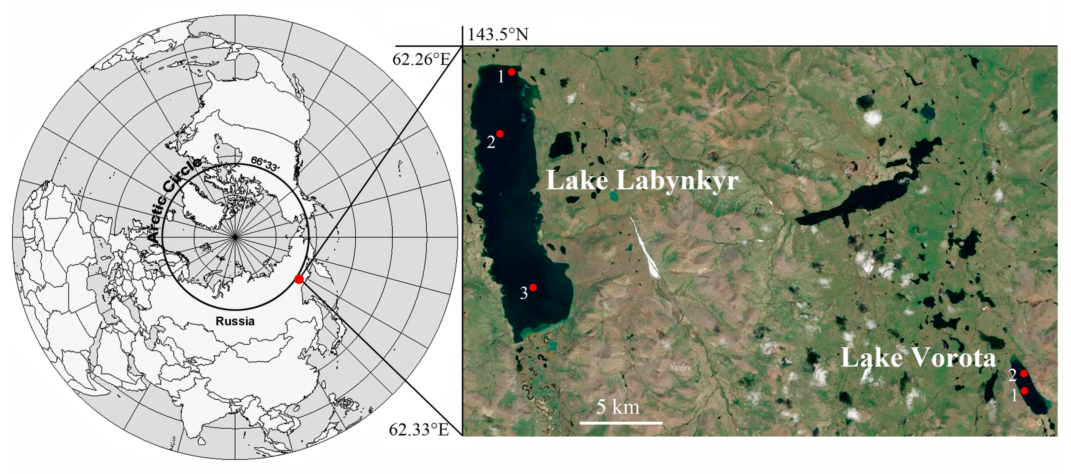
2.1. Study Site

Sampling was carried out in Lake Labynkyr on 2–4 April 2016 and in lakes Labynkyr and Vorota on 26–29 May 2016 and 3–8 June 2017 (Figure 1) from the ice, as described earlier [7].
Descriptions of geographical, ecological, and climatic conditions of the lakes Labynkyr and Vorota are given according to previously published data [7,12,13]. All measurements of concentrations of major ions and DOC were performed previously [7].
The cold season lasts 8–8.5 months [7,13]. During this time, the lakes remain frozen and are covered by snow [7]. The thickness of the ice cover on the lakes in the study period, from April to June, ranged from 80 to 111 cm. The thickness of the snow cover varied between 35 cm (April) and 0.5–5 cm (late May, June). The water temperature during the ice cover period of the lakes is relatively low, 1.2–5.6 °C [7]. The lakes are poorly mineralized and highly saturated with oxygen (during the ice cover period, the oxygen content at the bottom reached 9.8 mg/L) [7]. The pH of the water in Lake Labynkyr varied depending on the month of sampling. In April, the pH varied between 7.70 and 7.93 and was slightly alkaline. In May, the pH corresponded to an alkaline pH of 9.20–9.34. In June, in Lake Labynkyr, the pH dropped sharply to 6.80–6.98 and was neutral. In Lake Vorota, the pH of the water in May and June corresponded to a slightly alkaline level of 7.21–8.8 [7]. The total content of basic ions during the ice cover period in Lake Labynkyr varied from April to May in the range of 30.89–45.72 mg/L, and in Lake Vorota in the range of 33.8–54.75 μg/L [7]. Organic matter indicators of the lakes varied between 0.86 and 3.04 mgC/L; they were higher in April than in May and June [7] (Table S1). Nutrient concentrations were quite low [7].

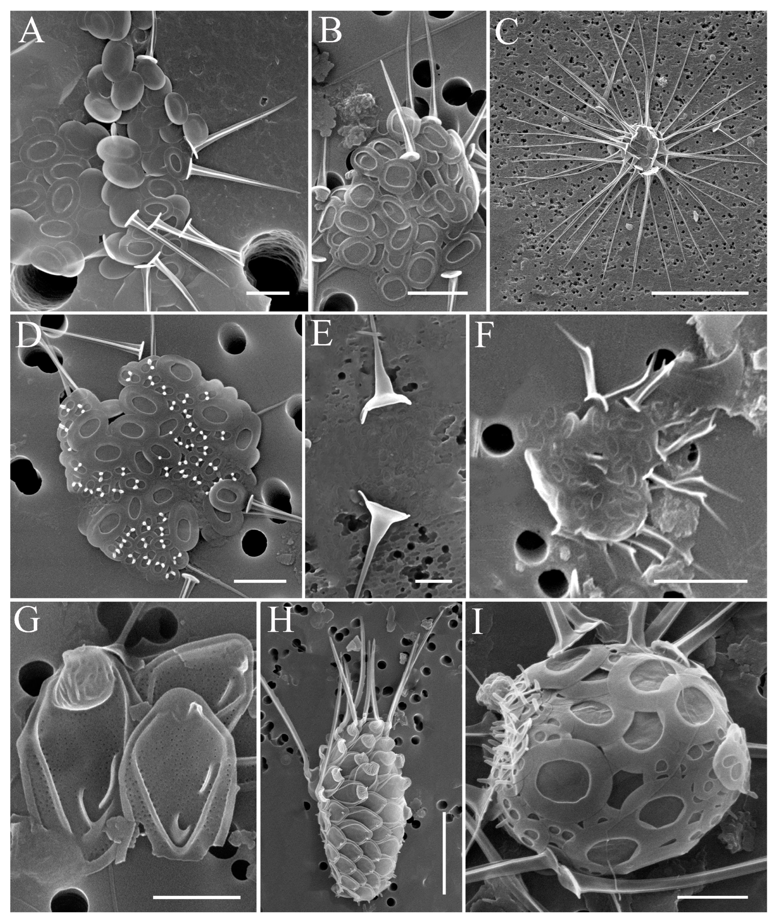
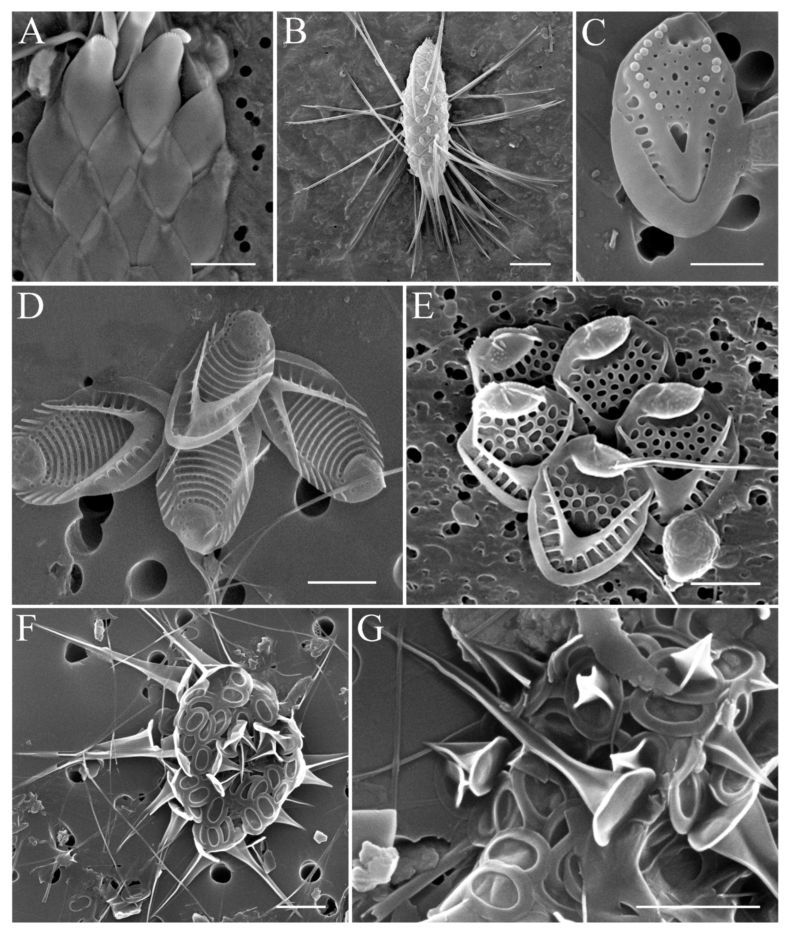
2.2. Study of Silica-Scaled Chrysophytes

Samples for the study of silica-scaled Chrysophyceae were prepared using the protocol described earlier (2019) [12]. Microscopic studies were carried out with Quanta-200 (FEI, USA). The distributions and relative abundances of each chrysophyte species was ranked by the amounts of scales observed on stab for SEM as follows: very rare (VR) 2–25 scales, rare (R) 26–50, common (C) 51–150, and abundant (A) if the number of scales was >150.

2.3. Statistical Analysis and Visualization

The spectrum of species in the samples, the values of species richness, species abundance, and cumulative species abundance by groups were visualized in the form of heat maps with the division of species into groups of photoautotrophic species, heterotrophic and phagotrophic species, and mixotrophic species, with columns (samples) clustered and grouped by similarity order (i.e., Jaccard or Euclidean distance metric and the complete-link clustering method). The analysis was performed using the “gplots” [14] and “vegan” [15] packages for R programming language.
To assess the relationship between the abundance of species from different groups with the months (May–April–June) of sampling and the physicochemical parameters of the environment, a one-way analysis of variance and correlation analysis (Kendall’s correlation coefficient for quantitative gradations) were carried out in the R programming language.
All primary data on species list, species abundance, and physicochemical parameters are available in the Supplementary Materials (see Table S1).

3. Results

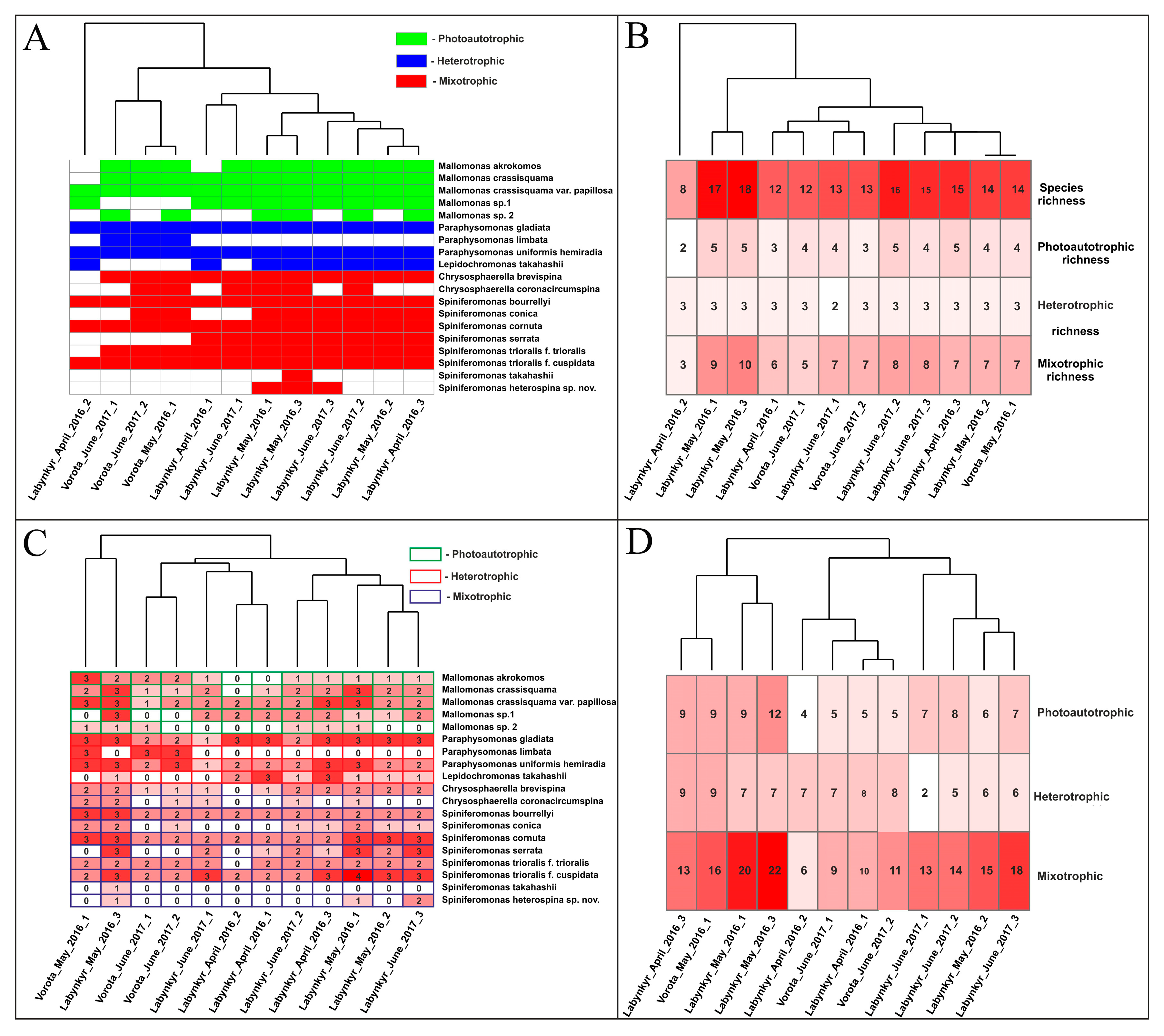
3.1. Features of the Species Composition of Silica-Scaled Chrysophytes in the Studied Lakes

In total, during the research period, 19 species of silica-scaled chrysophytes were found in the lakes Labynkyr and Vorota: Chrysosphaerella (2), Paraphysomonas (3), Lepidochromonas (1), Spiniferomonas (8), and Mallomonas (5) (Table 1; Figure 2, Figure 3 and Figure 4).
Despite the close location of the lakes, the species composition of the silica-scaled chrysophytes differed. Eighteen species were found in Lake Labynkyr during the research period; one species, Paraphysomonas limbata, is characteristic only of Lake Vorota. Fourteen species have been found in Lake Vorota; five species—Lepidochromonas takahashii, Spiniferomonas serrata, S. takahashii, Spiniferomonas heterospina and Mallomonas sp. 1—were found only in Lake Labynkyr (see Table 1). In Lake Labynkyr, the uneven nature of the distribution of scaly chrysophytes by the number of species and their relative abundance is noted. At coastal sampling points 1 and 3, species diversity and abundance are higher than at central point 2 (see Figure 1; Table 1).
The scales of Mallomonas sp. 1 and Mallomonas sp. 2 have previously been identified by us as Mallomonas kuzminii Gusev and Kulikovsky (Figure 33 and Figure 37 [12]) and M. striata Asmund (Figure 38 [12]). However, they differ from the original descriptions and probably represent new taxa to science. Their descriptions based on our observations are given below.
Mallomonas sp. 1 (Figure 3G,H).
Scales tripartite, oblong oval, 3.2–5.9 × 2.1–3.5 μm, with or without dome. Dome with permanent, numerous, and powerful ribs. V-rib acute or rounded, hooded, continuous with the anterior submarginal rib. Posterior flange with pores and without a secondary layer. Shield with ribs at the base and also in the central part. Pores evenly spaced on all the surface. A patch of small pores is present in the angle of the V-rib.
Mallomonas sp. 2 (Figure 4D).
Scales are 3.8–5.0 × 1.9–2.8 μm in size, oval in shape, with weak lateral incurving. The dome is smooth, or decorated with large pores (62 nm in diameter). The shield is marked by 9–12 evenly spaced transverse ribs reaching the back of the shield near the angle of the V-shaped rib. Between the transverse ribs are evenly spaced large pores, similar to those marked on the dome. The posterior part of the field near the angle of the V-rib is smooth. The anterior flanges are wide, with two to seven struts on each side. The anterior submarginal ribs are well developed. The V-rib on the scales is acutely angled, slightly hooded, and continues on wing-like extensions. The posterior rim is wide and smooth. The posterior flange contains wide approximately 14–20 struts and scattered large pores in the base-plate that are not evenly spaced.
According to our previous data, 16 species of the genus Spiniferomonas, namely S. abei Takahashi, S. abrubta Nielsen, S. alata Takahashi, S. bilacunosa Takahashi, S. bourrellyi, S. conica, S. cornuta, S. crusigera Takahashi, S. minuta Nicholls, S. serrata, S. septispina Nicholls, S. silverensis Nicholls, S. triangularis Siver, S. trioralis f. trioralis, S. trioralis f. cuspidata, and S. takahashii, out of 24 known in the world, inhabit the Russian reservoirs [16,17]. In Lake Labynkyr, damaged cells belonging to the genus Spiniferomonas were found, the morphology of which differs from the morphology of the scales described earlier. In our previous studies in Lake Vorota, we came across individual spines designated as S. trioralis f. trioralis ([12], M. striata, Figure 22 and Figure 23), which are a new species of the genus SpiniferomonasSpiniferomonas heterospina sp. nov.
Spiniferomonas heterospina sp. nov. Bessudova, Firsova and Kopyrina (Figure 4F,G).
Description: Cells are spherical, 4.5–8.8 μm in diameter (damaged cell). Cells are covered with two types of scales, plate-like and the spine-like scales. Plate-like scales are oval with a single lacuna, 0.9–1.1 μm long, 1.3–1.6 μm wide. Spine-like scales consist of a rounded base plate (1.4–2.3 μm in diameter) and a triangular spine tapered to a sharp apex. Long spines are 5.4–8.7 μm long, short spines are 1.0–2.7 μm long. Number of spine-like scales is 16–24. Stomatocysts unknown.
Type: A portion of a single gathering of scales on SEM stub No. 18073 (here designated, Figure 4F,G) was deposited at the Herbarium of the Limnological Institute, Siberian Branch of the Russian Academy of Sciences, Irkutsk.
Type locality: Lakes, Republic of Yakutia, Russia. Latitude/Longitude: N 62.412809–62.498777″, E 143.605418–143.605418″.
Etymology: The species was named because of the presence of different-sized spines.
Distribution: Scales were found in lakes Labynkyr (present study) and Vorota [10], at pH = 6.98–9.34, T = 2.6–9.7 °C, EC = 40.76–54.77 mS/cm (see Table S1 [10], Table 1).
Oval plate scales with one lacuna are similar to the plate scales of the species S. trioralis Takahashi and S. silverensis Nicholls. The spine scale is most similar to species with triangular spines, such as S. trioralis, S. involuta, and S. triangularis. The latter two species also have a large basal plate. A distinctive feature of the morphology of the new species Spiniferomonas heterospina sp. nov. is a combination of oval lamellar scales with a single lacuna and two types of triangular spines in the cross-section spine with a larger basal plate, long and short.
We have noticed interesting morphological features in some species. In Lake Labynkyr, on two of the four spines of the decayed Spiniferomonas bourrellyi cell (Figure 2I), pores 0.25 µm in diameter, not typical for the species, were located. The spines themselves were short, and the 3.8–4.2 µm spine base resembles a funnel. In Lake Labynkyr and Vorota, there were both typical S. trioralis cells with plate scales of the same type (Figure 3A), and similar to the species in structure and size of thorns but differing in the presence of two types of plate scales at once—oval and round (Figure 3B).

3.2. Development of Silica-Scaled Chrysophytes Depending on Environmental Factors

One-way analysis of variance showed (Table 2) that abundance in samples of photoautotrophic and mixotrophic species was significantly associated with the month of sampling (p value < 0.05). For the abundance of mixotrophic species, no relationship was found with the month of sampling (p value > 0.05). The relationship of the abundance in samples of photoautotrophic and mixotrophic species with the month of sampling is due to the state of the ice. In April and May, the ice was solid and transparent and let in enough light for the life of photoautotrophic and mixotrophic species; in June, as a result of temperature increase and melting, the ice became acicular and dark and did not let in enough light for the life of the species of this group.
Correlation analysis (Table 3) did not show a significant relationship (p value > 0.05) between the abundance of different species groups and the physicochemical parameters of the environment, except for the dependence of species relative abundances of mixotrophic species on snow cover thickness and water temperature (T °C). Species abundance of mixotrophic species significantly decreased with increasing snow cover thickness and increasing temperature (negative correlation). It can be concluded that a small snow cover thickness contributed to better light penetration, increasing the productivity of mixotrophic species.
An increase in temperature led to the melting (destruction) of ice, contributing to a decrease in its transparency and a deterioration in the penetration of sunlight, which contributed to a decrease in the abundance of mixotrophic species.
An analysis of the similarity of samples in terms of species spectra showed (Figure 5A) that, regardless of the month of the year (April or May or June), samples from Lake Vorota and Lake Labynkyr usually form two independent clusters (with the exception of one sample from Lake Labynkyr). The species composition of samples collected in one lake is more similar compared to the species composition of samples from different lakes. When comparing samples in terms of species richness and species abundance (Figure 5B,C), no clear clustering of samples by lake or month of sampling was observed, although more photoautotrophic and mixotrophic species were found on average in Lake Labynkyr samples compared to samples from Lake Vorota. In the analysis of cumulative abundance of species (Figure 5D), clustering identified a group of four April–May samples from Lake Labynkyr and Vorota, characterized by large abundances of photoautotrophic and mixotrophic species.
The result of the comparison of samples by cumulative species abundance confirms the conclusions of one-way analysis of variance, verifying the relationship of species abundance with the month of sampling through the state of ice cover.

4. Discussion

In two studied subarctic lakes of Yakutia, during the ice cover period, 19 species of silica-scaled chrysophytes were revealed. In previous studies, 24 species of silica-scaled chrysophytes were found in the Labynkyr and Vorota lakes during the open water period [12]. The previous list of discovered species has changed after the revision. The previously discovered spines, identified by us as Chrysosphaerella longispina Lauterborn, most likely belong to the species Chrysosphaerella brevispina, since we did not find lamellar scales of C. longispina in the long-term studies selected. The individual spines of C. brevispina found in the studied lakes have the shaft joining the two plates of the narrow, bobbin-like structure ([12], Figure 4). The scales identified by us as Mallomonas kuzminii and M. striata may be species that are new to science. Some of the M. striata scales found earlier actually belong to the species M. striata ([12], M. striata, Figure 39). This species remains on the general list of silica-scaled chrysophytes species found in lakes Labynkyr and Vorota after the conducted research. In total, as a result of these studies and research conducted earlier during the open water period [12], 29 species of silica-scaled chrysophytes live in the lakes. In relation to the trophic mode type, the lakes are inhabited by 17 mixotrophic species from the genera Spiniferomonas and Chrysosphaerella, 5 heterotrophic species of the genera Paraphysomonas and Lepidochromonas, and 7 phototrophic representatives from the genera Mallomonas and Synura (not found during the subglacial period). Spiniferomonas heterospina, a species new to science, has also been discovered.
During the ice cover period, from the point of view of relative abundance and species diversity from April to June, mixotrophic silica-scaled chrysophytes (Spiniferomonas) and heterotrophic silica-scaled chrysophytes (Paraphysomonas) predominate in the studied lakes. Photoautotrophic representatives of silica-scaled chrysophytes (Mallomonas) formed the maximum number in lakes in April–May, when a thick, solid, and transparent layer of ice and a small layer of snow were observed. During the same period, the maximum abundance and diversity of the chrysophytes were observed, especially of species belonging to the genera Spiniferomonas and Paraphysomonas. In June, when the ice became needle-like, dark, and saturated with water [7], none of the photoautotrophic species of the Mallomonas genus formed a high abundance. During the same period, there was a decrease in the relative abundance and species diversity of species of the genus Spiniferomonas. At the same time, three species of the genus Spiniferomonas (S. cornuta, S. serrata and S. trioralis f. cuspidata) were still in great relative abundances in Lake Labynkyr. Two representatives of the genus Paraphysomonas (heterotrophic silica-scaled chrysophytes) prevailed in Lake Vorota (P. limbata and P. uniformis hemiradia) and one in Lake Labynkyr (P. gladiata). Thus, there is a dependence on the illumination of the reservoir not only of photoautotrophic silica-scaled chrysophytes, but also of mixotrophic representatives. In our case, the illumination of the reservoir is directly related to the state of ice and snow covering the lakes. Earlier, during the open water period (July 2014), in Lake Labynkyr, mixotrophic and photoautotrophic silica-scaled chrysophytes over heterotrophic prevailed in terms of the number of species and relative abundances [12].
A distinctive feature of the species diversity of silica-scaled chrysophytes in reservoirs with a high level of nutrients is the predominance of photoautotrophic species of the genera Mallomonas and Synura [18,19], and in oligotrophic water basins, taxa of Spiniferomonas, Paraphysomonas, and Lepidochromonas successfully developed [20,21,22]. In the studied reservoirs, environmental factors such as the long period of ice cover (which disrupts the flow of organic matter from the atmosphere and land) and a low level of illumination under the ice limit the development of phytoplankton, making them ultraoligotrophic. In such conditions characteristic of northern reservoirs, silica-scaled chrysophytes genera Spiniferomonas and Paraphysomonas have a competitive advantage in conditions of lack of nutrients and low illumination. It is known that bacteria can serve as food for both colorless, phagotrophic representatives of the genus Paraphysomonas [23,24] and for species of the genus Spiniferomonas [25,26]. The high development and diversity of the bacterial community in the samples studied [7] probably contribute to the development of these organisms.
However, Paraphysomonas species were already assigned to subglacial plankton [9,10,17,27], while Spiniferomonas were previously considered as true autumnal species [17,27]. In reservoirs of moderate latitude—the Boguchansky reservoir and Lake Baikal—silica-scaled chrysophytes genera Spiniferomonas reach their maximum development in the autumn period. Thus, the food webs of subarctic, ultraoligotrophic lakes are more complex than we expected and require careful study.

5. Conclusions

Silica-scaled chrysophytes are the main component of the plankton of subarctic ultraoligotrophic lakes Labynkyr and Vorota, forming a high abundance due to the variety of trophic modes within the class. Silica-scaled chrysophytes have a competitive advantage in these ice-covered lakes. The leading role in changing the ratio of the various trophic modes of silica-scaled chrysophytes and their relative abundance is played by the illumination of the reservoir, which is directly affected by the state of the ice and the thickness of the snow cover. With a large thickness of snow cover and in the process of melting ice, with a decrease in its transparency, there was a decrease in the penetration of sunlight into the water column and a decrease in the species abundance of photoautotrophic and mixotrophic species. The effect of snow cover thickness and ice transparency on the species abundance of heterotrophic and photoautotrophic species has not been traced. Other hydrochemical parameters within the established variability (T, pH, electrical conductivity, PO4−3, TOC) did not significantly affect the abundance and species richness. A new species of Spiniferomonas heterospina was discovered in the studied lakes. A distinctive feature of the morphology of the new species Spiniferomonas heterospina sp. nov. is a combination of oval lamellar scales with a single lacuna and two types of triangular spines in the cross-section spine with a larger basal plate, long and short.

Supplementary Materials

The following supporting information can be downloaded at: https://figshare.com/articles/dataset/Table_S1_xls/21901035, Table S1: The main physicochemical parameters of the lakes Labynkyr and Vorota during the study period.

Author Contributions

A.B. and A.F., electron microscopy and identification of species; A.B., drafting, collection of literature, interpreting the results, and writing the first version of the manuscript; Y.B., statistical analysis; L.K. and Y.Z., sampling; Y.L., review and editing. All authors have read and agreed to the published version of the manuscript.

Funding

This work was supported by the project no. 0279-2021-0008 (Limnological Institute) and by the State Assignments of the Institute for Biological Problems of Cryolithozone, no. 0297-2021-0023, 21-121012190038-0; expeditions.

Institutional Review Board Statement

Not applicable.

Informed Consent Statement

Not applicable.

Data Availability Statement

The data presented in this study are openly available in the FigShare repository at https://doi.org/10.6084/m9.figshare.21901035.

Acknowledgments

The microscopy studies were performed at the Electron Microscopy Center of the Shared Research Facilities “Ultramicroanalysis” of Limnological Institute, https://www.lin.irk.ru/copp/ (accessed on 15 January 2023).

Conflicts of Interest

The authors declare no conflict of interest.

References

  1. Kristiansen, J.; Škaloud, P. Chrysophyta. Handbook of the Protists, 2nd ed.; Springer International Publishing: Cham, Switzerland, 2016; pp. 1–38. [Google Scholar]
  2. Kapustin, D.; Kulikovskiy, M. Chrysosphaerella septentrionalis sp. nov. (Chrysophyceae, Chromulinales), a New Species from the Arctic Including the Description of Chrysosphaerellaceae, fam. nov. Plants 2022, 11, 3166. [Google Scholar] [CrossRef]
  3. Kapustin, D.A.; Guiry, M.D. Reinstatement of Lepidochromonas Kristiansen (Lepidochromonadaceae fam. nov., Chrysophyceae). Phytotaxa 2019, 413, 49–53. [Google Scholar] [CrossRef]
  4. Bhatti, S.; Colman, B. Inorganic carbon acquisition by the chrysophyte alga Mallomonas papillosa. Can. J. Bot. 2005, 83, 891–897. [Google Scholar] [CrossRef]
  5. Scoble, J.M.; Cavalier-Smith, T. Scale evolution in Paraphysomonadida (Chrysophyceae): Sequence phylogeny and revised taxonomy of Paraphysomonas, new genus Clathromonas, and 25 new species. Eur. J. Protistol. 2014, 50, 551–592. [Google Scholar] [CrossRef] [Green Version]
  6. Olsen, N.E.; Poulsen, L.K.; Reuss, N.; Steinarsdottir, S.S. A new subspecies of Paraphysomonas punctata (Paraphysomonadaceae, Chrysophyceae). Nord. J. Bot. 1999, 19, 635–640. [Google Scholar] [CrossRef]
  7. Zakharova, Y.; Bashenkhaeva, M.; Galachyants, Y.; Petrova, D.; Tomberg, I.; Marchenkov, A.; Kopyrina, L.; Likhoshway, Y. Variability of Microbial Communities in Two Long-Term Ice-Covered Freshwater Lakes in the Subarctic Region of Yakutia, Russia. Microb. Ecol. 2021, 84, 958–973. [Google Scholar] [CrossRef]
  8. Eloranta, P. Biogeography of chrysophytes in Finnish lakes. Chrysophyte Algae: Ecology, Phylogeny and Development; Cambridge University Press: Cambridge, UK, 1995; pp. 214–231. [Google Scholar] [CrossRef]
  9. Ikävalko, J. On the presence of some selected Heterokontophyta (Chrysophyceae, Dictyochophyceae, Bicocoecidae) and cysts (“archaeomonads”) from sea ice—A synopsis. Nova Hedw. Beih. 2001, 122, 41–54. [Google Scholar]
  10. Lim, E.L.; Dennett, M.R.; Caron, D.A. The ecology of Paraphysomonas imperforata based on studies employing oligonucleotide probe identification in coastal water samples and enrichment cultures. Limnol. Oceanogr. 1999, 44, 37–51. [Google Scholar] [CrossRef] [Green Version]
  11. Siver, P.A. The distribution of chrysophytes along environmental gradients: Their use as biological indicators. Chrysophyte Algae; Cambridge University Press: Cambridge, UK, 1995; pp. 232–268. [Google Scholar]
  12. Bessudova, A.Y.; Tomberg, I.; Firsova, A.D.; Kopyrina, L.I.; Likhoshway, Y.V. Silica-scaled chrysophytes in lakes Labynkyr and Vorota of the Sakha (Yakutia) Republic, Russia. Nova Hedwig. Beih. 2019, 148, 35–48. [Google Scholar] [CrossRef]
  13. Kopyrina, L.; Firsova, A.; Rodionova, E.; Zakharova, Y.; Bashenkhaeva, M.; Usoltseva, M.; Likhoshway, Y. The insight into diatom diversity, ecology, and biogeography of an extreme cold ultraoligotrophic Lake Labynkyr at the Pole of Cold in the northern hemisphere. Extremophiles 2020, 24, 603–623. [Google Scholar] [CrossRef]
  14. Warnes, G.R.; Bolker, B.; Bonebakker, L.; Gentleman, R.; Liaw, W.H.A.; Lumley, T.; Maechler, M.; Magnusson, A.; Moeller, S.; Schwartz, M.; et al. Package “gplots”: Various R Programming Tools for Plotting Data; R Packag, Version 2.17.0; ScienceOpen: Berlin, Germany, 2015.
  15. Oksanen, J. Vegan: Ecological Diversity. R. Project. 2018. Available online: https://cran.r-project.org/web/packages/vegan/vignettes/diversity-vegan.pdf (accessed on 30 August 2022).
  16. Voloshko, L.N. Species of the genus Spiniferomonas (Chrysophyceae, Paraphysomonadaceae) in water bodies of the Russian North. Bot. Zhurn. 2013, 98, 848–858. (In Russian) [Google Scholar]
  17. Bessudova, A.Y.; Likhoshway, Y.V. Silica chrysophytes (Chrysophyceae) of the Boguchany reservoir. Modern science: Actual problems of theory and practice. Series “Natural and technical sciences (General biology). Acta Biol. Sib. 2017, 11, 4–11. (In Russian) [Google Scholar]
  18. Siver, P.A. Inferring the specific conductivity of lake water with scaled chrysophytes. Limnol. Oceanogr. 1993, 38, 1480–1492. [Google Scholar] [CrossRef]
  19. Kapustin, D.A.; Gusev, E.S. Silica-scaled chrysophytes from West Java (Indonesia) including description of a new Chrysosphaerella species. Nova Hedwig. Beih. 2019, 148, 11–20. [Google Scholar] [CrossRef]
  20. Kristiansen, J. Silica-scaled chrysophytes from West Greenland: Disko Island and the Søndre Strømfjord region. Nord. J. Bot. 1992, 12, 525–536. [Google Scholar] [CrossRef]
  21. Nemcova, Y.; Rott, E. Diversity of Silica-scaled Chrysophytes in High-altitude Alpine Sites (North Tyrol, Austria) Including a Description of Mallomonas pechlaneri sp. nov. Cryptogam. Algologie 2018, 39, 63–83. [Google Scholar] [CrossRef]
  22. Bessudova, A.; Gabyshev, V.; Firsova, A.; Gabysheva, O.; Bukin, Y.; Likhoshway, Y. Diversity Variation of Silica-Scaled Chrysophytes Related to Differences in Physicochemical Variables in Estuaries of Rivers in an Arctic Watershed. Sustainability 2021, 13, 13768. [Google Scholar] [CrossRef]
  23. Goldman, J.C.; Caron, D.A. Experimental studies on an omnivorous microflagellate: Implications for grazing and nutrient regeneration in the marine microbial food chain. Deep. Sea Res. Part A. Oceanogr. Res. Pap. 1985, 32, 899–915. [Google Scholar] [CrossRef]
  24. Goldman, J. Dennett Dynamics of prey selection by an omnivorous flagellate. Mar. Ecol. Prog. Ser. 1990, 59, 183–194. [Google Scholar] [CrossRef]
  25. Charvet, S.; Vincent, W.F.; Lovejoy, C. Chrysophytes and other protists in High Arctic lakes: Molecular gene surveys, pigment signatures and microscopy. Polar Biol. 2011, 35, 733–748. [Google Scholar] [CrossRef]
  26. Terrado, R.; Pasulka, A.L.; Lie, A.A.-Y.; Orphan, V.; Heidelberg, K.; A Caron, D. Autotrophic and heterotrophic acquisition of carbon and nitrogen by a mixotrophic chrysophyte established through stable isotope analysis. ISME J. 2017, 11, 2022–2034. [Google Scholar] [CrossRef] [Green Version]
  27. Bessudova, A.Y.; Domysheva, V.M.; Firsova, A.D.; Likhoshway, Y.V. Silica-scaled chrysophytes of Lake Baikal. Acta Biol. Sib. 2017, 3, 47–56. [Google Scholar] [CrossRef] [Green Version]
Figure 1. The map of the study regions and sampling site locations.
Diversity 15 00326 g001
Figure 2. Silica-scaled chrysophytes from lakes Labynkyr and Vorota. SEM: (A) Chrysosphaerella coronacircumspina; (B) C. brevispina; (C,D) Paraphysomonas limbata; (E) P. gladiata; (F,G) P. uniformis hemiradia; (H) Spiniferomonas bourrellyi; (I) S. cf. bourrellyi with pores; (J) Lepidochromonas takahashii. Scale bars: 2 µm.
Diversity 15 00326 g002
Figure 3. Silica-scaled chrysophytes from lakes Labynkyr and Vorota. SEM: (A) Spiniferomonas trioralis with oval plate scales; (B) Spiniferomonas sf. trioralis with oval and rounded plate scales; (C) S. trioralis f. cuspidata; (D) S. cornuta; (E) S. conica; (F) S. takahashii; (G,H) Mallomonas sp. 1; (I) S. serrata. Scale bars: (A,B,DG,I) 2 µm; (H) 10 µm; (C) 15 µm.
Diversity 15 00326 g003
Figure 4. Silica-scaled chrysophytes from lakes Labynkyr and Vorota SEM: (A) M. akrokomos; (B,C) M. crassisquama var. papillosa; (D) Mallomonas sp. 2; (E) M. crassisquama var. crassisquama; (F,G) Spiniferomonas heterospina sp. nov. Scale bars: (A,CG) 2 µm; (B): 10 µm.
Diversity 15 00326 g004
Figure 5. Analysis of similarities and differences between the studied samples (A) by species composition (clustering based on Jaccard distances); (B) by indicators of species richness of different groups of species (clustering based on Euclidean distances), where the species richness value is indicated in cells; (C) by indicators abundance of different groups of species (clustering based on Euclidean distances), where the cells indicate the value of the species abundance; and (D) by cumulative abundance of different species groups (clustering based on Euclidean distances), where the cells indicate the value of cumulative species abundance for species groups.
Diversity 15 00326 g005
Table 1. Species composition of silica-scaled chrysophytes in the studied lakes (very rare (VR) 2–25 scales, rare (R) 26–50, common (C) 51–150, and abundant (A) >150). Mixotrophic—blue, heterotrophic—red, photoautotrophic—green.
Lake Labynkyr
April 2016
Lake Labynkyr
May 2016
Lake Vorota May 2016Lake Labynkyr
June 2017
Lake Vorota
June 2017
Stations123123112312
No.Species
Paraphysomonas gladiata Preisig et HibberdCCCCCCCVRRCRR
P. limbata Preisig et Hibberd------C---CC
P. uniformis hemiradia Scoble et Cavalier-SmithRRCCRCCVRRRRC
Lepidochromonas takahashii (Cronberg et Kristiansen) Kapustin et Guiry CRCVRVRVR--VRVR--
Chrysosphaerella brevispina KorshikovVR-RRRRRVRRRVVR
C. coronacircumspina Wujek et Kristiansen---VR-RRVRVR--VR
Spiniferomonas bourrellyi TakahashiRRRRRCCRRRRR
S. conica Takahashi--VRRVRRR-VRVR-VR
S. cornuta BalonovRRRCCCCRRCRR
S. serrata NichollsVR-VRCRC-RRC--
S. trioralis f. trioralis TakahashiR-RRRRRRRRRR
S. trioralis f. cuspidata BalonovRRCACARCRCRR
S. takahashii Nicholls-----VR------
Spiniferomonas heterospina sp. nov.---VR-VR---R--
Mallomonas akrokomos Ruttner--VRVRVRRCVRVRVRRR
M. crassisquama (Asmund) Fott var. crassisquamaVR-RCRCRRRRVRVR
M. crassisquama var. papillosa Siver et SkogstadRRCCRCCRRRVRR
Mallomonas sp. 1RRRVRVRC-RRR--
Mallomonas sp. 2 --VRVR-VRVR-VR-VR-
Table 2. Results of one-way analysis of variance to assess the relationship of species abundance of different groups with sampling months (April, May, and June).
Trophic Mode TypeF Valuep Value
Photoautotrophic species1.3710.0424
Heterotrophic species0.5230.481
Mixotrophic species4.4120.0397
Table 3. Results of correlation analysis (Kendall correlation coefficient) to assess the relationship between species abundance of different groups and physicochemical parameters of the environment (see Table S1).
Trophic Mode TypeParameterr Valuep Value
Photoautotrophic speciesIce thickness (cm)−0.140.19
Snow cover thickness (cm)−0.150.15
T °C−0.150.15
pH0.080.43
Conductivity (μS/cm)0.050.57
PO43− (mg/L)−0.010.95
TOC (mgC/L)−0.030.74
Heterotrophic speciesIce thickness (cm)0.040.72
Snow cover thickness (cm)0.100.40
T °C0.100.40
pH0.160.13
Conductivity (μS/cm)0.150.17
PO43− (mg/L)0.010.98
TOC (mgC/L)0.060.57
Mixotrophic speciesIce thickness (cm)−0.050.42
Snow cover thickness (cm)−0.210.01
T °C−0.210.01
pH0.100.14
Conductivity (μS/cm)0.080.25
PO43− (mg/L)0.050.43
TOC (mgC/L)−0.060.39
Disclaimer/Publisher’s Note: The statements, opinions and data contained in all publications are solely those of the individual author(s) and contributor(s) and not of MDPI and/or the editor(s). MDPI and/or the editor(s) disclaim responsibility for any injury to people or property resulting from any ideas, methods, instructions or products referred to in the content.

