Article

Evaluation of the “Bottleneck” Effect in an Isolated Population of Microtus hartingi (Rodentia, Arvicolinae) from the Eastern Rhodopes (Bulgaria) by Methods of Integrative Analysis

by
Fedor N. Golenishchev
1,*,
Tanya A. Zorenko
2,
Tatyana V. Petrova
1,
Leonid L. Voyta
1,
Lyudmila Yu. Kryuchkova
3 and
Nasko Atanasov
4
1
Zoological Institute, Russian Academy of Sciences, Universitetkaya Emb. 1, Saint Petersburg 199034, Russia
2
Department of Zoology and Animal Ecology, University of Latvia, 1 Jelgava Street, LV-1004 Riga, Latvia
3
The Centre for XRD Studies, Saint Petersburg State University, Universitetkaya Emb. 7/9, Saint Petersburg 199155, Russia
4
Institute of Biodiversity and Ecosystem Research, Bulgarian Academy of Sciences, Tsar Osvoboditel Blvd. 1, 1000 Sofia, Bulgaria
*
Author to whom correspondence should be addressed.
Diversity 2022, 14(9), 709; https://doi.org/10.3390/d14090709
Submission received: 11 August 2022 / Revised: 22 August 2022 / Accepted: 23 August 2022 / Published: 26 August 2022

Abstract

:
An integrative analysis of an isolated population of Harting’s vole (Microtus hartingi) from the Eastern Rhodope Mountains (Bulgaria) was carried out by morphological and morphometric methods, computed tomography, Cytb variation data, and experimental hybridization. Substantial changes in the development of the skull and teeth were found. Nevertheless, those voles can live to the senex stage. A phylogenetic reconstruction based on Cytb sequences showed that M. hartingi from the Eastern Rhodopes forms a separate clade, which is a sister clade to the voles from Northeastern Greece (also from the foothills of the Rhodopes). M. hartingi from the Rhodopes is mostly isolated reproductively from M. h. ankaraensis from Anatolia (Turkey), as evidenced by a decrease in the proportion of pairs that started breeding, relatively high mortality of pups, and increased sterility of hybrid males. Possible time of isolation of the Rhodopean population and consequences of the “bottleneck” effect on its current state are discussed. At the same time, we believe that, at present, it is impossible to determine its taxonomic status. It is necessary to recognize M. hartingi from the Bulgarian Rhodopes as an endangered population and to include it in regional Red Lists of Bulgaria.

1. Introduction

Harting’s vole, Microtus hartingi Barret-Hamilton, 1903 is a member of the subgenus Sumeriomys Argyropulo, 1933 which, together with subgenera Microtus Schrank, 1798 and Hyrcanicola Nadachowski, 2007 constitute the genus Microtus proper. Relatively recently, M. hartingi was considered a subspecies of M. guentheri Danford et Alston, 1880. Nonetheless, in a Cytb gene analysis, it was found that M. guentheri consists of two allopatric forms: an eastern one (Syria and Israel) and a western one (the Balkans and Anatolia). In accordance with the priority rule, the eastern nominal form has retained the name M. guentheri, and it was proposed to name the western one as M. hartingi [1]. Morphological differences have previously been noted between Harting’s voles from Anatolia and M. guentheri topotypes [2]. The species status of M. hartingi has been confirmed by experimental hybridization with M. guentheri from terra typica [3,4]. These two species are separated by a geographical barrier in southeastern Turkey: the Taurus Mountains, i.e., the so-called Anatolian Diagonal. M. hartingi currently includes the following subspecies: M. h. hartingi Barret Hamilton, 1903 (Greece); M. h. lydius Blackler, 1916 (western part of Turkey); M. h. martinoi Petrov, 1939 (north Macedonia); M. h. strandzensis Markov, 1960 (Bulgaria); and M. h. ankaraensis Yiğit et Colak, 2002 (central Turkey). European and Asian forms of Harting’s vole, separated by the Sea of Marmara and narrow straits—the Bosphorus and the Dardanelles—diverged substantially during the Pleistocene and Holocene. When the European subspecies M. h. strandzensis was hybridized with the Asia Minor subspecies, M. h. ankaraensis from central Anatolia, viable and fertile F1 offspring were obtained; however, during subsequent crossing, including backcrossing, partial sterility of males and high mortality of pups began to manifest themselves [5]. For the first time, two males and three females of M. hartingi were captured in the Eastern Rhodopes in the 1990s and were studied karyologically [6] The present authors (T. Zorenko and N. Atanasov) surveyed the Eastern Rhodopes (Bulgaria) in 2014 and 2016 and documented the presence of an isolated population of M. hartingi, represented by small habitats that are distant from each other. The Rhodopean population significantly differs in linear parameters and brain shape from M. h. strandzensis and M. h. ankaraensis [7] and in the shape of spermatozoa from M. h. ankaraensis [8]. Due to the pronounced fragmentation of biotopes and a reduction in available localities for the settlement of young voles, M. hartingi from the Eastern Rhodopes shows a marked propensity for communal breeding [9].
The purpose of this study was an integrative assessment of the “bottleneck effect” in an isolated population of M. hartingi from the Eastern Rhodopes (Bulgaria) by the methods of morphology (using computed tomography), morphometry, and by molecular and reproductive-genetic differentiation of the Rhodopean population of M. hartingi.

2. Material and Methods

2.1. Morphology

2.1.1. Morphology and Morphometry

For morphometric analysis, we used data from collections of the following museums: the Zoological Institute of the Russian Academy of Sciences, St. Petersburg, Russia (ZIN); the Zoological Museum of Moscow State University, Moscow, Russia (ZMMU); the Biology Department, Faculty of Science, Ankara University, Ankara, Turkey (AUMAC); and the Zoology Department of the University of Latvia, Riga, Latvia (Table S1). In addition, we used voles caught by us in the following localities: M. h. ankaraensis in Kırşehir, Turkey (39°9′56.38″ N, 34°6′6.99″ E); and M. h. strandzensis in Gramatikovo, Bulgaria (42°3′14.83″ N, 27°39′44.41″ E) and M. hartingi from the Eastern Rhodopes: Meden Buk, Bulgaria (41°22′22.17″ N, 26°2′38.52″ E) and Mandrica, Bulgaria (41°23′29.39″ N, 26°7′47.72″ E) (Figure 1).
For exterior comparison, standard measurements were employed: L, body length (head and body length); C, the length of the tail (tail length); PL, foot length (foot length); and Au, ear length. For morphometric analysis, the following measurements were taken: CBL, condylobasal skull length; ZB, zygomatic breadth; DL, diastema length; OB, occipital breadth; OH, occipital height without bullae; TOH, occipital height with bullae; BB, bullae breadth; BL, bullae length; MBL, bullae length with the mastoideum; IB, interorbital breadth; RH, rostrum height; RB, rostrum breadth; M(1–3), upper molar alveolar length; m(1–3), lower molar alveolar length; and ML, mandible length.
To characterize bacula, standard measurements of the os penis were made: total length, the shaft’s length, width of the shaft’s base, length of the trident’s medial ossicle (=process), and length of the trident’s lateral ossicle (=process).

2.1.2. Statistical Analysis

Descriptive statistics were obtained in Microsoft Excel 2010. Principal component analysis (PCA) was performed on 15 log-transformed cranial and mandibular linear measurements in the PAST software, v3.18 [11]. Statistical analysis (ANOVA) did not detect sexual dimorphism for the measured cranial parameters; therefore, we combined samples from both sexes in all subsequent analyses.

2.1.3. High-Resolution X-ray Computed Microtomography

The analyzed material represents four vole skulls (skull + hemimandibles) of M. h. ankaraensis (ZIN 98971, male, age 5 months; vivarium sample, founders from Kırşehir, Turkey), M. h. strandzensis (ZIN 84905-471, male, age 5 months; vivarium sample, founders from Gramatikovo, Bulgaria), and from two M. hartingi individuals from the Rhodopes (ZIN 107168-10, ZIN 107168-11, males, age 8 and 11 months, respectively; vivarium sample). All analyses of the vole specimens were scanned on a SkyScan 1172 Scanner at the Resource Centre for X-ray Diffraction Studies of Saint Petersburg State University (Saint Petersburg, Russia). The SkyScan settings were as follows: ZIN 98971 (acceleration voltage 74 kV, resolution 5.58 µm, exposure 1500 ms), ZIN 84905-471 (74, 5.84, 1450, respectively), ZIN 107168-10 (74, 5.84, 1340), and ZIN 107168-11 (74, 5.84, 1340). All specimens were scanned in three-slice mode. Preprocessing was performed with DataViewer v1.5.4.0 (SkyScan, Bruker, Billerica, MA, USA). The main processing of tooth structure data (three-dimensional [3D] digital reconstruction and surface rendering) was performed by means of Avizo 2019.1 (FEI SAS). For all scanned specimens, 3D models (surfaces) of the whole molars, hemimandible, and skull were prepared for a comparison.

2.2. Molecular Analysis

2.2.1. Sampling and Laboratory Techniques

Four specimens were used: two specimens of M. hartingi from the Rhodopes, Bulgaria, one specimen of M. h. ankaraensis from Kırşehir, Anatolia, Turkey, and one specimen of M. g. philistinus from Qiryat Shemona, Israel (33°12′26.30″ N, 35°35′7.53″ E). We also analyzed voles transferred to us (for breeding in the vivarium and for further research) by Dr. Anna German (Institute of Plant Protection, A.R.O., The Volcani Center, Bet Dagan, Israel) and from Yvne, Israel (31°52′33.27″ N 34°45′22.82″ E). Genomic DNA was isolated from ethanol-preserved muscle tissues using the DNA-Ekstran Kit (Syntol, Moscow, Russia). PCRs were conducted with the PCR ScreenMix Kit (Evrogen, Russia). A fragment of the mitochondrial cytochrome b (Cytb) genes were amplified using primers and PCR conditions published by Abramson et al. [12]. The PCR products were purified on columns from the Omnix Kit (Omnix, St. Petersburg, Russia) and were sequenced in both directions with the help of the BigDye Terminator Cycle Sequencing Ready Reaction Kit on an ABI PRISM 3130 instrument (Applied Biosystems Inc., Foster City, CA, USA).
During a preliminary molecular analysis of the Cytb gene with standard primers, it was found that when phylogenetic trees were constructed based on the Cytb sequences of M. hartingi, the Rhodopean form does not cluster with the common clade of all voles of the subgenus Sumeriomys. Examination of the sequencing spectrograms of the gene fragment of interest suggested that in addition to the Cytb sequence, our PCR amplified pseudogene sequences. To verify this and to determine the exact phylogenetic position of M. hartingi from the Rhodopes, an analysis of individual sequences of the M. hartingi Cytb fragment was performed. For this purpose, the PCR product was cloned, and the resulting clones were sequenced. The amplicons were purified using the QIAquick PCR and Gel Cleanup Kit (Qiagen, Hilden, Germany). The purified DNA fragments were cloned into the pTZ57R/T vector by means of the Thermo Scientific InsTAclone PCR Cloning Kit (Thermo Fisher Scientific, Waltham, MA, USA). Then, the XL10-Gold bacterial strain was transformed with the ligation mixture. The transformants were plated on a solid LB medium supplemented with ampicillin (50 μg/mL) and IPTG/Xgal for white/blue selection. The plates with the medium were incubated for 14 h in a thermostat at 37 °C. From the colonies that emerged, white ones were chosen (harboring the pTZ57R/T plasmid with the insertion of the Cytb gene fragment), from which DNA was isolated (10 colonies for each vole tissue sample). DNA extraction was carried out by heating an aqueous cell suspension to 100 °C for 5 min. Next, PCR was carried out with primers UCBU-LM. After that, PCR was performed on the DNA isolated from the colonies using primers specific to the pTZ57R/T vector (M13 forw and M13 rev). The amplicons from this second PCR were sequenced.

2.2.2. Phylogenetic Analysis

Sequences were edited, assembled, and aligned with BioEdit v7.2.5 [13]. The level of genetic differentiation in terms of Cytb on the basis of p-distances was estimated in MEGA 7 [14]. In the current study, we amplified Cytb from four specimens (GenBank accession numbers ON324049–ON324052), and 59 sequences were downloaded from GenBank (Table 1).
Phylogenetic reconstruction was performed by Bayesian inference analysis (BI) in MrBayes v3.2.6 [16] with M. arvalis and M. rossiaemeridionalis as outgroups. Four Markov chains starting with random trees were implemented for five million generations with sampling every thousandth generation. BI both for Cytb was run via the mixed model with γ-distributed rate variation across sites, which is supposed to try out every possible nst value. Consensus trees were constructed based on the trees sampled after 25% burn-in from both independent runs. Bootstrap analysis involved 1000 replicates. The final tree was formatted with FigTree v1.4 (http://tree.bio.ed.ac.uk/software/figtree/, accessed on 26 November 2021).

2.3. Hybridization

Experimental hybridization was carried out between M. h. ankaraensis (abbreviated A) from Kırşehir, Anatolia, Turkey and M. hartingi (abbreviated H) from the Eastern Rhodopes, Bulgaria. Pairs were composed of sexually mature males and females of M. hartingi and M. h. ankaraensis at the age of 75–90 days. To estimate reproduction intensity during the hybridization, the following parameters were used: percent of females who started reproduction, average time interval between consequent litters, average number of newborns per litter, average number of surviving pups, mortality through the 45th day, the number of pups in every litter, and the parents’ and pups’ mortality. In case offspring were not obtained, the experiments were finished after 2.5–3.0 months from the moment of their coupling and placement together. We conducted 125 experiments on hybridization. Reproductively active (judging by the presence and motility of spermatozoa) hybrid males aged 4–6 months were subjected to sperm analysis. The males were killed by cervical dislocation. After dissection, the cauda epididymidis was cut out and placed in 1 mL of saline for 30 min at 37 °C to allow sperm cells to swim out. After the crossing ♂ A × ♀ H (Rhodopes), 63 hybrid males were analyzed, and after the crossing ♂ H × ♀ A, 53 hybrid males were analyzed.

3. Results

3.1. Morphology and Morphometric Analysis

3.1.1. Exterior

Externally, M. hartingi from the Eastern Rhodopes does not differ much from other forms of this vole species (Table 2).
Nonetheless, the coloration is somewhat darker than that in other subspecies of Harting’s vole and most closely resembles that of M. h. strandzensis. The back is dark fawn-colored; on the cheeks and along the sides, the dark fawn color turns into ochroid. The ventral side is light gray with slight yellowness, and the tail is unicolored or slightly two-color.

3.1.2. Morphometric Analysis

External examination of M. hartingi from the Eastern Rhodopes skulls revealed abnormalities in tooth growth. We hypothesized that such severe aberrations in the calcification of skull parts should affect morphometric parameters, as well. PCA of fifteen log-transformed cranial parameters yielded the first two principal components that explained 68.52% of variance, with the first principal component (PC1) explaining 50.48% of variance, and the second principal component (PC2) explaining 18.22% of variance (Figure 2).

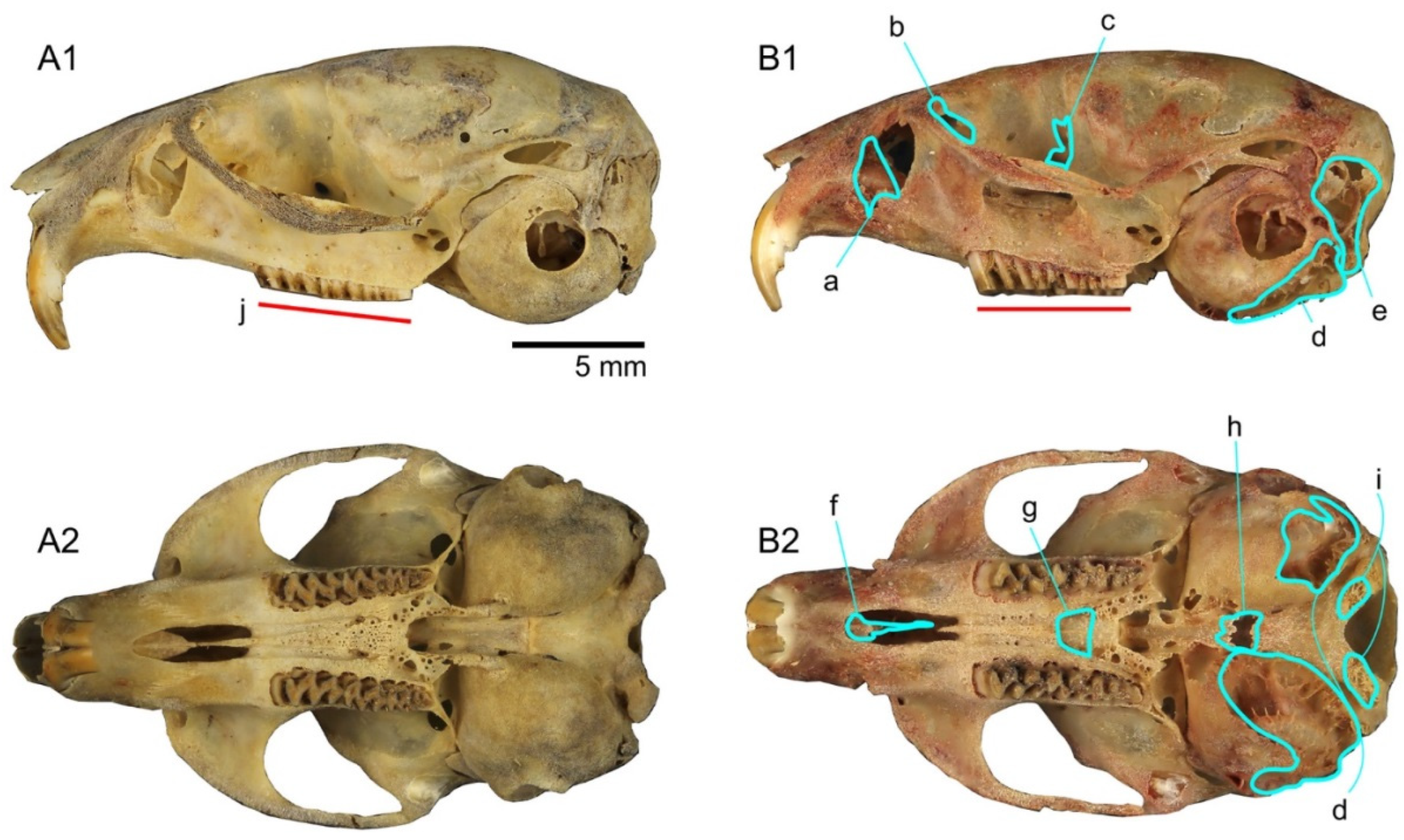
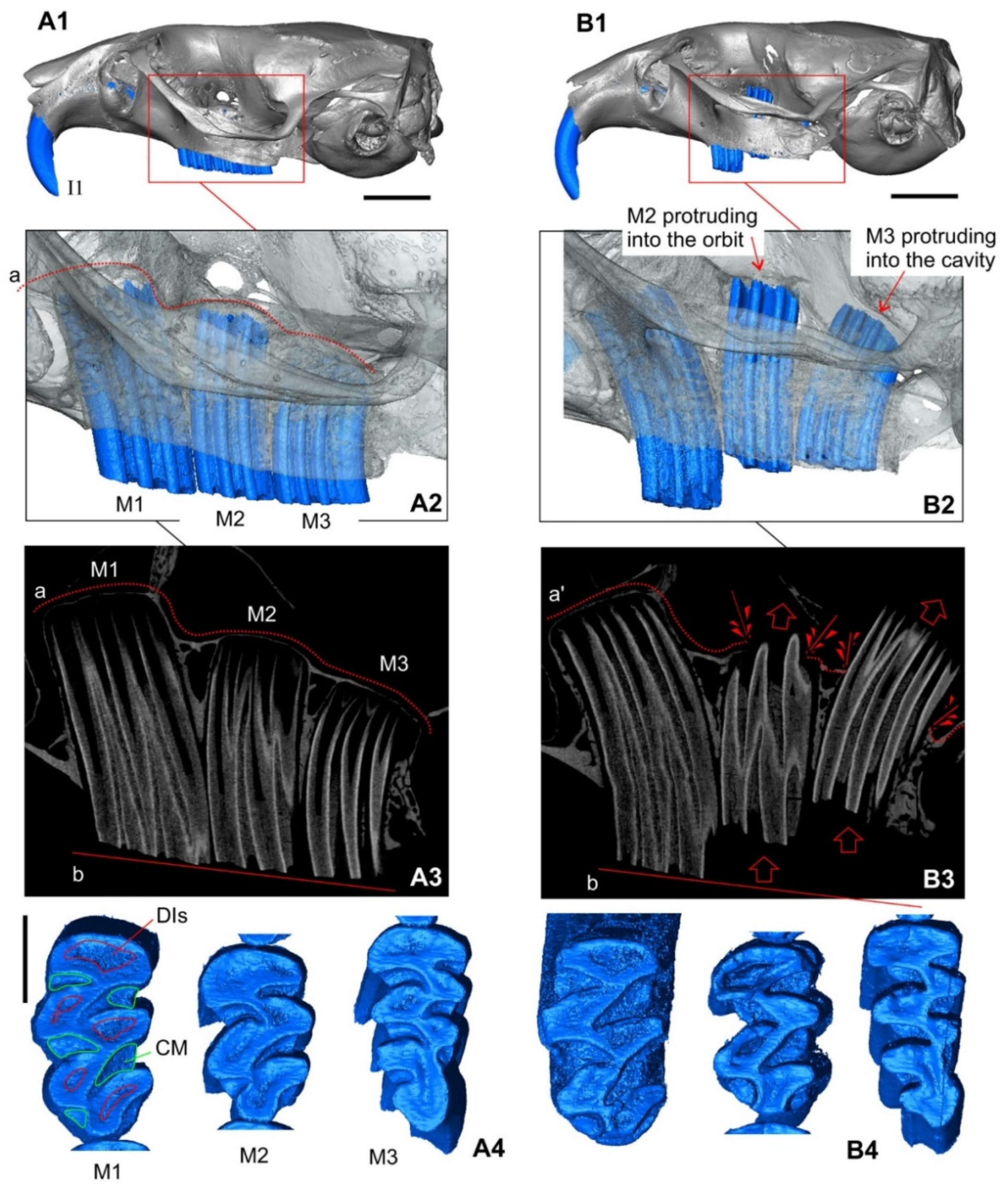
3.1.3. The Skull: Visual Comparisons and Computed Microtomographic Analysis

The visual comparison was performed on two age groups: (i) M. h. ankaraensis and M. hartingi (founders from the Eastern Rhodopes) at 1 month of age; and (ii) M. h. ankaraensis, M. hartingi (from the Eastern Rhodopes), and M. h. strandzensis at 5 months of age. The comparison revealed clear-cut differences between conventionally “normal” animals (judging by the condition of the skull, hemimandibles, and molars)—represented by M. h. ankaraensis and M. h. strandzensis—and Rhodopean animals considered “abnormal” in terms of the thickness of skull bones and symmetry of the skull. The skulls of 1-month-old voles from the Rhodopes have thinner and more friable bones as compared to age-matched voles of other subspecies (Figure 3).
The 5-month-old specimens from the Rhodopes have well-pronounced abnormality of the molar tooth row; namely, the second upper molar crown is lower by half than the first molar, and the third molar slightly protrudes at the alveolus margin or is fully submerged in the alveolus (Figure 4B1).
A preliminary analysis of the mature voles from the natural population of M. hartingi from the Eastern Rhodopes suggested strong aberrations in the development of the second and third upper molars because most the caught voles seemed to be “devoid of the third upper molar”. Computed microtomography revealed more or less normal development of upper molars, but there were pathological changes in the roof of the alveoli. The alveolar roof gradually becomes thinner and, apparently, at some point, breaks under the tooth pressure. In Figure 4, one can see the position of the apical part of M2 in orbits and M3 in the brain cavity (Figure 4B2,B3). Therefore, the second and third molars retain normal dimensions of the crown, and the effect of “abnormal tooth development” is linked with the thinness of bones. In addition, the flat worn occlusal surface of upper molars implies that the teeth had been engaged in regular chewing activity before the break in the alveolar roof (Figure 4B4). On the other hand, we can see moderate changes in the molar condition of M. hartingi from the Rhodopes in comparison with the teeth of M. h. ankaraensis or M. h. strandzensis. This change is supposedly related to disturbances of calcium metabolism, such as weak development of the cementum (friable consistency and low position along the enamel prisms) and friable condition of the dentine when the dentine islets are deep.
The analysis of the outer morphology of hemimandibles and of the lower molar row of 1-month-old animals did not reveal any abnormality in the specimen from M. hartingi Rhodopean founders, as was the case for both hemimandibles’ and molars’ proportions in 5-month-old specimen ZIN 107168-11. Another specimen, ZIN 107168-10, has an abnormal right first molar with unusual crown morphology and, correspondingly, an altered lower margin of the dentary (Figure 5B).
This molar has a swollen inflated lower part of the crown with a loss of prismatic structure (Figure 5B5) and a regular upper part with the flattened worn occlusal surface (Figure 5B4). Hence, we can theorize that changes in the functioning of the enamel tissue occurred at the mid-level of the m1 crown during maturation of the animal.

3.1.4. The Baculum

The baculum (os penis) of M. hartingi from the Eastern Rhodopes is much larger than that of M. hartingi from either the Asian part of Turkey or the European part of its range [15,16,17] (Figure 6, Table 3).

3.2. Analysis of Cytb Gene Variation

Across a 1140-bp-long alignment of the ingroup (excluding M. arvalis and M. rossiaemeridionalis), 282 nucleotide sites were found to be variable and 224 parsimony-informative.
Phylogenetic reconstruction based on Cytb sequences (Figure 7) revealed that two specimens of M. hartingi from the Eastern Rhodope (Bulgaria) form a separate branch, which is a sister clade to the clade of voles from northeastern Greece (also from the foothills of the Rhodopes, Paranesti), albeit with low support.
Based on calculated p-distances, M. hartingi individuals from the Eastern Rhodopes and from Northeastern Greece are equally different both from the European form M. h. hartingi and from the Asian one: M. h. ankaraensis (Table 4). This result suggests that populations from Southwestern Bulgaria and Northeastern Greece have stayed isolated for a long time and already have their own long evolutionary history.

3.3. Experimental Hybridization

It turned out that M. hartingi individuals from the Eastern Rhodopes easily produce hybrid offspring with M. h. ankaraensis as sexual partners and that the intensity of reproduction is high both in direct and reciprocal crosses. At the same time, the litters were large, but the viability of the young was noticeably lower. Offspring deaths occurred up to 45 days of age, but more often in the first 2 weeks of life. Mortality of hybrid pups was higher as compared to conspecific crosses of M. h. ankaraensis (by 4.4-fold) and of Rhodopean M. hartingi (by 9.5-fold; Table 5).
In backcrosses of ♂ M. h. ankaraensis with hybrid females, reproduction was also successful. Pup mortality was negligible if F1 females came from a cross of ♂ M. h. ankaraensis with ♀ M. hartingi; however, this parameter was 3.8 times higher in the opposite direction. F1 hybrid males rarely mated with M. h. ankaraensis females. The males of Rhodopean M. hartingi, when crossed with F1 females, bred successfully, and almost all pups survived. In contrast, F1 males, when crossed with M. hartingi females, produced offspring, but more than 50% of the backcrosses did not survive. The probability of reproduction of F1 hybrids among themselves depended on who their parents were. In case of ♂ A × ♀ H, 58.8% of pairs bred, whereas in the opposite direction, only 45.5% did. Pup mortality was 22.5% and 3.3%, respectively. Some F2 hybrids and backcrosses also bred among themselves, but only half of the pairs had offspring.
We identified male sterility by the absence of spermatozoa in the epididymis. The absence of spermatozoa in hybrids was noted in all types of crossing. After direct and reciprocal crosses of ♂ M. h. ankaraensis × ♀ M. hartingi, the rate of sterility of F1 hybrid males was 38% (8 out of 21 and 11 out of 29, respectively). The sterility of F2 males was just as high, especially if they derived from a cross of ♂ M. h. ankaraensis with ♀ M. hartingi (38.5%), but was considerably lower in reciprocal crosses (14.3%). The sterility of backcross males was high in both directions: 58.3% (♂ A × ♀ H) and 61.1% (♂ H × ♀ A). The highest sterility of males was noted in the third generation (♂ H × ♀ A): 71%. No experiments were conducted with the second crossing type owing to high sterility of F2 males and their low capacity for reproduction.

4. Discussion

According to mitochondrial genome analysis, differentiation of the genus Microtus into two subgeneric clades, Microtus s.str. and Sumeriomys, occurred approximately 1.87 million years ago (Mya), and the division of the latter into two groups “socialis” and “guentheri” took place approximately 1 Mya [20]. According to molecular analysis of only the Cytb gene in ref. [21], the time point of divergence between voles of the “socialis” group and ancestors of the “guentheri” group is 0.9 Mya (range: 0.6–1.2), in good agreement with the findings reported by Abramson et al. (2021) [20]. Within the latter group, a split occurred approximately 0.6 Mya (0.4–0.9), whereas M. hartingi diverged from a sister clade 0.4 Mya (0.2–0.7). According to other authors [15], who also analyzed the Cytb gene, the separation of European forms of Thracia M. hartingi from Asian forms of Anatolia M. hartingi happened ~0.73 Mya.
Paleontological data on the subgenus Sumeriomys are scarce. Microtus (Pallasiinus) praeguentheri Kretzoi, 1977 was found in a cave near Petralona (Greece). Presumably, a vole—Microtus bifrons—from the subgenus Sumeriomys from the Upper Pleistocene of southern France was described [22]. M. guentheri specimens were found in Middle–Late Pleistocene cave deposits in Israel (Qumm Qatafa) [23] and in Upper Pleistocene deposits of the Üçağızlı cave in southeastern Turkey [24]. There are interesting finds of voles identified as M. guentheri in the Middle Pleistocene deposits of Chios Island (Greece) [25] and the Yarimburgaz Cave in Thracia (Turkey) [26]. Apparently, these are the most ancient finds of M. hartingi. It should be pointed out that although M. hartingi went extinct on the island of Chios, this vole has survived to this day on Lesbos Island [21,27].
The European and Asian forms of Harting’s vole are separated from each other by the Sea of Marmara and by narrow straits—the Bosporus and the Dardanelles—which opened in the early Pleistocene (~2 Mya) and then closed repeatedly [28,29,30,31]. Harting’s vole entered southeastern Europe from Anatolia via a land corridor, beginning in the Middle Pleistocene [32]. Evidently, M. hartingi populations entered Europe from Anatolia several times, as evidenced by the rather complex subdivision into clades of the European part of the dendrogram. One of the first populations arose in the Rhodope Mountains (southwestern Bulgaria and northeastern Greece). During an ice age (24,000–17,000 years ago), steppes were less common in the Balkans than in Eastern Europe owing to its geographical location and more continental climate [33]. Later (<12.4 to ≥10.9 KYR B.P.), a Mediterranean mountain forest complex formed in the Balkans. Xerophytic forests of the Mediterranean (southwestern Balkans) alternated with areas of cereal–wormwood steppes [34]. The complex phylogeographic structure of M. hartingi is explained by the biogeographic barriers (the Pindus Mountains to the west, Stara Planina to the north, and the Isthmus of Corinth to the south) and by geographic range fragmentation due to the expansion of the forest zone in the European part of its geographic range. One of the fragments is northeastern Greece transitioning to the Bulgarian Rhodopes. From our point of view, this largest fragment could have emerged during the first period of colonization of Europe by Harting’s vole. It is bounded by the Maritsa (also known as Maritza) river. Geological formation of its basin began in the Paleogene and actively continued in the Neogene and Quaternary (http://www.inweb.gr/workshops2/sub_basins/13_14_15_Evros_Ardas_Ergene.html, accessed on 26 November 2021).
The filling of the geological channel with water flowing from the Rila Mountains could have already occurred in the Pleistocene [34]. Until that moment, Harting’s vole could penetrate Europe and reach Greece and the Rhodopes, but the formation of the river made it difficult to recolonize this region; therefore, Harting’s vole in this part of the geographic range can be regarded as the most ancient form.
In the Rhodopes, a special situation could have developed, different from that in Greece. Thessaly was an agricultural zone for many thousands of years of the Neolithic, while in the Rhodopes, only isolated areas where the vole could breed were preserved, isolated by slopes of mountains covered with forests. Intensive agriculture, apparently, was never practiced there. Therefore, the Rhodopean population was probably preserved in a small isolated area. Other very small fragments of the European range of M. hartingi persisted in north Macedonia and Serbia and also appear to be ancient isolates.
Due to the isolation of the vole population in the Rhodopes, the well-pronounced fragmentation of biotopes [35,36], and the lack of vacant territories for dispersal, these rodents encountered extreme conditions that enhanced the impact of stress on voles. The habitat conditions also contributed to inbreeding, which evidently has a fairly long history. Some experimental data show high tolerance of Rhodopean voles to inbreeding in comparison with M. h. ankaraensis, among which a taboo on incest was noted [9]. It is known that when a certain level of homozygosity is reached, communal breeding becomes common [37]. Rhodopean voles have a high propensity for communal breeding [9]. According to the hypothesis of Joly [38], inbreeding is the most important factor in microevolution and speciation.
We found that in the Rhodopean population of M. hartingi, specific disturbances of the growth and development of the skull and dental system have taken place probably due to anomalous phosphorus–calcium metabolism leading to the thinning of skull bones and then to maxillary-bone perforation during the growth of molars. It should be mentioned that despite such serious disturbances, voles continue to eat normally and live to adulthood and even to the senex stage (>1 year of age). Tooth growth disorders—molar apical elongation—have been documented in many voles of laboratory colonies: Mynomes ochrogaster [39], Mynomes californicus scirpensis [40], Mynomes pinetorum [41], Lagurus lagurus [42], and Alexandromys montebelli [43]. Nonetheless, the cause of these aberrations was an incorrectly selected diet for keeping laboratory colonies of voles. The lack of branch forage or simply pieces of wood for grinding down the ever-growing teeth results in molar apical elongation. We are not aware of such problems in voles of natural populations. In our study, the thinning of skull bones was observed due to an anomaly of phosphorus–calcium metabolism (not disturbances of uniform abrasion of the occlusal surface of teeth), causing molar apical elongation. Moreover, when bred under laboratory conditions, in individuals from natural populations of Rhodopean voles and in their descendants, there are alterations in the growth of incisors, an increase and deformation of the upper incisors, and a decrease in the lower incisors (Zorenko, nonpublished data). Disturbances in synchrony of the growth and development of teeth may be either a consequence of stress under extreme living conditions [9] or the result of inbreeding. Elucidating this issue requires further investigation. An interesting question concerns the adaptive value of the structure of teeth. To date, in explanations of evolutionary reasons for the complication of voles’ teeth, one can often see speculation about direct adaptive significance of the emergence of additional loops and protruding corners in teeth for the better grinding of food. As we see in M. hartingi from the Eastern Rhodopes, whole teeth (M2 and M3) are excluded from the functioning of the dentition, and yet this drawback does not prevent the animals from eating. It must be pointed out that for M. hartingi from northeastern Greece, no abnormalities in the structure of the teeth and skull were detected [44], although, according to Cytb analysis, this is a sister population to the Rhodopean population.
We also noticed the absence of calcification of lateral processes in the os penis. Usually, in voles of the genus Microtus, ossification of the medial and lateral processes ends by the puberty period of males (60–70 days of age) [45].
According to our data on experimental hybridization, M. hartingi from the Rhodopes is partially reproductively isolated from M. h. ankaraensis, as revealed by a lower proportion of hybrid pairs that started breeding, a relatively high mortality rate among the young, and the sterility of some hybrid males. On the other hand, hybridization between Rhodopean and Anatolian voles is much more successful than that between M. h. strandzensis from Strandzha and M. h. ankaraensis. Overall, for all crossing options, in the former case, 86.5% of pairs breed, while in the latter, only 43.7% do [5]. Mortality of hybrid offspring in the former case is 2.2 times lower than in the latter (25.5% and 55.7%, respectively). After the hybridization of voles from the Rhodopes and Anatolia, in all types of crossing, males were born (~40%) in which there were no spermatozoa in the epididymis of the testicles. Among hybrid males from the crossing of M. h. strandzensis with M. h. ankaraensis, the proportion of sterile males is greater: 60% [5]. In some males, we observed only a few sedentary spermatozoa present, which also explains the absence of reproduction in pairs of these voles. These findings imply an early disturbance of spermatogenesis in the hybrids and incomplete development of spermatozoa. The structure of spermatozoa in the hybrids may be abnormal, as well: tail deformation (twisting or angular deformation) and a higher proportion of spermatozoa without a head. These changes can lead to the loss of the abilities to fertilize and to advance in the female genital tract [46].
Evidently, between European and Asian forms of M. hartingi, reproductive isolation was established gradually, as in the case of voles of the “mystacinus” group of the subgenus Microtus, genus Microtus [47]. In an ancient and isolated population of M. hartingi from the Rhodopes, the isolation effect may have been a result of long term uninterrupted isolation, whereas in the southeastern Balkans, there have been repeated periods of colonization by M. hartingi from Asia Minor [35,48].
It should also be mentioned that the Rhodopean population has some distinct features of the X chromosome’s structure: in Rhodopean voles, the X-chromosome is acrocentric, whereas in M. h. strandzensis, it is submetacentric [6]. According to our data [17,49] in M. h. strandzensis, the X chromosome is subtelocentric, whereas in M. h. lydius from Isparta, this chromosome is acrocentric [16]. In general, M. hartingi is characterized by X chromosome polymorphism [50].

5. Conclusions

Thus, the results of our integrative analysis revealed high differentiation of unique Eastern Rhodopean M. hartingi in terms of morphological structure, variation in the Cytb gene, and the degree of reproductive isolation from M. h. ankaraensis. The uniqueness of the Rhodopean population of M. hartingi can be explained by the “bottleneck” effect. During the first colonization of Europe, M. hartingi probably fell into a kind of mountain “trap”, in which there were few suitable biotopes for settlement and reproduction and, as a consequence, the number of animals could be extremely small. The small population size, the stressful conditions, and inbreeding led to a violation of the development of the skull and teeth. Significant changes were found in the development of the skull and teeth, which arose as a result of a violation of phosphorus–calcium metabolism. The population could have gone through a “bottleneck”. Under the conditions of the mountain trap, two scenarios could ensue. One was likely to lead to the extinction of the population. Under the second scenario, the population could recover by adapting to the unfavorable conditions. It is this outcome that happened to the population of M. hartingi in the Eastern Rhodopes. For several thousand years, these voles have maintained good population size and successful reproduction. As noted above, communal reproduction and inbreeding tolerance have become the most important adaptations of this species under adverse conditions [8]. Inbreeding probably had a positive influence on the population because inbreeding eliminates harmful alleles, thereby possibly making the species stable even at low genetic diversity.
All the detected differences between the Rhodopean vole and other nominative forms of M. hartingi cannot be interpreted taxonomically because available data are still insufficient for comparing this form with the sister group of haplotypes from northeastern Greece to substantiate subspecies status; the degree of reproductive isolation is insufficient to validate species status.
In the future, it is necessary to recognize M. hartingi from the Bulgarian Rhodopes as an endangered population (EN) and to include it in regional Red Lists of Bulgaria.

Supplementary Materials

The following supporting information can be downloaded at: https://www.mdpi.com/article/10.3390/d14090709/s1, Table S1: The material of M. hartingi used in the article.

Author Contributions

Conceptualization, F.N.G. and T.A.Z.; methodology, F.N.G., T.A.Z. and L.L.V.; formal analysis, F.N.G., T.A.Z., L.L.V., T.V.P. and L.Y.K.; writing—original draft preparation, F.N.G., T.A.Z., L.L.V. and T.V.P.; writing—review and editing, F.N.G. and T.A.Z.; funding acquisition, F.N.G.; collecting material on voles in the field, T.A.Z., N.A. and F.N.G. All authors have read and agreed to the published version of the manuscript.

Funding

This work has been supported by grants from the Russian Science Foundation, RSF № 22-24-00782 (FNG).

Institutional Review Board Statement

The animal study protocol was approved by the Ethics Committee of Zoological Institute, Russian Academy of Sciences, Universitetkaya Emb. 1, Saint Petersburg 199034, Russia (protocol N 1-7/10-08-2022 code dated 10 August 2022) for studies involving animals.

Informed Consent Statement

Not applicable.

Data Availability Statement

Not applicable.

Acknowledgments

We thank Nuri Yiğit and Derya Çetintürk (Biology Department, Faculty of Science, Ankara University, Turkey) for providing us with the sequences of M. hartingi strandzensis from Turkish Thrace. Authors are grateful to Vladimir Malikov and Svetlana Sablina (Laboratory of Theriology, Zoological Institute, Russian Academy of Sciences, St. Petersburg, Russia) for their help in keeping and breeding voles.

Conflicts of Interest

The authors declare no conflict of interest.

References

  1. Kryštufek, B.; Bužan, E.V.; Vohralík, V.; Zareie, R.; Özkan, B. Mitochondrial cytochrome b sequence yields new insight into the speciation of social voles in south-west Asia. Biol. J. Linn. Soc. 2009, 98, 121–128. [Google Scholar] [CrossRef]
  2. Yiğit, N.; Çolak, E. On the distribution and taxonomic status of Microtus guentheri (Danford and Alston; 1880) and Microtus lydius Blackler; 1916 (Mammalia: Rodentia) in Turkey. Turk. J. Zool. 2002, 26, 197–204. [Google Scholar]
  3. Golenishchev, F.N.; Abramson, N.I. New data on phylogeography of voles’ subgenus Sumeriomys (Arvicolinae; Rodentia). In Proceedings of the International Conference “Mammals of Russia and Adjacent Territories” IX Congress of Theriological Society RAS, Moscow, Russia, 1–4 February 2011; KMK Scientific Press: Moscow, Russia, 2011; p. 117. (In Russian). [Google Scholar]
  4. Golenishchev, F.N.; Malikov, V.G. The systematics and distribution of the grey voles of the tribe Microtini (Rodentia; Arvicolinae) in the Caucasus and Asia Minor. In Proceedings of the International Conference “Biological Diversity and Conservation Problems of the Fauna of the Caucasus”, Yerevan, Armenia, 26–29 September 2011; LLC Spika: Yerevan, Armenia, 2011; pp. 101–105. (In Russian). [Google Scholar]
  5. Zorenko, T.A.; Atanasov, N.; Golenishchev, F.N. Behavioral differentiation and hybridization of the European and Asian forms of Harting’ vole Microtus hartingi (Rodentia; Arvicolinae). Russian J. Theriol. 2016, 15, 133–150. [Google Scholar] [CrossRef]
  6. Chassovnikarova, T.G.; Markov, G.G.; Atanasov, N.I.; Dimitrov, H.A. Sex chromosome polymorphism in Bulgarian populations of Microtus guentheri (Danford & Alston; 1880). J. Nat. Hist. 2008, 42, 261–267. [Google Scholar] [CrossRef]
  7. Zorenko, A.; Kagainis, U.; Baraškova, Ļ. Does the geometric and linear morphometry of the brain reflect the divergence in the “guentheri” group (Arvicolinae; Sumeriomys). Russian J. Theriol. 2020, 19, 45–57. [Google Scholar] [CrossRef]
  8. Zorenko, T.A.; Kagainis, U. Discrimination of the two subspecies of Microtus hartingi (Rodentia: Arvicolinae) by shape and linear features of the spermatozoon. Russian J. Theriol. 2021, 21, 143–157. [Google Scholar] [CrossRef]
  9. Zorenko, T.A. Communal reproduction of females of two subspecies of Harting’s vole; Microtus (Sumeriomys) hartingi (Rodentia; Arvicolinae); under experimental conditions. Zool. J. 2022, 101, 1048–1060. (In Russian) [Google Scholar]
  10. Kryštufek, B.; Shenbrot, G.I. Voles and Lemmings (Arvicolinae) of the Palaearctic Region; Maribor University Press: Maribor, Slovenia, 2022; p. 436. [Google Scholar] [CrossRef]
  11. Hammer, Ø.; Harper, D.A.T.; Ryan, P.D. PAST: Paleontological statistics software package for education and data analysis. Palaeontol. Electron. 2001, 4, 9. [Google Scholar]
  12. Abramson, N.I.; Lebedev, V.S.; Tesakov, A.S.; Bannikova, A.A. Supraspecies relationships in the subfamily Arvicolinae (Rodentia, Cricetidae): An unexpected result of nuclear gene analysis. Mol. Biol. 2009, 4, 834–846. [Google Scholar] [CrossRef]
  13. Hall, T.A. BioEdit: A user-friendly biological sequence alignment editor andanalysis program for Windows 95/98/NT. Nucleic Acids Symp. 1999, 41, 95–98. [Google Scholar] [CrossRef]
  14. Kumar, S.; Stecher, G.; Tamura, K. MEGA7: Molecular Evolutionary Genetics Analysis version 7.0 for bigger datasets. Mol. Biol. Evol. 2016, 33, 1870–1874. [Google Scholar] [CrossRef] [PubMed]
  15. Yiğit, N.; Çetintürk, D.; Çolak, E. Phylogenetic assessment of voles of the Guentheri Group (Mammalia: Microtus) in Turkish Thrace and Western Anatolia. Eur. Zool. J. 2017, 84, 252–260. [Google Scholar] [CrossRef] [Green Version]
  16. Ronquist, F.; Teslenko, M.; van der Mark, P.; Ayres, D.L.; Darling, A.; Höhna, S.; Larget, B.; Liu, L.; Suchard, M.A.; Huelsenbeck, J.P. MrBayes 3.2: Efficient Bayesian Phylogenetic Inference and Model Choice Across a Large Model Space. Syst. Biol. 2012, 61, 539–542. [Google Scholar] [CrossRef] [PubMed]
  17. Golenishchev, F.N.; Sablina, O.V.; Borodin, P.M.; Gerasimov, S. Taxonomy of voles of the subgenus Sumeriomys Argyropulo; 1933 (Rodentia; Arvicolinae; Microtus). Russ. J. Theriol. 2002, 1, 43–55. [Google Scholar] [CrossRef]
  18. Tabur, M.A.; Uğur, E.D.; Albayrak, I. Taxonomical and Karyological Features of Microtus hartingi (Barrett-Hamilton; 1903) (Mammalia: Rodentia) with Some Biological and Ecological Features. Pakistan J. Zool. 2015, 47, 691–698. Available online: http://acikerisim.sdu.edu.tr/xmlui/handle/123456789/62813 (accessed on 26 November 2021).
  19. Yiğit, N.; Markov, G.; Colak, E.; Kocheva, M.; Saygili, F.; Yuce, D.; Cam, P. Phenotypic features of the “guentheri” group Vole (Mammalia: Rodentia) in Turkey and Southeast Bulgaria: Evidence for its taxonomic detachment. Acta Zool. Bulg. 2012, 64, 23–32. [Google Scholar]
  20. Abramson, N.I.; Bodrov, S.Y.; Bondareva, O.V.; Genelt-Yanovskiy, E.A.; Petrova, T.V. A mitochondrial genome phylogeny of voles and lemmings (Rodentia: Arvicolinae): Evolutionary and taxonomic implications. PLoS ONE 2021, 16, e0248198. [Google Scholar] [CrossRef]
  21. Thanou, E.; Paragamian, K.; Lymberakis, P. Social but lonely: Species delimitation of social voles and the evolutionary history of the only Microtus species living in Africa. J. Zoolog. Syst. Evol. 2019, 58, 475–498. [Google Scholar] [CrossRef]
  22. Jeannet, M.; Fontana, L. Microtus (Sumeriomys) bifrons nov. sp. (Rodentia; Mammalia); a new vole in the French Upper Pleistocene identified at the Petits Guinards site (Creuzier-le-Vieux; Allier; France). Paleo Rev. D’archéologie Préhistorique 2015, 26, 59–77. [Google Scholar] [CrossRef]
  23. Tchernov, E. Succession of Rodent Faunas during the Upper Pleistocene of Israel. In Mammalia Depicta; Parey: Berlin, Germany, 1968; p. 152. [Google Scholar]
  24. Alpaslan, F.S. Some small mammal fossils of Üçağızlı Cave (Hatay; Turkey). Turk. J Zool. 2011, 35, 755–768. [Google Scholar] [CrossRef]
  25. Storch, G. Eine mittelpleistozäne Nager-Fauna von der Insel Chios, Ägäis (Mammalia: Rodentia). Senckenbergiana Biol. 1975, 56, 165–189. [Google Scholar]
  26. Santel, W.; Von Koenigswald, W. Preliminary report on the middle Pleistocene small mammal fauna from Yarimburgaz Cave in Turkish Thrace. EG Quat. Sci. J. 1998, 48, 162–169. [Google Scholar] [CrossRef]
  27. Masseti, M. Mammals of the Mediterranean islands: Homogenisationand the loss of biodiversity. Mammalia 2009, 73, 169–202. [Google Scholar] [CrossRef]
  28. Algan, O.; Çaǧatay, N.; Tchepalyga, A.; Ongan, D.; Eastoe, C.; Gökaşan, E. Stratigraphy of the Sediment Infil in Bosporus Strait: Water Exchange between the Black and Mediterranean Seas during the Last Glacial Holocene. J. Geophys. Res. 2001, 20, 209–218. [Google Scholar] [CrossRef]
  29. Çağatay, M.N.; Görür, N.; Algan, O.; Eastoe, C.; Tchapalyga, A.; Ongan, D.; Kuhn, T.; Kuşçu, I. Late Glacial-Holocene paleoceanography of the Sea of Marmara: Timing of connections with the Mediterranean and the Black seas. Mar. Geol. 2000, 167, 191–206. [Google Scholar] [CrossRef]
  30. Genov, I. Model of palaeoenvironmental evolution of the Black Sea region during the last glacial maximum—Holocene. Oceanology 2009, 49, 540–557. [Google Scholar] [CrossRef]
  31. Krijgsman, W.; Tesakov, A.; Yanina, T.; Lazarev, S.; Danukalova, G.; Van Baak, C.G.C.; Agustí, J.; Alçiçek, M.C.; Aliyeva, E.; Bista, D.; et al. Quaternary time scales for the Pontocaspian domain: Interbasinal connectivity and faunal evolution. Earth-Sci. Rev. 2019, 188, 1–40. [Google Scholar] [CrossRef]
  32. McHugh, C.M.G.; Gurung, D.; Giosan, L.; Ryan, W.B.F.; Mart, Y.; Sancar, U.; Buckles, L.; Çagatay, M.N. The last reconnection of the Marmara Sea (Turkey) to the World Ocean: A paleoceanographic and paleoclimatic perspective. Mar. Geol. 2008, 255, 64–82. [Google Scholar] [CrossRef]
  33. Markova, A.K.; van Kolfschoten, T.; Bohncke, S.J.P.; Kosintsev, P.A.; Mol, J.; Puzachenko, A.Y.; Simakova, A.N.; Smirnov, N.G.; Verpoorte, A.; Golovachev, I.B. Evolution of the European Ecosystems during Pleistocene—Holocene Transition (24–8 kyr BP); GEOS: Moscow, Russia, 2019; p. 279. [Google Scholar]
  34. Okay, N.; Okay, A. Tectonically induced Quaternary drainage diversion in the northeastern Aegean. J. Geol. Soc. 2002, 159, 393–399. [Google Scholar] [CrossRef]
  35. Markov, G.; Dimitrov, H. Habitat fragmentation and its implications for abundance of Guenther’s vole in southeastern Bulgaria (Strandzha mountain region). Biotechnolology Biotechnol. Equip. 2010, 24, 679–682. [Google Scholar] [CrossRef]
  36. Kryštufek, B.; Zorenko, T.; Bontzorlos, V.; Mahmoudi, A.; Atanasov, N.; Ivajnšič, D. Incipient road to extinction of a keystone herbivore in south-eastern Europe: Harting’s vole (Microtus hartingi) under climate change. Clim. Chang. 2018, 149, 443–456. [Google Scholar] [CrossRef]
  37. Lacey, E.A.; Sherman, P.W. Cooperative breeding in naked mole-rats: Implications for vertebrate and invertebrate sociality. In Cooperative Breeding in Mammals; Solomon, N.G., French, J.A., Eds.; Cambridge University Press: Cambridge, UK, 1997; pp. 267–301. [Google Scholar] [CrossRef]
  38. Joly, E. Essay: On the close relationship between speciation; inbreeding and recessive mutations. Nat. Preced. 2010. [Google Scholar] [CrossRef]
  39. Jheon, A.H.; Prochazkova, M.; Sherman, M.; Manoli, D.S.; Shah, N.M.; Carbone, L.; Klein, O. Spontaneous emergence of overgrown molar teeth in a colony of Prairie voles (Microtus ochrogaster). Int. J. Oral Sci. 2015, 7, 23–26. [Google Scholar] [CrossRef] [PubMed]
  40. Imai, D.M.; Pesapane, R.; Conroy, C.J.; Alarcon, C.N.; Allan, N.; Okino, R.A.; Fung, J.; Murphy, B.G.; Verstraete, F.J.M.; Foley, J.E. Apical elongation of molar teeth in captive Microtus voles. Vet. Pathol. 2018, 55, 572–583. [Google Scholar] [CrossRef] [PubMed] [Green Version]
  41. Harvey, S.B.; Alworth, L.C.; Blas-Machado, U. Molar malocclusions in pine voles (Microtus pinetorum). Am. Assoc. Lab. Anim. Sci. 2009, 48, 412–415. [Google Scholar]
  42. Imbschweiler, I.; Schauerte, N.; Henjes, C.; Fehr, M.; Baumgärtner, W. Odontogenic dysplasia in the molar teeth of steppe lemmings (Lagurus lagurus). Vet. J. 2011, 188, 365–368. [Google Scholar] [CrossRef]
  43. Sugita, S.; Uchiumi, O.; Fujiwara, K.; Shumpei, N.S.; Katsuhiro, F.K. Brain deformation caused by hyperplasia molar teeth (macrodonts) in the Japanese field vole (Microtus montebelli). Exp. Anim. 1995, 43, 769–772. [Google Scholar] [CrossRef]
  44. Fraguedakis-Tsolis, S.E.; Chondropoulos, B.P.; Stamatopoulos, C.V.; Giokas, S. Morphological variation of the five vole species of the genus Microtus (Mammalia; Rodentia; Arvicolinae) occurring in Greece. Acta Zool. 2009, 90, 254–264. [Google Scholar] [CrossRef]
  45. Aksenova, T.G. Comparative morphological analysis of the baculum structure of the voles from tribe Microtini (Rodentia, Cricetidae). Communication 1. Genus Lasiopodomys, Chionomys, Microtus (subgenus Microtus). Tr. Zool. Inst. RAN 1980, 99, 62–77. (In Russian) [Google Scholar]
  46. Zhabin, S.G.; Trechenkov, E.A.; Artifeksov, S.B.; Pavlenko, I.I.; Polukarov, A.N. Comparative evaluation of sperm DNA fragmentation and other sperm parameters. Probl. Reproduktsii 2015, 21, 121–124. [Google Scholar] [CrossRef]
  47. Bikchurina, T.I.; Golenishchev, F.N.; Kizilova, E.A.; Mahmoudi, A.; Borodin, P.M. Reproductive isolation between taxonomically controversial forms of the gray voles (Microtus, Rodentia; Arvicolinae): Cytological mechanisms and taxonomical implications. Front. Genet. 2021, 12, 653837. [Google Scholar] [CrossRef] [PubMed]
  48. Thanou, E.; Tryfonopoulos, G.; Chondropoulos, B.; Fraguedakis-Tsolis, S. Comparative phylogeography of the five Greek vole species infers the existence of multiple South Balkan subrefugia. Ital. J. Mammal. 2012, 79, 363–376. [Google Scholar] [CrossRef]
  49. Golenishchev, F.N.; Malikov, V.G.; Nazari, F.; Vaziri, S.; Sablina, O.V.; Polyakov, A.V. New species of vole of “guentheri” group (Rodentia; Arvicolinae; Microtus) from Iran. Russ. J. Theriol. 2003, 1, 117–123. [Google Scholar] [CrossRef]
  50. Zima, J.; Arslan, A.; Benda, P.; Macholán, M.; Kryštufek, B. Chromosomal variation in social voles: A robertsonian fusion in Günther’s vole. Acta Theriol. 2013, 58, 255–265. [Google Scholar] [CrossRef]
Figure 1. Distributional range of Harting’s social vole Microtus hartingi. The basis of the map is according to [10]. A: Part of Europe; B: Balkan and East Turkey. Colored circles mark the type of locality subspecies and M. hartingi from the Eastern Rhodopes. Abbreviations: brown dot, mrt, M. h. martinoi; gray dot, hrt, M. h. hartingi; red dot, rd, M. hartingi from the Eastern Rhodopes; green dot, lyd, M. h. lydius; blue dot, st, M. h. strandzensis; violet, ank, M. h. ankaraensis.
Diversity 14 00709 g001
Figure 2. The PCA plot for M. hartingi (the 1st axis versus the 2nd axis). Abbreviations: A, blow dots, M. h. ankaraensis; H, red dots, M. h. hartingi; L, green dots, M. h. lydius; M, brown dots, M. h. martinoi; R, violet dots, M. hartingi from the Eastern Rhodope Mountains; and S, black dots, M. h. strandzensis. Along the top and right margins of the plot, measurement abbreviations are displayed with values of the relation between a parameter and the axes.
Diversity 14 00709 g002
Figure 3. Skulls of 1-month-old M. h. ankaraensis (A) and M. hartingi from the Eastern Rhodopes (B) in lateral (A1,B1) and ventral (A2,B2) views. Abbreviations. a: the friable part of the maxilla (light blue outline; thin and friable bone lost through skeletonization by beetles); b: the friable part of the frontal and around the lacrimal bones; c: thin frontal bone with “fenestra”; d: a thin outer part of the tympanic bullae; e: a thin part of the occipital bone; f: a friable part of the vomer; g: a friable part of the palatine; h, i: friable parts of the occipital bone; j: the red line shows a continuous profile of the molar row. The scale bar is 5 mm.
Diversity 14 00709 g003
Figure 4. 3D models of the skulls and upper molars (M1, M2, M3) of M. h. ankaraensis (ZIN 98971; A) and M. hartingi (ZIN 107168-10 from the Eastern Rhodopes; B). (A1,B1) Models of skulls in lateral view. (A2,B2) Models of the orbital part of skulls in transparent view showing the molars’ positions. (A3,B3) X-ray images of molars in parasagittal cut view. (A4,B4) Models of upper molars in occlusal view. Abbreviations. a: the red dotted line indicates the approximate position of the alveolar roof under usual conditions; a’: broken alveolar roof; b: occlusal profile of the molar row; CM: cementum; DIs: dentine islet; red wide arrows: the direction of molars’ submersion. The scale bars are 5 mm (A1,B1) and 1 mm (A4,B4).
Diversity 14 00709 g004
Figure 5. 3D models of the hemimandibles and lower molars (m1, m2, m3) of M. h. ankaraensis (ZIN 98971; A) and M. hartingi (ZIN 107168-10 from the Eastern Rhodopes; B). (A1,B1) Models of the right hemimandibles in medial view. (A2,B2) Models of the dentary part in transparent view showing molars’ positions. (A3,B3) Models of isolated molars in medial view. (A4,B4) Models of lower molars in occlusal view. (B5) An X-ray image of the first molar in parasagittal cut view. Abbreviations. a: the red line shows the approximate border between “normal” prismatic structure of the crown and an “abnormal” part of the crown that lost prismatic structure; b: the red dotted line indicates the part without prisms. The scale bar is 5 mm.
Diversity 14 00709 g005
Figure 6. M. hartingi bacula. A: M. hartingi from the Eastern Rhodopes (A1: ZIN 107168-47, A2: ZIN 107168-26), B: M. h. strandzensis [17], C: M. h. lydius [18], and D: M. h. ankaraensis [19].
Diversity 14 00709 g006
Figure 7. The BI tree built using a Cytb fragment. Node values denote Bayesian posterior probabilities. Black circles indicate nodes with support above 0.95.
Diversity 14 00709 g007
Table 1. The materials subjected to the analysis of Cytb variation. Sequences obtained in the current study are boldfaced. The sequence (6488*) of M. hartingi strandzensis from Turkish Thrace was provided by the authors of ref. [15].
Species/SubspeciesLocalityGenbank Accession Numbers
Microtus h. hartingiGreeceFJ641110, FJ641116, FJ641119–FJ641121, FJ641127–FJ641130, FJ641132, FJ641137
Microtus h. hartingiGreece (south and Lesbos)MK631990–MK631993
M. h. ankaraensisTurkey, AnatoliaFJ767745–FJ767747, FJ767751, FJ767752, ON324051
M. h. martinoiNorth Greece, northern MacedoniaAY513804, FJ767744
M. h. strandzensisTurkey, Thrace6488*
M. hartingi ssp.?Bulgaria, the RhodopesON324049, ON324050
Northeastern GreeceFJ641135, FJ641136, FJ641141
M. dogramaciiTurkeyAY513793–AY513795, MK631997, MK631998
M. qazvinensisIranKM390973, KM390974, KM390979
M. guentheri philistinusSyria and IsraelAY513805–AY513807, FJ767743, KC953620, KC953621
MT381935, ON324052
M. mustersiLibyaMK631994–MK631996
M. socialisGeorgia and IranAY513830, AY513831
M. anatolicusTurkeyFJ767740, FJ767741, FJ767742
M. iraniTurkey and IranFJ767739, FJ767749, KM269337
M. paradoxusTurkmenistanKC953622-KC953624
M. schelkovnikoviAzerbaijanAM910619, KY754043
M. arvalis AM991041, AY220760, AY220762
M. rossiaemeridionalis KU957996, AY513821, MRU54495
Table 2. Body and skull measurements of the main forms of M. hartingi.
M. hartingi (Rhodopy) (n = 13)
m ± SE (Min–Max)
M. g. hartingi *M. h. hartingi
(n = 2)
M. h. strandzensis (n = 41)
m ± SE
(Min–Max)
M. h. martinoi
(n = 2)
M. h. lydius *M. h. lydius
(n = 17)
m ± SE
(Min–Max)
M. h. ankaraensis
(n = 28)
m ± SE
(Min–Max)
L118 ± 1.34
(111–125)
107100–117120.0 ± 1.13
(108–135)
105–124115152 ± 3.1
(140–171)
146 ± 3.5
(131–154)
C28.7 ± 1.32
(19–34)
2623–2727.4 ± 0.47
(23–33)
23.5–282629.3 ± 1.1
(24–36)
29.7 ± 1.7
(23–35)
PL20.0 ± 0.20
(19–21)
181918.6 ± 0.12
(17.5–20)
2–20.41821.3 ± 0.3
(20–23)
22.2 ± 0.6
(20–24)
Au12.7 ± 0.18
(12–14)
-12–1311.4 ± 0.12
(10–14)
10.5–10.61114.0 ± 0.4
(12–17)
13.7 ± 0.9
(10–16)
CBL30.1 ± 0.54
(26.2–32.1)
29.528,529.3 ± 0.16
(27.4–32.4)
26.3–29.226.629.0 ± 0.31
(26.6–31.1)
29.3 ± 0.22
(27.0–31.3)
ZB17.5 ± 0.58
(14.9–19.0)
17.115–1716.7 ± 0.11
(15.3–18.5)
15.0–16.115.216.8 ± 0.27
(14.5–18.5)
17.1 ± 0.17
(15.2–18.7)
DL9.3 ± 0.13
(8.2–9.8)
9.07.8–8.29.2 ± 0.07
(8.4–9.2)
7.8–8.18.78.8 ± 0.13
(7.9–9.7)
8.6 ± 0.09
(7.9–9.8)
OB14.4 ± 0.24
(12.4–15.2)
13.713.7–14.913.8 ± 0.08
(12.4–15.4)
13.1–14.2-14.7 ± 0.14
(13.5–15.6)
14.8 ± 0.11
(13.7–15.8)
OH9.3 ± 0.09
(8.9–9.9)
8.97.8–7.98.1 ± 0.05
(7.6–8.7)
8.78.18.2 ± 0.09
(7.5–8.9)
8.6 ± 0.09
(7.6–9.8)
TOH11.2 ± 0.14
(10.1–11.7)
10.110.3–10.810.4 ± 0.05
(9.6–11.2)
10.3–11.29.210.8 ± 0.12
(10.0–11.8)
10.9 ± 0.07
(10.3–11.9)
BB7.0 ± 0.12
(6.3–7.4)
6.66.6–7.26.4 ± 0.04
(5.8–7.2)
6.3–6.96.66.9 ± 0.07
(6.4–7.4)
7.1 ± 0.05
(6.5–7.6)
BL8.5 ± 0.24
(6.8–9.7)
-8.5–9.28.1 ± 0.06
(7.4–8.8)
7.7–9.0-8.4 ± 0.13
(7.8–9.5)
8.6 ± 0.11
(7.4–9.6)
MBL9.7 ± 0.20
(8.4–10.7)
9.09.5–9.89.4 ± 0.07
(8.4–10.4)
8.7–9.38.99.6 ± 0.09
(8.8–10.0)
9.8 ± 0.10
(8.7–11.0)
IB3.9 ± 0.04
(3.5–4.1)
3.63.63.5 ± 0.013
(3.4–3.7)
3.6–4.03.93.8 ± 0.05
(3.4–4.2)
3.8 ± 0.03
(3.3–4.1)
RH7.1 ± 0.13
(6.2–7.9)
6.96.4–7.07.0 ± 0.05
(6.4–7.7)
5.9–7.56.27.0 ± 0.11
(6.1–7.7)
7.1 ± 0.11
(6.2–8.4)
RB6.0 ± 0.08
(5.5–6.4)
5.14.9–5.25.9 ± 0.04
(5.2–6.7)
5.2–5.65.05.6 ± 0.10
(5.0–6.8)
5.6 ± 0.05
(5.1–6.0)
M(1–3)7.2 ± 0.09
(6.5–7.7)
6.77.1–7.36.8 ± 0.05
(6.2–7.6)
6.3–6.76.47.0 ± 0.12
(6.2–8.1)
7.0 ± 0.08
(6.0–7.9)
m(1–3)7.1 ± 0.13
(6.1–7.8)
7.06.8–7.16.7 ± 0.04
(6.2–7.2)
6.7–7.16.36.9 ± 0.12
(6.2–8.2)
6.9 ± 0.08
(6.1–7.7)
ML18.1 ± 0.33
(15.4–19.5)
17.917.2–17.317.7 ± 0.11
(16.5–19.7)
16.5–18.316.718.4 ± 0.23
(16.7–19.9)
18.5 ± 0.17
(16.6–20.1)
*—Measurements of the holotype from the Natural History Museum (London) are from the original description.
Table 3. Bacular measurements (mm) in M. hartingi. The number of samples (n), range (r), mean (m), and standard deviation (±SD) (* [17]; ** [2]; *** [18]).
Taxon and LocalitynTotal
Length
Shaft’s
Length
Width of the
Shaft’s Base
Length of the
Trident’s Medial
Ossicle (=Process)
Length of the
Trident’s Lateral
Ossicle (=Process)
M. hartingi (Rhodopes)25.30–5.323.52–3.721.63–2.211.58–1.741.20–1.25
M. h. strandzensis
(Gramatikovo, Bulgaria) *
54.43
(4.15–4.75)
3.15
(2.95–3.50)
1.92
(1.55–2.30)
1.24
(1.15–1.35)
1.08
(1.05–1.15)
M. h. ankaraensis
(Ankara, Turkey) **
14 2.9 ± 0.31.5 ± 0.3
M. h. lydius
(İzmir, Aydın, Turkey) **
7 2.3 ± 0.51.0 ± 0.4
M. h. lydius
(Isparta, Turkey) ***
11–124.18 ± 0.26
(3.58–4.68)
2.82 ± 0.14
(2.54–3.15)
1.33 ± 0.17
(1.05–1.55)
Table 4. p-Distances between intraspecific forms and species of the West “guentheri” group.
123456
1 M. hartingi hartingi0.015
2 M. h. ankaraensis0.0190.008
3 M. hartingi Rodopes and N-E Greece0.0180.0180.012
4 M. dogramacii0.0480.0450.0460.004
5 M. qazvinensis0.0520.0510.0500.0280.008
6 M. guentheri0.0650.0570.0690.0490.0560.004
Table 5. The results of crossing of intraspecific forms of M. hartingi and their F1 and F2 hybrids (A: M. h. ankaraensis, H: M. hartingi from the Eastern Rhodopes).
Crossbreeding TypesNumber of PairsBreeding Pairs (%)Days Before Birth of
Offspring
Number of
Newborn Pups
Average
Number of Newborns per Litter
Average Number of Surviving PupsMortality Through the 45th Day
♂ A × ♀ A3510031.2 ± 1.291584.5 ± 0.264.2 ± 0.237.6
♂ H × ♀ H3510026.2 ± 0.581895.4 ± 0.215.2 ± 0.232.7
♂ H × ♀ A1110031.3 ± 4.40423.8 ± 0.632.7 ± 0.5125.6
♂ A × ♀ H 1310029.3 ± 2.27654.6 ± 0.343.9 ± 0.5233.1
♂ A × ♀ F1 (♂ A × ♀ H)977.838.3 ± 4.54324.6 ± 0.574.1 ± 0.406.1
♂ A × ♀ F1 (♂ H × ♀ A)310024.3 ± 0.33113.7 ± 0.882.7 ± 0.6723.3
♂ F1 (♂ A × ♀H) × ♀ A 52027.08880
♂ F1 (♂ H × ♀ A) × ♀ A633.332.0 ± 6.073.5 ± 0.500100
♂ H × ♀ F1 (♂ H × ♀ A)783.332.0 ± 5.38244.8 ± 0.374.6 ± 0.404
♂ H × ♀ F1 (♂ A × ♀ H)54025.5 ± 0.5094.5 ± 0.504.5 ± 0.370
♂ F1 (♂ H × ♀ A) × ♀ H 105038.2 ± 7.79255.0 ± 0.712.8 ± 1.3953.4
♂ F1 × ♀ H (♂A × ♀H)85032.2 ± 4.07194.8 ± 0.752.5 ± 1.1954.2
♂ F1 × ♀ F1 (♂ A × ♀ H)1758.842.5 ± 5.38464.6 ± 0.273.6 ± 0.5022.5
♂ F1 × ♀ F1 (♂ H × ♀ A)1145.531.0 ± 3.75244.8 ± 0.804.4 ± 0.753.3
♂ F2 × ♀ F2 (♂ H × ♀ A)106041.8 ± 3.41294.8 ± 0.314.2 ± 0.4014.5
♂ B1 × ♀ B11546.742.0 ± 4.71314.4 ± 0.483.7 ± 0.6416.7
Publisher’s Note: MDPI stays neutral with regard to jurisdictional claims in published maps and institutional affiliations.

