Article

The Impact of Drying Temperature on Basidiospore Size

1
School of Science, Mae Fah Luang University, Chiang Rai 57100, Thailand
2
Yunnan Key Laboratory for Wild Plant Resources, Department of Economic Plants and Biotechnology, Kunming Institute of Botany, Chinese Academy of Sciences, Kunming 650201, China
3
Center of Excellence in Fungal Research, Mae Fah Luang University, Chiang Rai 57100, Thailand
4
Centre for Mountain Futures, Kunming Institute of Botany, Chinese Academy of Sciences, Kunming 650201, China
5
Center for Yunnan Plateau Biological Resources Protection and Utilization, College of Biological Resource and Food Engineering, Qujing Normal University, Qujing 655011, China
6
Postgraduate Institute of Science, University of Peradeniya, Peradeniya 20400, Sri Lanka
7
College of Biodiversity Conservation, Southwest Forestry University, Kunming 650201, China
*
Authors to whom correspondence should be addressed.
Diversity 2022, 14(4), 239; https://doi.org/10.3390/d14040239
Submission received: 7 March 2022 / Revised: 22 March 2022 / Accepted: 22 March 2022 / Published: 25 March 2022
(This article belongs to the Topic Fungal Diversity)

Abstract

:
Fungal taxonomy research, and specifically the study of macro and micro morphological characteristics, requires precise temperature control. This is because variation in temperature can affect macrofungal microstructures. Understanding the appropriate temperature range for drying macrofungal fruitbodies is crucial to ensure consistent reports between studies. In this study, three macrofungal species, viz. Agaricus bisporus, Lentinula edodes, and Pleurotus ostreatus, were selected to compare basidiospore sizes in dried and fresh macrofungal fruitbodies. All three were dehydrated within 24 h of harvesting at five different temperatures: 30 °C, 35 °C, 40 °C, 45 °C, and 50 °C, with dehydration lasting 48 h. We measured a total of 1000 basidiospores at each temperature for each species. A linear regression model was used to monitor the relationship between drying temperature and the length, width, and Q value of the basidiospores. We found that drying temperature was negatively related, while Q value was positively related to basidiospore length and width. Analysis of variance shows significant changes in basidiospore size among different drying temperatures. Our data indicate that the optimal method for drying macrofungal fruitbodies is to use a temperature of 30 °C for 48 h and subsequently preserve the specimens with silica gel. Standardizing drying temperature is crucial for the study of macrofungi as basidiospore size is used as a discriminative taxonomic characteristic in macrofungal identification.

1. Introduction

Basidiospores are the medium for the sexual reproduction of basidiomycete fungi. The enormous variation between basidiospores of different macrofungal species is of functional significance for the successful dispersal and germination of the spores. Mycologists accordingly rely upon the morphological features of basidiospores for identifying fungi. Spore properties, such as color, size, shape, and wall structures, are thus of critical importance for taxonomists [1]. Even though molecular techniques are powerful tools for use in fungal systematics, spore features remain crucial for species discrimination. Although spore shape and size may be characteristic of a certain species, these factors may also differ based on the size of the fruitbodies, age, harvest period, and microclimatic conditions [2]. Considering basidiospore variations remains a necessary part of accurate taxonomic work on basidiomycetes [3].
Generally, fresh macrofungal fruitbodies contain about 90% water and 10% dry matter [4]. Spores from fresh or dry specimens are used for traditional taxonomical work. Current methodologies require macrofungal specimens to be dried before subjecting them to macro and micro morphological study [5]. However, intensive drying, or using high temperatures during the drying process, can alter or collapse fungal spores, complicating species identification [2]. Evidence for this is provided in past studies on this topic, which have shown that spore size and surface structure vary under different drying methods [6,7]. Given the importance of spore size and shape for taxonomic use, and the considerable impact that drying temperatures can have on the microstructures of macrofungal fruitbodies, the standardization of drying temperature for macrofungal specimens is crucial. This will ultimately impact the reliability of data derived from voucher specimens [2].
Although the drying of macrofungal specimens is an important part of any taxonomic study, there is still no standardized optimal temperature for drying fruitbodies. Therefore, the aim of this study was to investigate the optimal temperature for drying macrofungal fruitbodies without losing important taxonomic characteristics related to basidiospores. Furthermore, we will compare results from this study and past studies to assess if the basidiospores of species used in previous work vary in size and shape.

2. Materials and Methods

Three macrofungal species, viz. Agaricus bisporus, Lentinula edodes, and Pleurotus ostreatus, were selected and used in this study. Fresh macrofungal fruitbodies were collected from Ciba Market in Panlong District, Kunming City, China in June 2021. Eighteen mature and clean fruitbodies of each species were selected, and the shapes of fresh basidiospores were recorded using scanning electron microscopy. The average water content of each species was monitored by randomly selecting, weighing, and drying five fruitbodies, at 105 °C for 24 h, in an oven. The fruitbody fresh and dry weights were recorded, and the water content was calculated according to Equation (1). The average water content of A. bisporus, L. edodes, and P. ostreatus fruitbodies was recorded as 90.0%, 87.1%, and 92.3%, respectively.
% W C = ( W f W d ) / W f × 100
where WC is water content, Wf is fresh weight, and Wd the dry weight of the fruitbodies.
In conjunction with our study of specimens, a literature review was conducted regarding basidiospore measurements of these three macrofungal species assessed in previous studies. We tabulated and compared basidiospore sizes as well as specimen conditions as reported in the literature.
Five Nesco FD-61 WHC food driers were used for the dehydration of fruitbodies at the following temperatures: 30 °C, 35 °C, 40 °C, 45 °C, and 50 °C. Three random mature fruitbodies from each species, totaling nine fruitbodies in total, were placed in each drier. Basidiospores from fresh specimens were observed on the same day they were purchased. Dry specimens were observed after drying for 48 h until the specimens reached a constant weight across each temperature.
Basidiospores from fresh and dry fruitbodies were examined in the mycology laboratory of the Kunming Institute of Botany, Chinese Academy of Sciences. Lamellae of fruitbodies were excised with a sharp blade under a dissecting microscope and were mounted with distilled water. Slides were examined under a DIC microscope at magnifications up to ×1000. Photographs of basidiospores were taken with a Nikon DS Ri2 camera fitted to the microscope, and the basidiospore measurements were obtained using NIS-Elements software D.5.10. Spore size was determined by recording the length and the width of each spore. The spore shape was characterized by the formula Q = length/width [8].
A total of 1000 basidiospores were measured for each species at each drying temperature. Basidiospore length and width ranges were shown as lmin–lmax × wmin–wmax. The Q value range of basidiospores was shown as Qmin–Qmax. For the data derived from past studies, the basidiospore size (length and width) ranges were calculated as lmin–lmax × wmin–wmax [9].
The raw basidiospore size data were processed with Excel 2007. One-way analysis of variance (ANOVA) and Dunnett’s T3 post hoc test were conducted in SPSS version 22.0. The relationship between the drying temperature of fruitbodies and basidiospore characteristics was calculated and plotted in SigmaPlot 12.5.

3. Results

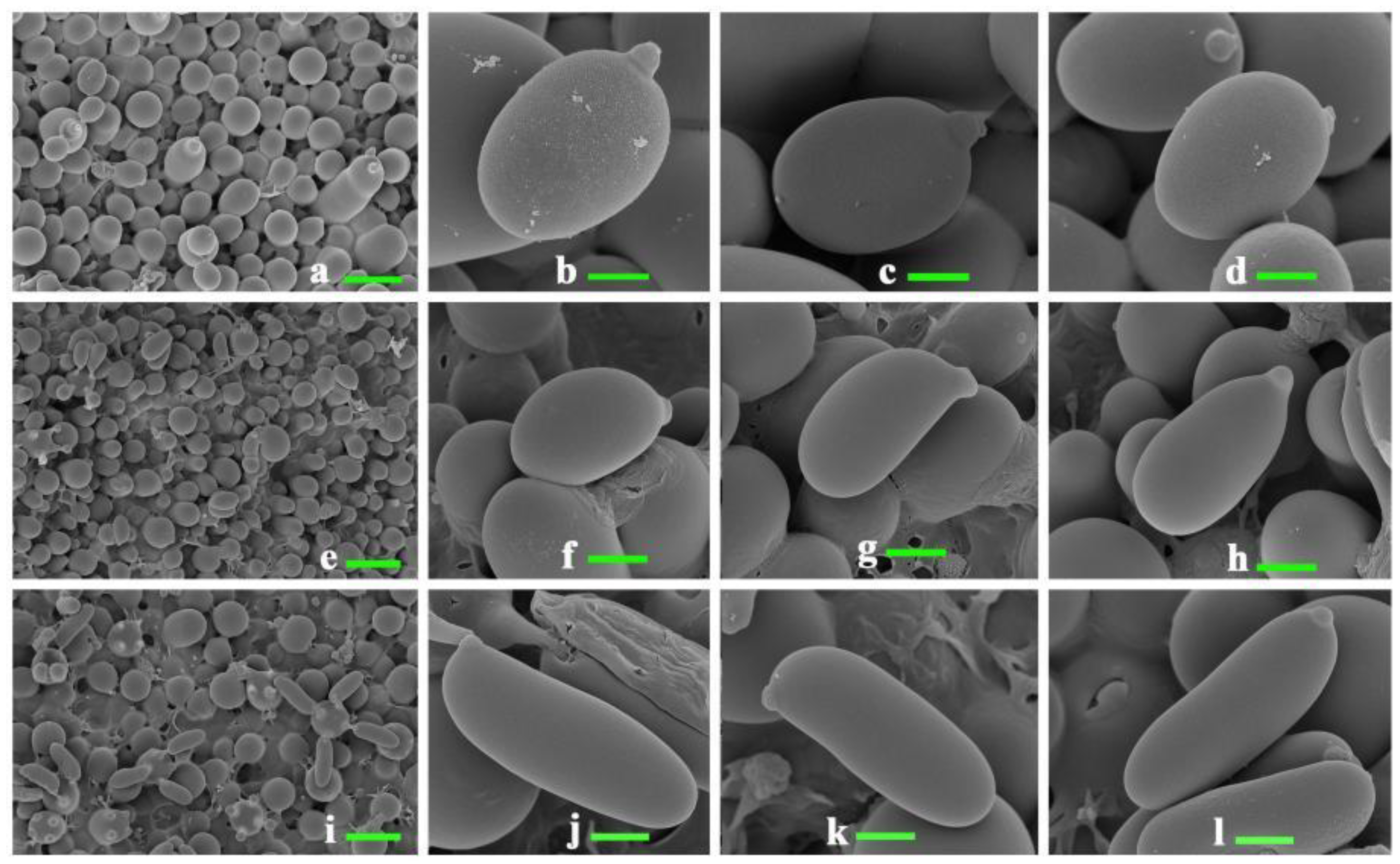
3.1. Shape and Size Range of Basidiospores

SEM images of fresh basidiospores are shown in Figure 1. The fresh basidiospores of A. bisporus (Figure 1a–d) were broadly ellipsoid, whereas those of L. edodes (Figure 1e–h) were oblong in shape, and the basidiospores of P. ostreatus (Figure 1i–l) were subcylindrical. The length range of fresh A. bisporus basidiospores was recorded as 6.31–8.01 μm (3.54–8.48 μm when dry); the width range 5.02–6.44 μm (3.27–6.65 μm when dry); and the Q value range 1.12–1.56 (0.66–2.03 when dry). The length range of fresh L. edodes basidiospores was recorded as 3.60–8.52 μm (2.71–8.36 μm when dry); the width range 2.68–4.00 μm (1.98–5.44 μm when dry); and the Q value range 1.03–2.52 (0.94–3.04 when dry). The length range of fresh P. ostreatus basidiospores was recorded as 4.92–10.85 μm (3.94–11.79 μm when dry); the width range as 2.47–3.78 μm (2.43–4.11 μm when dry); and the Q value range 1.68–3.83 (1.37–4.06 when dry) (Table 1).

3.2. Correlations between Drying Temperatures and Basidiospore Size

There were significant differences among the length and width of basidiospores of A. bisporus, L. edodes, and P. ostreatus with respect to different drying temperatures of fruitbodies (Table 1). The relationship between basidiospore length, width, Q value, and drying temperature was quantified and is shown in Figure 2.
For A. bisporus, fresh basidiospores had the greatest length (mean 7.13 μm) and width (mean 5.65 μm) when compared against dried basidiospores, followed by length (mean 6.87–6.96 μm) and width (mean 5.38–5.50 μm) of basidiospores dried at 30 °C or 35 °C. The shortest basidiospore length (mean 6.57–6.73 μm) and width (mean 5.10–5.15 μm) were found when drying temperatures were 40–50 °C. The length and the width of A. bisporus basidiospores decreased significantly upon drying, with a significant reduction in size starting at 35 °C; furthermore, drying did not affect the Q value significantly. Figure 2A shows the negative correlation between drying temperature and spore length in A. bisporus, and 67% of the variation was explained by this parameter; Figure 2B shows a negative correlation between drying temperature and spore width, and 92% of the negative correlation was explained in A. bisporus; Figure 2C shows positive correlations between drying temperature and the Q value, and 89% of the variation was explained by this parameter.
With regards to L. edodes, basidiospores were longest (mean 5.61–5.72 μm) and widest (mean 3.68 μm) when fresh or when dried at 45 °C; basidiospores had the smallest width (mean 2.81 μm) when dried at 50 °C. The largest Q value (mean 1.85) for L. edodes was recorded when the drying temperature was 50 °C, followed by dried basidiospores when the drying temperature was 30–40 °C; the smallest Q value was recorded when the drying temperature was 45 °C. The length and the width of the spores decreased significantly when dried, and the Q value increased significantly with the increasing temperatures, except for 45 °C, which resulted in a significant increase in the width and a decrease in the Q value. There was a negative correlation between spore length and drying temperature, and 24% of the variation was explained in L. edodes (Figure 2A); furthermore, there was a negative correlation between drying temperature and spore width (Figure 2B) and the negative correlation in L. edodes was expressed as 11% of the variation. Lastly, Figure 2C shows positive correlations between drying temperature and the Q value of L. edodes, and 8.5% of the variation was explained by this parameter.
For P. ostreatus, the greatest length (mean 8.74 μm) was recorded when the drying temperature was 30 °C, whereas the greatest width (mean 3.26–3.36) was recorded when the drying temperature was 30–40 °C; the shortest length (mean 7.87 μm) was recorded when the drying temperature was 45 °C; the shortest width (mean 3.07 μm) was recorded when the drying temperature was 50 °C. The length and width of the fresh basidiospores of P. ostreatus were smaller than those of the dried basidiospores. Drying P. ostreatus at 30 °C, 35 °C, and 40 °C resulted in the basidiospore length and width increasing significantly, while the length and width decreased significantly when the drying temperature was 45 °C or 50 °C. There was a negative correlation between drying temperature and spore length in P. ostreatus (21% of the variation was expressed by this parameter; Figure 2A) as well between drying temperature and spore width (20% of the variation was expressed by this parameter; Figure 2B). Lastly, drying temperature was not correlated against any changes in the Q value of P. ostreatus (Figure 2C).

3.3. Basidiospore Measurements Based on Previous Studies

A literature review focusing on the basidiospore measurements of A. bisporus, L. edodes, and P. ostreatus is summarized and presented in Table 2. The past studies we assessed failed to record drying temperatures for their specimens. However, we found that the length and the width of the basidiospores differed between studies for each of the species.

4. Discussion

This study conducted basidiospore measurements for three macrofungal species, comparing the characteristics of fresh spores and spores dried at a range of different temperatures. Spore size and shape are important characteristics in fungal taxonomy, and any factors that could potentially influence these attributes is relevant, as differing drying temperatures could result in variability in the reporting of spore morphology for a given species. The results of our study show that not only did the shape of the fresh basidiospores change once dried, but there was also a decrease in spore size, and that this decrease varied as the drying temperatures changed. Thus, there is a need for a consistent drying temperature to be adopted, or at least, drying temperatures should be reported, for all future taxonomic studies to avoid unnecessary variability across basidiospore descriptions.
The width and length of basidiospores from all three species used in our study decreased with drying; however, we saw conflicting results with regards to the Q value. Drying resulted in an increased Q value for L. edodes and A. bisporus, whereas the Q value of P. ostreatus remained unaffected. These findings are consistent with the work of Dramani et al. [2], who reported that the drying process led to a change in the morphology of basidiospores, including reduced spore size after drying, and in some cases an increased Q value. The study by Dramani et al. [2] compared the characteristics of fresh basidiospores with basidiospores dried at 50 °C, and clearly showed how drying impacted their size. Our research builds on their study in that we were able to explore a range of drying temperatures and determine how spore attributes change across a temperature range.
Basidiospores of the three macrofungal species under fresh conditions showed a different shape (Figure 1): broadly ellipsoid in A. bisporus, oblong in L. edodes, and subcylindrical in P. ostreatus. The shape of the basidiospores changed differently during the dehydration process. In agreement with suggestions from Dramani et al. [2], we speculate that the water content and the structure of basidiospore cell walls are key factors causing basidiospore transformation during the dehydration process. For example, A. bisporus and P. ostreatus have a higher water content (90–95%) than L. edodes (85–90%), and the basidiospore widths of A. bisporus and P. ostreatus declined rapidly upon drying. In addition, there was no significant difference in the Q value of A. bisporus and P. ostreatus between fresh and dried specimens, and this may be because the length and width of spores of these two species declined proportionally.
Our review of past studies highlighted the need for a more consistent approach to reporting in the methods of taxonomy-based manuscripts. Many studies failed to report whether specimens were dry or fresh, and if dried, at what temperatures they were dried. This is likely the reason behind the variability we observed in the spore characteristics between studies concerning the same species. For example, Mitchell and Walter [14] recorded the basidiospore size of A. bisporus as 6.8–8.5 × 5.6–6.6 μm, while Sarma et al. [12] recorded a smaller basidiospore size (4.2–6.2 × 3.2–4.4 μm). Pegler [17] recorded the basidiospore size of L. edodes as 5–7 × 3–3.7 μm, while Sarma et al. [12] recorded a bigger basidiospore size (7.25–8.5 × 5.2–6.7 μm). Lechner et al. [23] recorded the basidiospore size of P. ostreatus as 8–13 × 3.1–3.6 μm, whereas Venturella et al. [21] recorded a shorter length (6.2–8 μm) and longer width (4–6.5 μm). We also found that basidiospore length, width, and Q value differed significantly across the range of drying temperatures used in our study. Thus, basidiospore sizes in different but closely related species should be compared under the same drying conditions to avoid misidentification in future fungal taxonomy works.
The drying of macrofungal specimens and the subsequent depositing of these specimens in an herbarium are vital steps in fungal taxonomy. Hosaka and Uno [26] recommended a drying temperature of 35 °C for 48 h as ideal for small specimens, and larger specimens should be cut into thin slices before drying. However, these recommendations were based on maintaining reliable DNA quality for future analyses and not in reference to changes in spore size. Thus, it is clear that drying temperatures affect a range of factors within macrofungal specimens, and more consideration needs to be placed on the drying methods of specimens in the future. We recommend that a temperature of 30 °C for 48 h be used for drying specimens as this will minimize any changes in basidiospore characteristics and ensure DNA quality.

Author Contributions

Conceptualization, Y.H. and P.E.M.; methodology, S.C.K.; software, H.L.; validation, P.K. and P.E.M.; formal analysis, Y.H. and C.-L.Z.; investigation, M.C.A.G.; resources, S.C.K.; data curation, Y.H.; writing—original draft preparation, Y.H. and M.C.A.G.; writing—review and editing, S.C.K., H.L., C.-L.Z., P.K. and P.E.M.; visualization, Y.H. and H.L.; supervision, P.K.; project administration, S.C.K.; funding acquisition, S.C.K. and P.E.M. All authors have read and agreed to the published version of the manuscript.

Funding

This work was funded by the Key Research Project, Agroforestry System for Restoration and Bio-industry Technology Development (grant number 2017YFC0505101), Ministry of Sciences and Technology of China (grant number 2017YFC0505100); Yunnan Provincial Science and Technology Department (grant number 202003AD150004); ‘High-End Foreign Experts’ in the High-Level Talent Recruitment Plan of Yunnan Province, 2021; CAS President’s International Fellowship Initiative (PIFI) (number 2018PC0006); National Science Foundation of China (NSFC) (grant number 31750110478); National Sciences Foundation, China (NSFC) (grant number 41771063); and Thailand Science Research and Innovation grant Macrofungi diversity research from the Lancang-Mekong Watershed and surrounding areas (grant number DBG6280009).

Institutional Review Board Statement

Not applicable.

Informed Consent Statement

Not applicable.

Data Availability Statement

All data are available in the text.

Acknowledgments

Austin G. Smith at World Agroforestry (ICRAF), Kunming Institute of Botany, China, is also thanked for English editing.

Conflicts of Interest

The authors declare no conflict of interest.

References

  1. Halbwachs, H.; Bässler, C. Gone with the wind—A review on basidiospores of lamellate agarics. Mycosphere 2015, 6, 78–112. [Google Scholar] [CrossRef]
  2. Dramani, R.; Hegbe, A.D.M.T.; Tabe, A.; Badou, A.S.; Furneaux, B.; Ryberg, M.; Yorou, N.S. How are basidiospore size measurements affected by drying? Curr. Res. Environ. Appl. Mycol. 2020, 10, 63–70. [Google Scholar] [CrossRef]
  3. Decock, C.; Figueroa, S.H.; Robledo, G.; Castillo, G. Fomitiporia punctata (Basidiomycota, Hymenochaetales) and its presumed taxonomic synonyms in America: Taxonomy and phylogeny of some species from tropical/subtropical areas. Mycologia 2007, 99, 733–752. [Google Scholar] [CrossRef] [PubMed]
  4. Hoa, H.T.; Wang, C.L. The effects of temperature and nutritional conditions on mycelium growth of two Oyster mushrooms (Pleurotus ostreatus and Pleurotus cystidiosus). Mycobiology 2015, 43, 14–23. [Google Scholar] [CrossRef] [Green Version]
  5. Bruns, T.D.; Fogel, R.; Taylor, J.W. Amplification and sequencing of DNA from fungal herbarium specimens. Mycologia 1990, 82, 175–184. [Google Scholar] [CrossRef]
  6. Chin, S.K.; Law, C.L.; Supramaniam, C.V.; Cheng, P.G.; Mujumdar, A.S. Convective drying of Ganoderma tsugae Murrill and effect of temperature on basidiospores. Dry. Technol. 2008, 26, 1524–1533. [Google Scholar] [CrossRef]
  7. Yanaga, K.; Maekawa, N.; Shimomura, N.; Ishigaki, Y.; Nakamura, Y.; Takegami, T.; Tomosugi, N.; Miyazawa, S.; Kuwabata, S. Use of ionic liquid in fungal taxonomic study of ultrastructure of basidiospore ornamentation. Mycol. Prog. 2012, 11, 343–347. [Google Scholar] [CrossRef]
  8. Buyck, B.; Kauff, F.; Cruaud, C.; Hofstetter, V. Molecular evidence for novel Cantharellus (Cantharellales, Basidiomycota) from tropical African miombo woodland and a key to all tropical African chanterelles. Fungal Divers. 2013, 58, 281–298. [Google Scholar] [CrossRef]
  9. Cho, H.J.; Lee, H.; Park, M.S.; Kim, C.; Wisitrassameewong, K.; Lupala, A.; Park, K.H.; Kim, M.J.; Fong, J.J.; Lim, Y.W. Macrolepiota in Korea: New records and a new species. Mycobiology 2019, 47, 368–377. [Google Scholar] [CrossRef] [Green Version]
  10. Li, Y.; Liang, M.; Shu, X.; Liu, C.; Shu, J. Differentiation of basidiospores by MALDI-TOF lipid profiling. Int. J. Mass Spectrom. 2013, 352, 44–50. [Google Scholar] [CrossRef]
  11. Mata, G.; Medel, R.; Callac, P.; Billette, C.; Garibay-Orijel, R. First report of wild Agaricus bisporus (Basidiomycota, Agaricaceae) from Tlaxcala and Veracruz, Mexico. Rev. Mex. Biodivers. 2016, 87, 10–17. [Google Scholar] [CrossRef] [Green Version]
  12. Sarma, T.C.; Sarma, I.; Patiri, B.N. Wild edible mushrooms used by some ethnic tribes of Western Assam. The Bioscan 2010, 3, 613–625. [Google Scholar] [CrossRef]
  13. Callac, P.; Billette, C.; Imbernon, M.; Kerrigan, R.W. Morphological, genetic, and interfertility analyses reveal a novel, tetrasporic variety of Agaricus bisporus from the Sonoran Desert of California. Mycologia 1993, 85, 835–851. [Google Scholar] [CrossRef]
  14. Mitchell, A.D.; Walter, M. Species of Agaricus occurring in New Zealand. N. Z. J. Bot. 1999, 37, 715–725. [Google Scholar] [CrossRef]
  15. First Nature. Available online: https://www.first-nature.com/fungi/agaricus-bisporus.php (accessed on 5 December 2021).
  16. Mushroomexpert.com. Available online: http://www.mushroomexpert.com/agaricus_bisporus.html (accessed on 5 December 2021).
  17. Pegler, D.N. The genus Lentinula (Tricholomataceae tribe Collybieae). Sydowia 1983, 36, 227–239. [Google Scholar]
  18. Stamets, P. Growing Gourmet and Medicinal Mushroom, 3rd ed.; Ten Speed Press: Olympia, WA, USA; Berkeley, CA, USA, 2000; pp. 259–262. [Google Scholar]
  19. Asef, M.R. Intersterility groups of Pleurotus ostreatus complex in Iran. Mycology 2012, 3, 147–152. [Google Scholar] [CrossRef]
  20. Singh, R.K.; Pandey, S.K.; Singh, D.; Masurkar, P. First report of edible mushroom Pleurotus ostreatus from India with potential to kill plant parasitic nematodes. Indian Phytopathol. 2019, 72, 173–176. [Google Scholar] [CrossRef]
  21. Venturella, G.; Gargano, M.L.; Compagno, R. The genus Pleurotus in Italy. Flora Mediterr. 2015, 25, 143–155. [Google Scholar] [CrossRef]
  22. Junior, N.M.; Asai, T.; Capelari, M.; Paccola-Meirelles, L.D. Morphological and molecular identification of four Brazilian commercial isolates of Pleurotus spp. and cultivation on corncob. Braz. Arch. Biol. Technol. 2010, 53, 397–408. [Google Scholar] [CrossRef] [Green Version]
  23. Lechner, B.E.; Wright, J.E.; Albertó, E. The genus Pleurotus in Argentina. Mycologia 2004, 96, 845–858. [Google Scholar] [CrossRef]
  24. Nath, R.K.; Sarma, T.C. Edible macrofungi of Kaliabar sub-division of Nagaon district, Assam, India. Ann. Plant Sci. 2018, 7, 2161–2165. [Google Scholar] [CrossRef] [Green Version]
  25. Mushroomexpert.com. Available online: http://www.mushroomexpert.com/pleurotus_ostreatus.html (accessed on 5 December 2021).
  26. Hosaka, K.; Uno, K. Assessment of the DNA quality in mushroom specimens: Effect of drying temperature. Bull. Natl. Mus. Nat. Sci. Ser. B 2011, 37, 101–111. [Google Scholar]
Figure 1. Characteristics of fresh basidiospores under SEM. (ad) Agaricus bisporus; (eh) Lentinula edodes; (il) Pleurotus ostreatus. Scale bars: (a,e,i), 10 μm; (bd,fh,jl), 2 μm.
Diversity 14 00239 g001
Figure 2. Regression between observed basidiospore size and drying temperature of fruitbodies. (A): spore length and drying temperature; (B): spore width and drying temperature; (C): Q value and drying temperature. The regression equation was plotted using the mean value of the recorded basidiospore sizes for: AB, Agaricus bisporus; LE, Lentinula edodes; PO, Pleurotus ostreatus.
Diversity 14 00239 g002
Table 1. Basidiospore size ranges, length, width, and Q values. Parameters were determined for fresh basidiospores as well as basidiospores dried at different temperatures.
Macrofungal SpeciesDrying TemperatureBasidiospore Size Range (μm)Q Value RangeLength (μm)Width (μm)Q Value
Agaricus bisporusFresh6.31–8.01 × 5.02–6.441.12–1.567.13 ± 0.41 a5.65 ± 0.29 a1.27 ± 0.09 a
30 °C4.49–7.94 × 4.40–6.650.81–1.616.96 ± 0.67 ab5.50 ± 0.38 b1.27 ± 0.15 a
35 °C4.38–8.48 × 4.44–6.400.86–1.706.87 ± 0.58 b5.38 ± 0.37 b1.28 ± 0.14 a
40 °C4.69–8.22 × 3.27–6.460.86–2.036.57 ± 0.61 c5.15 ± 0.55 c1.29 ± 0.21 a
45 °C4.20–8.41 × 3.46–6.090.83–1.926.73 ± 0.66 bc5.14 ± 0.38 c1.32 ± 0.17 a
50 °C3.54–8.42 × 4.42–5.930.66–1.666.72 ± 0.75 bc5.10 ± 0.30 c1.32 ± 0.17 a
Lentinula edodesFresh3.60–8.52 × 2.68–4.001.03–2.525.72 ± 0.76 a3.49 ± 0.24 b1.65 ± 0.25 b
30 °C2.71–7.47 × 2.00–4.551.00–2.645.29 ± 0.72 b3.21 ± 0.40 c1.67 ± 0.30 b
35 °C3.93–6.70 × 2.16–3.751.28–2.695.26 ± 0.57 b3.09 ± 0.35 c1.73 ± 0.27 b
40 °C3.41–8.36 × 2.19–5.440.94–2.855.26 ± 0.73 b3.13 ± 0.49 c1.73 ± 0.36 b
45 °C4.24–6.96 × 2.96–4.441.11–2.045.61 ± 0.52 a3.68 ± 0.33 a1.53 ± 0.20 c
50 °C3.51–7.57 × 1.98–3.861.02–3.045.08 ± 0.68 b2.81 ± 0.38 d1.85 ± 0.40 a
Pleurotus ostreatusFresh4.92–10.85 × 2.47–3.781.68–3.838.14 ± 1.25 bc3.12 ± 0.27 b2.62 ± 0.43 a
30 °C5.87–11.79 × 2.70–3.991.55–4.068.74 ± 1.27 a3.34 ± 0.32 a2.64 ± 0.46 a
35 °C6.74–10.59 × 2.87–4.111.90–3.168.48 ± 0.77 ab3.36 ± 0.26 a2.54 ± 0.27 a
40 °C5.61–10.93 × 2.43–3.871.79–3.788.53 ± 1.02 ab3.26 ± 0.29 a2.64 ± 0.43 a
45 °C3.94–10.85 × 2.44–4.071.37–3.897.87 ± 1.40 c3.10 ± 0.32 b2.56 ± 0.53 a
50 °C5.71–9.88 × 2.58–3.611.78–3.338.09 ± 0.81 bc3.07 ± 0.24 b2.65 ± 0.33 a
Notes: Values with different bold letters across different temperatures of each species are significantly different under Dunnett’s T3 post hoc test at p < 0.05.
Table 2. Basidiospore size ranges (length and width, shown as lmin–lmax × wmin–wmax) of Agaricus bisporus, Lentinula edodes, and Pleurotus ostreatus reported from this study and previous studies.
Macrofungal SpeciesBasidiospore Size (μm)Status of SpecimenReferences
Agaricus bisporus6–8.5 × 5–6fresh[10]
6–9 × 5.5–9fresh[11]
4.2–6.2 × 3.2–4.4dry[12]
4.8–6.8 × 4.0–5.1dry[13]
6.8–8.5 × 5.6–6.6dry[14]
4–7.5 × 4–5.5ns[15]
6–8 × 5–6ns[16]
6.31–8.01 × 5.02–6.44freshThis study
Lentinula edodes5–7 × 3–3.7dry[17]
7.25–8.5 × 5.2–6.7dry[12]
5–6.5 × 3–3.5ns[18]
3.60–8.52 × 2.68–4.00freshThis study
Pleurotus ostreatus8–10.5 × 3–3.6fresh[19]
10–12 × 4–5fresh[20]
6.2–8 × 4–6.5dry[21]
8.7–11.2 × 3.7dry[22]
8–13 × 3.1–3.6dry[23]
5–7 × 3.5ns[24]
7–11 × 2–4ns[25]
4.92–10.85 × 2.47–3.78freshThis study
ns, not specified.
Publisher’s Note: MDPI stays neutral with regard to jurisdictional claims in published maps and institutional affiliations.

