Article

Morphological and Molecular Characterization Using Genitalia and CoxI Barcode Sequence Analysis of Afrotropical Mosquitoes with Arbovirus Vector Potential

by
Eddyson Montalvo-Sabino
1,10,
Ana Paula Abílio
2,
Milehna Mara Guarido
3,6,
Vera Valadas
1,
Maria Teresa Novo
1,
Ayubo Kampango
2,4,
Carla Alexandra Sousa
1,
José Fafetine
5,
Marietjie Venter
3,
Peter N. Thompson
6,
Leo Braack
7,8,
Anthony John Cornel
9,
Ricardo Parreira
1 and
António Paulo Gouveia de Almeida
1,*
1
Global Health and Tropical Medicine (GHTM), Institute of Hygiene and Tropical Medicine (IHMT), NOVA University Lisbon (NOVA), 1349-008 Lisboa, Portugal
2
Instituto Nacional de Saúde (INS), Marracuene 3943, Mozambique
3
Zoonotic Arbo- and Respiratory Virus Program, Centre for Viral Zoonoses, Department Medical Virology, Faculty of Health Sciences, University of Pretoria, Pretoria 0031, South Africa
4
Department of Zoology and Entomology, University of Pretoria, Pretoria 0002, South Africa
5
Centro de Biotecnologia, Universidade Eduardo Mondlane (UEM), Maputo 3453, Mozambique
6
Department of Production Animal Studies, Faculty of Veterinary Science, University of Pretoria, Onderstepoort 0110, South Africa
7
Malaria Consortium, Mahidol University, Bangkok 73170, Thailand
8
UP Institute for Sustainable Malaria Control, University of Pretoria, Pretoria 0031, South Africa
9
Mosquito Control Research Laboratory, Department of Entomology and Nematology and Vector Genetics Laboratory, Department of Pathology, Microbiology, and Immunology, University of California at Davis, 9240 South Riverbend Avenue, Parlier, CA 93648, USA
10
Escuela Académico Profesional de Medicina Veterinaria, Facultad de Medicina Veterinaria y Zootecnia, Universidad Nacional Hermilio Valdizán, Huanuco 10003, Peru
*
Author to whom correspondence should be addressed.
Diversity 2022, 14(11), 940; https://doi.org/10.3390/d14110940
Submission received: 26 September 2022 / Revised: 27 October 2022 / Accepted: 31 October 2022 / Published: 2 November 2022
(This article belongs to the Special Issue Diversity, Distribution and Phylogeny of Vector Insects)

Abstract

:
Potential arboviral Afrotropical mosquito vectors are underrepresented in public databases of CoxI barcode sequences. Furthermore, available CoxI sequences for many species are often not associated with voucher specimens to match the corresponding fine morphological characterization of specimens. Hence, this study focused on the characterization of Culicine mosquitoes from South Africa, Mozambique, and Angola and their classification using a complementary approach including a morphological analysis of specimens’ genitalia and phylogenetic study based on the analysis of CoxI barcode sequences using maximum likelihood and Bayesian phylogenetic inference methods, alongside Median-Joining Network and PCOORD analyses. Overall, 800 mosquitoes (652 males and 148 females) from 67 species, were analyzed. Genitalia from 663 specimens allowed the identification of 55 species of 10 genera. A total of 247 CoxI partial gene sequences corresponding to 65 species were obtained, 11 of which (Aedes capensis, Ae. mucidus, Culex andersoni, Cx. telesilla, Cx. inconspicuosus, Eretmapodites subsimplicipes, Er. quinquevittatus, Ficalbia uniformis, Mimomyia hispida, Uranotaenia alboabdominalis, and Ur. mashonaensis) are, to the best of our knowledge, provided here for the first time. The presence of Cx. pipiens ecotypes molestus and pipiens and their hybrids, as well as Cx. infula, is newly reported in the Afrotropical region. The rates of correct sequence identification using BOLD and BLASTn (≥95% identity) were 64% and 53%, respectively. Phylogenetic analysis revealed that, except for subgenus Eumelanomyia of Culex, there was support for tribes Aedini, Culicini, Ficalbiini, and Mansoniini. A divergence >2% was observed in conspecific sequences, e.g., Aedeomyia africana, Ae. cumminsii, Ae. unilineatus, Ae. metallicus, Ae. furcifer, Ae. caballus, and Mansonia uniformis. Conversely, sequences from groups and species complexes, namely, Ae. simpsoni, Ae. mcintoshi, Cx. bitaeniorhynchus, Cx. simpsoni, and Cx. pipiens were insufficiently separated. A contribution has been made to the barcode library of Afrotropical mosquitoes with associated genitalia morphological identifications.

Graphical Abstract

1. Introduction

Many mosquito species are important vectors of pathogens, including arboviruses, which can cause various febrile, neurological, and hemorrhagic diseases and, therefore, pose a considerable burden on human health and health systems [1]. While currently, the most important arboviruses transmitted by mosquitoes are dengue (DENV), Zika (ZIKV), Chikungunya (CHIKV), and yellow fever (YFV), outbreaks caused by West Nile (WNV), Rift Valley fever (RVFV), and Japanese Encephalitis (JEV) viruses have also been reported in recent years, becoming emerging health problems [2,3].
Mosquitoes (Diptera: Culicidae) are widely distributed throughout the world (except for Antarctica), with 3570 valid species and 130 subspecies thus far documented [1]. The correct identification of mosquito species that may be involved in pathogen transmission is the first step in the surveillance and control of mosquito-borne diseases and has been based on morphological analysis of mainly adult specimens, but also fourth instar larvae [4,5,6,7,8]. Furthermore, several mosquito species can only be identified based on morphological differences in the male genitalia (and occasionally on other male-specific structures), rendering the identification of their female counterparts sometimes unsolved. Nevertheless, the characteristics of male genitalia are structural, allowing accurate and reliable species identification, in addition to being less susceptible to general body damage that is so common in field samples. However, genitalia dissection is a fine and tedious process that requires specific and specialized training [9]. Furthermore, some mosquitoes form closely related, morphologically indistinguishable, cryptic species complexes, with each species having ecological and host preferences and reproductive isolation, constituting biological individual taxa. To overcome the difficulty in their identification, nucleic acids-based molecular identification methods are used for, for example, members of multiple Anopheles species complexes [10] and Culex (Culex) pipiens complex members [11].
So far, despite the medical importance of diseases such as dengue, yellow fever, West Nile fever, Zika, and Rift Valley fever, studies aimed at the molecular identification of vectors of arboviruses of African origin [12,13,14,15,16] are limited compared to those regarding the analysis of malaria vectors, or even arbovirus vectors of non-African origin [17,18,19,20,21,22,23,24,25,26,27,28]. The molecular identification of many species occurring in countries such as South Africa, Mozambique, and Angola that have high mosquito and viral richness are not available [12,29,30,31,32]. Cytochrome c oxidase subunit I (CoxI) barcode sequences of many Afrotropical mosquito vectors of arboviruses are lacking due to the underrepresentation of specimens in the largest public genomic sequence databases most frequently searched (BOLD and GenBank). Examination of the global representation of CoxI barcode Culicidae species sequences in BOLD clearly reveals the underrepresentation of African-derived taxa (https://www.boldsystems.org/index.php/Public_SearchTerms, accessed on 12 May 2022). Furthermore, it is essential to have reliable and comprehensively annotated reference databases of verified sequences that can be used for comparison for species identification [20]. Phylogenetic analyses based on some GenBank/BOLD records have suggested that some partial genomic sequences obtained from mosquitoes have been incorrectly assigned, a type of error that has already been identified in studies based on the CoxI marker [25] and internal transcribed spacer of nuclear ribosomal DNA (ITS) [33].
The objectives of this work were: (i) to morphologically characterize Afrotropical mosquitoes of the Culicinae subfamily, focusing on the analysis of genitalia of adult specimens, in order to have morphological vouchers associated with a matching mitochondrial CoxI sequence to be obtained sequently; (ii) to perform a phylogenetic reconstruction that would allow the identification of the sequences obtained in this work, but that would also (iii) shed light on the agreement between phylogenetic tree topology and the current morphology-based taxonomic arrangement.

2. Materials and Methods

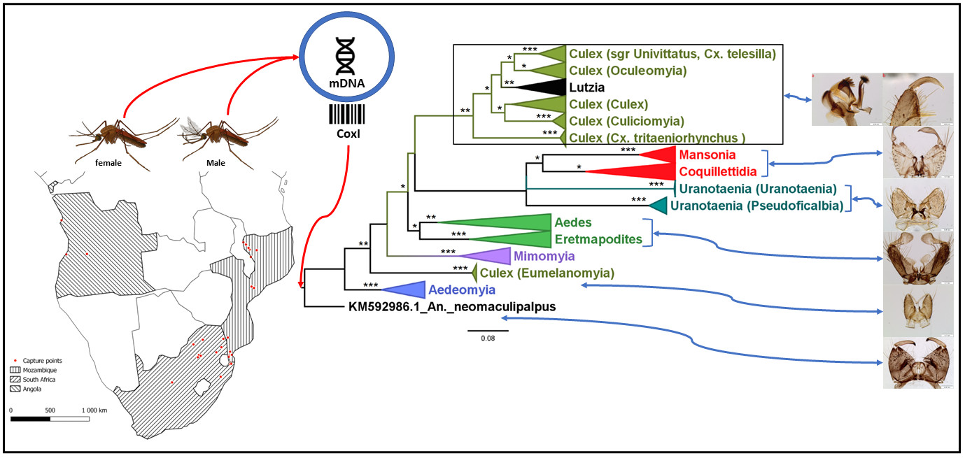
2.1. Mosquito Sampling and Preparation of Male Mosquito Genitalia

The mosquito collection analyzed in this work represented a convenience sample comprising specimens previously collected in three countries in southern Africa (Mozambique, South Africa, and Angola; Supplementary Material-SIV (File S-IV), Figure S1) between 2014 and 2018, within the scope of various scientific projects related to arbovirus detection and epidemiology assessments of arboviruses. After collection and subsequent transportation to the Institute of Hygiene and Tropical Medicine|NOVA University Lisbon (IHMT|NOVA), these mosquitoes, listed in Supplementary Material-I (File S-I), Table S1, were kept dehydrated in silica gel tubes at room temperature.
All mosquitoes were classified according to species, species complex, or species group (where possible) based on the analysis of their morphological features, following the keys of Edwards [4], Jupp [5], and Harbach [34]. The classification of the Aedini tribe followed that of Wilkerson et al. [35] and taxa nomenclature as in https://mosquito-taxonomic-inventory.myspecies.info/valid-species-list# (accessed on 12 May 2022).
The genitalia of all male, and some female, specimens were dissected and slide-mounted for careful examination. The terminal part of the mosquito abdomen was sectioned at the level of segment VII/VIII and immersed in Marc André’s solution for a minimum of 7 days at room temperature. Afterward, mosquito genitalia were placed on a slide with a drop of a polyvinyl-chloral-formo-phenol medium, dissected under a stereomicroscope, and covered with a coverslip [14]. Analysis of the different structures of the mosquito genitalia and (sometimes) of maxillary palps, were carried out using an Olympus microscope (BX5,1) and their identification and naming of parts followed the nomenclature of Harbach and Knight [9]. Photographs were taken with an Olympus SC30 digital camera and processed with the Zerene Stacker program (https://www.zerenesystems.com/, accessed on 12 May 2022). In Culex subgenus Oculeomyia, we relied on the description by Sirivanakarn [36] and Harbach [34] to confirm the identification based on the genitalia.

2.2. DNA Extraction, Partial Amplification of CoxI, and Culex Pipiens Complex Molecular Identification

Total genomic DNA was extracted from mosquito legs and abdomens, as previously described [14]. The barcode N-terminal region of the CoxI gene was amplified using the specific primers (LCO1490 and HCO2198), using reaction conditions described by Folmer et al. [37]. The amplified products of 658 bp were visualized under UV illumination after electrophoresis in 2% agarose gels. Whenever a specific amplification product was not observed, to obtain a CoxI-specific amplicon, an alternative strategy was used. This entailed the use of primers LCO1490 and TL2-N-3014 and the thermal profile previously described by Tchouassi et al. [38]. In case unsuccessful amplifications prevailed, a final attempt called for the design of new primers using multiple alignments of CoxI nucleotide sequences downloaded from the GenBank genomic database. These sequences were aligned using MAFFT v7 (https://mafft.cbrc.jp/alignment/server/, accessed 10 November 2021), and these alignments served as a starting point for the design of degenerate primers using the primer design-M tool (https://bio.tools/primerdesign-m, accessed 10 November 2021). The chosen primers (C_degF 5′-ACWTTATAYTTYATTTTYGG-3′ and C_degR 5′-GTTARWARTAT-WGTAATWGC-3′) were used at a final concentration of 500 nM in 20 μL PCR reactions containing 10 μL NZYTaq II 2x Green Master Mix (NYTech, Portugal), 2 μL of a 1:10 dilution of the original DNA extract, and 6 μL of nuclease-free water. The amplification conditions included one denaturation step at 95 °C for 5 min, followed by 40 cycles of amplification (denaturation: 30 s at 95 °C; annealing: 40 s at 43 °C; extension: 1 min at 72 °C) and a final extension step for 5 min at 72 °C.
A multiplex PCR assay that targets species-specific polymorphisms at the intron-2 of the acetylcholinesterase gene intron-2 (Ace-2) sequence of Cx. pipiens Linnaeus, 1758 and Cx. quinquefasciatus Say, 1823 was carried out with primers B1246s, ACEpip, and ACEquin, as described by Smith and Fonseca [11], yielding a PCR product of 610 bp for Cx. pipiens and 274 bp for Cx. quinquefasciatus. Differentiation of the Cx. pipiens ecotype molestus and Cx. pipiens ecotype pipiens followed the analysis of the CQ11 microsatellite flanking region, described by Bahnck and Fonseca [39], yielding a PCR-product approximately 200 bp in size for Cx. pipiens form pipiens and 250 bp for form molestus.

2.3. Amplicon Sequencing and Nucleotide Sequence Analyses

The amplified PCR products corresponding to partial sequences of the CoxI gene from each of the analyzed samples were purified and sequenced by the Sanger method (STABVida, Lda. 2825-182 Caparica, Portugal) using primers LCO1490 or C_deg_F, and the respective reverse primers when the obtained chromatogram lacked in quality. The sequences obtained were edited using the Chromas tool version 2.6.6 (https://technelysium.com.au/wp/, accessed on 10 November 2021). Low-quality sequences were excluded during the editing process. In these cases, a new amplification was performed from the same DNA extract. The purification and sequencing of the obtained amplification products were also repeated, as described above. All amplification products were sequenced, which ranged from 399–661 nucleotides. However, for phylogenetic and divergence analysis, only sequences greater than 500 nucleotides were considered.
The search for homologous sequences available in publicly accessible genomic databases (GenBank/ENA/DDBJ) was performed both with the BLASTn tool (https://blast.ncbi.nlm.nih.gov/Blast.cgi, accessed on 10 November 2021) and the taxonomy search engine in the BOLDSystems v4 database (https://www.boldsystems.org/index.php/IDS_OpenIdEngine, accessed on November 2021). These same tools were used for the identification/confirmation of the identity of our sequences. Multiple sequence alignments were constructed using the G-INS-i iterative refinement method as implemented in MAFFT v7. The obtained alignments were treated with Gblocks (http://phylogeny.lirmm.fr/phylo_cgi/one_task.cgi?task_type=gblocks, accessed 10 November 2021) after selecting the most permissive editing options. The evaluation of the phylogenetic signal of all used sequence datasets was carried out using the likelihood-mapping method, as implemented in the TREE-PUZZLE software [40].
For the phylogenetic sequence analysis, two different approaches were explored: the Maximum Likelihood optimization criterion (ML) and a Bayesian phylogenetic inference-based approach. For both, the first step of the analysis involved the choice of the best nucleotide substitution model to be used (GTR + Γ, GTR + I or GTR + Γ + I models: GTR-General Time Reversal; Γ-Gamma distribution; proportion I of invariant sites), using the IQtree software [41], which was also used for ML phylogenetic reconstruction. The topological support of the branches in the obtained trees was assessed with bootstrap analysis and an approximate likelihood ratio test [aLRT], also implemented in Iqtree. In either case, 1000 replicates of the original sequence data were used, and bootstrap or aLRT values ≥ 75 (% of the total number of replicates) were considered as indicating strong topological support.
Bayesian phylogenetic analyses were carried out using BEAST v1.10.4 software [42], using the same sequence data sets and evolutionary models adopted for the ML analyses. The Bayesian analyses consisted of two independent Markov chain Monte Carlo (MCMC) runs until 1 × 10⁸ states had been sampled at every 10,000th MCMC step (10% of which were later discarded as burn-in). In all cases, chain convergence was assessed using Tracer software v1.7.1 (http://beast.bio.ed.ac.uk/tracer, accessed on 10 November 2021), which was also used to check for an adequate effective sample size (ESS) higher than 200 (after the removal of the burn-in). The tree distribution was summarized using TreeAnnotator software v1.8.3 as a maximum clade credibility (MCC) tree, using median heights as the node heights in the tree. All the phylogenetic trees were visualized using FigTree v1.4.2 software (http://tree.bio.ed.ac.uk/software/figtree/, accessed 10 November 2021). At specific branches, a posterior probability value of ≥0.80 was considered as indicating strong topological support. In all trees, the sequence of the species An. neomaculipalpus Curry, 1931 (KM592986.1) was used as the outgroup. The trees obtained with maximum credibility (product of the Bayesian analysis) were selected to depict a topological organization of the branches more compatible with a priori taxonomic expectations. Specific branches were labeled with one to three “*” signs, according to the number of phylogenetic construction methods/tests that confirmed such topology (aLRT and bootstrap/ML + posterior probability/Bayesian analyses). The original trees can be found in Supplementary Material-III (File S-III).
The average intraspecific and interspecific genetic variation were calculated using genetic distances corrected with the Kimura 2-parameter model (K2P), as implemented in the MEGA X software.
Median Joining networks analysis was performed using SplitsTree5 5.0.0_alpha application with default options [43] for computing unrooted phylogenetic networks from alignments of sequences. The Neighbor Net method [44] was used (default options) to obtain compatible splits, and the Splits Network Algorithm method [45] was used (default options) to obtain split networks. Principal coordinates analysis was also carried out using the software available on the platform (https://www.hiv.lanl.gov/content/sequence/PCOORD/PCOORD.html, accessed on 10 April 2022).

3. Results

3.1. Mosquito Identification: Morphological and Molecular

A total of 800 mosquitoes, comprising 652 (81.5%) males and 148 (18.5%) females, were analyzed in this study. These included 73 specimens from Angola, 515 from South Africa, and 212 from Mozambique, representing 67 species belonging to 10 genera: Aedeomyia (2), Aedes (28), Coquillettidia (3), Culex (24), Eretmapodites (2), Ficalbia (1), Lutzia (1), Mansonia (2), Mimomyia (2), and Uranotaenia (2) (File S-I, Table S1).
Of these, genitalia from 652 male and 11 female specimens were dissected and their analysis confirmed the identification of 55 species (File S-IV, Table S1), a photographic record of which can be found in Supplementary Material-II (File S-II). The respective slides are deposited in the IHMT|NOVA Insect Collection.
From a subsample of genitalia-confirmed male Cx. pipiens and Cx. quinquefasciatus and females from the Cx. pipiens complex, Ace2 multiplex PCR allowed us to confirm 8 specimens (2 females and 6 males) as Cx. pipiens and 18 as Cx. quinquefasciatus (7 females and 11 males; File S-IV, Figure S2a). No hybrids were identified and four samples failed to react. Multiplex PCR for the CQ11 microsatellite flanking region identified one of the Cx. pipiens as the pipiens ecotype (EM305), two as the molestus ecotype (EM326 and EM332), and four as hybrids of the two ecotypes (EM300, EM302, EM303, EM304), while no amplification product was obtained for one male Cx. pipiens (EM306) (File S-IV, Figure S2b).
The amplification of the CoxI gene was successful in 247/333 specimens (74.2%). The majority (n = 184) of the CoxI amplicons were obtained with the Folmer et al. [37] protocol, while the remaining 63 sequences were obtained either with the Tchouassi et al. protocol (n = 14) [38] or using the degenerate primers/protocol here described (n = 49). A total of 65 species were identified through molecular analysis (File S-IV, Table S2). Not all species could be identified by both methods as in some, no males were available, and in others, no amplification was obtained, leaving the total number of species identified by either method as 67. Only 64% of the sequences obtained were correctly identified by the BOLD tool, i.e., corresponding to the genitalia-confirmed species, and 53% shared ≥95% identity with a given species-specific sequence using the BLASTn tool. For eleven of these species, and as far as we could ascertain, partial CoxI sequences are provided here for the first time. These species include Ae. (Albuginosus) capensis Edwards, 1924; Ae. (Mucidus) mucidus (Karsch, 1887); Cx. (Culex) andersoni Edwards, 1914; Cx. (Cux.) telesilla de Meillon and Lavoipierre, 1945; Cx. (Eumelanomyia) inconspicuosus (Theobald, 1908); Er. subsimplicipes Edwards, 1914; Er. quinquevittatus Theobald, 1901; Fi. uniformis (Theobald, 1904); Mi. (Mim) hispida (Theobald, 1910); Ur. (Uranotaenia) alboabdominalis Theobald, 1910; and Ur. (Pseudoficalbia) mashonaensis Theobald, 1901 (the male genitalia of which are presented in Figure 1).

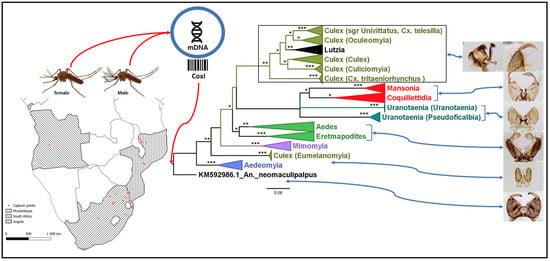
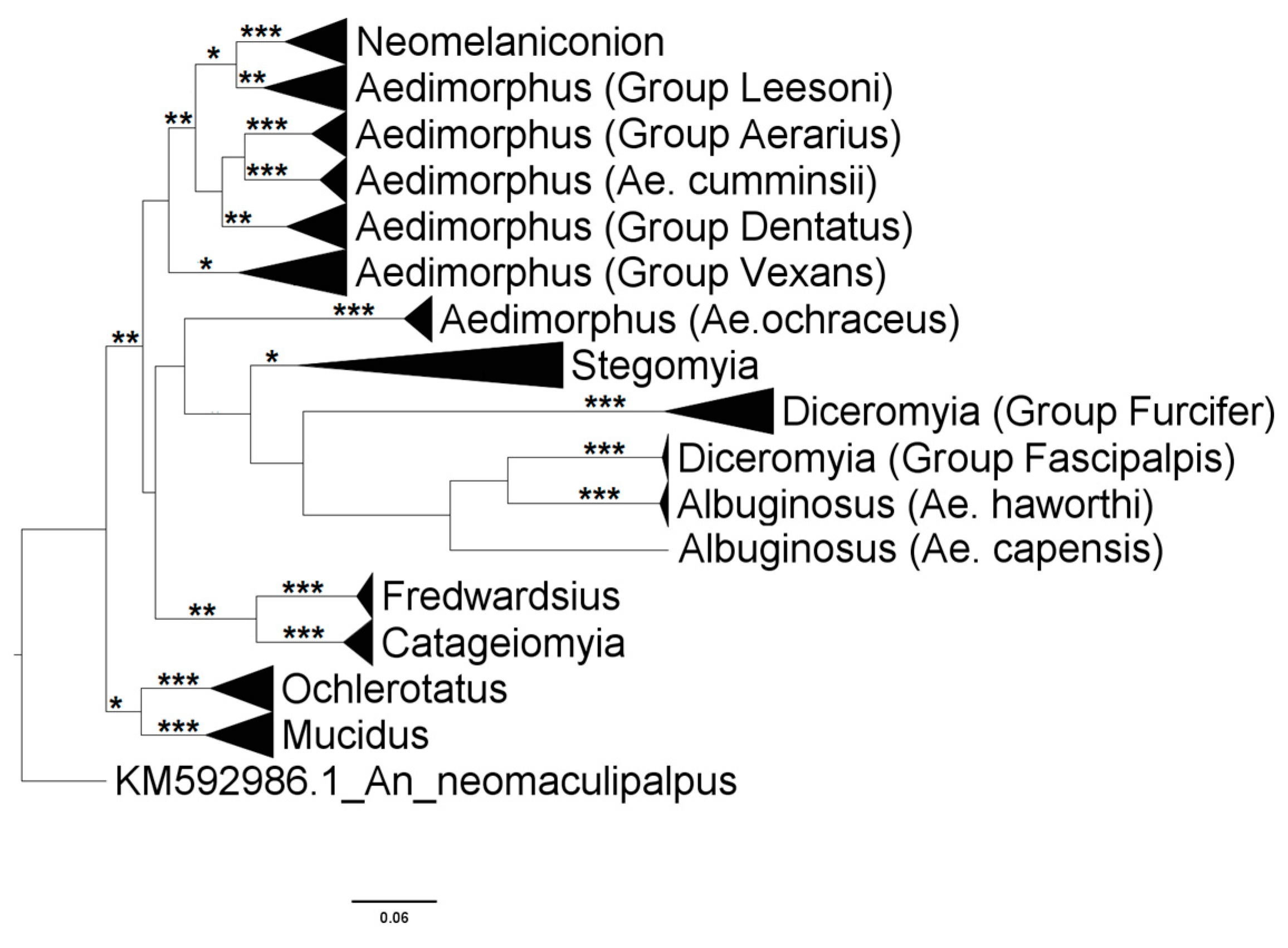
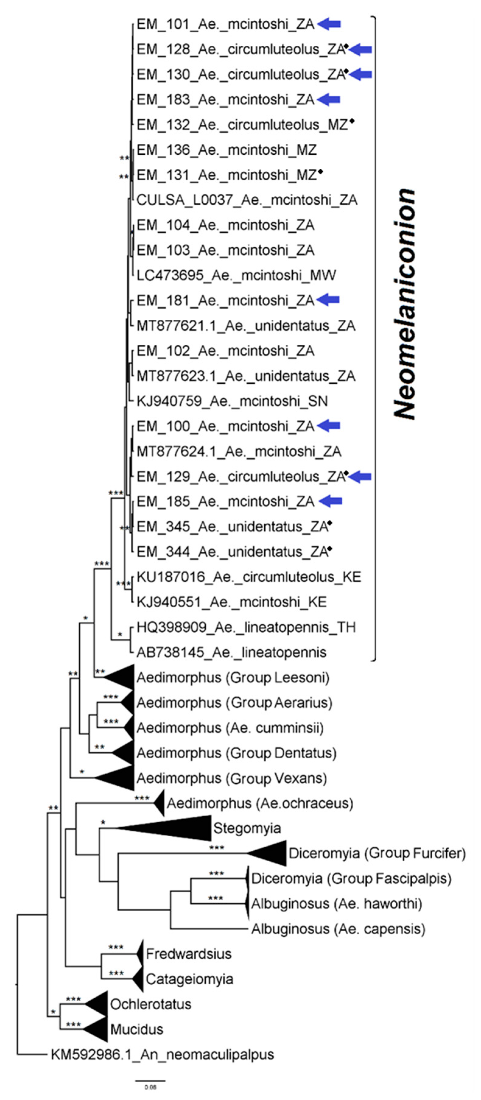
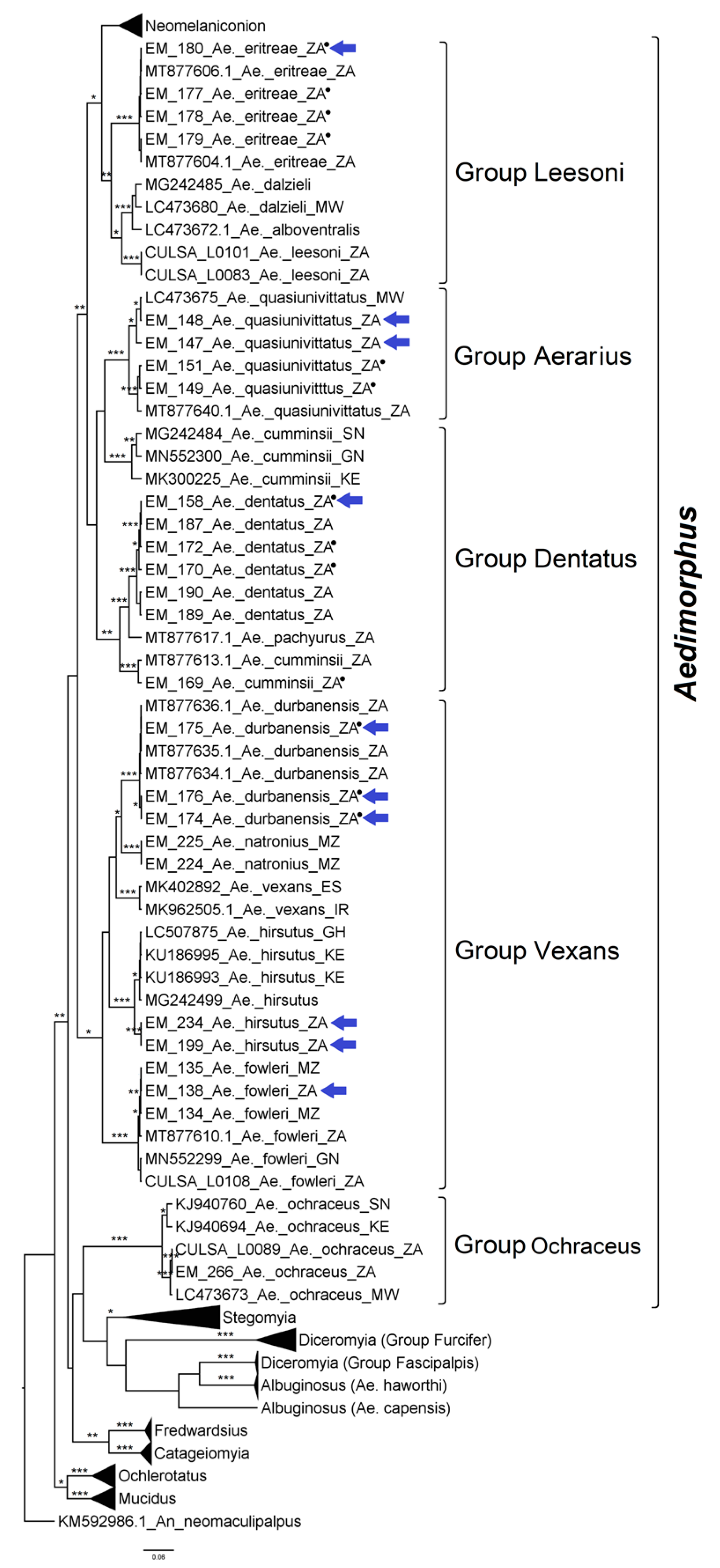
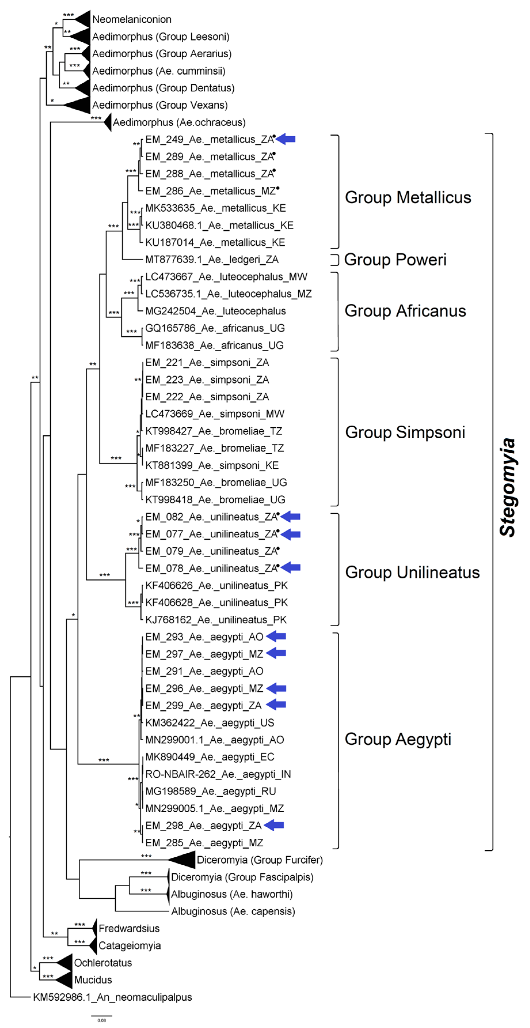
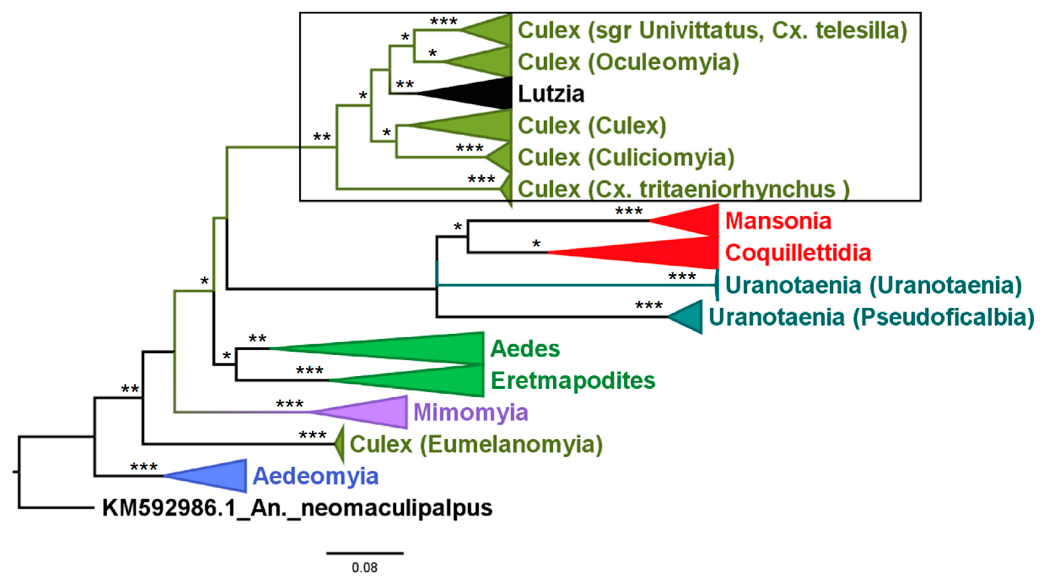
3.2. Mosquito Identification Using Phylogenetic Reconstruction

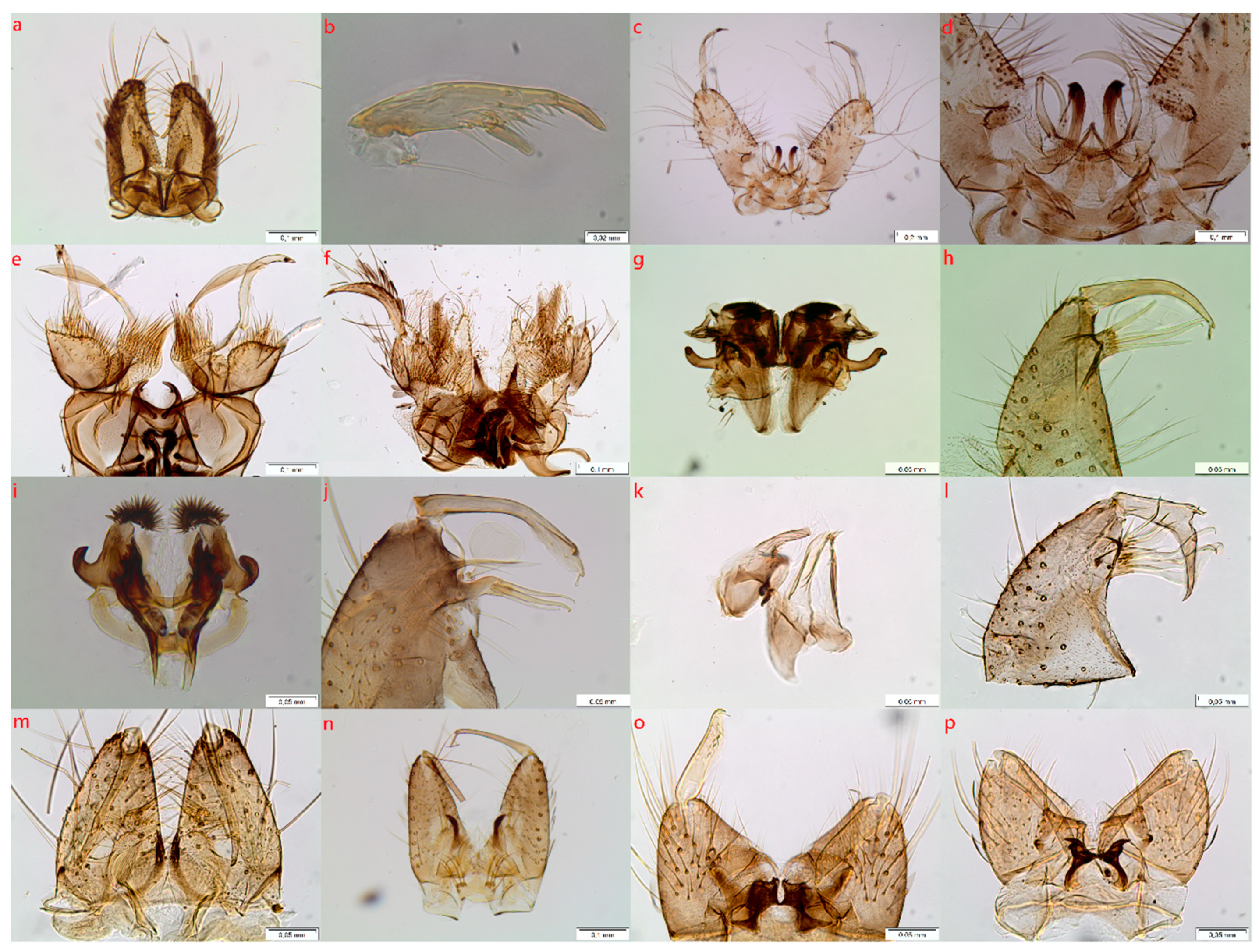
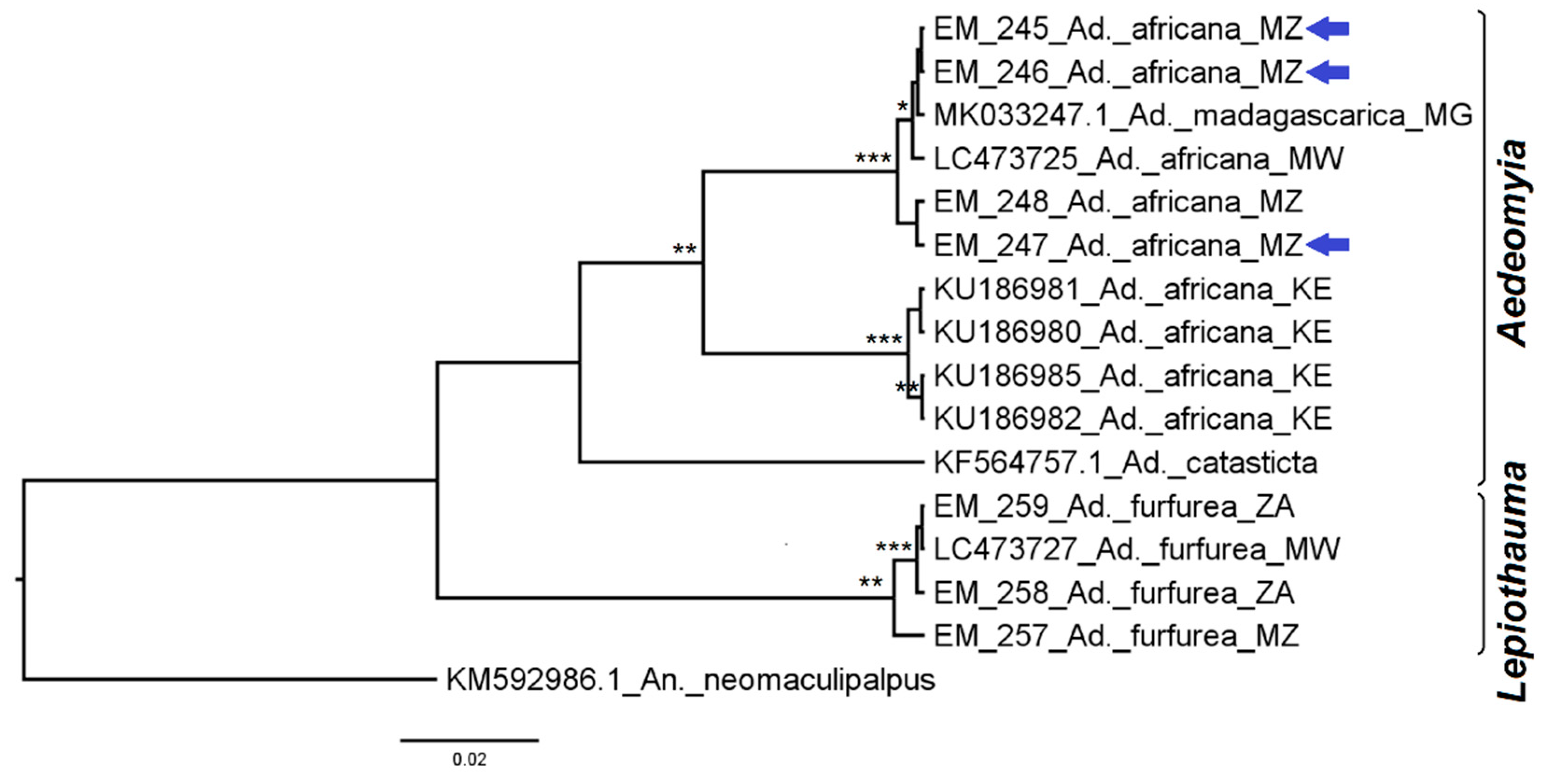
3.2.1. Genus Aedeomyia

Aedeomyia sequences were grouped phylogenetically according to their subgenera (Figure 2). Aedeomyia (Aedeomyia) africana Neveu-Lemaire, 1906 from Mozambique (File S-II Figure S1) was grouped according to a conspecific sequence from Malawi and another from Madagascar, Ad. (Ady) madagascarica Brunhes, Boussès & da Cunha Ramos, 2011. Those from Kenya formed their own clade with a divergence of 6.9% ± 1.3 between the two Ad. africana clades (Figure 2 and Supplementary Material File File S-IV, Table S3). Aedeomyia (Lepiothauma) furfurea (Enderlein, 1923), both from Mozambique and South Africa, formed a strong clade. The divergence between these two species was >10%. Networks and PCOORD analyses agreed with that topology (File S-IV, Figure S3).

3.2.2. Genus Aedes

Aedes sequences formed two main clusters, with species within subgenera Mucidus and Ochlerotatus forming a cluster separated from species representing all the other Aedes subgenera (Figure 3).
Species in the subgenus Neomelaniconion formed a single, monophyletic, strongly supported clade (Figure 4) in which Ae. (Neo.) mcintoshi Huang, 1985 (File S-II Figure S16), Ae. (Neo.) unidentatus McIntosh, 1971, and Ae. (Neo.) circumluteolus (Theobald, 1908) (File S-II Figure S15) were grouped in a clade with a variation of 1.2% ± 0.3 that overlapped the interspecific divergence (1.1–1.4%) (File S-IV, Table S4). Aedes (Neo.) lineatopennis (Ludlow, 1905) formed a sister clade, showing a divergence with the other species ≥ 5.4%. Networks and PCOORD analyses supported these results (File S-IV, Figure S4).
Most sequences of taxa in the subgenus Aedimorphus formed a polyphyletic clade, separating into subclades according to their morphologically based groupings designated by McIntosh [46] (Figure 5). However, Ae. (Adm.) cumminsii (Theobald, 1903) sequences from Kenya, Guinea, and Senegal shared an inter-group variation that ranged from 0.7–2.3%, according to the origin (File S-IV, Table S5), forming a clade distant from the conspecific sequences from South Africa, which joined the Dentatus group, Ae. (Adm.) dentatus (Theobald, 1904) (File S-II Figure S3) and Ae. (Adm.) pachyurus Edwards, 1936, to which they belong. The divergence between these two groups of Ae. cumminsii was ≥7.4%. Networks and PCOORD analyses corroborated this finding (File S-IV, Figure S5).
The Stegomyia subgenus formed a monophyletic clade in which most species formed well-supported species group clades (Figure 6). One exception was within the Simpsoni group where Ae. (Stg.) simpsoni (Theobald, 1905) and Ae. (Stg.) bromeliae (Theobald, 1911) formed a single clade with a variation of 1.1% ± 0.3, while the interspecific divergence of the species in the clade was 1.3% ± 0.4. Ae. (Stg.) unilineatus (Theobald, 1906) formed two monophyletic sister clades comprising sequences from either South Africa (File S-II Figure S20) or Pakistan, with a global intraspecific variation of 3.4% ± 0.6 and an inter-clade variation of 5.3% ± 0.9. Similarly, sequences from Ae. (Stg.) metallicus (Edwards, 1912) (Figure S-II S19) formed two monophyletic sister clades, with an inter-clade distance of 7.4% ± 1.0. These results were corroborated by the networks and PCOORD analyses, evidencing the near lack of separation of simpsoni/bromeliae, wider separation for the two groups of unilineatus, and even greater separation for metallicus (File S-IV, Figure S6).
Subgenus Diceromyia was paraphyletic, but the two species included—Ae. (Dic.) furcifer (Edwards, 1913) and Ae. (Dic.) fascipalpis (Edwards, 1912), both represented by specimens from South Africa (File S-II Figures S11 and S12)—formed species-specific clades with strong support (Figure 7), confirmed in networks and PCOORD analyses (File S-IV, Figure S7).
Ochlerotatus sequences formed a strong clade, with equally strong paraphyletic sub-clades; in these, Ae. (Och.) caballus (Theobald, 1912) (File S-II Figure S17) and Ae. (Och.) juppi McIntosh, 1973, both from South Africa, segregated into closer subclades, while Ae. caballus from Iran formed a separate cluster (Figure 7). Intraspecific variance within each of the three groups was low (≤0.5% ± 0.2); interspecific divergence between Ae. caballus and Ae. juppi from SA was 2.8% ± 0.7, and Ae. caballus from Iran had a divergence ≥ 3.6% ± 0.8 to either Ae. caballus or Ae. juppi from SA (File S-IV, Table S6). Networks and PCOORD analyses also placed Ae. caballus and Ae. juppi from SA closer to one another and farther apart from Ae. caballus from Iran (File S-IV, Figure S8).
The clade defining the subgenus Mucidus was strongly supported. Aedes (Muc.) sudanensis (Theobald, 1908) and Ae. (Muc.) scatophagoides (Theobald, 1901) were grouped in a single monophyletic cluster with an intra-clade variation of 0.9% ± 0.3 and an inter-specific divergence of 0.6% ± 0.2. The Ae. mucidus sequence from a Mozambique specimen segregated away from all Ae. scatophagoides with a divergence of 7.5% ± 1.2. Similarly, networks and PCOORD analyses placed Ae. mucidus sequences far from the sudanensis and scatophagoides, which were either pooled in an unsolved group or distributed along a single “dimension” without segregation (File S-IV, Figure S9).

3.2.3. Genus Eretmapodites

Eretmapodites sequences formed monophyletic clades separating the various species analyzed. Based on morphological features of male genitalia, Er. intermedius, Er. subsimilicipes (File S-II Figure S22), and Er. chrysogaster were very similar and considered members of the “Chysogaster group,” and separated quite distinctly from a clade consisting of a sequence of Er. quinquevittatus from Mozambique (File S-II Figure S21), which had quite different male genitalia and adult scutal patterns and a GenBank sequence denoted as Er. silvestris Ingram and de Meillon, 1927 (Figure 8). Eretmapodites subsimplicipes showed no intraspecific variation and diverged from Er. quinquevittatus by 9.3% ± 1.5 (File S-IV, Table S7). Similar results were obtained with networks and PCOORD analyses (File S-IV, Figure S10).

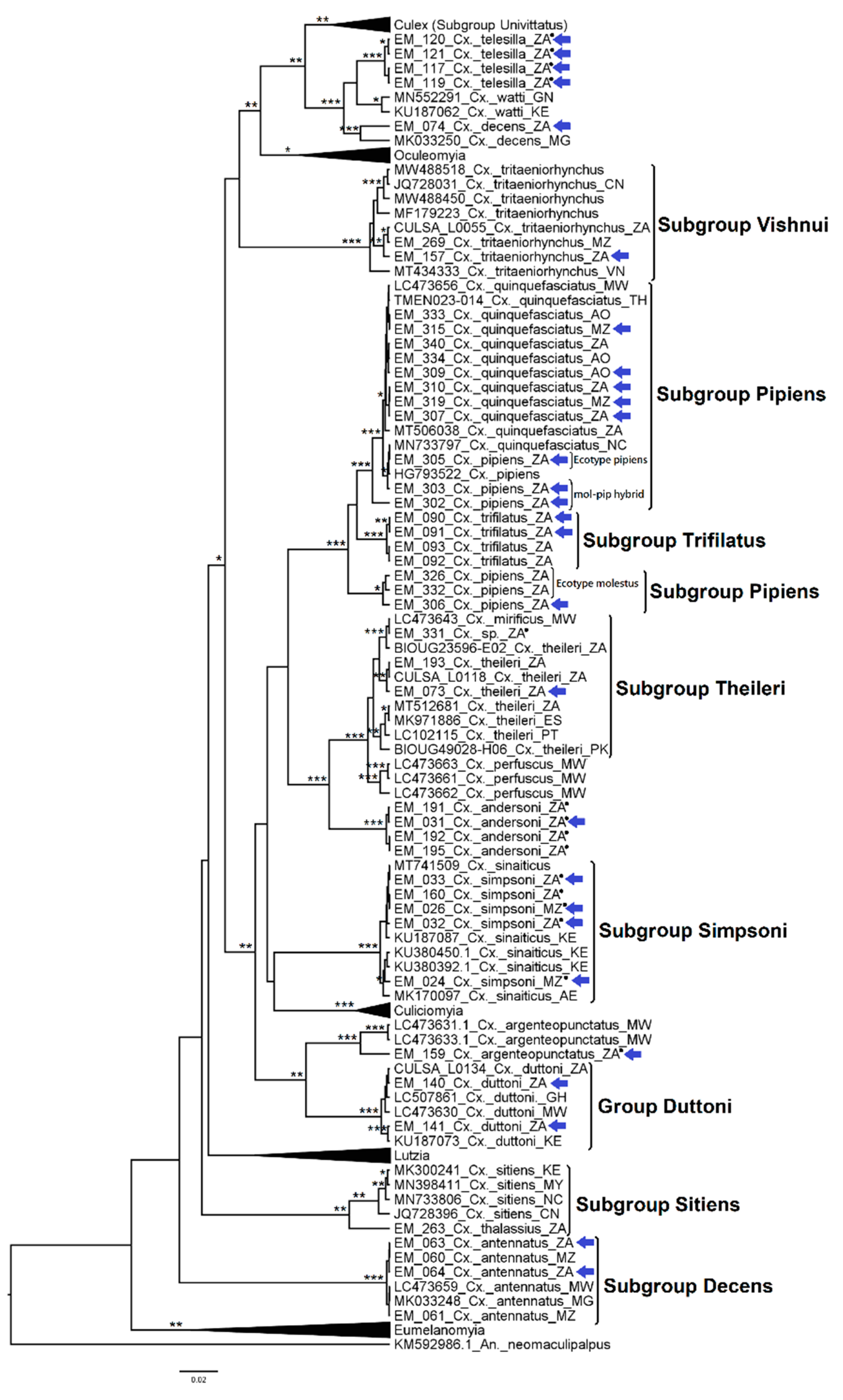
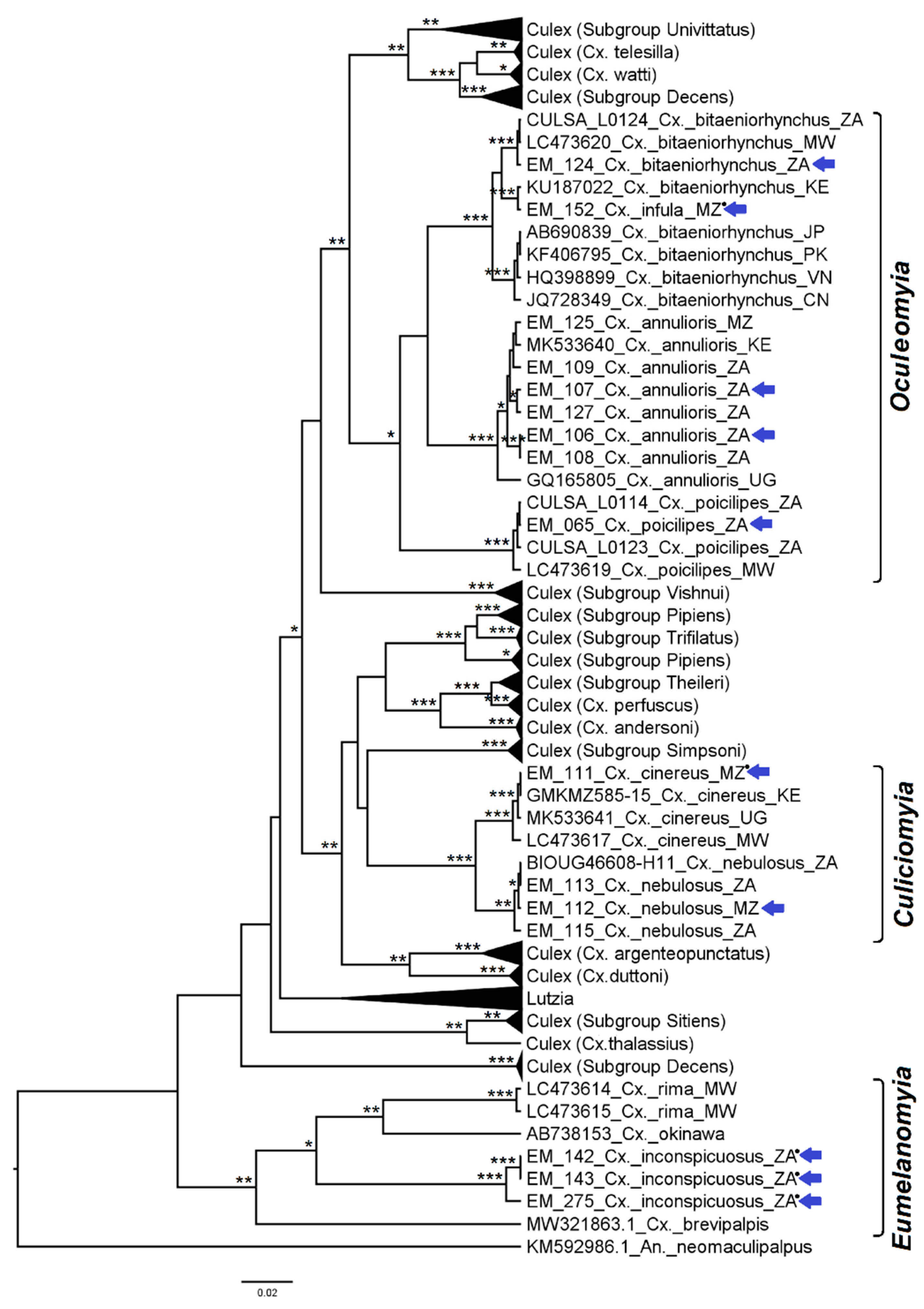
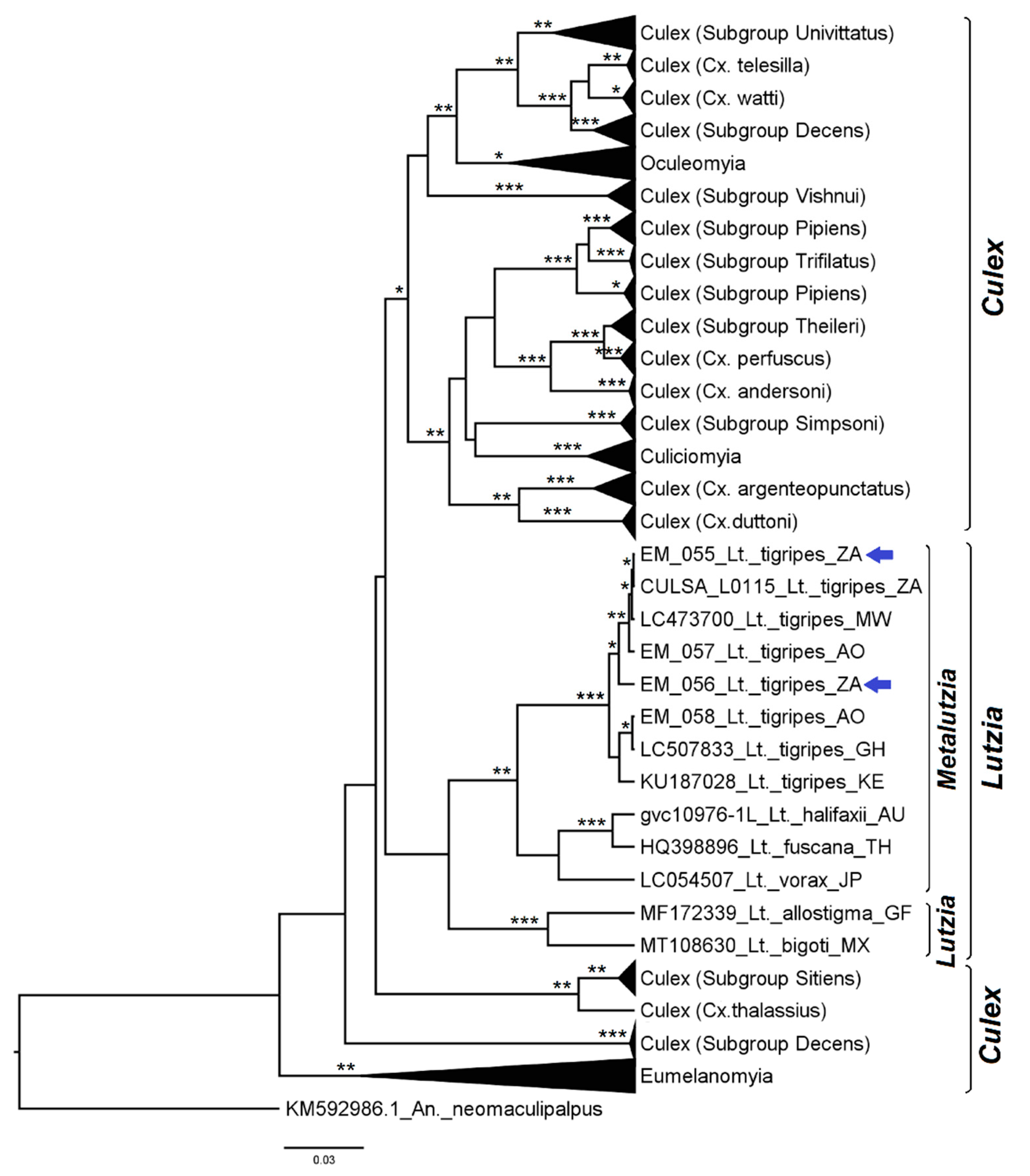
3.2.4. Genera Culex and Lutzia

Sequences from the genus Culex segregated into a highly polyphyletic topology, where most species of subgenus Culex segregated into clusters intermingled with members of other subgenera. Two major clades with support of one of the three methods were formed; the first contained species of the subgenus Culex, namely, some members of the groups Pipiens, Sitiens, the subgroup Vishnui, and the subgenus Oculeomyia; the second clade contained species of the subgenus Culex, namely, members of the Pipiens and Duttoni groups and the subgenus Culiciomyia. Other separate minor clades, without support among one another, were formed by species of subgroups Sitiens and Decens and the subgenus Eumelanomyia, with the genus Lutzia as a monophyletic clade (Figure 9).
The Univittatus subgroup formed a strongly supported clade, within which Cx. (Cux.) univittatus Theobald, 1901, Cx. (Cux.) neavei Theobald, 1906, and Cx. (Cux.) perexiguus Theobald, 1903, (File S-II Figures S27–S29) segregated into well-supported monophyletic clades (Figure 10). Sequences of Cx. perexiguus from South Africa and Mozambique clustered with sequences from other African countries, Europe, and the Middle East, with a divergence of 0.5% ± 0.2 between Cx. perexiguus from Europe and the Middle East and those from Africa (File S-IV, Table S8). Culex univittatus from Africa were segregated from those of European origin. Networks and PCOORD analyses confirmed these results (File S-IV, Figure S11).
Species of the pipiens complex, Cx. quinquefasciatus, and Cx. pipiens (File S-II Figures S31 and S32), and all those molecularly typed as Cx. quinquefasciatus, Cx. pipiens (pipiens ecotype plus hybrids of the pipiens and molestus ecotypes), formed a strongly supported clade (Figure 11). This monophyletic clade included Cx. (Cux.) trifilatus Edwards, 1914 (File S-II Figure S36), specimens of Cx. pipiens ecotype molestus, and one that could not be confirmed molecularly. The intra-clade variation supporting Cx. quinquefasciatus, Cx. pipiens (pipiens ecotype plus pipiens-molestus hybrids), and Cx. trifilatus was 1.6% ± 0.3, while the molestus ecotype diverged >2% in relation to the pipiens ecotype and Cx. quinquefasciatus, and Cx. trifilatus diverged ≥ 2.9% from any of the Pipiens subgroup members (File S-IV, Table S9). Similar results were obtained with networks and PCOORD analyses (File S-IV, Figure S12).
Sequences from South African Cx. (Cux.) theileri Theobald, 1903 (File S-II Figure S35), another from a female originally identified (by us) as Cx. sp. (EM331) and Cx. (Cux.) mirificus Edwards, 1913 from Malawi (sharing 100% CoxI identity with EM331), formed sister clades with other sequences of Cx. theileri from Spain, Portugal, and Pakistan, with an intraspecific variation of 0.8% ± 0.2 (Figure 11). These were joined by Cx. (Cux.) perfuscus Edwards, 1914 and Cx. andersoni (File S-II Figure S24), forming a larger, well-supported monophyletic clade, a pattern that was supported by networks and PCOORD analyses (File S-IV, Figure S13).
Culex spp. of the subgroup Simpsoni formed a strongly supported clade in which the sequences of Cx. (Cux.) simpsoni Theobald, 1905 from this study, which had been morphologically confirmed through the male genitalia (File S-II Figure S33), did not segregate from the sequences of Cx. (Cux.) sinaiticus Kirkpatrick, 1925 from GenBank (Figure 11). Intraclade, intraspecific and interspecies divergence values overlapped, ranging from 0.2% to 0.4% (±0.1–0.2). These species were neither segregated by networks nor PCOORD analyses (File S-IV, Figure S14).
Subgenus Oculeomyia formed a monophyletic clade with branch support in only one of three methods (Figure 12); Culex (Ocu.) bitaeniorhynchus Giles, 1901 (File S-II File S44), Cx. (Ocu.) infula Theobald, 1901 (File S-II Figure S43), Cx. (Ocu.) annulioris Theobald, 1901 (File S-II Figure S41), and Cx. (Ocu.) poicilipes (Theobald, 1903) (File S-II Figure S42) sequences formed sister clades. However, the clades containing Cx. bitaeniorhynchus and Cx. infula were not species-specific; rather, sequences were grouped according to geographic origin, separating African specimens from ones originating in Asia. Hence, to unravel the relation of these taxa, we performed a further phylogenetic reconstruction with a larger data set (File S-IV, Figure S15). Similarly, African sequences obtained in this work deviated from the large clade formed by sequences from Asia and the Middle East, without separation of Cx. bitaeniorhynchus and Cx. infula. The distance between the various groups of sequences from the various countries of origin, or of different species, did not surpass 3%, and the divergence of these clades ranged between 2.0–2.7% (File S-IV, Table S10a,b). Networks and PCOORD analyses (File S-IV, Figure S16) still failed to separate Cx bitaeniorhynchus from Cx. infula.
The subgenus Culiciomyia was grouped into a defined clade with strong branch support, where Cx. (Cui.) cinereus Theobald, 1901 and Cx. (Cui.) nebulosus Theobald, 1901 (File S-II Figures S38 and S39) formed equally strong monophyletic clades (Figure 12) with low intraspecific variation for each branch (≤0.4%), diverging by 3.6% ± 0.8.
Eumelanomyia sequences were grouped in an external clade of the remaining Culex subgenera (Figure 12), with Cx. inconspicuosus from South Africa (File S-II Figure S40) forming a strong clade with an intraspecific variation of 0.8% ± 0.3.
Sequences derived from Lutzia (Metalutzia) tigripes (de Grandpre & de Charmoy, 1901) from Angola and South Africa (File S-II Figure S45) were pooled with conspecific ones from other African countries (Figure 13). When analyzing the relationship of the genus Lutzia with the other genera studied in this work, it grouped within a strongly supported clade that combined it with species of the subgenera Culex, Oculeomyia, and Culiciomyia (Figure 14).

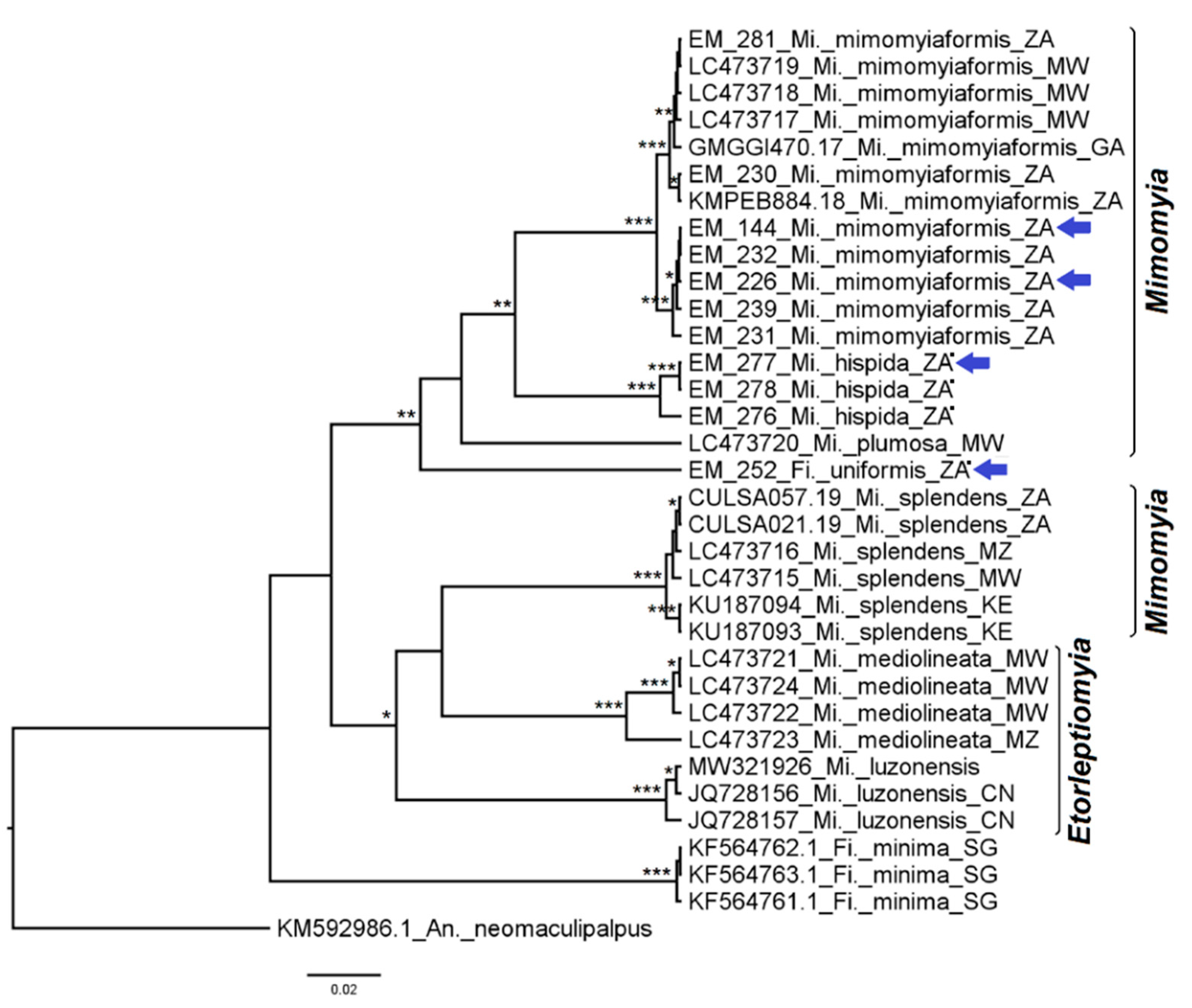
3.2.5. Genera Ficalbia and Mimomyia

The CoxI sequence obtained from Fi. uniformis (File S-II Figure S46) clustered inside the Mimomyia radiation, distant from the Fi. minima clade (Figure 15). Sequences of Mi. (Mimomyia) mimomyiaformis (Newstead, 1907) (File S-II Figure S47) and Mi. (Mim) hispida (File S-II Figure S48) clustered in a large clade, in which the former was organized into two strongly supported paraphyletic clades, with an overall intraspecific variation of 0.9% ± 0.3 (File S-IV, Table S11). Networks and PCOORD analyses revealed an identical pattern (File S-IV, Figure S17).

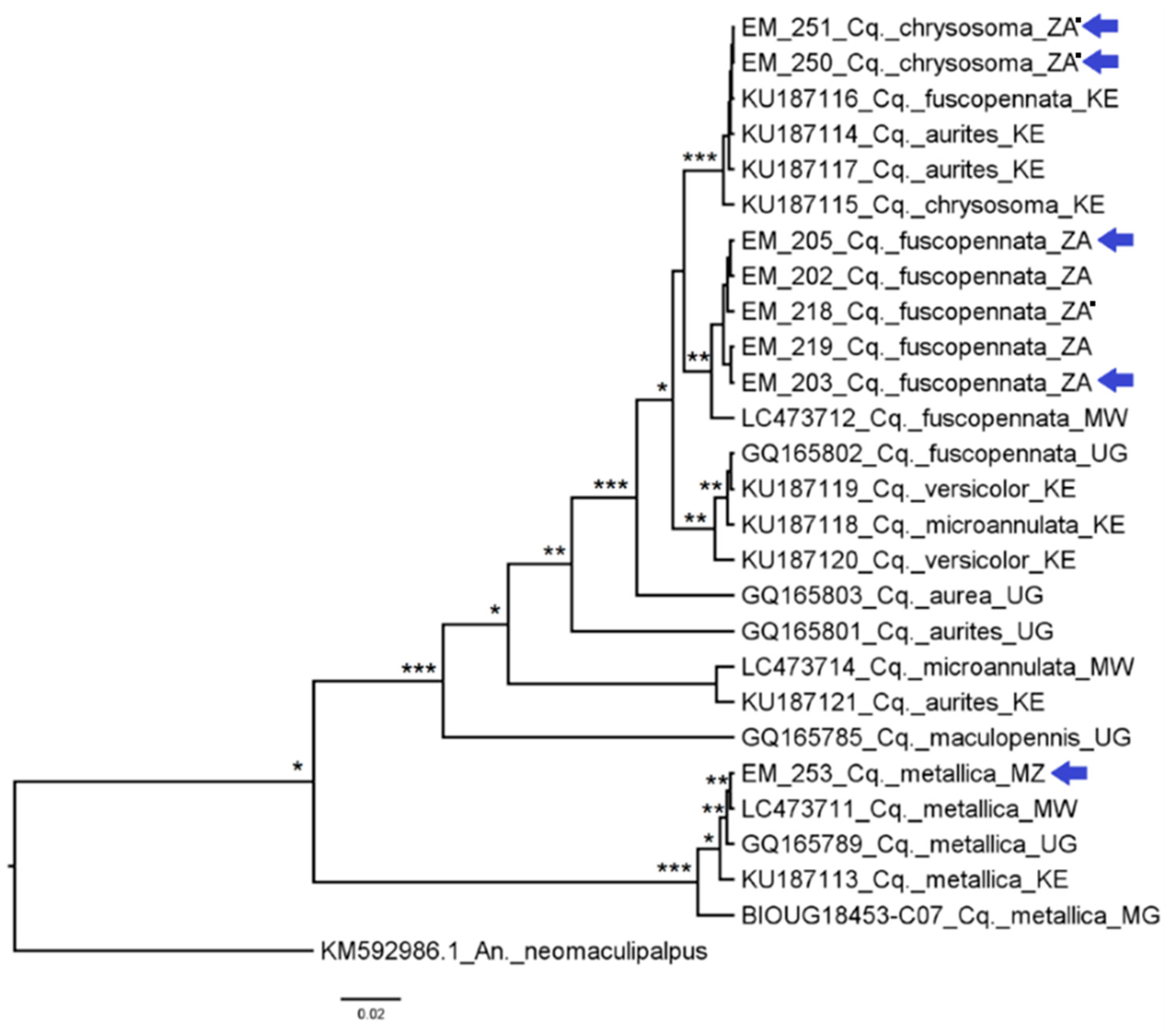
3.2.6. Genus Coquillettidia

Sequences from the South African Cq. (Coquillettidia) chrysosoma (Edwards, 1915) specimens (File S-II Figure S51) grouped with Cq. (Coq.) fuscopennata (Theobald, 1903), Cq. (Coq.) aurites (Theobald, 1901), and Cq. chrysosoma sequences from Kenya, with an intra-clade variation of 0.2% ± 0.1 (Figure 16). Sequences of Cq. fuscopennata from South Africa (File S-II Figure S49) clustered with a sequence from Malawi in a monophyletic clade with an intraspecific variation of 0.5% ± 0.2, while another clade clustered GenBank sequences from Cq. fuscopennata, Cq. (Coq.) versicolor (Edwards, 1913) and Cq. (Coq.) microannulata (Theobald, 1911). The sequence of Cq. (Coq.) metallica (Theobald, 1901) from Mozambique (File S-II Figure S50) clustered in a monophyletic clade with an intraspecific variation of 1.1% ± 0.3. This was confirmed by network and PCOORD analyses (File S-IV, Figure S18).

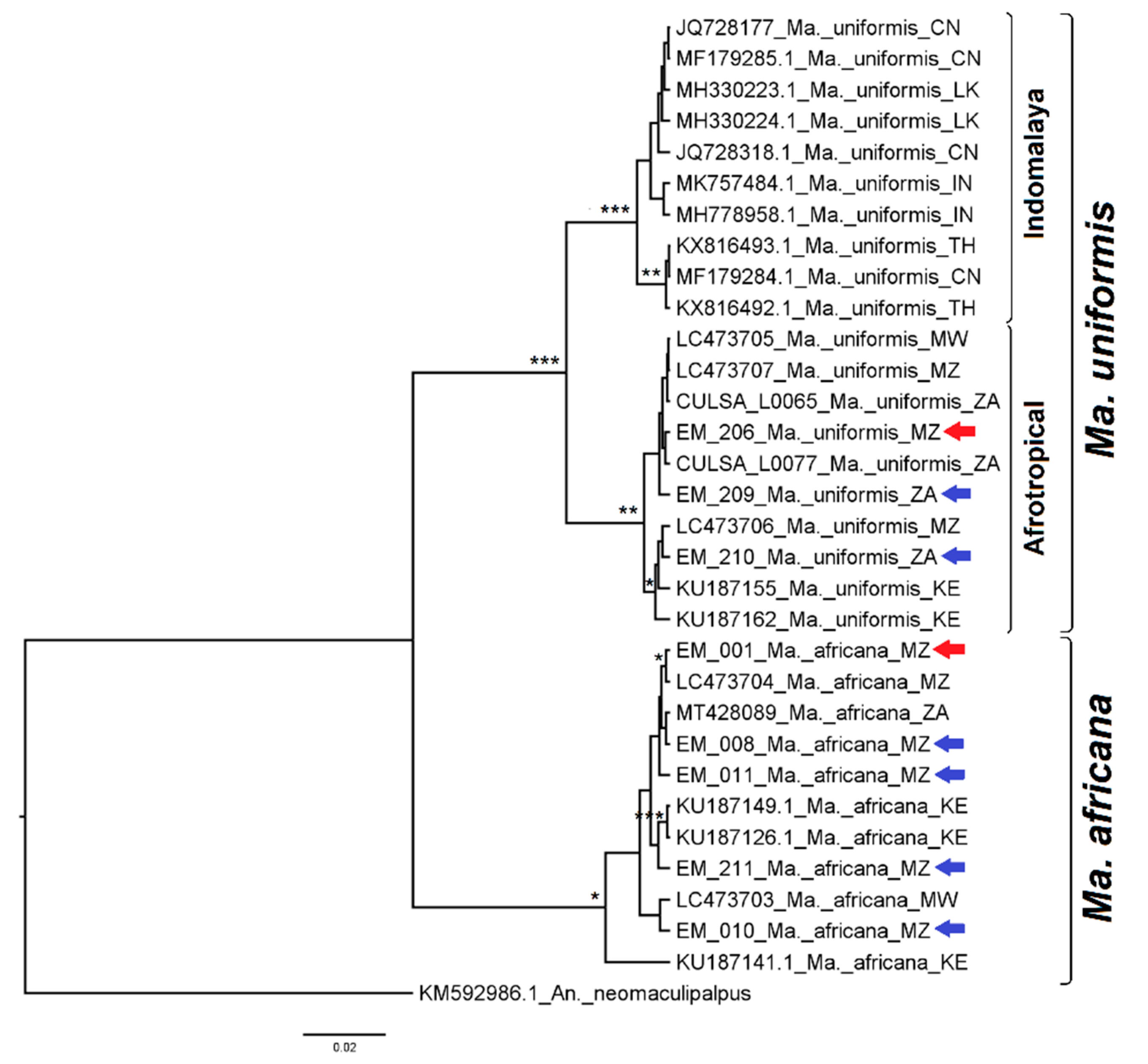
3.2.7. Genus Mansonia

Mansonia (Mansonioides) africana (Theobald, 1901) and Ma. (Mnd.) uniformis (Theobald, 1901) were identified in this study based on female and male genitalia structures (File S-II Figures S52 and S53). Sequences of Ma. uniformis, from the Afrotropical and Indomalayan regions, were placed in two sister clades (Figure 17) with low intra-clade variation (ranging from 0.4 to 0.7%) but diverging from one another by 4.1% ± 0.9. Mansonia africana joined conspecific sequences from various African origins, with a divergence from Ma. uniformis ≥9%. These results were congruent with the network and PCOORD analyses (File S-IV, Figure S19).

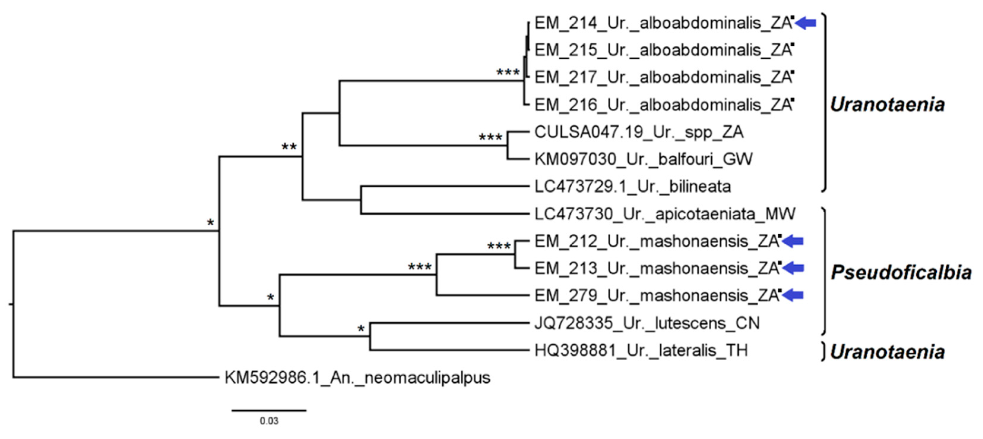
3.2.8. Genus Uranotaenia

Uranotaenia alboabdominalis (File S-II Figure S55) formed a strongly supported monophyletic clade (Figure 18) with an intraspecific variation of 0.2% ± 0.1. The sequences from Ur. mashonaensis (File S-II Figure S54) clustered into a monophyletic clade with strong support; however, the intraspecific variation was 4.1% ± 0.8, with a divergence of 5.6% ± 1.1 between the two branches.

4. Discussion

The genitalia of 663 mosquitoes (both male and female) were dissected and 55 species were identified; 247 partial sequences of the CoxI gene from 65 species were obtained and analyzed using complementary approaches, yielding a total of 67 species from 10 genera, identified by either method. This corresponded to circa 40% (60/150) of Culicinae mosquito fauna from South Africa and 34% (31/91) from Mozambique. Eleven of these partial CoxI sequences are, to the best of our knowledge, here published for the first time, with corresponding morphologic confirmation. Curiously, a considerable proportion of sequences that were generated failed to be identified either using the BOLD taxonomy tool (36%) or BLASTn (47%). In these cases, formal species assignment was carried out based on a fine morphological confirmation (genitalia) and/or by phylogenetic reconstruction.
Interspecific congeneric distances ranged between 1% and 20%, with mean values between 7% and 15% (Fiel S-IV Table S12). These values are mostly within the range observed for divergence in congeneric species, 2.3–21.8%, although the majority of cases are in the 4–11% interval (Ashfaq et al., 2014; Wang et al., 2012; refs. [21,27]). Low divergence values in congeneric taxa, such as 0.6–2.0%, can be interpreted as species of recent divergence [47]. Divergence between conspecific specimens typically ranges between 0% and 2.4% [21,27] or as high as 3% or 5.4% [18,48].
In some instances, divergence values > 2% in conspecific sequences were observed; hence, they were greater than expected in members of the same taxa; conversely, divergence values < 2% between taxa of different species were also obtained, revealing a failure of the CoxI marker to separate such taxa.
Higher than expected genetic diversity was observed in Ad. africana, where sequences from Malawi and Mozambique on the one hand and Kenya on the other formed separate clades with a divergence of 6.9%. Aedes cumminsii from South Africa fell within the Dentatus group as expected, jointly with Ae. dentatus from South Africa, a vector of RVFV and the Middelburg virus (MIDV) [2,3], while sequences from Kenya, Guinea, and Senegal formed a separate clade, diverging from the former by >7%. Aedes unilineatus, a monotypic member of the Unilineatus group [5] was considered a potential vector of ZIKV [49], with a very wide distribution in sub-Saharan Africa, the Middle East, and Asia [1], in which South African sequences and those originally from Pakistan had an interclade variation of 5.3%. Aedes metallicus, the monotypic member of the Metallicus group [5], an important vector of sylvatic YFV and potential vector of ZIKV in Africa [2], had high sequence divergence of 7.4% between South Africa and Mozambique versus that from Kenyan specimens. Aedes furcifer from South Africa, Kenya, and Senegal, had an intraspecific variation of 8.1%. Aedes caballus from South Africa clustered separately from those originating in Iran, with a divergence of ≥3.6% between them. Mansonia uniformis formed separate clades, according to African or Indomalayan-Asian origin, with a divergence of 4% between them. Uranotaenia mashonaensis from South Africa had a considerable variation of 4.1%.
Such divergence may be explained, in some cases, by comparing sequences of naturally different conspecific populations collected far apart geographically, such as in this study with Ae. unilineatus, Ae. caballus, and Ma. uniformis, which compared sequences of specimens originating from South Africa to those from the Asian region. However, others such as Ad. africana, Ae. cumminsii, Ae. metallicus, Ae. furcifer, and Ur. mashonaensis exhibited a large genetical divergence in sequences between specimens originating from a span of regional context in the African continent, e.g., South Africa, Mozambique, and Kenya.
It is not surprising that Aedes cumminsii, a vector of MIDV, Spondweni virus (SPOV), and RVFV [2], had considerable within-species sequence variations, as it is likely a sibling species complex. Throughout its broad savanna- and forest-dwelling distribution in Africa, many morphological variations have been noted (AJC personal communication and [4]). In addition, Ae. cumminsii has undergone some taxonomic confusion since it was originally described as a now designated valid subspecies, ssp mesostictus [1], which was originally named ssp mediopunctatus (Theobald, 1909) and later placed synonymously and elevated to a subspecies of Ae. cumminsii [50]. This subspecies was originally described from specimens collected in Ghana and differs from the typical form of Ae. cumminsii by the presence of small basal median whitish spots on the abdominal tergites in both sexes [4,5]; however, McIntosh [46] suggested that this subspecies occurs only in southern Africa. We identified Ae. cumminsii with the typical features of the subspecies mesostictus in northeastern South Africa, such as Guarido et al. [12], with a divergence of >7% from specimens from Kenya [15].
A lack of CoxI sequence separation of taxa of different species was found between Ad. madagascarica and Ad. africana; Ae. mcintoshi, Ae. circumluteolus, and Ae. unidentatus; Ae. simpsoni and Ae. bromeliae; Ae. scatophagoides and Ae. sudanensis; Cx. quinquefasciatus and Cx. pipiens; Cx. simpsoni and Cx. sinaiticus; and Cx. bitaeniorhynchus and Cx. infula. In these cases, they could not be correctly segregated into species-specific clusters either by traditional phylogenetic reconstruction, networks, or PCOORD analyses. Fortunately, although being morphologically similar species, they can still be differentiated by fine morphological details or male genitalia. Furthermore, genetic distance analysis disclosed overlapping intra- and inter-specific values, circa <2%, showing a limitation in their resolution capacity. Such overlap has been responsible for misidentifications and impossibilities of delimiting species based on pairwise distances [17,51]. One such example includes the segregation of Ad. africana from Mozambique (EM_245, LC662529) and Malawi (LC473725) with Ad. madagascarica (MK033247.1). Although Ad. madagascarica has only been described in Madagascar, the genetic divergence between this species and Ad. africana was only 0.2%. In a contrasting situation, Ad. africana CoxI sequences from neighboring countries were separated as aforementioned, raising the need for further clarification of the significance of both the similarity between sequences of Ad. madagascarica and Ad. africana from Mozambique and Malawi and the divergence of Ad. africana from Mozambique and Malawi versus Kenya.
Among the Aedes, the subgenus Neomelaniconion includes potential vectors of arboviruses, such as Ae. mcintoshi, a major vector of RVFV, and Ae. circumluteolus and Ae. unidentatus as potential RVFV vectors [2], but also potential vectors of the Shuni virus (SHUV) [31]. These three taxa could not be differentiated through phylogenetic reconstruction, in agreement with previous findings [12,13]. Evidence from Kenya suggests that Ae. mcintoshi forms a complex of morphologically indistinguishable species, with discordant results between CoxI and ITS markers [38], particularly ITS2, thereby failing to resolve species and species complexes in the subgenus Neomelaniconion in Madagascar [52].
Culex pipiens and Cx. quinquefasciatus, which are members of the Culex pipiens complex, not only display wide geographic distribution but are also highly relevant in the transmission of various pathogens, including arboviruses such as USUV, WNV, and SINV [1]. The female specimens of the two species are morphologically similar but differ in their vectorial efficiency and may occur sympatrically; additionally, hybridization has been reported in some locations [53,54,55], but not in South Africa [53]. In this study, the absence of pipiens-quinquefasciatus hybrids in southern Africa was also noted. Hybrids of the molestus and pipiens ecotypes, which, so far, have only been reported in the United States [56], Southern Europe [55], and North Africa [57], have also been described in this work as male Cx. pipiens specimens from South Africa. Curiously, while CoxI analyses could not resolve the closely convergent Cx. quinquefasciatus and Cx. pipiens into species-specific clusters, the molestus ecotype sequences clustered out, diverging 2.2–2.8% in relation to pipiens ecotype, pipiens-molestus hybrids, and Cx. quinquefasciatus. Other studies have reported an intraspecific variation in Cx. pipiens (3%), larger than the interspecific distance with Cx. quinquefasciatus (1.6%) and their lack of separation [18,24]. The CoxI gene has been successful in differentiating members of the Cx. pipiens complex, though with low variability; therefore, it may not be the better marker to infer the evolutionary relationship of such close taxa, and more polymorphic markers or a multilocus analysis may be more informative [58]. A lack of differences in CoxI between different species can also be explained by possible introgression of mitochondrial DNA after several interspecific crosses, which was proven for Culex species using several DNA markers [58].
Culex bitaeniorhynchus and Cx. infula belong to the Cx. bitaeniorhynchus complex of the subgenus Oculeomyia; Cx. bitaeniorhynchus has a wide distribution, being present in tropical and subtropical areas of the Afrotropical, Southern Palearctic, and Indomalayan regions, and on the mainland and islands of Southeast Asia and Australasia [1], and can be involved in the transmission of arboviruses [34]. Its status, as well as that of Cx. infula and Cx. ethiopicus Edwards, 1941, has been the subject of controversy [34,36]. The morphological characteristics of the specimens identified as Cx. bitaeniorhynchus (synonymous Cx. ethiopicus) corresponded to those described by Edwards [4] and Jupp [5], while the specimen of Cx. infula from Mozambique only allowed us to ascertain it as a Cx. sp., confirmed by the analysis of the genitalia. Malawian mosquitoes identified as Cx. ethiopicus were found to differ in the shape of the wing scales and diverge in the CoxI gene > 2% with Cx. bitaeniorhynchus from Asia [13]. In our study, the genetic divergence ranged from 2.0% (±0.6) to 2.7% (±0.7) with sequences from Asia, and the tree topology, networks, and PCOORD analysis suggest that CoxI does not have discriminating power for separating Cx. bitaeniorhynchus from Cx. infula. The Cx. infula CoxI sequence from Mozambique also deviated from Cx. infula from Asia by a similar range. So far, Cx. infula has only been described in Asia [1]; however, we were able to confirm its previous identification by Ribeiro in Angola of five male specimens which he designated as Cx. bitaeniorhynchus [59]. Furthermore, specimens collected in Africa continue to be classified as Cx. bitaeniorhynchus and Cx. ethiopicus, according to Edwards’ [4] nomenclature [13,29]. Altogether, based on the evidence presented, we believe that most of the specimens identified as Cx. bitaeniorhynchus in Africa actually are Cx. infula, a situation that Harbach [34] had already anticipated.
Aedes aegypti Linnaeus, 1762 is one of the most important vectors of several global health impact arboviruses, such as DENV, ZIKV, CHIKV, and YFV, not only in Africa but globally [1,2,60]. In the phylogenetic analysis, all sequences of Ae. aegypti grouped into a single clade with strong branch support. Although this clade was divided into two branches, albeit only one with reasonable support, and both with a very small distance, there were no data to support the notion that these may correspond to either subspecies Ae. aegypti aegypti or Ae. aegypti formosus Walker, 1848. Our samples and data set were not adequate for such a separation as no clear morphological differentiation was noticed in our specimens. A small set of sequences (N = 10) was analyzed, and for low variability data, as in this case, the phylogenetic study performed was not that indicated, and haplotype analyses of markers such as mtDNA ND4 [61] or microsatellites [62] were necessary.
As to the arrangement of genera, on the whole, Aedes sequences formed clusters that mainly corresponded to the subgenera, in agreement with the morphology-based taxonomy, including the informal groups proposed by McIntosh for the species of Subgenus Aedimorphus [5,46]. Subgenera Neomelaniconion, Stegomyia, Catageiomyia, Fredwardsius, Ochlerotatus, and Mucidus formed monophyletic clades, but Diceromyia, Aedimorphus, and Albuginosus did not. Subgenus Diceromyia was represented by Ae. furcifer and Ae. fascipalpis, which, although well separated, yielded a paraphyletic arrangement. Genus Aedes is a highly complex entity, the taxonomy of which is in dire need of clarification [35], and that mitogenome evolutionary analysis has shown to be paraphyletic [63].
Genus Culex segregated into a highly paraphyletic topology, where most species of subgenus Culex coincided with the informal groups and subgroups proposed by Harbach [64]; however, mitogenome phylogenetics has found genus Culex to be monophyletic [63]. To the best of our knowledge, this is the first phylogenetic study including African members of the subgenus Culiciomyia. While sequences of only two species were included in this study, the monophyly of Culiciomyia was well supported, and the CoxI marker performed well in the discrimination of Cx. nebulosus and Cx. cinereus. Multiple species of Culiciomyia occur in Africa and all have identical female and male external morphologies, relying solely on male genitalia structures for species identification [65]. Separation of species by CoxI gene sequences may be fruitful in the case of Culiciomyia.
Lutzia tigripes is the only representative of the genus Lutzia in the Afrotropical region [1], clustering as a monophyletic assemblage within the Culex radiation, as previously found with CoxI and ITS2 [20,22,66]. Morphological data from adults and larvae support different patterns of relationships between Lutzia and Culex [67], while a recent analysis of the complete mitochondrial genome concluded that Lutzia forms a monophyletic group with genus status [68], emphasizing the limitations of phylogenetic studies with a single marker. However, the classification controversy is not limited to the genus Lutzia. Support for the monophyly of Culicini generic-level groups is granted for all except subgenera Culex, Eumelanomyia, and Neoculex [67]. Our analysis could not confirm subgenus Culex as a monophyletic group, while Eumelanomyia formed a clade distant from the remaining Culex, in agreement with previous works [23,28,69]. Nevertheless, the study of all genera together yielded some interesting results; except for subgenus Eumelanomyia of Culex, there was support for tribes Aedini, Culicini, Ficalbiini, and Mansoniini, in agreement with the monophyly of genera Mansonia, Coquillettidia, and Culex, through mitochondrial phylogenomics [63].
In most of these cases, representative studies involving more taxa, a higher number of specimens per taxa sampled over a wider geographic range, and merging morphological and molecular characterization are needed to unravel the specific status of different populations and characterize species complexes in Africa and their relationship with their members elsewhere and/or the monophyly/paraphyly of some subgenera or genera. The systematics within the Culicini tribe cannot be resolved with morphological data alone [67], stressing the relevance of obtaining new molecular data.
Circa 40% (36–47%) of the sequences obtained in this study could not be correctly identified using BOLD and BLASTn as identification tools; this was because (i) the sequence was obtained for the first time, (ii) they had been obtained from members of species complexes, or (iii) there was an incorrect assignment, including at the genus level, such as sequences from Cx. inconspicuosus and Ae. durbanensis that were identified in BOLD as Ae. argenteopuntatus and Cx. tritaeniorhynchus, respectively, with >99% probability. Although studies that associate morphological and CoxI barcode-based molecular identifications are increasing, few include a definite diagnostic identification [20,24,48]. This absence is a potential source of error, as many species are only distinguishable by subtle morphological differences in the male genitalia. In fact, we have detected sequences from the barcode fragment of the CoxI gene from GenBank and Boldsystems which, given their phylogenetic signal, suggested the possibility of misassignment to another species. Examples of such are the Coquillettidia heterospecific sequences that clustered with our sequence of Cq. chrysosoma and the sequence ascribed to Er. Silvestris, which had complete identity with our sequence of Er. quinquevittatus, given that in both cases we had the morphology-based identification to the level of male genitalia for our sequences. This type of error has already been identified in other studies based on the CoxI marker [25,26] and ITS [33]. Such species assignment errors are perpetuated and amplified when authors consider only genetic similarities with previous GenBank entries. CoxI-based barcoding should complement morphologically-based identification [20], rather than species identification being based only on genetic similarities with existing sequences in the GenBank database [15].
Incorrect assignments also cause irregular situations in the BINs (barcode index numbers) assigned to what the BOLD system defines as operational taxonomic units, ideally corresponding to different species. For example, as in the case of the Aedeomyia species from the Afrotropical region, where three BINs were identified, the first for Ad. furfurea from Malawi (BOLD:AEH5592), the second BIN was shared between Ad. madagascarica and Ad. africana (BOLD:ADV5603) and the third was shared between Ad. africana and Ad. furfurea (BOLD:ACK8488). In the third case, there may have been an incorrect assignment of certain sequence(s) to the species Ad. furfurea, a situation that phylogenetic analysis was able to resolve. In other cases, more than one species clustering into one BIN have been registered, and another species has been split into more than one BIN [26].

5. Conclusions

Our study has contributed to the barcode library of Afrotropical mosquitoes, some of which are known potential vectors of arboviruses [2,3] or have recently been found to be so, or carriers of insect specific flavivirus [30,31,32,70]. This was achieved by associating careful morphologically identified referenced voucher specimens to specific molecular marker CoxI partial sequences. However, partial CoxI sequences have been shown to fail in unambiguously discriminating some proximal species or members of species complexes in addition to overestimating the diversity of Culex spp. [17]. Hence, it will be necessary to use alternative molecular markers, including nuclear, such as Ace2 [11], microsatellites [54,55,56], or mitochondrial, such as ITS, to molecularly delineate species. However, that may prove to not always be sufficient [52] and other markers such as 16S [26], ND4 [71], or the complete mitochondrial genome [63,68] may be required.

Supplementary Materials

The following supporting information can be downloaded at: https://www.mdpi.com/article/10.3390/d14110940/s1, File S-I—Table SI-1; File S-II—Photos of mounted genitalia; File S-III—Original phylogenetic trees + likelihood mapping; File S-IV—Extra figures + tables.

Author Contributions

A.P.G.d.A., R.P.—study conceptualization; A.P.G.d.A., R.P., M.V., L.B., A.J.C., A.P.A., J.F.—study design and methodology; A.P.G.d.A., A.P.A., M.M.G., A.K., A.J.C., L.B., P.N.T., C.A.S.—field surveys and sample collections; E.M.-S., A.P.A., M.M.G., A.K., A.P.G.d.A., A.J.C., V.V., M.T.N., R.P.—sample processing and analysis; E.M.-S., R.P., A.P.G.d.A.—phylogenetics and statistical analysis; E.M.-S., A.P.G.d.A., R.P.—manuscript drafting. All authors have read and agreed to the published version of the manuscript.

Funding

Eddyson Montalvo-Sabino was recipient of a grant from “Programa Nacional de Becas y Crédito Educativo” (PRONABEC), 2019—Beca Generacion del Bicentenario, from the “Ministerio de Educación” of Peru. A.P. Abilio was a recipient of a grant from Wellcome Trust (Grant WT087546MA) through SACIDS RVF and NPHI-Phase-II from the National Institute for Health of Mozambique through a cooperative agreement number [5NU14GH001237-03-00]. Marietjie Venter was a recipient of a sub-award from the Global Disease Detection Program, US-CDC award 5U19GH000571-02 with the NICD and University of Pretoria that funded vector surveillance in South Africa (2012–2015) and by the Cooperative Agreement Number (5 NU2GGH001874-02-00) with the University of Pretoria (2014–2017). Milehna M. Guarido received a studentship through this grant. A.P.G. Almeida has been a recipient of the Visiting Professor Programme by the University of Pretoria for the work in South Africa. This work received financial support from the Global Health and Tropical Medicine Center (GHTM|IHMT|NOVA), which is funded through FCT contract UID/Multi/04413/2013, Portugal. The findings and conclusions expressed in this manuscript are those of the author(s) and do not necessarily represent the official position of the funding agencies.

Institutional Review Board Statement

Collection of mosquitoes in South Africa was cleared under Section 20 approval by the Department of Agriculture Land Reform and Rural Development. Informed consent was obtained from the head of the household or property owners in Mozambique and South Africa.

Data Availability Statement

The slides with the mounted dissected genitalia of the mosquitoes in this study are deposited in the Institute of Hygiene and Tropical Medicine|NOVA University Lisbon (IHMT|NOVA) Insect Collection, Lisbon, Portugal.

Acknowledgments

We are grateful to Lapalala Wilderness, Marataba Conservation, South African National Parks, and private farm owners for logistical assistance and permission to collect mosquitoes on their properties. Carla A. Sousa acknowledges Filomeno Fortes, “Diretor nacional do programa de combate às grandes endemias”, and his team, responsible for the “Missão de apoio técnico especializado no âmbito do combate ao surto epidémico de febre-amarela, 2016”, under which mosquito collections in Angola took place.

Conflicts of Interest

The authors declare no conflict of interest. The funders had no role in the design of the study; collection, analysis, or interpretation of data; writing of the manuscript; or decision to publish the results.

References

  1. Wilkerson, R.; Linton, Y.-M.; Strickman, D. Mosquitoes of the World; Johns Hopkins University Press: Baltimore, MA, USA, 2021; Volume 1. [Google Scholar]
  2. Braack, L.; De Almeida, A.P.G.; Cornel, A.J.; Swanepoel, R.; De Jager, C. Mosquito-Borne Arboviruses of African Origin: Review of Key Viruses and Vectors. Parasites Vectors 2018, 11, 29. [Google Scholar] [CrossRef] [PubMed]
  3. Venter, M. Assessing the Zoonotic Potential of Arboviruses of African Origin. Curr. Opin. Virol. 2018, 28, 74–84. [Google Scholar] [CrossRef] [PubMed] [Green Version]
  4. Edwards, F.W. Mosquitoes of the Ethiopian Region. III.-Culicine Adults and PupAe; British Museum (N.H.): London, UK, 1941. [Google Scholar]
  5. Jupp, P.G. Mosquitoes of Southern Africa: Culicinae and Toxorhynchitinae; Ekogilde Publishers: New York, NY, USA, 1996. [Google Scholar]
  6. Huang, Y. Aedes (Stegomyia) Simpsoni Complex in the Ethiopian Region with Lectotype Designation for Simpsoni (Theobald) (Diptera: Culicidae). Mosq. Syst. 1979, 11, 221–234. [Google Scholar]
  7. Huang, Y. Aedes (Stegomyia) Bromeliae (Diptera: Culicidae), the Yellow Fever Virus Vector in East Africa. J. Med. Entomol. 1986, 23, 196–200. [Google Scholar] [CrossRef] [PubMed]
  8. Huang, Y. Notes on the Aedes (Diceromyia) Furcifer Group, with a Description of a New Species (Diptera: Culicidae). Proc. Entomol. Soc. Washingt. 1986, 88, 634–649. [Google Scholar]
  9. Harbach, R.E.; Knight, K.L. Taxonomists’ Glossary of Mosquito Anatomy; Plexus Publishing, Inc.: Marlton, NJ, USA, 1980; ISBN 9780937548004. [Google Scholar]
  10. Walton, C.; Sharpe, R.G.; Pritchard, S.J.; Thelwell, N.J.; Butlin, R.K. Molecular Identification of Mosquito Species. Biol. J. Linn. Soc. 1999, 68, 241–256. [Google Scholar] [CrossRef]
  11. Smith, J.L.; Fonseca, D.M. Rapid Assays for Identification of Members of the Culex (Culex) Pipiens Complex, Their Hybrids, and Other Sibling Species (Diptera: Culicidae). Am. J. Trop. Med. Hyg. 2004, 70, 339–345. [Google Scholar] [CrossRef] [Green Version]
  12. Guarido, M.M.; Riddin, M.A.; Johnson, T.; Braack, L.E.O.; Schrama, M.; Gorsich, E.E.; Brooke, B.D.; Almeida, A.P.G.; Venter, M. Aedes Species (Diptera: Culicidae) Ecological and Host Feeding Patterns in the North-Eastern Parts of South Africa, 2014–2018. Parasites Vectors 2021, 14, 339. [Google Scholar] [CrossRef]
  13. Maekawa, Y.; Pemba, D.; Kumala, J.; Gowelo, S.; Higa, Y.; Futami, K.; Sawabe, K.; Tsuda, Y. DNA Barcoding of Mosquitoes Collected through a Nationwide Survey in 2011 and 2012 in Malawi, Southeast Africa. Acta Trop. 2021, 213, 105742. [Google Scholar] [CrossRef]
  14. Mixão, V.; Bravo Barriga, D.; Parreira, R.; Novo, M.T.; Sousa, C.A.; Frontera, E.; Venter, M.; Braack, L.; Almeida, A.P.G. Comparative Morphological and Molecular Analysis Confirms the Presence of the West Nile Virus Mosquito Vector, Culex Univittatus, in the Iberian Peninsula. Parasites Vectors 2016, 9, 601. [Google Scholar] [CrossRef] [Green Version]
  15. Makanda, M.; Kemunto, G.; Wamuyu, L.; Bargul, J.; Muema, J.; Mutunga, J. Diversity and Molecular Characterization of Mosquitoes (Diptera: Culicidae) in Selected Ecological Regions in Kenya. F1000Research 2018, 7, 262. [Google Scholar] [CrossRef] [Green Version]
  16. Ajamma, Y.U.; Villinger, J.; Omondi, D.; Salifu, D.; Onchuru, T.O.; Njoroge, L.; Muigai, A.W.T.; Masiga, D.K. Composition and Genetic Diversity of Mosquitoes (Diptera: Culicidae) on Islands and Mainland Shores of Kenya’s Lakes Victoria and Baringo. J. Med. Entomol. 2016, 53, 1348–1363. [Google Scholar] [CrossRef] [PubMed]
  17. Hemmerter, S.; Šlapeta, J.; Beebe, N.W. Resolving Genetic Diversity in Australasian Culex Mosquitoes: Incongruence between the Mitochondrial Cytochrome c Oxidase I and Nuclear Acetylcholine Esterase 2. Mol. Phylogenet. Evol. 2009, 50, 317–325. [Google Scholar] [CrossRef]
  18. Hernández-Triana, L.M.; Brugman, V.A.; Nikolova, N.I.; Ruiz-Arrondo, I.; Barrero, E.; Thorne, L.; de Marco, M.F.; Krüger, A.; Lumley, S.; Johnson, N.; et al. DNA Barcoding of British Mosquitoes (Diptera, Culicidae) to Support Species Identification, Discovery of Cryptic Genetic Diversity and Monitoring Invasive Species. Zookeys 2019, 2019, 57–76. [Google Scholar] [CrossRef] [Green Version]
  19. Awedoba, A.K. West Africa. Afr. Yearb. 2020, 16, 32–47. [Google Scholar] [CrossRef]
  20. Chan, A.; Chiang, L.P.; Hapuarachchi, H.C.; Tan, C.H.; Pang, S.C.; Lee, R.; Lee, K.S.; Ng, L.C.; Lam-Phua, S.G. DNA Barcoding: Complementing Morphological Identification of Mosquito Species in Singapore. Parasites Vectors 2014, 7, 569. [Google Scholar] [CrossRef]
  21. Ashfaq, M.; Hebert, P.D.N.; Mirza, J.H.; Khan, A.M.; Zafar, Y.; Mirza, M.S. Analyzing Mosquito (Diptera: Culicidae) Diversity in Pakistan by DNA Barcoding. PLoS ONE 2014, 9, e97268. [Google Scholar] [CrossRef] [PubMed] [Green Version]
  22. Demari-Silva, B.; Vesgueiro, F.T.; Sallum, M.A.M.; Marrelli, M.T. Taxonomic and Phylogenetic Relationships between Species of the Genus Culex (Diptera: Culicidae) from Brazil Inferred from the Cytochrome c Oxidase I Mitochondrial Gene. J. Med. Entomol. 2011, 48, 272–279. [Google Scholar] [CrossRef]
  23. Kumar, N.P.; Rajavel, A.R.; Natarajan, R.; Jambulingam, P. DNA Barcodes Can Distinguish Species of Indian Mosquitoes (Diptera: Culicidae). J. Med. Entomol. 2007, 44, 1–7. [Google Scholar] [CrossRef]
  24. Laurito, M.; De Oliveira, T.M.P.; Almiron, W.R.; Sallum, M.A.M. COI Barcode versus Morphological Identification of Culex (Culex) (Diptera: Culicidae) Species: A Case Study Using Samples from Argentina and Brazil. Mem. Inst. Oswaldo Cruz 2013, 108, 110–122. [Google Scholar] [CrossRef]
  25. Pagac, B.B.; Spring, A.R.; Stawicki, J.R.; Dinh, T.L.; Lura, T.; Kavanaugh, M.D.; Pecor, D.B.; Justi, S.A.; Linton, Y.M. Incursion and Establishment of the Old World Arbovirus Vector Aedes (Fredwardsius) Vittatus (Bigot, 1861) in the Americas. Acta Trop. 2021, 213, 105739. [Google Scholar] [CrossRef] [PubMed]
  26. Talaga, S.; Leroy, C.; Guidez, A.; Dusfour, I.; Girod, R.; Dejean, A.; Murienne, J. DNA Reference Libraries of French Guianese Mosquitoes for Barcoding and Metabarcoding. PLoS ONE 2017, 12, e0176993. [Google Scholar] [CrossRef] [PubMed]
  27. Wang, G.; Li, C.; Guo, X.; Xing, D.; Dong, Y.; Wang, Z.; Zhang, Y.; Liu, M.; Zheng, Z.; Zhang, H.; et al. Identifying the Main Mosquito Species in China Based on DNA Barcoding. PLoS ONE 2012, 7, e47051. [Google Scholar] [CrossRef] [PubMed]
  28. Weeraratne, T.C.; Surendran, S.N.; Karunaratne, S.H.P.P. DNA Barcoding of Morphologically Characterized Mosquitoes Belonging to the Subfamily Culicinae from Sri Lanka. Parasites Vectors 2018, 11, 266. [Google Scholar] [CrossRef] [PubMed] [Green Version]
  29. Johnson, T.; Braack, L.; Guarido, M.; Venter, M.; Gouveia Almeida, A.P. Mosquito Community Composition and Abundance at Contrasting Sites in Northern South Africa, 2014–2017. J. Vector Ecol. 2020, 45, 104–117. [Google Scholar] [CrossRef] [PubMed]
  30. Abílio, A.P.; Silva, M.; Kampango, A.; Narciso, I.; Gudo, E.S.; Das Neves, L.C.B.; Sidat, M.; Fafetine, J.M.; De Almeida, A.P.G.; Parreira, R. A Survey of RNA Viruses in Mosquitoes from Mozambique Reveals Novel Genetic Lineages of Flaviviruses and Phenuiviruses, as Well as Frequent Flavivirus-like Viral DNA Forms in Mansonia. BMC Microbiol. 2020, 20, 225. [Google Scholar] [CrossRef]
  31. Guarido, M.M.; Motlou, T.; Riddin, M.A.; MacIntyre, C.; Manyana, S.C.; Johnson, T.; Schrama, M.; Gorsich, E.E.; Brooke, B.D.; Almeida, A.G.P.; et al. Potential Mosquito Vectors for Shuni Virus, South Africa, 2014–2018. Emerg. Infect. Dis. 2021, 27, 3142–3146. [Google Scholar] [CrossRef]
  32. Guarido, M.M.; Govender, K.; Riddin, M.A.; Schrama, M.; Gorsich, E.E.; Brooke, B.D.; Almeida, A.P.G.; Venter, M. Detection of Insect-Specific Flaviviruses in Mosquitoes (Diptera: Culicidae) in Northeastern Regions of South Africa. Viruses 2021, 13, 2148. [Google Scholar] [CrossRef]
  33. Marrelli, M.T.; Sallum, M.A.M.; Marinotti, O. The Second Internal Transcribed Spacer of Nuclear Ribosomal DNA as a Tool for Latin American Anopheline Taxonomy—A Critical Review. Mem. Inst. Oswaldo Cruz 2006, 101, 817–832. [Google Scholar] [CrossRef] [Green Version]
  34. Harbach, R.E. The Mosquitoes of the Subgenus Culex in Southwestern Asia and Egypt (Diptera: Culicidae). Contrib. Am. Entomol. Inst. 1988, 24, 1. [Google Scholar]
  35. Wilkerson, R.C.; Linton, Y.M.; Fonseca, D.M.; Schultz, T.R.; Price, D.C.; Strickman, D.A. Making Mosquito Taxonomy Useful: A Stable Classification of Tribe Aedini That Balances Utility with Current Knowledge of Evolutionary Relationships. PLoS ONE 2015, 10, e0133602. [Google Scholar] [CrossRef] [PubMed]
  36. Sirivanakarn, S. Medical Entomology Studies—A Revision of the Subgenus Culex in the Oriental Region (Diptera: Culicidae). Contrib. Am. Entomol. Inst. 1976, 12, 1–272. [Google Scholar]
  37. Folmer, O.; Black, M.; Hoeh, W.; Lutz, R.; Vrijenhoek, R. DNA Primers for Amplification of Mitochondrial Cytochrome c Oxidase Subunit I from Diverse Metazoan Invertebrates. Mol. Mar. Biol. Biotechnol. 1994, 3, 294–299. [Google Scholar] [PubMed]
  38. Tchouassi, D.P.; Bastos, A.D.S.; Sole, C.L.; Diallo, M.; Lutomiah, J.; Mutisya, J.; Mulwa, F.; Borgemeister, C.; Sang, R.; Torto, B. Population Genetics of Two Key Mosquito Vectors of Rift Valley Fever Virus Reveals New Insights into the Changing Disease Outbreak Patterns in Kenya. PLoS Negl. Trop. Dis. 2014, 8, e3364. [Google Scholar] [CrossRef]
  39. Bahnck, C.M.; Fonseca, D.M. Rapid Assay to Identify the Two Genetic Forms of Culex (Culex) Pipiens L. (Diptera: Culicidae) and Hybrid Populations. Am. J. Trop. Med. Hyg. 2006, 75, 251–255. [Google Scholar] [CrossRef] [Green Version]
  40. Schmidt, H.A.; von Haeseler, A. Maximum-Likelihood Analysis Using TREE-PUZZLE. Curr. Protoc. Bioinform. 2007, 17, 6.6.1–6.6.23. [Google Scholar] [CrossRef] [Green Version]
  41. Trifinopoulos, J.; Nguyen, L.T.; von Haeseler, A.; Minh, B.Q. W-IQ-TREE: A Fast Online Phylogenetic Tool for Maximum Likelihood Analysis. Nucleic Acids Res. 2016, 44, W232–W235. [Google Scholar] [CrossRef] [Green Version]
  42. Suchard, M.A.; Lemey, P.; Baele, G.; Ayres, D.L.; Drummond, A.J.; Rambaut, A. Bayesian Phylogenetic and Phylodynamic Data Integration Using BEAST 1.10. Virus Evol. 2018, 4, vey016. [Google Scholar] [CrossRef] [Green Version]
  43. Huson, D.H.; Bryant, D. Application of Phylogenetic Networks in Evolutionary Studies. Mol. Biol. Evol. 2006, 23, 254–267. [Google Scholar] [CrossRef]
  44. Bryant, D.; Moulton, V. Neighbor-Net: An Agglomerative Method for the Construction of Phylogenetic Networks. Mol. Biol. Evol. 2004, 21, 255–265. [Google Scholar] [CrossRef]
  45. Dress, A.W.M.; Huson, D.H. Constructing Splits Graphs. IEEE/ACM Trans. Comput. Biol. Bioinf. 2004, 1, 109–115. [Google Scholar] [CrossRef] [PubMed]
  46. McIntosh, B.M. A Taxonomic Revision of Certain Aedes Species (Diptera: Culicidae) of the Subgenus Aedimorphus in Southern Africa. J. Entomol. Soc. S. Afr. 1975, 38, 251–287. [Google Scholar]
  47. Hebert, P.D.N.; Cywinska, A.; Ball, S.L.; DeWaard, J.R. Biological Identifications through DNA Barcodes. Proc. R. Soc. B Biol. Sci. 2003, 270, 313–321. [Google Scholar] [CrossRef] [Green Version]
  48. Torres-Gutierrez, C.; Bergo, E.S.; Emerson, K.J.; de Oliveira, T.M.P.; Greni, S.; Sallum, M.A.M. Mitochondrial COI Gene as a Tool in the Taxonomy of Mosquitoes Culex Subgenus Melanoconion. Acta Trop. 2016, 164, 137–149. [Google Scholar] [CrossRef]
  49. Diagne, C.T.; Diallo, D.; Faye, O.; Ba, Y.; Faye, O.; Gaye, A.; Dia, I.; Faye, O.; Weaver, S.C.; Sall, A.A.; et al. Potential of Selected Senegalese Aedes Spp. Mosquitoes (Diptera: Culicidae) to Transmit Zika Virus. BMC Infect. Dis. 2015, 15, 2–7. [Google Scholar] [CrossRef] [PubMed]
  50. Harbach, R.E.; Howard, T.M. Corrections in the Status and Rank of Names Used to Denote Varietal Forms of Mosquitoes (Diptera: Culicidae). Zootaxa 2007, 48, 35–48. [Google Scholar] [CrossRef] [Green Version]
  51. Meier, R.; Shiyang, K.; Vaidya, G.; Ng, P.K.L. DNA Barcoding and Taxonomy in Diptera: A Tale of High Intraspecific Variability and Low Identification Success. Syst. Biol. 2006, 55, 715–728. [Google Scholar] [CrossRef] [PubMed] [Green Version]
  52. Kengne, P.; Goff, G.L.; Fontenille, D. Molecular Genetic Investigation of Morphological Species of Members of the Neomelaniconion Subgenus (Diptera: Culicidae: Aedini) from Madagascar Using Ribosomal Internal Transcribed Spacer 2. J. Med. Entomol. 2009, 46, 403–407. [Google Scholar] [CrossRef]
  53. Cornel, A.J.; Mcabee, R.D.; Rasgon, J.; Stanich, M.A.; Scott, T.W.; Coetzee, M. Differences in Extent of Genetic Introgression between Sympatric Culex Pipiens and Culex Quinquefasciatus (Diptera: Culicidae) in California and South Africa. J. Med. Entomol. 2003, 40, 36–51. [Google Scholar] [CrossRef]
  54. Gomes, B.; Alves, J.; Sousa, C.A.; Santa-Ana, M.; Vieira, I.; Silva, T.L.; Almeida, A.P.G.; Donnelly, M.J.; Pinto, J. Hybridization and Population Structure of the Culex Pipiens Complex in the Islands of Macaronesia. Ecol. Evol. 2012, 2, 1889–1902. [Google Scholar] [CrossRef]
  55. Gomes, B.; Sousa, C.A.; Novo, M.T.; Freitas, F.B.; Alves, R.; Côrte-Real, A.R.; Salgueiro, P.; Donnelly, M.J.; De Almeida, A.P.G.; Pinto, J. Asymmetric Introgression between Sympatric Molestus and Pipiens Forms of Culex Pipiens (Diptera: Culicidae) in the Comporta Region, Portugal. BMC Evol. Biol. 2009, 9, 262. [Google Scholar] [CrossRef] [PubMed] [Green Version]
  56. Huang, S.; Molaei, G.; Andreadis, T.G. Genetic Insights into the Population Structure of Culex Pipiens (Diptera: Culicidae) in the Northeastern United States by Using Microsatellite Analysis. Am. J. Trop. Med. Hyg. 2008, 79, 518–527. [Google Scholar] [CrossRef] [PubMed]
  57. Amraoui, F.; Tijane, M.; Sarih, M.; Failloux, A.B. Molecular Evidence of Culex Pipiens Form Molestus and Hybrids Pipiens/Molestus in Morocco, North Africa. Parasites Vectors 2012, 5, 2010–2013. [Google Scholar] [CrossRef] [PubMed] [Green Version]
  58. Shaikevich, E.V.; Vinogradova, E.B.; Bouattour, A.; De Almeida, A.P.G. Genetic Diversity of Culex Pipiens Mosquitoes in Distinct Populations from Europe: Contribution of Cx. Quinquefasciatus in Mediterranean Populations. Parasites Vectors 2016, 9, 47. [Google Scholar] [CrossRef] [Green Version]
  59. Ribeiro, H. Research on the Mosquitoes of Angola (Diptera: Culicidae): II–Some New Culicine Records. An. Do Inst. Med. Trop. 1966, 23, 163–166. [Google Scholar]
  60. Weetman, D.; Kamgang, B.; Badolo, A.; Moyes, C.L.; Shearer, F.M.; Coulibaly, M.; Pinto, J.; Lambrechts, L.; McCall, P.J. Aedes Mosquitoes and Aedes-Borne Arboviruses in Africa: Current and Future Threats. Int. J. Environ. Res. Public Health 2018, 15, 220. [Google Scholar] [CrossRef]
  61. Moore, M.; Sylla, M.; Goss, L.; Burugu, M.W.; Sang, R.; Kamau, L.W.; Kenya, E.U.; Bosio, C.; de Munoz, M.L.; Sharakova, M.; et al. Dual African Origins of Global Aedes Aegypti s.l. Populations Revealed by Mitochondrial DNA. PLoS Negl. Trop. Dis. 2013, 7, e2175. [Google Scholar] [CrossRef] [Green Version]
  62. Gloria-Soria, A.; Ayala, D.; Bheecarry, A.; Calderon-Arguedas, O.; Chadee, D.D.; Chiappero, M.; Coetzee, M.; Elahee, K.B.; Fernandez-Salas, I.; Kamal, H.A.; et al. Global Genetic Diversity of Aedes Aegypti. Mol. Ecol. 2016, 25, 5377–5395. [Google Scholar] [CrossRef] [Green Version]
  63. da Silva, A.F.; Machado, L.C.; de Paula, M.B.; da Silva, P.V.C.J.; de Morais, B.R.V.; de Melo, S.M.A.V.; Wallau, G.L. Culicidae Evolutionary History Focusing on the Culicinae Subfamily Based on Mitochondrial Phylogenomics. Sci. Rep. 2020, 10, 18823. [Google Scholar] [CrossRef]
  64. Harbach, R.E. Classification within the Cosmopolitan Genus Culex (Diptera: Culicidae): The Foundation for Molecular Systematics and Phylogenetic Research. Acta Trop. 2011, 120, 1–14. [Google Scholar] [CrossRef]
  65. Cornel, A.J.; Mayi, M.P.A.; Kowo, C.; Foncha, D.; Andongma, E.; Anong, D.N.; Elad, M.; Djomo, C.; Tchuinkam, T.; Brisco, K.K.; et al. New Species of Culex (Culiciomyia) (Diptera: Culicidae) from Talangaye Forest in Cameroon and Descriptions and Identification Keys for Males of the Afrotropical Species of the Subgenus. Zootaxa 2020, 4858, zootaxa-4858. [Google Scholar] [CrossRef]
  66. Vesgueiro, F.T.; Demari-Silva, B.; dos Santos, M.R.; Sallum, M.A.M.; Marrelli, M.T. Intragenomic Variation in the Second Internal Transcribed Spacer of the Ribosomal DNA of Species of the Genera Culex and Lutzia (Diptera: Culicidae). Mem. Inst. Oswaldo Cruz 2011, 106, 1–8. [Google Scholar] [CrossRef] [PubMed] [Green Version]
  67. Kitching, I.J.; Culverwell, C.L.; Harbach, R.E. The Phylogenetic Conundrum of Lutzia (Diptera: Culicidae: Culicini): A Cautionary Account of Conflict and Support. Insect Syst. Evol. 2015, 46, 269–290. [Google Scholar] [CrossRef] [Green Version]
  68. Sun, L.; Li, T.J.; Fu, W.B.; Yan, Z.T.; Si, F.L.; Zhang, Y.J.; Mao, Q.M.; Demari-Silva, B.; Chen, B. The Complete Mt Genomes of Lutzia Halifaxia, Lt. Fuscanus and Culex Pallidothorax (Diptera: Culicidae) and Comparative Analysis of 16 Culex and Lutzia Mt Genome Sequences. Parasites Vectors 2019, 12, 368. [Google Scholar] [CrossRef] [PubMed] [Green Version]
  69. Harbach, R.E.; Kitching, I.J.; Culverwell, C.L.; Dubois, J.; Linton, Y.M. Phylogeny of Mosquitoes of Tribe Culicini (Diptera: Culicidae) Based on Morphological Diversity. Zool. Scr. 2012, 41, 499–514. [Google Scholar] [CrossRef]
  70. van den Bergh, C.; Thompson, P.N.; Swanepoel, R.; Almeida, A.P.G.; Paweska, J.T.; van Vuren, P.J.; Wilson, W.C.; Kemp, A.; Venter, E.H. Detection of Rift Valley Fever Virus in Aedes (Aedimorphus) Durbanensis, South Africa. Pathogens 2022, 11, 125. [Google Scholar] [CrossRef]
  71. Andreeva, Y.V.; Khrabrova, N.V.; Alekseeva, S.S.; Abylkassymova, G.M.; Simakova, A.V.; Sibataev, A.K. First Record of the Invasive Mosquito Species Aedes Koreicus (Diptera, Culicidae) in the Republic of Kazakhstan. Parasite 2021, 28, 52. [Google Scholar] [CrossRef]
Figure 1. Genitalia of male mosquito species whose partial sequence of the CoxI gene was obtained for the first time in this study: (a,b) Ae. (Alb.) capensis, (c,d) Ae. (Muc.) mucidus, (e) Er. subsimplicipes, (f) Er. quinquevittatus, (g,h) Cx. (Cux.) andersoni, (i,j) Cx. (Cux.) telesilla, (k,l) Cx. (Eum.) inconspicuosus, (m) Fi. uniformis, (n) Mi. hispida, (o) Ur. (Ura.) alboabdominalis, (p) Ur. (Pfc.) mashonaensis. Most photographs represent the whole genitalia, with the exception of (b) detail of gonostylus, (d) detail of basal dorsomesal lobe, claspettes, and proteger, (g,i,k) phallosome, and (h,j,l) gonocoxite with gonostylus.
Diversity 14 00940 g001
Figure 2. Phylogenetic analysis of 15 partial coxI nucleotide sequences from Aedeomyia mosquitoes. At specific branches, the number of * indicates the tree topological support revealed by the different phylogenetic reconstruction methods, assuming relevant bootstrap and aLRT values above 75% and posterior probability values above 0.80. The sequences obtained in this work are designated with the “EM” code, while those with associated genitalia are indicated with horizontal blue arrows. Reference sequences downloaded from the public databases are shown by their respective access codes (Boldsystems) or accession numbers (GenBank), as well as the country of origin [South Africa (ZA), Madagascar (MG), Malawi (MW), Mozambique (MZ), Kenya (KE)]. Vertical lines mark the Aedeomyia and Lepiothauma subgenera.
Diversity 14 00940 g002
Figure 3. Phylogenetic analysis of 172 partial coxI nucleotide sequences from Aedes mosquitoes. At specific branches, the number of * indicates the tree topological support revealed by the different phylogenetic reconstruction methods, assuming relevant bootstrap and aLRT values above 75% and posterior probability values above 0.80. In the collapsed branches are the species of the subgenera and/or informal groups of the subgenera.
Diversity 14 00940 g003
Figure 4. Phylogenetic analysis of 172 partial coxI nucleotide sequences from mosquitoes of the genus Aedes, presenting the subgenus Neomelaniconion. At specific branches, the number of * indicates the tree topological support revealed by the different phylogenetic reconstruction methods, assuming relevant bootstrap and aLRT values above 75% and posterior probability values above 0.80. The sequences obtained in this work are indicated with the “EM” code, and those with associated genitalia are indicated by horizontal blue arrows. The sequences downloaded from GenBank and Boldsystems are indicated by their respective accession numbers and access codes (respectively); the symbol “.” after the code of our sequence indicates that said sequence was not identified by Boldsystems. Their country of origin [South Africa (ZA), Malawi (MW), Mozambique (MZ), Kenya (KE), Senegal (SN), Thailand (TH)] are also indicated. The vertical line marks the subgenus Neomelaniconion and the collapsed branches indicate the species of the subgenera and/or informal groups of the subgenera.
Diversity 14 00940 g004
Figure 5. Phylogenetic analysis of 172 partial coxI nucleotide sequences from mosquitoes of the genus Aedes, presenting the subgenus Aedimorphus. At specific branches, the number of * indicates the tree topology support revealed by the different phylogenetic reconstruction methods, assuming relevant bootstrap and aLRT values above 75% and posterior probability values above 0.80. The sequences obtained in this work have the “EM” code and those with associated genitalia are indicated by horizontal blue arrows. The sequences downloaded from GenBank and Boldsystems are indicated by their respective accession numbers and access codes (respectively); the symbol “.” after the code of our sequence indicates that said sequence was not identified by Boldsystems. Their country of origin [South Africa (ZA), Spain (ES), Ghana (GH), Guinea (GN), Iran (IR), Malawi (MW), Mozambique (MZ), Kenya (KE), Senegal (SN)] are also indicated. The vertical lines mark the informal groups and the subgenus Aedimorphus; the collapsed branches are the species of the subgenera and/or informal groups of the subgenera.
Diversity 14 00940 g005
Figure 6. Phylogenetic analysis of 172 partial coxI nucleotide sequences from mosquitoes of the genus Aedes, presenting the subgenus Stegomyia. At specific branches, the number of * indicates the tree topological support revealed by the different phylogenetic reconstruction methods, assuming relevant bootstrap and aLRT values above 75% and posterior probability values above 0.80. The sequences obtained in this work are indicated with the “EM” code and those with associated genitalia are indicated by horizontal blue arrows. The sequences downloaded from GenBank and Boldsystems are indicated by their respective accession numbers and access codes (respectively); the symbol “.” after the code of our sequence indicates that said sequence was not identified by Boldsystems. Their country of origin [South Africa (ZA), Angola (AO), United States (US), Ecuador (EC), Russian Federation (RU), India (IN), Malawi (MW), Mozambique (MZ), Pakistan (PK), Kenya (KE), Tanzania (TZ), Uganda (UG)] are also indicated. The vertical lines mark the informal groups and the subgenus Stegomyia; the collapsed branches are the species of the subgenera and/or informal groups of the subgenera.
Diversity 14 00940 g006
Figure 7. Phylogenetic analysis of 172 partial coxI nucleotide sequences from mosquitoes of the genus Aedes, presenting the subgenera Diceromyia, Albuginosus, Fredwardsius, Catageiomyia, Ochlerotatus, and Mucidus. At specific branches, the number of * indicates the tree topological support revealed by the different phylogenetic reconstruction methods, assuming relevant bootstrap and aLRT values above 75% and posterior probability values above 0.80. The sequences obtained in this work are indicated with the “EM” code and those with associated genitalia are indicated by horizontal blue arrows. The sequences downloaded from GenBank and Boldsystems are indicated by their respective accession numbers and access codes (respectively); the symbol “.” after the code of our sequence indicates that said sequence was not identified by Boldsystems. Their country of origin [South Africa (ZA), Belgium (BE), China (CN), Spain (ES), Ghana (GH), Guinea (GN), India (IN), Iran (IR), Malawi (MW), Mozambique (MZ), Portugal (PT), Kenya (KE)] are also indicated. The vertical lines mark the subgenera shown; the collapsed branches are the species of the subgenera and/or informal groups of the subgenera.
Diversity 14 00940 g007
Figure 8. Phylogenetic analysis of 10 partial coxI nucleotide sequences from mosquitoes of the genus Eretmapodites. At specific branches, the number of * indicates the tree topological support revealed by the different phylogenetic reconstruction methods, assuming relevant bootstrap and aLRT values above 75% and posterior probability values above 0.80. The sequences obtained in this work are indicated with the “EM” code and those with associated genitalia are indicated by horizontal blue arrows; the “Grp” indicated group is marked by vertical lines. The sequences downloaded from GenBank and Boldsystems are indicated by their respective accession numbers and access codes (respectively); the symbol “.” after the code of our sequence indicates that said sequence was not identified by Boldsystems. Their country of origin [South Africa (ZA), Ghana (GH), Guinea (GN), Mozambique (MZ), Kenya (KE), Uganda (UG)] are also indicated.
Diversity 14 00940 g008
Figure 9. Phylogenetic analysis of 170 partial coxI nucleotide sequences from Culex and Lutzia mosquitoes. At specific branches, the number of * indicates the tree topological support revealed by the different phylogenetic reconstruction methods, assuming relevant bootstrap and aLRT values above 75% and posterior probability values above 0.80. In the collapsed branches there are species of the genus Lutzia and the subgenus and informal subgroups of the genus Culex; the vertical lines mark the informal groups.
Diversity 14 00940 g009
Figure 10. Phylogenetic analysis of 170 partial coxI nucleotide sequences from Culex and Lutzia mosquitoes, presenting the subgroup Univittatus. At specific branches, the number of * indicates the tree topological support revealed by the different phylogenetic reconstruction methods, assuming relevant bootstrap and aLRT values above 75% and posterior probability values above 0.80. The sequences obtained in this work are indicated by the “EM” code and those with associated genitalia are indicated by horizontal blue arrows. The sequences downloaded from GenBank and Boldsystems are indicated by their respective accession numbers and access codes (respectively); the symbol “.” After the code of our sequence indicates that said sequence was not identified by Boldsystems. Their country of origin [South Africa (ZA), Angola (AO), United Arab Emirates (AE), Spain (ES), Madagascar (MG), Malawi (MW), Mozambique (MZ), Pakistan (PK), Portugal (PT), Kenya (KE), Turkey (TR)] are also indicated. The vertical line marks the subgroup Univittatus; the collapsed branches are the species of the genus Lutzia and subgenera and/or informal groups of the genus Culex.
Diversity 14 00940 g010
Figure 11. Phylogenetic analysis of 170 partial coxI nucleotide sequences from Culex and Lutzia mosquitoes, presenting the informal groups of the Culex subgenus. At specific branches, the number of * indicates the tree topological support revealed by the different phylogenetic reconstruction methods, assuming relevant bootstrap and aLRT values above 75% and posterior probability values above 0.80. The sequences obtained in this work are indicated by the “EM” code and those with associated genitalia are indicated by horizontal blue arrows. The sequences downloaded from GenBank and Boldsystems are indicated by their respective accession numbers and access codes (respectively); the symbol “.” after the code of our sequence indicates that said sequence was not identified by Boldsystems. Their country of origin [South Africa (ZA), Angola (AO), China (CN), Ghana (GH), Guinea (GN), United Arab Emirates (AE), Spain (ES), New Caledonia (NC), Madagascar (MG), Malaysia (MY), Malawi (MW), Mozambique (MZ), Pakistan (PK), Portugal (PT), Kenya (KE), Thailand (TH), Vietnam (VN)] are also indicated. The vertical lines mark the informal subgroups; the collapsed branches are the species of the genus Lutzia and subgenera and/or informal groups of the genus Culex, in addition to the ecotypes of Cx. pipiens.
Diversity 14 00940 g011
Figure 12. Phylogenetic analysis of 170 partial coxI nucleotide sequences from Culex and Lutzia mosquitoes, presenting the subgenera Oculeomyia, Culiciomyia, and Eumelanomyia. At specific branches, the number of * indicates the tree topological support revealed by the different phylogenetic reconstruction methods, assuming relevant bootstrap and aLRT values above 75% and posterior probability values above 0.80. The sequences obtained in this work are indicated by the “EM” code and those with associated genitalia are indicated by horizontal blue arrows. The sequences downloaded from GenBank and Boldsystems are indicated by their respective accession numbers and access codes (respectively); the symbol “.” after the code of our sequence indicates that said sequence was not identified by Boldsystems. Their country of origin [South Africa (ZA), China (CN), Japan (JP), Malawi (MW), Mozambique (MZ), Pakistan (PK), Kenya (KE), Vietnam (VN), Uganda (UG)] are also indicated. The vertical lines mark the subgenera of Culex; the collapsed branches are the species of the genus Lutzia and informal groups of the genus Culex.
Diversity 14 00940 g012
Figure 13. Phylogenetic analysis of 170 partial coxI nucleotide sequences from mosquitoes of the Culex and Lutzia genera, presenting the Lutzia genus. At specific branches, the number of * indicates the tree topological support revealed by the different phylogenetic reconstruction methods, assuming relevant bootstrap and aLRT values above 75% and posterior probability values above 0.80. The sequences obtained in this work are indicated by the “EM” code and those with associated genitalia are indicated by horizontal blue arrows. The sequences downloaded from GenBank and Boldsystems are indicated by their respective accession numbers and access codes (respectively). Their country of origin [South Africa (ZA), Angola (AO), Australia (AU), Ghana (GH), French Guiana (GF), Japan (JP), Malawi (MW), Mexico (MX), Kenya (KE), Thailand (TH)] are also indicated. The vertical lines mark the Culex genus and the Lutzia genus and its subgenera.
Diversity 14 00940 g013
Figure 14. Phylogenetic analysis of 179 partial coxI nucleotide sequences from mosquitoes of the genera Aedeomyia, Aedes, Coquillettidia, Culex, Lutzia, Mimomyia, and Uranotaenia. At specific branches, the number of * indicates the tree topological support revealed by the different phylogenetic reconstruction methods, assuming relevant bootstrap and aLRT values above 75% and posterior probability values above 0.80. The collapsed branches indicate the different genera; “sgr” indicates the subgroup.
Diversity 14 00940 g014
Figure 15. Phylogenetic analysis of 33 partial coxI nucleotide sequences from mosquitoes of the Ficalbia and Mimomyia genera. At specific branches, the number of * indicates the tree topological support revealed by the different phylogenetic reconstruction methods, assuming relevant bootstrap and aLRT values above 75% and posterior probability values above 0.80. The sequences obtained in this work are indicated by the “EM” code and those with associated genitalia are indicated by horizontal blue arrows. The sequences downloaded from GenBank and Boldsystems are indicated by their respective accession numbers and access codes (respectively); the symbol “.” after the code of our sequence indicates that said sequence was not identified by Boldsystems. Their country of origin [South Africa (ZA), China (CN), Gabon (GA), Malawi (MW), Mozambique (MZ), Kenya (KE)] are also indicated. The vertical lines mark the subgenera of Mimomyia and Etorleptiomyia.
Diversity 14 00940 g015
Figure 16. Phylogenetic analysis of 26 partial coxI nucleotide sequences from mosquitoes of the genus Coquillettidia. At specific branches, the number of * indicates the tree topological support revealed by the different phylogenetic reconstruction methods, assuming relevant bootstrap and aLRT values above 75% and posterior probability values above 0.80. The sequences obtained in this work are indicated by the “EM” code and those with associated genitalia are indicated by horizontal blue arrows. The sequences downloaded from GenBank and Boldsystems are indicated by their respective accession numbers and access codes (respectively); the symbol “.” after the code of our sequence indicates that said sequence was not identified by Boldsystems. Their country of origin [South Africa (ZA), Madagascar (MG), Malawi (MW), Mozambique (MZ), Kenya (KE), Uganda (UG)] are also indicated.
Diversity 14 00940 g016
Figure 17. Phylogenetic analysis of 31 partial coxI nucleotide sequences from mosquitoes of the genus Mansonia. At specific branches, the number of * indicates the tree topological support revealed by the different phylogenetic reconstruction methods, assuming relevant bootstrap and aLRT values above 75% and posterior probability values above 0.80. The sequences obtained in this work are indicated by the “EM” code and those with associated genitalia are indicated by horizontal arrows (blue = males, red = females). The sequences downloaded from GenBank and Boldsystems are indicated by their respective accession numbers and access codes (respectively). Their country of origin [South Africa (ZA), China (CN), India (IN), Malawi (MW), Mozambique (MZ), Kenya (KE), Sri Lanka (LK), Thailand (TH)] are also indicated.
Diversity 14 00940 g017
Figure 18. Phylogenetic analysis of 13 partial coxI nucleotide sequences from mosquitoes of the genus Uranotaenia. At specific branches, the number of * indicates the tree topological support revealed by the different phylogenetic reconstruction methods, assuming relevant bootstrap and aLRT values above 75% and posterior probability values above 0.80. The sequences obtained in this work are indicated by the “EM” code and those with associated genitalia are indicated by horizontal blue arrows. The sequences downloaded from GenBank and Boldsystems are indicated by their respective accession numbers and access codes (respectively); the symbol “.” after the code of our sequence indicates that said sequence was not identified by Boldsystems. Their country of origin [South Africa (ZA), China (CN), Guinea-Bissau (GW), Malawi (MW), Thailand (TH)] are also indicated. The vertical lines mark the subgenera Uranotaenia and Pseudoficalbia.
Diversity 14 00940 g018
Publisher’s Note: MDPI stays neutral with regard to jurisdictional claims in published maps and institutional affiliations.

