Article

Impacts of Cd Pollution on the Vitality, Anatomy and Physiology of Two Morphologically Different Lichen Species of the Genera Parmotrema and Usnea, Evaluated under Experimental Conditions

by
Alex Marcelino dos Santos
1,
Luciana Cristina Vitorino
1,*,
Bárbara Gonçalves Cruvinel
1,
Roniel Geraldo Ávila
2,
Sebastião de Carvalho Vasconcelos Filho
3,
Priscila Ferreira Batista
4 and
Layara Alexandre Bessa
2
1
Laboratory of Agricultural Microbiology, Instituto Federal Goiano, Campus Rio Verde, Highway Sul Goiana, Km 01, Rio Verde 75901-970, Brazil
2
Laboratory of Metabolism and Genetics of Biodiversity, Instituto Federal Goiano, Campus Rio Verde, Rio Verde 75901-970, Brazil
3
Laboratory of Plant Anatomy, Instituto Federal Goiano, Campus Rio Verde, Rio Verde 75901-970, Brazil
4
Graduate Program in Biodiversity and Conservation (PPGBio), Instituto Federal Goiano, Campus Rio Verde, Rio Verde 75901-970, Brazil
*
Author to whom correspondence should be addressed.
Diversity 2022, 14(11), 926; https://doi.org/10.3390/d14110926
Submission received: 28 September 2022 / Revised: 24 October 2022 / Accepted: 25 October 2022 / Published: 29 October 2022

Abstract

:
The heavy metal Cd accumulates in trophic chains, constituting a toxic element for photosynthesizing organisms, including the algal photobionts of lichen. Thus, as lichens respond differently to heavy metal toxicity, we hypothesized that the species Parmotrema tinctorum and Usnea barbata, commonly sampled in the Cerrado ecoregion, could be sensitive to Cd and, therefore, be used to biomonitor the dispersion of this metal. We also aimed to indicate the responsiveness of biological markers to Cd in these species by exposing the thalli to simulated rainfall with increasing metal concentrations. We observed that both lichen species are responsive to Cd stress; however, different pathways are accessed. The synthesis of carotenoids by P. tinctorum and the production of antioxidant enzymes by U. barbata seem to constitute relevant response strategies to Cd-induced stress. The lichen morphoanatomy, cell viability, photobiont vitality index, chlorophyll a fluorescence, and chlorophyll a synthesis were efficient biomarkers for the effects of increasing Cd exposure in P. tinctorum, being the variables primarily associated with damage to the photobiont. For U. barbata, the lichen morphoanatomy, photochemistry, and antioxidant enzyme activity (catalase, superoxide dismutase and ascorbate peroxidase) were essential to reflect Cd toxicity. However, the species P. tinctorum was characterized as the most sensitive to Cd toxicity, constituting a good bioindicator for the presence of this metal. It can be used in the diagnosis of air quality in urban and industrial areas or even in forest areas influenced by Cd in phosphate fertilizers.

1. Introduction

High levels of heavy metals in the environment can pose a danger to the biota owing to their adverse effects on virtually all living organisms. Thus, these metals constitute a problem for biodiversity preservation as they accumulate throughout the trophic chain [1,2,3]. Furthermore, Cd is a non-essential toxic element that is diffused into the environment by various anthropic activities, such as waste incineration and urban traffic. It is a waste product of the metallurgical and mining industry and a by-product of phosphate fertilizers [4,5].
Studies have shown that Cd is a toxic element for photosynthesizing organisms, affecting plants and algae, including the photobionts of lichens, e.g., [6,7,8]. This is because its ions are easily absorbed, affecting growth, photosynthetic activity, and nutrition [9]. Cd interferes with many cellular functions, mainly by forming complexes with external groups of organic compounds, such as proteins, inhibiting essential activities [10]. In addition, Cd bioaccumulation can induce lipid peroxidation and disrupt the antioxidant defense system, which results in the generation of reactive oxygen species (ROS) [11,12]. Moreover, because it is a potent oxidizing agent, this metal can affect chlorophyll a fluorescence patterns and, consequently, photosynthetic performance [13].
One way to avoid this element’s effects on living organisms is by biomonitoring its presence in pollutant atmospheric dispersions. As Cd affects lichen photobionts and lichens are classically recognized as efficient bioindicators [14], we hypothesized that the lichens Parmotrema tinctorum (Despr. Ex Nyl.) Hale and Usnea barbata (L.) Weber ex FH Wigg, species commonly found in the Cerrado ecoregion, could be sensitive to Cd and, therefore, biomonitor the dispersion of this metal to urban areas or vegetational fragments immersed in agricultural matrices. The species P. tinctorum and U. barbata have already been indicated as potential bioindicators for air pollution [15,16,17], and in previous studies, it was found that P. tinctorum is morphologically and physiologically affected by agricultural pollution in areas on the edge of vegetation fragments [18]. Associated with this, the areas of the Cerrado ecoregion correspond to the areas of highest agricultural productivity in Brazil and, therefore, to high consumption of phosphate fertilizers rich in Cd [19,20]. In addition, studies have indicated that fertilizers, pesticides, and their derivatives can disperse to nearby fragments, impacting lichen communities [21]. In Brazil, this drift effect is even more drastic, given that the legislation for the presence of contaminants in natural fertilizers allows the content of 0.75 mg of Cd per 1% of P2O5, or up to 450 mg k−1 of Cd in the total mass of fertilizers [22].
In general, lichens do not have roots; therefore, their mineral nutrition depends mainly on the atmosphere, from where they absorb nutrients and heavy metals that are directly accumulated in the thalli [23]. The absence of cuticle and waxy stomata allows contaminants, transported by air or rainwater, to be absorbed over the entire thalli surface [24], to interact with the tissues, and to modify the anatomical layers. Many studies have shown that various heavy metals can also interfere with the physiology of different lichen species, especially the integrity of chlorophyll and the photosynthetic activity of the algal component [25,26]. Therefore, at the cellular level, heavy metals can cause anatomical and photosynthetic changes and oxidative stress in lichens, e.g., [11,27].
P. tinctorum and U. barbata are lichens of the Parmeliaceae family, with lichenized fungi associated with unicellular green algae of the genus Trebouxia, such as Trebouxia corticola and Trebouxia sp. [28,29,30]. As we know that lichens can respond in different ways to the impacts of heavy metals, e.g., [31,32], we also aimed to identify biological markers responsive to Cd action by exposing P. tinctorum and U. barbata thalli to simulated rainfall. To achieve this, we evaluated the effect of increasing Cd concentrations on morphoanatomy, the viability and vitality of the photobiont, and the photochemical efficiency and oxidative metabolism of the thalli. We chose to evaluate classical techniques, routinely implementable in laboratories, or metrics that can be easily obtained using portable equipment, e.g., [33,34,35]. Moreover, P. tinctorum is a foliose lichen species while U. barbata is a fruticose lichen species; thus, response and tolerance patterns may differ in these morphotypes. So, when we decided to compare a foliose and fruticose species, we were wondering if the concept that fruticose species are always more sensitive than foliose would be true [36,37]. As the choice of a bioindicator as an environmental tool should take into account biological sensitivity [38], our objective was also to indicate a potentially sensitive species, susceptible enough to be used in the biomonitoring of Cd dispersion to urban areas and forest fragments immersed in agricultural matrices or influenced by industrial areas.

2. Materials and Methods

2.1. Lichen Material and Experimental Conditions

We used two species of lichen widely distributed and commonly found in areas of the Cerrado ecoregion; one has a foliose morphotype (Parmotrema tinctorium (Despr. ex Nyl.) Hale) and the other a fruticose morphotype (Usnea barbata (L.) Weber ex FH Wigg). The material was collected at a specific point located within the Emas National Park (18°15′33.6″ S and 52°53′13.7″ W) in an area characterized by gallery forest-type vegetation. The samples were carefully removed from tree trunks with a spatula and stored separately in trays covered with moist paper. After collection, the material was taken, under conditions of light and ambient temperature, to the metabolism and biodiversity genetics Laboratory of the Instituto Federal Goiano, Rio Verde campus, for immediate processing.
The experiment involved exposing the lichens to a model of abiotic stress based on simulated rainfall [39], which consisted of spraying the lichens with different concentrations of Cd, simulating the dispersion of particles occurring in nature. For exposure, the lichens were distributed in 40 × 30 cm plastic trays, completely covering the bottom, and 25 mL of solution was sprayed on the thalli, with no need for drainage. The thalli were kept in the trays, in a growth chamber, under a relative humidity of 65%, temperature of 25 °C, and a photoperiod of 12 h (light and dark), with light being provided by 40-Watt fluorescent lights, and the analyses were performed after 24 h. Seven concentrations of the metal were tested: 0, 10, 25, 50, 100, 250, and 500 µM. Each solution was prepared in a total volume of 1000 mL, and the metal was supplied in divalent form (Cd2+) as chloride.

2.2. Assessment of Algal Cell Viability and Tolerance Index

The photobiont cells were stained using a neutral red dye. One hundred cells per sample were classified as living, dead, or plasmolyzed [40]. Counting occurred under an Olympus microscope (BX61, Tokyo, Japan), using magnifications of 40–100×. The photobiont vitality index (PVI) was calculated considering the numbers of living, dead, and plasmolyzed cells. The formula can be expressed as PVI = [V + (Pl/2)/M + (Pl/2)] where V = the number of living cells, Pl = the number of plasmolyzed cells, and M = the number of dead cells [41,42].
The tolerance index was calculated to express the tolerance of each lichen species to Cd as a function of the doses assessed, revealing which lichen species was more sensitive to the action of this heavy metal. For this, the percentage of living cells observed in the thalli subjected to the highest concentration of Cd (500 µM) was divided by the percentage of living cells observed in the control treatment thalli adapted from [43].

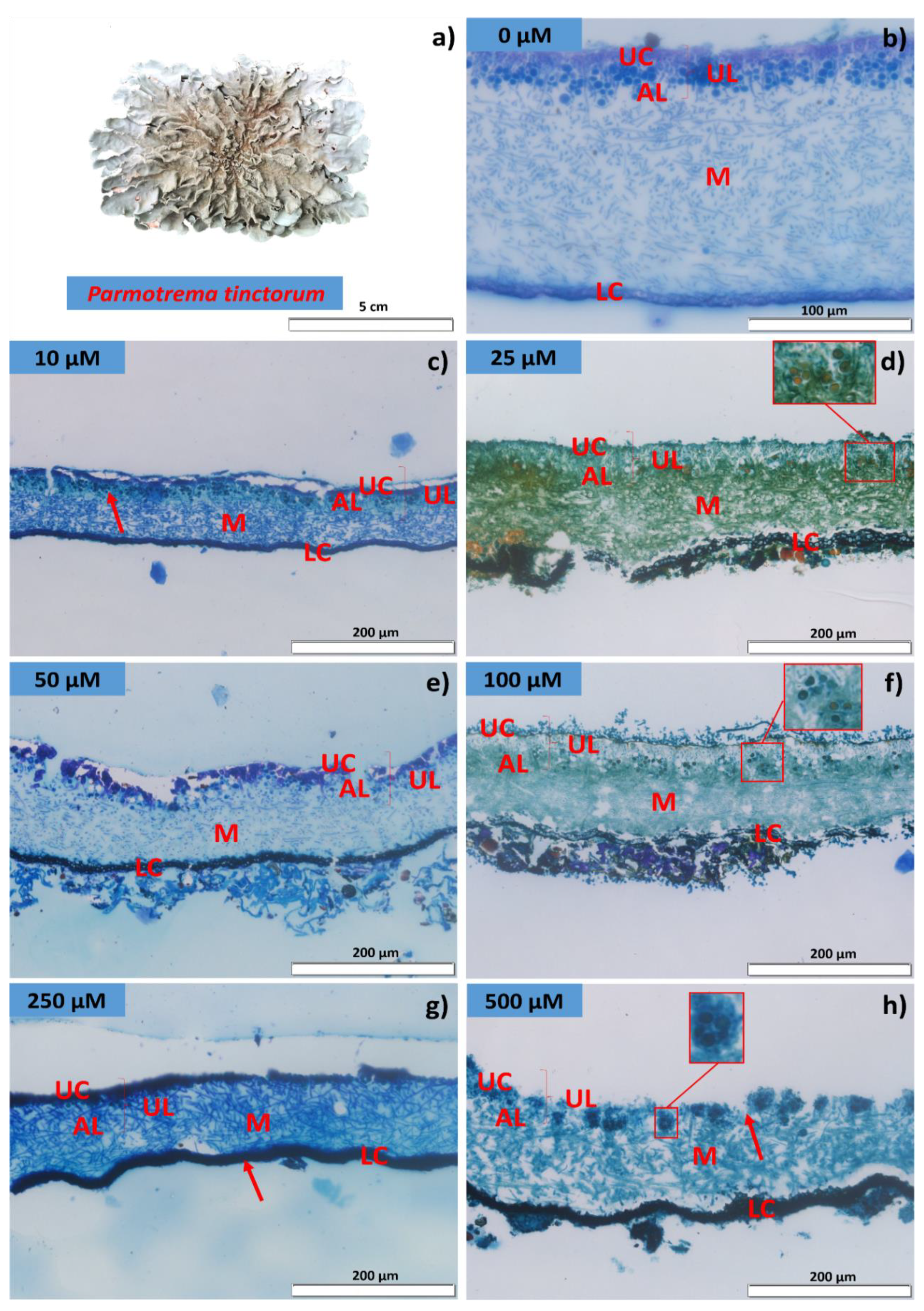
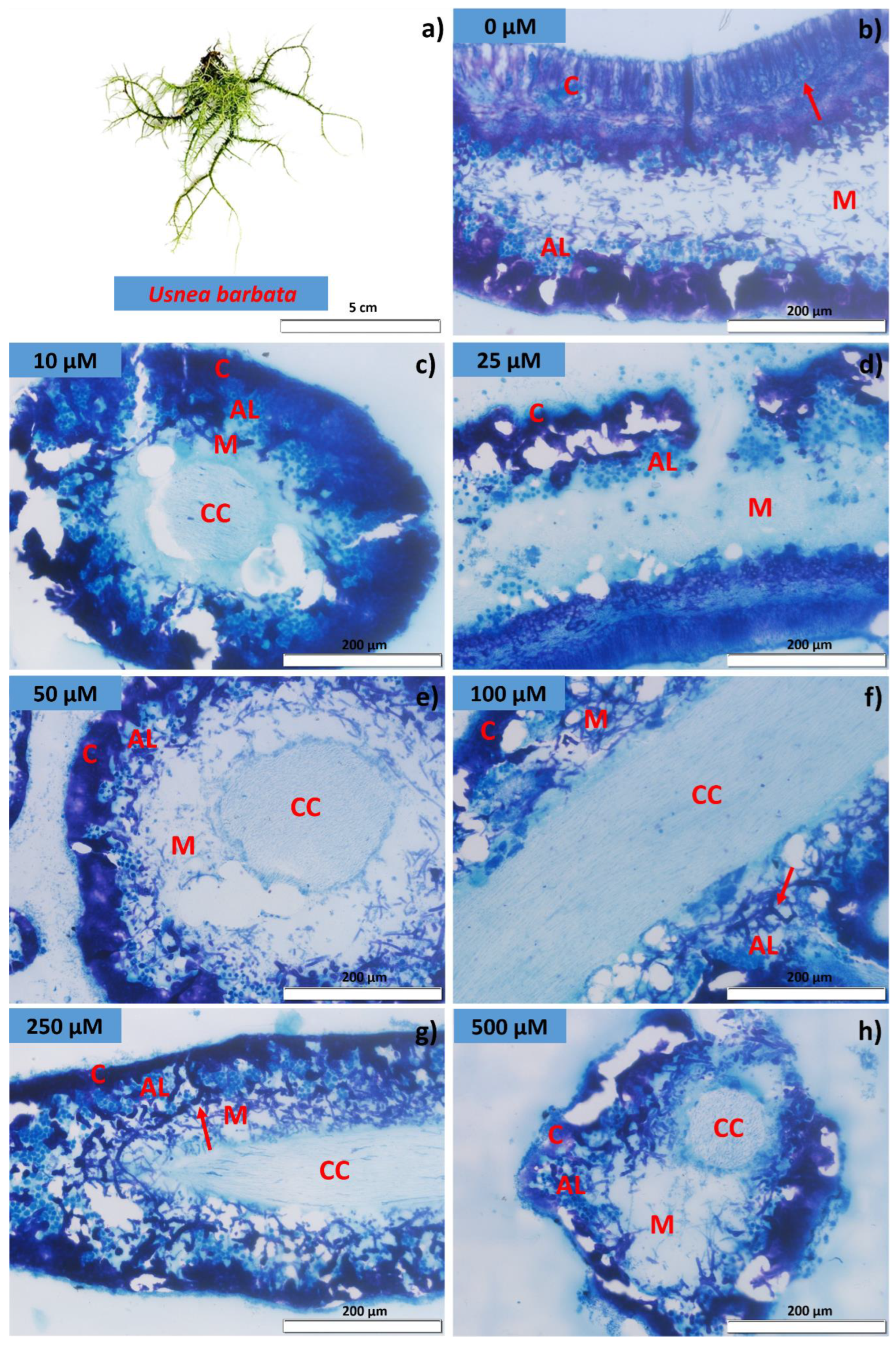
2.3. Membrane Integrity and Lichen Anatomy

The damage caused by Cd to cell membranes, suggesting tissue deterioration, was quantified by electrolyte leakage, and measured by the electrical conductivity test. Thus, we adapted the methods proposed by Brandão Jr. et al. [44] to measure the electrical conductivity in the thalli. The thalli were first weighed, and 2.0× g were used per repetition. Subsequently, they were placed to soak in plastic cups (200 mL) containing 75 mL of deionized water (<2.0 µS cm−1). These samples were kept in an oven at 25 °C for 24 h. After the conditioning period, the electrical conductivity of the solution was measured using a Digimed conductivity meter, model CD-21, and the results were expressed in µS cm−1 g−1.
The anatomy of the lichens was evaluated to identify tissue damage caused by glyphosate exposure. Thus, samples of the thalli were embedded in HistoResin following a four-step process—fixation (FAA50), dehydration in an increasing ethyl series, pre-infiltration, and infiltration in HistoResin (Leica Microsystems, Mensheim, Germany)—according to the manufacturer’s recommendations. Subsequently, the samples were cross-sectioned on a rotary microtome (Model 1508R, Logen Scientific, Shangjiao Dashi, Guangzhou, China) into 5-µm thick sections. The sections were stained with toluidine blue—polychromatic staining at 0.05% in a 0.1 M phosphate buffer, pH 6.8 [45]—and permeabilized in Canada balsam. Three slides were mounted for each treatment, and each slide contained 10 histological sections from which the integrity of the anatomical layers was evaluated.

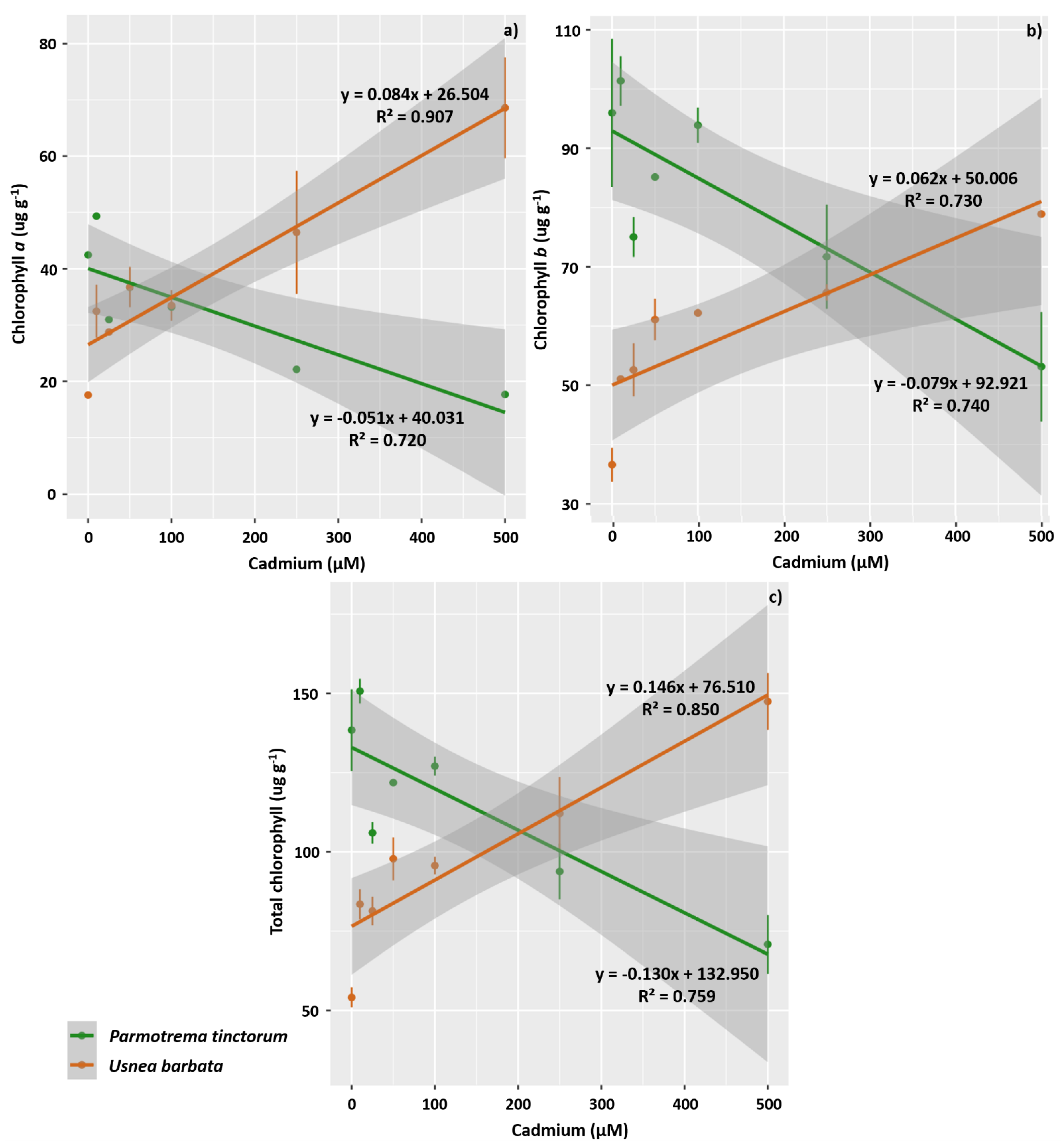
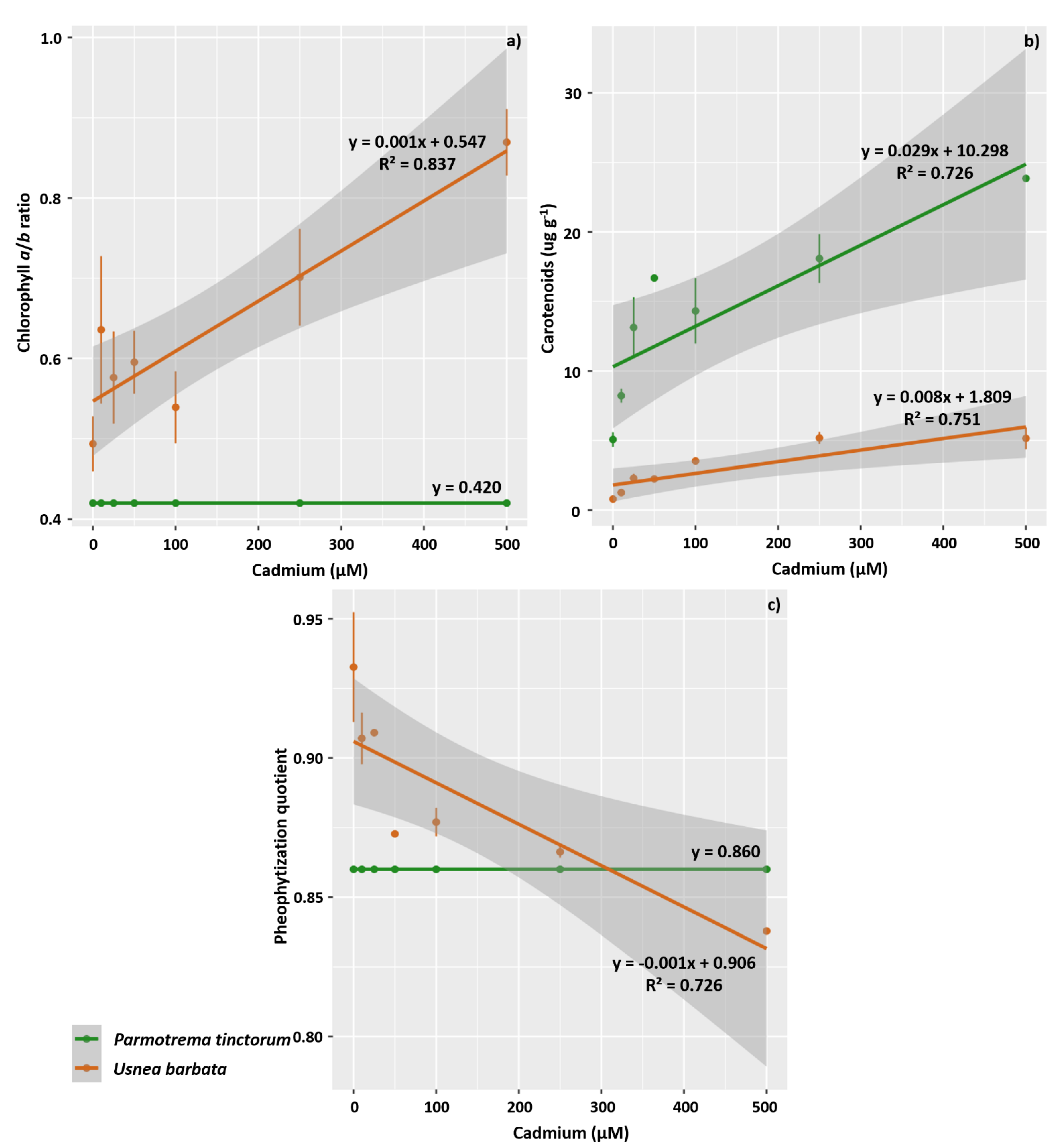
2.4. Evaluation of Photosynthetic Pigments

The concentration of photosynthetic pigments (chlorophyll a, chlorophyll b, total chlorophyll, the chlorophyll a to chlorophyll b ratio, and carotenoids) in the thalli was evaluated. In lichens, high concentrations of acidic substances can increase the pheophytization of chlorophyll. Therefore, the thalli were previously washed in 100% CaCO3-saturated acetone to avoid this effect. The chloroplast pigments were extracted in an extraction solution consisting of dimethylsulfoxide (DMSO) and polyvinylpolypyrrolidone (PVPP) at 2.5 mg mL−1. The thalli were covered with 5 mL of the extraction solution. The vials were then sealed, covered with aluminum foil, and kept at 65 °C in the dark for 40 min. The absorption spectrum was measured in a spectrophotometer for wavelengths 665 and 648 nm, calibrating against a blank containing only extraction solution. Turbidity was checked at 750 nm, and in cases where the value was higher than 0.01 optical density, the extract was centrifuged (2000× g) for 90 s, and the supernatant was reevaluated. The pigments were quantified based on the work and methods of Wellburn [46].
The pheophytization quotient was expressed as the ratio obtained by the absorbances at 435 and 415 nm (OD435/OD415) [47].

2.5. Chlorophyll a Fluorescence Parameters

The OJIP transient fluorescence of chlorophyll a was determined using a portable fluorometer FluorPen FP 100 (Photon Systems Instruments; Drasov, Czech Republic). The thalli of all the sample units were previously adapted to the dark for 30 min to allow the complete oxidation of the photosynthetic electron transport system. Subsequently, they were subjected to a pulse of 3000 µmol m−2 s−1 of blue light. The minimum fluorescence (F0) was measured at 50 μs when all PSII reaction centers were open (defined as the O step), followed by the J step (at 2 ms), the I step (at 30 ms), and the maximum fluorescence (Fm), when all PSII reaction centers were closed (the P step). These values were used for the estimation of various bioenergetic indices of PSII according to Strasser et al. [48]. We estimated values for the following parameters: the specific light absorption flux per reaction center (ABS/RC); the trapped energy flux per reaction center at t = 0 (TR0/RC); the electron transport flux per reaction center (ET0/RC); the specific energy dissipation flux at the level of the chlorophylls of the antenna complex (DI0/RC); the photosynthetic performance index (PiAbs) that incorporates the energy cascade processes from the first uptake events to the reduction in PQ; the maximum quantum yield of primary photochemistry (PHIP0); the probability that an exciton moves an electron down the electron transport chain after the quinone (PHI0), and the quantum yield of electron transport (PHIE0), after adaptation of the thallus to the dark (30 min).
Images of chlorophyll a fluorescence were obtained with an Imaging-PAM modulated fluorometer (Imaging-PAM M Series, Wals) and analyzed using ImagingWin v2.41a software. Initially, initial fluorescence (F0) and maximum fluorescence (Fm) were determined in thallus pre-adapted to the dark for 30 min. From this, it was possible to calculate the potential quantum yield of photosystem II (FSII) (FV/FM = (Fm − F0)/Fm). The variables from the slow phase of fluorescence induction were obtained sequentially: the fluorescence in a light-adapted sample before the saturation pulse (F) and Fm in a light-adapted sample (Fm′). The effective quantum yield of photochemical energy conversion in FSII, ΦII = (Fm′ − F)/Fm′, and the quantum yield of unregulated energy dissipation, ΦNO = F/Fm, were calculated.

2.6. Enzyme Activity of Antioxidant Metabolism, Hydrogen Peroxide, and Malondialdehyde

For the quantification of the antioxidant enzyme activity, samples were collected, conditioned in liquid nitrogen, and stored in an ultra-freezer at −80 °C. The enzymes were extracted by macerating 200 mg of lichen tissue in liquid nitrogen with 50% PVPP and following the extraction protocol proposed by Biemelt et al. [49]. The extraction buffer comprised 100 mM potassium phosphate buffer (pH 7.8), 0.1 mM EDTA, and 10 mM ascorbic acid. The extract was then centrifuged at 13,000× g for 10 min at 4 °C, and the supernatants were used to evaluate the catalase (CAT), ascorbate peroxidase (APX), and superoxide dismutase (SOD) activity.
The CAT activity was evaluated according to the methods proposed by Havir and McHale [50]. For this, an aliquot of the enzyme extract was added to an incubation medium containing 100 mM potassium phosphate (pH 7.0) and 12.5 mM hydrogen peroxide (H2O2). Enzyme activity was determined based on the consumption of H2O2 every 15 s for 3 min at 240 nm in a spectrophotometer. The molar extinction coefficient used was 36 mM−1 cm−1. The CAT activity was quantified in µmol H2O2 min−1 mg−1 protein.
The APX activity was evaluated based on the methods of Nakano and Asada [51], in which the oxidation rate of ascorbate at 290 nm is monitored every 15 s for 3 min. Thus, an aliquot of the enzymatic extract was added to a medium containing a 100 mM potassium phosphate buffer (pH 7.0), 0.5 mM ascorbic acid, and 0.1 mM hydrogen peroxide. The molar extinction coefficient used was 2.8 mM−1 cm−1. APX activity was determined as µmol AsA min−1 mg−1 protein.
The SOD activity was determined based on the methods of Giannopolitis and Ries [52], in which the enzyme’s capacity to inhibit the photoreduction of nitrotetrazolium blue (NBT) is evaluated. For this, an aliquot of the enzyme extract was incubated in a medium containing 50 mM of potassium phosphate (pH 7.8), 14 mM methionine, 0.1 µM EDTA, 75 µM NBT, and 2 µM riboflavin. The samples and incubation medium were illuminated with a 20 W fluorescent lamp for 7 min. Readings were performed in a spectrophotometer at 560 nm. The SOD activity was determined in U mg−1 protein, where 1 U corresponds to the amount of enzyme necessary to inhibit NBT photoreduction by 50%.
The concentration of H2O2 and malondialdehyde (MDA) in the thalli was determined by macerating 200 mg of lichen tissue in liquid nitrogen and PVPP, followed by homogenization in 0.1% (w/v) trichloroacetic acid (TCA), and centrifugation at 10,000× g for 15 min at 4 °C. The concentration of H2O2 was obtained by spectrophotometry according to the method of Velikova et al. [53]. The concentration of MDA was determined using the methods proposed by Buege and Aust [54].

2.7. Experimental Design and Statistical Analyses

The experiments were conducted in an entirely randomized design, considering seven concentrations of Cd (0, 10, 25, 50, 100, 250, and 500 µM). The lichen species (P. tinctorium and U. barbata) were analyzed separately, and all the analyses were conducted in triplicate. For the analyses of enzymatic activity, H2O2, and MDA, triplicates of the triplicates were analyzed.
The data obtained from each response variable for each lichen were submitted to one-way ANOVA to verify the effect of Cd concentration on the vitality, anatomy, photochemistry, physiology, oxidative metabolism enzymes, and synthesis of H2O2 and MDA in the lichen thalli. The data were also submitted to regression analysis, and the effect of increasing heavy metal concentration was evaluated by fitting linear models. The adjustment of the model was evaluated based on the determination coefficient, the regression significance coefficient, and the t-test at the 5% probability level. The tolerance index was used to compare the lichen species used, and the means were analyzed by Student’s t-test at a 0.05% (p < 0.05 **) significance level.
Afterwards, all the variables were evaluated together for both lichens. This evaluation aimed to analyze the behavior of each variable for each concentration of Cd tested. These variables were analyzed using a correlation matrix and combined in a principal component analysis (PCA). As they had different measurement units, correlation PCAs were performed and constructed using standardized data with a mean of 0 and a standard deviation of 1. The number of components was chosen according to the eigenvalues (>1.0) and the explained variance (above 80%). All analyses were conducted in R 4.2.1 [55].

3. Results

3.1. Algal Cell Viability, Electrical Conductivity, and Lichen Anatomy

In both lichens evaluated, we observed a linear reduction in the percentage of living algal cells as the tested concentration of Cd increased (Figure 1a). However, an effect of Cd concentration on the percentage of dead cells was observed only in P. tinctotum, where this percentage increased linearly (Figure 1b). Furthermore, the percentage of plasmolyzed cells was also affected by Cd concentration in both species, increasing linearly as a function of concentration increase (Figure 1c). Therefore, P. tinctorum seems to be more sensitive to Cd than U. barbata, which can be verified by the fitting of linear models with greater slopes for the percentage decrease in living cells (β = −0.011) and the percentage increases in dead cells (β = 0.007) and plasmolyzed cells (β = 0.004).
Exposure to heavy metal also affected membrane integrity in both species, with an increase in electrolyte leakage owing to increasing concentrations (Figure 1d). Therefore, the fitting of a linear model with a greater slope for U. barbata (β = 0.0002) represents a more effective response by this species for this variable.
The increase in Cd concentration also affected the integrity of the anatomical layers of the P. tinctorum and U. barbata thalli. In P. tinctorum, there was a significant reduction in the number of cells composing the algal layer and an increase in the disintegration of the hyphae that make up the upper cortex (Figure 2). Therefore, the upper cortex appeared damaged, even in the lowest concentration evaluated (Figure 2c). Moreover, under Cd treatment, the algal cells and the inferior cortex, where the rhizines are located, appear intensely stained, indicating that these areas are reactive to the accumulation of Cd in P. tinctorum. Adversely, Cd treatment seemed to modify the pattern of pigment synthesis by the thallus, with a differential accumulation of orange pigments related to the algal cells exposed to this metal (Figure 2d,f,h).
In the lichen U. barbata, the anatomical sections also showed a reduced cell content in the algal layer (Figure 3), especially for concentrations of 25 µM and higher (Figure 3d). In the control treatment, the spore-producing structures appear whole, but they are deconfigured under the action of Cd; it is not possible to distinguish the presence of asci and ascospores in the cortex region in the treated samples, given the degradation of the anatomical structures. In fact, in this lichen, the cortex and hyphae belonging to the medulla were highly responsive to the action of Cd. Therefore, the cortex was intensely stained, while the hyphae of the medulla also became heavily stained under high metal concentrations (see Figure 3g,h). Thus, intense disaggregations were seen in the pith as the exposure concentration increased. However, the integrity of the central cylinder was maintained in all treatments, suggesting that the other layers protect this layer effectively.

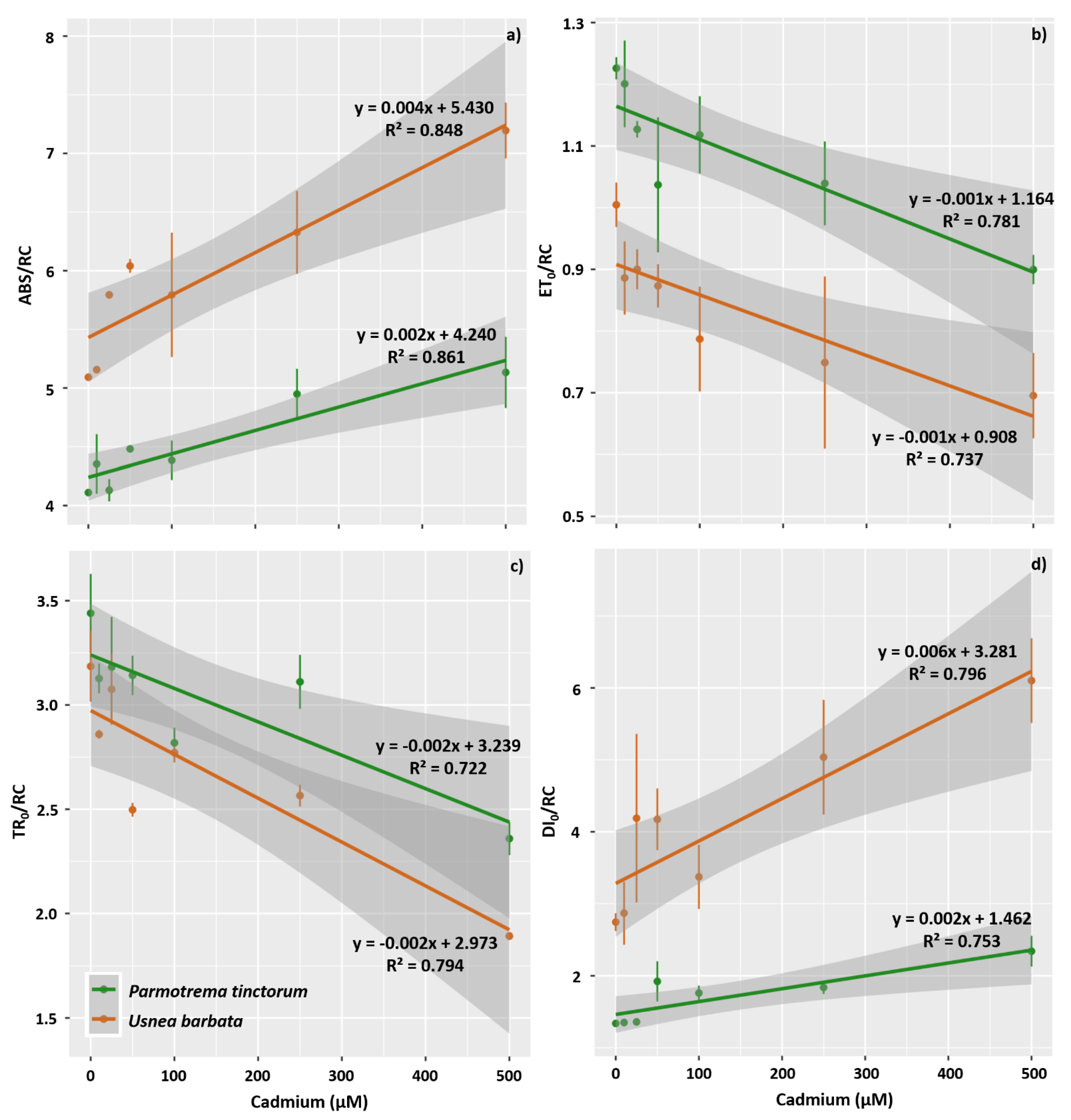
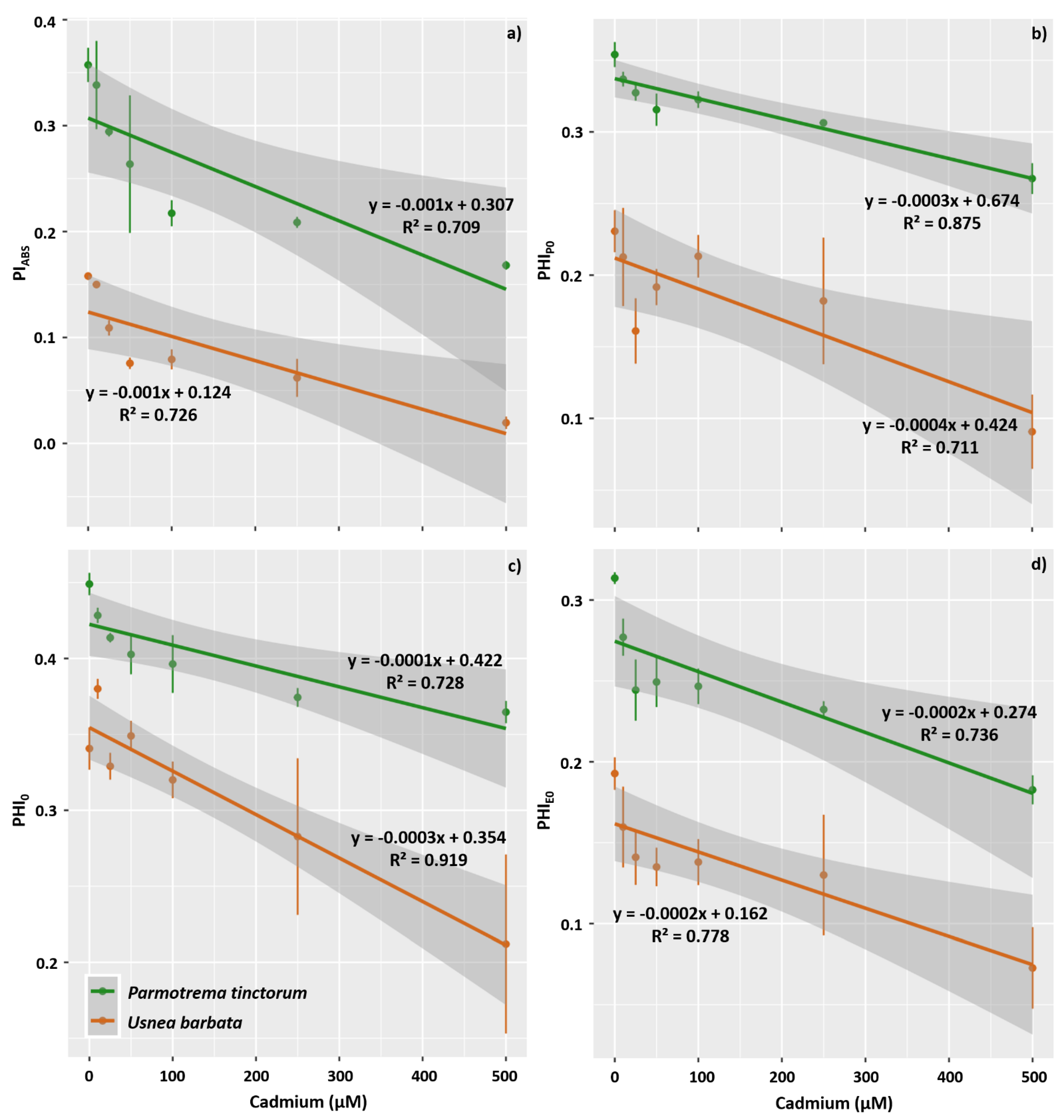
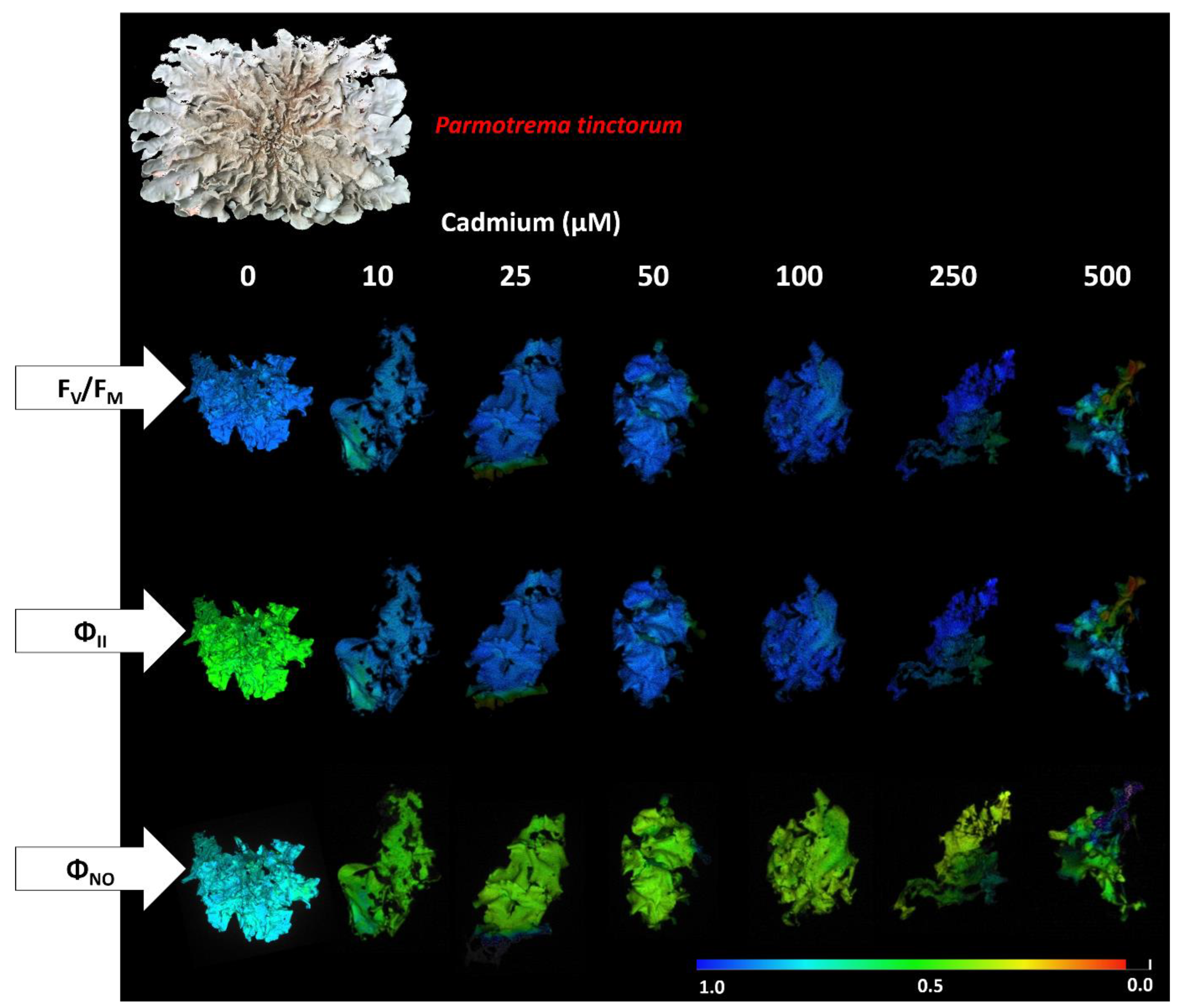
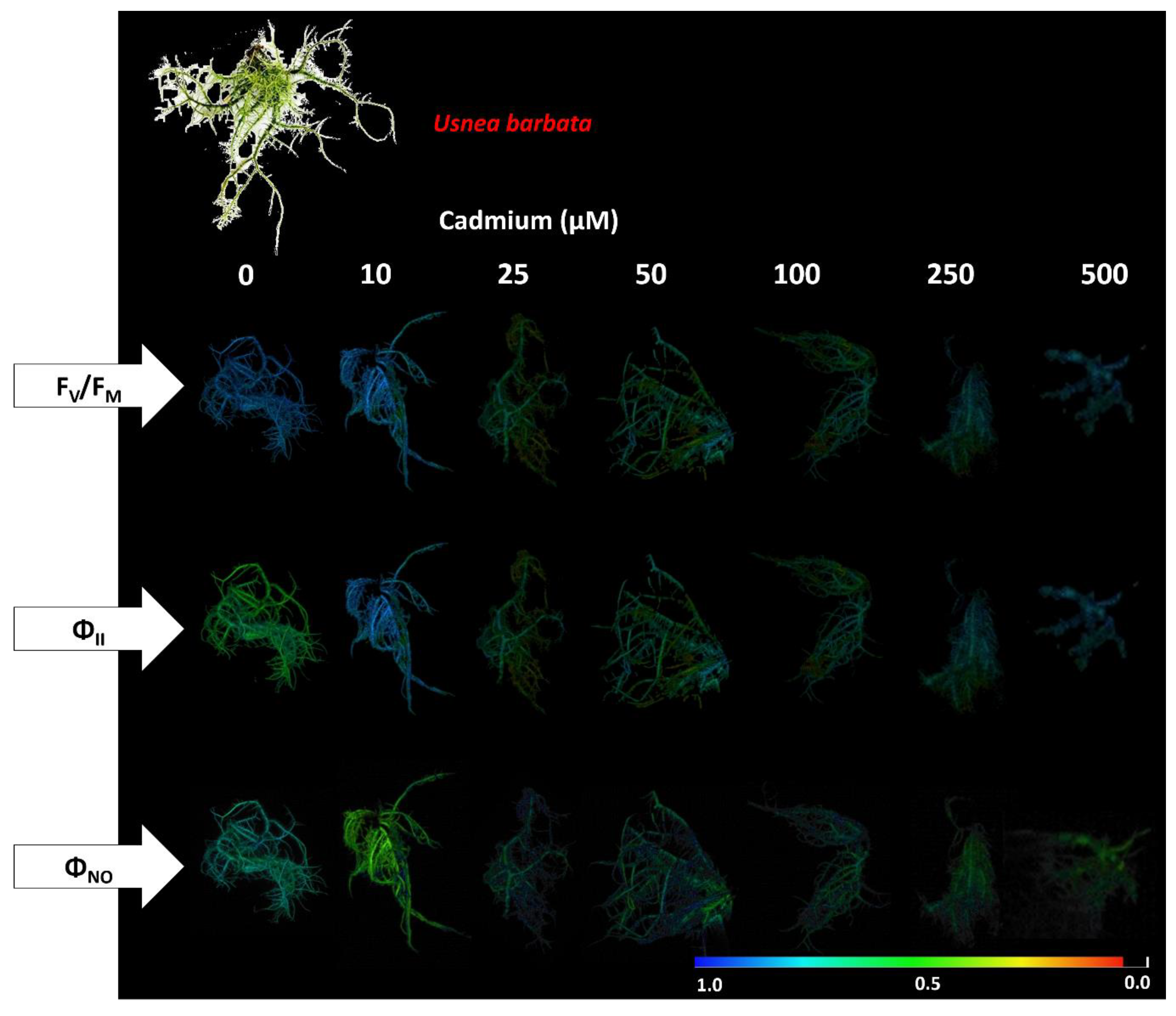
3.2. Photosynthetic Pigment Concentration and Chlorophyll a Fluorescence in the Photobiont

Exposure to different concentrations of Cd differentially affected the concentration of photosynthetic pigments in the lichen species photobionts. Surprisingly, while P. tinctorum showed a linear reduction in the content of chlorophyll a, chlorophyll b, and total chlorophyll in the thalli, in U. barbata, the mean values observed for these pigments increased as the tested Cd concentrations increased (Figure 4a–c).
When we evaluated the effect of Cd exposure concentration on the chlorophyll a/b ratio, we did not observe any effect on the photobionts of P. tinctorum. However, we observed an increase in the means of this ratio for U. barbata, indicating that in the thalli of this lichen, the synthesis of chlorophyll a tended to be higher than that of chlorophyll b in response to increasing Cd concentrations (Figure 5a). The concentration of carotenoids tended to increase linearly as a function of Cd in the thalli of both lichens evaluated (Figure 5b). Nevertheless, the fitting of a linear model with a greater slope for P. tinctorum (β = 0.029) demonstrates that the synthesis of these pigments was more expressive in the photobiont of this foliose lichen, indicating that this may be an essential response mechanism to the action of Cd.
The pheophytization quotient was not affected by increasing Cd exposure concentration in P. tinctorum. However, in U. barbata, we observed a reduction of this quotient, with a negative slope model fit (β = −0.001) (Figure 5c). This data correlates directly with the observed increase in chlorophyll synthesis in the thalli of this species.
The increasing concentrations of Cd also affected the algal photochemistry in the P. tinctorum and U. barbata thalli, with linear increases in the parameters ABS/RC and DI0/RC, consistent with stress responses (Figure 6a,d). However, for these variables, the U. barbata thallus seem to have been more strongly affected, which is supported by the more positive slopes observed in the curves obtained for ABS/RC (β = 0.004) and DI0/RC (β = 0.006) in this lichen. However, for ET0/RC and TR0/RC, we observed the opposite behavior from that shown by the previous variables, i.e., the means observed for these parameters decreased with increasing Cd exposure concentration (Figure 6b,c). The reduction patterns were similar in both lichens, which can be observed in the equal negative slope values in the curves of ET0/RC (β = −0.001) and TR0/RC (β = −0.002).
The performance and photochemical yield parameters were negatively affected by the increasing doses of Cd in the tested lichen algae, shown in the linear reduction in the PiAbs, PHIP0, PHI0, and PHIE0 means of the thalli (Figure 7a–d). Furthermore, the fitting of linear models with greater decrease slopes for the parameters PHIP0 (β = −0.0004) and PHI0 (β = −0.0003) also indicate the greater photochemical sensitivity of the algae of U. barbata to increasing doses of the heavy metal.
Fluorescence image analysis visually demonstrated the effect of Cd concentrations on the primary photochemistry of P. tinctorum and U. barbata. Indeed, photochemical damage was already evident at the lowest metal concentration (Figure 8 and Figure 9). The values of FV/FM and ΦII were reduced as a function of increasing exposure concentration in both lichens. However, a diffusion of the energy destined for ΦII and ΦNO was observed in the thalli.

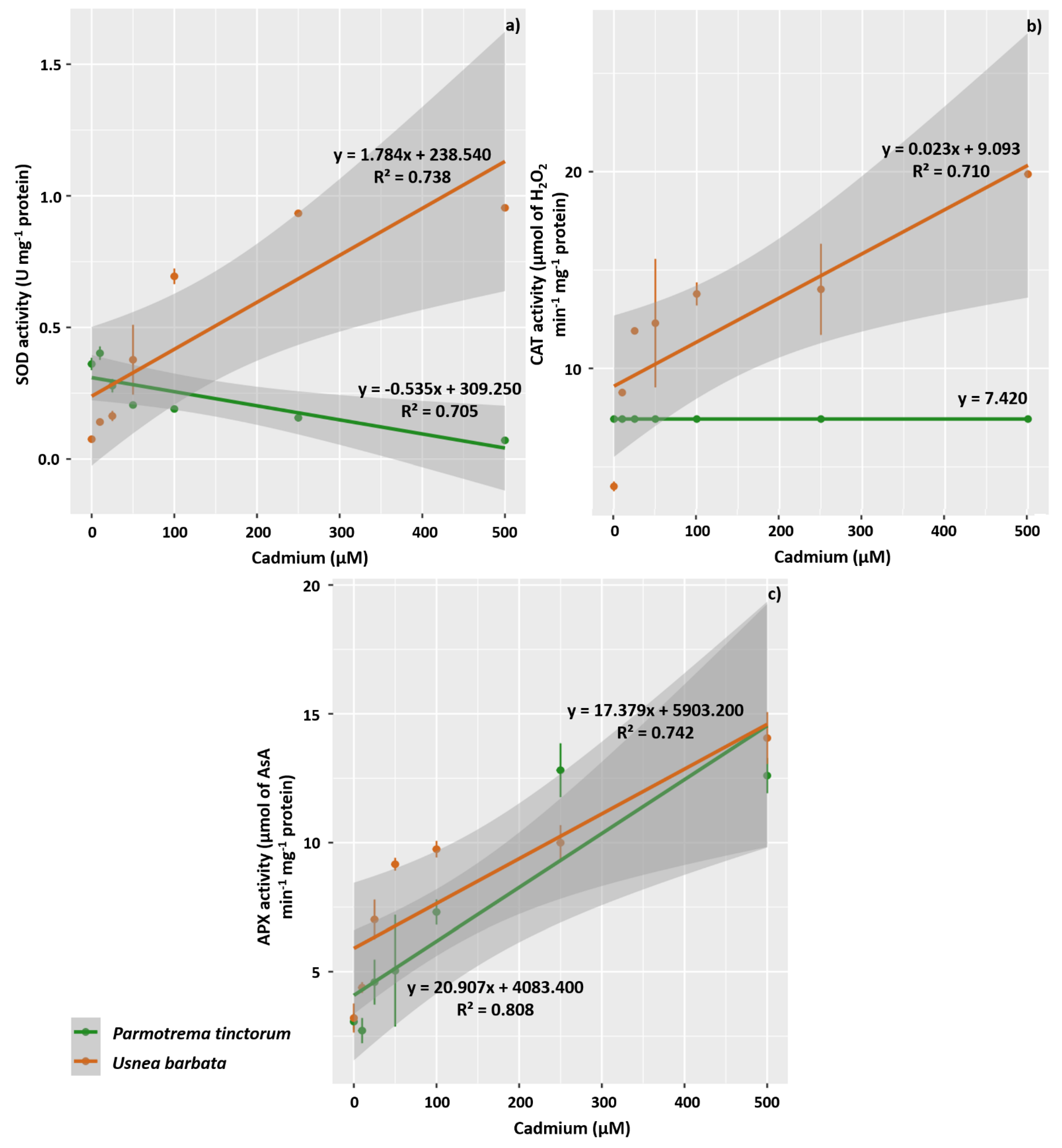
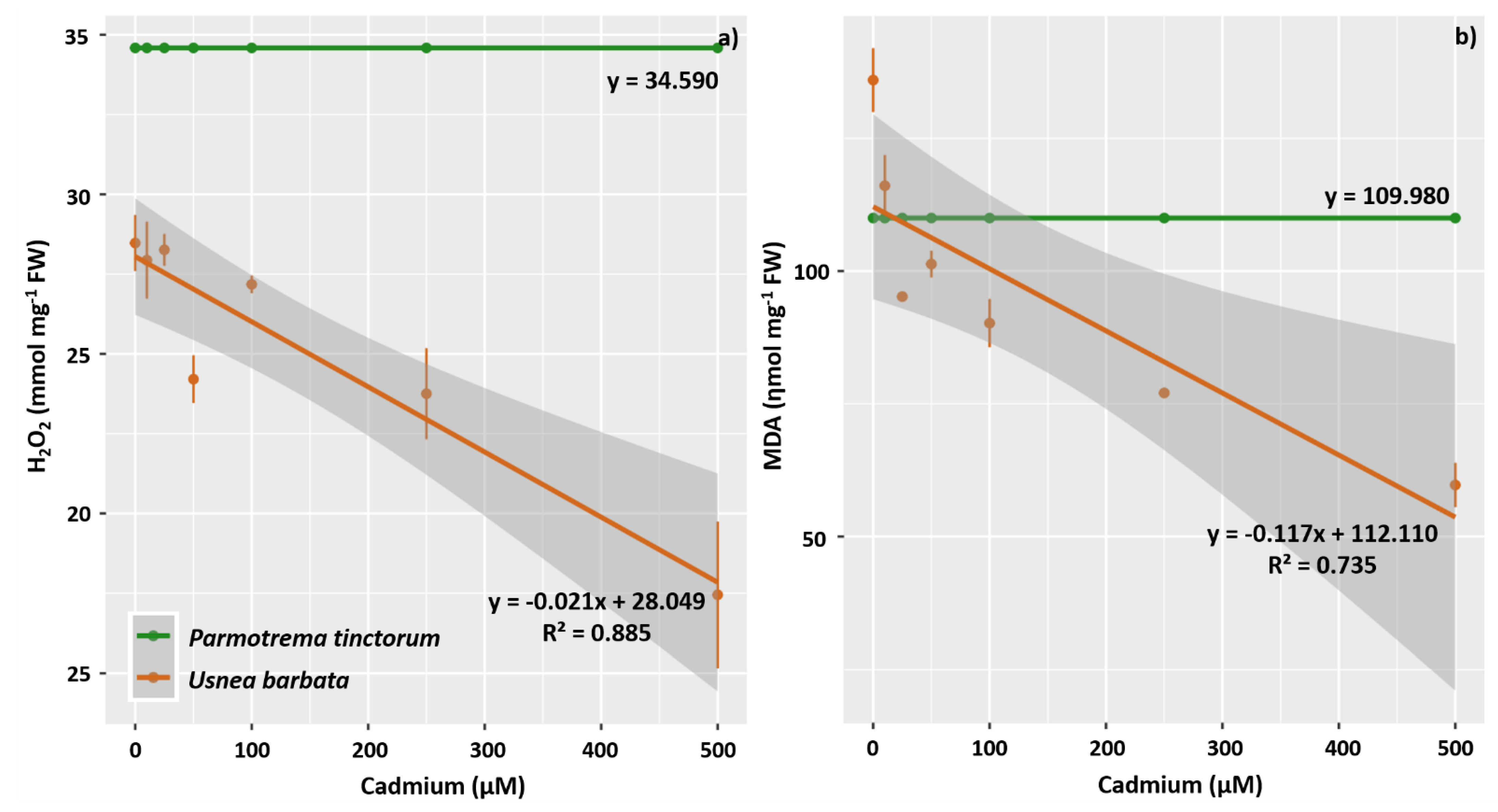
3.3. Enzyme Activity of Antioxidant Metabolism, H2O2, and MDA

Distinct patterns were verified for the SOD enzyme activity in the lichen species thalli; the activity of this enzyme increased as a function of Cd concentration in U. barbata (β = 1.784) but decreased in P. tinctorum (β = −0.535) (Figure 10a). In U. barbata, the activity observed for the CAT enzyme also increased in response to increasing metal exposure concentration, although concentration did not affect the activity of this enzyme in P. tinctorum thallus (Figure 10b). Moreover, the patterns observed for the APX enzyme activity were similar for both lichens, with a linear increase in activity as a function of the exposure concentration (Figure 10c). In summary, the U. barbata thallus responded to Cd by increasing the activity of these three enzymes of the antioxidant system.
The concentration of H2O2 in U. barbata thallus decreased linearly with increasing Cd concentration (β = −0.021), and this pattern is consistent with the observed activity of antioxidant enzymes in this lichen. However, in P. tinctorum thallus, the concentration of this reactive species was not affected by increasing concentrations of heavy metal exposure (Figure 11a). Similar behavior to that observed for H2O2 was verified for the curves corresponding to the concentration of MDA in the thalli, which show a linear decrease as a result of the increase in Cd concentration in U. barbata thallus (β = −0.117) but no effect of these concentrations in P. tinctorum (Figure 11b).

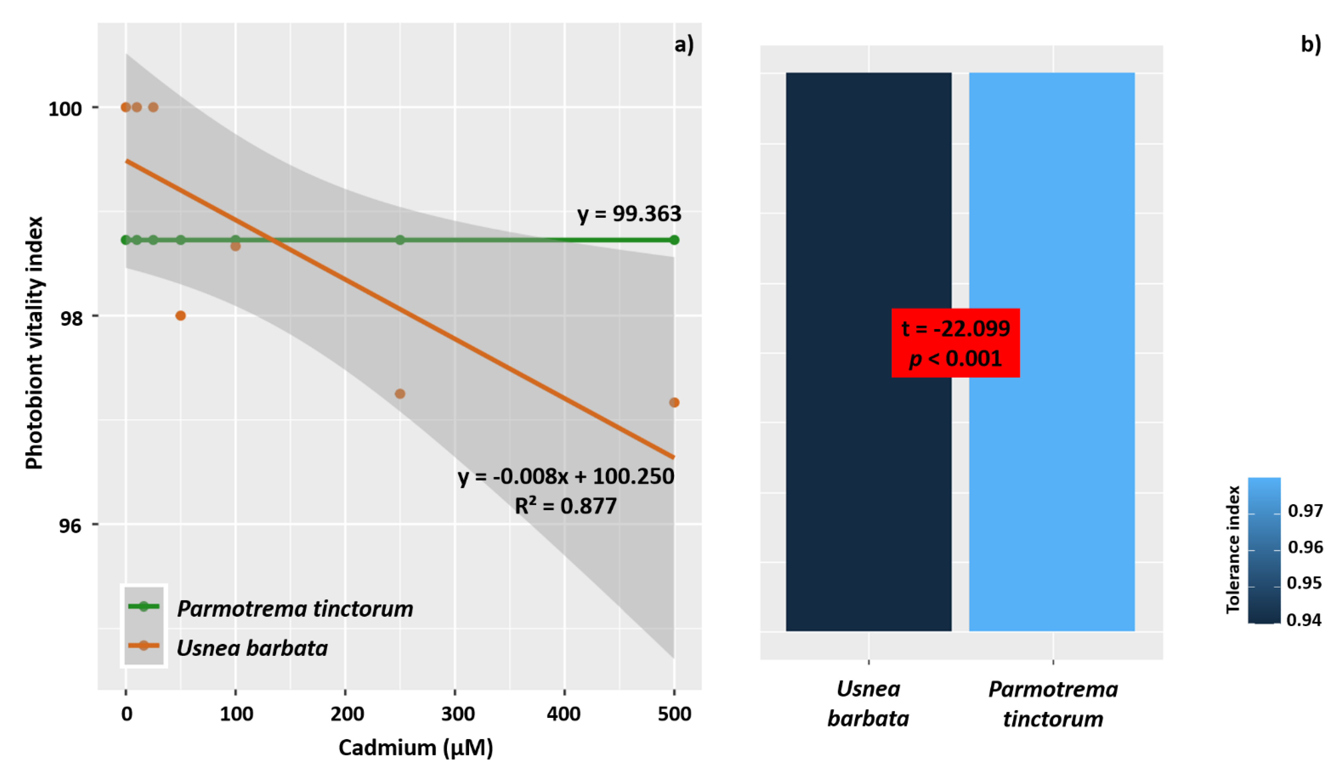
3.4. PVI and Tolerance Index

When we evaluated the lichen species using PVI, we found a linear model fit only for P. tinctorum, where the model revealed a reduction in vitality owing to increasing Cd concentration (β = −0.008). However, in the U. barbata photobiont, increasing metal concentration did not affect algal vitality (Figure 12a). The PVI seemed to anticipate what was observed for the tolerance index, because when comparing the tolerance of the lichen species to the highest concentration of Cd, we found that P. tinctorum is more sensitive than U. barbata (Figure 12b). The tolerance index of P. tinctorum was 0.946, and that of U. barbata was 0.980.

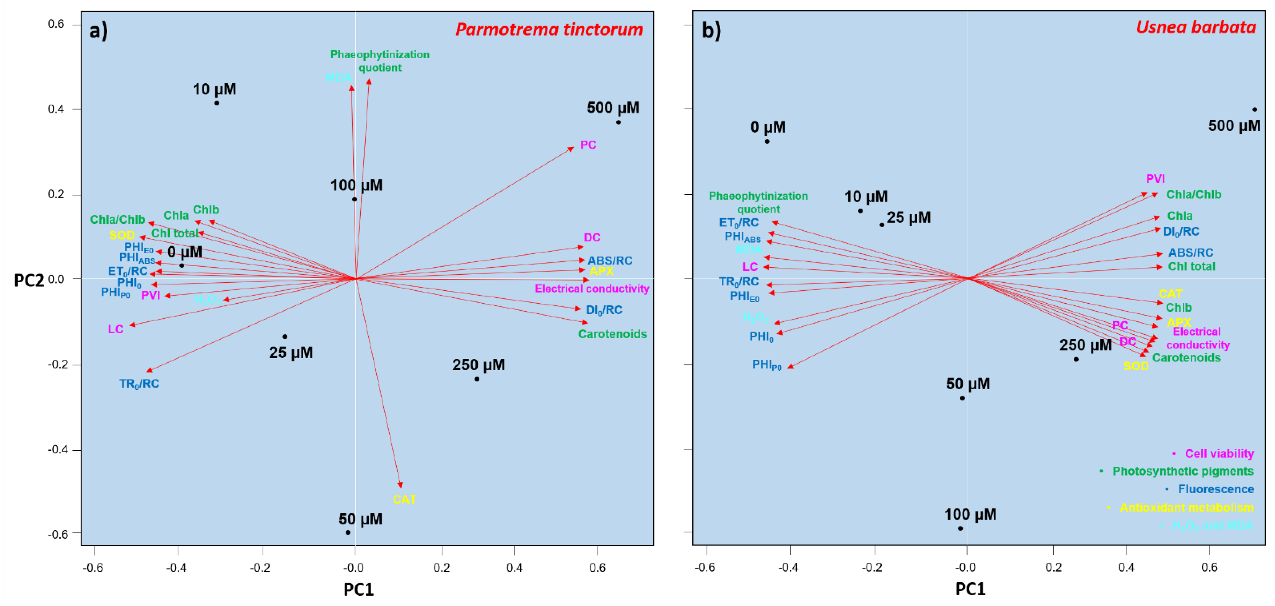
3.5. Analysis of Variables as Biomarkers of the Effect of Cd

In general, when we jointly plotted the observed data for P. tinctorum, we found that cell viability and PVI are excellent biomarkers for the damage caused by increasing concentrations of Cd exposure (Figure 13a). Thus, the percentage of living cells significantly defined the differences observed between the control and the highest Cd concentration tested. Similarly, the percentage of dead and plasmolyzed cells and the electrical conductivity were essential to defining the differences observed between the higher and control concentrations. We also found that chlorophyll a fluorescence constitutes a good effect marker since stress indicators such as ABS/RC and DI0/RC were associated with higher Cd concentrations, while indicators of photochemical efficiency, such as PiAbs, PHIP0, PHI0, and PHIE0, were more strongly associated with the control treatment. Furthermore, the concentrations of chlorophyll a, chlorophyll b, and total chlorophyll, and the chlorophyll a/b ratio were also strongly associated with the control. Carotenoid pigments and APX enzyme activity were associated with higher concentrations of the heavy metal. In contrast, the MDA content, CAT activity, and pheophytization quotient were not crucial in defining the effect of concentration (Figure 13a).
When we plotted the evaluated data for U. barbata, cell viability, especially the percentage index of living cells, behaved as an essential biomarker associated with the control treatment (Figure 13b). In contrast, the percentages of plasmolyzed and dead cells and the electrical conductivity tended to be associated with the higher tested Cd concentrations. Unexpectedly, PVI, as well as photosynthetic pigments, such as chlorophyll a, chlorophyll b, total chlorophyll, carotenoids, and the Chla/Chlb ratio, were also associated with higher Cd concentrations; thus, they are not suitable biomarkers for the stress effects caused by exposure to increasing Cd concentrations. The pheophytization quotient showed the opposite behavior to the accumulation of chlorophyll. As expected, the photochemical stress parameters, ABS/RC and DI0/RC, were associated with higher Cd concentrations, while the performance indicators, PiAbs, PHIP0, PHI0, and PHIE0, were related to lower concentrations. The CAT, APX, and SOD enzymes were efficient in marking the stress effects of thalli exposure to increasing concentrations of Cd, but MDA and H2O2 demonstrated the opposite behavior to these enzymes (Figure 13b).

4. Discussion

4.1. Cd Exposure Affects Photobiont Viability and Membrane Integrity, and Promotes Damage and Morphoanatomical Changes in P. tinctorum and U. barbata

The lichenized fungi that constitute P. tinctorum and U. barbata associate with unicellular green algae (Chlorophyta), and other studies have confirmed that Cd exposure can inhibit cell division and cause the development of deformed cells in chlorophytes. La Rocca et al. [56] found that at 5 ppm, Cd stopped cell growth and algal cells suffered lysis, while Di Toppi et al. [57] provided evidence that Cd can promote ultrastructural damage and decrease cell density in the photobiont of Trebouxia impressa. We found a decrease in living cells and an increase in plasmolyzed cells in P. tinctorum and U. barbata and the adjustment to the linear model was highly reliable. Our anatomical observation results also confirm that Cd reduces the number of cells in the algal layer. These data were accompanied by increases in electrical conductivity, signaling that this metal affects the integrity of lichen membranes. Rihab et al. [58] showed that high concentrations of Cd could induce lipid peroxidation even in algae tolerant to this metal. Furthermore, since electrical conductivity signals electrolyte leakage from damaged membranes, e.g., [59,60,61], we conclude that increasing Cd exposure concentration negatively affects cell membrane structure in P. tinctorum and U. barbata.
Morphoanatomical damage from Cd action was observed in the upper cortex of P. tinctorum and the cortex and pith of U. barbata. In fact, heavy metal exposure generally affects the integrity of lichen cell membranes, e.g., [62]. Owing to increasing exposure concentration, these effects can be seen through the disaggregation of anatomical layers. The lower cortex region of P. tinctorum and the cortex and hyphae of the medulla of U. barbata appeared heavily stained, indicating the deposition of electron-dense material in the mycobiont cell wall, which may constitute an important mechanism used by these lichens to tolerate Cd exposure. Similar results were observed by Bora and Sarma [63] when they evaluated the ultrastructure of Ceratopteris pteridoides under Cd stress. Pawlik-Skowrońska et al. [64] found that lichen thalli can synthesize phytochelatin in response to some metals, including Cd, which tends to form strong complexes with S-containing peptides and proteins. Daimari et al. [65] also observed substantial anatomical damage in the mycobiont hyphae of P. tinctorum when this lichen was exposed to urban pollution, and these results are consistent with the increased accumulation of Cd and other heavy metals in the thalli.
Heavy metals are retained in the thallus by particle trapping, physicochemical processes such as ion exchange, and passive and active intracellular uptake [66,67]. They can then interact with tissues, modifying structures. The secondary metabolites of lichens may be involved in the production of chelates with heavy metals. Studies have confirmed that lichens colonized by green algae of the genus Trebouxia, also present in P. tinctorum, can synthesize an orange-colored anthraquinone pigment called parietin, which is deposited as tiny crystals in the upper cortex [68]. This pigment seems to constitute a resistance strategy of the lichen to various stresses, such as UV radiation and heavy metals [69,70]. Furthermore, Kalinowska et al. [71] provided evidence that parietin can protect the photobiont cells in Xanthoria parietina from excess Cd ions. We also observed the differential deposition of Trebouxia-related orange pigments in P. tinctorum, suggesting that this may be a protective mechanism used by this species in response to Cd exposure. Thus, as we verified the concentration-dependent toxic effect of Cd on the photobiont viability and morphoanatomy of P. tinctorum and U. barbata, these changes can be considered as efficient biomarkers for Cd toxicity analyses.

4.2. Cd Exposure Negatively Affects the Concentration of Chlorophyll a, Chlorophyll b, and Total Chlorophyll in the Photobionts of P. tinctorum and Increases the Synthesis of Carotenoids

The chlorophyte Trebouxia cortícola, symbiotic to P. tinctorum, responds to increasing Cd exposure concentration by reducing chlorophyll and increasing carotenoid synthesis. Indeed, several studies have tested the impact of Cd pollution on the vitality of various lichen photobiont species by measuring photosynthetic pigment concentrations in the thalli, e.g., [57,72,73]. Lichen photobionts exposed to metal-containing solutions generally experience a significant reduction in their chlorophyll and carotenoid content, e.g., [70,74,75,76]. Certain metals may be responsible for chlorophyll degradation and/or cause disturbances in synthesizing this pigment. Under field conditions, chlorophyll degradation in lichens from polluted sites correlated with the accumulation of metals in the thallus [77].
The synthesis of carotenoids also seems to constitute a strategy used by P. tinctorum (and U. barbata) to resist the oxidative stress caused by the presence of Cd, although the reliability observed in the determination of the coefficients for this variable has been low. A similar effect was suggested by Dobroviczká et al. [78], evidencing the singular importance of the presence of carotenoids in the tissues. Carotenoids play a structural role in the organization of photosynthetic membranes and participate in light harvesting, energy transfer, and the interception of free oxygen radicals [79]. Our results are similar to those of Bačkor et al. [80], who observed that, when the lichens Peltigera rufescens and Cladina arbuscula subsp. mitis were subjected to Cd concentrations between 0 and 500 µM for 24 h, their carotenoid content was not affected. Thus, we characterized the degradation of chlorophyll as an efficient metabolic pathway biomarker of Cd action in P. tinctorum.

4.3. Cd Exposure Affects Primary Photochemistry in Photobionts of P. tinctorum and U. barbata

From the lowest concentration of Cd tested, the photochemistry of the thalli was negatively affected, with an increase in the values of stress parameters as the exposure concentration increased. However, as expected, the opposite behavior was verified for the photochemical efficiency parameters. The reduced Fv/Fm values induced by Cd action indicate chronic photoinhibition of photosystem II [81,82]. The portion of energy harnessed in the photochemical step (ΦII) also indicates damage to the photobiont. With increasing stress, the portion of electrons destined for the photochemical step tends to decrease, owing to increases in the dissipation of excess energy in other pathways, such as photorespiration [83]. This behavior can be demonstrated by the observed increases in the DI0/RC values, although the reliability in the determination of the coefficients was low. In this case, the anatomical damage caused by Cd seems to compromise the diffusion of CO2 into the thalli. When small amounts of CO2 reach the photobiont layer, it inhibits the Calvin cycle, and the photochemical dissipation is impaired by the slow regeneration of its substrates (ADP and NADP+) [84]. However, it is known that the sum of the quantum yields tends to be equal to 1. Therefore, we observed the diffusion of the energy intended for ΦII and ΦNO in the thallus.
Interaction with Cd affected the primary photochemistry in U. barbata more than that in P. tinctorum, and the thallus type may explain this. In foliose lichens, such as P. tinctorum, the upper cortex is formed by hyphae that protect the algal layer, while the lower cortex and the medulla form an extensive layer that any pollutant must pass through to reach the photobiont. In fruticose lichens, such as U. barbata, a single cortex protects the algal layer, which leaves it more exposed to the action of pollutants and other stressing agents, such as excess light and heat. We observed intense staining in the cortex of U. barbata, indicating the deposition of electron-dense material. By blocking the passage of CO2, light, and water through the thallus, this electron-dense material affects the algal photochemistry. Schroeter [85] evaluated apparent electron flux rates through PSII and CO2 rates in the photobiont green algae of Placopsis contortuplicata and found that gas exchange was suboptimal after hydration with water vapor only. Maximum rates could only be observed when the thallus was saturated with liquid water, attesting to the importance of algal access to water for optimal photochemical performance.
Although lichen photobionts are considered vital elements of lichen sensitivity, further research is needed to elucidate this point. Studies show that secondary metabolites, such as cortical depside atranorin and dibenzofuran usnic acid, may be essential to protect the primary photochemistry of lichens of the family Parmeliaceae, such as P. tinctorum and U. barbata, e.g., [86]. However, despite the possible action of these and other metabolites in the thalli, the primary photochemistry of P. tinctorum and U. barbata was negatively affected by Cd, with chlorophyll a fluorescence being a consistent biomarker of Cd stress.

4.4. The Activity of Antioxidant Enzymes Increases in U. barbata Thalli Exposed to Increasing Concentrations of Cd

Although Cd is a non-redox active metal and, therefore, unable to directly stimulate ROS formation, ROS production and increased lipid peroxidation have already been recorded [11]. Indeed, Cd can promote significant physiological and molecular changes in photobionts of the genus Trebouxia; for example, ROS formation and the expression of genes related to antioxidant enzyme synthesis [32]. The activity of antioxidant enzymes appears to constitute an important pathway by which U. barbata responds to Cd stress. Cuny et al. [87] evaluated the effect of heavy metals (Cd, Pb, and Zn) on the lichen Diploschistes muscorum (Scop.) R. Sant. and also verified membrane damage and increased SOD activity. In addition, studies have shown that lichens exposed to pollution have increased activity of SOD and other antioxidant enzymes, such as APX and glutathione (GSH) reductase [88,89,90].
SOD catalyzes the dismutation of superoxide into molecular oxygen and H2O2, a substrate for catalase, which is an enzyme that reacts efficiently in peroxisomes with H2O2 to form water and molecular oxygen [91]. We found that, in U. barbata, high CAT activity was consistently associated with low peroxide concentrations. In this lichen, low concentrations of H2O2 were also related to high APX activity. This enzyme can catalyze the breakdown of peroxide into H2O [92] and this may constitute an alternative pathway for eliminating this antioxidant from the thalli. Similarly, Monnet et al. [27] found increased APX activity in the thalli of the lichen Dermatocarpon luridum when exposed to copper toxicity.
The increasing concentrations of Cd decreased the MDA content in U. barbata thalli, indicating that despite the increase observed in electrolyte leakage, lipid peroxidation, indicated by MDA [93], was efficiently suppressed by the action of the antioxidant system. In addition to the enzymatic system, which proved to be an effective biomarker of Cd stress in U. barbata, the presence of antioxidant metabolites, such as usnic acid, may have assisted in suppressing the oxidative effects of the metal, e.g., [94,95,96].

4.5. Parmotrema tinctorum Is More Sensitive to Cd Stress Than Usnea barbata

Although the photobiont of U. barbata suffered more Cd-induced photochemical damage than that in P. tinctorum, we verified that, in the fruticose species, the photobiont was insensitive to increased Cd concentrations; it did not lose vitality, and it increased the synthesis of photosynthetic pigments and antioxidant enzymes, resulting in higher efficiency in the elimination of H2O2 and decreased lipid peroxidation. Thus, we verified the increased sensitivity of P. tinctorum to stress induced by exposure of the thalli to increasing concentrations of the heavy metal. These results were surprising, given the classical conception that fruticose lichens are more sensitive than foliose species [97,98]. According to Martins-Mazzitelli et al. [99], when compared to foliose lichens, fruticose lichens are the most sensitive to air pollution, being the first to disappear in heavily polluted areas. Regarding Cd pollution, however, given the dispersion of industrial pollutants or agricultural fertilizers, P. tinctorum seems more suitable than U. barbata for air monitoring studies in urban and forest areas immersed in agricultural matrices.

4.6. Biomarkers for the Effect of Cd-Induced Stress in P. tinctorum and U. barbata

Besides the lichen morphoanatomy parameter, which was effective in biomarking the stress caused by increased concentrations of Cd in the two lichens evaluated, for P. tinctorum, the cell viability, PVI, chlorophyll a fluorescence, and chlorophyll concentration parameters were also efficient Cd stress biomarkers. These parameters are important because they are directly associated with the most sensitive component of the lichen, the algal component. In another way, our results are significant because the techniques associated with obtaining the vitality data and photosynthetic pigments are simple and inexpensive, based on dyes and extractants, e.g., [40,46,100,101]. Therefore, they could easily be implemented in laboratories aiming to use lichens in biomonitoring Cd dispersal from pollutant sources. For example, Port et al. [41] evaluated photobiont vitality and metal concentration in P. tinctorum samples in urban and forest areas and concluded that vitality and chlorophyll content are essential parameters for urban pollution biomonitoring. Similarly, Raimundo-Costa et al. [15] concluded that the chlorosis seen in the thalli of this species in urban and industrial environments, where the concentration of heavy metals in the air is high, is an indication of impaired pigment synthesis.
In U. barbata, the photobiont cell viability, the primary photochemistry, and the activity of the antioxidant enzymes CAT, SOD, and APX were efficient in biomarking the stress induced by exposure to increasing concentrations of Cd. Studies have shown that Usnea thallus can concentrate high activity of antioxidant enzymes and inhibit lipid peroxidation [102]. Unexpectedly, Cd stress did not affect PVI and pigment synthesis in this lichen. Di Toppi et al. [57] showed that GSH and phytochelatins, as well as proteins produced under stress, efficiently protect the cells of the chlorophyte Trebouxia impressa against Cd damage. Indeed, phytochelatins synthesized by the mycobiont seem to act in Cd detoxification processes [64]. Phytochelatins are synthesized from GSH with the help of phytochelatin synthase, activated by heavy metals [103]. Thus, the presence of an effective enzymatic antioxidant system and usnic acid synthesis [104,105] may have contributed to protecting the U. barbata photobiont from the toxic effects of Cd.
In general, for the two lichens evaluated, the MDA content and the pheophytization index were considered less effective parameters in biomarking the effect of Cd. During lipid peroxidation, the conjugation of the ethylene groups of polyunsaturated fatty acids can be observed, which increases hydroperoxy-conjugated dienes (HPDCs). Rodriguez et al. [106] suggest that HPDCs are better estimators of lipid peroxidation than MDA. MDA is usually measured by its reaction with TBA, yielding MDA–TBA2, which is detectable by spectrophotometry. However, this method shows poor specificity because TBA can react with various compounds, leading to overestimating MDA values [107]. Thus, the MDA values of tissues may not correctly reflect their oxidative stress state.
Moreover, there was no evidence of increased chlorophyll pheophytization in the thalli as a function of the degree of stress imposed, especially in U. barbata. Pheophytization reactions, which include the replacement of magnesium at the center of the chlorophyll molecule by hydrogen and the removal of the phytol chain, forming chlorophyllide or pheophorbide, are the most critical chlorophyll degradation pathways [108]. However, as our lichens were treated with acetone precisely to prevent chlorophyll pheophytization [109,110], the observed values of this index were not efficient in biomarking the algal stress caused by Cd.

5. Conclusions

The lichens Parmotrema tinctorum and Usnea barbata respond to Cd stress, generating the idea of using these lichens to verify the dispersion of atmospheric pollutants containing Cd. For P. tinctorum, the lichen morphoanatomy, cell viability, PVI, chlorophyll a fluorescence, and chlorophyll a synthesis are efficient biomarkers for the effects of exposure to increasing concentrations of Cd, as these variables are primarily associated with the photobiont. For U. barbata, the lichen morphoanatomy, photochemistry, and activity of the antioxidant enzymes CAT, SOD, and APX were essential to reflect Cd toxicity. Parmotrema tinctorum was characterized as the most sensitive to the action of Cd and is considered a good bioindicator for the presence of this metal and the diagnosis of air quality in urban and industrial areas, or even in forest areas influenced by Cd in fertilizers. Considering this, it is crucial to remember that the lichen’s function as a bioindicator is to signal the dispersion of this metal, ensuring the necessary decision-making to minimize the impact of heavy metal pollution on people and plant communities.

Author Contributions

Conceptualization, L.C.V. and L.A.B.; methodology, L.C.V., R.G.Á., S.d.C.V.F. and P.F.B.; formal analysis, A.M.d.S.; investigation, A.M.d.S. and B.G.C.; resources, A.M.d.S. and B.G.C.; writing—original draft preparation, A.M.d.S.; writing—review and editing, L.C.V. and L.A.B.; visualization, L.C.V. and P.F.B.; supervision, L.A.B.; project administration, L.A.B.; funding acquisition, L.C.V. and L.A.B. All authors have read and agreed to the published version of the manuscript.

Funding

This research received no external funding.

Institutional Review Board Statement

Not applicable.

Informed Consent Statement

Not applicable.

Data Availability Statement

Not applicable.

Acknowledgments

The authors thank C.N.P.q. for their financial support, the Rio Verde campus of the Instituto Federal Goiano (Federal Institute Goiano) for the infrastructure of the Laboratory of Agricultural Microbiology used for the analyses, and the students involved in this study.

Conflicts of Interest

The authors declare no conflict of interest. The funders had no role in the design of the study; in the collection, analyses, or interpretation of data; in the writing of the manuscript, or in the decision to publish the results.

References

  1. Melo, E.E.C.; Costa, E.T.S.; Guilherme, L.R.G.; Faquin, V.; Nascimento, C.W.A. Accumulation of arsenic and nutrients by castor bean plants grown on an As-enriched nutrient solution. J. Hazard. Mater. 2009, 168, 479–483. [Google Scholar] [CrossRef] [PubMed]
  2. Gall, J.E.; Boyd, R.S.; Rajakaruna, N. Transfer of heavy metals through terrestrial food webs: A review. Environ. Monit Assess. 2015, 187, 201. [Google Scholar] [CrossRef] [Green Version]
  3. Suhani, I.; Sahab, S.; Srivastava, V.; Singh, R.P. Impact of cadmium pollution on food safety and human health. Curr. Opin. Toxicol. 2021, 27, 1–7. [Google Scholar] [CrossRef]
  4. Yuan, Z.; Luo, T.; Liu, X.; Hua, H.; Zhuang, Y.; Zhang, X.; Ren, J. Tracing anthropogenic cadmium emissions: From sources to pollution. Sci. Total Environ. 2019, 676, 87–96. [Google Scholar] [CrossRef] [PubMed]
  5. Niño-Savala, A.G.; Zhuang, Z.; Ma, X.; Fangmeier, A.; Li, H.; Tang, A.; Liu, X. Cadmium pollution from phosphate fertilizers in arable soils and crops: An overview. Front. Agric. Sci. Eng. 2019, 6, 419–430. [Google Scholar] [CrossRef] [Green Version]
  6. Bačkor, M.; Váczi, P.; Barták, M.; Buďová, J.; Dzubaj, A. Uptake, photosynthetic characteristics and membrane lipid peroxidation levels in the lichen photobiont Trebouxia erici exposed to copper and cadmium. Bryologist 2007, 110, 100–107. [Google Scholar] [CrossRef]
  7. Huybrechts, M.; Cuypers, A.; Deckers, J.; Iven, V.; Vandionant, S.; Jozefczak, M.; Hendrix, S. Cadmium and plant development: An agony from seed to seed. Int. J. Mol. Sci. 2019, 20, 3971. [Google Scholar] [CrossRef] [Green Version]
  8. Jamers, A.; Blust, R.; De Coen, W.; Griffin, J.L.; Jones, O.A. An omics based assessment of cadmium toxicity in the green alga Chlamydomonas reinhardtii. Aquat. Toxicol. 2013, 126, 355–364. [Google Scholar] [CrossRef]
  9. Mishra, V.K.; Tripathi, B.D. Concurrent removal and accumulation of heavy metals by the three aquatic macrophytes. Bioresour. Technol. 2008, 99, 7091–7097. [Google Scholar] [CrossRef]
  10. Metwally, A.; Finkemeier, I.; Georgi, M.; Diezt, K.-J. Salicylic acid alleviates the cadmium toxicity in barley seedlings. Plant Physiol. 2003, 132, 272–281. [Google Scholar] [CrossRef] [PubMed]
  11. Kováčik, J.; Dresler, S.; Babula, P.; Hladký, J.; Sowa, I. Calcium has protective impact on cadmium-induced toxicity in lichens. Plant Physiol. Biochem. 2020, 156, 591–599. [Google Scholar] [CrossRef]
  12. Hassan, M.; Mansoor, S. Oxidative stress and antioxidant defense mechanism in mung bean seedlings after lead and cadmium treatments. Turk. J. Agric. For. 2014, 38, 55–61. [Google Scholar] [CrossRef]
  13. Wang, S.; Wufuer, R.; Duo, J.; Li, W.; Pan, X. Cadmium caused different toxicity to photosystem i and photosystem ii of freshwater unicellular algae Chlorella pyrenoidosa (Chlorophyta). Toxics 2022, 10, 352. [Google Scholar] [CrossRef]
  14. Conti, M.E.; Cecchetti, G. Biological monitoring: Lichens as bioindicators of air pollution assessment—A review. Environ. Pollut. 2001, 114, 471–492. [Google Scholar] [CrossRef]
  15. Raimundo-Costa, W.; Ferreira, D.C.; Anhê, A.C.B.M.; Senhuk, A.P.M.D.S. The use of Parmotrema tinctorum (Parmeliaceae) as a bioindicator of air pollution. Rodriguésia 2021, 72, e01872019. [Google Scholar] [CrossRef]
  16. Viana, C.O.; Vaz, R.P.; Cano, A.; Santos, A.P.; Cançado, L.G.; Ladeira, L.O.; Junior, A.C. Physiological changes of the lichen Parmotrema tinctorum as result of carbon nanotubes exposition. Ecotoxicol. Environ. Saf. 2015, 120, 110–116. [Google Scholar] [CrossRef] [PubMed]
  17. Conti, M.E.; Pino, A.; Botrè, F.; Bocca, B.; Alimonti, A. Lichen Usnea barbata as biomonitor of airborne elements deposition in the Province of Tierra del Fuego (southern Patagonia, Argentina). Ecotoxicol. Environ. Saf. 2009, 72, 1082–1089. [Google Scholar] [CrossRef] [PubMed]
  18. Palharini, K.M.Z.; Vitorino, L.C.; Bessa, L.A.; Vasconcelos Filho, S.C.; Silva, F.G. Parmotrema tinctorum as an indicator of edge effect and air quality in forested areas bordered by intensive agriculture. Environ. Sci. Pollut. Res. 2021, 28, 68997–69011. [Google Scholar] [CrossRef] [PubMed]
  19. Bizarro, V.G.; Meurer, E.J.; Tatsch, F.R.P. Cadmium contents of phosphate fertilizers marketed in Brazil. Ciênc Rural. 2008, 38, 247–250. [Google Scholar] [CrossRef] [Green Version]
  20. Gomes, L.; Simões, S.J.; Dalla Nora, E.L.; Sousa-Neto, E.R.; Forti, M.C.; Ometto, J.P.H. Agricultural expansion in the Brazilian Cerrado: Increased soil and nutrient losses and decreased agricultural productivity. Land 2019, 8, 12. [Google Scholar] [CrossRef]
  21. Palharini, K.M.Z.; Vitorino, L.C.; Menino, G.C.D.O.; Bessa, L.A. Edge effects reflect the impact of the agricultural matrix on the corticolous lichens found in fragments of cerrado savanna in central Brazil. Sustainability 2020, 12, 7149. [Google Scholar] [CrossRef]
  22. MAPA. Ministério da Agricultura, Pecuária e Abastecimento. SDA Normative Instruction: Limits of Phytotoxic Agents, Pathogens to Humans, Animals and Plants, Toxic Heavy Metals, Pests and Weeds Admitted in Fertilizers, Correctives, Inoculants and Biofertilizers. 2006. Available online: http://extranet.agricultura.gov.br/sislegisconsulta/consultarLegislacao.do?operacao=visualizar&id=16951 (accessed on 23 October 2022).
  23. Rola, K. Insight into the pattern of heavy-metal accumulation in lichen thalli. J. Trace Elem. Med. Biol. 2020, 61, 126512. [Google Scholar] [CrossRef]
  24. Vannini, A.; Tedesco, R.; Loppi, S.; Di Cecco, V.; Di Martino, L.; Nascimbene, J.; Barbante, C. Lichens as monitors of the atmospheric deposition of potentially toxic elements in high elevation Mediterranean ecosystems. Sci. Total Environ. 2021, 798, 149369. [Google Scholar] [CrossRef] [PubMed]
  25. Dzubaj, A.; Bačkor, M.; Tomko, J.; Peli, E.; Tuba, Z. Tolerance of the lichen Xanthoria parietina (L.) Th. Fr. to metal stress. Ecotoxicol. Environ. Saf. 2008, 70, 319–326. [Google Scholar] [CrossRef] [PubMed]
  26. Galun, M.; Ronen, R. Interaction of lichens and pollutants. In Handbook of Lichenology; CRC Press: Boca Raton, FL, USA, 2019; pp. 55–74. [Google Scholar]
  27. Monnet, F.; Bordas, F.; Deluchat, V.; Baudu, M. Toxicity of copper excess on the lichen Dermatocarpon luridum: Antioxidant enzyme activities. Chemosphere 2006, 65, 1806–1813. [Google Scholar] [CrossRef] [PubMed]
  28. Ohmura, Y.; Takeshita, S.; Kawachi, M. Photobiont diversity within populations of a vegetatively reproducing lichen, Parmotrema tinctorum, can be generated by photobiont switching. Symbiosis 2019, 77, 59–72. [Google Scholar] [CrossRef]
  29. Ohmura, Y.; Kawachi, M.; Kasai, F.; Watanabe, M.M.; Takeshita, S. Genetic combinations of symbionts in a vegetatively reproducing lichen, Parmotrema tinctorum, based on ITS rDNA sequences. Bryologist 2006, 109, 43–59. [Google Scholar] [CrossRef]
  30. Balarinová, K.; Váczi, P.; Barták, M.; Hazdrová, J.; Forbelská, M. Temperature-dependent growth rate and photosynthetic performance of Antarctic symbiotic alga Trebouxia sp. cultivated in a bioreactor. Czech Polar Rep. 2013, 3, 19–27. [Google Scholar] [CrossRef] [Green Version]
  31. Mohamed, E.; Mohamed, L. Using calcicolous and corticolous lichens to assess lead and cadmium air pollution of the Moroccan Atlantic Coast Safi-Essaouira. Pol. J. Environ. Stud. 2019, 29, 779–787. [Google Scholar] [CrossRef]
  32. Vingiani, G.M.; Gasulla, F.; Barón-Sola, Á.; Sobrino-Plata, J.; Henández, L.E.; Casano, L.M. Physiological and molecular alterations of phycobionts of genus Trebouxia and Coccomyxa exposed to cadmium. Microb. Ecol. 2021, 82, 334–343. [Google Scholar] [CrossRef] [PubMed]
  33. Xavier, W.D.; Castoldi, G.; Cavalcante, T.J.; Rodrigues, C.R.; Trindade, P.R.; Luiz, I.A.; Damin, V. Portable chlorophyll meter for indirect evaluation of photosynthetic pigments and nitrogen content in sweet Sorghum. Sugar Tech. 2021, 23, 560–570. [Google Scholar] [CrossRef]
  34. Ruiz-Espinoza, F.H.; Murillo-Amador, B.; Garcia-Hernandez, J.L.; Fenech-Larios, L.; Rueda-Puente, E.O.; Troyo-Dieguez, E.; Beltran-Morales, A. Field evaluation of the relationship between chlorophyll content in basil leaves and a portable chlorophyll meter (SPAD-502) readings. J. Plant Nutr. 2010, 33, 423–438. [Google Scholar] [CrossRef]
  35. Rascher, U.; Liebig, M.; Lüttge, U. Evaluation of instant light-response curves of chlorophyll fluorescence parameters obtained with a portable chlorophyll fluorometer on site in the field. Plant Cell Environ. 2000, 23, 1397–1405. [Google Scholar] [CrossRef]
  36. Akyiğit, F.; Demir, M.; Öztürk, Ş.; Oran, S. Changes of photosynthetic pigment content in lichens collected from urban and rural localities in Bursa province. J. Biol. Environ. Sci. 2019, 13, 65–69. [Google Scholar]
  37. Beckett, P.J. Lichens: Sensitive indicators of improving air quality. In Restoration and Recovery of an Industrial Region; Gunn, J.M., Ed.; Springer: New York, NY, USA, 1995; pp. 81–91. [Google Scholar] [CrossRef]
  38. Ferreira, R.L.; Olivati, F.N. A utilização de bioindicadores como instrumento de perícia ambiental. Meio Ambiente Sustent. 2014, 5, 3. [Google Scholar]
  39. Rodrigues, D.A.; de Fátima Sales, J.; Vasconcelos Filho, S.C.; Rodrigues, A.A.; Teles, E.M.G.; Costa, A.C.; Reis, E.L.; Andrade de Carvalho Silva, T.; Müller, C. Bioindicator potential of Ricinus communis to simulated rainfall containing potassium fluoride. PeerJ 2020, 8, e9445. [Google Scholar] [CrossRef]
  40. Zetsche, E.M.; Meysman, F.J. Dead or alive? Viability assessment of micro-and mesoplankton. J. Plankton Res. 2012, 34, 493–509. [Google Scholar] [CrossRef]
  41. Port, R.K.; Käffer, M.I.; Schmitt, J.L. Morphophysiological variation and metal concentration in the thallus of Parmotrema tinctorum (Despr. ex Nyl.) Hale between urban and forest areas in the subtropical region of Brazil. Environ. Sci. Pollut. Res. 2018, 25, 33667–33677. [Google Scholar] [CrossRef] [PubMed]
  42. Käffer, M.I.; Dantas, R.V.; Martins, S.M.A. Characterization of the epiphytic lichen vegetation in a riparian forest in southern Brazil. Plant Ecol. Evol. 2016, 149, 92–100. [Google Scholar] [CrossRef]
  43. Souza, T.C.; Castro, E.M.; Magalhães, C.P.; Lino, L.O.; Alves, E.T.; Albuquerque, P.E.P. Morphophysiology, morphoanatomy, and grain yield under field conditions for two maize hybrids with contrasting response to drought stress. Acta Physiol. Plant. 2013, 35, 1007–1738. [Google Scholar] [CrossRef] [Green Version]
  44. Brandão, J.R.; Ribeiro, D.S.; Bernardino Filho, D.C.A.; Vieira, J.R. Adequação do teste de condutividade elétrica para determinar a qualidade fisiológica de sementes. Inf. Abrates. 1997, 7, 184. [Google Scholar]
  45. O’brien, T.P.; Mccully, M.E. The Study of Plant Structure: Principles and Selected Methods; Termarcarphy Pry Ltd.: Melbourne, Australia, 1981. [Google Scholar]
  46. Wellburn, A.R. The spectral determination of chlorophylls a and b, as well as total carotenoids, using various solvents with spectrophotometers of different resolutions. J. Plant Physiol. 1994, 144, 307–313. [Google Scholar] [CrossRef]
  47. Ronen, R.; Galun, M. Pigment extraction from lichens with dimethyl sulphoxide (DMSO) and estimation of chlorophyll degradation. Environ. Exp. Bot. 1984, 24, 239–245. [Google Scholar] [CrossRef]
  48. Strasser, R.J.; Srivastava, A.; Tsimilli-Michael, M. The fluorescence transient as a tool to characterize and screen photosynthetic samples. In Probing Photosynthesis: Mechanism, Regulation and Adaptation; Yunus, M., Pathre, U., Mohanty, P., Eds.; Taylor and Francis: New York, NY, USA, 2000; pp. 445–483. [Google Scholar]
  49. Biemelt, S.; Keetman, U.; Albrecht, G. Re-aeration following hypoxia or anoxia leads to activation of the antioxidative defense system in roots of wheat seedlings. Plant Physiol. 1998, 116, 651–658. [Google Scholar] [CrossRef] [PubMed] [Green Version]
  50. Havir, E.A.; Mchale, N.A. Biochemical and developmental characterization of multiple forms of catalase in tobacco leaves. Plant Physiol. 1987, 84, 450–455. [Google Scholar] [CrossRef] [Green Version]
  51. Nakano, Y.; Asada, K. Hydrogen peroxide is scavenged by ascorbate-specific peroxidase in spinach chloroplasts. Plant Cell Physiol. 1981, 22, 867–880. [Google Scholar] [CrossRef]
  52. Giannopolitis, C.N.; Ries, S.K. Superoxide dismutases: I. Occurrence in higher plants. Plant Physiol. 1977, 59, 309–314. [Google Scholar] [CrossRef] [PubMed]
  53. Velikova, V.; Yordanov, I.; Edreva, A. Oxidative stress and some antioxidant systems in acid rain-treated bean plants: Protective role of exogenous polyamines. Plant Sci. 2000, 151, 56–66. [Google Scholar] [CrossRef]
  54. Buege, J.A.; Aust, S.D. Microsomal lipid peroxidation. In Methods in enzymology; Qin, P.Z., Ed.; Academic Press: New York, NY, USA, 1978. [Google Scholar] [CrossRef]
  55. R Core Team. R: A Language and Environment for Statistical Computing. R Foundation for Statistical Computing, Vienna, Austria. 2021. Available online: https://www.R-project.org/ (accessed on 20 February 2022).
  56. La Rocca, N.; Andreoli, C.; Giacometti, G.Á.; Rascio, N.; Moro, I. Responses of the Antarctic microalga Koliella antarctica (Trebouxiophyceae, Chlorophyta) to cadmium contamination. Photosynthetica 2009, 47, 471–479. [Google Scholar] [CrossRef]
  57. Di Toppi, L.S.; Pawlik-Skowrońska, B.; Vurro, E.; Vattuone, Z.; Kalinowska, R.; Restivo, F.M.; Skowroński, T. First and second line mechanisms of cadmium detoxification in the lichen photobiont Trebouxia impressa (Chlorophyta). Environ. Pollut. 2008, 151, 280–286. [Google Scholar] [CrossRef]
  58. Rihab, B.A.; Sabrine, B.O.; Lina, C.; Imed, M.; Hatem, B.O.; Ali, O. Cadmium effect on physiological responses of the tolerant Chlorophyta specie Picocystis sp. isolated from Tunisian wastewaters. Environ. Sci. Pollut. Res. 2017, 24, 1803–1810. [Google Scholar] [CrossRef] [PubMed]
  59. El Rasafi, T.; Oukarroum, A.; Haddioui, A.; Song, H.; Kwon, E.E.; Bolan, N.; Rinklebe, J. Cadmium stress in plants: A critical review of the effects, mechanisms, and tolerance strategies. Crit. Rev. Environ. Sci. Technol. 2022, 52, 675–726. [Google Scholar] [CrossRef]
  60. Anjum, S.A.; Tanveer, M.; Hussain, S.; Bao, M.; Wang, L.; Khan, I.; Shahzad, B. Cadmium toxicity in Maize (Zea mays L.): Consequences on antioxidative systems, reactive oxygen species and cadmium accumulation. Environ. Sci. Pollut. Res. 2015, 22, 17022–17030. [Google Scholar] [CrossRef]
  61. Skórzyńska-Polit, E.; Drążkiewicz, M.; Krupa, Z. Lipid peroxidation and antioxidative response in Arabidopsis thaliana exposed to cadmium and copper. Acta Physiol. Plant. 2010, 32, 169–175. [Google Scholar] [CrossRef]
  62. Sujetovienė, G.; Česynaitė, J. Assessment of air pollution at the indoor environment of a shooting range using lichens as biomonitors. J. Toxicol. Environ. Health Part A 2021, 84, 273–278. [Google Scholar] [CrossRef]
  63. Bora, M.S.; Sarma, K.P. Anatomical and ultrastructural alterations in Ceratopteris pteridoides under cadmium stress: A mechanism of cadmium tolerance. Ecotoxicol. Environ. Saf. 2021, 218, 112285. [Google Scholar] [CrossRef]
  64. Pawlik-Skowrońska, B.; Di Toppi, L.S.; Favali, M.A.; Fossati, F.; Pirszel, J.; Skowroński, T. Lichens respond to heavy metals by phytochelatin synthesis. New Phytol. 2002, 156, 95–102. [Google Scholar] [CrossRef]
  65. Daimari, R.; Bhuyan, P.; Hussain, S.; Nayaka, S.; Mazumder, M.A.; Hoque, R.R. Anatomical, physiological, and chemical alterations in lichen (Parmotrema tinctorum (Nyl.) Hale) transplants due to air pollution in two cities of Brahmaputra Valley, India. Environ. Monit. Assess. 2021, 193, 101. [Google Scholar] [CrossRef] [PubMed]
  66. Tyler, G. Uptake, retention and toxicity of heavy metals in lichens. A brief review. Water Air. Soil. Poll. 1989, 47, 321–333. [Google Scholar] [CrossRef]
  67. Aprile, G.G.; Catalano, I.; Migliozzi, A.; Mingo, A. Monitoring epiphytic lichen biodiversity to detect environmental quality and air pollution: The case study of Roccamonfina Park (Campania Region—Italy). In Air Pollution--New Developments; Moldoveanu, A.M., Ed.; In Tech Publisher: Rijeka, Croatia, 2011; pp. 227–244. [Google Scholar] [CrossRef] [Green Version]
  68. Solhaug, K.A.; Gauslaa, Y. Photosynthates stimulate the UV-B induced fungal anthraquinone synthesis in the foliose lichen Xanthoria parietina. Plant Cell Environ. 2004, 27, 167–176. [Google Scholar] [CrossRef]
  69. Solhaug, K.A.; Gauslaa, Y.N.; Line, B.W. UV-induction of sun-screening pigments in lichens. New Phytol. 2003, 158, 91–100. [Google Scholar] [CrossRef]
  70. Bačkor, M.; Fahselt, D.; Davidson, R.D.; Wu, C.T. Effects of copper on wild and tolerant strains of the lichen photobiont Trebouxia erici (Chlorophyta) and possible tolerance mechanisms. Arch. Environ. Contam. Toxicol. 2003, 45, 159–167. [Google Scholar] [CrossRef] [PubMed]
  71. Kalinowska, R.; Bačkor, M.; Pawlik-Skowrońska, B. Parietin in the tolerant lichen Xanthoria parietina (L.) Th. Fr. increases protection of Trebouxia photobionts from cadmium excess. Ecol. Indic. 2015, 58, 132–138. [Google Scholar] [CrossRef]
  72. Sujetovienė, G.; Šliumpaitė, I. Effects of cadmium on physiological parameters of the lichen Evernia prunastri and Ramalina fastigiata. E3S Web Conf. 2013, 1, 29007. [Google Scholar] [CrossRef] [Green Version]
  73. Rustichelli, C.; Visioli, G.; Kostecka, D.; Vurro, E.; di Toppi, L.S.; Marmiroli, N. Proteomic analysis in the lichen Physcia adscendens exposed to cadmium stress. Environ. Pollut. 2008, 156, 1121–1127. [Google Scholar] [CrossRef]
  74. Bačkor, M.; Kováčik, J.; Dzubaj, A.; Bačkorová, M. Physiological comparison of copper toxicity in the lichens Peltigera rufescens (Weis) Humb. and Cladina arbuscula subsp. mitis (Sandst.) Ruoss. J. Plant Growth Regul. 2009, 58, 279–286. [Google Scholar] [CrossRef] [Green Version]
  75. Vantová, I.; Bačkor, M.; Klejdus, B.; Bačkorová, M.; Kováčik, J. Copper uptake and copper-induced physiological changes in the epiphytic lichen Evernia prunastri. Plant Growth Regul. 2013, 6, 1–9. [Google Scholar] [CrossRef]
  76. Rola, K.; Latkowska, E.; Myśliwa-Kurdziel, B.; Osyczka, P. Heavy-metal tolerance of photobiont in pioneer lichens inhabiting heavily polluted sites. Sci. Total Environ. 2019, 679, 260–269. [Google Scholar] [CrossRef]
  77. Garty, J.; Weissman, L.; Tamir, O.; Beer, S.; Cohen, Y.; Karnieli, A.; Orlovsky, L. Comparison of five physiological parameters to assess the vitality of the lichen Ramalina lacera exposed to air pollution. Physiol. Plant. 2000, 109, 410–418. [Google Scholar] [CrossRef] [Green Version]
  78. Dobroviczká, T.; Piršelová, B.; Meszaros, P.; Blehová, A.; Libantova, J.; Moravčiková, J.; Matušíková, I. Effects of cadmium and arsenic ions on content of photosynthetic pigments in the leaves of Glycine max (L.) Merrill. Pak. J. Bot. 2013, 45, 105–110. [Google Scholar]
  79. Young, A.J.; Britton, G. Carotenoids and oxidative stress. In Current Research in Photosynthesis; Baltscheffsky, M., Ed.; Kluwer: Dordrecht, The Netherlands, 1990; pp. 587–590. [Google Scholar]
  80. Bačkor, M.; Kováčik, J.; Piovár, J.; Pisani, T.; Loppi, S. Physiological aspects of cadmium and nickel toxicity in the lichens Peltigera rufescens and Cladina arbuscula subsp. mitis. Water Air Soil Pollut. 2010, 207, 253–262. [Google Scholar] [CrossRef]
  81. Carmo Araújo, S.A.; Deminicis, B.B. Fotoinibição da fotossíntese. Rev. Bras. Biociênc. 2009, 7, 4. [Google Scholar]
  82. Öquist, G.; Chow, W.S.; Anderson, J.M. Photoinhibition of photosynthesis represents a mechanism for the long-term regulation of photosystem II. Planta 1992, 186, 450–460. [Google Scholar] [CrossRef] [PubMed]
  83. Sunil, B.; Saini, D.; Bapatla, R.B.; Aswani, V.; Raghavendra, A.S. Photorespiration is complemented by cyclic electron flow and the alternative oxidase pathway to optimize photosynthesis and protect against abiotic stress. Photosynth. Res. 2019, 139, 67–79. [Google Scholar] [CrossRef]
  84. Sharma, S.; Joshi, J.; Kataria, S.; Verma, S.K.; Chatterjee, S.; Jain, M.; Brestic, M. Regulation of the Calvin cycle under abiotic stresses: An overview. Plant Life Chang. Environ. 2020, 1, 681–717. [Google Scholar] [CrossRef]
  85. Schroeter, B. In situ photosynthetic differentiation of the green algal and the cyanobacterial photobiont in the crustose lichen Placopsis contortuplicata. Oecologia 1994, 98, 212–220. [Google Scholar] [CrossRef]
  86. Maslać, A.; Maslać, M.; Tkalec, M. The impact of cadmium on photosynthetic performance and secondary metabolites in the lichens Parmelia sulcata, Flavoparmelia caperata and Evernia prunastri. Acta Bot. Croat. 2016, 75, 186–193. [Google Scholar] [CrossRef] [Green Version]
  87. Cuny, D.; Van Haluwyn, C.; Shirali, P.; Zerimech, F.; Jérôme, L.; Haguenoer, J.M. Cellular impact of metal trace elements in terricolous lichen Diploschistes muscorum (Scop.) R. Sant.–identification of oxidative stress biomarkers. Water Air Soil Pollut. 2004, 152, 55–69. [Google Scholar] [CrossRef]
  88. Deltoro, V.I.; Gimeno, C.; Calatayud, A.; Barreno, E. Effects of SO2 fumigations on photosynthetic CO2 gas exchange, chlorophyll a fluorescence emission and antioxidant enzymes in the lichens Evernia prunastri and Ramalina farinacea. Physiol. Plant. 1999, 105, 648–654. [Google Scholar] [CrossRef]
  89. Silberstein, L.; Siegel, B.Z.; Siegel, S.M.; Mukhtar, A.; Galun, M. Comparative studies on Xanthoria parietina, a pollution-resistant lichen, and Ramalina duriaei, a sensitive species. II. Effects of possible air pollution protection mechanisms. Lichenologist 1996, 28, 367–383. [Google Scholar] [CrossRef]
  90. Weissman, L.; Fraiberg, M.; Shine, L.; Garty, J.; Hochman, A. Responses of antioxidants in the lichen Ramalina lacera may serve as an early-warning bioindicator system for the detection of air pollution stress. FEMS Microbiol. Ecol. 2006, 58, 41–53. [Google Scholar] [CrossRef] [PubMed] [Green Version]
  91. Panchal, F.H.; Ray, S.; Munshi, R.P.; Bhalerao, S.S.; Nayak, C.S. Alterations in lipid metabolism and antioxidant status in lichen planus. Indian J. Dermatol. 2015, 60, 439. [Google Scholar] [PubMed]
  92. Caverzan, A.; Passaia, G.; Rosa, S.B.; Ribeiro, C.W.; Lazzarotto, F.; Margis-Pinheiro, M. Plant responses to stresses: Role of ascorbate peroxidase in the antioxidant protection. Genet. Mol. Biol. 2012, 35, 1011–1019. [Google Scholar] [CrossRef] [PubMed] [Green Version]
  93. Devasagayam, T.P.A.; Boloor, K.K.; Ramasarma, T. Methods for estimating lipid peroxidation: An analysis of merits and demerits. Indian J. Biochem. Biophys. 2003, 40, 300–308. [Google Scholar]
  94. Araújo, A.A.S.; Melo, M.G.D.; Rabelo, T.K.; Nunes, P.S.; Santos, S.L.; Serafini, M.R.; Gelain, D.P. Review of the biological properties and toxicity of usnic acid. Nat. Prod. Res. 2015, 29, 2167–2180. [Google Scholar] [CrossRef]
  95. Oran, S.; Sahin, S.; Sahinturk, P.; Ozturk, S.; Demir, C. Antioxidant and antimicrobial potential, and HPLC analysis of stictic and usnic acids of three Usnea species from Uludag mountain (Bursa, Turkey). Iran. J. Pharm. Res. 2016, 15, 527. [Google Scholar]
  96. Cakmak, K.C.; Gülçin, İ. Anticholinergic and antioxidant activities of usnic acid-an activity-structure insight. Toxicol. Rep. 2019, 6, 1273–1280. [Google Scholar] [CrossRef]
  97. Santos, R.K.; Gomes, N.C.; Assis, O.G.; Silva, J.J.R.; Alvarenga, C.A.; Belardi, R.M. Lichens used as bioindicator of air quality in mining town of Itabira. Res. Soc. Dev. 2018, 7, e4712480. [Google Scholar] [CrossRef]
  98. Raven, P.H.; Evert, R.F.; Eichhorn, S.E. Biologia Vegetal; Editora Guanabara Koogan: Rio de Janeiro, BR, Brazil, 2007. [Google Scholar]
  99. Martins-Mazzitelli, S.M.A.; Mota Filho, F.O.; Pereira, E.C.; Figueira, R. Utilização de liquens no biomonitoramento da qualidade do ar. In Biologia de Liquens; Xavier Filho, L., Legaz, M.E., Córdoba, C.V., Pereira, E.C., Eds.; Âmbito Cultural: Rio de Janeiro, BR, Brazil, 2006; pp. 101–133. [Google Scholar]
  100. Le Blanc, F.S.C. Possibilities and methods for mapping air pollution on the basis of lichen sensitivity. Vienna Forstl Bundes Vers. Mariabrunn Mitt 1971, 92, 103–126. [Google Scholar]
  101. Barnes, J.D.; Balaguer, L.; Manrique, E.; Elvira, S.; Davison, A.W. A reappraisal of the use of DMSO for the extraction and determination of chlorophylls a and b in lichens and higher plants. Environ. Exp. Bot. 1992, 32, 85–100. [Google Scholar] [CrossRef]
  102. Sepahvand, A.; Studzińska-Sroka, E.; Ramak, P.; Karimian, V. Usnea sp.: Antimicrobial potential, bioactive compounds, ethnopharmacological uses and other pharmacological properties; a review article. J. Ethnopharmacol. 2021, 268, 113656. [Google Scholar] [CrossRef] [PubMed]
  103. Grill, E.; Löffler, S.; Winnacker, E.L.; Zenk, M.H. Phytochelatins, the heavy-metal-binding peptides of plants, are synthesized from glutathione by a specific γ-glutamylcysteine dipeptidyl transpeptidase (phytochelatin synthase). Proc. Natl. Acad. Sci. USA 1989, 86, 6838–6842. [Google Scholar] [CrossRef] [PubMed] [Green Version]
  104. Ingolfsdottir, K. Usnic acid. Phytochemistry 2002, 61, 729–736. [Google Scholar] [CrossRef]
  105. Popovici, V.; Bucur, L.; Popescu, A.; Caraiane, A.; Badea, V. Determination of the content in usnic acid and polyphenols from the extracts of Usnea barbata L. and the evaluation of their antioxidant activity. Farmacia 2018, 66, 337–341. [Google Scholar]
  106. Rodriguez, J.H.; Carreras, H.A.; Pignata, M.L.; Gonzalez, C.M. Nickel exposure enhances the susceptibility of lichens Usnea amblyoclada and Ramalina celastri to urban atmospheric pollutants. Arch. Environ. Contam. Toxicol. 2007, 53, 533–540. [Google Scholar] [CrossRef]
  107. Abeyrathne, E.D.N.S.; Nam, K.; Ahn, D.U. Analytical methods for lipid oxidation and antioxidant capacity in food systems. Antioxidants 2021, 10, 1587. [Google Scholar] [CrossRef]
  108. Streit, N.M.; Canterle, L.P.; Canto, M.W.D.; Hecktheuer, L.H.H. The chlorophylls. Cienc. Rural. 2005, 35, 748–755. [Google Scholar] [CrossRef]
  109. Karakaş, V.E.; Öztürk, Ş.; Oran, S. Comparison of photosynthetic pigment contents in lichen samples were collected from different localities in Bursa. J. Biol. Environ. Sci. 2017, 11, 121–127. [Google Scholar]
  110. Lucadamo, L.; Corapi, A.; Loppi, S.; Paoli, L.; Gallo, L. Spatial variation of eco-physiological parameters in the lichen Pseudevernia furfuracea transplanted in an area surrounding a cement plant (S Italy). Environ. Monit. Assess. 2015, 187, 500. [Google Scholar] [CrossRef]
Figure 1. Percentage of living (a), dead (b), and plasmolyzed (c) cells observed for the photobiont, and electrical conductivity (d) observed for the thalli, of the lichen species Parmotrema tinctorum and Usnea barbata subjected to different concentrations of the heavy metal cadmium (0, 10, 25, 50, 100, 250, and 500 µM). The straight lines represent the fitted model, and the prediction intervals (95%) are in gray.
Diversity 14 00926 g001
Figure 2. Anatomical sections showing the tissue organization of the thallus of the lichen Parmotrema tinctorum subjected to different concentrations of the heavy metal cadmium (0, 10, 25, 50, 100, 250, and 500 µM). Parmotrema tinctorum (a), anatomical layers (b,e), UC = upper cortex, AL = algal layer, UL = upper layer (upper cortex + photobiont layer), M = pith, and LC = lower cortex. In (c,g), arrows indicate intensely stained algal cells and the lower cortex. In (h) the arrow indicates anatomical disaggregation of the upper cortex. In (d,f,h), the smaller frames highlight the differential orange pigmentation.
Diversity 14 00926 g002
Figure 3. Anatomical sections show the tissue organization of Usnea barbata thallus subjected to different concentrations of the heavy metal cadmium (0, 10, 25, 50, 100, 250, and 500 µM). Usnea barbata (a). In (b), the arrow indicates the presence of ascospores stored in asci. In (bh), C = cortex, AL = algal layer, M = medulla, and CC = central cylinder. In (f,g), the arrow indicates the intensely stained medullary hyphae.
Diversity 14 00926 g003
Figure 4. Photosynthetic pigment concentrations chlorophyll a (a), chlorophyll b (b), and total chlorophyll (c) observed for the photobionts of the lichen species Parmotrema tinctorum and Usnea barbata subjected to different concentrations of Cd (0, 10, 25, 50, 100, 250, and 500 µM). The straight lines represent the fitted model, and the prediction intervals (95%) are gray.
Diversity 14 00926 g004
Figure 5. Chlorophyll a/b ratio (a), carotenoid concentration (b), and pheophytization quotient (c) observed for the photobionts of the lichen species Parmotrema tinctorum and Usnea barbata subjected to different concentrations of Cd (0, 10, 25, 50, 100, 250, and 500 µM). The straight lines represent the fitted model, and the prediction intervals (95%) are gray.
Diversity 14 00926 g005
Figure 6. Chlorophyll a fluorescence parameters: ABS/RRC (a), ET0/RC (b), TR0/RC (c), and DI0/RC (d) observed for the photobiont of the lichen species Parmotrema tinctorum and Usnea barbata subjected to different concentrations of Cd (0, 10, 25, 50, 100, 250, and 500 µM). The straight lines represent the fitted model, and the prediction intervals (95%) are gray.
Diversity 14 00926 g006
Figure 7. Chlorophyll a fluorescence parameters: PiAbs (a), PHIP0 (b), PHI0 (c), and PHIE0 (d) observed for the photobiont of the lichen species Parmotrema tinctorum and Usnea barbata subjected to different concentrations of Cd (0, 10, 25, 50, 100, 250, and 500 µM). The straight lines represent the fitted model, and the prediction intervals (95%) are gray.
Diversity 14 00926 g007
Figure 8. Chlorophyll a fluorescence parameters: FV/FM (maximum quantum efficiency of PSII photochemistry), ΦII (effective quantum yield of photochemical energy conversion in PSII), and ΦNO (quantum yield of unregulated energy dissipation) observed for the photobiont of the lichen Parmotrema tinctorum subjected to different concentrations of Cd (0, 10, 25, 50, 100, 250, and 500 µM).
Diversity 14 00926 g008
Figure 9. Chlorophyll a fluorescence parameters: FV/FM (maximum quantum efficiency of PSII photochemistry), ΦII (effective quantum yield of photochemical energy conversion in PSII), and ΦNO (quantum yield of unregulated energy dissipation) observed for the photobiont of the lichen Usnea barbata subjected to different concentrations of Cd (0, 10, 25, 50, 100, 250, and 500 µM).
Diversity 14 00926 g009
Figure 10. Activities of the enzymes superoxide dismutase (SOD) (a), catalase (CAT) (b), and ascorbate peroxidase (APX) (c) observed in the thalli of the lichen species Parmotrema tinctorum and Usnea barbata subjected to different concentrations of Cd (0, 10, 25, 50, 100, 250, and 500 µM). The straight lines represent the fitted model, and the prediction intervals (95%) are gray.
Diversity 14 00926 g010
Figure 11. Concentrations of peroxide (H2O2) (a) and malondialdehyde (MDA) (b) observed in the thalli of the lichen species Parmotrema tinctorum and Usnea barbata subjected to different concentrations of Cd (0, 10, 25, 50, 100, 250, and 500 µM). The straight lines represent the fitted model, and the prediction intervals (95%) are gray.
Diversity 14 00926 g011
Figure 12. Photobiont vitality index (PVI) (a) and tolerance index (b) to high cadmium concentrations (500 µM) observed for the thalli of the lichen species Parmotrema tinctorum and Usnea barbata subjected to the action of Cd. Differences analyzed by Student’s t-test at a 0.05% significance level.
Diversity 14 00926 g012
Figure 13. Principal component analysis of the cell viability, photobiont vitality index (PVI), photosynthetic pigment concentrations, electrical conductivity, chlorophyll a fluorescence, antioxidant metabolism enzyme activity, peroxide (H2O2), and malondialdehyde (MDA) in the thalli of the lichens Parmotrema tinctorum (a) and Usnea barbata (b), analyzed as a function of different concentrations of Cd (0, 10, 25, 50, 100, 250, and 500 µM). LC = living cells, PC = plasmolyzed cells, DC = dead cells, ABS/RC = the specific light absorption flux per reaction center, TR0/RC = the energy flux captured per reaction center at t = 0, ET0/RC = the electron transport flux per reaction center, DI0/RC = the specific energy dissipation flux at the level of the chlorophylls of the antenna complex, PiAbs = the photosynthetic performance index, PHIP0 = maximum quantum yield of primary photochemistry, PHI0 = probability that an exciton moves an electron down the electron transport chain after the quinone, PHIE0 = quantum yield of electron transport, Chla = chlorophyll a, Chlb = chlorophyll b, Chl total = total chlorophyll, CAT = catalase, APX = ascorbate peroxidase, and SOD = superoxide dismutase.
Diversity 14 00926 g013
Publisher’s Note: MDPI stays neutral with regard to jurisdictional claims in published maps and institutional affiliations.

