Article

Multi-Gene Phylogeny and Taxonomy of Hypoxylon (Hypoxylaceae, Ascomycota) from China

1
Institute of Tropical Bioscience and Biotechnology, Chinese Academy of Tropical Agricultural Sciences, Haikou 571101, China
2
Hainan Institute for Tropical Agricultural Resources, Chinese Academy of Tropical Agricultural Sciences, Haikou 571101, China
3
Hainan Key Laboratory of Tropical Microbe Resources, Haikou 571101, China
4
College of Plant Protection, Jilin Agricultural University, Changchun 130000, China
5
College of Forestry, Hainan University, Haikou 571101, China
6
College of Biodiversity Conservation, Southwest Forestry University, Kunming 650224, China
*
Author to whom correspondence should be addressed.
Haixia Ma and Zikun Song contributed equally to this work.
Diversity 2022, 14(1), 37; https://doi.org/10.3390/d14010037
Submission received: 1 December 2021 / Revised: 4 January 2022 / Accepted: 4 January 2022 / Published: 7 January 2022
(This article belongs to the Special Issue Diversity and Evolution of Fungi)

Abstract

:
The Hypoxylon species play an important ecological role in tropical rainforest as wood-decomposers, and some might have benefical effects on their hosts as endophytes. The present work concerns a survey of the genus Hypoxylon from Hainan Tropical Rainforest National Park of China. Four new species: H. wuzhishanense, H. hainanense, H. chrysalidosporum, and H. cyclobalanopsidis, were discovered based on a combination of morphological characteristics and molecular data. Hypoxylon wuzhishanense is characterized by Rust pulvinate stromata, amyloid apical apparatus and brown ascospores, with most of the perispore being indehiscent in 10% KOH. Hypoxylon hainanense has effused–pulvinate and Violet stromata, amyloid apical apparatus, light-brown to brown ascospores with straight germ slit and dehiscent perispore. Hypoxylon chrysalidosporum is distinguished by glomerate to pulvinate stromata, highly reduced or absent inamyloid apical apparatus, and light-brown to brown ascospores with very conspicuous coil-like ornamentation. Hypoxylon cyclobalanopsidis has Livid Purple pulvinate stromata, highly reduced amyloid apical apparatus, faint bluing, brown ascospores and dehiscent perispore, and it grows on dead branches of Cyclobalanopsis. Detailed descriptions, illustrations, and contrasts with morphologically similar species are provided. Phylogenetic analyses inferred from ITS, RPB2, LSU, and β-tubulin sequences confirmed that the four new species are distinct within the genus Hypoxylon.

1. Introduction

Hypoxylon Bull., described by Bulliard in 1791 [1], is a genus that contains primarily saprotrophs and endophytes of angiospermous plants [2,3]. The genus Hypoxylon, together with Annulohypoxylon Y.M. Ju, J.D. Rogers, H.M. Hsieh and Daldinia Ces., De Not., are all closely associated with both dicots and, infrequently, monocots in forest ecosystems [4]. Most hypoxylaceous fungi have a strong capacity to degrade cellulose and lignin and are important elements in forest ecosystems, playing a key ecological role in carbon circulation [5]. In addition, the endophytic stages of these fungi may even benefit their host plants by protecting them from pathogens [6,7].
The type genus Hypoxylon is the largest genus in the Hypoxylaceae, with more than 200 species [8] and 1173 epithets in the Index Fungorum (http://www.indexfungorum.org/names/names.asp, accessed on 1 November 2021). Members of the genus have a world-wide distribution, but they display a higher diversity in the tropics and subtropics [4,6,9,10]. In the 20th century, the generic concept of Hypoxylon was based only on morphological characteristics [1,4,11,12,13,14,15]. Currently, morphological, phylogenetic, and chemotaxonomic evidence, has also been used to infer species limits in inter- and intra-genera in Hypoxylaceae [3,6,10] and to segregate some new genera such as Annulohypoxylon [16], Hypomontagnella [17], Jackrogersella, and Pyrenopolyporus [18] from the genus Hypoxylon. The genus Hypoxylon is quite common in China; however, the occurrence of the species in China has not been confirmed by molecular phylogenetic analyses, and the species diversity and distribution of the genus in China are unclear [19,20,21,22].
Hainan Tropical Rainforest National Park is located in south-central Hainan province, between 18°33′16″–19°14′16″ N and 108°44′32″–111°04′43″ E and has a tropical monsoon climate. More than 3577 plant species, 1142 genera, and 220 families have been reported in the rainforest park (http://www.hntrnp.com, accessed on 15 November 2021), including abundant hypoxylaceous fungi. During investigations on Xylariales from Hainan Province, China, some specimens of Hypoxylaceae were collected. These collections were carefully studied using both morphological and phylogenetic methods, and four undescribed species of Hypoxylon were identified. The aims of this study were to confirm the taxonomic status of the new species, explore the species diversity of Hypoxylon in Hainan Tropical Rainforest National Park, and infer the evolutionary relationships of the genus Hypoxylon.

2. Materials and Methods

2.1. Sample Sources

The studied specimens were collected from Hainan Tropical Rainforest National Park, China, in 2020. These specimens were deposited at the Fungarium of the Institute of Tropical Bioscience and Biotechnology, Chinese Academy of Tropical Agricultural Sciences (FCATAS).

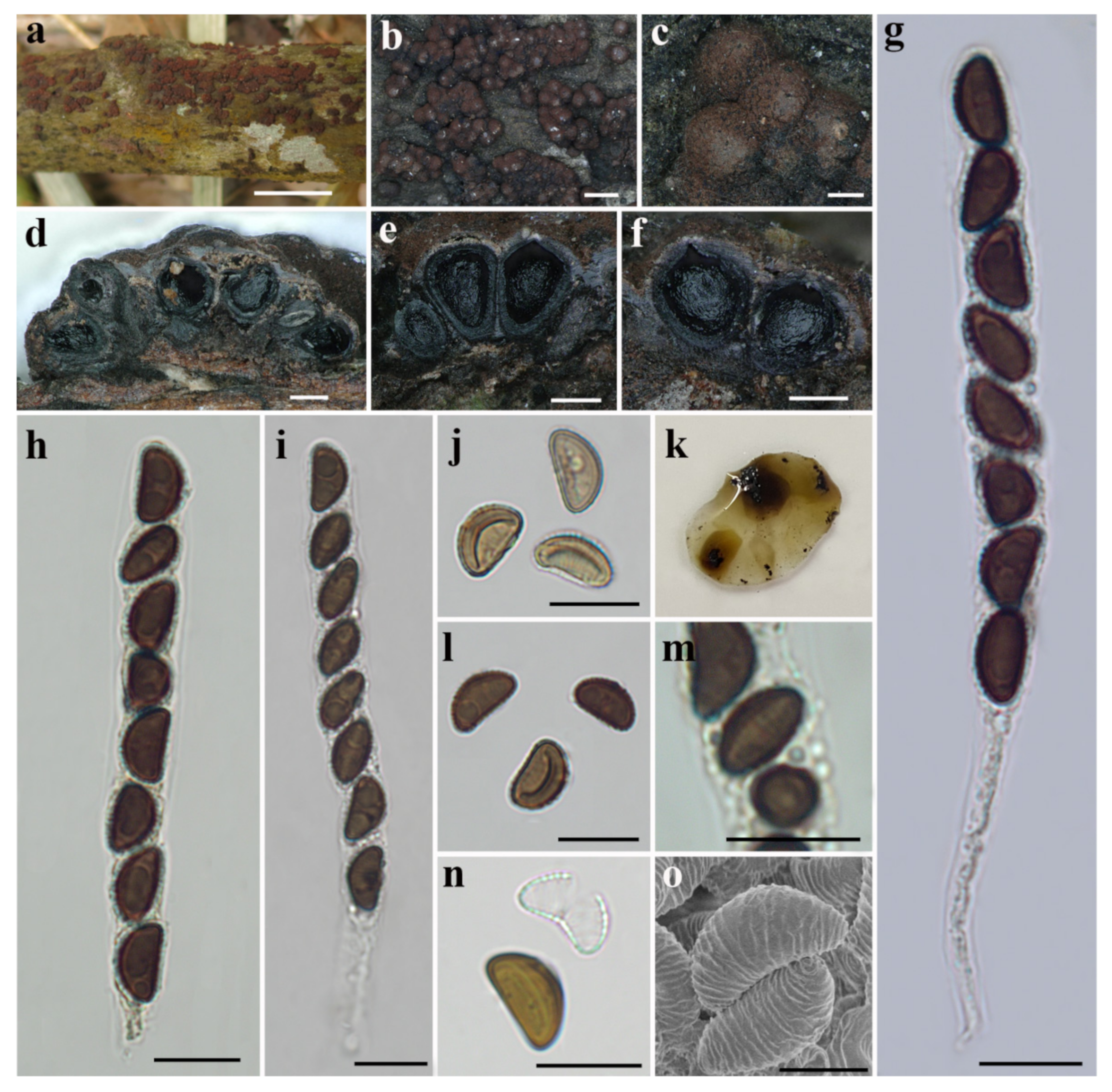
2.2. Morphological Characterization

The micromorphological observations, micrographs, and measurements were obtained using an Olympus IX73 inverted fluorescence microscope (Tokyo, Japan) with the laser capture microdissection system of model MMI CellCut Plus (Zurich, Switzerland), while the same processes for observing the morphological characteristics of stromatal sufaces and perithecia were performed using a VHX-600E microscope from the Keyence Corporation. The photographs of ascospores were examined by scanning electron microscope (SEM) (Hitachi Corporation, Tokyo, Japan). Sexual structures were microscopically observed in water, 10% KOH, and Melzer’s reagent, as determined by Ju and Rogers [4]. The color codes appearing in this article refer to Rayner [23]. In the text, the following abbreviations are used: KOH = 10% potassium hydroxide, n = number of ascospores measured from a given number of specimens, M = arithmetical average of sizes of all ascospores.

2.3. DNA Extraction, PCR Amplification, and Sequencing

Following the instructions of the manufacturer, total genomic DNA of studied samples was extracted using an improved cetyltrimethylammonium bromide (CTAB) rapid extraction kit for plant genomes (Aidlab Biotechnologies, Beijing, China) and a Thermo Scientific Phire Plant Direct PCR Kit (Thermo Fisher Scientific, Waltham, MA, USA). Four DNA loci of ITS (internal transcribed spacer regions), nLSU (nuclear large subunit ribosomal DNA), RPB2 (RNA polymerase II second largest subunit), and β-tubulin (beta-tubulin) were amplified by polymerase chain reaction (PCR) using HS Taq Mix (Dongsheng Biotech, Guangzhou, China). The 40 µL PCR mixtures contained 16 µL of ddH2O, 20 µL of 2 × HS™ Mix, 2 µL of DNA template, and 1 µL of each forward and reverse primer. The primer pairs ITS5/ITS4, LR0R/LR5, fRPB2-7CR/fRPB2-5F, and T1/T22 were used to amplify ITS, LSU, RPB2, and β-tubulin, respectively [24,25,26,27,28]. The PCR thermal cycling program for ITS was set as initial denaturation at 95 °C for 3 min, followed by 30 cycles of denaturation at 94 °C for 40 s, annealing at 55.8 °C for 45 s, extension at 72 °C for 1 min, and a final extension at 72 °C for 10 min. For generation of LSU sequence data, the following program was used: initial denaturation at 94 °C for 3 min, 36 cycles of 1 min at 94 °C, 50 s at 52 °C and 1 min at 72 °C, with a final extension period of 10 min at 72 °C. The PCR amplification of RPB2 and β-tubulin was set up as follows: initial denaturation at 95 °C for 3 min, followed by 35 cycles at 94 °C for 1 min, 52 °C for 50 s, 72 °C for 1 min, with a final extension of 72 °C for 10 min [29]. PCR sample purification and DNA sequencing were carried out by BGI, Guangzhou, China. The generated sequences were submitted to GenBank to acquire accession numbers.

2.4. Phylogenetic Analysis

All newly generated sequences and relevant sequences of closely related species within the genus Hypoxylon and among genera of the family Hypoxylaceae and some related genera of the Xylariales based on ITS, LSU, RPB2, and β-tubulin were obtained in the phylogenetic analysis (Table 1). The other genera included Annulohypoxylon, Daldinia, Hypomontagnella, Jackrogersella, Pyrenopolyporus, Rhopalostroma, and Thamnomyces. The phylogenetic trees were rooted with Xylaria hypoxylon (L.) Grev. and Biscogniauxia nummularia (Bull.) Kuntze as outgroups.
The sequence alignment was conducted using fast Fourier transformation (MAFFT) online (http://mafft.cbrc.jp/alignment/server/, accessed on 22 November 2021) [48]. Further sequence processing was conducted using BioEdit 7.0.5, and the concatenation of four DNA loci of ITS, LSU, RPB2, and β-tubulin was completed using MEGA 6.0 [29,49,50].
A combined matrix of ITS–LSU–RPB2–β-tubulin was used to construct phylogenetic trees and analyzed by two methods: Maximum Likelihood (ML) and Bayesian Inference (BI). Maximum Likelihood (ML) analysis was performed using raxmlGUI 2.0 with rapid bootstrap search executing 1000 replicates, setting the substitution model as GTRGAMMA+G [45,51]. Bayesian analysis was performed using MrBayes v.3.2.6, based on using jModelTest 2 to conduct model discrimination [52]. The selected applicable model implemented the Markov Chain Monte Carlo (MCMC) algorithm, which determined posterior probabilities (PP) [53]. Six simultaneous Markov chains were run for 1000000 generations, from which every 100th generation was sampled as a tree [54,55,56,57,58]. Phylogenetic trees were viewed in FigTree 1.4.2 [59]. The phylogenetic tree and multi-gene sequence alignment were deposited in TreeBASE (www.treebase.org/treebase-web/home.html, accessed on 20 December 2021) with accession number S29126.

3. Results

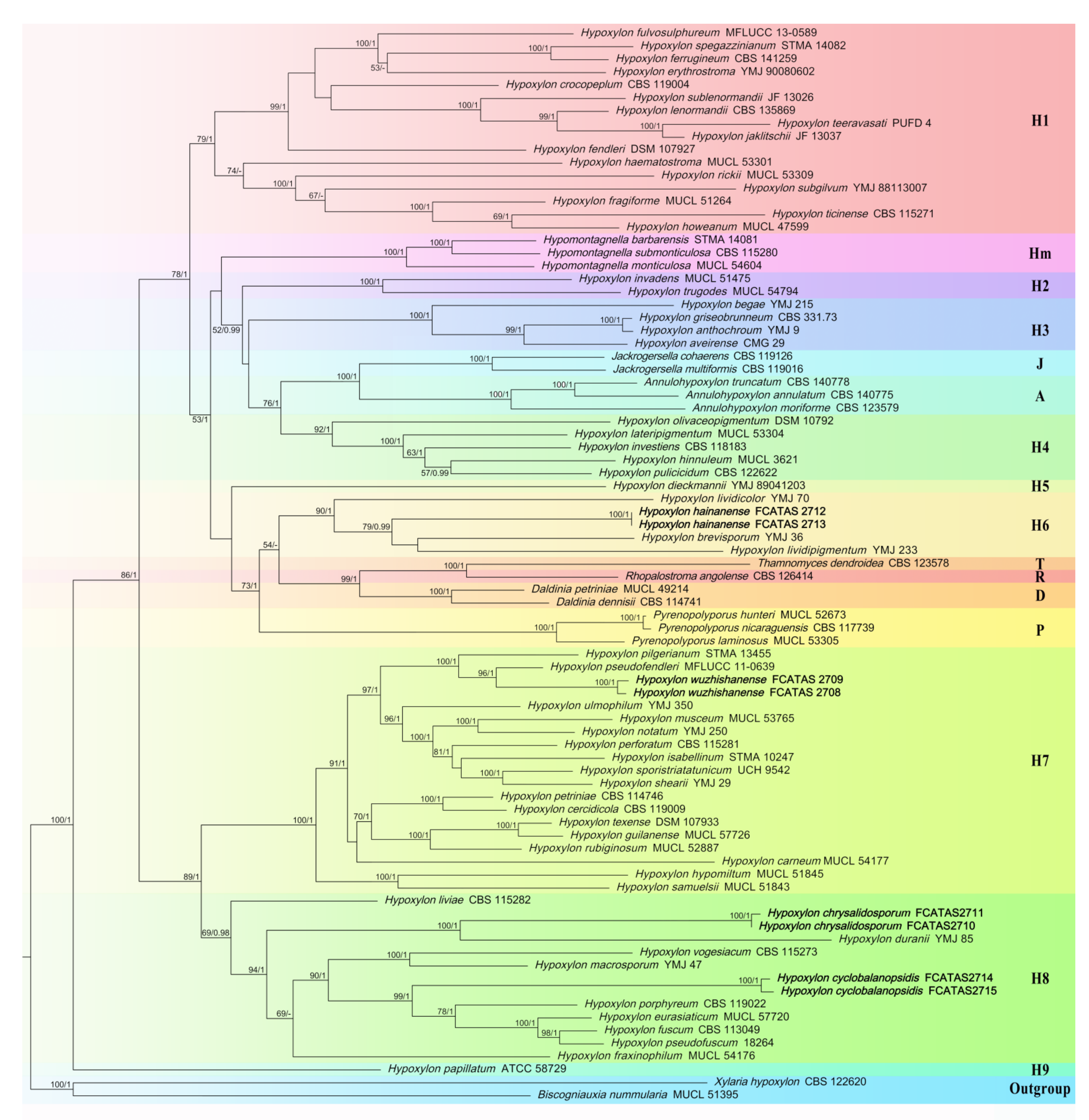
3.1. Phylogenetic Analysis

The sequence datasets for the contributions of the molecular phylogenetic trees consisted of 82 ITS, 57 LSU, 58 RPB2, and 81 β-tubulin sequences. All of the 278 sequences came from 79 strains including 4 newly described Hypoxylon taxa, 58 known Hypoxyplon taxa, 3 Annulohypoxylon taxa, 2 Daldinia taxa, 3 Hypomontagnella taxa, 2 Jackrogersella taxa, 3 Pyrenopolyporus taxa, 1 Rhopalostroma taxon and 1 Thamnomyces taxon, as well as the X. hypoxylon and B. nummularia as outgroups.
After aligned by MAFFT online, the sequence datasets contained 2046 character positions for ITS alignment, 3320 character positions for LSU alignment, 1285 character positions for RPB2 alignment, and 2298 character positions for β-tubulin alignment. With less informative positions trimmed, and four DNA loci connected, the generated multi-gene alignment (MGA) had an aligned length of 3836 characters, of which 1977 characters were parsimony-informative. Phylogenetic trees generated from BI and ML analyses of the combined dataset of ITS–LSU–RPB2–β-tubulin were highly similar in topology. Only the ML tree is shown in Figure 1, with ML bootstrap values ≥ 50% and Bayesian posterior probabilities ≥ 0.95 labelled along the branches.
The phylogenies reveal a paraphyly of Hypoxylon, with the genera Annulohypoxylon, Daldinia, Hypomontagnella, Jackrogersella, Pyrenopolyporus, and Thamnomyces embedded within the former. The phylogeny inferred from the ITS–LSU–RPB2–β-tubulin sequences demonstrated that the four new species, i.e., H. wuzhishanense, H. hainanense, H. chrysalidosporum, and H. cyclobalanopsidis, formed distinct well-supported lineages (Figure 1).

3.2. Taxonomy

Hypoxylon chrysalidosporum Hai X. Ma, Z.K. Song, sp. nov., Figure 2.
MycoBank: MB 841956.
Diagnosis. Differs from H. duranii and H. notatum in its KOH-extractable pigments, highly reduced or absent inamyloid apical apparatus, and smaller ascospores with straight germ slit.
Etymology.Chrysalidosporum (Lat.): referring to the chrysalis-shaped ascospores.
Holotype. CHINA: Hainan Province, Ledong County, Hainan Tropical Rainforest National Park, Jianfengling National Natural Reserve, approximately 108°51′ E and 18°47′ N, elevation approximately 700 m, saprobic on surface of dead corticated branches, 24 October 2020, Haixia Ma, Col. J214 (FCATAS 2710).
Teleomorph. Stromata glomerate to effused–pulvinate, with conspicuous perithecial mounds, 0.1–0.7 cm long × 0.1–0.3 cm broad × 0.3–0.5 mm thick; surface Bay (6), Rust (39), Dark Brick (60) and Livid Purple (81); with pale brown to dull reddish brown granules immediately beneath the surface and between perithecia; yielding Pale Luteous (11), Honey (60) and Ochreous (44) pigments in 10% KOH; tissue below the perithecial layer black, inconspicuous, 0.1–0.4 mm thick. Perithecia spherical to obovoid, black, 0.2–0.4 mm broad × 0.3–0.4 mm high. Ostioles umbilicate, encircled with a paler area, opening lower than the stromatal surface. Asci cylindrical, eight-spored, uniseriate, 75–139 µm total length × 6.6–11.9 µm broad; the spore-bearing portion 49–82 µm long, and stipes 19–71 µm long, with inamyloid apical apparatus highly reduced or absent, not bluing in Melzer’s reagent. Ascospores light-brown to brown, unicellular, ellipsoid-inequilateral, with slightly broad rounded ends, 8–10.6(–11.1) × 4.1–6.3(–7.1) µm (n = 60, M = 9.2 × 5.3 µm), with conspicuously straight spore-length germ slit on the convex side; perispore dehiscent in 10% KOH, with very conspicuous coil-like ornamentation in SEM; epispore smooth.
Additional specimens examined. CHINA: Hainan Province, Ledong County, Hainan Tropical Rainforest National Park, Jianfengling National Natural Reserve, approximately 108°49′ E and 18°50′ N, elevation approximately 750 m, saprobic on surface of dead corticated branches, 24 October 2020, Haixia Ma, Col. J1059 (FCATAS 2711).
Hypoxylon cyclobalanopsidis Hai X. Ma, Z.K. Song, sp. nov., Figure 3.
MycoBank: MB 841957.
Diagnosis. Differs from H. porphyreum in its larger ascospores, KOH-extractable pigments, host plant and distribution. Differs from H. eurasiaticum, H. fuscum, and H. pseudofuscum in its smaller apical apparatus, host plant and tropical distribution.
Etymology.Cyclobalanopsidis (Lat.): referring to the host genus Cyclobalanopsis which the fungus inhabits.
Holotype. CHINA: Hainan Province, Ledong County, Hainan Tropical Rainforest National Park, Jianfengling National Natural Reserve, Mingfeng Valley, approximately 108°53′ E and 18°43′ N, elevation approximately 720 m, saprobic on dead corticated branches of Cyclobalanopsis, 23 October 2020, Haixia Ma, Col. J217 (FCATAS 2714).
Teleomorph. Stromata pulvinate to effused–pulvinate, 0.1–2 cm long × 0.1–0.6 cm broad × 0.25–0.45 mm thick; with inconspicuous perithecial mounds; surface Livid Purple (81), Livid Vinaceous (83) and Violet (32), with colored coating worn off exposing Dark Purple (36) areas, sometimes with tiny cracks appearing; with pale-brown to orange-brown granules immediately beneath the surface and between perithecia; yielding Amber (47) and Ochreous (44) to Fulvous (43) pigments in 10% KOH; tissue below the perithecial layer inconspicuous and pale-brown to black. Perithecia ovoid to obovoid, black, 0.1–0.3 mm broad × 0.1–0.4 mm high. Ostioles umbilicate, encircled with a white area, opening lower than the stromatal surface. Asci cylindrical, eight-spored, uniseriate, short-stipitate, 76–117 µm total length × 6.9–12.9 µm broad, the spore-bearing portion 65–93 µm long, and stipes 6–28 µm long, with apical apparatus highly reduced and minute, faintly bluing in Melzer’s reagent. Ascospores brown to dark-brown, unicellular, ellipsoid-inequilateral, with narrowly to broadly rounded ends, 11–15.2 × 5.1–7 µm (n = 60, M = 13 × 6.3 µm), with more sigmoid to less straight spore-length germ slit on the convex side; perispore dehiscent in 10% KOH, with conspicuous coil-like ornamentation in SEM; epispore smooth.
Additional specimens examined. CHINA: Hainan Province, Ledong County, Hainan Tropical Rainforest National Park, Jianfengling National Natural Reserve, Mingfeng Valley, approximately 108°51′ E and 18°45′ N, elevation approximately 700 m, saprobic on dead corticated branches of Cyclobalanopsis, 23 October 2020, Haixia Ma, Col. J200 (FCATAS 2715).
Hypoxylon hainanense Hai X. Ma, Z.K. Song, sp. nov., Figure 4.
MycoBank: MB 841955.
Diagnosis. Differs from H. brevisporum in having larger and wider ascospores, spherical to obovoid perithecia, and slightly larger apical apparatus. Differs from H. lividicolor in its thiner stromata, spherical to obovoid perithecia, and smaller ascospores.
Etymology.Hainanense (Lat.): referring to the holotype locality of species in Hainan Province.
Holotype. CHINA: Hainan Province, Ledong County, Hainan Tropical Rainforest National Park, Jianfengling National Natural Reserve, approximately 108°48′ E and 18°45′ N, elevation approximately 650 m, saprobic on surface of dead decorticated wood, 29 December 2020, Haixia Ma, Col. J233 (FCATAS 2712).
Teleomorph. Stromata effused–pulvinate, 0.8–7.2 cm long × 0.6–3.3 cm broad × 0.7–1.5 mm thick; with inconspicuous to conspicuous perithecial mounds; surface Violet (32), Livid Purple (81) and Dark Violet (33); highly carbonaceous black granules immdediately beneath surface and between perithecia; yielding Pale Vinaceous (85) to Livid Vinaceous (83) and Vinaceous Purple (101) pigments in 10% KOH; tissue below the perithecial layer black, conspicuous, 0.3–0.8 mm thick. Perithecia spherical to obovoid, black, 0.2–0.5 mm broad × 0.3–0.6 mm high, occasionally with pale-brown perithecial contents. Ostioles opening at the same level or slightly higher than the stromatal surface. Asci cylindrical, eight-spored, uniseriate, 70–139 µm total length × 4.6–6.8 µm broad, the spore-bearing portion 47–61 µm long, and stipes 19–83 µm long, with amyloid apical apparatus bluing in Melzer’s reagent, discoid, 0.6–1.1 µm high × 1.3–1.8 µm broad. Ascospores light-brown to brown, unicellular, ellipsoid-inequilateral, with slightly broad rounded ends, 6.1–9.6 × 3.2–5 µm (n = 60, M = 7.7 × 4 µm), with conspicuously straight germ slit less than spore-length on the convex side; perispore dehiscent in 10% KOH, with inconspicuous coil-like ornamentation in SEM; epispore smooth.
Additional specimens examined. CHINA: Hainan Province, Ledong County, Hainan Tropical Rainforest National Park, Jianfengling National Natural Reserve, approximately 108°50′ E and 18°46′ N, elevation approximately 600 m, saprobic on surface of dead decorticated wood, 29 December 2020, Haixia Ma, Col. J1058 (FCATAS 2713).
Hypoxylon wuzhishanense Hai X. Ma, Z.K. Song, sp. nov., Figure 5.
MycoBank: MB 841954.
Diagnosis. Differs from H. pseudofendleri by having smaller prithecia, larger ascospores and lower ostioles. Differs from H. pilgerianum in having larger apical apparatus, larger and wider ascospores, and most of perispore indehiscent in 10% KOH.
Etymology.Wuzhishanense (Lat.): referring to the holotype locality of species in Wuzhishan National Natural Reserve.
Holotype. CHINA: Hainan Province, Wuzhishan City, Hainan Tropical Rainforest National Park, Wuzhishan National Natural Reserve, approximately 109°38′ E and 18°55′ N, elevation approx. 600 m, saprobic on surface of dead Bamboo sp., 30 December 2020, Haixia Ma, Col. W2 (FCATAS 2708).
Teleomorph. Stromata pulvinate, 0.8–14 cm long × 0.4–3.2 cm broad × 0.2–0.3 mm thick; with inconspicuous-to-conspicuous perithecial mounds; surface Rust (39), Livid Purple (81) to Dark Brick (60), with colored coating worn off exposing Dark Purple (36) areas, with yellowish-brown granules immediately beneath the surface and between perithecia; yielding Amber (47) and Ochreous (44) to Fulvous (43) pigments in 10% KOH; tissue below the perithecial layer inconspicuous. Perithecia spherical, black, 0.2–0.3 mm broad × 0.1–0.2 mm high. Ostioles umbilicate, opening slightly lower than the stromatal surface. Asci cylindrical, eight-spored, uniseriate, 71–106 µm total length × 6.4–9 µm broad, the spore-bearing portion 58–91 µm long, and stipes 9–28 µm long, with amyloid apical apparatus bluing in Melzer’s reagent, discoid, 1–1.9 µm high × 2.2–3.4 µm broad. Ascospores light-brown to brown, unicellular, ellipsoid-inequilateral, with narrowly rounded ends, (9.5–)10–14 × 5.4–6.7 µm (n = 60, M = 11.4 × 6 µm), with straight to less frequently sigmoid germ slit spore-length on the convex side; most of perispore indehiscent in 10% KOH, occasionally dehiscent, with inconspicuous coil-like ornamentation in SEM; epispore smooth.
Additional specimens examined. CHINA: Hainan Province, Wuzhishan City, Hainan Tropical Rainforest National Park, Wuzhishan National Natural Reserve, approximately 109°38′ E and 18°55′ N, elevation approximately 600 m, saprobic on surface of dead Bamboo sp., 30 December 2020, Haixia Ma, Col. X469 (FCATAS 2709).
Key to Hypoxylon species from China and related species around the world
1. Ostioles barely to slightly higher than the stromatal surface .......................................... 2
1. Ostioles lower than the stromatal surface .......................................................................... 8
2. Ascospores nearly equilateral .............................................................................................. 3
2. Ascospores inequilateral ....................................................................................................... 4
3. Stromata glomerate to pulvinate; ascospores 8.5–12(–13.5) × 4–5 µm .......... H. croceum
3. Stromata pulvinate to effused–pulvinate; ascospores 11–14.5 × (4.5–)5–6.5 µm ................................................................................................................... H. parksianum
4. Perithecia tubular.................................................................................................................... 5
4. Perithecia subglobose, spherical to obovoid ...................................................................... 6
5. Perithecia 0.5–0.85 mm broad × 0.35–0.5mm high ............................... H. pseudofendleri
5. Perithecia 0.2–0.3 mm broad × 0.4–0.6 mm high ................................. H. lienhwacheense
6. Ascospores light-brown to brown ................................................................ H. hainanense
6. Ascospores brown to dark-brown ....................................................................................... 7
7. Perithecia 0.3–0.5(–0.6) mm broad; ascospores 9.5–15(–16) × 4–6.5(–7) µm .................................................................................................................. H. lenormandii
7. Perithecia 0.1–0.2 mm broad; ascospores 7.5–9.5 × 3.5–4.5 µm ....................... H. rutilum
8. Ascospores nearly equilateral .............................................................................................. 9
8. Ascospores inequilateral ..................................................................................................... 14
9. Perispore dehiscent in 10% KOH ................................................................ H. hypomiltum
9. Perispore indehiscent in 10% KOH ................................................................................... 10
10. KOH-extractable pigment Orange (7) .................................................... H. cinnabarinum
10. Without apparent KOH-extractable pigments or with other colors ............................ 11
11. Without apparent KOH-extractable pigments or with dilute Grayish Sepia (106) to blackish pigments ........................................................................................ H. dieckmannii
11. KOH-extractable pigments greenish to olivaceous ........................................................ 12
12. Perithecia tubular to long tubular, 0.3–0.4 mm broad × 0.5–1 mm high .. H. investiens
12. Perithecia spherical to obovoid, less than 0.3 mm broad .............................................. 13
13. Stromatal surface Vinaceous Gray (116), Purplish Gray (128), Livid Vinaceous (83), Dark Vinaceous (82), or Brown Vinaceous (83), becoming blackish when aged; dull reddish-brown granules immediately beneath surface and between perithecia; asci with apical apparatus bluing in Melzer’s reagent; ascospores dark-brown to blackish-brown, pyriform to obovoid, (11.5–)12–15(–16) × 5.5–7 µm ........................................................................................................... H. fuscopurpureum
13. Stromatal surface Fawn (87) or Umber (9); blackish granules immediately beneath surface and between perithecia; asci with apical apparatus lightly bluing in Melzer’s reagent; ascospores brown, ellipsoid, 7–8.5 × 4–4.5 µm .............………. H. gilbertsonii
14. Sigmoid germ slit ....................................................................................................……… 15
14. Straight or slightly sigmoid germ slit ............................................................................... 17
15. Stromata glomerate, with conspicuous perithecial mounds; KOH-extractable pigments Pure Yellow (14) with Citrine (13) tone, Greenish Olivaceous (90), or Orange (7) .......................................................................................................... H. musceum
15. Stromata pulvinate or effused–pulvinate, with inconspicuous to conspicuous perithecial mounds; KOH-extractable pigments with other colors ............................ 16
16. Asci with apical apparatus bluing in Melzer’s reagent, 0.5–1.2 µm high × 1.8–2.5 µm broad; KOH-extractable pigment Orange (7) ..................……........................ H. fendleri
16. Asci with apical apparatus bluing in Melzer’s reagent, 0.5–0.8 µm high × 3–3.4 µm broad; KOH-extractable pigment Vinaceous Purple (101) ........…............. H. fuscoides
17. Straight germ slit …............................................................................................................. 18
17. Straight or slightly sigmoid germ slit ............................................................................... 26
18. Straight germ slit slightly less than spore length; perispore infrequently dehiscent in 10% KOH ................................................................................................................. H. dengii
18. Straight spore-length germ slit; perispore dehiscent in 10% KOH .............................. 19
19. Perithecia long tubular …................................................................................................... 20
19. Perithecia spherical to obovoid …..................................................................................... 21
20. Stromatal surface Fulvous (43), Sienna (8), or Rust (39); stromata containing orange-red granules, with KOH-extractable pigments Orange (7) or Scarlet (5); asci with apical apparatus bluing in Melzer’s reagent, 1–2(–2.5) µm high × 3–4 µm broad ......................….......................................................................….. H. haematostroma
20. Stromatal surface Brown Vinaceous (84), Dark Brick (60), Sepia (63), or Chestnut (40); stromata containing dark-reddish-brown or blackish granules, with KOH-extractable pigments Olivaceous (48), Greenish Olivaceous (90), Isabelline (65), or Dull Green (70), or infrequently without apparent pigments; asci with apical apparatus bluing in Melzer’s reagent, 0.5–1 µm high × 2.5–3 µm broad ............................. H. placentiforme
21. KOH-extractable pigments Olivaceous Gray (12), Greenish Olivaceous (90), or Gray Olivaceous (107) ......................................................................................... H. brevisporum
21. KOH-extractable pigments with other colors ................................................................. 22
22. Conspicuous coil-like ornamentation of perispore; asci with apical apparatus not bluing in Melzer’s reagent ................................................................................................ 23
22. Smooth or with inconspicuous coil-like ornamentation of perispore; asci with apical apparatus bluing to lightly bluing in Melzer’s reagent ................................................ 24
23. KOH-extractable pigments Orange (7), Sienna (8), and Amber (47). H. baihualingense
23. KOH-extractable pigments Pale Luteous (11), Citrine (13) and Honey (64) ....................................................................................................... H. chrysalidosporum
24. Stromata on bamboo .................................................................................... H. pilgerianum
24. Stromata on dicot wood ..................................................................................................... 25
25. Stromata effused–pulvinate, plane, or with inconspicuous to conspicuous perithecial mounds; perithecia 0.2–0.5 mm broad × 0.3–0.6 mm high; smooth or with inconspicuous coil-like ornamentation of perispore ............................. H. rubiginosum
25. Stromata pulvinate with conspicuous perithecial mounds; perithecia 0.1–0.2 mm broad × 0.2–0.3 mm high; smooth perispore ................................ H. vinosopulvinatum
26. Perithecia 0.5–0.7 mm broad ..................................................................... H. wujiangensis
26. Perithecia less than 0.3 mm broad .................................................................................... 27
27. Perispore indehiscent in 10% KOH ...................................................... H. wuzhishanense
27. Perispore dehiscent in 10% KOH ...................................................................................... 28
28. Conspicuous coil-like ornamentation of perispore ........................................................ 29
28. Smooth or with inconspicuous coil-like ornamentation of perispore ......................... 32
29. Stromata glomerate to hemispherical .............................................................................. 30
29. Stromata pulvinate to effused–pulvinate ........................................................................ 31
30. Stromata glomerate, restricted-pulvinate to effused–pulvinate, 0.1–6 cm long × 0.1–1.5 cm broad; asci with apparatus bluing in Melzer’s reagent; ascospores with straight or slightly sigmoid spore-length germ slit ........................................ H. duranii
30. Stromata hemispherical, pulvinate to effused–pulvinate, up to 12 cm long × 0.2–2 cm broad; asci with apical apparatus bluing in Melzer’s reagent; ascospores with more sigmoid or less straight spore-lenth germ slit ....................................... H. eurasiaticum
31. Asci with apical apparatus highly reduced, minute, faintly bluing in Melzer’s reagent; ascospores 11–15.2 × 5.1–7 µm, with more sigmoid or less straight spore-length germ slit ........................................................................................................ H. cyclobalanopsidis
31. Asci with apical apparatus bluing in Melzer’s reagent, 0.5–1 µm high × 2–2.5 µm broad; ascospores (9–)9.5–12 × 4.5–5 µm, with straight or slightly sigmoid spore-length germ slit .................................................................................................... H. retpela
32. Stromata hemispherical to spherical ................................................................................ 33
32. Stromata pulvinate to effused–pulvinate ........................................................................ 36
33. KOH-extractable pigments Orange (7) or Rust (39) .................................. H. howeanum
33. KOH-extractable pigments with other colors ................................................................. 34
34. Asci with apical apparatus highly reduced or lacking, not bluing in Melzer’s reagent; ascospores (11–)12–16 × (5.5–)6–7.5 µm .......................................................... H. notatum
34. Asci with apical apparatus bluing in Melzer’s reagent ................................................. 35
35. Perithecia spherical to obovoid, 0.1–0.3(–0.4) mm broad × 0.2–0.5 mm high; ascospores 8–20 × 4–8 µm, with slightly sigmoid germ slit ............................. H. fuscum
35. Perithecia spherical, 0.1-0.3 mm diameter; ascospores (8–)9–12(–13) × 4–6 µm, with straight or slightly sigmoid germ slit ......................................................... H. perforatum
36. Ascospore length less than 11 µm .................................................................................... 37
36. Ascospore length more than 11 µm ................................................................................. 39
37. KOH-extractable pigments Pure Yellow (14) or Amber (47) ....................... H. trugodes
37. KOH-extractable pigment Orange (7) .............................................................................. 38
38. Stromatal surface Fulvous (43), Ochreous (44), or Apricot (42); asci with apical apparatus 0.2–0.5 µm high × 1–1.5 µm broad; ascospores 8–9.5(–11) × 4–5 µm; Periconiella-like conidiogenous structure ..................................................... H. jecorinum
38. Stromatal surface Umber (9), Sepia (63), Rust (39), Sienna (8), Dark Brick (60), or Bay (6); asci with apical apparatus 0.3–1 µm high × 1.5–2.2 µm broad; ascospores 7–11 × 3.5–5 µm; Nodulisporium-like conidiogenous structure ............................. H. subgilvum
39. KOH-extractable pigment Orange (7) ...................................................... H. crocopeplum
39. KOH-extractable pigments with other colors ................................................................. 40
40. KOH-extractable pigment Dark Livid (80) ...................................................................... 41
40. KOH-extractable pigments greenish to olivaceous ........................................................ 42
41. Stromata 2.5 mm thick; ascospores 11–12.5 × 4.5–5 µm ............................ H. lividicolor
41. Stromata 0.8–1 mm thick; ascospores 10–13.5(–15) × 4.5–6 µm ..... H. lividipigmentum
42. Perithecia obovoid to tubular ................................................................... H. anthochroum
42. Perithecia spherical to obovoid ......................................................................................... 43
43. Perithecia 0.18–0.35 mm broad × 0.28–0.42 mm high; ascospores (9–)10–13.5 × 4–5 μm .................................................................................................................. H. porphyreum
44. Perithecia 0.12–0.28 mm broad × 0.19–0.36mm high; ascospores 11–16 × 4.5–7.3 µm ............................................................................................................... H. pseudofuscum

4. Discussion

Hainan Tropical Rainforest National Park is primarily tropical lowland and tropical mountain rainforest, enjoying a tropical island monsoon climate moderated by a hot and moist climate with annual rainfall often over 2200 mm (http://www.hntrnp.com, accessed on 15 November 2021). The pattern raises the high diversity and the high number of endemic species of vegetation and fungi in the region. Hypoxylon is a cosmopolitan genus, but in tropical and subtropical regions it displays a higher diversity [4]. In the present study, four new species of Hypoxylon from Hainan Tropical Rainforest National Park are described, based on morphological characteristics and phylogenetic analyses of the ITS, LSU, RPB2, and β-tubulin sequences. The secondary metabolite profiles generated from chemotaxonomic studies provide strong support for identifying species. However, chemotaxonomic data were not generated in this study [3].
Phylogenetically, H. chrysalidosporum is closely related to H. duranii J. D. Rogers, based on a combined ITS–LSU–RPB2–β-tubulin dataset. Hypoxylon duranii was originally described from Mexico, but the holotype lacked phylogenetic data. Sequence data for H. duranii collected from China were referenced in this study [4,16]. Morphologically, H. duranii is similar to H. chrysalidosporum, sharing glomerate and effused–pulvinate stromata, spherical to obovoid perithecia, and dehiscent perispore with conspicuous coil-like ornamentation. However, H. duranii can be distinguished from H. chrysalidosporum by its KOH-extractable pigments Isabelline or Amber, amyloid apical apparatus, bluing in Melzer’s reagent, and slighly larger ascospores [9.5–13(–14.5) × 4.5–6.5 µm] with a straight or slightly sigmoid germ slit [4]. Hypoxylon chrysalidosporum resembles H. notatum Berk., M. A. Curtis apud Berk. and H. shearii Y.-M. Ju, J. D. Rogers in having a similar stromatal morphology, apical apparatus being highly reduced or absent, not bluing in Melzer’s reagent, and having dehiscent perispore [4]. However, the type of H. notatum was selected by Miller (1961) from the southern United States, and differs in having KOH-extractable pigments Pure Yellow with Greenish Yellow tone and Dark Brown, and larger ascospores [(11–)12–16 × (5.5–)6–7.5 µm] which are strongly curved [1,4]. H. shearii has a buff or fawn stromatal surface, with Luteous KOH-extractable pigments, larger prithecia (0.4–0.7 mm diameter), and dark-brown, larger ascospores [12–14 × 5.5–6.5(–7) µm] [4].
Hypoxylon cyclobalanopsidis is closely related to H. porphyreum Granmo, H. eurasiaticum Pourmoghaddam, Krisai-Greilhuber, Khodap., H. fuscum (Pers.: Fr.) Fr., and H. pseudofuscum Pourmoghaddam, Khodap., Krisai-Greilhuber in the phylogenetic analyses (Figure 1). Hypoxylon porphyreum differs from H. cyclobalanopsidis in its smaller ascospores [(9–)10–13.5 × 4–5 µm, M = 11.4 × 4.8 µm], KOH-extractable pigments Brown with a Greenish tone, and growing on Quercus from southeastern Norway, Sweden, France and the USA [60,61]. Hypoxylon eurasiaticum can be distinguished from H. cyclobalanopsidis by its larger discoid apical apparatus (0.5–1.5 µm high × 2.5–3.5 µm wide), smaller ascospores (9–12.5 × 4–6 µm), and by growing on Quercus castaneifolia from Iran [33]. Hypoxylon fuscum is primarily distinguished from H. cyclobalanopsidis by its hemispherical to pulvinate stromata with dull orange, dull orange-brown, or dull reddish-brown granules immediately beneath the surface and between perithecia, larger discoid apical apparatus (0.5–2 µm high × 1.2–3.5 µm wide), slightly larger ascospores (12.5–15.5 × 5–7 µm), and it frequently occurs on Corylus avellana in Europe [4,62,63]. Hypoxylon pseudofuscum has larger discoid apical apparatus (0.5–1.5 µm high × 2–3.5 µm wide), KOH-extractable pigments Isabelline, or Hazel, slightly larger ascospores (11–16 × 4.5–7.3 µm), and it grows on Alnus and Salix from Germany and Iran [33].
Hypoxylon hainanense is closely related to H. brevisporum Y.M. Ju, J.D. Rogers, H. lividipigmentum F. San Martín, Y.M. Ju, J.D. Rogers, and H. lividicolor Y.-M. Ju, J. D. Rogers, with a weak bootstrap value according to ML phylogenetic analyses (Figure 1). Hypoxylon brevisporum differs from H. hainanense in having KOH-extractable pigment Olivaceous Gray, obovoid to tubular perithecia, smaller and thiner ascospores (5.5–8 × 2.5–3.5 µm), and slightly smaller apical apparatus (0.2–0.4 µm high × 1.2–1.5 µm broad) [4]. Hypoxylon lividicolor is distinguished by its thicker, chestnut stromata with KOH-extractable pigment Dark Livid, tubular to long tubular perithecia, and larger dark-brown ascospores (11–12.5 × 4.5–5 µm) with straight or slightly sigmoid germ slit [4]. Hypoxylon lividipigmentum differs in having tubular to long tubular perithecia and larger, dark-brown ascospores [10–13.5(–15) × 4.5–6 µm] [4].
Hypoxylon wuzhishanense is closely related to H. pseudofendleri D.Q. Dai, K.D. Hyde in our phylogenetic analyses (Figure 1). Unfortunately, RPB2 and β-tubulin sequences of H. pseudofendleri are not available for phylogenetic analysis in GenBank. Morphologically, H. pseudofendleri is similar to H. wuzhishanense in having large, purplish-brown stromata, yellowish-brown granules beneath the surface and between perithecia, similar asci and apical apparatus. However, H. pseudofendleri differs from H. wuzhishanense in having larger perithecia (500–850 µm broad × 350–500 µm high), with ostioles slightly higher than the stromatal surface, and slightly smaller ascospores (9–11.5 × 4.5–6.5 µm, M = 10.2 × 5.7 µm) with slighly pointed at the ends and smooth wall [42]. There are no descriptions of stromatal pigments in 10% KOH, germination site of ascospores, or perispore in 10% KOH for H. pseudofendleri, so we cannot compare these characteristics between the two species. The two species group together with H. pilgerianum Henn. Hypoxylon pilgerianum was originally described from Brazil on Chusquea sp., with ascospores 10–12 × 4–5 µm (M = 11 × 4.5 µm), and reinstated by Ju and Rogers on dead culms of bamboo, with ascospores 8.5–12(–13.5) × 4–5(–5.5) µm (M = 10.3 × 4.5 µm) [4,9,64]. Fournier et al. described two collections from Martinique as H. cf. pilgerianum sp. 1 and H. cf. pilgerianum sp. 2, with ascospores (7.6–)7.9–9.1(–10) × (3.4–)3.7–4.3(–4.4) μm (M = 8.5 × 4 µm) and (10.3–)10.9–12.5(–12.8) × (4.9–)5.2–6.1(–6.7) μm (M = 11.6 × 5.7 µm), respectively [9]. Hypoxylon pilgerianum s. Ju, Rogers resembles H. wuzhishanense in stromatal morphology, but the former has slighly smaller apical apparatus (0.5–1 µm high × 2.5 µm broad), and smaller and thinner ascospores [8.5–12(–13.5) × 4–5(–5.5) µm], with perispore dehiscent in 10% KOH [4]. Hypoxylon fuscopurpureum (Schwein.) M. A. Curtis somewhat resembles H. wuzhishanense, sharing its stromatal morphology, but differs in having greenish KOH-extractable pigments and larger asci (115–150 × 8–10 µm) [4].
Most species of Hypoxylon play an important ecological role in tropical rainforests as wood-decomposers [3], and some might have benefical effects on their hosts during their endophytic life stage [65]. In addition, many species have been found to produce highly bioactive secondary metabolites [41,43,66,67,68,69,70]. Although approximately 33 species of Hypoxylon have been recorded in China [4,19,20,21,29,71], species diversity, evolution, population dynamics, and the host–fungus interactions of this genus are still obscure. Therefore, comprehensive studies on the diversity, phylogeny, evolution, host–fungus interactions, and secondary metabolites of the genus Hypoxylon are needed in the future.

5. Conclusions

The current study revealed four new taxa of Hypoxylon from Hainan Tropical Rainforest National Park based on morphological characteristics, ecological distributions, and a combined ITS–LSU–RPB2–β-tubulin phylogeny.

Author Contributions

H.M. designed the research; H.M., Z.S., X.P., Z.Y., and Z.Q. prepared the samples; Z.S. conducted the molecular experiments and analyzed the data; Z.S. and H.M. drafted the manuscript; Y.L. revised the language of the text. All authors have read and agreed to the published version of the manuscript.

Funding

The research was supported by the National Natural Science Foundation of China (No. 31972848, 31770023), Key Research and Development Program of Hainan (ZDYF2020062), and Hainan Basic and Applied Research Project for Cultivating High-Level Talents (2019RC305).

Institutional Review Board Statement

Not applicable.

Data Availability Statement

Publicly available datasets were analyzed in this study. All resulting alignments were deposited in TreeBASE (http://www.treebase.org; accession number S29126). All newly generated sequences were deposited in GenBank (https://www.ncbi.nlm.nih.gov/genbank/; Table 1). All new taxa were deposited in MycoBank (https://www.mycobank.org/; MycoBank identifiers follow new taxa).

Acknowledgments

We express our gratitude to Shuang-hui He (Beijing Forestry University, China) for help during field collections. Hainan Tropical Rainforest National Park Service is thanked for providing collecting facilities in Jianfengling and Wuzhishan National Natural Reserve.

Conflicts of Interest

The authors declare no conflict of interest. The funders had no role in the design of the study; in the collection, analyses, or interpretation of data; in the writing of the manuscript, or in the decision to publish the results.

References

  1. Miller, J.H. A Monograph of the World Species of Hypoxylon; University Georgia Press: Athens, Greece, 1961; p. 158. [Google Scholar]
  2. Rogers, J.D. The Xylariaceae: Systematic, biological and evolutionary aspects. Mycologia 1979, 71, 1–42. [Google Scholar] [CrossRef]
  3. Stadler, M. Importance of secondary metabolites in the Xylariaceae as parameters for assessment of their taxonomy, phylogeny, and functional biodiversity. Curr. Res. Envion. Appl. Mycol. 2011, 1, 75–133. [Google Scholar] [CrossRef]
  4. Ju, Y.M.; Rogers, J.D. A revision of the Genus Hypoxylon; American Phytopathological Society Press: St. Paul, MN, USA, 1996; p. 365. [Google Scholar]
  5. Rogers, J.D. Thoughts and musings on tropical Xylariaceae. Mycol. Res. 2000, 104, 1412–1420. [Google Scholar] [CrossRef]
  6. Kuhnert, E.; Fournier, J.; Per, D.; Luangsaard, J.J.D.; Stadler, M. New Hypoxylon species from Martinique and new evidence on the molecular phylogeny of Hypoxylon based on ITS rDNA and β-tubulin data. Fungal Divers. 2014, 64, 181–203. [Google Scholar] [CrossRef]
  7. Hyde, K.D.; Norphanphoun, C.; Maharachchikumbura, S.S.N.; Bhat, D.J.; Jones, E.B.G.; Bundhun, D. Refined families of Sordariomycetes. Mycosphere 2020, 11, 305–1059. [Google Scholar] [CrossRef]
  8. Pourmoghaddam, M.J.; Lambert, C.; Surup, F.; Khodaparast, S.A.; Krisai-Greilhuber, I.; Voglmayr, H.; Stadler, M. Discovery of a new species of the Hypoxylon rubiginosum complex from Iran and antagonistic activities of Hypoxylon spp. Against the Ash Dieback pathogen, Hymenoscyphus fraxineus, in dual culture. MycoKeys 2020, 66, 105–133. [Google Scholar] [CrossRef] [PubMed] [Green Version]
  9. Fournier, J.; Lechat, C.; Courtecuisse, R. The genus Hypoxylon (Xylariaceae) in Guadeloupe and Martinique (French West Indies). Ascomycete.org 2016, 7, 145–212. [Google Scholar]
  10. Sir, E.B.; Kuhnert, E.; Lambert, C.; Hladki, A.I.; Romero, A.I.; Stadler, M. New species and reports of Hypoxylon from Argentina recognized by a polyphasic approach. Mycol. Prog. 2016, 15, 42. [Google Scholar] [CrossRef]
  11. Rogers, J.D. Two new Hypoxylon species from Gabon. Can. J. Bot. 1981, 59, 1363–1364. [Google Scholar] [CrossRef]
  12. Rogers, J.D.; Candoussau, F. A new variety of Hypoxylon cohaerens from France. Mycologia 1980, 72, 826–829. [Google Scholar] [CrossRef]
  13. Rogers, J.D. Hypoxylon weldenii var. microsporum and H. punctidiscum. Mycologia 1980, 72, 829–832. [Google Scholar] [CrossRef]
  14. Rogers, J.D.; Ju, Y.M.; Hemmes, D.E. Hypoxylon rectangulosporum sp. nov., Xylaria psidii sp. nov., and comments on taxa of Podosordaria and Stromatoneurospora. Mycologia 1992, 84, 166–172. [Google Scholar] [CrossRef]
  15. Gucht, K.V.; Ju, Y.M.; Rogers, J.D. New Hypoxylon species from Papua New Guinea and notes on some other taxa. Mycologia 1997, 89, 503–511. [Google Scholar] [CrossRef]
  16. Hsieh, H.; Ju, Y.M.; Rogers, J.D. Molecular phylogeny of Hypoxylon and closely related genera. Mycologia 2005, 97, 844–865. [Google Scholar] [CrossRef]
  17. Lambert, C.; Wendt, L.; Hladki, A.I.; Stadler, M.; Sir, E.B. Hypomontagnella (Hypoxylaceae): A new genus segregated from Hypoxylon by a polyphasic taxonomic approach. Mycol. Prog. 2019, 18, 187–201. [Google Scholar] [CrossRef]
  18. Wendt, L.; Sir, E.B.; Kuhnert, E.; Heitkämper, S.; Lambert, C.; Hladki, A.I.; Romero, A.I.; Luangsaard, J.J.; Srikitikulchai, P.; Per, D.; et al. Resurrection and emendation of the Hypoxylaceae, recognised from a multigene phylogeny of the Xylariales. Mycol. Prog. 2018, 17, 115–154. [Google Scholar] [CrossRef] [Green Version]
  19. Teng, S.C. Fungi of China; Science Press: Beijing, China, 1963; pp. 182–191. [Google Scholar]
  20. Tai, F.L. Sylloge Fungorum Sinicorum; Science Press: Beijing, China, 1979; pp. 163–167. [Google Scholar]
  21. Ma, H.X. Taxonomy and Molecular Phylogeny of Several Genera of Xylariaceae from China. Ph.D. Thesis, Jilin Agricultural University, Changchun, China, June 2011. [Google Scholar]
  22. Chi, S.Q.; Xu, J.; Lu, B.S. Three New Chinese Records of Hypoxylon. J. Fungal Res. 2016, 14, 218–221. [Google Scholar]
  23. Rayner, R.W. A Mycological Colour Chart; Commonwealth Mycological Institute: London, UK, 1970. [Google Scholar]
  24. Gardes, M.; Bruns, T.D. ITS primers with enhanced specificity for basidiomycetes-application to the identification of mycorrhizae and rusts. Mol. Ecol. 1993, 2, 113–118. [Google Scholar] [CrossRef]
  25. O’donnell, K.; Cigelnik, E. Two divergent intragenomic rDNA ITS2 types within a monophyletic lineage of the fungus Fusarium are nonorthologous. Mol. Phylogenet. Evol. 1997, 7, 103–116. [Google Scholar] [CrossRef]
  26. White, T.J.; Bruns, T.D.; Lee, S.; Taylor, J.W. Amplification and Direct Sequencing of Fungal Ribosomal RNA Genes for Phylogenetics—Science Direct; Academic Press: San Diego, CA, USA, 1990; pp. 315–322. [Google Scholar]
  27. Vilgalys, R.; Hester, M. Rapid genetic identification and mapping of enzymatically amplified ribosomal DNA from several Cryptococcus species. J. Bacteriol. 1990, 172, 4238–4246. [Google Scholar] [CrossRef] [Green Version]
  28. Liu, Y.J.; Whelen, S.; Hall, B.D. Phylogenetic relationships among ascomycetes: Evidence from an RNA polymerse II subunit. Mol. Biol. Evol. 1999, 16, 1799–1808. [Google Scholar] [CrossRef] [PubMed]
  29. Ma, H.X.; Qiu, J.Z.; Xu, B.; Li, Y. Two Hypoxylon species from Yunnan Province based on morphological and molecular characters. Phytotaxa 2018, 376, 027–036. [Google Scholar] [CrossRef]
  30. Kuhnert, E.; Sir, E.B.; Lambert, C.; Hyde, K.D.; Hladki, A.I.; Romero, A.I.; Rohde, M.; Stadler, M. Phylogenetic and chemotaxonomic resolution of the genus Annulohypoxylon (Xylariaceae) including four new species. Fungal Divers. 2017, 85, 1–43. [Google Scholar] [CrossRef]
  31. Stadler, M.; Læssøe, T.; Fournier, J.; Decock, C.; Schmieschek, B.; Tichy, H.V.; Peršoh, D. A polyphasic taxonomy of Daldinia (Xylariaceae). Stud. Mycol. 2014, 77, 1–143. [Google Scholar] [CrossRef] [Green Version]
  32. Vicente, T.F.L.; Goncalves, M.F.M.; Brandao, C.; Fidalgo, C.; Alves, A. Diversity of fungi associated with macroalgae from an estuarine environment and description of Cladosporium rubrum sp. nov. and Hypoxylon aveirense sp. nov. Int. J. Syst. Evol. Microbiol. 2021, 71, 004630. [Google Scholar] [CrossRef]
  33. Lambert, C.; Pourmoghaddam, M.J.; Cedeño-Sanchez, M.; Surup, F.; Khodaparast, S.A.; Krisai-Greilhuber, I.; Voglmayr, H.; Stradal, T.E.B.; Stadler, M. Resolution of the Hypoxylon fuscum complex (Hypoxylaceae, Xylariales) and discovery and biological characterization of two of its prominent secondary metabolites. J. Fungi 2021, 7, 131. [Google Scholar] [CrossRef]
  34. Sir, E.B.; Becker, K.; Lambert, C.; Bills, G.F.; Kuhnert, E. Observations on Texas hypoxylons, including two new Hypoxylon species and widespread environmental isolates of the H. croceum complex identified by a polyphasic approach. Mycologia 2019, 11, 832–856. [Google Scholar] [CrossRef]
  35. Friebes, G.; Wendelin, I. Studies on Hypoxylon ferrugineum (Xylariaceae), a rarely reported species collected in the urban area of Graz (Austria). Ascomycete.org 2016, 8, 83–90. [Google Scholar] [CrossRef]
  36. Daranagama, D.A.; Camporesi, E.; Tian, Q.; Liu, X.Z.; Chamyuang, S.; Stadler, M.; Hyde, K.D. Anthostomella is polyphyletic comprising several genera in Xylariaceae. Fungal Divers. 2015, 73, 203–238. [Google Scholar] [CrossRef]
  37. Sir, E.B.; Kuhnert, E.; Surup, F.; Hyde, K.D.; Stadler, M. Discovery of new mitorubrin derivatives from Hypoxylon fulvosulphureum sp. nov. (Ascomycota, Xylariales). Mycol. Prog. 2015, 14, 28. [Google Scholar] [CrossRef]
  38. Vu, D.; Groenewald, M.; Vries, M.; Gehrmann, T.; Stielow, B.; Eberhardt, U.; Al-Hatmi, A.; Groenewald, J.Z.; Cardinali, G.; Houbraken, J.; et al. Large-scale generation and analysis of filamentous fungal DNA barcodes boosts coverage for kingdom fungi and reveals thresholds for fungal species and higher taxon delimitation. Stud. Mycol. 2019, 92, 135–154. [Google Scholar] [CrossRef]
  39. Bitzer, J.; Læssøe, T.; Fournier, J.; Kummer, V.; Decock, C.; Tichy, H.V.; Piepenbring, M.; Peršoh, D.; Stadler, M. Affinities of Phylacia and the daldinoid Xylariaceae, inferred from chemotypes of cultures and ribosomal DNA sequences. Mycol. Res. 2008, 112, 251–270. [Google Scholar] [CrossRef]
  40. Becker, K.; Lambert, C.; Wieschhaus, J.; Stadler, M. Phylogenetic assignment of the fungicolous Hypoxylon invadens (Ascomycota, Xylariales) and investigation of its secondary metabolites. Microorganisms 2020, 8, 1397. [Google Scholar] [CrossRef] [PubMed]
  41. Kuhnert, E.; Surup, F.; Sir, E.B.; Lambert, C.; Hyde, K.D.; Hladki, A.I.; Romero, A.I.; Stadler, M. Lenormandins A—G, new azaphilones from Hypoxylon lenormandii and Hypoxylon jaklitschii sp. nov., recognised by chemotaxonomic data. Fungal Divers. 2015, 71, 165–184. [Google Scholar] [CrossRef]
  42. Dai, D.Q.; Phookamsak, R.; Wijayawardene, N.N.; Li, W.J.; Bhat, D.J.; Xu, J.C.; Taylor, J.E.; Hyde, K.D.; Chukeatirote, E. Bambusicolous fungi. Fungal Divers. 2017, 82, 1–105. [Google Scholar] [CrossRef]
  43. Bills, G.F.; González-Menéndez, V.; Martín, J.; Platas, G.; Fournier, J.; Peršoh, D.; Stadler, M. Hypoxylon pulicicidum sp. nov. (Ascomycota, Xylariales), a pantropical insecticide-producing endophyte. PLoS ONE 2012, 7, e46687. [Google Scholar] [CrossRef] [PubMed]
  44. Stadler, M.; Kuhnert, E.; Peršoh, D.; Fournier, J. The Xylariaceae as model example for a unified nomenclature following the “One Fungus-One Name” (1F1N) concept. Mycology 2013, 4, 5–21. [Google Scholar]
  45. Cedeño-Sanchez, M.; Wendt, L.; Stadler, M.; Mejía, L.C. Three new species of Hypoxylon and new records of Xylariales from Panama. Mycosphere 2020, 11, 1457–1476. [Google Scholar] [CrossRef]
  46. Phookamsak, R.; Hyde, K.D.; Jeewon, R.; Bhat, D.J.; Jones, E.B.G.; Maharachchikumbura, S.S.; Raspe, O.; Karunarathna, S.C.; Wanasinghe, D.N.; Hongsanan, S.; et al. Fungal diversity notes 929–1035: Taxonomic and phylogenetic contributions on genera and species of fungi. Fungal Divers. 2019, 95, 1–273. [Google Scholar] [CrossRef] [Green Version]
  47. Stadler, M.; Fournier, J.; Laessøe, T.; Chlebicki, A.; Lechat, C.; Flessa, F.; Rambold, G.; Peršoh, D. Chemotaxonomic and phylogenetic studies of Thamnomyces (Xylariaceae). Mycoscience 2010, 51, 189–207. [Google Scholar] [CrossRef]
  48. Katoh, K.; Rozewicki, J.; Yamada, K.D. MAFFT online service: Multiple sequence alignment, interactive sequence choice and visualization. Brief. Bioinform. 2019, 20, 1160–1166. [Google Scholar] [CrossRef] [Green Version]
  49. Felsenstein, J. Evolutionary trees from DNA sequences: A maximum likelihood approach. J. Mol. Evol. 1981, 17, 368–376. [Google Scholar] [CrossRef]
  50. Tamura, K.; Peterson, D.; Peterson, N.; Stecher, G.; Nei, M.; Kumar, S. MEGA5: Molecular evolutionary genetics analysis using maximum likelihood, evolutionary distance, and maximum parsimony methods. Mol. Biol. Evol. 2011, 28, 2731–2739. [Google Scholar] [CrossRef] [PubMed] [Green Version]
  51. Stamatakis, A. Raxml version 8: A tool for phylogenetic analysis and post-analysis of large phylogenies. Bioinformatics 2014, 9, 1312–1313. [Google Scholar] [CrossRef]
  52. Darriba, D.; Taboada, G.L.; Doallo, R.; Posada, D. jModelTest 2: More models, new heuristics and parallel computing. Nat. Methods 2012, 9, 772. [Google Scholar] [CrossRef] [Green Version]
  53. Rannala, B.; Yang, Z. Probability distribution of molecular evolutionary trees: A new method of phylogenetic inference. J. Mol. Evol. 1996, 43, 304–311. [Google Scholar] [CrossRef] [PubMed]
  54. Huelsenbeck, J.P.; Ronquist, F. MRBAYES: Bayesian inference of phylogenetic trees. Bioinformatics 2001, 17, 754–755. [Google Scholar] [CrossRef] [PubMed] [Green Version]
  55. Hsieh, H.; Lin, C.; Fang, M.; Rogers, J.D.; Fournier, J.; Lechat, C.; Ju, Y.M. Phylogenetic status of Xylaria subgenus Pseudoxylaria among taxa of the subfamily Xylarioideae (Xylariaceae) and phylogeny of the taxa involved in the subfamily. Mol. Phylogenet. Evol. 2010, 54, 957–969. [Google Scholar] [CrossRef]
  56. Ronquist, F.; Teslenko, M.; Mark, P.; Ayres, D.L.; Darling, A.; Höhna, S.; Larget, B.; Liu, L.; Suchard, M.A.; Huelsenbeck, J.P. Mrbayes 3.2: Efficient bayesian phylogenetic inference and model choice across a large model space. Syst. Biol. 2012, 61, 539–542. [Google Scholar] [CrossRef] [Green Version]
  57. Daranagama, D.A.; Hyde, K.D.; Sir, E.B.; Thambugala, K.M.; Stadler, M. Towards a natural classification and backbone tree for Graphostromataceae, Hypoxylaceae, Lopadostomataceae and Xylariaceae. Fungal Divers. 2018, 88, 1–165. [Google Scholar] [CrossRef]
  58. Long, Q.D.; Liu, L.L.; Zhang, X.; Wen, T.C.; Kang, J.C.; Hyde, K.D.; Shen, X.C.; Li, Q.R. Contributions to species of Xylariales in China-1. Durotheca species. Mycol. Prog. 2019, 18, 495–510. [Google Scholar] [CrossRef]
  59. Rambaut, A. Figtree Version 1.4.2. Available online: http://tree.bio.ed.ac.uk/software/figtree/ (accessed on 19 January 2018).
  60. Granmo, A. Morphotaxonomy and Chorology of the Genus Hypoxylon (Xylariaceae) in Norway. Sommerfeltia 1999, 26, 1–81. [Google Scholar] [CrossRef]
  61. Stadler, M.; Fournier, J.; Beltrán-Tejera, E.; Granmo, A. The “red Hypoxylons” of the temperate and subtropical Northern Hemisphere. N. Am. Fungi 2008, 3, 73–125. [Google Scholar] [CrossRef] [Green Version]
  62. Stadler, M.; Fournier, J. Pigment chemistry, taxonomy and phylogeny of the Hypoxyloideae (Xylariaceae). Rev. Iberoam. Micol. 2006, 23, 160–170. [Google Scholar] [CrossRef]
  63. Stadler, M.; Fournier, J.; Quang, D.N.; Akulov, A.Y. Metabolomic studies on the chemical ecology of the Xylariaceae (Ascomycota). Nat. Prod. Commun. 2007, 2, 287–304. [Google Scholar] [CrossRef] [Green Version]
  64. Hennings, P. Fungi mattogrossenses a Dr. R. Pilger collecti 1899. Hedwigia. 1900, 39, 134–139. [Google Scholar]
  65. Wibberg, D.; Stadler, M.; Lambert, C.; Bunk, B.; Spröer, C.; Rückert, C.; Kalinowski, J.; Cox, R.J.; Kuhnert, E. High quality genome sequences of thirteen Hypoxylaceae (Ascomycota) strengthen the phylogenetic family backbone and enable the discovery of new taxa. Fungal Divers. 2021, 106, 7–28. [Google Scholar] [CrossRef]
  66. Kuhnert, E.; Surup, F.; Herrmann, J.; Huch, V.; Müller, R.; Stadler, M. Rickenyls A-E, antioxidative terphenyls from the fungus Hypoxylon rickii (Xylariaceae, Ascomycota). Phytochemistry 2015, 118, 68–73. [Google Scholar] [CrossRef] [PubMed]
  67. Kuhnert, E.; Navarro-Muñoz, J.C.; Becker, K.; Stadler, M.; Collemare, J.; Cox, R.J. Secondary metabolite biosynthetic diversity in the fungal family Hypoxylaceae and Xylaria hypoxylon. Stud. Mycol. 2021, 99, 100118. [Google Scholar] [CrossRef]
  68. Leman-Loubiere, C.; Le, G.G.; Debitus, C.; Ouazzani, J. Sporochartines A-E, a new family of natural products from the marine fungus Hypoxylon monticulosum isolated from a Sphaerocladina sponge. Front. Mar. Sci. 2017, 4, 1–9. [Google Scholar] [CrossRef] [Green Version]
  69. Surup, F.; Kuhnert, E.; Lehmann, E.; Heitkämper, S.; Hyde, K.D.; Fournier, J.; Stadler, M. Sporothriolide derivatives as chemotaxonomic markers for Hypoxylon monticulosum. Mycology 2014, 5, 110–119. [Google Scholar] [CrossRef] [PubMed] [Green Version]
  70. Surup, F.; Kuhnert, E.; Böhm, A.; Pendzialek, T.; Solga, D.; Wiebach, V.; Engler, H.; Berkessel, A.; Stadler, M.; Kalesse, M. The rickiols: 20-, 22-, and 24-membered macrolides from the ascomycete Hypoxylon rickii. Chemistry Eur. J. 2018, 24, 2200–2213. [Google Scholar] [CrossRef] [PubMed]
  71. Ma, H.X.; Vasilyeva, L.; Li, Y. Hypoxylon from China-2: H. dengii sp. nov and H. crocopeplum new to China. Mycotaxon 2012, 122, 1–5. [Google Scholar] [CrossRef]
Figure 1. ML phylogram of the Hypoxylon species based on the multi-gene alignment of ITS–LSU–RPB2–β-tubulin. Support values of ML and BI analyses (bootstrap support ≥ 50%, posterior probabilities value ≥ 0.95) are labelled above or below the respective branches (ML/BI). New species are labelled in bold.
Diversity 14 00037 g001
Figure 2. Hypoxylon chrysalidosporum (holotype FCATAS 2710). (a) Stromata on dead corticated branch. (b,c) Stromatal surface. (df) Stroma in vertical section showing the perithecia and tissue below the perithecial layer. (g,h) Ascus in Melzer’s reagent. (i) Ascus in water. (j,l) Ascospore in water. (k) KOH-extractable pigments. (m) Ascospores in Melzer’s reagent showing germ slit. (n) Ascospores in 10% KOH. (o) Ascospore under SEM. Scale bars: (a) = 1cm; (b) = 1 mm; (cf) = 200 µm; (gj,ln) = 10 µm; (o) = 5 µm.
Diversity 14 00037 g002
Figure 3. Hypoxylon cyclobalanopsidis (holotype FCATAS 2714). (a) Stromata on branches. (b) Stromatal surface and ostioles. (c,d) Stroma in vertical section showing the perithecia and tissue below the perithecial layer. (e) Mature ascus in water showing germ slit. (f) Ascospore under SEM. (g) Ascospore in 10% KOH. (h) KOH-extractable pigments. (i) Apical apparatus in Melzer’s reagent. (j) Ascospore in water showing germ slit. (k) Ascus in Melzer’s reagent. Scale bars: (a) = 1cm; (bd) = 200 µm; (e,g,ik) = 10 µm; (f) = 5 µm.
Diversity 14 00037 g003
Figure 4. Hypoxylon hainanense (holotype FCATAS 2712). (a) Stromata on wood. (b) Stromatal surface. (c,d) Stroma in vertical section showing the perithecia and tissue below the perithecial layer. (e) Ascus in Melzer’s reagent. (f) Ascus in water. (g) Ascospore in 10% KOH. (h) Apical apparatus in Melzer’s reagent. (i) Ascospore in water. (j) Ascospore in 10% KOH showing germ slit. (k) Ascospore under SEM. (l) KOH-extractable pigments. Scale bars: (a) = 1cm; (bd) = 200 µm; (ej) = 10 µm; (k) = 5 µm.
Diversity 14 00037 g004
Figure 5. Hypoxylon wuzhishanense (holotype FCATAS 2708). (a) Stromata on dead Bamboo sp. (b) Stroma in vertical section showing the perithecia and tissue below the perithecial layer. (c) Stromatal surface and ostioles. (d) Ascus in 10% KOH. (e) Ascospores in Melzer’s reagent showing germ slit. (f) Ascus in water. (g) Ascus in Melzer’s reagent. (h) KOH-extractable pigments. (i,j) Apical apparatus in Melzer’s reagent. (k) Ascospore in water. (l,m) Ascospore in 10% KOH. (n) Ascospore under SEM. Scale bars: (a) = 1 mm; (b,c) = 200 µm; (dg,im) = 10 µm; (g) = 5 µm.
Diversity 14 00037 g005
Table 1. A list of species, specimen numbers, locality, GenBank accession numbers, and references used in this study. Holotype and epitype specimens are labelled as T and ET respectively. Species highlighted in bold were derived from this study. N/A: not available.
Species NameSpecimen No.LocalityGenBank Accession No.StatusReference
ITSLSURPB2β-tubulin
Annulohypoxylon annulatumCBS 140775USAKU604559KY610418KY624263KX376353ET[10,18,30]
A. moriformeCBS 123579MartiniqueKX376321KY610425KY624289KX271261 [30]
A. truncatumCBS 140778USAKX376329KY610419KY624277KX376352ET[18,30]
Daldinia dennisiiCBS 114741AustraliaJX658477KY610435KY624244KC977262T[6,18,31]
D. petriniaeMUCL 49214AustriaJX658512KY610439KY624248KC977261ET[6,18,31]
Hypomontagnella barbarensisSTMA 14081ArgentinaMK131720MK131718MK135891MK135893T[17]
Hypom. monticulosaMUCL 54604French GuianaKY610404KY610487KY624305KX271273ET[18]
Hypom. submonticulosaCBS 115280FranceKC968923KY610457KY624226KC977267 [6,18]
Hypoxylon anthochroumYMJ 9MexicoJN660819N/AN/AAY951703 [16]
H. aveirenseCMG 29PortugalMN053021N/AN/AMN066636T[32]
H. begaeYMJ 215USAJN660820N/AN/AAY951704 [16]
H. brevisporumYMJ 36Puerto RicoJN660821N/AN/AAY951705 [16]
H. carneumMUCL 54177FranceKY610400KY610480KY624297KX271270 [18]
H. cercidicolaCBS 119009FranceKC968908KY610444KY624254KX271270 [6,18]
H. chrysalidosporumFCATAS2710ChinaOL467294OL615106OL584222OL584229TThis study
H. chrysalidosporumFCATAS2711ChinaOL467295OL615107OL584223OL584230 This study
H. crocopeplumCBS 119004FranceKC968907KY610445KY624255KC977268 [18]
H. cyclobalanopsidisFCATAS2714ChinaOL467298OL615108OL584225OL584232TThis study
H. cyclobalanopsidisFCATAS2715ChinaOL467299OL615109OL584226OL584233 This study
H. dieckmanniiYMJ 89041203ChinaJN979413N/AN/AAY951713 [16]
H. duraniiYMJ 85ChinaJN979414N/AN/AAY951714 [16]
H. erythrostromaYMJ 90080602ChinaJN979416N/AN/AAY951716 [16]
H. eurasiaticumMUCL 57720IranMW367851N/AMW373852MW373861 [33]
H. fendleriDSM 107927USAMK287533MK287545MK287558MK287571 [34]
H. ferrugineumCBS 141259AustriaKX090079N/AN/AKX090080 [35]
H. fragiformeMUCL 51264GermanyKM186294KM186295KM186296KM186293ET[36]
H. fraxinophilumMUCL 54176FranceKC968938N/AN/AKC977301ET[6]
H. fulvosulphureumMFLUCC 13-0589ThailandKP401576N/AN/AKP401584T[37]
H. fuscumCBS 113049FranceKY610401KY610482KY624299KX271271ET[18]
H. griseobrunneumCBS 331.73IndiaKY610402MH872399KY624300KC977303T[6,18,38]
H. guilanenseMUCL 57726IranMT214997MT214992MT212235MT212239T[8]
H. haematostromaMUCL 53301MartiniqueKC968911KY610484KY624301KC977291ET[6,18]
H.hainanenseFCATAS2712ChinaOL467296OL616132OL584224OL584231TThis study
H.hainanenseFCATAS2713ChinaOL467297N/AN/AN/A This study
H. hinnuleumMUCL 3621USAMK287537MK287549MK287562MK287575T[36]
H. howeanumMUCL 47599GermanyAM749928KY610448KY624258KC977277 [6,18,39]
H. hypomiltumMUCL 51845GuadeloupeKY610403KY610449KY624302KX271249 [18]
H. invadensMUCL 51475FranceMT809133MT809132MT813037MT813038T[40]
H. investiensCBS 118183MalaysiaKC968925KY610450KY624259KC977270 [6,18]
H. isabellinumSTMA10247MartiniqueSTMA10247N/AN/AKC977295T[6]
H. jaklitschiiJF 13037Sri LankaKM610290N/AN/AKM610304T[41]
H. lateripigmentumMUCL 53304MartiniqueKC968933KY610486KY624304KC977290T[6,18]
H. lenormandiiCBS 135869CameroonKY610390KY610453KY624262KM610295 [18,41]
H. liviaeCBS 115282NorwayNR155154N/AN/AKC977265ET[6]
H. lividicolorYMJ 70ChinaJN979432N/AN/AAY951734 [16]
H. lividipigmentumYMJ 233MexicoJN979433N/AN/AAY951735 [16]
H. macrosporumYMJ 47CanadaJN979434N/AN/AAY951736 [16]
H. musceumMUCL 53765GuadeloupeKC968926KY610488KY624306KC977280 [6,18]
H. notatumYMJ 250USAJQ009305N/AN/AAY951739 [16]
H. olivaceopigmentumDSM 10792USAMK287530MK287542MK287555MK287568T[34]
H. papillatumATCC 58729USANR155153KY610454KY624223KC977258T[6,18]
H. perforatumCBS 115281FranceKY610391KY610455KY624224KX271250 [18]
H. petriniaeCBS 114746FranceNR155185KY610491KY624279KX271274T[18]
H. pilgerianumSTMA 13455MartiniqueKY610412N/AKY624308KY624315 [18]
H. porphyreumCBS 119022FranceKC968921KY610456KY624225KC977264 [6,18]
H. pseudofendleriMFLUCC 11-0639ThailandKU940156KU863144N/AN/A [42]
H. pseudofuscum18264GermanyMW367857MW367848MW373858MW373867T[33]
H. pulicicidumCBS 122622MartiniqueJX183075KY610492KY624280JX183072T[18,43]
H. rickiiMUCL 53309MartiniqueKC968932KY610416KY624281KC977288ET[18]
H. rubiginosumMUCL 52887GermanyKC477232KY610469KY624266KY624311ET[18,44]
H. samuelsiiMUCL 51843GuadeloupeKC968916KY610466KY624269KC977286ET[6,18]
H. sheariiYMJ 29MexicoEF026142N/AN/AAY951753 [16]
H. spegazzinianumSTMA 14082ArgentinaKU604573N/AN/AKU604582T[10]
H. sporistriatatunicumUCH9542PanamaMN056426N/AN/AMK908140T[45]
H. subgilvumYMJ 88113007ChinaJQ009315N/AN/AAY951755 [16]
H. sublenormandiiJF 13026Sri LankaKM610291N/AN/AKM610303T[41]
H. teeravasatiPUFD 4IndiaN/AMF385274MG986895MG986894 [46]
H. texenseDSM 107933USAMK287536MK287548MK287561MK287574T[34]
H. trugodesMUCL 54794Sri LankaKF234422NG066380KY624282KF300548ET[6,18]
H. ulmophilumYMJ 350RussiaJQ009320N/AN/AAY951760 [16]
H. vogesiacumCBS 115273FranceKC968920KY610417KY624283KX271275 [18]
H.wuzhishanenseFCATAS2708ChinaOL467292OL615104OL584220OL584227TThis study
H.wuzhishanenseFCATAS2709ChinaOL467293OL615105OL584221OL584228 This study
Jackrogersella cohaerensCBS 119126GermanyKY610396KY610497KY624270KY624314 [18]
J. multiformisCBS 119016GermanyKC477234KY610473KY624290KX271262ET[6,18]
Pyrenopolyporus hunteriMUCL 52673Ivory CoastKY610421KY610472KY624309KU159530ET[18,30]
P. laminosusMUCL 53305MartiniqueKC968934KY610485KY624303KC977292T[6,18]
P. nicaraguensisCBS 117739Burkina FasoAM749922KY610489KY624307KC977272 [6,18,39]
Rhopalostroma angolenseCBS 126414Ivory CoasKY610420KY610459KY624228KX271277 [18]
Thamnomyces dendroideaCBS 123578French GuianaFN428831KY610467KY624232KY624313T[18,47]
Xylaria hypoxylonCBS 122620SwedenKY610407KY610495KY624231KX271279ET[18]
Biscogniauxia nummulariaMUCL 51395FranceKY610382KY610427KY624236KX271241 [18]
Publisher’s Note: MDPI stays neutral with regard to jurisdictional claims in published maps and institutional affiliations.

