Article

Endosymbiotic Green Algae in Paramecium bursaria: A New Isolation Method and a Simple Diagnostic PCR Approach for the Identification

1
Research Department for Limnology Mondsee, Leopold-Franzens-University of Innsbruck, A-5310 Mondsee, Austria
2
Albrecht-von-Haller-Institute of Plant Sciences, Experimental Phycology and Culture Collection of Algae, Georg-August-University of Göttingen, D-37073 Göttingen, Germany
3
Applied Ecology and Phycology, University of Rostock, D-18059 Rostock, Germany
*
Author to whom correspondence should be addressed.
Diversity 2020, 12(6), 240; https://doi.org/10.3390/d12060240
Submission received: 15 May 2020 / Revised: 6 June 2020 / Accepted: 10 June 2020 / Published: 12 June 2020
(This article belongs to the Special Issue Biodiversity of Ciliates and their Symbionts)

Abstract

:
Paramecium bursaria is a single-celled model organism for studying endosymbiosis among ciliates and green algae. Most strains of P. bursaria bear either Chlorella variabilis or Micractinium conductrix as endosymbionts. Both algal genera are unicellular green algae characterized by cup-shaped chloroplasts containing a single pyrenoid and reproduction by autospores. Due to their size and only few morphological characteristics, these green algae are very difficult to discriminate by microscopy only. Their cultivation is laborious and often unsuccessful, but we developed a three-step isolation method, which provided axenic cultures of endosymbionts. In addition to the time-consuming isolation, we developed a simple diagnostic PCR identification method using specific primers for C. variabilis and M. conductrix that provided reliable results. One advantage of this approach was that the algae do not have to be isolated from their host. For a comparative study, we investigated 19 strains of P. bursaria from all over the world (new isolates and available laboratory strains) belonging to the five known syngens (R1–R5). Six European ciliate strains belonging to syngens R1 and R2 bore M. conductrix as endosymbiont whereas C. variabilis was discovered in syngens R1–R5 having worldwide origins. Our results reveal the first evidence of C. variabilis as endosymbiont in P. bursaria in Europe.

Graphical Abstract

1. Introduction

Symbiosis of green algae with protists and invertebrates has been studied for more than 100 years ([1] and references therein). Living in a mutualistic relationship has advantages for both ciliate and algae. The ciliate can survive starvation under nutrient limitation and prevent against damages induced by solar irradiation. The algae are protected against infection by chloroviruses, which lyse the endosymbionts outside of their hosts [2,3,4]. Such mixotrophic ciliates are widely distributed in many freshwater habitats and belong to different phylogenetic lineages [5,6,7]. The endosymbiotic green algae are commonly assigned to Chlorella-like organisms or simply named as zoochlorellae [1,8,9]. So far, Paramecium bursaria is the most studied model ciliate in endosymbiosis research because of its easiness of cultivating and cloning and it can be identified rather easily from its morphology [10,11]. P. bursaria contains up to 500 green algal endosymbionts, which are located in perialgal vacuoles [12]. In contrast, identification of the endosymbionts of P. bursaria based solely on morphology is almost impossible (Figure 1), and the isolation and cultivation of these zoochlorellae is quite difficult and time-consuming, which was already recognized by Pringsheim [13] and Loefer [14]. The few algal strains available in public culture collections were characterized by Pröschold et al. [1] using an integrative approach based on their morphology and phylogenetic analyses. Pröschold et al. [1] identified four different green algal species isolated from different Paramecium bursaria strains: Chlorella variabilis, C. vulgaris, Micractinium conductrix, and an unidentified species of Scenedesmus. However, most strains of Paramecium bursaria bore either Chlorella variabilis or Micractinium conductrix as endosymbionts and they were assigned to an American (or Southern) and a European (or Northern) group by Gaponova et al. [15], Hoshina et al. [16,17,18], and Hoshina and Imamura [19,20], respectively. A clear identification of the endosymbionts is of special interests since the discovery of highly specific chloroviruses, which infected these green algae when they were released from their hosts ([21] and references therein).
The aim of this study was to develop (i) a protocol for the isolation of green algal endosymbionts and (ii) a quick and precise identification method without previously isolating them from their hosts. The isolation method focuses on the separation of green algal endosymbionts from other free-living organisms and the special nutrient requirement for the growth of these zoochlorellae. The easy diagnostic PCR approach uses species-specific primers, which focused on the internal transcribed spacer region 2 (ITS-2) of the nuclear ribosomal operon, often used for species delimitation among the Chlorellaceae before ([1,22] and references therein). Moreover, the ITS-2 region was selected on the basis of the exact delineation at the species level using the compensatory base change (CBC) concept introduced by Coleman [23]. This concept uses the CBCs in the conserved region of the ITS-2 secondary structure. Coleman [23,24,25] found that if two specimens differed in at least one CBC in the conserved region of ITS-2 (helices II and III), both were not able to mate and therefore represented two different biological species. Pröschold et al. [1] demonstrated that the endosymbiotic species had several CBCs in their ITS-2 secondary structures and can be clearly distinguished. In addition, Pröschold et al. [1] raised the question if the occurrence of a green algal taxon was correlated with the geographical origin of its host or/and with the affiliation to a certain ciliate syngen (= biological species). Bomford [26] investigated the mating behavior among several isolates of P. bursaria and discovered six syngens by conjugation experiments. Greczek-Stachura et al. [9] confirmed the syngen pattern by sequencing of the nuclear ITS rDNA, the mitochondrial cytochrome c oxidase subunit I (COI), and the histone H4 gene. However, they focused only on the ciliate phylogeny and therefore, which green algal endosymbionts were present in the different syngens remains unknown.

2. Material and Methods

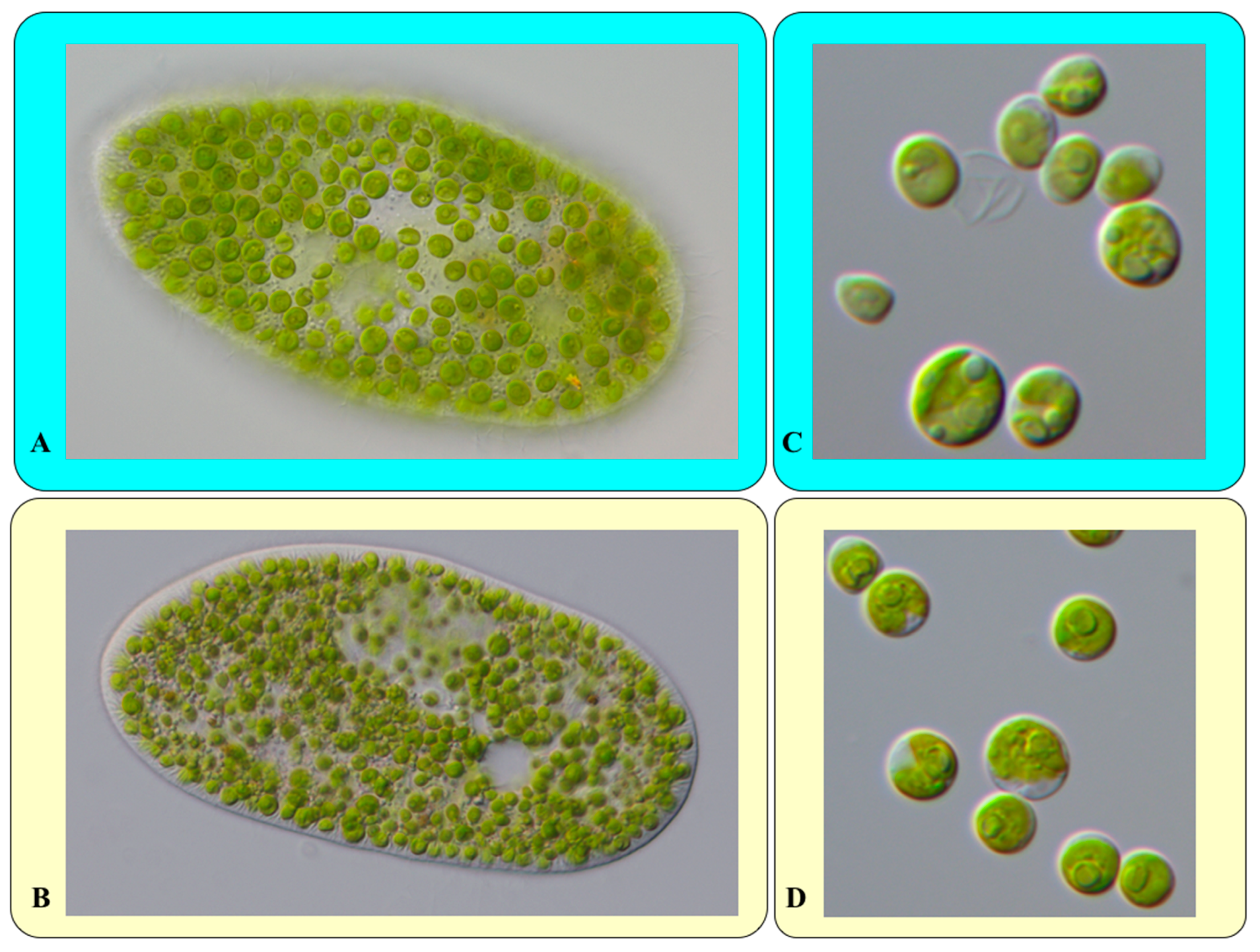
2.1. Cultivation and Molecular Characterization of Paramecium bursaria

The investigated strains were collected from around the world and cultivated in modified Bold Basal Medium (3N-BBM+V; medium 26a in [27]) with the addition of 30 mL of soil extract per liter final medium (called S/BBM). The soil extract was prepared as described in Schlösser [28]. Origin and details about the investigated Paramecium bursaria strains are listed in Table 1. All cultures were maintained at 15–21 °C under a light:dark cycle of 12:12 h (photon flux rate up 50 μmol m−2 s−1). Genomic DNA of the green algae was extracted using the DNeasy Plant Mini Kit (Qiagen GmbH, Hilden, Germany). The SSU and ITS rDNA were amplified using the Taq PCR Mastermix Kit (Qiagen GmbH, Hilden, Germany) with the primers EAF3 and ITS055R [29]. The SSU and ITS rDNA sequences of the Paramecium bursaria strains were aligned according to the secondary structures, resulting in a dataset of 19 sequences (2197 bp). GenBank accession numbers of all newly deposited sequences were given in the phylogenetic tree and Table 1. For the phylogenetic analyses, we calculated the log-likelihood values of 56 models using the automated selection tool implemented in PAUP version 4.0b167 [30] to test which evolutionary model fitted best for the dataset. The best model according to the Akaike criterion by PAUP was chosen for the analyses. The settings of the best model are given in the figure legend. The following methods were used for the phylogenetic analyses: Distance, maximum parsimony, and maximum likelihood, all included in PAUP version 4.0b167 [30].
The secondary structures were folded using the software mfold [31], which uses the thermodynamic model (minimal energy) for RNA folding. The visualization of the structures was done using the program PseudoViewer 3 [32].

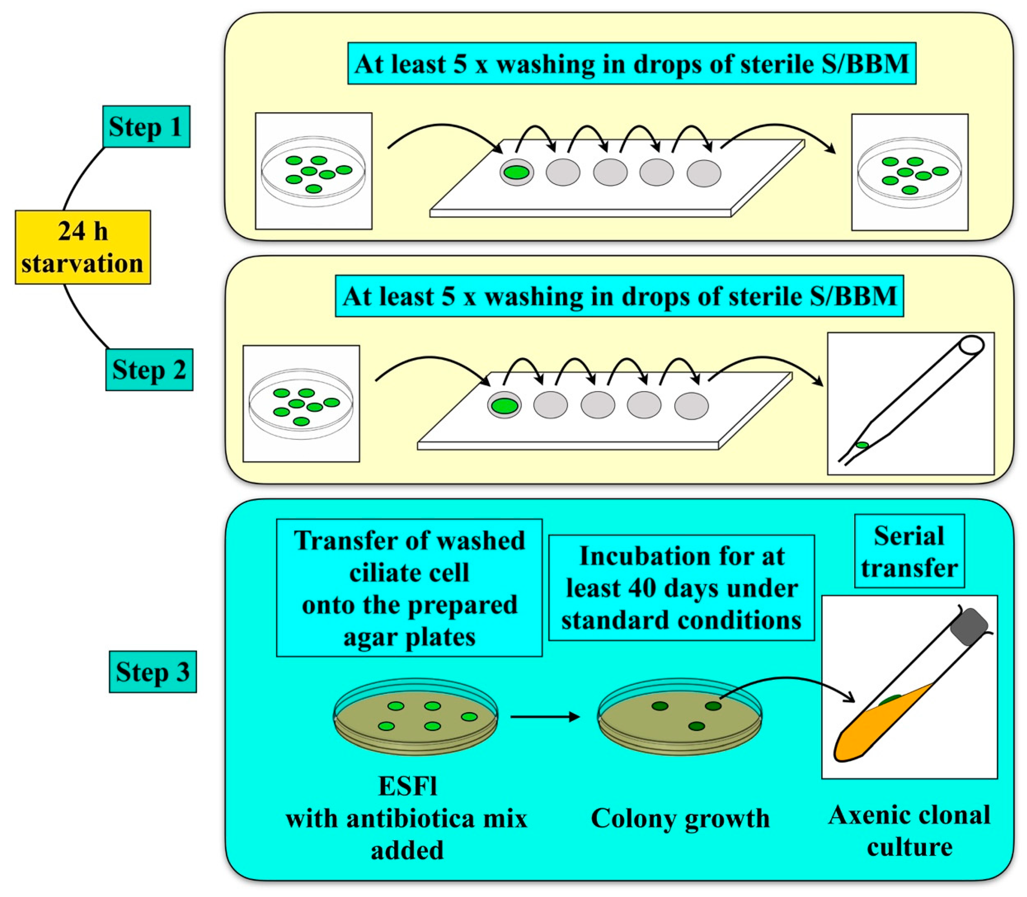
2.2. Isolation of the Green Algal Endosymbionts

The endosymbiotic green algae are often not easy to distinguish from free-living green algae in the surrounding media. Therefore, a special isolation method needed to be developed to avoid that free-living algae, or contaminations grow during the isolation process. With the following procedure as demonstrated in the work scheme (Figure 2), we obtained several axenic clonal strains in culture from different mixotrophic ciliates, such as Paramecium bursaria.
For the isolation of their green algal endosymbionts, single ciliate cells were washed several times and transferred into fresh S/BBM medium (step 1). After starvation and digestion of any food, after approximately 24 h, cells were washed again (step 2) and the ciliates were transferred onto agar plates containing basal medium with beef extract (ESFl; medium 1a according to Schlösser, [28]). Before placement of the ciliates onto agar plates, 50 µL of an antibiotic mix (mixture of 1% penicillin G, 0.25% streptomycin, and 0.25% chloramphenicol) were added to prevent bacterial growth. The agar plates were kept under the same conditions as described. After growth (~40 days), the algal colonies were transferred onto agar slopes (1.5%) containing ESFl medium and were kept under the described culture conditions (step 3).

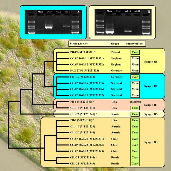
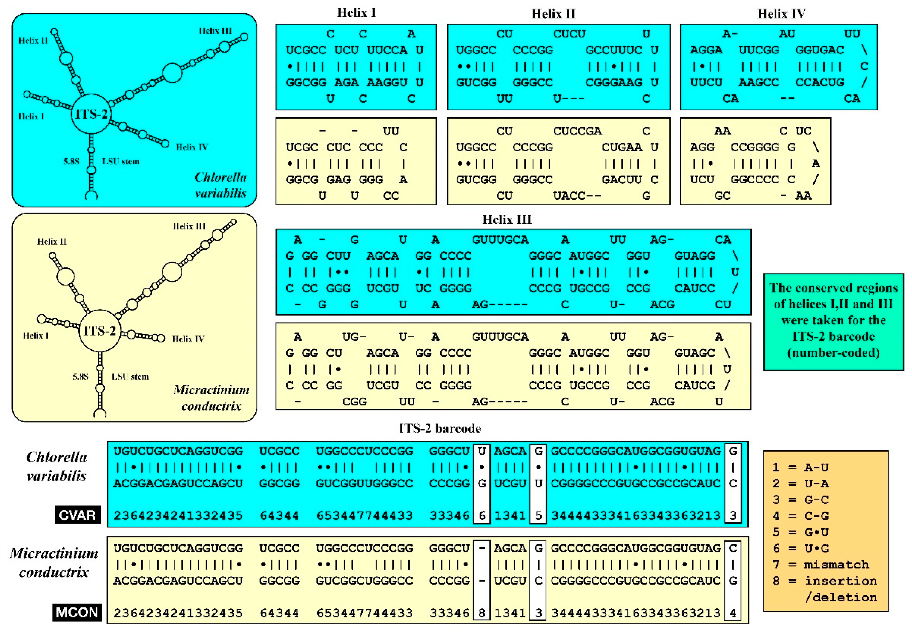
2.3. Diagnostic PCR Amplification
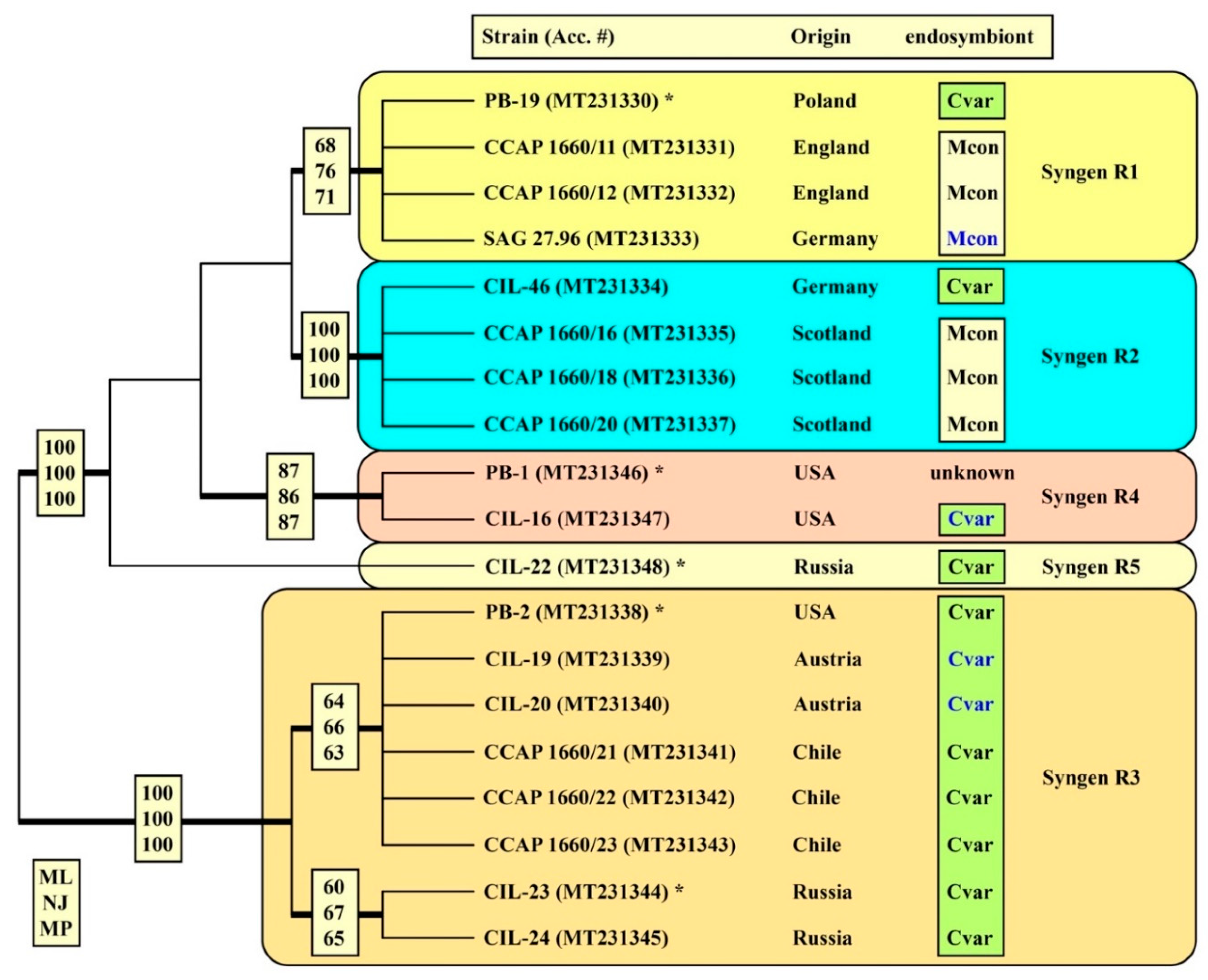
The isolation of the green algal endosymbionts is time-consuming and not always successful, especially if Chlorella variabilis is the endosymbiont. To investigate which green algal endosymbiont was present in a set of P. bursaria strains without isolating them, we developed a diagnostic PCR method using species-specific primers. Pröschold et al. [1] demonstrated that the SSU and ITS rDNA of Chlorella variabilis and Micractinium conductrix contain three introns and one intron, respectively, which makes an easy PCR amplification not possible. Therefore, we amplified the ITS-2 sequences for both C. variabilis and M. conductrix, a sequence which is diagnostic. For designing species primers, the internal transcribed spacer 1 (ITS-1) rDNA sequences of all representatives (58 species) belonging to the Chlorellaceae were compared to find characteristic positions. The following diagnostic primers for both species were designed based on the SSU and ITS rDNA sequences (C. variabilis, NC64A = CCAP 211/84; FN298923, and M. conductrix, SAG 241.80, FM205851; see Figure 3): CvarF: CTCGTGCTGTCTCACTTCGGTG and MconF: GTAGGCGCAGCCTCGTGTTGTGAC.
These primers were tested in combination with the reverse primer ITS055R [29], both on the isolated axenic algal cultures and their hosts. For identification of the endosymbionts, both primer combinations (CvarF/ITS055R and MconF/ITS055R) were tested on 18 investigated Paramecium bursaria strains. All PCR amplifications were done on a thermocycler with the following program: 5 min initial denaturation at 95 °C, followed by 30 cycles (1 min at 95 °C, 2 min at 55 °C and 3 min at 68 °C), and final synthesis for 10 min at 68 °C.

3. Results

The isolation of the green algal endosymbionts using the method described above (Figure 2) was successful for some of the Paramecium bursaria strains (SAG 27.96, CIL-16, CIL-19, and CIL-20). However, it was not always successful and was too time-consuming. Therefore, we used the diagnostic PCR approach for the identification of the endosymbionts. The primer combinations CvarF/ITS055R and MconF/ITS055R were highly species-specific as shown in Figure 4 for both the isolated algae CCAP 211/84 (=NC64A) Chlorella variabilis and SAG 241.80 Micractinium conductrix and their host organisms CIL-16 and SAG 27.96.
We tested this approach using these primer combinations on 18 Paramecium bursaria strains from different geographical origin and various syngens (see Figure 5 for the origin of the strains). Additionally, we sequenced the SSU and ITS rDNA sequences of the ciliate strains (the accession numbers are given in Figure 5). Despite their low genetic variability among the isolates (only 1.7%), the phylogenetic analyses (Figure 5) of the SSU and ITS rDNA sequences confirmed their subdivision of P. bursaria strains into five lineages representing the known syngens R1–R5 [9], which was well or moderately supported in all bootstrap analyses.
Among the different syngens, both endosymbionts were discovered using the diagnostic PCR approach. All ciliate syngens but not all strains harbored C. variabilis, whereas M. conductrix was only present in syngens R1 and R2, which originated from Europe (Figure 5). The ITS-2 sequences of these Chlorella variabilis (12 obtained with the primers CvarF/ITS055R) and Micractinium conductrix (6 obtained with the primers MconF/ITS055R) were identical to those of the reference strains NC64A = CCAP 211/84 (FN298923; [1]) and SAG 241.80 (FM205851; [1]), respectively.

4. Discussion

The green algal endosymbionts are often designated as Chlorella-like organisms or as simple zoochlorellae because of the difficulties in identifying them based solely on the morphology (see Figure 1). Especially, identification at the species level is almost impossible using light microscopic observations. The isolation of clonal cultures from the ciliate hosts is quite difficult and time-consuming, but the described method above resulted in some axenic strains of green algal endosymbionts. The main problem for isolating endosymbionts from their hosts is the slow growth and the requirement of additional nutrients, which is included in supplements, such as soil and beef extract. Especially, Chlorella variabilis requires organic compounds and vitamins [33,34]. Several attempts have been undertaken to isolate green algal endosymbionts from their hosts. Loefer [14] was the first to obtain the endosymbiont in culture. He isolated this alga by taking green algae from the sediment of an axenic P. bursaria culture and spreading it on agar plates containing tap water with unknown organic compounds. Since then, several methods for the isolation of endosymbionts have been described [35,36,37,38]. Similar to our approach, washing of the ciliate and the usage of antibiotics and transfer onto agar plates were used in different variants. However, the crucial points of our approach are the starvation of the ciliate for 24 h before rupture on agar plates and the microscopical check during all steps, providing some security that the isolated algae are the endosymbionts of Paramecium bursaria. Especially, the last point is of great importance. For example, Hoshina and Imamura [20] described that the strain CCAP 1660/13 of P. bursaria had an additional endosymbiont (Coccomyxa sp.); however, Pröschold et al. [1] revealed that this alga was not an endosymbiont and represented only a free-living alga co-occurring in the culture of this P. bursaria strain.
Another critical point is the choice of culture media for the endosymbionts. As highlighted, most of them need organic compounds for growth. Therefore, it is mostly likely that the three protocols provided in Achilles-Day and Day [39] resulted in the cultivation of free-living green algae, which are co-cultivated with the hosts. As described in Achilles-Day and Day [39], all green algal endosymbionts grew on media without organic nutrients. In contrast, the three steps of our method (Figure 2) rely on the microscopical control at each step as well as the elimination of contaminants and free-living algae growing outside in the medium. This method can result in axenic clonal cultures of green algal endosymbionts when they are needed for further investigations.
If it is only required to know which endosymbiont species is in a strain of Paramecium bursaria, the presented diagnostic PCR approach revealed an easy and fast method for species identification. Diagnostic PCR approaches have been successfully established in several approaches, such as for the identification of harmful algae ([40] and references therein). In P. bursaria, Tanaka et al. [41] used a PCR-based approach to demonstrate the success of the elimination of green algal endosymbionts from their hosts. This was based on the small subunit of the rubisco gene (rbcS) gene and did not focus on the identification of the endosymbionts. Here, we used the ITS-2 for our diagnostic PCR approach because this gene has been used for species delimitation as described above, and for all described species belonging to the Chlorellaceae, ITS rDNA sequences are available in GenBank, which is the only reliable dataset for species identification within this group until now ([22] and references therein). With species-specific primers (Figure 3) in PCR amplifications, Chlorella variabilis and Micractinium conductrix could be exactly identified, which were confirmed by ITS-2 sequencing (Figure 4 and Figure 5). Both endosymbionts were differently distributed among the five syngens of P. bursaria. Whereas the syngens R3–R5 exclusively had C. variabilis as an endosymbiont, both endosymbionts could be discovered in strains belonging to syngens R1 and R2, which originated exclusively from Europe. Gaponova et al. [15] also found M. conductrix in P. bursaria isolates collected in north Karelia (Russia). It appears that this green algal endosymbiont occurred only in Europe, whereas C. variabilis was distributed worldwide. Consequently, the subdivision into the "American" and "European" endosymbionts groups, i.e., C. variabilis and M. conductrix, respectively, as proposed by Hoshina and Imamura [20] and the references therein needs to be revised as C. variabilis was also found in P. bursaria isolates originating from Europe: Ciliate strains PB-19 (Poland, syngen R1), CIL-46 (Germany, syngen R2), CIL-19, and CIL-20 (both from Austria, syngen R3). In contrast to our findings here, Summerer et al. [42,43] described that ciliate isolates from the two locations about 50 km apart (called PbPIB and PbW) bore Chlorella sp. as endosymbionts based on ITS-1 rDNA sequences. Unfortunately, neither the cultures nor the DNA of these endosymbionts are available anymore for comparative investigations. However, in our investigations, both Austrian P. bursaria strains and both algal strains revealed C. variabilis, which are the first European isolates. Considering the findings of Jeanniard et al. [4], who demonstrated that specific chloroviruses infected these algal species were widely distributed, indicating that hosts containing C. variabilis and M. conductrix also occurred in these freshwater habitats. As demonstrated above, our diagnostic PCR approach provided a quick and precise identification of the endosymbiotic green algae occurring in P. bursaria. This promising method will discover new endosymbionts not only in the model ciliate P. bursaria but also in other ciliate and invertebrate hosts and finally elucidate the biogeographic patterns of endosymbiotic green algal species.
The characteristics of both endosymbionts found in Paramecium bursaria are summarized in Pröschold et al. [1]. Both species differed in the ITS-2 secondary structures and their ITS-2 barcode (Figure 6). Despite the variations in the helices I–IV, both species were differentiated by one compensatory base change (CBC) and one hemi-CBC (one-sided base change) in the conserved region (ITS-2 barcode). The genomes of both species were sequenced [44,45]. The genome of the strain CCAP 211/84 (NC64A) Chlorella variabilis (ITS-2 barcode: CVAR in Figure 6) has a size of 46.2 Mb and contains 12 chromosomes [44]. Micractinium conductrix, strain SAG 241.80 (ITS-2 barcode: MCON in Figure 6), has a larger genome (60.8 Mb with more than 13 chromosomes; [45]). The chloroplast and mitochondrial genomes of both species are similar in size (125 vs. 129 Kb and 78 vs. 75 Kb; respectively; [45]). Fan et al. [46] questioned the separation of both species into two different genera based on a comparison of the chloroplast and mitochondrial genomes. However, only a few taxa of Chlorella and Micractinium were included in this study. Before generic revision can be taken into account, more species of both genera need to be investigated using an integrative approach.

5. Conclusions

The endosymbiotic green algae of Paramecium bursaria can be clearly identified at the species level using the diagnostic PCR approach. This approach is also applicable for other mixotrophic ciliates if species-specific primers were designed. For the design of these primers, it is necessary to know which green algal endosymbionts occur in the green algal–ciliate association. This can be provided by isolation of the endosymbionts using the three-step method described above. Axenic cultures of isolated endosymbionts allow further genomic studies, such as the sequencing of whole genomes and plastomes.

Author Contributions

Conceptualization, T.P. and T.D.; methodology, C.S., T.B. and T.P.; software, T.P.; validation, T.P., T.D. and B.S.; formal analysis, T.P.; investigation, C.S., T.B. and T.P.; resources, T.P.; data curation, T.P., T.D. and B.S.; writing—original draft preparation, C.S., T.D., B.S. and T.P.; writing—review and editing, T.D., B.S. and T.P.; visualization, T.P. and T.D.; supervision, T.P.; project administration, B.S. and T.P.; funding acquisition, B.S. and T.P. All authors have read and agreed to the published version of the manuscript.

Funding

This research was funded by the Austrian Science Fund (FWF): grant number P28333-B25.

Conflicts of Interest

The authors declare no conflict of interest.

References

  1. Pröschold, T.; Darienko, T.; Silva, P.C.; Reisser, W.; Krienitz, L. The systematics of “Zoochlorella” revisited employing an integrative approach. Environ. Microbiol. 2011, 13, 350–364. [Google Scholar] [CrossRef] [PubMed]
  2. Muscatine, L.; Karakashian, S.J.; Karakashian, M.W. Soluble extracellular products of algae symbiotic with a ciliate, a sponge and a mutant Hydra. Comp. Biochem. Physiol. 1967, 20, 1–12. [Google Scholar] [CrossRef]
  3. Sommaruga, R.; Sonntag, B. Photobiological aspects of the mutualistic association between Paramecium bursaria and Chlorella. Microbiol. Monogr. 2009, 12, 111–130. [Google Scholar]
  4. Jeanniard, A.; Dunigan, D.D.; Gurnon, J.R.; Agarkova, I.V.; Kang, M.; Vitek, J.; Duncan, G.; McClung, O.W.; Larsen, M.; Claverie, J.-M.; et al. Towards defining the chloroviruses: A genomic journey through a genus of large DNA viruses. BMC Genom. 2013, 14, 158. [Google Scholar] [CrossRef] [Green Version]
  5. Foissner, W.; Berger, H.; Schaumburg, J. Identification and ecology of limnetic plankton ciliates. Bayer. Landesamt Wasserwirtsch. Munich Inf. 1999, 3/99, 1–793. [Google Scholar]
  6. Kreutz, M.; Foissner, W. The Sphagnum ponds of Simmelried in Germany: A biodiversity hot-spot for microscopic organisms. Protozool. Monogr. 2006, 3, 1–267. [Google Scholar]
  7. Lynn, D.H. The Ciliated Protozoa. Characterization, Classification, and Guide to the Literature; Springer: Dordrecht, The Netherlands; Heidelberg, Germany; London, UK; New York, NY, USA, 2008. [Google Scholar]
  8. Hoshina, R.; Hayashi, S.; Imamura, N. Intraspecific genetic divergence of Paramecium bursaria and re-construction of the Paramecian phylogenetic tree. Acta Protozool. 2006, 45, 377–386. [Google Scholar]
  9. Greczek-Stachura, M.; Potekhin, A.; Przybos, E.; Rautian, M.; Skobio, I.; Tracz, S. Identification of Paramecium bursaria syngens through molecular markers—Comparative analysis of three loci in the nuclear and mitochondrial DNA. Protist 2012, 163, 671–685. [Google Scholar] [CrossRef]
  10. Fujishima, M. Endosymbionts in Paramecium. Microbiol. Monogr. 2009, 12, 1–252. [Google Scholar]
  11. Kreutz, M.; Stoeck, T.; Foissner, W. Morphological and molecular characterization of Paramecium (Viridoparamecium nov. subgen.) chlorelligerum Kahl 1935 (Ciliophora). J. Eukaryot. Microbiol. 2012, 59, 548–563. [Google Scholar] [CrossRef] [PubMed] [Green Version]
  12. Takahashi, T. Simultaneous evaluation of life cycle dynamics between a host Paramecium and the endosymbionts of Paramecium bursaria using capillary flow cytometry. Sci. Rep. 2016, 6, 31638. [Google Scholar] [CrossRef] [PubMed] [Green Version]
  13. Pringsheim, E.G. Physiologische Untersuchungen an Paramecium bursaria: Ein Beitrag zur Symbioseforschung. Arch. Protistenkd. 1928, 64, 289–418. [Google Scholar]
  14. Loefer, J.B. Isolation and growth characteristics of the “Zoochlorella” of Paramecium bursaria. Am. Midl. Nat. 1936, 70, 184–188. [Google Scholar] [CrossRef]
  15. Gaponova, I.N.; Andronov, E.E.; Migunova, A.V.; Vorobyev, K.P.; Chizhevskaja, E.P.; Kvitko, K.V. Genomic dactyloscopy of Chlorella sp., symbionts of Paramecium bursaria. Protistology 2007, 4, 311–317. [Google Scholar]
  16. Hoshina, R.; Kamako, S.; Imamura, N. Phylogenetic position of endosymbiotic green algae in Paramecium bursaria Ehrenberg from Japan. Plant Biol. 2004, 6, 447–453. [Google Scholar] [CrossRef]
  17. Hoshina, R.; Kato, Y.; Kamako, S.; Imamura, N. Genetic evidence of “American” and “European” type symbiotic algae of Paramecium bursaria Ehrenberg. Plant Biol. 2005, 7, 526–532. [Google Scholar] [CrossRef]
  18. Hoshina, R.; Iwataki, M.; Imamura, N. Chlorella variabilis and Micractinium reisseri sp. nov. (Chlorellaceae, Trebouxiophyceae): Redescription of the endosymbiotic green algae of Paramecium bursaria (Peniculia, Oligohymenophorea) in the 120th year. Phycol. Res. 2010, 58, 188–201. [Google Scholar] [CrossRef]
  19. Hoshina, R.; Imamura, N. Multiple origins of the symbioses in Paramecium bursaria. Protist 2008, 159, 53–63. [Google Scholar] [CrossRef]
  20. Hoshina, R.; Imamura, N. Origins of algal symbionts of Paramecium bursaria. Microbiol. Monogr. 2009, 12, 1–29. [Google Scholar]
  21. Kang, M.; Dunigan, D.D.; Van Etten, J.L. Chlorovirus: A genus of Phycodnaviridae that infects certain Chlorella-like green algae. Mol. Plant Pathol. 2005, 6, 213–224. [Google Scholar] [CrossRef] [Green Version]
  22. Krienitz, L.; Bock, C. Present state of the systematics of planktonic coccoid green algae of inland waters. Hydrobiologia 2012, 698, 295–326. [Google Scholar] [CrossRef]
  23. Coleman, A.W. The significance of a coincidence between evolutionary landmarks found in mating affinity and a DNA sequence. Protist 2000, 151, 1–9. [Google Scholar] [CrossRef]
  24. Coleman, A.W. ITS2 is a double-edged tool for eukaryote evolutionary comparisons. Trends Genet. 2003, 19, 370–375. [Google Scholar] [CrossRef]
  25. Coleman, A.W. Pan-eukaryote ITS2 homologies revealed by RNA secondary structure. Nucleic Acid Res. 2007, 35, 3322–3329. [Google Scholar] [CrossRef] [PubMed] [Green Version]
  26. Bomford, R. The syngens of Paramecium bursaria: New mating types and intersyngenic mating reactions. J. Protozool. 1966, 13, 497–501. [Google Scholar] [CrossRef] [PubMed]
  27. Schlösser, U.G. Additions to the culture collections of algae since 1994. Bot. Acta 1997, 110, 424–429. [Google Scholar] [CrossRef]
  28. Schlösser, U.G. SAG-Sammlung von Algenkulturen at the University of Göttingen. Bot. Acta 1994, 107, 424–429. [Google Scholar]
  29. Marin, B.; Palm, A.; Klingberg, M.; Melkonian, M. Phylogeny and taxonomic revision of plastid-containing euglenophytes based on SSU rDNA sequence comparisons and synapomorphic signatures in the SSU rRNA secondary structure. Protist 2003, 154, 99–145. [Google Scholar] [CrossRef]
  30. Swofford, D.L. PAUP* Phylogenetic Analysis Using Parsimony (*and other Methods), Version 4.0b10; Sinauer Associates: Sunderland, MA, USA, 2002. [Google Scholar]
  31. Zuker, M. Mfold web server for nucleic acid folding and hybridization prediction. Nucleic Acid Res. 2003, 31, 3406–3615. [Google Scholar] [CrossRef]
  32. Byun, Y.; Han, K. PseudoViewer3: Generating planar drawings of large-scale RNA structures with pseudoknots. Bioinformatics 2009, 25, 1435–1437. [Google Scholar] [CrossRef]
  33. Shihira, I.; Krauss, R.W. Chlorella. Physiology and Taxonomy of Forty-One Isolates; University of Maryland: College Park, MD, USA, 1969. [Google Scholar]
  34. Kessler, E.; Huss, V.A.R. Biochemical taxonomy of symbiotic Chlorella strains from Paramecium and Acanthocystis. Bot. Acta 1990, 103, 140–142. [Google Scholar] [CrossRef]
  35. Siegel, R.W. Hereditary endosymbiosis in Paramecium bursaria. Exp. Cell Res. 1960, 19, 239–252. [Google Scholar] [CrossRef]
  36. Weis, D.S. Correlation of infectivity and Concanavalin A, agglutinability of algae exsymbiotic from Paramecium bursaria. J. Protozool. 1978, 25, 366–370. [Google Scholar] [CrossRef]
  37. Reisser, W. The endosymbiotic unit of Stentor polymorphus and Chlorella sp., morphological and physiological studies. Protoplasma 1981, 105, 273–284. [Google Scholar] [CrossRef]
  38. Nishihara, N.; Horiike, S.; Takahashi, T.; Kosaka, T.; Shigenaka, Y.; Hosoya, H. Cloning and characterization of endosymbiotic algae isolated from Paramecium bursaria. Protoplasma 1998, 203, 91–99. [Google Scholar] [CrossRef]
  39. Achilles-Day, U.E.M.; Day, J.G. Isolation of clonal cultures of endosymbiotic green algae from their ciliate hosts. J. Microbiol. Meth. 2013, 92, 355–357. [Google Scholar] [CrossRef]
  40. Jedlicki, A.; Fernández, G.; Astorga, M.; Oyarzún, P.; Toro, J.E.; Navarro, J.M.; Martinez, V. Molecular detection and species identification of Alexandrium (Dinophyceae) causing harmful algal blooms along the Chilean coastline. AoB Plants 2012, 2012, pls033. [Google Scholar] [CrossRef]
  41. Tanaka, M.; Murata-Hori, M.; Kadono, T.; Yamada, T.; Kawano, T.; Kosaka, T.; Hosoya, H. Complete elimination of endosymbiotic algae from Paramecium bursaria and its confirmation by diagnostic PCR. Acta Protozool. 2002, 41, 255–261. [Google Scholar]
  42. Summerer, M.; Sonntag, B.; Sommaruga, R. An experimental test of the symbiosis specificity between the ciliate Paramecium bursaria and strains of the unicellular green alga Chlorella. Environ. Microbiol. 2007, 9, 2117–2122. [Google Scholar] [CrossRef] [PubMed]
  43. Summerer, M.; Sonntag, B.; Sommaruga, R. Ciliate-symbiont specificity of freshwater endosymbiotic Chlorella (Trebouxiophyceae, Chlorophyta). J. Phycol. 2008, 44, 77–84. [Google Scholar] [CrossRef] [PubMed]
  44. Blanc, G.; Duncan, G.; Agarkova, I.; Borodovsky, M.; Gurnon, J.; Kuo, A.; Lindquist, E.; Lucas, S.; Pangilinan, J.; Polle, J.; et al. The Chlorella variabilis NC64A genome reveals adaptation to photosymbiosis, coevolution with viruses, and cryptic sex. Plant Cell 2010, 22, 2943–2955. [Google Scholar] [CrossRef] [Green Version]
  45. Arriola, M.; Velmurugan, N.; Zhang, Y.; Plunkett, M.H.; Hondzo, H.; Barney, B.M. Genome sequences of Chlorella sorokiniana UTEX 1602 and Micractinium conductrix SAG 241.80: Implications to maltose excretion by a green alga. Plant J. 2018, 93, 566–586. [Google Scholar] [CrossRef] [Green Version]
  46. Fan, W.; Guo, W.; Van Etten, J.L.; Mower, J.P. Multiple origins of endosymbionts in Chlorellaceae with no reductive effects on the plastid or mitochondrial genomes. Sci. Rep. 2017, 7, 10101. [Google Scholar] [CrossRef] [Green Version]
Figure 1. Morphology of Chlorella variabilis and Micractinium conductrix in their host Paramecium bursaria strains and cultivated as axenic strains. (A,B) Paramecium bursaria strains CIL-16 (A) and SAG 27.96 (B), respectively, (C) CCAP 211/84 = NC64A; (D) SAG 241.80.
Diversity 12 00240 g001
Figure 2. Working scheme for isolating the green algal endosymbionts of Paramecium bursaria.
Diversity 12 00240 g002
Figure 3. ITS-1 secondary structure of Chlorella variabilis (blue) and Micractinium conductrix (yellow). The positions of the diagnostic primers CvarF and MconF are highlighted in white boxes in the structures.
Diversity 12 00240 g003
Figure 4. Diagnostic PCR using the primer combinations CvarF/ITS055R (A) and MconF/ITS055R (B). Mcon—Micractinium conductrix (SAG 241.80); Cvar—Chlorella variabilis (CCAP 211/84); reference hosts: Paramecium bursaria strains SAG 27.96 and strain CIL-16.
Diversity 12 00240 g004
Figure 5. Molecular phylogeny of the Paramecium bursaria based on SSU and ITS rDNA sequence comparisons. The phylogenetic tree shown was inferred using the maximum likelihood method based on the datasets (2197 aligned positions of 19 taxa) using computer program PAUP 4.0a167. For the analyses, the best model was calculated by PAUP 4.0a167. The setting of the best model was given as follows: GTR + I (base frequencies: A 0.2994, C 0.1832, G 0.2278, T 0.2896; rate matrix A-C 2.4955, A-G 4.2183, A-U 4.9756, C-G 0.7513, C-U 9.6155, G-U 1.0000) with the proportion of invariable sites (I = 0.9303). The branches in bold are highly supported in all bootstrap analyses (bootstrap values > 50% calculated with PAUP using the maximum likelihood, neighbor-joining, and maximum parsimony). The clades are named after the syngens (color-coded) proposed by Greczek-Stachura et al. [9]. The accession numbers are given after the strain numbers. The endosymbiotic green algae identified using the diagnostic PCR are highlighted (Mcon—Micractinium conductrix and Cvar—Chlorella variabilis) after the origin of the Paramecium bursaria strains. The reference strain of each syngen is marked with an asterisk. The green algal endosymbionts isolated and available for further investigations are highlighted in blue.
Diversity 12 00240 g005
Figure 6. ITS-2 secondary structure of Chlorella variabilis (blue) and Micractinium conductrix (yellow). The ITS-2 barcodes of both species (CVAR and MCON) is given as number codes.
Diversity 12 00240 g006
Table 1. Strain of Paramecium bursaria investigated in this study.
Strain NumberSyngenOriginAccession NumberEndosymbiont
PB-19R1Poland: Biebrza National ParkMT231330Cvar
CCAP 1660/11R1England: Cambridge, Cavendish PondMT231331Mcon
CCAP 1660/12R1England: Cambridge, Cavendish PondMT231332Mcon
SAG 27.96R1Germany: Göttingen, pond in Old Botanical GardenMT231333Mcon
CIL-46R2Germany: Seeburger See near GöttingenMT231334Cvar
CCAP 1660/16R2Scotland: Loch Inverawe, InveraweMT231335Mcon
CCAP 1660/18R2Scotland: Loch Lily, InveraweMT231336Mcon
CCAP 1660/20R2Scotland: Loch Lily, InveraweMT231337Mcon
PB-2R3USA: Massachusetts, BostonMT231338Cvar
CIL-19R3Austria: Piburger SeeMT231339Cvar
CIL-20R3Austria: WildbichlMT231340Cvar
CCAP 1660/21R3Chile: Concepción, artificial pond at University campusMT231341Cvar
CCAP 1660/22R3Chile: Concepción, artificial pond at University campusMT231342Cvar
CCAP 1660/23R3Chile: Concepción, artificial pond at University campusMT231343Cvar
CIL-23R3Russia: Khabarovsk region, Amur riverMT231344Cvar
CIL-24R3Russia: Primorie, KiparisovoMT231345Cvar
PB-1R4USA: Massachusetts, BostonMT231346unknown
CIL-16R4USA: North Carolina, BurlingtonMT231347Cvar
CIL-22R5Russia: Astrakhan Nature ReserveMT231348Cvar

