Article

News from the Sea: A New Genus and Seven New Species in the Pleosporalean Families Roussoellaceae and Thyridariaceae

Mycotheca Universitatis Taurinensis, Department of Life Sciences and Systems Biology, University of Torino, Viale Mattioli 25, 10125 Torino, Italy
*
Author to whom correspondence should be addressed.
Diversity 2020, 12(4), 144; https://doi.org/10.3390/d12040144
Submission received: 19 March 2020 / Revised: 1 April 2020 / Accepted: 3 April 2020 / Published: 6 April 2020
(This article belongs to the Special Issue Fungal Diversity in the Mediterranean Area)

Abstract

:
Nineteen fungal strains associated with the seagrass Posidonia oceanica, with the green alga Flabellia petiolata, and the brown alga Padina pavonica were collected in the Mediterranean Sea. These strains were previously identified at the family level and hypothesised to be undescribed species. Strains were examined by deep multi-loci phylogenetic and morphological analyses. Maximum-likelihood and Bayesian phylogenies proved that Parathyridariella gen. nov. is a distinct genus in the family Thyriadriaceae. Analyses based on five genetic markers revealed seven new species: Neoroussoella lignicola sp. nov., Roussoella margidorensis sp. nov., R. mediterranea sp. nov., and R. padinae sp. nov. within the family Roussellaceae, and Parathyridaria flabelliae sp. nov., P. tyrrhenica sp. nov., and Parathyridariella dematiacea gen. nov. et sp. nov. within the family Thyridariaceae.

1. Introduction

Marine fungi are a relevant and active component of the microbial communities that inhabit the oceans [1]. Fungi in the marine environment live as mutualists, parasites, pathogens and saprobes, and are pivotal to marine food webs because of the recycling of recalcitrant substrata [2]; besides, these widely dispersed organisms are a source of novel bioactive compounds [3].
Marine fungi have been recovered worldwide from a broad range of biotic and abiotic substrata, such as driftwood algae, sponges, corals, sediments, etc. [4,5]. Following the definition of Pang et al [6] that considered “a marine fungus” to be any fungus retrieved repeatedly from marine environment and that reproduces in the marine environment, Jones et al. [7] listed 1680 fungal species belonging to 693 genera, 223 families, 87 orders, 21 classes and six phyla. However, considering that the total number of marine fungi has been estimated to exceed 10,000 taxa [8], fungal diversity remains largely undescribed. With more than 900 species [9], the Ascomycota are the dominant fungal phylum in the sea; the most represented lineages include the order Pleosporales (class Dothideomycetes) with 36 families, 95 genera and 194 species described to date (www.marinefungi.org).
In recent surveys aimed to uncover the underwater fungal diversity, 19 unidentified Roussoellaceae were isolated from several substrates, as follows: 12 from the brown alga Padina pavonica (L.) Thivy [10], 4 from the green alga Flabellia petiolata (Turra) Nizamuddin [11], 2 from the seagrass Posidonia oceanica (L.) [12] Delile, and 1 from the Atlantic sponge Dysidea fragilis (Montagu) [13]. The Roussoellaceae is a well-resolved family in the Pleosporales [14]. Others [15] have treated the family Roussoellaceae as a synonym of Thyridariaceae, based on phylogenetic affinities. However, following the discovery of new genera in this group, delineated by high resolution multi-locus phylogenetic analyses, the Roussoellaceae and Thyridariaceae are now recognized as two distinct but closely related families [16,17,18,19,20].
Many new species of Roussoellaceae and Thyridariaceae have recently been described on terrestrial plants including bamboo, palms and mangroves [14,17,20,21]. This paper provides a more precise phylogenetic placement of the 19 strains isolated from marine substrata together with morphological insights of those strains that represent new species within these two families.

2. Materials and Methods

2.1. Fungal Isolates

The fungal isolates analyzed in this paper were retrieved in the Mediterranean Sea from P. oceanica (2), collected in Riva Trigoso bay and Elba island, P. pavonica (12), and F. petiolata (3) from the coastal waters of Elba island [10,11,12]. A single isolate was previously retrieved in association with D. fragilis in the Atlantic Ocean [13] (Table 1).
The strains investigated were originally isolated on Corn Meal Agar medium supplemented with sea salts (CMASS; 3.5% w/v sea salt mix, Sigma-Aldrich, Saint Louis, USA, in ddH2O) and are preserved at the Mycotheca Universitatis Taurinensis (MUT), Italy.

2.2. Morphological Analysis

All isolates were pre-grown on Malt Extract Agar-sea water (MEASW; 20 g malt extract, 20 g glucose, 2 g peptone, 20 g agar in 1 L of sea water) for one month at 24 °C prior to inoculation in triplicate onto new Petri dishes (9 cm Ø) containing (i) MEASW, (ii) Oatmeal Agar-sea water (OASW; 30 g oatmeal, 20 g agar in 1 L of sea water), or iii) Potato Dextrose Agar-sea water (PDASW; 4 g potato extract, 20 g dextrose, 20 g agar in 1 L of sea water). Petri dishes were incubated at 15 and/or 24 °C. The colony growth was monitored periodically for 28 days. Macroscopic and microscopic traits, were assessed for strains grown on MEASW at the end of the incubation period.
In an attempt to induce sporulation, sterile pieces of Quercus ruber cork and Pinus pinaster wood (species autochthonous to the Mediterranean area) were placed on 3 week old fungal colonies grown on MEASW ([22], modified). Petri dishes were further incubated for 4 weeks at 24 °C. Subsequently, cork and wood pieces were transferred to 50 mL tubes containing 20 mL of sterile sea water. Samples were incubated at 24 °C for one month. In parallel, the strains were also plated on Syntetic Nutrient Agar-sea water (SNASW; 1 g KH2PO4, 1 g KNO3, 0.5 g MgSO4 • 7H2O, 0.5 g KCl, 0.2 g glucose, 0.2 g sucrose, 20 g agar in 1 L of sea water) supplemented with sterile pine needles. Petri dishes were incubated at 24 °C for one month.
Morphological structures were observed, and images captured using an optical microscope (Leica DM4500B, Leica microsystems GmbH, Wetzlar) equipped with a camera (Leica DFC320, Leica microsystems GmbH, Wetzlar). Macro- and microscopic features were compared with the available description of Roussoellaceae and Thyridariaceae [14,15,17,18,20].

2.3. DNA Extraction, PCR Amplification, and Data Assembling

Genomic DNA was extracted from about 100 mg of fresh mycelium grown on MEASW plates. Mycelium was disrupted by the mean of a MM400 tissue lyzer (Retsch GmbH, Haan, Germany) and DNA extracted using a NucleoSpin kit (Macherey Nagel GmbH, Duren, DE, USA) following the manufacturer’s instructions. The quality and quantity of DNA were measured spectrophotometrically (Infinite 200 PRO NanoQuant; TECAN, Männedorf); DNA was stored at −20 °C.
The partial sequences of five genetic markers were amplified by PCR. Primer pairs ITS1/ITS4 [23], LR0R/LR7 [24], NS1/NS4 [23] were used to amplify the internal transcribed spacers, including the 5.8S rDNA gene (nrITS), 28S large ribosomal subunit (nrLSU) and 18S small ribosomal subunit (nrSSU). The translation elongation factor (TEF1α) and RNA polymerase II subunit (RPB2) were amplified by using primer pairs EF1-1018F/EF1-1620R [25] and fRPB2-5F/fPB2-7R [26].
Amplifications were run in a T100 Thermal Cycler (Bio-Rad, Hercules, CA, USA) programmed as described in Table 2. Reaction mixtures consisted of 20–40 ng DNA template, 10× PCR Buffer (15 mM MgCl2, 500 mM KCl, 100 mM Tris-HCl, pH 8.3), 200 µM each dNTP, 1 μM each primer, 2.5 U Taq DNA Polymerase (Qiagen, Chatsworth, CA, USA), in 50 μL final volume. For problematic cases, additional MgCl2 and/or 2.5% DMSO facilitated the reaction.
Amplicons, together with a GelPilot 1 kb plus DNA Ladder, were visualized on a 1.5% agarose gel stained with 5 mL 100 mL−1 ethidium bromide; PCR products were purified and sequenced at the Macrogen Europe Laboratory (Madrid, Span). The resulting Applied Biosystem (ABI) chromatograms were inspected, trimmed and assembled to obtain consensus sequences using Sequencer 5.0 (GeneCodes Corporation, Ann Arbor, Michigan, USA http://www.genecodes.com). Newly generated sequences were deposited in GenBank (Table 1).

2.4. Sequence Alignment and Phylogenetic Analysis

A dataset consisting of nrSSU, nrITS, nrLSU, TEF1α and RPB2 was assembled on the basis of BLASTn results and of recent phylogenetic studies focused on Roussoellaceae and Thyridariaceae [18,20]. Reference sequences were retrieved from GenBank (Table 1).
Sequences were aligned using MUSCLE (default conditions for gap openings and gap extension penalties), implemented in MEGA v. 7.0 (Molecular Evolutionary Genetics Analysis), visually inspected and trimmed by TrimAl v. 1.2 (http://trimal.cgenomics.org) to delimit and discard ambiguously aligned regions. Since no incongruence was observed among single-loci phylogenetic trees, alignments were concatenated into a single data matrix with SequenceMatrix [27]. The best evolutionary model under the Akaike Information Criterion (AIC) was determined with jModelTest 2 [28].
Phylogenetic inference was estimated using Maximum Likehood (ML) and Bayesian Inference (BI) criteria. The ML analysis was generated using RAxML v. 8.1.2 [29] under GTR + I + G evolutionary model and 1000 bootstrap replicates. Support values from bootstrapping runs (MLB) were mapped on the globally best tree using the “-f a” option of RAxML and “-x 12345” as a random seed to invoke the novel rapid bootstrapping algorithm. BI was performed with MrBayes 3.2.2 [30] with the same substitution model (GTR + I + G). The alignment was run for 10 million generations with two independent runs each containing four Markov Chains Monte Carlo (MCMC) and sampling every 100 iterations. The first 25% of generated trees were discarded as “burn-in”. A consensus tree was generated using the “sumt” function of MrBayes and Bayesian posterior probabilities (BPP) were calculated. Consensus trees were visualized in FigTree v. 1.4.2 (http://tree.bio.ed.ac.uk/software/figtree).
Two strains of Occutibambusa bambusae (Occultibambusaceae) were used to root the tree. Due to topological similarity of the two resulting trees, only ML analysis with MLB and BPP values was reported (Figure 1).
DNA diagnostic characters were visually identified by the presence of heterozygous bases. For each locus, aligned sequences of the individual clusters containing new species, were inspected. Nucleotide diversities of the novel species were annotated when occurred (Tables S1–S18).
Sequence alignments and phylogenetic trees were deposited in TreeBASE (http://www.treebase.org, submission number S24773).
Following phylogenetic tree inspection, isolates that clustered in the same group and that derived from the same substrate were subjected to PCR-fingerprinting by using the micro- and mini-satellite primers (GTG)5 and M13 [31,32] to exclude duplicates from further analysis. DNA fingerprints were visualized with 1.5% agarose gel stained with 5 mL 100 mL−1 ethidium bromide while a GelPilot 1 kb plusDNA Ladder was used as a reference. Images were acquired with a Gel Doc1000 System (Bio-Rad, Hercules, CA, USA) and fingerprints analyzed using Bionumerics v 7.6 (http://www.applied-maths.com).

3. Results

3.1. Phylogenetic Inference
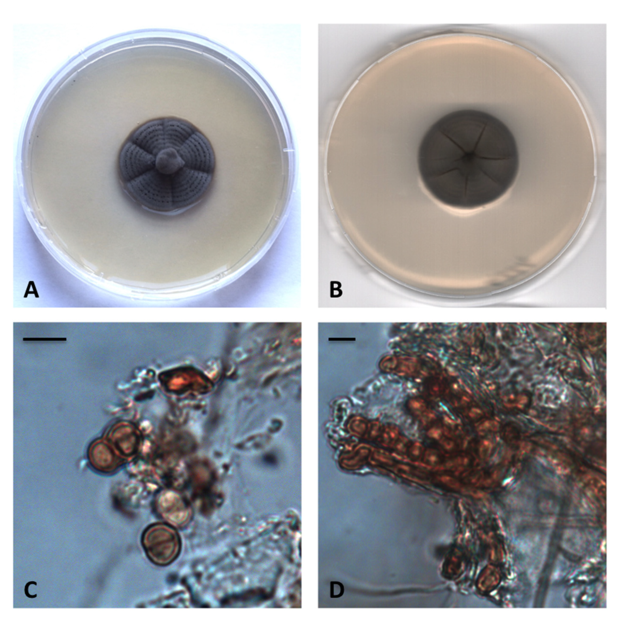
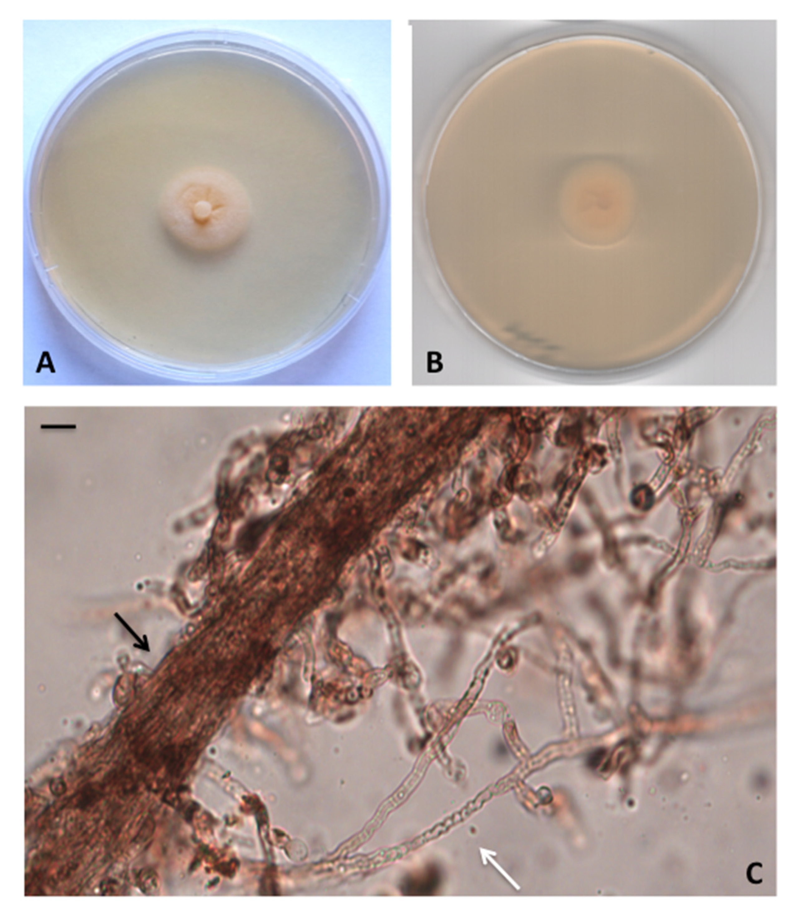
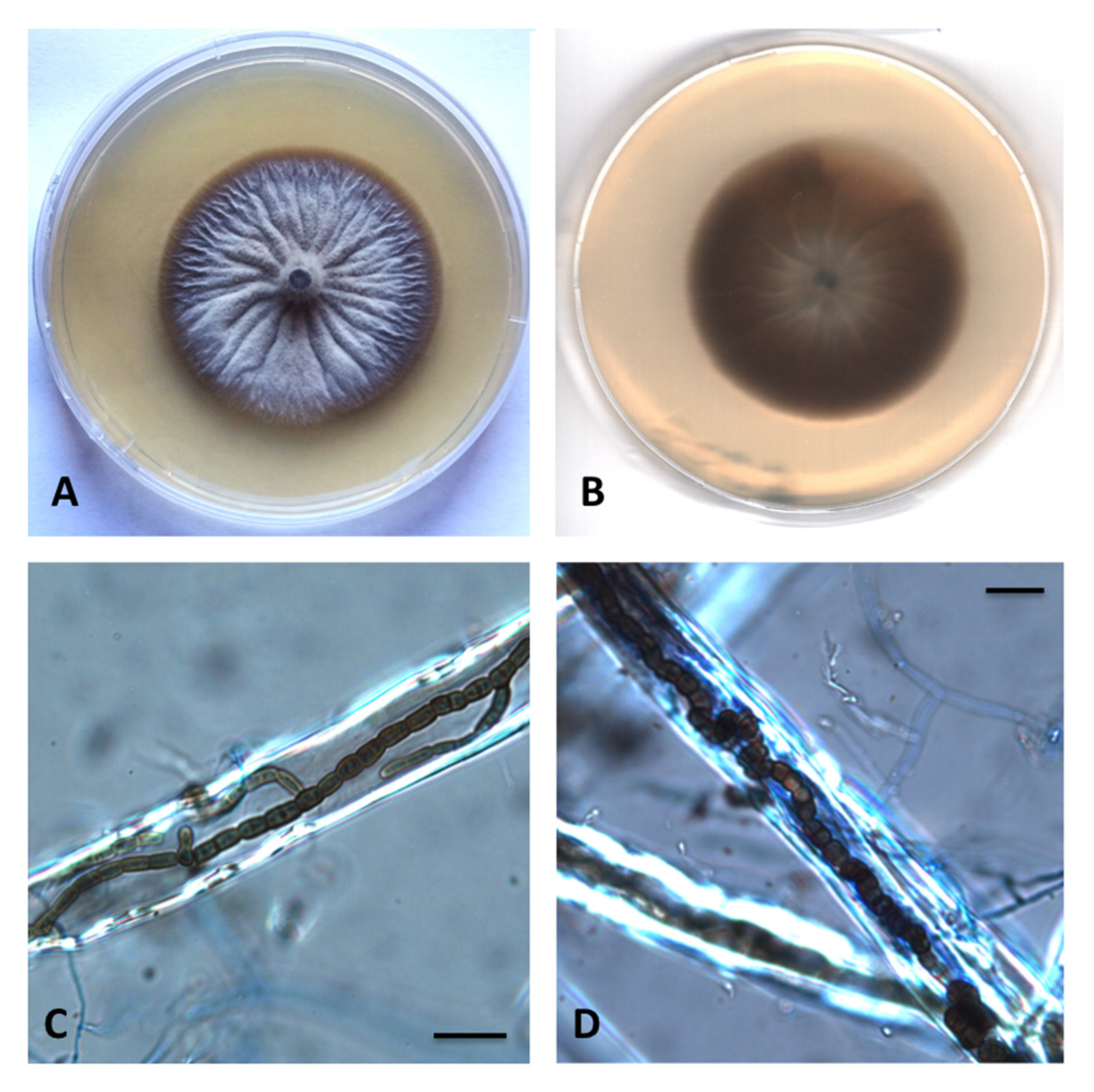
Preliminary analyses carried out individually with nrITS, nrSSU, nrLSU, TEF1α and RPB2 denoted no incongruence in the topology of the single-locus trees. The combined five-markers dataset—built on the basis of BLASTn results and of recent phylogenetic studies [18,20]—consisted of 81 taxa (including MUT isolates) that represented 16 genera and 56 species (Table 1). A total of 63 sequences (2 nrITS, 8 nrSSU, 13 nrLSU, 20 TEF1α and 20 RPB2) were newly generated while 261 were retrieved from GenBank.
The combined dataset had an aligned length of 3390 characters, of which 1683 were constant, 657 were parsimony-uninformative and 1050 parsimony informative (TL = 218, CI = 0.422018, RI = 0.825243, RC = 0.348267, HI = 0.877952).
Strains MUT 4893 and MUT 2452 were identified as Parathyridaria robiniae, the rest of the strains represented seven new species and one new genus (Figure 1). Parathyridaria tyrrhenica sp. nov. (MUT 5371 and MUT 4966) formed a sister clade to Parathyridaria flabelliae sp. nov. (MUT 4859 and MUT 4886) with high statistical support (BYPP = 1.00; MLB = 100%); these two novel species are closely related to P. ramulicola (BYPP = 1.00; MLB = 100%) and clustered with other Parathyridaria species in the Thyridariaeae family. Within this family, four isolates (MUT 5310, MUT 5381, MUT 4419 and MUT 4884) clustered together with the genera Thyridariella, Liua and Cycasicola, and formed a strongly supported monophyletic lineage (BYPP = 1.00; MLB = 100%). Therefore, we have introduced the novel genus Parathyridariella, typified by the new species Parathyridariella dematiacea sp. nov.
The three strains, MUT 4904, MUT 5373 and MUT 5008, represented a novel species Neoroussoella lignicola sp. nov. and formed an independent and robust clade (BYPP = 1.00; MLB = 100%), within the Neoroussoella group in the Roussoellaceae.
Two sister clades within the Roussoella group were represented by the new species Roussoella padinae sp. nov. (MUT 5503, MUT 5341 and MUT 5365) and Roussoella mediterranea sp. nov. (MUT 5306 and MUT 5369). Finally, MUT 5329 Roussoella margidorensis sp. nov. clustered together with R. nitidula, R. pseudohysterioides, R. thailandica and R. tubercolata (BYPP = 0.99; MLB = 71%) but was phylogenetically distant from these species.
Nucleotide divergence between each novel species and members of the same clusters were annotated for each locus, when occurred (Tables S1–S18).

3.2. Taxonomy

Parathyridariella gen. nov. V. Prigione, A. Poli, E. Bovio and G.C. Varese
MYCOBANK: MB 832836
Type species. Parathyridariella dematiacea sp. nov.
Etymology. In reference to the phylogenetic proximity to the genus Thyridariella.
Phylogenetic placement. Thyridariaceae, Sordariomycetes, Ascomycota. The genus Parathyridariella gen. nov. clusters together with genera Cycasicola, Liua and Thyridariella (Figure 1).
Parathyridariella dematiacea sp. nov. V. Prigione, A. Poli, E. Bovio and G.C. Varese
MYCOBANK: MB 832837
Type. Italy, Tuscany, Mediterranean Sea, Elba Island (LI), Ghiaie ISL, 14–15 m depth, 42°49’04”N, 10°19’20”E, form the green alga Flabellia petiolata, 20 March 2010, R. Mussat-Sartor and N. Nurra, MUT 4884 holotype, living culture permanently preserved in metabolically inactive state by deep-freezing at Mycotheca Universitatis Taurinensis (MUT).
Additional material examined. Italy, Ligury, Mediterranean Sea, Riva Trigoso, Punta Manara (GE), 5–21 m depth, 44°15′08.62″N 9°24′17.64″E, from the seagrass Posidonia oceanica, March 2008, MUT 4419.
Etymology. In reference to the color of the colony on culture media.
Description. Growing actively on Pinus pinaster and Quercus ruber cork. Showing a floccose growth mainly on Pinus pinaster. Hyphae 2.8–4.8 μm wide, septate, hyaline to lightly pigmented. Chlamydospores numerous, mostly in chain, intercalary or solitary, globose to subglobose, from brownish to dark brown, 7–10 × 6–8 μm diameter.
Sexual morph not observed. Asexual morph with differentiated conidiogenesis not observed.
Colony description. Colonies on MEASW attaining 28–34 mm diam after 28 days at 24 °C, mycelium from dark grey/black to dark green, dense with radial grooves and concentric rings, submerged edges; reverse dark green. Brown exudate present above the concentric rings. Growth on OASW reaching 40–54 mm diam at 24 °C and 21–29 mm diam at 15 °C; colonies on PDA attaining 36–49 mm diam and 15.5–22.5 mm diam at 24 °C and 15 °C, respectively.
Parathyridaria tyrrhenica sp. nov. A. Poli, V. Prigione, E. Bovio and G.C. Varese
MYCOBANK: MB 832838
Type. Italy, Tuscany, Mediterranean Sea, Elba Island (LI), Ghiaie ISL, 14–15 m depth, 42°49’04”N, 10°19’20”E, from the brown alga Padina pavonica, March 2010, R. Mussat-Sartor and N. Nurra, MUT 5371 holotype, living culture permanently preserved in metabolically inactive state by deep-freezing at MUT.
Additional material examined. Italy, Tuscany, Mediterranean Sea, Elba Island (LI), Ghiaie ISL, 14–15 m depth, 42°49’04”N, 10°19’20”E, from the green alga Flabellia petiolata, March 2010, R. Mussat-Sartor and N. Nurra, MUT 4966.
Etymology. In reference to Tyrrhenian Sea.
Description. Growing actively on Pinus pinaster wood and Quercus ruber cork. Hyphae 5 μm diameter, septate, hyaline to brownish, sometimes wavy or swollen, forming hyphal strands.
Sexual morph not observed. Asexual morph with differentiated conidiogenesis: not observed.
Colony description. Colonies growing on MEASW, reaching 10 mm diam after 28 days, at 21 °C, mycelium funiculose, yellowish, lightly ochre at the edges; reverse light yellow, lighter at the edges. Growth on OASW reaching 48–50 mm diam at 24 °C and 26–29 mm diam at 15 °C; colonies on PDA attaining 31–46 mm diam and 16–19 mm diam at 24 °C and 15 °C, respectively.
Parathyridaria flabelliae sp. nov. E. Bovio, A. Poli, V. Prigione and G.C. Varese
MYCOBANK: MB 832839
Type. Italy, Tuscany, Mediterranean Sea, Elba Island (LI), Ghiaie ISL, 14–15 m depth, 42°49’04”N, 10°19’20”E, from the green alga Flabellia petiolata, March 2010, R. Mussat-Sartor and N. Nurra, MUT 4859 holotype, living culture permanently preserved in metabolically inactive state by deep-freezing at MUT.
Additional material examined. Italy, Tuscany, Mediterranean Sea, Elba Island (LI), Ghiaie ISL, 14–15 m depth, 42°49’04”N, 10°19’20”E, from the green alga Flabellia petiolata, March 2010, R. Mussat-Sartor and N. Nurra, MUT 4886.
Etymology. In reference to the original substratum, the green alga Flabellia petiolata.
Description. Growing actively on Pinus pinaster and on Quercus ruber cork. Hyphae 2.6−5 μm wide, septate and hyaline. Chlamydospores numerous, globose or subglobose, from light to dark brown, unicellular (4 × 5 µm diameter) and multicellular (up to four-celled; 8 × 12 µm diameter).
Sexual morph not observed. Asexual morph with differentiated conidiogenesis not observed.
Colony description. Colonies growing on MEASW, reaching 37–44 mm diam after 28 days at 21 °C, funiculose, whitish with submerged edges; reverse brown in the middle, lighter at edges. Growth on OASW reaching 60 mm diam at 24 °C and 33–35 mm diam at 15 °C; colonies on PDA attaining 53–64 mm diam and 23–24 mm diam at 24 °C and 15 °C, respectively.
Neoroussoella lignicola sp. nov. A. Poli, E. Bovio, V. Prigione and G.C. Varese
MYCOBANK: MB 832840
Type. Italy, Tuscany, Mediterranean Sea, Elba Island (LI), Margidore ISL, 14–15 m depth, UTM WGS84 42°45’29’’N, 10°18’24’’E, from the brown alga Padina pavonica, March 2010, R. Mussat-Sartor and N. Nurra, MUT 5373 holotype, living culture permanently preserved in metabolically inactive state by deep-freezing at MUT.
Additional material examined. Italy, Tuscany, Mediterranean Sea, Elba Island (LI), Margidore ISL, 14–15 m depth, UTM WGS84 42°45’29’’N, 10°18’24’’E, from the brown alga Padina pavonica, March 2010, R. Mussat-Sartor and N. Nurra, MUT 4904.
Italy, Tuscany, Mediterranean Sea, Elba Island (LI), Margidore ISL, 14–15 m depth, UTM WGS84 42°45’29’’N, 10°18’24’’E, from the seagrass Posidonia oceanica, March 2010, R. Mussat-Sartor and N. Nurra, MUT 5008.
Etymology. In reference to the lignicolous behavior.
Description. Growing efficiently on Pinus pinaster wood. Hyphae 2–4.4 μm wide, septate, hyaline, assuming toruloid aspect when growing into wood vessels and forming chains of two-celled chlamydospores which, at maturity, protrude from the vessels. Chlamydospores 7.4 × 5.2 μm, from light to dark brown, globose or subglobose.
Sexual morph not observed. Asexual morph with differentiated conidiogenesis not observed.
Colony description. Colonies growing on MEASW, reaching 28–29 mm diam after 28 days at 21° C, from grey to dark green, floccose with irregular edges, reverse dark grey. Clear exudate often present. Growth on OASW reaching 27–40 mm diam at 24 °C and 14.5–26 mm diam at 15 °C; colonies on PDA attaining 38–45 mm diam and 19–29 mm diam at 24 °C and 15 °C, respectively.
Roussoella margidorensis sp. nov. E. Bovio, V. Prigione, A. Poli and G.C. Varese
MYCOBANK: MB 832841
Type. Italy, Tuscany, Mediterranean Sea, Elba Island (LI), Margidore ISL, 14–15 m depth, UTM WGS84 42°45’29’’N, 10°18’24’’E, from the brown alga Padina pavonica, March 2010, R. Mussat-Sartor and N. Nurra, MUT 5329 holotype, living culture permanently preserved in metabolically inactive state by deep-freezing at MUT.
Etymology. In reference to the area of origin, Margidore.
Description. Growing actively on Pinus pinaster wood. Hyphae approx. 2 μm wide, septate, brownish.
Sexual morph not observed. Asexual morph and differentiated conidiogenesis not observed.
Colony description. Colonies growing on MEASW, attaining 33–34 mm diam after 28 days at 21 °C; whitish, lighter to the edge, umbonate in the middle, reverse ochre. Caramel diffusible pigment produced. Growth on OASW reaching 45 mm diam at 24 °C and 27 mm diam at 15 °C; colonies on PDA attaining 45 mm diam and 23 mm diam at 24 °C and 15 °C, respectively.
Roussoella mediterranea sp. nov. A. Poli, E. Bovio, V. Prigione, and G.C. Varese
MYCOBANK: MB 832842
Type. Italy, Tuscany, Mediterranean Sea, Elba Island (LI), Margidore ISL, 14–15 m depth, UTM WGS84 42°45’29’’N, 10°18’24’’E, from the brown alga Padina pavonica, March 2010, R. Mussat-Sartor and N. Nurra, MUT 5369 holotype, living culture permanently preserved in metabolically inactive state by deep-freezing at MUT.
Additional material examined. Italy, Tuscany, Mediterranean Sea, Elba Island (LI), Margidore ISL, 14–15 m depth, UTM WGS84 42°45’29’’N, 10°18’24’’E, from the brown alga Padina pavonica, March 2010, R. Mussat-Sartor and N. Nurra, MUT 5306 (identical to MUT 5306 on the basis of micro- and minisatellite analyses)
Etymology. In reference to the geographical origin, Mediterranean Sea.
Description in culture. Growing actively on Pinus pinaster wood and poorly colonizing Quercus ruber cork. Hyphae 2.4 μm wide, septate, dematiaceous. Chlamydospores 4.5 × 5.7 μm, from unicellular to 4-celled; branched chains of light to dark brown chlamydospores often present.
Sexual morph not observed. Asexual morph with differentiated conidiogenesis not observed.
Colony description. Colonies growing on MEASW, reaching 55 mm diam after 28 days at 21 °C, light grey, floccose, with umbonate area in the middle, reverse brown with lighter edges. Dark exudate present. Growth on OASW reaching 67–72 mm diam at 24 °C and 33–38 mm diam at 15 °C; colonies on PDA attaining 69–76 mm diam and 32.5–39 mm diam at 24 °C and 15 °C, respectively.
Roussoella padinae sp. nov. V. Prigione, E. Bovio, A. Poli and G.C. Varese
MYCOBANK: MB 832843
Type. Italy, Tuscany, Mediterranean Sea, Elba Island (LI), Margidore ISL, 14–15 m depth, UTM WGS84 42°45’29’’N, 10°18’24’’E, from the brown alga Padina pavonica, March 2010, R. Mussat-Sartor and N. Nurra, MUT 5503 holotype, living culture permanently preserved in metabolically inactive state by deep-freezing at MUT.
Additional material examined. Italy, Tuscany, Mediterranean Sea, Elba Island (LI), Margidore ISL, 14–15 m depth, UTM WGS84 42°45’29’’N, 10°18’24’’E, from the brown alga Padina pavonica, March 2010, R. Mussat-Sartor and N. Nurra, MUT 5341 and MUT 5345 (identical to MUT 5503 on the basis of micro- and minisatellite analyses)
Etymology. In reference to the original substratum, Padina pavonica.
Description in culture. Growing efficiently on Quercus ruber cork and poorly colonizing Pinus pinaster wood. Hyphae 3 μm wide, septate, brownish, assuming toruloid aspect when growing into wood vessels and forming chains of two-celled chlamydospores which, at maturity, protrude from the vessels. Chlamydospores 5–7 × 4 μm, from light to dark brown, subglobose, ellipsoidal or cylindrical.
Sexual morph not observed. Asexual morph n with differentiated conidiogenesis not observed.
Colony description. Colonies growing on MEASW, reaching 53 mm diam after 28 days at 21 °C, from grey to dark green, floccose in the middle, with radial grooves, fimbriate edges, reverse brown. Growth on OASW reaching 57.5–65 mm diam at 24 °C and 30–35 mm diam at 15 °C; colonies on PDA attaining 60–69 mm diam and 30–34 mm diam at 24 °C and 15 °C, respectively.

4. Discussion

The description of these new taxa was particularly challenging because neither asexual nor sexual reproductive structures developed in axenic conditions. Therefore, we were unable to describe the range of anatomical variations and diagnostic features among these newly recognized phylogenetic lineages. Indeed, strictly vegetative growth without sporulation is a common feature of many marine fungal strains [10,11,33]. Possibly, these organisms rely on hyphal fragmentation for their dispersal, or alternatively, the differentiation of reproductive structures may be obligatorily dependent on the peculiar environmental conditions under which they live (e.g., wet-dry cycles, high salinity, low temperature, high pressure, etc.). During the study of these fungi, we tried to mimic the saline environment by using different culture media supplemented with natural sea water or sea salts. Although these culture methods were applied to induce sporulation, we observed that only media supplemented with sea water supported a measurable growth of vegetative mycelium (data not shown). The method introduced by Panebianco et al. [22] to induce sporulation by placing wood and cork specimens on the colony surface with their subsequent transfer into sea water, was only partially successful: out of seven species, three (P. dematiacea, P. flabelliae, R. mediterranea) developed chlamydospores in the mycelium above the wood surface, two (N. lignicola, R. padinae) gave rise to resting spores inside wood vessels. Most of the strains preferred to colonize P. pinaster wood rather than Q. ruber cork. These structures were interpreted as “chlamydospores” instead of “conidia” for the following reasons: (i) They were characterized by a very thick cell wall, a typical feature of resting spores; (ii) conidiogenous cells were never observed. Additional efforts to force the development of reproductive structures by using SNASW and pine needles, were also unsuccessful.
Both R. padinae and N. lignicola displayed a similar lignicolous behavior, growing and producing chlamydospores inside wooden vessels, although of different size and shape. The ability to form hyphae and to grow inside the wood vessels has been reported for a number of dark septate endophyte fungi in terrestrial environment [34] and, recently, for Posidoniomyces atricolor Vohník and Réblová, a marine endophyte that lives in association with the roots of P. oceanica [35]. By definition, endophytes live inside living plant tissues. To induce sporulation, sterilized specimens of dead wood were employed, therefore R. padinae and N. lignicola were inferred to be “lignicolous fungi” rather than “endophytes”. The observation of this growth characteristic in two different genera, may find its reason in an evolutionary adaptation to marine life in association with lignocellulosic matrices. Therefore, we can hypothesize their ecological role as saprobes involved in degrading organic matter.
Notwithstanding the lack of exhaustive descriptions of morphological features, the strongly supported phylogenetic and molecular analysis, conducted with five different genetic markers (nrSSU, nrITS, nrLSU, TEF1α and RPB2) undoubtedly pointed out the differences among these species and their belonging to new taxa. This is also supported by the DNA diagnostic characters identified in the individual loci (Tables S1–S18). In particular, the present study introduces four new species of Roussoellaceae and three new species of Thyridariaceae. Indeed, only MUT 2452 and MUT 4893 were ascribable to the previously described P. robiniae (Figure 1). In the case of MUT 4884, the holotype of P. dematiacea, a novel genus was proposed since it formed a defined cluster with MUT 5310 and MUT 4419, well separated by the genera Cycasicola, Liua and Thyridariella.
Most of the Roussoellaceae and Thyridariaceae described to date are associated with terrestrial plants, especially bamboo and palm species [15,16]. In fact, only two species, R. mangrovei and R. nitidula have been retrieved from the marine environment (www.marinefungi.org). However, considering the present study, we can infer that these families are well represented in the sea, thus improving our knowledge on the largely unexplored fungal marine biodiversity.

Supplementary Materials

The following are available online at https://www.mdpi.com/1424-2818/12/4/144/s1, Table S1: The eight variable sites detected in the nrITS region among P. dematiacea and its neighbor species, Table S2: The single variable site detected in the nrLSU region among P. dematiacea and its neighbor species, Table S3: The five variable sites detected in the nrSSU region among P. dematiacea and its neighbor species, Table S4: The six variable sites detected in the TEF1α partial gene among P. dematiacea and its neighbor species, Table S5: The six variable sites detected in the nrITS region among P. tyrrhenica, P. flabelliae and their neighbor species, Table S6: The eight variable sites detected in the nrLSU region among P. tyrrhenica, P. flabelliae and their neighbor species, Table S7: The eight variable sites detected in the TEF1α partial gene among P. tyrrhenica, P. flabelliae and their neighbor species, Table S8: The 33 variable sites detected in the RPB2 partial gene among P. tyrrhenica, P. flabelliae and their neighbor species, Table S9: The two variable sites detected in nrITS region among R. mediterranea, R. padinae and the neighbor species, Table S10: The single variable site detected in nrLSU region among R. mediterranea, R. padinae, and the neighbor species, Table S11: The six sites detected in the TEF1α partial gene among R. mediterranea, R. padinae and the neighbor species, Table S12: The six sites detected in the RPB2 partial gene among R. mediterranea, R. padinae and the neighbor species, Table S13: The eight variable sites detected in the nrITS region among N. lignicola and its neighbor species, Table S14: The three variable sites detected in the nrLSU region among N. lignicola and its neighbor species, Table S15: The eight variable sites detected in the nrSSU region among N. lignicola and its neighbor species, Table S16: The ten sites detected in the TEF1α partial gene among N. lignicola and its neighbor species, Table S17: The three variable sites detected in the nrITS region among R. margidoriensis and its neighbor species, Table S18: The 29 variable sites detected in the TEF1α partial gene among R. margidoriensis and its neighbor species

Author Contributions

Conceptualization, A.P., E.B., V.P., G.C.V.; methodology, A.P., E.B., V.P., G.C.V.; software, A.P.; validation, A.P., E.B., V.P., L.R., G.C.V.; formal analysis, A.P., E.B., V.P., L.R.; investigation, A.P., E.B., V.P., L.R., G.C.V.; resources, V.P., G.C.V.; data curation, A.P., V.P.; writing—original draft preparation, A.P., V.P.; writing—review and editing, A.P., E.B., V.P., G.C.V.; visualization, A.P., V.P.; supervision, V.P., G.C.V.; project administration, A.P., E.B., V.P., G.C.V.; funding acquisition, G.C.V. All authors have read and agreed to the published version of the manuscript.

Funding

This research was funded by Fondazione CRT, Torino, Italy and by the University of Torino (ex 60%).

Acknowledgments

The authors are grateful to Pelagosphera s.c.r.l. for harvesting algal and seagrass samples.

Conflicts of Interest

The authors declare no conflict of interest.

References

  1. Richards, T.A.; Jones, M.D.; Leonard, G.; Bass, D. Marine fungi: Their ecology and molecular diversity. Ann. Rev. Mar. Sci. 2012, 4, 495–522. [Google Scholar] [CrossRef] [PubMed]
  2. Amend, A.; Burgaud, G.; Cunliffe, M.; Edgcomb, V.P.; Ettinger, C.L.; Gutierrez, M.H.; Heitman, J.; Hom, E.F.Y.; Ianiri, G.; Jones, A.C.; et al. Fungi in the marine environment: Open questions and unsolved problems. Mbio 2019, 10, 15. [Google Scholar] [CrossRef] [PubMed] [Green Version]
  3. Carroll, A.R.; Copp, B.R.; Davis, R.A.; Keyzers, R.A.; Prinsep, M.R. Marine natural products. Nat. Prod. Rep. 2019, 36, 122–173. [Google Scholar] [CrossRef] [PubMed] [Green Version]
  4. Jones, E.G.; Pang, K.-L. Marine fungi and fungal-like organisms; Jones, E.G., Pang, K.-L., Eds.; Walter de Gruyter: Berlin, Germany, 2012. [Google Scholar]
  5. Raghukumar, S. Fungi in Coastal and Oceanic Marine Ecosystems: Marine Fungi; Springer: Cham, Switzerland, 2017. [Google Scholar]
  6. Pang, K.L.; Overy, D.P.; Jones, E.B.G.; Calado, M.D.; Burgaud, G.; Walker, A.K.; Johnson, J.A.; Kerr, R.G.; Cha, H.J.; Bills, G.F. ‘Marine fungi’ and ‘marine-derived fungi’ in natural product chemistry research: Toward a new consensual definition. Fungal Biol. Rev. 2016, 30, 163–175. [Google Scholar] [CrossRef]
  7. Jones, E.B.G.; Pang, K.-L.; Abdel-Wahab, M.A.; Scholz, B.; Hyde, K.D.; Boekhout, T.; Ebel, R.; Rateb, M.E.; Henderson, L.; Sakayaroj, J.; et al. An online resource for marine fungi. Fungal Divers. 2019, 96, 347–433. [Google Scholar] [CrossRef]
  8. Jones, E.B.G. Fifty years of marine mycology. Fungal Divers. 2011, 50, 73–112. [Google Scholar] [CrossRef]
  9. Jones, E.B.G.; Suetrong, S.; Sakayaroj, J.; Bahkali, A.H.; Abdel-Wahab, M.A.; Boekhout, T.; Pang, K.L. Classification of marine Ascomycota, Basidiomycota, Blastocladiomycota and Chytridiomycota. Fungal Divers. 2015, 73, 1–72. [Google Scholar] [CrossRef]
  10. Garzoli, L.; Poli, A.; Prigione, V.; Gnavi, G.; Varese, G.C. Peacock’s tail with a fungal cocktail: First assessment of the mycobiota associated with the brown alga Padina pavonica. Fungal Ecol. 2018, 35, 87–97. [Google Scholar] [CrossRef]
  11. Gnavi, G.; Garzoli, L.; Polil, A.; Prigione, V.; Burgaud, G.; Varese, G.C. The culturable mycobiota of Flabellia petiolata: First survey of marine fungi associated to a Mediterranean green alga. PLoS ONE 2017, 12. [Google Scholar] [CrossRef]
  12. Panno, L.; Bruno, M.; Voyron, S.; Anastasi, A.; Gnavi, G.; Miserere, L.; Varese, G.C. Diversity, ecological role and potential biotechnological applications of marine fungi associated to the seagrass Posidonia oceanica. New Biotechnol. 2013, 30, 685–694. [Google Scholar] [CrossRef]
  13. Bovio, E.; Garzoli, L.; Poli, A.; Prigione, V.; Firsova, D.; McCormack, G.; Varese, G. The culturable mycobiota associated with three Atlantic sponges, including two new species: Thelebolus balaustiformis and T. spongiae. Fungal Syst. Evol. 2018, 1, 141–167. [Google Scholar] [CrossRef] [Green Version]
  14. Liu, J.-K.; Phookamsak, R.; Dai, D.-Q.; Tanaka, K.; Jones, E.; Xu, J.-C.; Chukeatirote, E.; Hyde, K.D. Roussoellaceae, a new pleosporalean family to accommodate the genera Neoroussoella gen. nov., Roussoella and Roussoellopsis. Phytotaxa 2014, 181, 1–33. [Google Scholar] [CrossRef] [Green Version]
  15. Jaklitsch, W.M.; Voglmayr, H. Hidden diversity in Thyridaria and a new circumscription of the Thyridariaceae. Stud. Mycol. 2016, 85, 35–64. [Google Scholar] [CrossRef] [Green Version]
  16. Tibpromma, S.; Hyde, K.D.; Jeewon, R.; Maharachchikumbura, S.S.N.; Liu, J.K.; Bhat, D.J.; Jones, E.B.G.; McKenzie, E.H.C.; Camporesi, E.; Bulgakov, T.S.; et al. Fungal diversity notes 491–602: Taxonomic and phylogenetic contributions to fungal taxa. Fungal Divers. 2017, 83, 1–261. [Google Scholar] [CrossRef]
  17. Devadatha, B.; Sarma, V.V.; Jeewon, R.; Wanasinghe, D.N.; Hyde, K.D.; Jones, E.B.G. Thyridariella, a novel marine fungal genus from India: Morphological characterization and phylogeny inferred from multigene DNA sequence analyses. Mycol. Prog. 2018, 17, 791–804. [Google Scholar] [CrossRef]
  18. Jayasiri, S.; Hyde, K.; Jones, E.; McKenzie, E.; Jeewon, R.; Phillips, A.; Bhat, D.; Wanasinghe, D.; Liu, J.; Lu, Y.; et al. Diversity, morphology and molecular phylogeny of Dothideomycetes on decaying wild seed pods and fruits. Mycosphere 2019, 10, 1–186. [Google Scholar] [CrossRef]
  19. Phookamsak, R.; Hyde, K.D.; Jeewon, R.; Bhat, D.J.; Jones, E.B.G.; Maharachchikumbura, S.S.N.; Raspe, O.; Karunarathna, S.C.; Wanasinghe, D.N.; Hongsanan, S.; et al. Fungal diversity notes 929–1035: Taxonomic and phylogenetic contributions on genera and species of fungi. Fungal Divers. 2019, 95, 1–273. [Google Scholar] [CrossRef] [Green Version]
  20. Jiang, H.B.; Hyde, K.D.; Jayawardena, R.S.; Doilom, M.; Xu, J.C.; Phookamsak, R. Taxonomic and phylogenetic characterizations reveal two new species and two new records of Roussoella (Roussoellaceae, Pleosporales) from Yunnan, China. Mycol. Prog. 2019, 18, 577–591. [Google Scholar] [CrossRef]
  21. Dai, D.Q.; Phookamsak, R.; Wijayawardene, N.N.; Li, W.J.; Bhat, D.J.; Xu, J.C.; Taylor, J.E.; Hyde, K.D.; Chukeatirote, E. Bambusicolous fungi. Fungal Divers. 2017, 82, 1–105. [Google Scholar] [CrossRef]
  22. Panebianco, C.; Tam, W.Y.; Jones, E.B.G. The effect of pre-inoculation of balsa wood by selected marine fungi and their effect on subsequent colonisation in the sea. Fungal Divers. 2002, 10, 77–88. [Google Scholar]
  23. White, T.J.; Bruns, T.; Lee, S.; Taylor, J. Amplification and direct sequencing of fungal ribosomal RNA genes for phylogenetics. In PCR protocols: A guide to methods and applications; Academic Press: New York, NY, USA, 1990; Volume 18, pp. 315–322. [Google Scholar]
  24. Vilgalys, R.; Hester, M. Rapid genetic identification and mapping of enzymatically amplified ribosomal DNA from several Cryptococcus species. J. Bacteriol. 1990, 172, 4238–4246. [Google Scholar] [CrossRef] [Green Version]
  25. Stielow, J.; Lévesque, C.; Seifert, K.; Meyer, W.; Iriny, L.; Smits, D.; Renfurm, R.; Verkley, G.; Groenewald, M.; Chaduli, D. One fungus, which genes? Development and assessment of universal primers for potential secondary fungal DNA barcodes. Persoonia 2015, 35, 242–263. [Google Scholar] [CrossRef] [Green Version]
  26. Liu, Y.J.; Whelen, S.; Hall, B.D. Phylogenetic relationships among ascomycetes: Evidence from an RNA polymerse II subunit. Mol. Biol. Evol. 1999, 16, 1799–1808. [Google Scholar] [CrossRef]
  27. Vaidya, G.; Lohman, D.J.; Meier, R. SequenceMatrix: Concatenation software for the fast assembly of multi-gene datasets with character set and codon information. Cladistics 2011, 27, 171–180. [Google Scholar] [CrossRef]
  28. Darriba, D.; Taboada, G.L.; Doallo, R.; Posada, D. jModelTest 2: More models, new heuristics and parallel computing. Nat. Methods 2012, 9, 772. [Google Scholar] [CrossRef] [Green Version]
  29. Stamatakis, A. RAxML version 8: A tool for phylogenetic analysis and post-analysis of large phylogenies. Bioinformatics 2014, 30, 1312–1313. [Google Scholar] [CrossRef]
  30. Ronquist, F.; Teslenko, M.; van der Mark, P.; Ayres, D.L.; Darling, A.; Hohna, S.; Larget, B.; Liu, L.; Suchard, M.A.; Huelsenbeck, J.P. MrBayes 3.2: Efficient Bayesian Phylogenetic Inference and Model Choice Across a Large Model Space. Syst. Biol. 2012, 61, 539–542. [Google Scholar] [CrossRef] [Green Version]
  31. Lieckfeldt, E.; Meyer, W.; Borner, T. Rapid identification and differentiation of yeasts by DNA and PCR fingerprinting. J. Basic Microbiol. 1993, 33, 413–426. [Google Scholar] [CrossRef]
  32. Poli, A.; Lazzari, A.; Prigione, V.; Voyron, S.; Spadaro, D.; Varese, G.C. Influence of plant genotype on the cultivable fungi associated to tomato rhizosphere and roots in different soils. Fungal Biol. 2016, 120, 862–872. [Google Scholar] [CrossRef] [Green Version]
  33. Poli, A.; Vizzini, A.; Prigione, V.; Varese, G.C. Basidiomycota isolated from the Mediterranean Sea-Phylogeny and putative ecological roles. Fungal Ecol. 2018, 36, 51–62. [Google Scholar] [CrossRef]
  34. Oses, R.; Valenzuela, S.; Freer, J.; Sanfuentes, E.; Rodriguez, J. Fungal endophytes in xylem of healthy Chilean trees and their possible role in early wood decay. Fungal Divers. 2008, 33, 77–86. [Google Scholar]
  35. Vohnik, M.; Borovce, O.; Kolarikova, Z.; Sudova, R.; Reblova, M. Extensive sampling and high-throughput sequencing reveal Posidoniomyces atricolor gen. et sp. nov. (Aigialaceae, Pleosporales) as the dominant root mycobiont of the dominant Mediterranean seagrass Posidoniaoceanica. Mycokeys 2019, 55, 59–86. [Google Scholar] [CrossRef] [PubMed]
Figure 1. Phylogram generated from RAxML analysis based on a combined dataset of nrITS, nrSSU, nrLSU, TEF1α and RPB2 partial sequences. The tree is rooted to Occultibambusa bambusae. Branch numbers indicate BYPP/MLB values; Bar = expected changes per site (0.06).
Diversity 12 00144 g001
Figure 2. Parathyridariella dematiacea sp. nov. 28-days-old colony at 21 °C on MEASW (A) and reverse (B); solitary (C) and in chain (D) chlamydospores. Scale bars: 10 μm (C, D).
Diversity 12 00144 g002
Figure 3. Parathyridaria tyrrhenica sp. nov. 28-days-old colony at 21 °C on MEASW (A) and reverse (B); mycelium (C), black and white arrows indicate hyphal strands and wavy hyphae, respectively. Scale bar: 10 μm.
Diversity 12 00144 g003
Figure 4. Parathyridaria flabelliae sp. nov. 28-days-old colony at 21 °C on MEASW (A) and reverse (B); unicellular and multicellular chlamydospores (C). Scale bar: 10 μm.
Diversity 12 00144 g004
Figure 5. Neoroussoella lignicola sp. nov. 28-days-old colony at 21 °C on MEASW (A) and reverse (B); two-celled chlamydospores inside wood vessels (C). Scale bar: 10 μm.
Diversity 12 00144 g005
Figure 6. Roussoella margidorensis sp. nov. 28-days-old colony at 21 °C on MEASW (A) and reverse (B); chlamydospores (C). Scale bar: 10 μm.
Diversity 12 00144 g006
Figure 7. Roussoella mediterranea sp. nov. 28-days-old colony at 21 °C on MEASW (A) and reverse (B); unicellular and multicellular chlamydosporesn indicated by a black arrow (C). Scale bar: 10 μm.
Diversity 12 00144 g007
Figure 8. Roussoella padinae sp. nov. 28-days-old colony at 21 °C on MEASW (A) and reverse (B); toruloid hyphae (C) and two-celled chlamydospores (D) inside wood vessels. Scale bars: 10 μm.
Diversity 12 00144 g008
Table 1. Dataset used for phylogenetic analysis. Genbank sequences including newly generated nrITS, nrLSU, nrSSU, TEF1-α and RPB2 amplicons relative to the novel species of Roussoellaceae and Thyridariaceae, to Parathyridaria robiniae MUT 2452 and MUT 4893 and to Parathyridaria ramulicola MUT 4397.
SpeciesStrain CodeSourcenrITSnrSSUnrLSUTEF-1αRPB2
Roussoellaceae
Arthopyrenia salicis MassalCBS 368.94Salix barkKF443410AY538333AY538339KF443404KF443397
Neoroussoella bambusae Liu and HydeMFLUCC 11-0124Dead branch of BambusaKJ474827--KJ474839KJ474848KJ474856
N. alishanense Karunarathna, Kuo, Phookamsak and HydeAKTW 03 FU31016Pennisetum purpureumMK503816MK503828MK503822MK336181MN037756
AKTW 11 FU31018Pennisetum purpureumMK503818MK503830MK503824MK336182MN037757
N. entadae Jones and HydeMFLUCC 18-0243 Leucaena sp.MK347786MK347893MK348004MK360065MK434866
N. heveae Senwanna, Phookamsak and HydeMFLUCC 17-1983Twig of Hevea brasiliensisMH590693--MH590689----
N. leucaenae Jones and HydeMFLUCC 18-1544Decaying pod of LeucaenaMK347767MK347874MK347984MK360067MK434876
MFLUCC 17-0927Pterocarpus sp.MK347733MK347841MK347950MK360066MK434896
Neoroussoella lignicolasp. nov.MUT 4904P. pavonicaKT699129MN556307*MN556319*MN605894*MN605914*
MUT 5008P. oceanica leavesMN556317*MN556308*MN556320*MN605895*MN605915*
MUT 5373P. pavonicaKU314953KU314954MN556321*MN605896*MN605916*
Pararoussoella juglandicola Crous and SchumachCBS 145037 MK442607--MK442543MK442699MK442671
P. mukdahanensis (Phookamsak, Dai and Hyde) CrousMFLUCC 11-0201BambooKU940129KU872121KU863118----
P. rosarum Jones and HydeMFLUCC 17-0796Rosa sp.MG8289391MG829154MG829048MG829224--
Pseudoneoconiothyrium rosae (Phukhams., Camporesi and Hyde) Phukhams., Camporesi and HydeMFLUCC 15-0052Dead aerial spines of Rosa caninaMG828922MG829138MG829032----
Roussoella chiangraina Phookamsak, Liu and HydeMFLUCC 10-0556Dead branch of bambooKJ474828--KJ474840KJ474849KJ474857
R. doimaesalongensis Thambug. and HydeMFLUCC 14–0584Dead branch of bambooKY026584--KY000659KY651249KY678394
R. elaeicola Konta. and HydeMFLUCC 15-0276aDead petiole of Elaeis guineensisMH742329--MH742326----
MFLUCC 15-0276bDead petiole of Elaeis guineensisMH742330--MH742327----
R. euonymi Crous and AkulovCBS 143426Fallen branches of Euonymus europaeusMH107915--MH107961--MH108007
R. hysterioides (Ces.) Höhn.CBS 546.94PhyllostachysKF443405AY642528KF443381KF443399KF443392
R. intermedia Ju, Rogers and HuhndorfCBS 170.96BambooKF443407KF443390KF443382KF443398KF443394
R. japanensisKaz. Tanaka, Liu and HydeMAFF 239636Twigs of Sasa veitchii KJ474829AB524480AB524621AB539114AB539101
R. kunmingensis Jiang, Phookamsak and HydeKUMCC 18-0128Dead bambooMH453491--MH453487MH453480MH453484
R. mangrovei Phukhams. and HydeMFLUCC 16-0424Dead branches of RhizophoraMH025951--MH023318MH028246MH028250
Roussoella margidorensis sp. nov.MUT 5329P. pavonicaKU314944MN556309*MN556322*MN605897*MN605917*
Roussoella mediterranea sp. nov.MUT 5306P. pavonicaKU255054MN556310*MN556323*MN605898*MN605918*
MUT 5369P. pavonicaKU314947KU314948MN556324*MN605899*MN605919*
R. mexicana Crous and Yáñez-Mor.CPC 25355Leaf spots of Coffea arabicaKT950848--KT950862----
R. neopustulans Dai, Liu and HydeMFLUCC 11-0609BambooKJ474833--KJ474841KJ474850
MFLUCC 12-0003BambooKU940130KU872122KU863119----
R. nitidula Sacc. and Paol.MFLUCC 11-0182BambooKJ474835--KJ474843KJ474852KJ474859
MFLUCC 11-0634BambooKJ474834--KJ474842KJ474851KJ474858
Roussoella padinae sp. nov.MUT 5341P. pavonicaKU158153KU158176MN556325*MN605900*MN605920*
MUT 5365P. pavonicaKU158170KU158179MN556326*MN605901*MN605921*
MUT 5503P. pavonicaKU158170MN556312*MN556327*MN605902*MN605922*
R. pseudohysterioides Dai and HydeMFLUCC 13-0852BambooKU940131KU872123KU863120KU940198--
R. pustulans (Ellis and Everh.) Ju, Rogers and HuhndorfKT 1709Culms of Sasa kurilensis KJ474830AB524482AB524623AB539116AB539103
R. scabrispora (Höhn.) AptrootMFLUCC 11-0624BambooKJ474836--KJ474844KJ474853KJ474860
RSCBambooKX650566--KX650566KX650537--
R. siamensis Phookamsak, Liu and HydeMFLUCC 11-0149BambooKJ474837KU872125KJ474845KJ474854KJ474861
R. thailandica Dai, Liu and HydeMFLUCC 11-0621BambooKJ474838--KJ474846--
R. tuberculata Dai and HydeMFLUCC 13-0854BambooKU940132KU872124KU863121KU940199
R. verrucispora Kaz. Tanaka, Liu and HydeCBS 125434Sasa kurilensisKJ474832AB52448AB524622AB539115--
R. yunnanensis Jiang, Phookamsak and HydeKUMCC 18-0115Dead bambooMH453492--MH453488MH453481--
Roussoellopsis macrospora (Hino and Katum.) Hinoand KatumMFLUCC 12-0005BambooKJ739604KJ739608KJ474847KJ474855KJ474862
Ro. tosaensis (Hino and Katum.) Hino and KatumKT 1659Culms of bamboo--AB524484AB524625AB539117AB539104
Thyridariaceae
Cycasicola goaensis Jones and HydeMFLUCC 17-0754Cycas sp.MG828885MG829112MG829001MG829198--
C. leucaenae Jones and HydeMFLUCC 17-0914Leucaena leucocephalaMK34772MK347833MK347942MK360046MK434900
Liua muriformis Phookamsak, Jiang and HydeKUMCC 18-0177Dead hanging branches of Lonicera maackiiMK433599MK433595MK433598MK426798MK426799
Parathyridaria percutanea (Ahmed, Stevens, van de Sande and de Hoog) Jaklitsch and VoglmayrCBS 868.95HumanKF322118KF366451KF366449KF407987KF366452
CBS 128203HumanKF322117KF366450KF366448KF407988KF366453
P. ramulicola Jaklitsch, Fourn and VoglmayrCBS 141479Twigs of Ribes rubrum NR_147657KX650514KX650565KX650536KX650584
MUT 4397P. oceanicaKC339235MN556311*KF636775MN605913*MN605933*
P. robiniaeMapook, Camporesi and HydeMFLUCC 14-1119Dead branch of Robinia pseudoacaciaKY511142--KY511141KY549682--
MUT 2452Dysidea fragilisMG813183MN556312*MG816491MN605903*MN605923*
MUT 4893P. pavonicaKM355998KM355993MN556328*MN605904*MN605924*
Parathyridaria flabelliae sp. nov.MUT 4859F. petiolataKR014355KT587315KP671716MN605909*MN605929*
MUT 4886F. petiolataKR014358KT587317KP671720MN605910*MN605930*
Parathyridaria tyrrhenica sp. nov.MUT 4966F. petiolata,KR014366KT587309KP671740MN605911*MN605931*
MUT 5371P. pavonicaKU314951KU314952MN556329*MN605912*MN605932*
Parathyridariella dematiacea sp. nov.MUT 4419P. oceanica rhizomesKC339245MN556313*KF636786MN605905*MN605925*
MUT 4884F. petiolataMN556317*KT587329KP671726MN605906*MN605926*
MUT 5310P. pavonicaKU255057MN556314*MN556330*MN605907*MN605927*
MUT 5381P. pavonicaKU314959KU314960MN556331*MN605908*MN605928*
Thyridaria acaciae (Crous and Wingf.) Jaklitsch and VoglmayrCBS 138873Leaves of Acacia tortilisKP004469--KP004497----
T. broussonetiae (Sacc.) TraversoTBHippocrepis emerusKX650567--KX650567KX650538KX650585
TB1Amorpha fruticosaKX650568KX650515KX650568KX650539KX650586
Thyridariella mahakoshae Devadatha, Sarma, Wanas., Hyde and JonesNFCCI 4215Decaying wood
Avicennia marina
MG020435MG020441MG020438MG023140MG020446
Th. mangrovei Devadatha, Sarma, Hyde, Wanas. and JonesNFCCI 4213Decaying wood
Avicennia marina
MG020434MG020440MG020437MG020443MG020445
NFCCI 4214Decaying wood
Avicennia marina
MG020436MG020442MG020439MG020444MG020447
Occultibambusaceae
Occultibambusa bambusae Dai and HydeMFLUCC 11-0394BambooKU940124--KU863113KU940194KU940171
MFLUCC 13-0855BambooKU940123KU872116KU863112KU940193KU940170
Ohleriaceae
Ohleria modesta FuckelMGC KX650562--KX650562KX650533KX650582
OMBranches of Chamaecytisus proliferusKX650563KX650513KX650563KX650534KX650583
Torulaceae
Dendryphion europaeum Crous and SchumacherCPC 22943Heracleum sphondyliumKJ869146--KJ869203----
Torula herbarum (Pers.) LinkCBS 111855n.a.KF443409KF443391KF443386KF443403KF443396
CBS 595.96 KF443408KF443387KF443385KF443402KF443395
Torula hollandica CrousCBS 220.69Delphinium
dead stem
KF443406--MH877717----
* = newly generated sequences; n.a. = not available.
Table 2. Primers and PCR conditions used to amplify specific gene marker.
Forward and Reverse PrimersThermocycler ConditionsReferences
ITSITS1- ITS495 °C: 5 min, (95 °C: 40 s, 55 °C: 50 s,
72 °C: 50 sec) × 35 cycles; 72 °C: 8 min; 4 °C: ∞
[23]
LSULR0R-LR795 °C: 5 min, (95 °C: 1 min, 50 °C: 1 min,
72 °C: 2 min) × 35 cycles; 72 °C: 10 min; 4 °C: ∞
[24]
SSUNS1-NS495 °C: 5 min, (95 °C: 1 min, 50 °C: 1 min,
72 °C: 2 min) × 35 cycles; 72 °C: 10 min; 4 °C: ∞
[23]
TEF-1α1018F/1620R95 °C: 5 min, (95 °C: 1 min, 50 °C: 1 min;
72 °C: 2 min) × 40 cycles, 72 °C: 10 min; 4 °C: ∞
[25]
RPB2fRPB2-5F/fPB2-7cR94 °C: 3 min, (94 °C: 30 s; 55 °C: 30 s; 72 °C: 1 min) × 40 cycles, 72 °C: 10 min; 4 °C: ∞[26]

