Article

Two New Marine Free-Living Nematodes from Jeju Island Together with a Review of the Genus Gammanema Cobb 1920 (Nematoda, Chromadorida, Selachinematidae)

1
Department of Invertebrate Zoology, Faculty of Biology, Moscow State University, 119991 Moscow, Russia
2
Department of Life Science, Hanyang University, Seoul 04763, Korea
*
Author to whom correspondence should be addressed.
Diversity 2020, 12(1), 19; https://doi.org/10.3390/d12010019
Submission received: 19 November 2019 / Revised: 13 December 2019 / Accepted: 18 December 2019 / Published: 1 January 2020
(This article belongs to the Special Issue Meiofauna Biodiversity and Ecology)

Abstract

:
In the context of exploration of meiofauna in a sandy intertidal zone of Jeju Island (South Korea), over 70 nematode species are identified, some which have been proven to be new for science. Two new free-living marine nematode species of the family Selachinematidae (Chromadorida, Selachinematidae, Choniolaiminae) are described from the intertidal sandy sediments of Jeju Island (South Korea). Gammanema okhlopkovi sp. n. is closest to Gammanema anthostoma (Okhlopkov, 2002) and differs by having longer cephalic setae (8.5–19 μm in G. okhlopkovi versus 6–7.5 μm in G. anthostoma) and by the presence of precloacal supplementary organs. The genus diagnosis of Gammanema is updated. The genus includes fourteen valid species, while three species are considered species inquirendae due to incomplete diagnoses and illustrations impeding their correct recognition. An annotated list of valid and invalid Gammanema species is provided. A pictorial key for valid Gammanema species is constructed, which consists of two components: (1) simplified images of heads, and (2) a table summarizing most of the significant measured and numeric characters between species. Latronema obscuramphis sp. n. differs from its related species Latronema aberrans (Allgén 1934), Latronema annulatum (Gerlach, 1953), and Latronema spinosum (Andrássy, 1973) by body size, number of supplementary organs, tail shape, length of spicules, and cuticle ornamentation.

1. Introduction

Data on Jeju marine free-living nematodes have been presented by Rho and Kim [1,2], Rho et al. [3], and Lim and Chang [4]. However, the exploration of the rich nematofauna of Jeju is still in the early stages. In connection with our project on Jeju intertidal meiofauna, we have recorded almost 70 nematode species at four points along a transect through the sandy intertidal zone at Shinyang, in the eastern corner of Jeju Island. A taxonomic survey of Jeju nematodes started with the family Thoracostomopsidae by Jeong et al. [5,6]. Now, we present a communication on the family Selachinematidae.
The selachinematid nematodes are usually characterized by a stout cylindrical body with a truncate anterior end. They have a complicated buccal armament of rhabdions, denticles, or heavy mandibles, enabling them to capture nematode prey. The selachinematids have been known for a long time as carnivores in the strict sense, ingesting other smaller nematodes [7,8]. The percentage of selachinematids and other predator taxa in nematode assemblages are usually low in mud and silty sediment, but more abundant in sandy sediment with higher species diversity [9,10]. Particularly, most species of Gammanema and Latronema are confined to sandy sediments of upper shelf zones.

2. Materials and Methods

Quantitative samples were taken in June 2018, in the intertidal zone at Shinyang, close to the eastern corner of Jeju Island (Figure 1). Sediments were fixed in 5% neutralized formalin solution and brought back to the laboratory. Meiofauna were extracted using the Ludox method [11], and postfixed with 70% ethanol dyed with Rose bengal. Nematodes were counted and specimens were packed into a Syracuse glass filled with mixture of glycerin, 95% ethanol, and distilled water (1:29:70). The glass was placed in a drying oven set to 40 °C for 1–2 days until completely dehydrated, as in the glycerin-ethanol method [12]. Specimens were mounted in a drop of anhydrous glycerin within a wax-paraffin ring. Specimens were identified, measured, photographed, and drawn under a Leica DM5000 light microscope equipped with Leica Application Suite Version 3.8.0 software and a Leica DFC 425 C digital camera. All the measured sizes are given in μm. For scanning electron microscopy, specimens were dehydrated in a series: 40% ethanol, 70% ethanol, 95% ethanol, 95% ethanol + acetone (50:50), acetone I, and acetone II, and then critical point dried. Once dried, specimens were mounted on a stub to be coated with platinum–palladium alloy and examined with Cam Scan S-2.
Type specimens were deposited in National Institute of Biological Resources (South Korea).

3. Results—Systematics

3.1. Review of Genus Gammanema

Order Chromadorida Chitwood, 1933
Family Selachinematidae Cobb, 1915
Subfamily Choniolaiminae Schuurmans-Stekhoven and Adam, 1931
Genus Gammanema Cobb, 1920

3.1.1. Diagnosis

Updated after Leduc 2013 and Tchesunov 2014 [13,14].
Selachinematidae, Choniolaiminae. Cuticle with homogenous annulations, without longitudinal ridges or lateral differentiation. Six inner and six outer labial sensilla, either setose or papillose; four cephalic setae, often slender and longer; outer labial and cephalic sensilla combined in common circle of 10, with dorso- and ventrosublateral sensilla arranged in four pairs with cephalic sensilla. Amphideal fovea spiral or loop-shaped, usually noticeably larger in males than in females. Somatic setae in irregular longitudinal rows; anterior cervical setae may be as long as cephalic setae. Mouth opening surrounded by partly fused lips shaping a circumoral membrane with fine longitudinal striation. There are twelve projections, from small and inconspicuous to prominent and elaborate, at the rim of the mouth opening. Buccal cavity (pharyngostome) consists of two chambers, anterior cup-shaped and posterior cylindrical; walls of each chamber are strengthened with three cuticularized rhabdions; the rhabdions of the anterior chamber terminate posteriorly in minute denticles. Pharynx cylindroids, evenly muscular and devoid of a terminal bulb. Alimentary tract terminates by rectum and anus. Precloacal midventral supplementary organs sucker-like, cup-shaped, tubular, or absent. Tail short, conical, cuticle of its terminal cone levigated or smooth.
Type species: Gammanema ferox (Cobb, 1920).
Annotated species list (valid species in bold)
  • Gammanema agglutinans Leduc, 2013. Leduc 2013:21‒25, Figures 11‒13, Table 2 [13] (males, females); SW Pacific, region to the east of New Zealand, Chatham Rise, depths 350‒1238 m.
  • Gammanema anthostoma Okhlopkov, 2002. Okhlopkov 2002:42‒45, Figures 2 and 3 [15] (males and females); White Sea (Northern Russia), Kandalaksha Bay, depth 14 m.
  • Gammanema cancellatum Gerlach, 1955. Gerlach 1955:271‒272, Abbildung. 6 [16] (female); San Salvador, coastal groundwater. Remark: the species is retained here as valid, despite the absence of males in diagnosis, since it can be distinguished sound from other congeners by peculiar cuticle ornamentation with rods.
  • Gammanema conicauda Gerlach, 1953. Gerlach 1953:553‒555, Abbildungen 17 a‒f [17] (males, females, juvenile); Tyrrhenian Sea, area of Naples, surf zone; North Sea coast.
  • Gammanema curvata Gagarin et Klerman, 2007. Gagarin and Klerman 2007:780, Figure 2, Table 2 [18] (male, females); Mediterranean coast of Israel, depth 50 m, sandy sediment.
  • Gammanema fennicum (Gerlach, 1953) Gerlach, 1964. Gerlach 1953:22–23, Abbildung 6 [19] (as Halichoanolaimus fennicus) (female, juvenile); east Baltic Sea. Gerlach 1964:37 [20] (as junior synonym of Gammanema rapax). Okhlopkov 2002:45‒46, Figures 4 and 5 [15] (males, females, juvenile); White Sea (Northern Russia), Kandalaksha Bay, depth 16 m.
  • Gammanema ferox Cobb, 1920 sp. inq. Cobb 1920:291‒293, Figure 74 [21] (male, female); New Hebrides, coral sand. Though the species is described for both genders, the description lacks some necessary measurements, such as anterior setae and spicules; amphids are not depicted and not mentioned in the text (amphideal fovea possibly inconspicuous or reduced); the only illustration presents a head without an indication of gender and position in slide (possibly sublateral). Restitution of valid state is possible after redescription of new specimens from the type area; the species can be recognized through the prominent anterior projections of rugae.
  • Gammanema kosswigi Gerlach, 1964. Gerlach 1964:38‒39, Abbildungen 11 a‒d [19] (male, juvenile); Maldive Islands, coastal groundwater, intertidal zone.
  • Gammanema magnum Shi et Xu, 2018. Shi and Xu 2018, 3–9, Figures 1‒4, Table 1 [22] (males, females); East China Sea, intertidal sandy sediment.
  • Gammanema mediterraneum Vitiello, 1970. Vitiello 1970:491‒493, planche XV, 30 a‒e [23] (as Gammanema mediterranea) (females, juveniles); West Mediterranean, depths 310‒650 m.
  • Gammanema menzelii (Ditlevsen, 1918) Gerlach, 1964 sp. inq. Ditlevsen 1918:172‒173, Plate VI, Figure 2; Plate VII, Figures 1 and 8 [24] (as Halichoanolaimus Menzelii) (one male); Danish Belt Sea, depth 30 m, clean sand. Gerlach 1964:31 [19] (transfer to Gammanema).
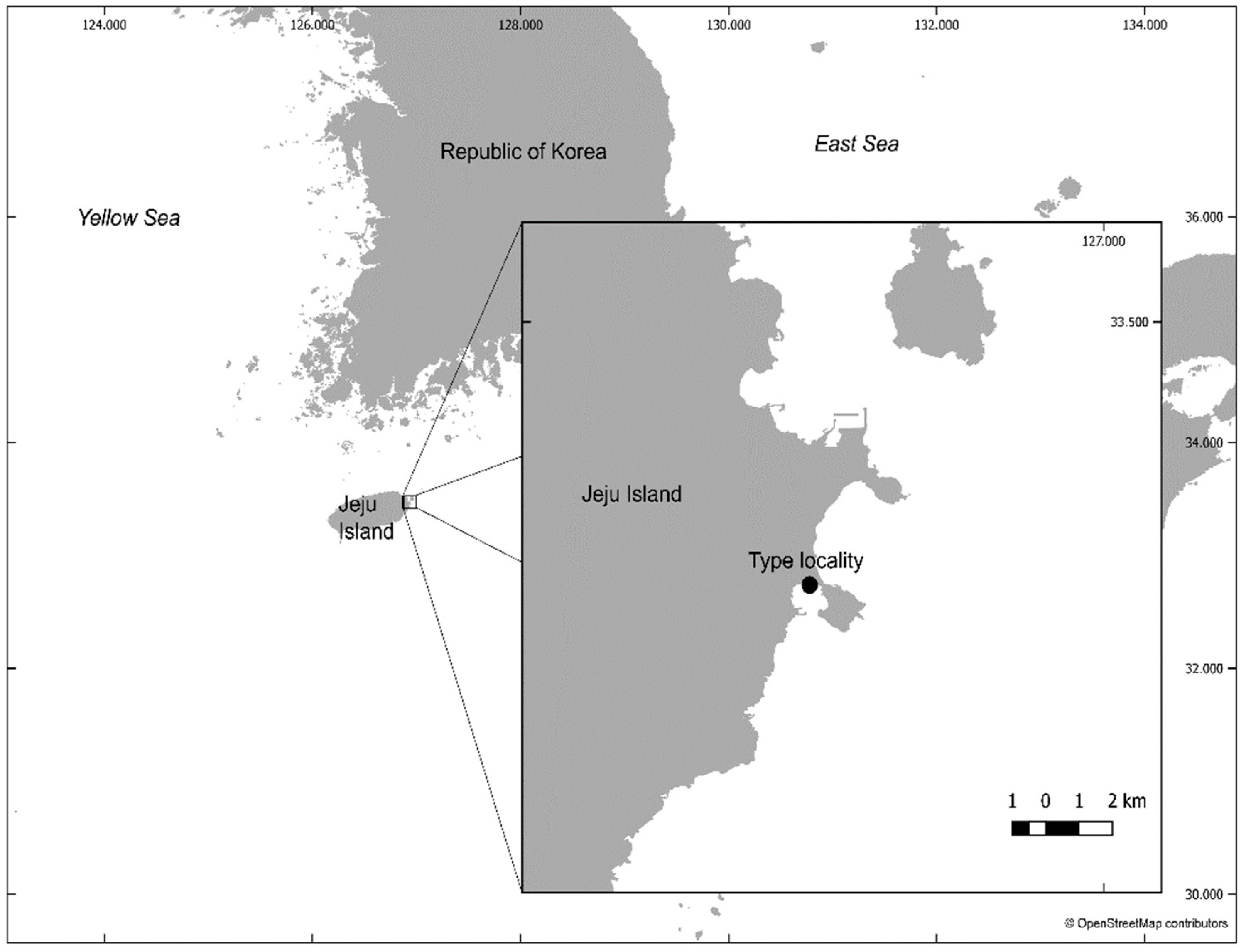
  • Gammanema okhlopkoviTchesunov, Jeong et Lee sp. n. Present paper.
  • Gammanema polydonta Murphy, 1965. Murphy 1965:176‒179, Figures 3A‒C and 4A‒H [25] (males, females); Chilean coast, depth 45 m.
  • Gammanema rapax (Ssaweljev, 1912) Gerlach 1964 sp. inq. Ssaweljev 1912:122 [26] (as Halichoanolaimus rapax) (male, female); Barents Sea, Kolafjord, Palafjord, Mogilnojesee (lake with an intermediate layer of sea water on the Kildin Island in the Barents Sea), sand. Murphy (1965: 179 [25]) considered the species; dubious because the original description is not illustrated and type specimens are evidently lacking. Gerlach (1964 [20]) synonymized Halichoanolaimus menzeli Ditlevsen 1918, Gammanema ferox Cobb, 1920 and Gammanema fennicum (Gerlach, 1953) with Gammanema rapax.
  • Gammanema smithi Murphy, 1964. Murphy 1964: 194‒198, Figure 3A,B,D,E, Figure 4, Figure 5B [27] (male); Puget Sound (NE Pacific), intertidal sand. Remark: there are some discrepancies in the original diagnosis. Caudal glands are described and depicted ([27], Figure 4) as incaudal (i.e., located entirely within tail), while the glands in Figure 5A are definitely excaudal (i.e., gland cell bodies protrude anteriorly and lie along the midgut). Supplementary organs are depicted from a medio-ventral view in Figure 3C, while the hind body in Figure 4 is shown laterally. Taking into account that the diagnosis is based on only one male (holotype), we consider Figures 3C and 5A as wrong and not pertaining to G. smithi.
  • Gammanema tchesunovi Gagarin et Klerman, 2007. Gagarin and Klerman 2007:782‒785, Figure 3, Table 3 [18] (males, females); Mediterranean coast of Israel, depths 50‒55 m, sand.
  • Gammanema uniformis (Cobb, 1920) Tchesunov et Okhlopkov, 2006. Cobb 1920:293‒294, Figure 75 [21] (as Trogolaimus uniformis) (seemingly, one male); Atlantic coast of the United States, New Hampshire. Tchesunov and Okhlopkov 2006:40‒43, Figure 12, Table 6 [28] (males, females); Atlantic coast of the United States, Maine.

3.1.2. Remarks on the Species List and Synonymy

Murphy (1965:179 [25]) considered the species Gammanema rapax (Ssaweljev, 1912) as dubious because the original description is not depicted, and type specimens are evidently lacking. Gerlach (1964 [20]) synonymized Gammanema menzelii (Ditlevsen, 1918), Gammanema ferox Cobb, 1920, and Gammanema fennicum (Gerlach, 1953) with G. rapax. We find this broad synonymization questionable. G. rapax is a large species, measuring 5800–6100 μm in body length (Ssaweljev 1912 [26]), while all other species and specimens are at least half the size (except G. menzelii) and found far from the type locality. G. menzelii corresponds to G. rapax in body length (male 5900 μm) but differs in its higher number of preanal midventral supplementary organs (35 versus 20–22). Diagnosis of G. menzelii also lacks some important details, such as sizes of anterior sensilla, amphideal fovea, and spicules. Descriptions of G. ferox (New Hebrides [21]), G. rapax (North Sea [20]), and G. rapax (White Sea [15]) differ from the original diagnosis of Ssaweljev (1912 [26]) in their much smaller body size. Hence, we do not accept all these described specimens to be conspecific. Okhlopkov [15] redescribed females of G. fennicum and for the first time described males of G. fennicum. Though the specimens have not been sampled in the area of the type locality (Gulf of Finland, east Baltic Sea), but instead in the White Sea, the newly found females correspond to the original diagnosis in most details and morphometrics. Hence, G. fennicum is restored as a valid species.

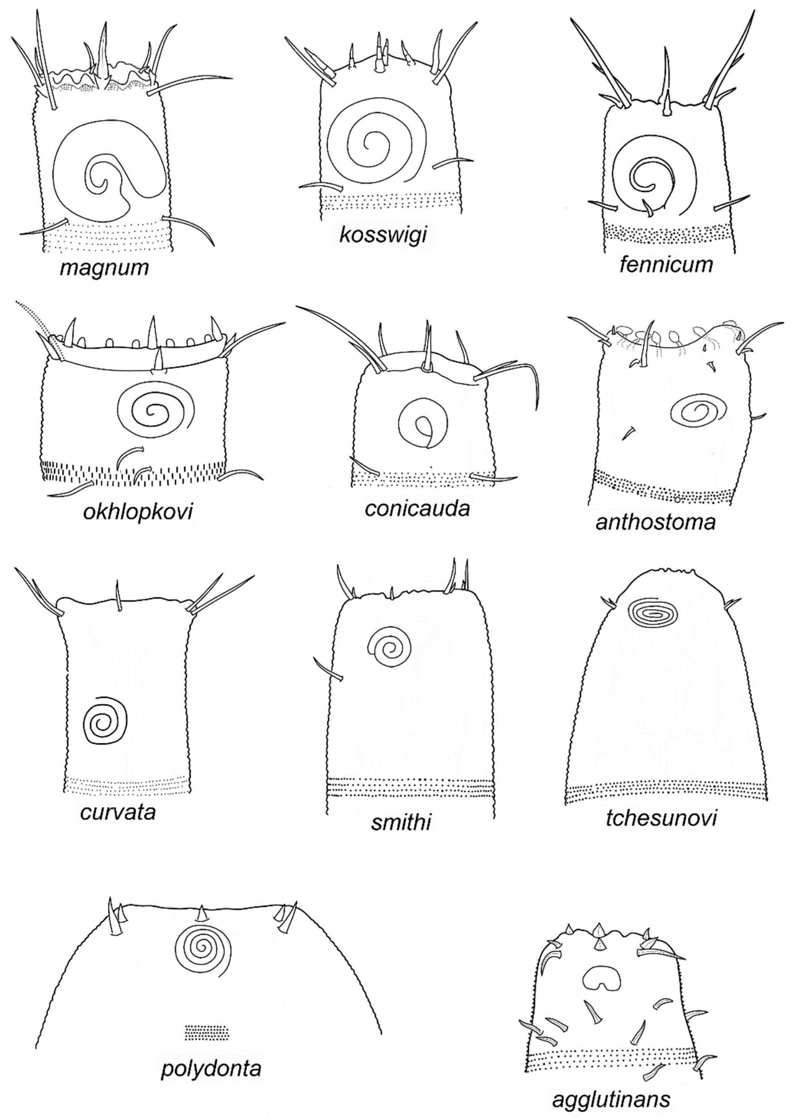
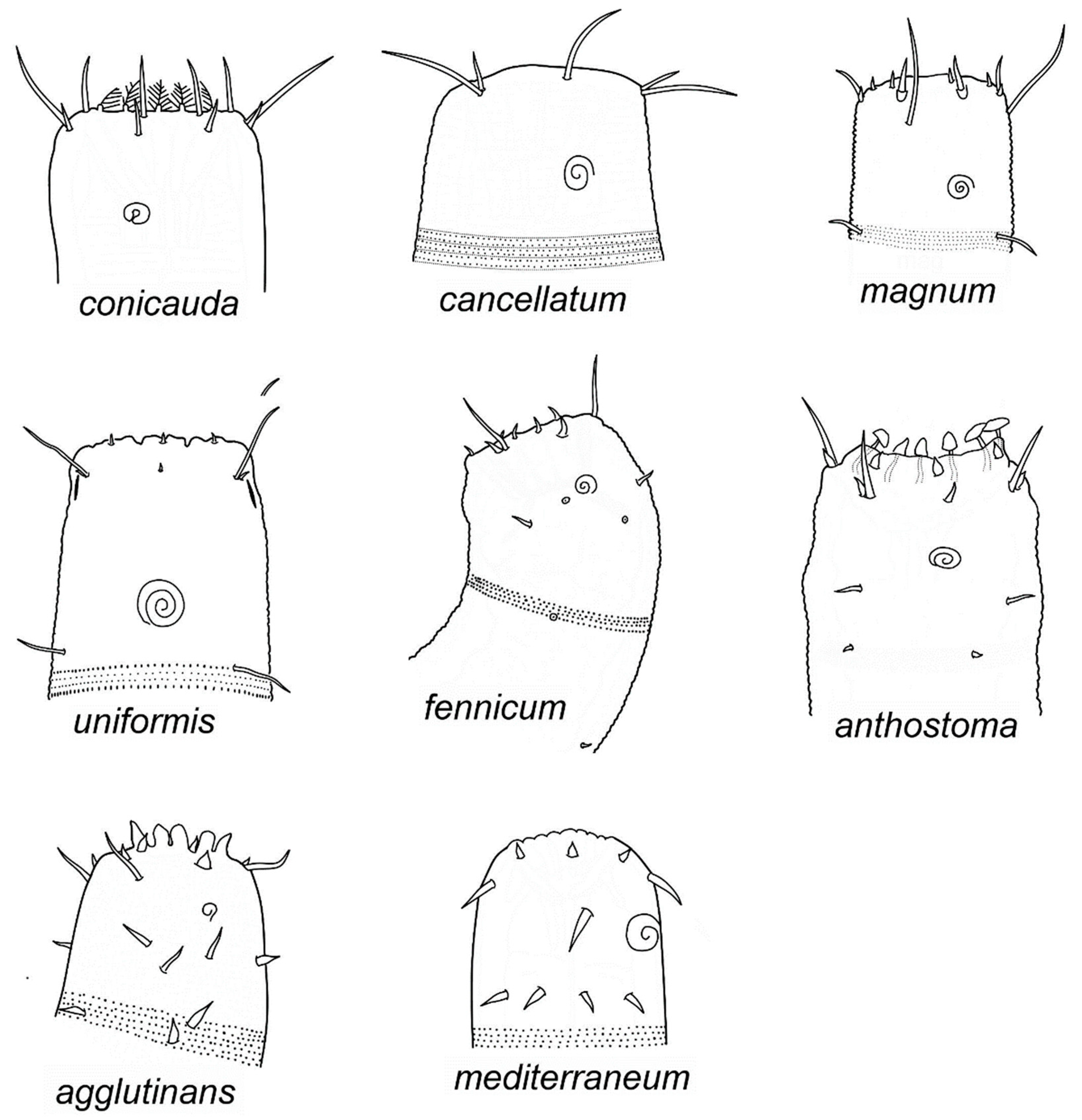
3.1.3. Identification of Species of Gammanema

Since Gammanema species show evident sexual dimorphism in size and shape of amphids and other structures, it is highly desirable to have both sexes (or at least males) for species identification. Due to deficiency of material, identification based on males is more reliable than identification solely based on females, since the males possess additional significant characteristics, such as supplementary organs and spicules.
For species identification of Gammanema, we propose a pictorial key constructed according to the principles of Platt [29], who first introduced pictorial keys in marine nematology. The pictorial key consists of two components, simplified images (caricatures) of heads (Figure 2 and Figure 3), and Table 1 and Table 2, with some metric and numeric characters being most important for species differentiation. Because of considerable sexual dimorphism, male and female head images are arranged in two separate plates, where the species are ordered in rows of gradual decrease of amphideal fovea (males) and cephalic setae (females), from top left to bottom right position.

3.2. Description of New Species of Gammanema

Gammanema okhlopkovi sp. n.

3.2.1. Etymology

The species is named in honor of Yuri R. Okhlopkov, a PhD student who published a few papers on Selachinematidae, but unfortunately passed away in 2014 by mischance.

3.2.2. Material Examined
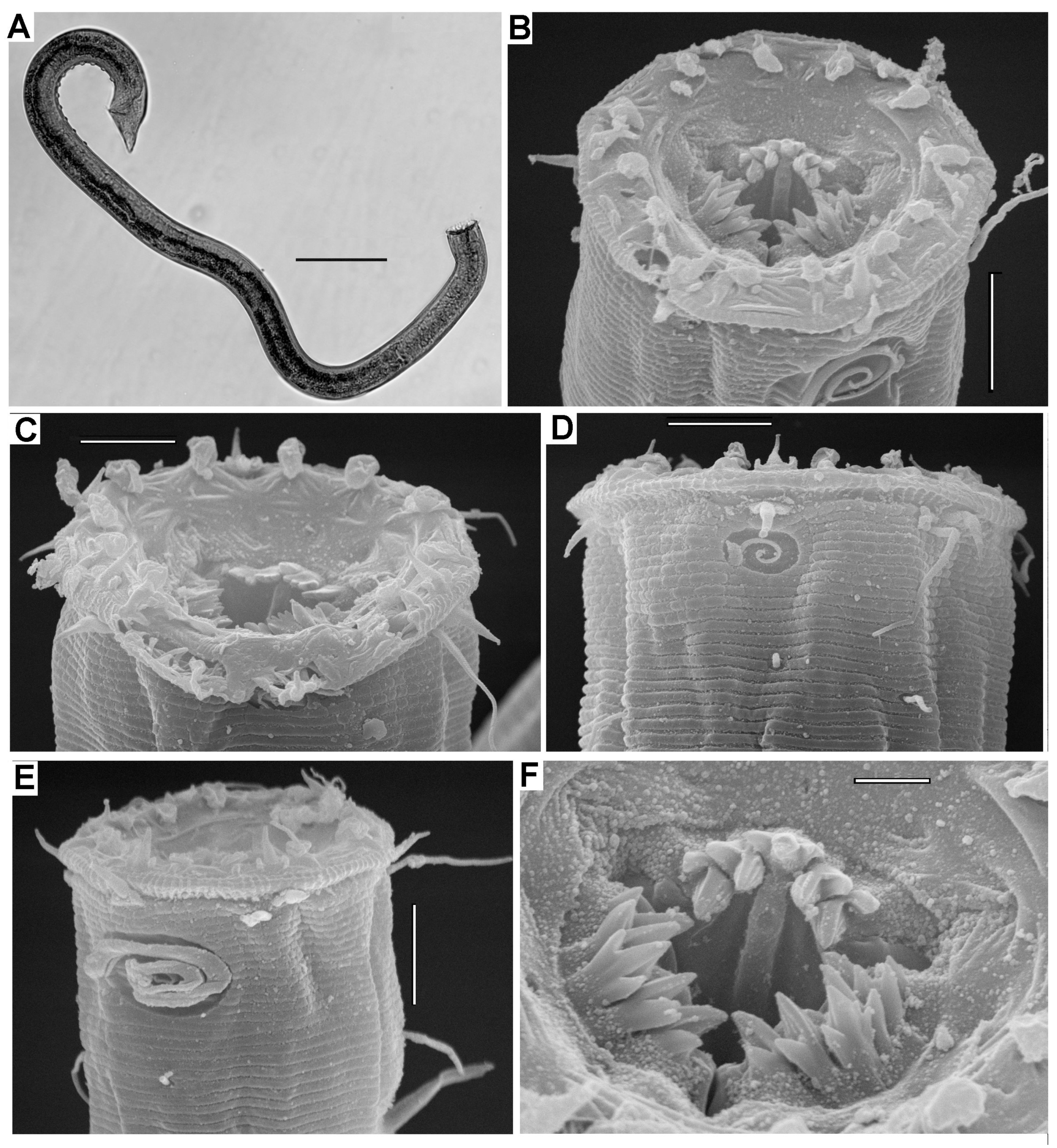
One holotype and seven paratype males were deposited in the National Institute of Biological Resources (South Korea). Inventory numbers of the holotype and paratypes are NIBRIV0000861671 and NIBRIV000086172-NIBRIV0000861677, respectively.
Type locality: Intertidal zone at coast of Jeju Island, South Korea (33°26′05″ N, 126°55′15″ E), sandy beach, June 2018.

3.2.3. Description

Body cylindrical, with truncate anterior end and short conical tail. Cuticle annulated and punctuated. There are ca. 13 annules within a 20 µm area, everywhere along the body. Cuticle punctuation homogeneous, without lateral differentiation, the dots arranged in chessboard order. The dots are actually struts in the median zone of the cuticle; the struts are composed of two elements, as seen in sites of damaged cuticles.
Truncate anterior end bordered by a membranous fringe with indistinct sectioning in lips. The fringe is marked by longitudinal striations, with about 15 fine, short ribs or rugae between lateral outer labial sensilla and cephalic seta. There are twelve prominent peg-like projections around the rim of the anterior end.
Anterior sensilla arranged in 6 + 10 pattern. Six inner labial sensilla as pointed conical papillae situated just outside or the peg-like projections. Six outer labial sensilla situated at the base of the striated fringe; they are nearly equal in shape and size to the inner labial sensilla. Four cephalic setae at the same level with latero-median outer labial sensilla in tight pairs; within pair, the cephalic seta disposed laterally of the adjacent outer labial sensilla. Somatic sensilla represented by a few irregular postamphideal setae and two subsequent lateral papillae further posteriad. Somatic sensilla on the posterior body and tail region are represented by papillae sparsely arranged in latero-ventral, lateral, and dorso-lateral rows. Amphideal fovea large, transversally oval in outer outline, spirally coiled in three turns.
The mouth opening is situated on the bottom of a funnel-shaped depression on the truncate anterior end. Acute teeth arranged in three radiating groups are visible, even in a semi-open mouth. Cheilostome is narrow and indistinct. Pharyngostome consists of two chambers. Anterior chamber is cup-shaped. Its walls are strengthened by three sclerotized rhabdions terminating posteriorly, with a group or five small cusps or denticles projected freely into the buccal cavity. The anterior rhabdions spread widely, thus making the anterior chamber bowl-shaped. In each group, the median tooth is tridentate, while teeth of two lateral pairs are bidentate. Posterior chamber cylindrical and narrow; its walls are strengthened by three sclerotized rhabdions. Pharynx cylindrical and evenly strongly muscular along its entire length.
Reproductive system is diorchic. Anterior outstretched testis situated ventrally to the intestine; posterior reflexed testis situated to the right of the vas deferens and intestine. Spicules paired, arcuate, distally pointed, and proximally very slightly cephalated. Gubernaculum as paired, nearly straight bars directed dorsally. Midventral supplementary organs, ten to thirteen in number, shaped as a nail-head 5–6 µm in diameter; the posteriormost organ situated at a distance 39 µm from the cloacal opening. A short conical midventral precloacal seta is present.
Tail very short; its conical tip turned left in all the specimens.

3.2.4. Diagnosis

Gammanema with short and stout body (L 961‒1150 μm, a 25‒31). Twelve prominent peg-like projections protruded outward from inner side of the circumoral membranous fringe. Set of anterior sensilla composed of six inner labial setae 4‒5 μm, six outer labial setae 5‒7 μm, and cephalic setae 9‒19 μm long. Amphideal fovea spirally coiled in three turns, 32‒48 μm wide. Rhabdion of anterior pharyngostome chamber 13‒17 μm, rhabdion of posterior pharyngostome chamber 11‒16 μm long. Spicules 32‒39 μm long. Midventral preanal row of supplementary organs consists of precloacal seta and 10–13 nail-head organs. Tail short and conical, c’ 1‒1.6.

3.2.5. Differential Diagnosis

G. okhlopkovi sp. n. is closest to G. anthostoma, sharing similar body length and body proportions a, b, and c, and similar size of amphids and spicules. The most obvious differences between the two species are longer cephalic setae (8.5‒19 μm in G. okhlopkovi versus 6‒7.5 μm in G. anthostoma), as well as a lack of precloacal supplementary organs in G. anthostoma (present in G. okhlopkovi).

3.3. Description of New Species of Latronema

Genus Latronema (Wieser 1954)
Diagnosis of the genus has been recently updated by Leduc and Zhao [30]. A detailed review of the taxon is intended in forthcoming work on a peculiar diversity of Latronema species on an Atlantic seamount.
Latronema obscuramphis sp. n.

3.3.1. Etymology

The species name reflects the indistinctness of the amphids (from Latin ”obscurus”, inconspicuous).

3.3.2. Material Examined

One holotype male, two paratype females, and four juveniles were deposited in National Institute of Biological Resources (South Korea). Inventory numbers of the holotype and paratypes are NIBRIV0000861678 and NIBRIV0000861679–NIBRIV0000861680, respectively.

3.3.3. Description

Body stout cylindrical, with truncate anterior end and short conical tail. Body cuticle finely cross-striated (16 annules in 20 µm in light microscope, 12 annules in 10 µm, from scanning electron micrographs) in the midbody laterally. The cuticle is not punctated under light microscope but marked with longitudinal wings. The wings number approximately five, visible on the lateral body side. Anteriorly, short and weak extra wings are added between the main wings, close to the head.
Labial region is presented by circular labial membrane with fine longitudinal ribbing (about ten ribs in between two adjacent setae of cephalic crown counted under light microscope, or seven to ten in SEM). Since the edges of the labial membrane are rolled inward, it is impossible to discern whether the membrane is divided into labial lobes. Labial region is encircled by a light cuticular ridge (collar).
Six slender inner labial setae are situated at the edge of the circular labial membrane. Ten equal setae located at the edge of the collar; they are about equal in length to one another and equal to inner labial setae, but wider basally; all the setae of the anterior circle look delicate and nearly transparent, and hence may be hardly visible against the labial cuticle. Fine ribs (striae) on the circumoral membrane may be arranged in fan-like bundles at sites opposite to cheilostomatal lobes. Quite possible the circle of ten equal setae comprises six outer labial and four cephalic setae, but all the setae are located at the same distance between them and do not make close pairs. Somatic setae comparable in length with anterior setae are distributed along the body in irregular longitudinal sublateral rows. Amphids in males are scarcely discernible under light microscope but detected in SEM as a small pit with simplified spiral structure. Amphids in females are not discernible at all under light microscope.
Buccal cavity consists of comparatively narrow cheilostome surrounded by labial membrane and voluminous pharyngostome composed of two compartments, with the anterior being cup-shaped and the posterior cylindrical. Cheilostome walls are differentiated interiorly into twelve rounded lobes that do not continue into anterior free projections, unlike that of Gammanema. Walls of the anterior pharyngostome are strengthened by three rhabdions—one dorsal and two lateroventral. The rhabdions terminate posteriorly with about seven acute denticles (one median denticle and two sets of three lateral denticles on both sides of it). Anterior pharyngostome is opened widely, and its rhabdions move apart at obtuse angle. Posterior pharyngostome short and wide, its rhabdions slightly sclerotized, while intermediate cuticle between them thin and soft.
Pharynx cylindrical and strongly muscular. Nerve ring hardly discernible. Intestine is composed of convex enterocytes bulging in the gut lumen. Gut content in the holotype male is a long, fine-granular body, possibly a digested prey nematode in the posterior half of the intestine.
Female reproductive system is didelphic, both ovaries short and small, antidromously reflexed; in one female, anterior ovary to the right and posterior ovary to the left of the intestine, in other female anterior ovary to the left and posterior to the right of the intestine. Both spermathecas and uteri branch are filled with spermatozoa. Posterior ovary contains large oocyte with coarsely granulated cytoplasm and large nucleolated nucleus in the center. Somatic cuticle around vulva is thickened and distinctly set off as a vulvar plate.
Male reproductive system is diorchic. Both anterior outstretched and posterior reflexed testes situated ventrally and to the right of the intestine. Spicules equal, slightly arcuate, distally narrowed, proximally located, with very weakly developed knobs. Gubernaculum as a pair of short bars parallel to posterodistal edge of spicules. Preanal set of midventral supplementary organs composed of a midventral preanal seta and a row of about eight nail-head-shaped organs. The latter are not quite distinctly observed because of their position on the concave body side and covering by foreign particles.
Tail short and conical; its terminal cone covered with smooth cuticle and curved to the left.

3.3.4. Diagnosis

Latronema with small and stout body (L 591‒718 μm, a 13.9‒19). All the anterior setae (i.e., inner labial setae, outer labial setae, and cephalic setae) are similar in length, ca 9‒12 μm in males, 6‒6.5 μm in females. Amphideal fovea small and obscure. Anterior pharyngostome rhabdion 12‒12.5 μm long, posterior rhabdions 7.5‒11 μm. Spicules arcuate, 38 μm long, gubernaculum 12 μm. Preanal midventral row of supplementary organs made up of preanal conical seta and twelve sucker-like organs. Tail very short (c 14.4, c’ 1.6), with terminal cone ca 16 μm long usually turned left.

3.3.5. Differential Diagnosis

L. obscuramphis sp. n. differs considerably from most Latronema species by combination of body size, anterior setae length, size and shape of amphideal fovea, size of spicules, number of preanal supplementary organs. L. aberrans (Allgén, 1934), L. annulatum (Gerlach, 1953), and L. spinosum (Andrássy, 1973) are closest to L. obscuramphis in characters cited above. L. obscuramphis differs from L. aberrans (the species known from the west Baltic Sea and White Sea) by smaller body length (male, L 591 versus 960 μm; females, L 666‒718 versus 1140 μm) and lesser number of preanal supplementary organs (8 versus 13); from L. annulatum (Madagascar, littoral) by tail shape (in male c’ 1.64 versus 2.4‒2.8), longer spicules (38 μm versus 27 μm), and lesser number of supplements (8 versus 12); and from L. spinosum (Cuba, only female known) by no spinose body cuticular rings, smaller body (666‒718 versus 850‒1062 μm), and lesser index b (3.8‒4.4 versus 4.5‒5).

4. Conclusions

The nematofauna of the intertidal sandy beach of Jeju Island is very rich, including over 70 species representing nearly all the families of marine free-living nematodes. Even described species of Thoracostomopsidae [5,6] and Selachinematidae (present paper) constitute only about one-third of the actual diversity of those families from the studied Jeju beach. In forthcoming works, we will try to identify at least the most common nematode species, aiming in the future to reveal fauna composition and species distribution from upper to lower intertidal horizons, as well as vertical distribution in sediment columns.

Author Contributions

Conceptualization, A.T. and W.L.; investigation, A.T. and R.J.; data curation, R.J. and W.L.; writing—original draft preparation, A.T.; writing—review and editing, R.J. and W.L.; funding acquisition, W.L. and A.T. All authors have read and agreed to the published version of the manuscript.

Funding

The study was supported by a grant 18-504-51026 Russian Fund of Basic Research, a grant NRF-2017K2A9A1A06051528 from the Korea Research Foundation, a grant NIBR202000000 entitled “Study of Undiscovered Invertebrate Species from Korea” from the National Institute of Biological Resources (NIBR) funded by the Ministry of Environment (MOE) of the Republic of Korea, as well as by the Marine Biotechnology Program of the Korea Institute of Marine Science and Technology Promotion (KIMST) funded by the Ministry of Oceans and Fisheries (MOF) (No. 20170431).

Acknowledgments

The authors thank Vadim Mokievsky (P.P. Shirshov Institute of Oceanology, Moscow) and Jungho Hong (Biodiversity Institute, Marine Act Co., Seoul) for their help in the field work sampling in June 2018. The authors are grateful to both anonymous reviewers for their remarks and advice, which helped us amend the manuscript.

Conflicts of Interest

The authors declare no conflict of interest.

Abbreviations

abody length divided by body diameter at midbody
bbody length divided by pharynx length
cbody length divided by tail length
c’tail length divided by anal body diameter
CVcoefficient of variation (SD divided by mean, in %)
SDstandard deviation
Vdistance from anterior end to vulva in % of entire body length

References

  1. Rho, H.S.; Kim, W. Paradraconema jejuense, a new species of genus Paradraconema (Nematoda: Draconematidae) from Korea. Korean J. Syst. Zool. 2004, 21, 81–91. [Google Scholar]
  2. Rho, H.S.; Kim, W. A new free-living marine nematode species of the genus Dracogalerus Allen and Noffsinger (Nematoda: Draconematidae) from a shallow subtidal zone of Jeju Island, Korea. Integr. Biosci. 2005, 9, 113–122. [Google Scholar] [CrossRef]
  3. Rho, H.S.; Decraemer, W.; Sørensen, M.V.; Min, W.G.; Jung, J.; Kim, W. Megadraconema cornutum, a new genus and species from Korea, with a discussion of its classification and relationships within the family Draconematidae (Nematoda, Desmodorida) based on morphological and molecular characters. Zool. Sci. 2011, 28, 68–84. [Google Scholar] [CrossRef]
  4. Lim, H.W.; Chang, C.Y. First record of Desmoscolex Nematoda (Desmoscolecida: Desmoscolecidae) from Korea. Integr. Biosci. 2006, 10, 219–225. [Google Scholar] [CrossRef] [Green Version]
  5. Jeong, R.; Tchesunov, A.V.; Lee, W. Bibliographic revision of Mesacanthion Filipjev, 1927 (Nematoda: Thoracostomopsidae) with description of a new species from Jeju Island, South Korea. PeerJ 2019, 7, e8023. [Google Scholar] [CrossRef] [PubMed]
  6. Jeong, R.; Tchesunov, A.V.; Lee, W. Two species of Thoracostomopsidae (Nematoda: Enoplida) from Jeju Island, South Korea. PeerJ, submitted, under review.
  7. Okhlopkov, J.R. Feeding of free-living nematodes of the families Selachinematidae and Richtersiidae in the White Sea. Proc. Pertsov White Sea Biol. Stn. 2003, 9, 127–139. (In Russian) [Google Scholar]
  8. Moens, T.; Braeckman, U.; Derycke, S.; Fonseca, F.; Galucci, F.; Gingold, R.; Guilini, K.; Ingels, J.; Leduc, D.; Vanaverbeke, J.; et al. Ecology of free-living marine nematodes. In Handbook of Zoology; Schmidt-Rhaesa, A., Ed.; De Gruyter: Berlin, Germany, 2014; Volume 2, pp. 109–152. [Google Scholar]
  9. Warwick, R.M. Nematode associations in the Exe estuary. J. Mar. Biol. Assoc. UK 1971, 51, 439–454. [Google Scholar] [CrossRef] [Green Version]
  10. Gallucci, F.; Steyaert, M.; Moens, T. Can field distributions of marine predacious nematodes be explained by sediment constraints on their foraging success? Mar. Ecol. Prog. Ser. 2005, 304, 167–178. [Google Scholar] [CrossRef] [Green Version]
  11. Burgess, R. An improved protocol for separating meiofauna from sediments using colloidal silica soils. Mar. Ecol. Prog. Ser. 2001, 214, 161–165. [Google Scholar] [CrossRef]
  12. Seinhorst, J. A rapid method for the transfer of nematodes from fixative to anhydrous glycerin. Nematologica 1959, 4, 67–69. [Google Scholar] [CrossRef] [Green Version]
  13. Leduc, D. Two new genera and five new species of Selachinematidae (Nematoda, Chromadorida) from the continental slope of New Zealand. Eur. J. Taxon. 2013, 63, 1–32. [Google Scholar] [CrossRef] [Green Version]
  14. Tchesunov, A.V. Order Chromadorida Chitwood, 1933. In Handbook of Zoology; Schmidt-Rhaesa, A., Ed.; Gastrotricha, Cycloneuralia and Gnathifera, Volume 2: Nematoda; De Gruyter: Berlin, Germany, 2014; pp. 373–398. [Google Scholar]
  15. Okhlopkov, J.R. Free-living nematodes of the families Selachinematidae and Richtersiidae in the White Sea (Nematoda, Chromadoria). Zoosyst. Ross. 2002, 11, 41–55. [Google Scholar]
  16. Gerlach, S.A. Zur Kenntnis der freilebenden marinen Nematoden von San Salvador. Z. für Wiss. Zool. 1955, 158, 249–303. [Google Scholar]
  17. Gerlach, S.A. Die Nematodenbesiedlung des Sandstrandes und des Küstengrundwassers an der italienischen Küste. I. Systematischer Teil. Arch. Zool. Ital. 1953, 37, 517–640. [Google Scholar]
  18. Gagarin, V.G.; Klerman, A.K. New species of predatory chromadorids (Nematoda, Chromadorida) from the Mediterranean Sea. Zool. Zhurnal 2007, 86, 778–786, (In Russian with English Abstract). [Google Scholar]
  19. Gerlach, S.A. Die Nematodenfauna der Uferzonen und des Küstengrundwassers am finnischen Meeresbusen. Acta Zool. Fenn. 1953, 73, 1–32. [Google Scholar]
  20. Gerlach, S.A. Revision der Choniolaimidae und Selachinematidae (Freilebende Meeres-Nematoden); Kosswig-Festschrift; Mitteilungen aus dem Hamburgischen Zoologischen Museum und Institut: Hamburg, Germany, 1964; pp. 23–50. [Google Scholar]
  21. Cobb, N.A. One hundred new nemas (type species of 100 new genera). Contrib. Sci. Nematol. 1920, 9, 217–343. [Google Scholar]
  22. Shi, B.; Xu, K. Two new rapacious nematodes from intertidal sediments, Gammanema magnum sp. nov. and Synonchium caudatubatum sp. nov. (Nematoda, Selachinematidae). Eur. J. Taxon. 2018, 405, 1–17. [Google Scholar] [CrossRef] [Green Version]
  23. Vitiello, P. Nématodes libres marins des vases profondes du Golfe du Lion. II. Chromadorida. Tethys 1970, 2, 449–500. [Google Scholar]
  24. Ditlevsen, H. Marine freeliving nematodes from Danish waters. Vidensk. Meddelelser Dansk Naturhistorisk Foren. I Kjobenhavn 1918, 70, 147–214. [Google Scholar]
  25. Murphy, D.G. Chilean marine nematodes. Veröff. Inst. für Meeresforsch. Bremerhav. 1965, 9, 173–203. [Google Scholar]
  26. Ssaweljev, S. Zur Kenntnis der freilebenden Nematoden des Kolafjords und des Relictensee Mogilnoje. Trav. Soc. Imp. Nat. St. Pétersbg. 1912, 43, 108–126. [Google Scholar]
  27. Murphy, D.G. Free-living marine nematodes, I. Southerniella youngi, Dagda phinneyi, and Gammanema smithi, new species. Proc. Helminthol. Soc. Wash. 1964, 31, 190–198. [Google Scholar]
  28. Tchesunov, A.V.; Okhlopkov, Y.R. On some selachinematid nematodes (Chromadorida: Selachinematidae) deposited in the collection of the Smithsonian National Museum of Natural History. Nematology 2006, 8, 21–44. [Google Scholar]
  29. Platt, H.M. Pictorial taxonomic keys: Their construction and use for the identification of free-living marine nematodes. Cah. Biol. Mar. 1984, 25, 83–91. [Google Scholar]
  30. Leduc, D.; Zhao, Z. Latronema whataitai sp. n. (Nematoda: Selachinematidae) from intertidal sediments of New Zealand, with notes on relationships within the family based on preliminary 18S and D2-D3 phylogenetic analyses. Nematology 2015, 17, 941–952. [Google Scholar] [CrossRef]
Figure 1. Map of sampling locality. This map is made with QGIS software v.2.18.14, a free and open-source geographic information system (https://qgis.org).
Diversity 12 00019 g001
Figure 2. Caricatured male heads of valid Gammanema species arranged from top left to bottom right along decrease of amphid size and cephalic setae in relation to head diameter.
Diversity 12 00019 g002
Figure 3. Caricatured female heads of valid Gammanema species arranged from top left to bottom right along decrease of cephalic setae in relation to head diameter.
Diversity 12 00019 g003
Figure 4. Gammanema okhlopkovi sp.n., entire view and anterior body: (A) holotype, male, entire view; (B) apical view, male, scanning electron micrograph; (C) subapical view, female, scanning electron micrograph; (D) head, lateral view, female, scanning electron micrograph; (E) head, sublateral view, male, scanning electron micrograph; (F) mouth opening, male, scanning electron micrograph. Scale bars: (A) 100 μm, (B) 10 μm, (C) 10 μm, (D) 10 μm, (E) 10 μm, (F) 3 μm.
Diversity 12 00019 g004
Figure 5. Gammanema okhlopkovi sp. n., head of the holotype male: (A) surface view; (B) optical section. Scale bars: 20 μm.
Diversity 12 00019 g005
Figure 6. Gammanema okhlopkovi sp. n., posterior body, males: (A) hind body; (B) spicule tips, precloacal seta, and supplementary organs, lateral view, scanning electron micrograph; (C) posterior-most supplementary organ, scanning electron micrograph; (D) cloacal opening and precloacal seta, subventral view, scanning electron micrograph; (E) tail with sensory papillae; (F) damaged cuticle near a supplement displays struts in the median zone, scanning electron micrograph. Scale bars: (A) 50 μm, (B) 10 μm, (C) 3 μm, (D) 3 μm, (E) 10 μm, (F) 3 μm.
Diversity 12 00019 g006
Figure 7. Gammanema okhlopkovi sp. n., posterior body of the holotype male. Scale bar: 20 μm.
Diversity 12 00019 g007
Figure 8. Latronema obscuramphis sp. n., entire body of the holotype male and surface cuticular structure: (A) holotype male, entire view; (B) entire view of a female, scanning electron micrograph; (C) head of a female laterally, scanning electron micrograph; (D) amphideal fovea of a female, scanning electron micrograph; (E) body cuticle rings and alae laterally, scanning electron micrograph. Scale bars: (A) 50 μm, (B) 30 μm, (C,E) 10 μm, (D) 3 μm.
Diversity 12 00019 g008
Figure 9. Latronema obscuramphis sp. n., male holotype structures: (A) surface view of the head; (B) sagittal optical section of the head; (C) posterior body. Scale bars: 20 μm.
Diversity 12 00019 g009
Table 1. Gammanema valid species, comparison of males.
SpeciesCharacters
Body LengthaInner Labial Setae LengthOuter Labial Setae LengthCephalic Setae LengthAmphid WidthSupplements ShapeNumber of SupplementsSpicule SengthSome Particular Bold Features
agglutinans544‒69610‒112210‒1110tubular≥634‒36Cuticle with minute spines, somatic setae leaf-shaped
anthostoma1002‒195022–295‒65‒86‒814‒26absentabsent62‒87Prominent circumoral projections each consisted of peduncle and triangular cap
conicauda1985‒248441‒48161730‒3315conical papillae2233‒40Prominent circumoral hair-like projections
curvata328676not indicated17138‒8.5cup-shaped1628-
fennicum2165‒260028‒4917‒248‒1134‒5028‒31sucker-like13‒2162‒64-
kosswigi120029810‒122030weak setose439Outer labial setae jointed
magnum2900–408548–5215–1622–2531–3542–45absentabsentleft 51‒56, right 62‒69Spicules slightly unequal
okhlopkovi961‒115025‒314‒53‒78‒1911‒18sucker-shaped10‒1332‒39Prominent circumoral Projections peg-like
polyodonta3200‒420021‒27not indicatedpapillae1642‒45cup-shaped43‒52135Very massive appearance; Anterior half of body opaque because of densely pigmented intestine
smithi221041not indicated52014cup-shaped2078-
tchesunovi1603‒188915‒16not indicated58‒815‒18cup-shaped23‒2485‒87Tail rounded and conical, terminal cone not developed
uniformis2700‒295045‒611‒22‒29‒1010‒11cup-shaped19‒2282‒87Cephalic cuticle thickened and internally sclerotized around anterior chamber of pharyngostome
Table 2. Gammanema valid species, comparison of females.
SpeciesCharacters
Body LengthaInner Labial SetaeOuter Labial SetaeCephalic SetaeAmphid WidthSome Particular Bold Features
agglutinans763‒91811‒152211‒195Cuticle with minute spines, somatic setae leaf-shaped
anthostoma1487‒178217‒245‒6.55‒7.515‒199.5‒10.5Prominent circumoral projections; each consisted of peduncle and triangular cap
cancellatum246524no data40158Complex ornamentation of cuticle consists of dots and longitudinally arranged short rods
conicauda2492‒372430‒35161230‒338‒9Prominent circumoral hair-like projections
curvata2392‒263964‒71no data15‒1712‒138‒8.5-
fennicum1672‒278213‒259‒1310‒1316‒379.5‒11-
magnum2540‒314528‒3310‒1420‒2140‒459‒10-
mediterraneum660‒74518‒20papillaepapillae7.55‒6-
polyodonta3730‒405023‒31papillaepapillae1620Very massive appearance; anterior half of body opaque because of densely pigmented intestine
tchesunovi1562‒168211‒12papillae57‒816‒18Tail rounded and conical, terminal cone not developed
uniformis2260‒279040‒571‒22‒29‒129‒10Cephalic cuticle thickened and internally sclerotized around anterior chamber of pharyngostome
Table 3. Morphometrics of males Gammanema okhlopkovi sp. n. (holotype and paratypes).
CharacterHolotypeTotal NumberMin.–Max.MeanSDCV%
Body length9618961‒1150108261.85.71
pharynx length1788164‒20718515.18.15
tail length48742‒57.550.45.611.1
body diameter at level of cs37736.9‒43.739.22.406.12
body diameter at level of nerve ring35833.1‒40.036.82.326.30
body diameter at level of cardia35834.1‒41.036.52.456.72
body diameter at level of midbody37837.0‒42.038.82.546.65
body diameter at level of cloaca36836.0‒42.538.22.546.65
a26825.6‒31.127.82.177.80
b5.485.40–6.565.880.416.97
c20718.5‒24.621.62.3210.7
c’1.3371.01‒1.531.310.1612.2
inner labial setae length3.5‒464.0‒5.04.270.399.13
lateral outer labial setae length3.763.0‒7.25.221.6331.2
latero-median outer labial setae length555.0‒7.05.880.7612.9
cephalic setae length1268.5‒19.014.14.0128.5
anterior cervical setae length7.677.6‒15.610.82.5623.8
amphid width16711.0‒18.014.82.019.13
amphid width/corresponding body diameter43832.5‒47.737.75.2213.9
distance amphid—cephalic apex8.687.3‒11.38.951.3715.3
anterior pharyngostome rhabdion length15813.0‒16.914.91.4910.0
posterior pharyngostome rhabdion length14811.2‒16.013.51.4010.3
spicule length (arc)34632.0‒39.035.22.868.12
spicule ‘length (chord)28629.0‒36.032.92.47.29
number of supplements12610‒1311.51.059.13
tail terminal cone length17416.8–23.519.62.9315.0
Table 4. Latronema obscuramphis sp. n., morphometry.
CharacterHolotype MaleParatype Female 1Paratype Female 2
Body length591666718
a15.213.919.0
b5.094.383.78
c14.4tail not measurabletail not measurable
c’1.60tail not measurabletail not measurable
V, %-57.470.0
body diameter at level of cephalic setae30.038.033.1
body diameter at level of nerve ring30.5?30.6
body diameter at level of cardia35.041.533.8
body diameter at level of midbody39.048.037.0
body diameter at level of cloaca/anus25.630.0?
inner labial setae length8.77.76.0
latero-median outer labial setae length11.6not measurablenot measurable
lateral outer labial setae length10.05.56.3
cephalic setae length8.8not measurablenot measurable
anterior cervical setae length10.012.0not measurable
anterior pharyngostome rhabdion length12.012.012.4
posterior pharyngostome rhabdion length8.07.510.7
spicules length (arc)38.0--
spicules length (chord)33.0--
gubernaculum length12.0--
tail terminal cone length16not measurablenot measurable

