Article

Planktotrophic Brachiopod Larvae from the Pacific and Caribbean of Panama

by
Rachel Collin
1,*,
Dagoberto E. Venera-Pontón
1,
Amy C. Driskell
2,
Kenneth S. Macdonald III
2 and
Michael J. Boyle
3
1
Smithsonian Tropical Research Institute, Apartado Postal 0843-03092, Balboa Ancon, Panama
2
Laboratories of Analytical Biology, National Museum of Natural History, Smithsonian Institution, Washington, DC 20560, USA
3
Smithsonian Marine Station, Fort Pierce, Florida, FL 34949, USA
*
Author to whom correspondence should be addressed.
Diversity 2019, 11(1), 2; https://doi.org/10.3390/d11010002
Submission received: 14 October 2018 / Revised: 19 December 2018 / Accepted: 21 December 2018 / Published: 26 December 2018
(This article belongs to the Section Marine Diversity)

Abstract

:
Lingulids and discinids are the only brachiopods that exhibit life histories that include a feeding planktonic stage usually referred to as a “larva”. We collected planktotrophic brachiopod larvae from the Pacific and Caribbean coasts of Panama and took a DNA barcoding approach with mitochondrial cytochrome c oxidase subunit I (COI), mitochondrial ribosomal 16S, and nuclear ribosomal 18S genes to identify those larvae and to estimate their diversity in the region. We observed specimens from both coasts with distinct morphologies typical of lingulid and discinid larvae. COI and 16S were sequenced successfully for the lingulid larvae but failed consistently for all discinid larvae. 18S was sequenced successfully for larvae from both families. Sequence data from each gene revealed one lingulid operational taxonomic unit (OTU) from Bocas del Toro on the Caribbean coast, and one lingulid OTU from the Bay of Panama on the Pacific coast. These OTUs differed by >20% for COI, >10% for 16S and ~0.5% for 18S. Both OTUs clustered with GenBank sequences of Glottidia species, the only genus of lingulids in the Americas, but were distinct from G. pyramidata the only species reported for the Caribbean. Analysis of 18S sequence data for discinid larvae recovered 2 OTUs, one exclusively from the Pacific and one with a mixture of Pacific and Caribbean larvae. The 18S marker does not provide enough resolution to distinguish between species, and comparisons with GenBank sequences suggest that one OTU includes Pelagodiscus species, while the other may include Discradisca species. When compared with other marine invertebrates, our surveys of brachiopod larvae through DNA barcoding show relatively low levels of diversity for Panama.

1. Introduction

Within extant Brachiopoda, species in only two families (Lingulidae and Discinidae) have life histories with planktotrophic (planktonic, feeding) developmental stages [1]. These are generally referred to as larvae (a convention which we follow), although morphologically the shelled stages are planktonic juveniles [2,3,4,5]. Brachiopod larvae from other families lack functional guts and are lecithotrophic (non-feeding). Observations of the species with planktotrophic larvae have contributed significantly to ideas about the evolution of mode of development, the mechanisms of evolutionary loss and gain of feeding larvae, and provide a basis to infer the mode of development in fossil brachiopods [5,6,7]. Despite this interest, very little information has been published on the diversity and ecology of planktotrophic brachiopod larvae with the exception of detailed studies on Glottidia pyramidata [8], Lingula anatina [2,9] and Discinisca [10,11]. No information has been published on brachiopod larvae from Central America, and information on adult brachiopods from this region is limited.
As part of a larger effort to document the diversity of marine invertebrate larvae on both coasts of Panama, we collected, photographed, and DNA barcoded planktotrophic brachiopod larvae from the Bay of Panama on the Pacific coast, and the Bocas del Toro Archipelago on the Caribbean coast. We used DNA sequences to determine how many operational taxonomic units (OTUs) were present in samples from each ocean and compared these OTUs to sequences in GenBank or BoLD in order to identify the larvae. Subsequently, we compared these OTUs to morphological observations and photographs with the aim of identifying characteristics that can distinguish larval OTUs. Our more structured sampling in the Caribbean also provided information on density and seasonality of these larvae.
The potential diversity of planktotrophic brachiopod larvae in the waters around Panama is low. On the Pacific coast, the lingulids Glottidia albida (Hinds, 1844) and G. audebarti (Broderip, 1835) have both been reported from the coast of Costa Rica [12]. G. audebarti has also been reported on the Pacific coast of Panama [13,14]. Among discinids, Discradisca strigata (Broderip, 1834) is abundant along the Pacific coasts of Costa Rica and Panama [12,15]. Pelagodiscus atlanticus (King, 1868) has also been reported from the Pacific coast of Costa Rica [12] and is shown as occurring in Panama, along with Discradisca cumingi (Broderip, 1833) in the Brachiopoda Database [16].
Thus far, few brachiopods are reported for the Caribbean coast of Panama. Glottidia pyramidata (Stimpson, 1860) has been reported in the eastern Caribbean and Gulf of Mexico [13,14,17] but not, as far as we know, in Panama. Discradisca antillarum (d’Orbigny, 1845) ranges from Florida to Southern Brazil, and throughout the Gulf of Mexico [16,17], including the San Blas Islands on the eastern Caribbean coast of Panama [13].

2. Materials and Methods

2.1. Sample Collection

Caribbean samples were collected in the Bahia Almirante in Bocas del Toro Province and Pacific samples were collected in the Bay of Panama. Details of our collections are given in reference [18]. Briefly, Caribbean larvae were collected with a 0.5 m diameter 125 µm mesh plankton net towed behind a small boat that was moving with the engine in neutral. This method maintained the net at 10–20 m depth. The volume of water sampled was measured using a flow meter (General Oceanics) tethered within the mouth of the net. Structured quantitative sampling involved four collection campaigns evenly spaced over one year (August 2015, November 2015, February–March 2016, and June 2016). Each campaign consisted of 3 or 4 tows over an interval of 9–10 days. Each tow in the Caribbean was conducted between 07:00 and 09:00 in the channel between Isla Colon and Isla Cristobal. Longitude ranged from 09°20′8.9″ N to 09°20′36.3″ N and latitude ranged from 82°15′41.0″ W to 82°15′50.0″ W. Additional samples were obtained in 2013 during the Larval Invertebrate Diversity, Form and Function short-course at the Smithsonian Tropical Research Institute’s Bocas del Toro Research Station (BRS). These samples were collected in a similar way, but included sites from throughout Bahia Almirante. Samples from the Pacific were collected from the northern region of the Bay of Panama, adjacent to Taboga and/or Contadora Islands between 2013 and 2016. Pacific tows were exploratory, and nets were towed for approximately 20 min, using the same net as was used in the Caribbean; however, the boat engine was periodically moved in and out of gear to sample a depth-range of approximately 5 to 20 m. Sampling sites and dates were selected opportunistically as weather and sea conditions permitted.
Live plankton samples were sorted using a stereomicroscope and all brachiopod larvae were moved to dishes of filtered sea water. In Bocas del Toro, each sample was sorted exhaustively, providing data on larval density per tow. For samples from the Bay of Panama, larvae were also sorted using a stereomicroscope but no effort was made to ensure that all larvae were detected and counted. Only one tow was sorted exhaustively to count the numbers of larvae. Representative larvae from each tow, and from each coast, were individually photographed live in seawater through a stereomicroscope prior to preservation for DNA sequencing. We recorded the overall appearance, morphological details, and approximate size of each larva when viewed with epi-illumination in front of a black background, with some larvae also observed with transmitted light.

2.2. DNA Sequencing

Individual larvae were preserved in 150 µL of M2 extraction buffer (AutoGen) in 96-well plates, stored frozen at −20 °C, and shipped to the Smithsonian’s Laboratories of Analytical Biology (LAB) for DNA extraction and Sanger sequencing. DNA from each larval sample was extracted with an AutoGenprep 965 extraction robot after overnight digestion with proteinase-K in M2 buffer. We attempted to sequence 3 gene fragments using the primer combinations given in Table 1. Cytochrome oxidase subunit I (COI) and the 16S rRNA gene, both commonly used barcode fragments, were sequenced with the aim of identifying larva to species level, while 18S, which evolves much more slowly, was sequenced with the aim of confirming the family-level or possibly genus-level identity. In addition to the primers listed in Table 1, the phoronid-specific primers described in reference [18] were tested but failed to amplify the 16S fragment from samples that did not amplify with the other primer pairs. The PCR cocktail included 5 µL GoTaq Hot Start Mix (Promega), 0.1 µL 20 µg/µL BSA, 0.3 µL each 10 mM primer and 0.5 µL dNTPs (2.5 mM each) in a total volume of 10 µL. The cocktail for 16S used Biolase Taq (Bioline) with the addition of 0.5 µL 50 mM MgCl2. The annealing temperature for nearly all reactions for all three gene regions was 50 °C, although occasionally it was raised to 52 °C in an attempt to increase priming specificity when it appeared that co-amplification was occurring.

2.3. Sequence Analysis

Sequences were screened for quality and contigs of forward and reverse sequences were produced using Sequencher 5.4.6 (Gene Codes). Only COI sequences of more than 450 bp in length and with a Phred quality score of at least 30 for more than 85% of the bases were combined into contigs and used for analyses. For both 16S and 18S, sequences greater than 400 bp were analyzed. To check for potential contamination, all sequences were compared internally with all other larvae sequenced in our project within the BoLD project workbench (www.boldsystems.org) and also compared to other publicly available sequences using BLAST searches in GenBank. Sequences that had identity >95% to species in other phyla were eliminated from subsequent analyses.
Because DNA barcoding is a distance-based approach, we constructed neighbor joining trees (BIONJ, [23]) with Jukes-Cantor distances from our sequences combined with every lingulid and discinid brachiopod COI, 16S, or 18S sequence available in GenBank as of 10 March 2018. COI alignments were performed with the BoLD aligner (amino acid based Hidden Markov Model [24]) whereas 16S and 18S alignments were performed with the Kalign algorithm [25]. Alignments were subsequently corrected manually when necessary. We stopped correcting the alignment when no other region in which the differences among sequences appeared to be caused by the position of gaps, rather than by actual nucleotide differences was found, or when regions in which the number and extension of the gaps could not be reduced without causing more nucleotide differences. Operational Taxonomic Units (OTUs) were identified with the Automatic Barcode Gap Discovery method [26]. DNA sequences have been deposited in GenBank (Table 2; COI: MK092033-MK092062; 16S: MK073386-MK073417; 8S: MK073419-MK073480), and datasets and specimen information are available in BoLD (dataset: doi.org/10.5883/DS-BRACHIOP).

3. Results

A total of 73 brachiopod larvae (38 from the Bahia Almirante and 35 from the Bay of Panama) were collected to sequence (Figure 1 and Figure 2). We found larvae with morphology typical of both lingulids and discinids on both coasts. Structured sampling in Bahia Almirante produced 0 to 11 brachiopods per tow with an average density of 2.26 (s.d. = 0.63) individuals per m3 of seawater sampled, densities similar to lingulid densities reported by Hammond [40]. Larvae were collected during all four sampling periods and were not obviously more abundant during any particular season. In the Caribbean, all of the larvae had attained the shelled stage. In the Pacific, very small, early stage discinids with multiple ciliated lobes and long setae, but lacking a shell, were occasionally abundant (>15 in some tows) although shelled stages were easier to detect in the samples. Brachiopod larvae were abundant (66 in the single sample we collected using the same procedure as for the Caribbean) in the Pacific, but density estimates cannot be given as the volume of water sampled was not quantified for most tows at these sites. We did not find lecithotrophic brachiopod larvae in any sample.

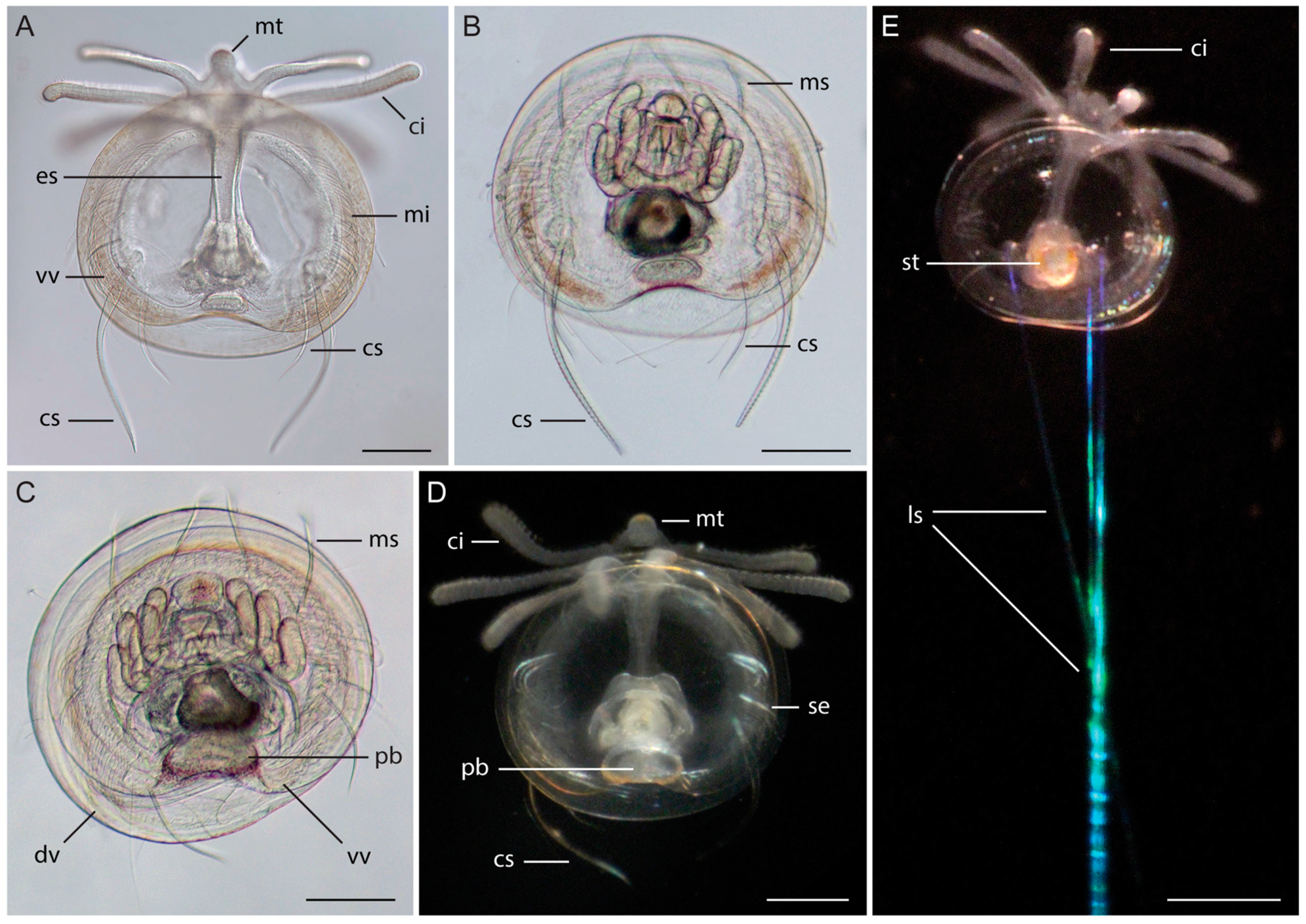
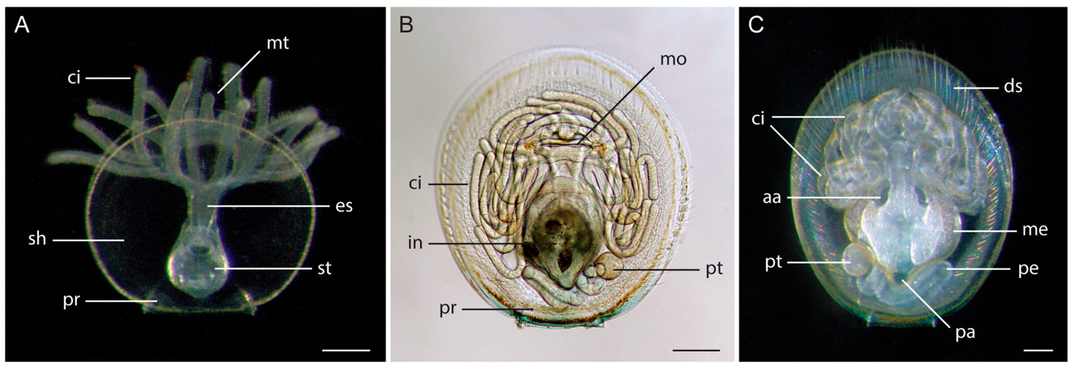
3.1. Morphology of Discinid Larvae

The morphology of all Panamanian discinid larvae was typical of morphologies previously described for discinid larvae (Figure 1) [9,10,11,41,42,43]. The early un-shelled larval stages observed in the Bay of Panama had four pairs of cirri and extremely long (~600–800 microns) setae whose birefringence makes the tiny (<150 microns) larvae relatively easy to see under a stereomicroscope with epi-illumination. These larvae could not be easily imaged and they had such little tissue we did not attempt to sequence them. We did not find early un-shelled stages in our Caribbean samples. Presence of very early stages in the Pacific plankton sample supports Chuang’s [11] conclusion that early stages are not brooded or benthic, while little can be concluded by their absence in the Caribbean, where all stages of larvae were uncommon.
Small, early-stage shelled larvae had an overall transparent appearance, a shell length of ~200 microns and one bundle of long setae on each side (referred to as embryonic setae by Chuang [11], although they appear to be homologous to larval setae of articulate brachiopods [4]). The three larvae from the Caribbean collected at this stage had at least four long setae on each side, and one individual had five on one side. The only larva collected from the Pacific at this stage had two setae on one side and one on the other side (Figure 1E). The larvae from both locations had four pairs of cirri (p.c.) with two pairs on each side of the median tentacle, a pattern that is typical of discinid larvae [1]. In all four larvae, the relatively larger, curved, principal larval setae were already visible. Their shells were slightly wider than they were long, and the distinctive posterior shell embayment on the ventral valve that is characteristic of discinid larvae had not yet developed.
Later developmental stages that had lost the long setae were more common in our samples (Figure 1A–D). These larvae had a shell length of 300–600 microns, an embayment on the posterior margin of the ventral shell, and clear development of the pedicle posterior to the gut and anterior to the shell embayment. All of these larvae possessed a number of small setae around the edge of the shell. These setae were difficult to distinguish in darkfield micrographs and variable in number in the brightfield micrographs we obtained for a subset of larvae (Figure 1A–C). These larvae all had four pairs of cirri, and in some larvae very subtle rust color was observed on the median tentacle (e.g., Figure 1C) and near the shell embayment under reflected light. Two red spots were visible within the tissue lateral to the gut. These appear coincident or slightly anterior to the statocyst. These larvae also had greenish-yellow pigment granules in a distinct band around the perimeter of the mantle, which appeared dark under transmitted light (Figure 1A–C).

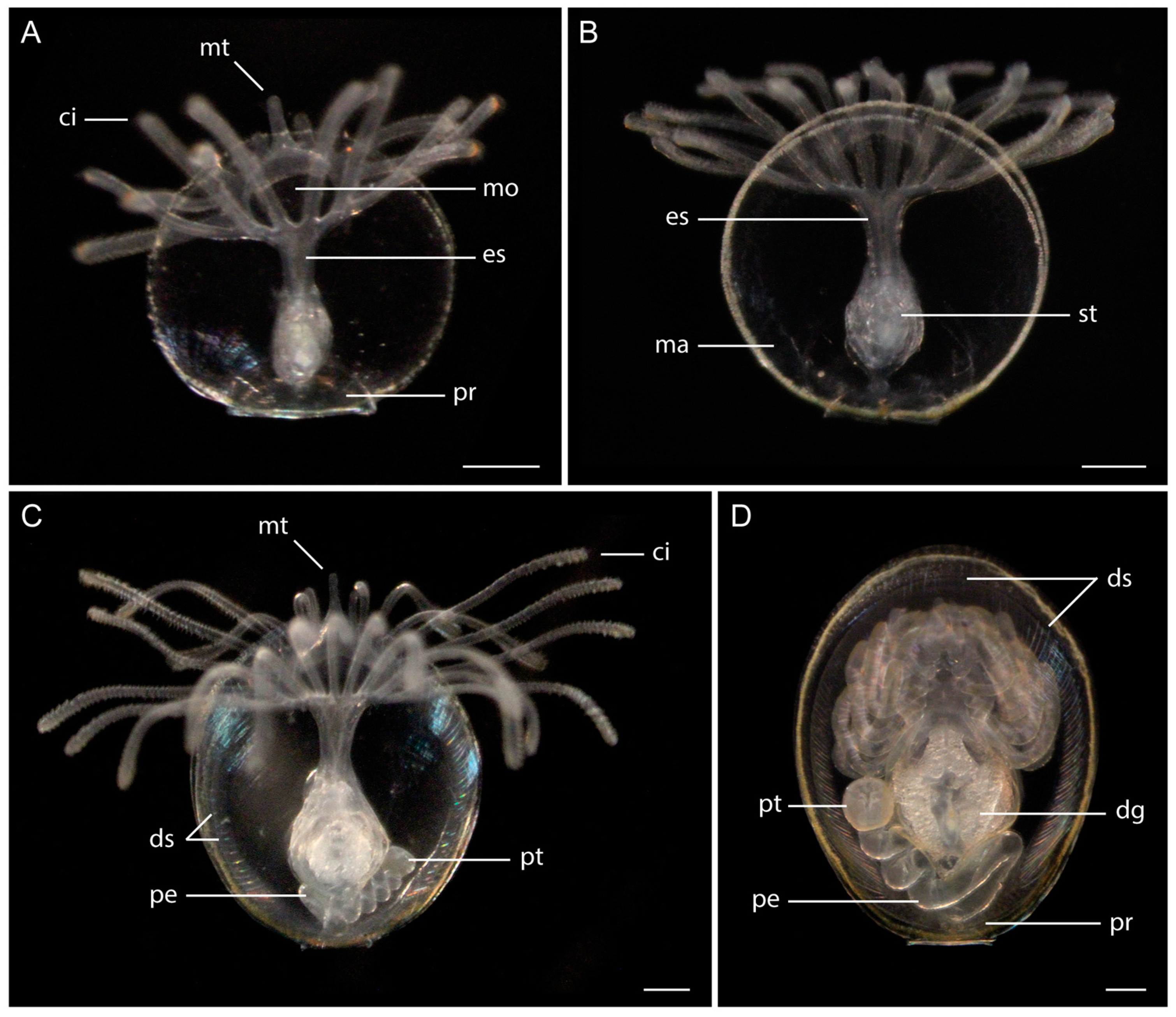
3.2. Morphology of Glottidia Larvae
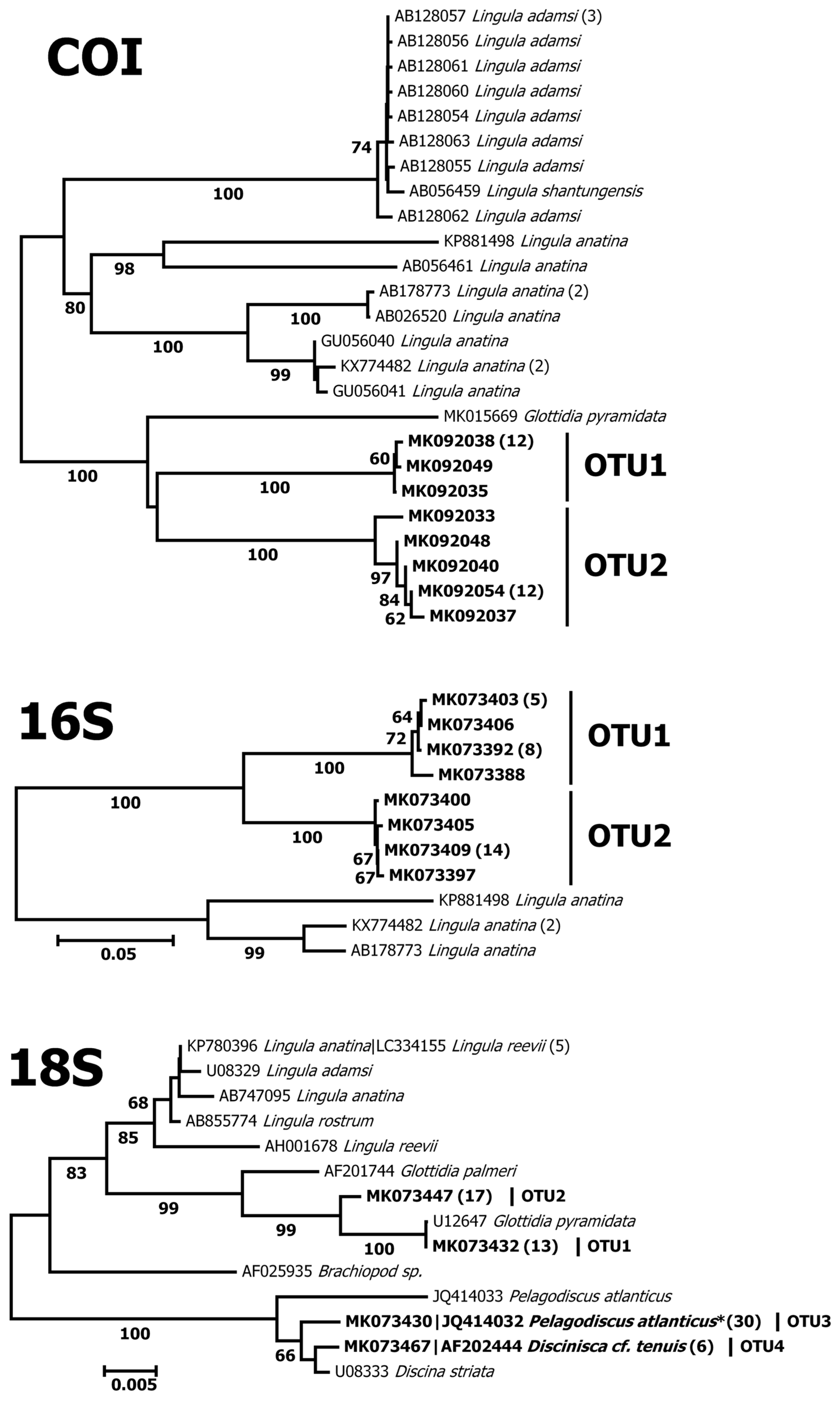
All extant lingulids from the Americas are currently classified in the genus Glottidia [16,44]. Therefore, the lingulid larvae we collected are almost certainly Glottidia species. Larval morphology as described below was similar for specimens collected from both oceans and very similar to those described in the literature [8]. These larvae can be distinguished from discinid larvae by their straight posterior shell margin, larger size, and the absence of long setae projecting outwards from the shell. In our samples they ranged from early stages with a D-shape and a long straight posterior shell margin to stages where this early shell was still evident as the protegulum (Figure 2A and Figure 3A), to larger oval-shaped larvae with a well-developed pedicle coiled inside the posterior region of the shell (Figures 2B,C and 3C,D). These larvae were semitransparent overall, but with an intense ring of yellowish pigment along the margin of the otherwise transparent mantle tissue.
The Glottidia from Bocas del Toro (Figure 2) ranged from 300 microns to approximately 1.0 mm in length. At 300 microns, the band of yellow highlighting the mantle margin was clearly visible but the pedicle had not yet begun to develop (Figure 2A). At 500–600 microns, the larvae had 6–8 pairs of cirri. At 800 microns, an array of shell setae was visible around the margin of the oval shell, and at 900 microns there were ten pairs of cirri. The pedicle was well-developed in the 1.0 mm long larvae (Figures 2B and C), which also had sparse brownish-orange pigment around the gut and on the tentacle (noted on some dates but not others), and at two distinct spots at the corners of the mouth (Figure 2B). Larvae were reluctant to extend their lophophores under the microscope, which limited our ability to count the number of cirri in most of the largest individuals we studied.
Glottidia larvae from the Pacific (Figure 3) were generally similar to those from Bocas del Toro. In addition to yellow pigment along the mantle margin, the cirri were commonly tipped with orange pigment (Figure 3A) and in large larvae, there were light brown spots at each side of the mouth (apparently visible in only one orientation). When we were able to count the cirri, larvae measuring 1.0 mm typically had up to 14 pairs. The largest larvae we captured were larger than those from the Caribbean, with some measuring 1.2 mm in length with a large pedicle (Figure 3D) and some possessing large posterior setae in addition to definitive shell setae around the aperture.

3.3. DNA Barcoding

Of the 73 larvae, 30 (14 from the Bahia Almirante and 16 from the Bay of Panama; Table 2) were sequenced successfully for COI. The success rate for sequencing Glottidia larvae was high (94% for COI and 100% for 16S), while discinids completely failed to amplify for these markers. For Glottidia, the analyses of COI and 16S sequences both produced 2 distinct OTUs (OTU1 and OTU2; Figure 3), with one OTU including larvae only from one ocean (Figure 4). These OTUs differed from each other by more than 20% Jukes Cantor distance in COI and more than 10% in 16S. They also differed from Glottidia pyramidata from Florida by a similar amount in COI (Figure 4).
The 18S marker was sequenced with 84% overall success and produced four OTUs; two for each type of larva (discinid and lingulid). The two 18S OTUs for Glottidia coincided with the OTUs recovered with COI and 16S. Both OTUs of Glottidia larvae clustered most closely with the other available Glottidia sequences in GenBank. 18S sequences from OTU1 from Bocas del Toro differed at one site from the available sequence of Glottidia pyramidata from Florida, while the sequences from OTU2 differed from these at several sites (Figure 4). The discinid 18S sequences also produced two OTUs (OTU3 and OTU4). OTU4 contained four individuals from the Pacific and was identical to a previously published sequence of Discinisca cf. tenuis from Namibia [33,38]. Only two of these four larvae were photographed successfully (Figure 1C,D), and no definitive morphological differences could be discerned to distinguish them from larvae in OTU3. Discinid larvae in OTU3 contained a mix of individuals from the Pacific (10 individuals) and the Caribbean (18 individuals), and were identical to two GenBank sequences: One from Discinisca cf. tenuis from the Bay of Panama [38] and the other from Pelagodiscus atlanticus from the Tropical East Pacific [39].

4. Discussion

It is common for DNA barcoding studies of larvae to detect more or different species than have been detected in surveys of adult diversity (e.g., [18,44,45,46]). This has been the case with surveys of phoronid and nemertean larvae in Panama using the same methods as described here [18,47]. However, our study of brachiopod larvae appears to be an exception. Documented ranges of adult brachiopods suggest that we may expect to find a minimum of 5 species on the Pacific coast and 2 species on the Caribbean coast. These estimates are based on incidental observations in published literature, and not from focused efforts to collect or document actual brachiopod occurrences. Therefore, the expected diversity may under-estimate the actual diversity of the region. We recovered fewer larval OTUs than this.
As is common with studies of marine invertebrates, the paucity of identified sequences in public databases such as GenBank limits our ability to identify larvae with DNA barcoding data. Our results suggest that mitochondrial markers could be useful in systematic and population genetic surveys of Glottidia, as they have been for Lingula in Asia [29,35], and that lingulids are likely to be recovered in meta-barcoding studies using 16S and COI markers. With the data at hand, we can draw only one conclusion about the identity of the lingulid larvae we collected. Glottidia pyramidata has been reported in the eastern Caribbean [14] and the GenBank COI sequence from the Gulf coast of Florida clearly shows our Caribbean OTU to be related to, but distinct from, this species, while the 18S provides insufficient resolution to distinguish them (Figure 4). This indicates there are at least two species of Glottidia in the Greater Caribbean region. A number of other invertebrates show cryptic diversity with genetic or phylogenetic breaks between the eastern and western, or northern and southern Caribbean [48,49,50]. Sampling designed to test for such a genetic break in Glottidia from the Caribbean and Gulf of Mexico is necessary to clarify the ranges of these two species, and comparisons of adult morphology will be necessary to clarify the taxonomy. Larval morphology is unlikely to contribute to this taxonomic challenge, as we did not find any obvious diagnostic differences between the larvae of the two OTUs. Maximum size did appear to differ, as did the number of cirri at large shell sizes, however, the relationship between cirri and shell size is thought to vary within species [40], therefore use of this feature should only be implemented after statistical verification that these features differ significantly between species.
Discinids present more of a challenge for genetic studies as primers that amplify mitochondrial markers in these animals have not been developed. Our 18S sequences could provide some indication of the generic identity of the OTUs; however, the slow rate of evolution in this gene combined with poor taxon coverage and taxonomic uncertainty in GenBank make it challenging to come to a clear conclusion. Our results revealed that the rare OTU4 contains larvae exclusively found in the Pacific of Panama, and are identical matches to GenBank sequences of adult Discinisca. cf. tenuis collected in Namibia. The common OTU3 includes larvae collected from the Pacific and the Caribbean, and are identical matches to GenBank sequences from D. cf. tenuis adults collected in the Bay of Panama [38] and to Pelagodiscus atlanticus from the Tropical Eastern Pacific [39]. Presumably, one of these OTUs is the species that has been previously identified as Discradisca strigata [15], which is abundant in the rocky intertidal of the Bay of Panama.
Our 18S results could be explained by a number of scenarios. If the samples from OTU3 belong to closely related geminate species or at least to the same genus, the slow rate of evolution in 18S could mean that these data fail to distinguish between the species. This could explain why it appears that OTU3 occurred in both the Pacific and Caribbean samples—the occurrence of multiple species in OTU3 is masked by insufficient sequence divergence. The only congeneric adults that are reported to occur on both coasts of Panama belong to Discradisca (D. strigata and D. cumingi in the Pacific and D. antillarum in the Caribbean). In addition, Discradisca strigata is extremely abundant in the intertidal near our collecting sites making it reasonable to conclude that OTU3 may encompass Discradisca species. Unfortunately, the sequences for OTU3 are identical to GenBank sequences for both Pelagodiscus and Discinisca, and there are no 18S sequences for Discradisca in GenBank. If OTU3 is Discradisca, OTU4 would likely be from the deep-water Pelagodiscus atlanticus, the only other known species in the Pacific of Panama (although it matches sequence(s) labelled as Discinisca in GenBank). Alternately, the common larval OTU3 could be Pelagodiscus atlanticus, which is also reported to occur in the Caribbean, and the uncommon OTU4 could be some combination of Discradisca species. This is possible as larval abundance does not always closely track adult abundance.
Unfortunately, larval morphology of these species cannot be of additional help, as definitive larvae of Pelagodiscus have not been described in detail. In general larval descriptions are based on wild-caught larvae which are inferred to belong to the local species with the most abundant adults, resulting in tentative identifications. Nevertheless, published descriptions suggest that both the placement of and order of appearance of setae may be useful in distinguishing larval types [41]. Setal patterns seemed to vary within our OTUs and the lack of resolution from the DNA sequence data and small sample size of larvae in OTU4 leaves the taxonomic utility of this feature open for further study.
In conclusion, using DNA barcoding of planktonic larvae we documented two species of Glottidia in Panama. Identification of these species was not possible as the Caribbean specimens did not match Glottidia pyramidata, the only species previously reported for the Caribbean, and no published sequences are available for G. audebarti or G. albida, the two species reported as adults from the Pacific. We also documented at least two (probably three) species of discinids, but low resolution of 18S sequence data and taxonomic confusion in the few published sequences prevents identification of these larvae.

Author Contributions

Conceptualization, R.C. and M.J.B.; methodology, R.C., M.J.B., A.C.D., D.E.V.-P. and K.S.M.III; validation, R.C., A.C.D. and D.E.V.-P.; formal analysis, D.E.V.-P.; investigation, R.C., M.J.B., A.C.D., D.E.V.-P. and K.S.M.III; resources, R.C. and A.C.D.; data curation, A.C.D. and D.E.V.-P.; writing—original draft preparation, R.C.; writing—review and editing, R.C., D.E.V.-P., A.C.D. and M.J.B.; visualization, M.J.B.; supervision, R.C. and A.C.D.; funding acquisition, R.C.

Funding

This work was supported by the 1923 Fund, Paul Peck, and the Smithsonian Institution.

Acknowledgments

This work was performed with permission from the Panamanian Ministry of the Environment (MiAmbiente). We thank the student participants of the Larval Invertebrate Diversity, Form and Function short-course at the Smithsonian Tropical Research Institute’s Bocas del Toro Research Station in 2013, especially Karen Kit-Yu Chan and Leyre Villotta Nieva for help collecting some of the larvae, and Kevin Kocot for providing the COI sequence of G. pyramidata. All molecular laboratory work was conducted in and with the support of the Laboratories of Analytical Biology facilities of the National Museum of Natural History, Smithsonian Institution. This publication is Smithsonian Marine Station contribution No. 1101.

Conflicts of Interest

The authors declare no conflicts of interest.

References

  1. Pennington, J.T.; Stricker, S.A. Phylum Brachiopoda. In Atlas of Marine Invertebrate Larval Forms; Young, C.M., Sewell, M.A., Rice, M.E., Eds.; Academic Press: San Diego, CA, USA, 2002; pp. 441–461. [Google Scholar]
  2. Yatsu, N. On the development of Lingula anatina. J. Coll. Sci. Imp. Univ. Tokyo 1902, 17, 1–112. [Google Scholar]
  3. Long, J.A.; Stricker, S.A. Brachiopoda. In Reproduction of Marine Invertebrates, Volume VI, Echinoderms and Lophophorates; Giese, A.C., Pearse, J.S., Pearse, V., Eds.; The Boxwood Press: Pacific Grove, CA, USA, 1991; pp. 47–84. [Google Scholar]
  4. Lüter, C. Brachiopod larval setae—A key to the phylum’s ancestral life cycle? In Brachiopods; CRC Press: Boca Raton, FL, USA, 2003; pp. 60–69. [Google Scholar]
  5. Strathmann, R.R. Progressive vacating of adaptive types during the Phanerozoic. Evolution 1978, 32, 907–914. [Google Scholar] [CrossRef] [PubMed]
  6. Valentine, J.W.; Jablonski, D. Larval adaptations and patterns of brachiopod diversity in space and time. Evolution 1983, 37, 1052–1061. [Google Scholar] [CrossRef] [PubMed]
  7. Freeman, G.; Lundelius, J.W. Changes in the timing of mantle formation and larval life history traits in linguliform and craniiform brachiopods. Lethaia 1999, 32, 197–216. [Google Scholar] [CrossRef]
  8. Paine, R.T. Ecology of the brachiopod Glottidia pyramidata. Ecol. Monogr. 1963, 33, 187–213. [Google Scholar] [CrossRef]
  9. Ashworth, J.H. On the larvae of Lingula and Pelagodiscus (Discinisca). Trans. R. Soc. Edinb. 1915, 51, 45–69. [Google Scholar] [CrossRef]
  10. Chuang, S.H. The larvae of a discinid (Inarticulata, Brachiopoda). Biol. Bull. 1968, 135, 263–272. [Google Scholar] [CrossRef]
  11. Chuang, S.H. Larval development in Discinisca (inarticulate brachiopod). Am. Zool. 1977, 17, 39–53. [Google Scholar] [CrossRef]
  12. Emig, C.C. Brachiopods. In Marine Biodiversity of Costa Rica, Central America; Whertmann, I.S., Cortés, J., Eds.; Springer: Berlin, Germany, 2008; pp. 417–420. ISBN 978-1-4020-8278-8. [Google Scholar]
  13. Cooper, G.A. Brachiopods from the Caribbean Sea and adjacent waters. Stud. Trop. Oceanogr. Miami 1977, 14, 1–211. [Google Scholar]
  14. Emig, C.C. Taxonomie du genre Glottidia (Brachiopodes Inarticulés). Bull. Mus. Natl. Hist. Nat. Paris 1983, 4, 469–489. [Google Scholar]
  15. Labarbera, M. Mechanisms of spatial competition of Discinisca strigata (Inarticulata: Brachiopoda) in the intertidal of Panama. Biol. Bull. 1985, 168, 91–105. [Google Scholar] [CrossRef]
  16. Emig, C.C.; Bitner, M.A.; Alvarez, F. Brachiopoda Database. Available online: http://paleopolis.rediris.es/brachiopoda_database/index.html (accessed on 30 December 2018).
  17. Santagata, S.; Tunnell, J.W., Jr. Brachiopoda of the Gulf of Mexico. In Gulf of Mexico Origin, Waters, and Biota: Biodiversity, 1st ed.; Texas A&M University Press: College Station, TX, USA, 2009; Volume 1, pp. 1137–1141. ISBN 1603440941. [Google Scholar]
  18. Collin, R.; Venera-Pontón, D.E.; Driskell, A.C.; Chan, K.-Y.K.; MacDonald, K.S.; Boyle, M.J. Understanding Neotropical diversity of phoronids with DNA barcoding of planktonic larvae. Invertebr. Biol. 2018. accepted. [Google Scholar]
  19. Geller, J.; Meyer, C.; Parker, M.; Hawk, H. Redesign of PCR primers for mitochondrial cytochrome c oxidase subunit I for marine invertebrates and application in all-taxa biotic surveys. Mol. Ecol. Resour. 2013, 13, 851–861. [Google Scholar] [CrossRef]
  20. Meyer, C.P. Molecular systematics of cowries (Gastropoda: Cypraeidae) and diversification patterns in the tropics. Biol. J. Linn. Soc. Lond. 2003, 79, 401–459. [Google Scholar] [CrossRef] [Green Version]
  21. Palumbi, S.; Martin, A.; Romano, S.; McMillan, W.O.; Stice, L.; Grabowski, G. The Simple Fool’s Guide to PCR, Version 2; Department of Zoology and Kewalo Marine Laboratory, University of Hawaii: Honolulu, HI, USA, 1991. [Google Scholar]
  22. Medlin, L.; Elwood, H.J.; Stickel, S.; Sogin, M.L. The characterization of enzymatically amplified eukaryotic 16S-like rRNA-coding regions. Gene 1988, 71, 491–499. [Google Scholar] [CrossRef] [Green Version]
  23. Gascuel, O. BIONJ: An improved version of the NJ algorithm based on a simple model of sequence data. Mol. Biol. Evol. 1997, 14, 685–695. [Google Scholar] [CrossRef] [PubMed]
  24. Ratnasingham, S.; Hebert, P.D.N. BOLD: The barcode of life data system (http://www.barcodinglife.org). Mol. Ecol. Notes 2007, 7, 355–364. [Google Scholar] [CrossRef]
  25. Lassmann, T.; Sonnhammer, E.L.L. Kalign—An accurate and fast multiple sequence alignment algorithm. BMC Bioinform. 2005, 6, 298. [Google Scholar] [CrossRef]
  26. Puillandre, N.; Lambert, A.; Brouillet, S.; Achaz, G. ABGD, Automatic Barcode Gap Discovery for primary species delimitation. Mol. Ecol. 2012, 21, 1864–1877. [Google Scholar] [CrossRef]
  27. Saito, M.; Kojima, S.; Endo, K. Mitochondrial COI sequences of brachiopods: Genetic code shared with protostomes and limits of utility for phylogenetic reconstruction. Mol. Phylogenet. Evol. 2000, 15, 331–344. [Google Scholar] [CrossRef]
  28. Wu, L.J.; Sheng, G.L.; Lai, X.L.; Hou, X.; Yuan, J. Geographical pattern analysis of the brachiopod Lingula anatina based on mitochondrial COI gene sequences. Geol. Sci. Technol. Inform. 2010, 29, 17–22. [Google Scholar]
  29. Endo, K.; Ozawa, T.; Kojima, S. Nuclear and mitochondrial gene sequences reveal unexpected genetic heterogeneity among northern Pacific populations of the brachiopod Lingula anatina. Mar. Biol. 2001, 139, 105–112. [Google Scholar] [CrossRef]
  30. Endo, K.; Noguchi, Y.; Ueshima, R.; Jacobs, H.T. Novel repetitive structures, deviant protein-encoding sequences and unidentified ORFs in the mitochondrial genome of the brachiopod Lingula anatina. J. Mol. Evol. 2005, 6, 36–53. [Google Scholar] [CrossRef] [PubMed]
  31. Luo, Y.J.; Satoh, N.; Endo, K. Mitochondrial gene order variation in the brachiopod Lingula anatina and its implications for mitochondrial evolution in lophotrochozoans. Mar. Genomics 2015, 24, 31–40. [Google Scholar] [CrossRef] [PubMed]
  32. Mackey, L.Y.; Winnepenninckx, B.; De Watcher, R.; Backeljau, T.; Emschermann, P.; Garey, J.R. 18S rRNA suggests that entoprocta are protostomes, unrelated to ectoprocta. J. Mol. Evol. 1996, 42, 552–559. [Google Scholar] [CrossRef]
  33. Cohen, B.L.; Gawthrop, A.; Cavalier–Smith, T. Molecular phylogeny of brachiopods and phoronids based on nuclear–encoded small subunit ribosomal RNA gene sequences. Philos. Trans. R. Soc. Lond. B Biol. Sci. 1998, 353, 2039–2061. [Google Scholar] [CrossRef] [Green Version]
  34. Field, K.G.; Olsen, G.J.; Lane, D.J.; Giovannoni, S.J.; Ghiselin, M.T.; Raff, E.C.; Pace, N.R.; Raff, R.A. Molecular phylogeny of the animal kingdom. Science 1988, 239, 748–753. [Google Scholar] [CrossRef]
  35. Sato, S.; Endo, K.; Yamashita, H. Morphological and genetic comparisons between Lingula adamsi Dall, 1873 from South Korea and Japan. Jpn. J. Benthol. 2004, 59, 13–18. [Google Scholar] [CrossRef]
  36. Cohen, B.L. Monophyly of brachiopods and phoronids: Reconciliation of molecular evidence with Linnaean classification (the subphylum Phoroniformea nov.). Proc. R. Soc. Lond. B 2000, 267, 225–231. [Google Scholar] [CrossRef]
  37. Halanych, K.M.; Bacheller, J.D.; Aguinaldo, A.M.; Liva, S.M.; Hillis, D.M.; Lake, J.A. Evidence from 18S ribosomal DNA that the lophophorates are protostome animals. Science 1995, 267, 1641–1643. [Google Scholar] [CrossRef]
  38. Cohen, B.L.; Weydmann, A. Molecular evidence that phoronids are a subtaxon of brachiopods (Brachiopoda: Phoronata) and that genetic divergence of metazoan phyla began long before the early Cambrian. Org. Divers. Evol. 2005, 5, 253–273. [Google Scholar] [CrossRef] [Green Version]
  39. Cohen, B.L. Rerooting the rRNA tree reveals phoronids to be ’brachiopods without shells’; dangers of wide taxon samples in metazoan phylogenetics (Phoronida; Brachiopoda). Zool. J. Linn. Soc. 2013, 167, 82–92. [Google Scholar] [CrossRef]
  40. Hammond, L.S. Breeding season, larval development and dispersal of Lingula anatina (Brachiopoda, Inarticulata) from Townsville, Australia. J. Zool. 1982, 198, 183–196. [Google Scholar] [CrossRef]
  41. Hammond, L.S. The larvae of a discinid (Brachipoda: Inarticulata) from inshore waters near Townsville, Australia, with revised identifications of previous records. J. Natl. Hist. 1980, 14, 647–661. [Google Scholar] [CrossRef]
  42. Chuang, S.H. The inarticulate brachiopod larvae of the International Indian Ocean Expedition. J. Mar. Biol. Ass. India 1973, 15, 538–544. [Google Scholar]
  43. Fagetti, E.G. Nota sobre larvas de Brachiopoda Discinidae de la costa Chilena. Montemar 1964, 4, 195–200. [Google Scholar]
  44. Williams, A.; Cohen, B.L.; Cusack, M.; Long, S.L. Provenance of Atlantic lingulid brachiopods. Palaeontology 2000, 43, 999–1018. [Google Scholar] [CrossRef]
  45. Barber, P.; Boyce, S.L. Estimating diversity of Indo-Pacific coral reef stomatopods through DNA barcoding of stomatopod larvae. Proc. R. Soc. Lond. B 2006, 273, 2053–2061. [Google Scholar] [CrossRef] [Green Version]
  46. Mahon, A.R.; Thornhill, D.J.; Norenburg, J.L.; Halanych, K.M. DNA uncovers Antarctic nemertean biodiversity and exposes a decades-old cold case of asymmetric inventory. Polar Biol. 2010, 33, 193–202. [Google Scholar] [CrossRef]
  47. Maslakova, S.A.; Ellison, C.; Moss, N.D.; Dillenburg, B.; Howland, C.; Robbins, K.; Schwartz, M.L.; Partridge, M.; Zattara, E.; Collin, R.; et al. DNA-barcoding of benthic and planktonic life history stages reveals a large amount of undescribed, cryptic, and unsampled diversity of nemerteans in the Caribbean Sea. In preparation.
  48. Lee, T.; Ó Foighil, D. Placing the Floridian marine genetic disjunction into a regional evolutionary context using the scorched mussel, Brachidontes exustus, species complex. Evolution 2007, 59, 2139–2158. [Google Scholar] [CrossRef]
  49. Cowen, R.K.; Paris, C.B.; Srinivasan, A. Scaling of connectivity in marine populations. Science 2006, 311, 522–527. [Google Scholar] [CrossRef] [PubMed]
  50. Díaz-Ferguson, E.; Haney, R.; Wares, J.; Silliman, B. Population genetics of a trochid gastropod broadens picture of Caribbean Sea connectivity. PLoS ONE 2010, 5, e12675. [Google Scholar] [CrossRef]
Figure 1. Larval discinids from the Pacific and Caribbean coasts of Panama. (A) Larva in operational taxonomical unit (OTU)3 from Bocas del Toro; (B) Larva in OTU3 from the Bay of Panama; (C) Larva in OTU4 from the Bay of Panama; (D) Darkfield micrograph of larva in OTU4 from the Bay of Panama; (E) Young larva from Bay of Panama with long iridescent larval setae. No sequence data was generated for this specimen. Abbreviations: ci, cirri; cs, curved seta; ds, dorsal valve; es, esophagus; ls, larval setae; mi, minor setae; ms, major setae; mt, median tentacle; pb, pedicle bud; se, seta; st, stomach; vv, ventral valve. Scale bars = 100 µm.
Diversity 11 00002 g001
Figure 2. Larval Glottidia from Bocas del Toro. (A) Larval stage with eight pairs of cirri a defined protegulum, and no pedicle; (B) Larva with nine pairs of cirri, setae around the margin of the shell, and a well-developed pedicle; (C) Larva with an elongate shell, setae extending from the anterior shell margin and a large pedicle. Abbreviations: aa, anterior adductor; ci, cirri; ds, definitive setae; es, esophagus; in, intestine; me, metacoel; mo, mouth; mt, median tentacle; pa, posterior adductor; p, pedicle; pr, protegulum; pt, pedicle tip; sh, shell; st, stomach. Scale bars = 100 µm.
Diversity 11 00002 g002
Figure 3. Larval Glottidia from the Bay of Panama. (A) Early stage larva with six pairs of cirri, a defined protegulum, and no pedicle; (B) Larva with ten pairs of cirri and initiation of the pedicle bud; (C) Larva with twelve pairs of cirri, elongate shell, definitive setae around the shell margin and a pedicle; (D) Late larva with definitive setae and a well-developed pedicle. Abbreviations: dg, digestive glands; ds, definitive setae; es, esophagus; ma, mantle; mo, mouth; mt, median tentacle; pe, pedicle; pr, protegulum; pt, pedicle tip; te, tentacle. Scale bars = 100 µm.
Diversity 11 00002 g003
Figure 4. Neighbor-Joining trees for cytochrome c oxidase subunit I (COI), 16S and 18S sequences from brachiopod larvae in this study with every brachiopod sequence available in GenBank. The tree shows only unique haplotypes, followed by the number of individuals with this haplotype in parentheses if ≥2. The Jukes-Cantor distance between haplotypes is proportional to the length of the branches separating them, as shown in the scale bars at the bottom left. Bootstrap support values over 60% are shown below the corresponding branches. OTUs comprised of our sequences are indicated in bold. * This OTU had 100% sequence identity with one sequence from material identified as Pelagodiscus atlanticus and with one sequence from material identified as Discinisca cf. tenuis.
Diversity 11 00002 g004
Table 1. Summary of primers and DNA fragments used in this study.
GenePrimersFragment Length (bp)References
COI Barcode fragmentjgLCO1490: TITCIACIAAYCAYAARGAYATTGG
jgHCO2198: TAIACYTCIGGRTGICCRAARAAYCA
or
dgLCO1490: GGTCAACAAATCATAAAGAYATYGG
dgHCO2198: TAAACTTCAGGGTGACCAAARAAYCA
654[19,20]
16S16Sar CGCCTGTTTATCAAAAACAT
16Sbr CCGGTCTGAACTCAGATCACGT
~525[21]
18SEukF (modified Primer A): AACCTGGTTGATCCTGCCAGT
SR7: GTTCAACTACGAGCTTTTTAA
~600[22]
Table 2. Summary of OTUs and GenBank sequences used in this study.
OTU/Species 1Genbank # COIGenbank # 16SGenbank # 18SLocationCollection Dates
New Data
Larval OTU1—GlottidiaMK092035, 36, 38–39, 41, 47,49–51, 55–58, 61MK073388, 89, 91–93, 95, MK073401, 03–04, 06, 10–13, 16MK073422, 26,32–33, 38, 49, 53, 55, 58, 68, 69, 70, 73Bahía AlmiranteJul 2013, August & November 2015, February-March & June 2016
Larval OTU2—GlottidiaMK092033, 34, 37,40 42–46, 48, 52–54, 59–60, 62 MK073386, 87, 90, 94, 96–99, MK073400, 02, 05, 07–09, 14–15, 17MK073419, 20, 28, 34, 39, 40, 42–43, 47–48, 52, 62–63, 65, 74–75, 79Bay of PanamaApril-June & November 2014
Larval OTU3—Discinid----------------MK073423, 24–25, 27, 29,30–31, 35–37, 41, 44–46, 50–51, 54, 56–57, 59, 61, 64, 66, 72, 76–78, 80Bahía Almirante and Bay of PanamaJuly-August 2013, August 2015, November 2014 & 2015, March & June 2016
Larval OTU4—Discinid------------------MK073421, 60, 67, 71Bay of PanamaAugust 2013, March 2014
Published Data Reference
Lingulids
Lingula anatinaAB026520 Japan[27]
Lingula anatinaGU056040-41 China[28]
Lingula anatinaAB056460 Japan[29]
Lingula anatinaAB056461, AB056462 Hong Kong[29]
Lingula anatinaAB178773AB178733 Japan[30]
Lingula anatinaKX774482, NC_036679KX774482, NC_036679 South KoreaKaragozlu & Kim, Unpublished 2
Lingula anatinaKP881498KP881498KP780396Japan[31]
Lingula anatina X81631Hong Kong[32]
Lingula anatina AB747095Japan[31]
Lingula anatina U08331New Caledonia[33]
Lingula rostrum AB855774Japan[31]
Lingula reevii AB747096Japan[31]
Lingula reevii AH001678unknown[34]
Lingula reevii LC334155 JapanKurita et al. Unpublished
Lingula shantungensisAB056459 Japan[29]
Lingula adamsiAB128054-63 South Korea[35]
Lingula adamsi U08329New Caledonia[33]
Glottidia palmeri AF201744Baja California[36]
Glottidia pyramidata U12647Florida, Gulf Coast (K Halanych, pers.com)[37]
Glottidia pyramidataMK015669 Florida, Gulf Coast K. Kocot, unpublished
Discinids
Discinisca cf. tenuis AF202444Namibia[36]
Discinisca cf. tenuis U08327Namibia[33]
Discinisca cf. tenuis AY842020Panama Bay, Panama[38]
Discina striata U08333Gambia[33]
Pelagodiscus atlanticus JQ414032Antarctica [39]
Pelagodiscus atlanticus JQ414033Uncertain 3[39]
Brachiopod sp. AF025935Guam, Pacific[33]
1 Taxonomy follows GenBank records and reports the species name listed in the organism field of each record. 2 Sequences are listed as unpublished if they are listed as such in GenBank and our literature review failed to find a publication that reports those sequences. 3 The published paper [39] and the GenBank record report the locality as North of Galapagos, but the coordinates they provide are offshore of Costa Rica in the Tropical Eastern Pacific.

