Review

Cell Membrane-Coated Nanoparticles for Precision Medicine: A Comprehensive Review of Coating Techniques for Tissue-Specific Therapeutics

by
Andrés Fernández-Borbolla
1,2,
Lorena García-Hevia
1,2 and
Mónica L. Fanarraga
1,2,*
1
The Nanomedicine Group, Institute Valdecilla-IDIVAL, 39011 Santander, Spain
2
Molecular Biology Department, Faculty of Medicine, Universidad de Cantabria, 39011 Santander, Spain
*
Author to whom correspondence should be addressed.
Int. J. Mol. Sci. 2024, 25(4), 2071; https://doi.org/10.3390/ijms25042071
Submission received: 6 December 2023 / Revised: 1 February 2024 / Accepted: 6 February 2024 / Published: 8 February 2024
(This article belongs to the Special Issue Nano & Micro Materials in Healthcare 2.0)

Abstract

:
Nanoencapsulation has become a recent advancement in drug delivery, enhancing stability, bioavailability, and enabling controlled, targeted substance delivery to specific cells or tissues. However, traditional nanoparticle delivery faces challenges such as a short circulation time and immune recognition. To tackle these issues, cell membrane-coated nanoparticles have been suggested as a practical alternative. The production process involves three main stages: cell lysis and membrane fragmentation, membrane isolation, and nanoparticle coating. Cell membranes are typically fragmented using hypotonic lysis with homogenization or sonication. Subsequent membrane fragments are isolated through multiple centrifugation steps. Coating nanoparticles can be achieved through extrusion, sonication, or a combination of both methods. Notably, this analysis reveals the absence of a universally applicable method for nanoparticle coating, as the three stages differ significantly in their procedures. This review explores current developments and approaches to cell membrane-coated nanoparticles, highlighting their potential as an effective alternative for targeted drug delivery and various therapeutic applications.

1. Introduction

Nanoencapsulation for in vivo administration provides numerous benefits, such as enhancing effectiveness and safety by protecting the substances from degradation or elimination [1,2]. This technique contributes to increased absorption and improved bioavailability, optimizing distribution, and extending circulation time while simultaneously reducing toxicity [2,3]. Some nanomaterials offer advantages such as enhanced solubility and loading capacity, improved delivery efficiency, and protection from degradation due to the stability provided by the nanocarriers [1,2,3]. However, nanoparticle delivery has many limitations. Nanocarriers are very prone to interact with biomolecules in the bloodstream, creating the so-called “biocorona” [4], which results in recognition by the immune system [5]. Upon arrival to the target cells, many nanocarriers are trapped in endocytic vesicles and end up being degraded by lysosomes, diminishing the drug delivery efficiency [6].
Recent studies suggest that nanocarriers show an average efficiency of delivering to the desired target of less than 1% [7,8], leaving space for a significant improvement in targeted delivery. As a result, nanoparticles coated with cell membranes have been proposed as a way to address these problems, as they show a combination of the advantages present in natural nanomaterials such as cell membrane-derived nanomaterials, and artificial nanocarriers, such as the aforementioned polymeric or inorganic nanocarriers [9,10,11,12].
Cell membrane-coated nanoparticles are biomimetic nanoparticles that are constituted by a cell membrane cover and synthetic nanoparticles [5]. They offer several advantages over bare nanomaterials, such as increased biocompatibility, due to the similarity of biological membranes to cellular materials, reducing the risk of immune system rejection [13]. The presence of biological membranes enhances biodistribution by guiding nano-vectored materials to target cells, utilizing membrane receptors recognizable by the target cells. This aspect represents a significant area of study, applicable to immune system cells [13], central nervous system [14], as well as a large number of cancer cells (Table 1). Additionally, coated nanocarriers demonstrate improved drug release control and efficiency, as the biological membranes can degrade or fuse with target cells, releasing the drug at the desired location [9,15]. Specifically, the biological camouflage provided by these membranes protects nanoparticles from the body’s defense systems, extending their lifespan and reducing the risk of premature elimination [13,15,16,17,18,19,20]. The ability to target particles to specific cells, facilitated by the presence of receptors on biological membranes, is a key advantage that positions nanomaterials coated with biological membranes as a promising option for targeted delivery.
As this pioneering methodology is still in its nascent stages, our study aims to comprehensively review the recent advancements in this technology. Specifically, we delve into various studies conducted to date, focusing on elucidating the techniques employed for obtaining cell membrane fragments. We provide detailed insights into the processes involved in isolating these membranes and coating nanoparticles with them. The ultimate goal of this review is to examine technology which has been developed recently to generate cell membrane-coated nanoparticles, showcasing their potential for achieving tissue-specific targeting. This review aims to clearly outline the significance of the study within the broader context of this emerging field.

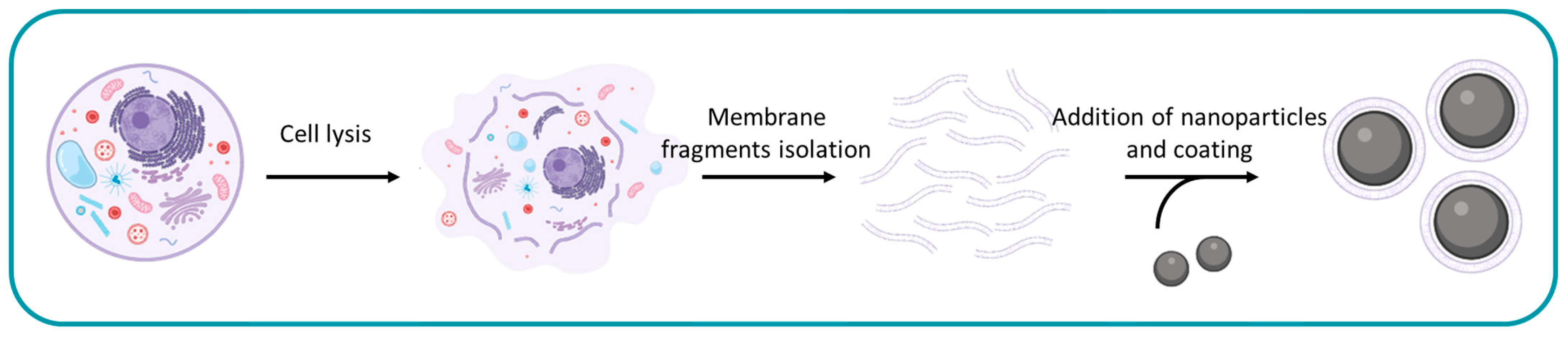
2. General Procedure

To obtain cell membrane-coated nanoparticles three pivotal and indispensable steps must be undertaken. These steps encompass the cell lysis and fragmentation of the membranes, the isolation of these membrane fragments, and the coating of the selected nanocarriers (Figure 1).
The choice of materials for each of these crucial steps depends on the specific tissue being targeted and the nature of the treatment under investigation. The selection is tailored to optimize compatibility with the intended biological environment and enhance the efficacy of the experimental approach.

3. Membrane Donor Cells

The selection of a specific cell type is contingent upon the target tissue or application. Typically, cancer cells are employed to specifically target the corresponding cancerous tissue, while white or red blood cells may be used for applications with less specific targets. Most of these cell types were employed to facilitate the precise targeting of nanoparticles to specific tissues. However, some of these cells served a dual purpose by inducing immune stimulation against cancer.
M any different cell types have been used for nanoparticle membrane coating (Table 1). Notably, a range of cancer lines has been used, including cervical and ovarian cancers [21,22,23,24], multiple myeloma [25], melanoma [12,26,27,28,29,30,31,32], leukemia [23,33,34,35,36,37,38,39,40,41,42,43,44], breast cancer [6,37,40,45,46,47,48,49,50,51,52,53,54,55,56], neuroblastoma [79], colon carcinoma [23,57], head and neck squamous cell carcinoma [58,59,60,61], lung cancer [54,62], glioma [63,64], glioblastoma [65,66], prostate cancer [67], and liver cancer [68]. Furthermore, beyond cancer cells, a multitude of non-cancer cells has also been utilized, such as leukocytes [92,93], macrophages [92,94,95,96,97,98,99], erythrocytes [19,29,46,48,80,81,82,83,84,85,86,87,88,89,90,91], dendritic cells [100], neutrophils [89,101,102,103,104], mesenchymal stem cells [74,75,76,77,78], platelets [48,86,87,93,105,106,107], fibroblasts [49,69], embryonic kidney cells [70], vaginal endothelial cells [71], neural stem cells [72], microglial cells [66], and keratinocytes [73].
Cervical and ovarian cancer cells were used to favor the cytosolic delivery of cargo inside living cells [21] or for homologous targeting [22]. Multiple myeloma cells were chosen to target their equivalent counterparts, ensuring specificity in cargo delivery [25]. In the case of melanoma cells, their use was geared towards promoting the delivery and internalization of therapeutic or antigenic materials [12], or for photoimmunotherapy [26]. Leukemia cells were employed to deliver cargo into leukemia cells [34] or were genetically modified to express a protein that can specifically target a tissue [36]. Neuroblastoma cells were employed for their capacity to capture neurotoxins effectively [79]. Breast cancer cells were used to target homologous cells and deliver cargo [6]. Similarly, colon carcinoma [57] head and neck squamous cell carcinoma [58], lung cancer [54], glioma [63,64], glioblastoma [65,66], prostate cancer [67], and liver cancer [68] cells were selected for homologous targeting, ensuring precision in cargo delivery to specific tissues.
In the case of non-cancer cells, leukocytes were harnessed for their capacity to target specific tissues effectively [92]. Erythrocytes were used to target cancer tissues, due to their elasticity and capacity to diffuse into the tumor extracellular matrix [81]. Dendritic cells were employed to promote tumor immune effects [100]. Vaginal endothelial cells were used to protect the cells from a toxin [71]. Neural stem cells were used to cross the blood-brain barrier and specific targeting [72]. Neutrophils [101], mesenchymal stem cells [74], fibroblasts [49,69], embryonic kidney cells [70], microglial cells [66], and keratinocytes [73] were also used for specific targeting.
Some investigations opted to combine membranes from different cells so that the coated nanoparticles benefited from the characteristics of both types of source cells. When hybrid membrane-coated nanoparticles were developed by combining two cell types, leukocytes were chosen to mitigate immune recognition [93], platelets were selected for their notable ability to bind to cancer cells [93], and erythrocytes due to their long circulation times [48] and immune-evasion capability [29]. Additionally, breast cancer cells [46,48], were incorporated in hybrid membrane coating to ensure precise targeting of homologous cells.

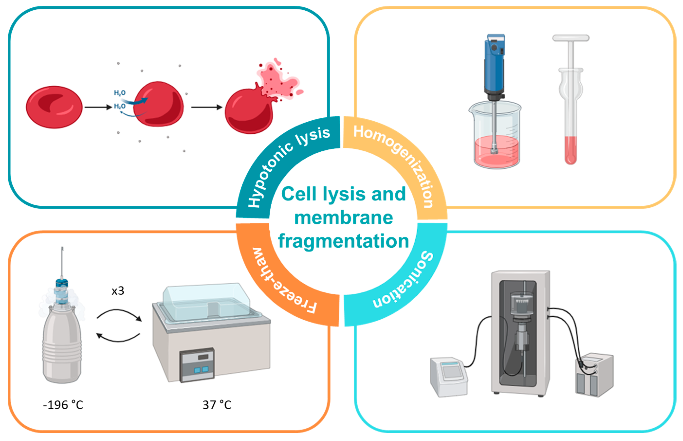
4. Fragmentation of Cell Membranes

The initial crucial step in the preparation of cell membrane-coated nanoparticles involves the obtention of purified cell membrane fragments. Various techniques are employed to produce these membrane fragments, with hypotonic lysis, homogenization, freeze-thaw, and sonication emerging as the most commonly utilized methods (Figure 2). Often, these methods are used together to enhance results, such as combining hypotonic lysis, homogenization, and freeze-thaw for improved outcomes.

4.1. Hypotonic Lysis

Most researchers employed hypotonic lysis in their studies [6,12,19,21,22,23,24,25,26,27,29,30,31,32,33,34,35,36,38,39,41,42,43,44,45,48,49,50,51,54,55,56,57,58,60,61,62,63,64,65,66,67,69,70,71,72,73,74,75,76,77,79,81,82,84,87,88,89,90,91,92,93,95,97,98,100,101,102,103,104]. This lysis method involves resuspending the utilized cells in a hypotonic solution containing low concentrations of salts and protease or phosphatase inhibitors. Several authors used a hypotonic lysis buffer with 20 mM Tris-HCl pH 7.5, 10 mM KCl, 2 mM MgCl2, and 1 EDTA-free mini protease inhibitor tablet per 10 mL of solution [12,23,25,34,44,45,51,54,56,58,67,72,95]. Parodi et al. drew upon the use of the same salts as Qu et al., but adding 25 mM of sucrose and using PMSF and trypsin-chymotrypsin inhibitors [92]. A similar buffer was used by Li et al. (Tris-HCl, 20 mM KCl, 2 mM MgCl2, and EDTA-free-microprotease inhibitor) [62]. Other authors utilized Tris-HCl, sucrose, and D-mannitol in combination with phosphatase and protease inhibitor cocktails [27,30,31,71]. In contrast, a handful of researchers used these components along with EGTA (IB-1 buffer) [38,50,98,101,102], while Nie et al. used this IB-1 buffer with 0.5% (w/v) BSA [6]. Ma et al. opted for the commercial RIPA Lysis Buffer (50 mM Tris-HCl pH 7.6, 150 mM NaCl, 1% NP-40, 0.5% sodium deoxycholate, 0.1% SDS) in addition to a protein inhibitor cocktail [100]. Others used simpler Tris-HCl lysis buffers, such as Bu et al. (50 mM Tris-HCL pH 7.4) [73], Ma et al. and Zou et al. (10 mM Tris and 10 mM MgCl2 EDTA free protease inhibitor) [63,77], or Liu et al. (Tris-HCl pH 7.4, 10 mM MgCl2, 1× PMSF, 0.2 mM EDTA and phosphatase inhibitor cocktail) [69].
Other variations in hypotonic buffers were observed, such as the use of a hypotonic buffer with 0.25X PBS [61,76,81,82,88,90,91] containing a protease inhibitor cocktail [21] or PMSF [46]. PBS was also used in combination with EDTA-2Na [87]. Jiang et al. and Rao et al. used Hepes B buffer (10 mM Hepes, 5 mM MgCl2, 1 mM EDTA, 1 mM DTT, 10 mM KCl, pH 7.6) mixed with protease inhibitor tablets [48,93], and Li et al. used a similar homogenization medium with 20 mM HEPES-NaOH, 1 mM EDTA, and 0.25 M of sucrose with PMSF [22]. As an alternative to EDTA-containing buffers, Wang et al. and Park et al., employed EGTA in combination with a phosphatase and protease inhibitor [35,79]. Jiang et al. opted for a NaHCO3 based buffer (1 mM NaHCO3, 0.2 mM EDTA∙2Na, 1 mM PMSF and 1 × PIC in H2O) [39], while Du et al. used a similar buffer [64]. Li et al. used double distilled water [103]. Some articles did not specify the exact buffer composition but indicated the use of a low-osmotic lysis buffer containing membrane protein extraction reagents and PMSF [26]. Wu et al. subjected the cell mix to only a membrane protein extraction buffer [33] or with the addition of a protease or phosphatase inhibitor such as PMSF [46,57]. Deng et al. and Wang et al. added Membrane Protein Extraction Reagent A containing PMSF [29,32,36,42,60,70]. Others only said that they had performed hypotonic lysis but didn’t describe any component of the buffer [24,41,43,49,65,66,75,89,97,104]. These buffers are shown in Table 2.

4.2. Homogenization

More than half of the articles employing hypotonic lysis treatment incorporated homogenization to optimize the extraction of membrane fragments [12,23,24,25,26,27,30,31,33,34,35,38,41,42,43,45,48,51,56,58,60,64,65,72,79,92,93,95,101,102]. In most of these studies [12,23,24,25,26,33,34,38,41,42,43,45,51,56,58,60,64,65,72,92,93,95,101,102] the common approach involved introducing lysed membrane fragments into a Dounce homogenizer. The fragments then underwent several passes or mechanical disruptions. The number of passes varied across experiments, ranging from 20 to 100. Notably, Kroll et al., Park et al., Jiang et al., and Wang et al. used a different system. They homogenized using a Polytron homogenizer for 15 [27,30,31,35] or 20 passes [79]. Jiang et al. homogenized instead the cells three times with an IKA T10 basic homogenizer [48].

4.3. Freeze-Thaw

While not a widely adopted strategy for this purpose, freeze-thaw has been employed in certain experiments [28,37,40,53,59,78,86,93,94,105,106]. This technique involves subjecting the cell suspension to multiple cycles of freezing and thawing, with the addition of only a phosphatase inhibitor to the suspension. In some cases, it has been utilized in combination with hypotonic lysis, submitting the lysed cells to several cycles of freezing in liquid nitrogen or at -80 °C and subsequent thawing at 37 °C [21,32,57,63,70]. Yao et al. performed a freeze-thaw treatment followed by sonication, without any previous hypotonic lysis [107].

4.4. Sonication

To harvest cell membrane fragments, a sonication treatment can be employed, which may involve the use of a bath sonicator [21,39,54,68,80,86,87,107] or ultrasonication with an ultrasonication device [22,36,44,50,52,69,73,90,99]. Soprano et al. utilized this method following hypotonic lysis and freeze-thaw treatments, placing the cells in a bath sonicator for 5 min [21]. Zhou et al. and Zhang et al. sonicated samples in a bath sonicator for 10 min after a hypotonic lysis treatment [80,87]. Nie et al. and Gan et al. applied repeated sonication steps in an ice bath [6,54]. Li et al. applied sonication after hypotonic lysis, subjecting the cells to 10 cycles of 3 s of ultrasonication at 150 W [22], while others homogenized the cell suspension using an ultrasonic disruptor [36,44,50,69,90]. Ultrasonication of the lysed membranes was used by several authors [52,73,99]. Dehaini et al. sonicated the cell suspension after a freeze-thaw treatment in a bath sonicator at 42 kHz and 100 W [86].

4.5. Other Methods

Another method employed for obtaining cell membrane fragments, either used alone or in combination with other techniques, is extrusion. In the studies by Chen et al. and Liu et al. extrusion is applied in conjunction with hypotonic lysis, occurring after the lysis process and before centrifugation to remove other cell components [95,104].

4.6. Summary

Upon reviewing all of the compiled articles, hypotonic lysis coupled with homogenization stands out as the overwhelmingly predominant method employed for membrane fragmentation in cells designated for coating. This approach has been consistently applied across a diverse range of cell types, encompassing both cancer and normal cells, and is independent of the specific cell type under investigation.
The hypotonic lysis buffer composed of 20 mM Tris-HCl pH 7.5, 10 mM KCl, 2 mM MgCl2, and 1 EDTA-free mini protease inhibitor tablet per 10 mL of solution, emerged as the most prevalent lysis buffer. Remarkably, this buffer was applied across various cell types, including melanoma, myeloma, triple-negative breast cancer, leukemia, and macrophages. Numerous other studies adopted similar lysis buffers based on Tris-HCl, either in combination with other compounds or inhibitors. Nevertheless, buffers incorporating Tris-HCl predominated, demonstrating their widespread usage and satisfactory results. In contrast, homogenization was predominantly carried out using a Dounce homogenizer, underscoring the effectiveness of this device in the membrane fragmentation process. The less commonly employed methods for membrane fragmentation were also applied to various cell types. Freeze-thaw was utilized for the fragmentation of macrophages, melanoma cells, erythrocytes, and platelets, while sonication was applied to cervical cancer, erythrocytes, and macrophages. These findings collectively suggest that there is no singular method universally valid for membrane fragmentation. Instead, there exist several reliable methods for this procedure, irrespective of the cell type chosen for coating. The selection of a specific method appears to be influenced by the availability of required materials in each laboratory. The advantages and disadvantages of each technique are detailed in Table 3.

5. Membrane Fragments Isolation

After the membranes have been fragmented, the next step involves recovering and isolating these fragments for their subsequent use in coating nanoparticles. Typically, the isolation stage includes 1 to 3 centrifugation steps to separate the remaining membrane materials. This process may be preceded or followed by a gradient separation to move other components away from the membrane fragments. Once the membrane fragments are obtained, they can undergo washing and/or lyophilization, or they may be directly resuspended if the nanoparticle coating process is scheduled immediately after isolation.

5.1. Centrifugation

To isolate membrane fragments from other cell components, most of the studies employed 1 to 3 cycles of centrifugation. Typically, a two-step process is followed. In the first centrifugation step, the mix undergoes a lower g force, approximately 3000× g, to precipitate the remaining cell components, and the supernatant was collected for the subsequent step. Some studies performed only this single centrifugation [28,29,37,46,48,74,76,81,82,84,94,106], whereas some others did a single centrifugation at higher g forces, such as 14,000× g [66], 15,000× g [90] or 21,000× g [107]. Others, seeking increased efficiency, resuspended the pellet, homogenized it, and subjected it to one or two additional centrifugations to recover more membrane fragments [12,23,25,34,38,45,56,58,62,73,90,92]. Numerous studies conducted the first centrifugation at 7000× g [63], 10,000× g [24,27,30,31,35,38,50,65], 16,000× g [87] or 20,000× g [41,72,101]. The second step involved one or two extra centrifugations of the supernatants from the first step to precipitate the membranes. This second step involved centrifugations at 3000× g to 8000× g [51,56,71,77] 10,000× g to 20,000× g [6,23,26,29,33,34,36,39,40,42,43,44,45,46,52,53,57,58,60,61,62,63,67,69,70,73,75,89,98,99,100,102], 30,000× g to 40,000× g [12,22,25,48,92], 100,000× g [24,41,47,65,72,79,101] or 150,000× g [27,30,31,35,38,50]. A final centrifugation or ultracentrifugation of the previous supernatant at around 15,000× g [51,77], 30,000× g to 40,000× g [45,48,71,92], 70,000× g [69], 80,000× g [58,61,95], or around 100,000× g [6,12,23,25,34,44,56,62,67,98,99,102] was also carried out in some cases.

5.2. Gradient

Certain experiments incorporated a gradient to enhance the performance of membrane fragments between the first and second centrifugations. This gradient took the form of a discontinuous sucrose density gradient, with weight/volume ratios of 55%, 40%, and 30%. The interface between 40% and 30% was then collected [48,92,93].

5.3. Washing

After isolation, the membrane fragments were at times washed in a 0.5–2 mM EDTA solution [12,25,27,31,35,58,102], sometimes with the addition of 10 mM Tris-HCl (pH 7.5) [12,25,58,102]. Alternatively, some studies washed the fragments with 1× PBS [28,36,66,74], HEPES [23], or 0.25 M sucrose [52].

5.4. Other Methods

In two investigations, a lyophilization step was implemented following the centrifugations. Parodi et al. lyophilized the isolated membranes before rehydrating them and storing them at 4 °C [92]. On the other hand, Bai et al. and Nie et al. directly lyophilized the membranes and stored them at −80 °C for future use [6,40,57,99].

5.5. Summary

In the isolation of membrane fragments, the predominant approach involved subjecting the fragments to one, two, or three centrifugation steps. Some experiments sought to enhance efficiency by incorporating additional steps such as resuspensions in lysis buffer and homogenizations, or by utilizing a sucrose gradient. However, the fundamental procedure typically comprised a combination of one to three centrifugation steps along with the washing of the isolated cell membrane fragments. The optimal number of centrifugations and the inclusion of a gradient appeared to be experiment-specific. While three-step centrifugation with additional lysis and homogenization steps might seem advantageous at first glance, it may not be universally necessary, and in some cases, omitting these extra steps could enhance efficiency. The decision on the specific approach likely depends on the unique requirements and outcomes of each experiment.
The different centrifugation steps and the g forces applied in each are dependent on which cell components are wanted and which ones need to be discarded. Centrifugation around 3000× g served to remove the nuclei and unbroken cells. Centrifugation steps at ca. 10,000× g or 20,000× g are used to remove mitochondria and other organelles. Finally, ultracentrifugation steps are performed to obtain the isolated cell membrane fragments. If the procedure does not require the elimination of organelles, the ultracentrifugation step can be omitted.

6. Nanoparticle Cores

Various types of nanoparticles were employed for coating, as shown in Table 4. Poly(lactic-co-glycolic acid) (PLGA) was overwhelmingly the most common choice in several studies [12,19,22,27,28,30,31,35,38,40,43,45,47,62,63,65,69,74,79,80,82,84,85,86,87,100,101,102,103,104,107]. However, the variety of nanoparticulate cores employed for membrane coating is extensive (Table 4).
Among the nanoparticles mentioned, hollow gold, hollow copper sulfide, melanin, and Fe3O4 nanoparticles, as well as NaYF4:Yb,Er core nanoparticles, serve a specific function beyond being carriers for cargo. The former are employed in photothermal therapy, where they are heated with light to generate hyperthermia, effectively killing the cancer cells targeted with the membrane coating [48]. On the other hand, the latter are utilized for photodynamic therapy, generating reactive oxygen species (ROS) when exposed to light [95]. MPBzyme ischemic stroke therapy, CoFc Ros production (Fenton reaction) to kill the tumor.

6.1. Cargoes Loaded into the Particles

In certain cases, the coated nanoparticles did not carry any additional load, as the nanoparticle itself was responsible for the desired therapeutic effect. For instance, in the study conducted by Jiang et al., melanin nanoparticles were employed for photothermal therapy without the need for an additional payload [49]. In the majority of other cases, nanoparticles were loaded with diverse substances tailored to the specific objectives of each research, as detailed in Table 5. These objectives ranged from chemotherapy to inhibiting molecular pathways, silencing genes, immune adjuvation (Figure 3a) or photosensitizing. The loaded substances included dexamethasone [22,35,47,89], doxorubicin [6,24,29,33,34,40,46,64,68,78,81,83,85,91,92,106], paclitaxel [62,74,76,94], cisplatin (Pt) [58], docetaxel [90], dacarbazine [55], SN-38 (primary active derivative of the pivot-al chemotherapeutic agent CPT-11, with enhanced efficacy in colorectal cancer) [98], methyl-triazeno-imidazole-carboxamide (MTIC) [66], KLA peptide (KLAKLAKKLAKLAK) [104], temozolomide [63,65], epirubicin [50], bortezomib (Figure 3b) [25], carfilzomib (CFZ) [102], ABT-737 [45], rapamycin [100], TPI-1 [33], mefuparib hydrochloride [6], hydroxychloroquine [60], NLG919 [53], aPD-1 [61], MLN4924 [43], R837 [28,67], L-γ-glutamyl-p-nitroanilide (GPNA) [52], bexarotene [72], siCdk4 [57], siRNASur [105], Ca2+ tar-geting siRNAs [24], mRNA transcripts for EGFP and CLuc [31], L-7, a TLR7 agonist [26], CpG oligodeoxynucleotide 1826 (CpG) [27], tetrakis(4-carboxyphenyl)porphyrin (TCPP), indocyanine green (ICG) [53,83,107], glucose oxidase [50], hemin [50], calcitriol [90], cannabidiol [101], Elamipretide [107], hySF (secreted factors from hypoxic adipose derived mesenchymal stem cells) [87], bone morphogenetic protein-2 (BMP-2) [75], minocycline hydrochloride (Mino) [36], low-molecular-weight fucoidan (LMWF) [103], bisphosphonate [56], Ag2S nanodots [37], AgAuSe quantum dots [72], uricase [96], recombinant human hyaluronidase, PH20 (rHuPH20) [80], 1,1′-dioctadecyl-3,3,3′3′-tetramethylindocarbocyanine perchlorate (DiI) [32,39,106], 1,1′-dioctadecyl-3,3,3′,3′-tetramethylindodicarbocyanine,4-chlorobenzenesulfonate salt (DiD) [19,30,82], 1,1′-dioctadecyl-3,3,3′,3′-tetramethylindotricarbocyanine iodide (DiR) [38], 3,3′-dioctadecyloxacarbocyanine perchlorate (DiO) [32,38] and IR780 [42].

6.2. Summary

PLGA has emerged as the overwhelmingly preferred material for nanoparticles, primarily due to its notable biocompatibility [22], biodegradability [45] versatile loading capabilities with various cargoes [12]. These characteristics make PLGA one of the most suitable materials for nanoparticle coating. The stabilization induced by the coating itself further enhances its utility since both cell membrane fragments and PLGA alone exhibit instability in physiological conditions. However, when united as a coated nanoparticle, their amalgamation remains stable until reaching the target cell. Upon reaching the target cell, the nanoparticle can be released to deliver the cargo effectively [12].
The selection of alternative nanoparticles might hinge on the therapeutic goal. For instance, if phototherapy or radiotherapy is desired, melanin nanoparticles, hollow gold, or copper sulfide nanoparticles may be better suited for the task. The choice of cargo for nanoparticles is entirely dependent on the therapeutic goal. Doxorubicin and dexamethasone stand out as the most frequently employed cargoes, owing to their well-established roles in cancer treatments, leveraging their chemotherapeutic [34] and anti-inflammatory [35] capabilities, respectively.

7. Membrane Coating of Nanoparticles

The coating of nanoparticle cores with isolated membrane fragments can be achieved through various methods, with extrusion and sonication being the most common. However, other techniques have also been employed, including a combination of sonication and extrusion. Additionally, in some studies, membrane vesicles were formed before being added to nanoparticles. The ratio of membrane to nanoparticles varies in each experiment, depending on the preceding steps and specific goals of the study. The general protocol for hybrid membrane-coated nanoparticles only derives from the standard on when the coating is applied, as both membrane fragments are mixed (Figure 4).

7.1. Coating after Vesicle Formation

Vesicle formation is achieved employing different methods that include extrusion and/or sonication [12,19,21,23,25,26,33,34,43,44,45,46,48,51,58,66,67,74,77,82,88,91,93,106]. Nanoparticle coating with vesicles involved similar methods to those described for membrane fragments, including extrusion [12,23,25,26,34,44,45,48,51,52,58,62,66,67,77], sonication [82,103], and a combination of sonication and extrusion [19,21,33,43,46,74,88,91,93,106]. In these cases, the sonication process typically involved using a bath sonicator for 2 [82], 5 [19,74,88,93,106] or 10 [21,46] minutes or ultrasonicated for 3 [91], 5 [103] or 15 min [43], whereas the extrusion methods entailed passing the fragments through 200 nm [66] or 400 nm polycarbonate porous membranes [12,21,23,25,26,33,43,44,45,46,51,58,62,67,77,88], or sequentially through 400 and 200 nm membranes [48,52,91,93,106] or 400 and 100 nm [19]. The coated nanoparticles were produced in a manner consistent with the rest of the process once these vesicles were formed.

7.2. Sonication

Sonication proved to be nearly as prevalent as extrusion, featuring independently in almost half of the procedures [23,24,27,29,30,31,32,33,35,37,38,40,47,50,52,54,57,59,68,71,72,78,79,82,85,86,89,99,101,102,104,107] and in combination with extrusion in some others [21,22,39,43,48,60,61,70,74,81,83,87,88,91,94,98,103]. The coating method typically involved sonication of the mixture of nanoparticles and membrane fragments in a bath sonicator for varying durations, of 2 [27,30,31,40,47,59,72,79,82,85,86,98,101,104], 3 [35,38], 5 [37,94,106], 6 [78,81], 10 [29,32,39,87,89,107], 20 [54] or 30 min [60,68,83]. In other studies, an ultrasonicator was utilized, sonicating the mixture in various intervals of a few seconds [33,50,57,102] or in a single treatment for 150 s [61] or 3 min [24].
Sonication was also employed in cases where membrane vesicles had been previously generated. In these instances, vesicles were sonicated along with nanoparticles in a bath sonicator for 30 s [88], or 2 [82,103], 3 [74], 5 [74], 10 [52] or 40 min [43], at a frequency of 53 kHz and a power of 100 W, or an amplitude of 50%.

7.3. Extrusion

Extrusion emerged as the predominant method for coating nanoparticles with isolated cell membrane fragments. This method is used in more than half of the investigations reviewed [6,12,19,23,25,26,34,36,41,42,44,45,46,49,51,53,58,62,63,64,65,66,67,69,70,73,76,77,80,84,90,93,94,96,97,100,105,106]. Additionally, in some other studies, extrusion was combined with sonication [21,22,39,43,48,60,61,70,74,81,83,87,88,91,94,98,103]. The coating procedure typically in-volved the coextrusion of both nanoparticles and membrane fragments, either in their fragmented state or having been previously transformed into vesicles. This coextrusion was performed for several cycles through a 100 nm [41,74,80,84], 200 nm [6,42,46,49,53,60,63,65,70,80,83,97,98], 220 nm [90], 400 nm [87], 800 nm [39] or 2 μm [73] polycarbonate membrane, or sequentially through polycarbonate membranes with pore sizes of 1000, 400, and 200 nm [64,105], 800, 400 and 200 nm [61], 400 and 200 nm [22,25,69,100], or 400 and 100 nm [94].
Extrusion was also employed in cases where membrane the vesicles had been previ-ously generated. In these instances, vesicles were coextruded with nanoparticles through 100 nm [19], 200 nm [12,23,26,44,51,58,66,67,93,106], 400 nm [34,43,45,62,88,103] or 800 nm [21] polycarbonate membranes or sequentially through 1000, 400, and 200 nm [48] or 200 and 100 nm [91] polycarbonate porous membranes.

7.4. Sonication-Extrusion

Several other studies employed a combination of both systems, involving a sonication treatment before implementing a standard extrusion procedure [21,22,39,43,48,60,61,70,74,81,83,87,88,91,94,98,103]. Two of those procedures performed the extrusion stage preceding the sonication of the mix [43,103].

7.5. Summary

Regarding nanoparticle coating, both sonication and extrusion appear to be valid methods. The frequency with which each method is employed suggests that they yield comparable results. However, a combination of both techniques could potentially enhance efficiency by combining the advantages of each. The advantages and disadvantages of these methods are shown in Table 6.

8. Discussion

An interesting factor to analyze after reviewing the methods is the membrane isolation efficiency, but almost none of the researchers gave information about it. Zou et al. mentioned how easy the erythrocytes were to isolate [77], while Fang et al. stated that their membrane isolation was successful [12]. Only Ferreira et al. gave specific results of the membrane isolation efficiency, reporting that 80% of the membrane was retained after isolation [65].
The coating efficiency is also an important factor to analyze since it shows how successful the coating was. In this regard, most of the researchers report a successful coating, showing the complete coating of the particles with TEM imaging or the analysis of zeta potential comparing the potential of the coated nanoparticles with those of the nude nanoparticle and the isolated membrane. Only 3 of the reviewed articles gave an exact value of coating efficiency. Liu et al. reported a 90.21% efficiency [104], which is in line with the reports of complete or almost complete coating given by all of the investigations that analyzed it with TEM and zeta potential. Conversely, Li et al. report a 21% coating efficiency with a sonication method [50], and Liu et al. measured the coating with a fluorescence quenching essay where they used a quencher that cannot cross membranes and therefore only affects their uncoated parts, state that up to 90% of the nanoparticles are only partially coated and 60% of them are only 20% coated [23]. These results open the door for future improvements to the coating techniques.
Most of the coatings caused an increment of around 10 to 30 nm to the diameter of the nanoparticles. But there were many cases where the increase was notably higher, such as Liu et al. (66 nm) [99], Ren et al. (59 nm) [52], Li et al. (56 nm) [103], Bu et al. (80 nm) [73], or Li et al. (140 nm) [53]. These results can be attributed to an imperfect membrane coating of the nanocarriers, either by having more than one layer of membrane fragments or by the creation of aggregates of those fragments on the surface of the particle. Conversely, Huang et al. report a more exceptional result where they observed a reduction of the size of the nanoparticles, diminishing from 150.1 to 137.3 nm [66]. The researches attribute this decrease in size to the pressures to which the particles are subjected to during the extrusion process [66].
The particle-membrane interactions were covered by only a handful of the reviewed articles, since most of them were focused on the effects of the cargo loaded on the nanoparticles on the cells. Despite that, some articles give interesting information about these interactions. Ferreira et al. and Scully et al. explain that the coating is achieved by electrostatic interactions that favor the right-side orientation of the membrane [45,65]. Chen et al. and Liu et al. also state that negatively charged nanoparticles give better results than positively charged nanocarriers due to their electrostatic interactions [5,23]. Luk et al. stated that the negatively charged cores created a more subtle interaction, allowing the membranes to retain their structure and fluidity, whereas the positively charged cores created strong electrostatic interactions that can cause the collapse of the membrane and thus create aggregates of nanoparticles and membrane fragments [109]. Mornet et al. went further and analyzed the effect of differently charged membranes on the coating. They observed that highly negative membranes didn’t achieve a successful coating, but moderately negatively charged membranes were able to completely coat the nanoparticles [110]. Xia et al. attribute these interactions to the presence of dense negatively charged sialic acid moiety present in the outer membrane side, that allows the right side of the membrane to coat the nanoparticles when a negatively charged core is used but causes the formation of aggregates when positively charged nanoparticles are used due to these negative charges located in the outer side of the membrane [111]. Zhao et al. and Zhang et al. state that a higher concentration of H+ in the tumoral microenvironment favors the dissociation of the membrane and the nanoparticle, allowing for a faster release of the cargo [24,94].
The biological and micro/nano interactions responsible for tissue-specific therapeutics using these nanoparticles are very diverse. The most common approach was to take profit from the homotypic targeting allowed by the “self-recognition” molecules present on the target tissue [45], especially among those who wanted to target cancers with patient-derived tumor cells, since cancer cells have surface antigens that allow multicellular aggregation through homophilic adhesion domains [100]. Some of them rely on the presence of proteins in the membrane coat of the nanocarriers that attach to receptors of the target cells, allowing thus their internalization via endocytosis, such as Tiwari et al. [55], who relied on the presence of heparanase, syndecan-1 and glypican-1, that target HSPG receptors, unchaining the endocytosis. The particles that were designed to avoid immune recognition profited from immune and other blood cells components, especially from macrophages and erythrocytes, respectively, such as macrophages’ SIRPα receptor, to which the CD47 proteins of the membranes of the donor cell bind to be recognized by the macrophages and avoid phagocytosis [112]. Some opted for the decoration of membranes with targeting molecules, such as aptamers, that target the tumors [78]. Another alternative was to genetically modify the donor cells to overexpress a protein that targets a specific protein from the target tissue, such as the rabies viral glycoprotein used by et al. to target acetylcholine receptors on cerebrovascular endothelial cells and nerve cells [72]. Another example of this is the use of antibodies linked to the membrane, designed to target the aimed cells [39].
The release kinetics were given by almost all of the reviewed articles, but most of them only studied the difference of released cargo at different pH values. As expected, more cargo was released and also in a faster way when the coated nanocarriers were in more acidic conditions, such as those present at the tumor microenvironments, than in normal physiological conditions (i.e., pH 7.4) [55,78]. But among those who actually compared coated and non-coated particles, there were different results. Some researchers such as Qi et al., Zhang et al., Li et al., and Lin et al. report similar release kinetics between both types of carriers, with a minimal difference in speed and total release, as coated nanoparticles were a bit slower and released a bit less cargo than their non-coated counterparts [22,87,91,98]. Conversely, Ma et al. observed that coated nanoparticles released less cargo at pH 7.4 but at pH 5.5 were more effective in the release than the non-coated ones [100]. Others, such as Li et al. and Chen et al. observed that coated nanoparticles released 10% less of the total cargo than those that were not coated during the first 12–24 h, but in the long term (5–7 days) both end up releasing the same amount of cargo [60,62]. Tian et al. observed a great difference in released cargo between coated and non-coated nanoparticles (16.85% against 40.1%), releasing thus less cargo during circulation and improving drug delivery [74]. A similar result is reported by Qu et al., who observed a similar difference but both coated and non-coated nanoparticles release higher amounts of cargo (33% versus 50%) [25], and by Scully et al., who reported a 12% release of cargo after 24 h and 16% after 48 h in coated nanocarriers, whereas the non-coated released 30 and 37%, respectively [45]. Parodi et al. studied the release kinetics of two different cargos (doxorubicin and BSA) [92]. There were very significant differences in the release of both cargos between coated and non-coated carriers, being 20% against 45% release of doxorubicin after 3 h, and 15 versus 25% after 3 h and 80 versus 90% after 48 h, respectively [92]. In Liu et al.’s study, non-coated cores were able to deliver the whole cargo after 72 h, but their coated counterparts only released 50% of it in those 72 h, requiring 120 h to release 90% of the cargo [101]. Liu stated that the use of PEG and the membrane coating improved the stabilization of the nanoparticles, allowing the reported better retention of the cargo in the nanocarriers [101]. Du et al. saw almost no difference in release between coated and non-coated nanocarriers at pH 7.4 (both around 11%) but noticed a significant 16% difference at pH 5.0 [64]. Despite not releasing less at physiological conditions and being less efficient at tumor conditions, the low release at pH 7.4 allows for an enhanced cargo accumulation at tumor sites and a reduction of toxicity to other tissues [64]. Xie et al. noted that at pH 7.4 coated carriers released much less cargo than the non-coated ones (24.3% against 37.9%), but at pH 5.5, both released more similar amounts (76.1% versus 84.1%) [76]. Gong et al. reported a bigger difference at pH 7.4 (40% against 65%), but at pHs 5.5 and 4.7 those differences are reduced significantly, especially at pH 4.7, where the difference is almost negligible [40]. These results from Xie et al. and Gong et al. show that the coating protects the nanoparticles and avoids the loss of cargo before arriving at the tumor, improving thus the loading capacity and the drug release behavior [76].
These coating techniques were evaluated through a comparison between cell membrane-coated nanoparticles and their non-coated counterparts and/or free cargoes. In all studies conducting cellular uptake analyses, improvements were consistently observed compared to non-coated nanocarriers and free substances. While some studies reported a twofold increase in uptake, others, such as Fang et al., noted a remarkable 40-fold improvement [12]. Certain investigations extended their analysis by comparing uptake in the target cell type with other cell types to assess specificity. For instance, Bai et al. observed significantly higher uptake in the target cells compared to other cell types [57]. Furthermore, certain studies prioritized investigating immune avoidance, noting a reduction in phagocytosis of coated nanoparticles by macrophages compared to non-coated nanocarriers [38,82,92,101]. In summary, cell membrane-coated nanoparticles consistently demonstrated improvements in uptake, specificity, or immune evasion compared to their non-coated counterparts.

9. Future Directions

While this review primarily concentrates on the cell membrane coating of nanoparticles designed for combating cancers, the application of biomimicry extends beyond oncology. This promising technique has found utility in diverse areas such as gene editing, including the induction of gene expression [31], gene silencing [57], detoxification [79,82], ischemic stroke therapy [101], immune modulation [113], and antibacterial vaccination [36,114,115]. The versatility of biomimicry underscores its potential across various fields of research and therapeutic applications.
Further research is needed to enhance the effectiveness of cell membrane-coated nanoparticles, as well as to improve their coating efficiency. While they are already more effective than naked nanoparticles, improvements in targeting ability and residence time are areas of focus. Several novel methods have been explored for this purpose, including modified lipid insertion, membrane hybridization, and genetic modification of source cells [116,117]. Modified lipid insertion involves introducing modified lipids into the coated nanoparticles to enhance their fusion and ligand binding properties. For instance, modified lipid insertion has been shown to improve fusion properties [21] and ligand binding properties [84]. Membrane hybridization combines characteristics from different source cell membrane fragments [86]. For example, creating an erythrocyte-melanoma hybrid coat provides the coated nanoparticles with both the prolonged circulation time of erythrocytes and the homotypic targeting capabilities of melanoma cell membranes [29,46]. Genetic modification of source cells involves introducing specific membrane proteins or lipids not present in the original, non-modified cells. This genetic modification provides the coated nanoparticles with the ability to specifically target new ligands, thereby improving their targeting ability [30].

Author Contributions

A.F.-B. wrote the manuscript and prepared figures and tables. L.G.-H. revised the manuscript and prepared figures. M.L.F. conceived the idea and revised the manuscript. All authors discussed, reviewed, and contributed to the final version. All authors have read and agreed to the published version of the manuscript.

Funding

This research was funded by financial support from the European Union FEDER funds and the Spanish Instituto de Salud Carlos iii under Projects ref. PI22/00030 and PI23/00261, as well as the Project “From waste to wealth” Ref. TED2021-129248B-I00 funded by MCIN/AEI/10.13039/501100011033 and by the European Union Next Generation EU/PRTR. L.G.H wants to thank the Ministry of Innovation and Science of Spain for her Juan de la Cierva Incorporación grant (IJC2020-043746-I).

Institutional Review Board Statement

Not applicable.

Informed Consent Statement

Not applicable.

Data Availability Statement

Not applicable.

Acknowledgments

Figure 1 and Figure 2 have been created with BioRender software (BioRender.com, License ID: 9519A1C8-0002).

Conflicts of Interest

The authors declare there are no conflicts of interest.

References

  1. Wolfram, J.; Ferrari, M. Clinical Cancer Nanomedicine. Nano Today 2019, 25, 85–98. [Google Scholar] [CrossRef] [PubMed]
  2. Wang, C.; Zhang, S. Advantages of Nanomedicine in Cancer Therapy: A Review. ACS. Appl. Nano Mater. 2023, 6, 22594–22610. [Google Scholar] [CrossRef]
  3. Aftab, S.; Shah, A.; Nadhman, A.; Kurbanoglu, S.; Aysıl Ozkan, S.; Dionysiou, D.D.; Shukla, S.S.; Aminabhavi, T.M. Nanomedicine: An Effective Tool in Cancer Therapy. Int. J. Pharm. 2018, 540, 132–149. [Google Scholar] [CrossRef] [PubMed]
  4. Jiao, M.; Zhang, P.; Meng, J.; Li, Y.; Liu, C.; Luo, X.; Gao, M. Recent Advancements in Biocompatible Inorganic Nanoparticles towards Biomedical Applications. Biomater. Sci. 2018, 6, 726–745. [Google Scholar] [CrossRef] [PubMed]
  5. Chen, H.Y.; Deng, J.; Wang, Y.; Wu, C.Q.; Li, X.; Dai, H.W. Hybrid Cell Membrane-Coated Nanoparticles: A Multifunctional Biomimetic Platform for Cancer Diagnosis and Therapy. Acta. Biomater. 2020, 112, 1–13. [Google Scholar] [CrossRef] [PubMed]
  6. Nie, D.; Dai, Z.; Li, J.; Yang, Y.; Xi, Z.; Wang, J.; Zhang, W.; Qian, K.; Guo, S.; Zhu, C.; et al. Cancer-Cell-Membrane-Coated Nanoparticles with a Yolk-Shell Structure Augment Cancer Chemotherapy. Nano Lett. 2020, 20, 936–946. [Google Scholar] [CrossRef] [PubMed]
  7. Wilhelm, S.; Tavares, A.J.; Dai, Q.; Ohta, S.; Audet, J.; Dvorak, H.F.; Chan, W.C.W. Analysis of Nanoparticle Delivery to Tumours. Nature Rev. Mater. 2016, 1, 1–12. [Google Scholar] [CrossRef]
  8. Mahmoudi, M. Debugging Nano–Bio Interfaces: Systematic Strategies to Accelerate Clinical Translation of Nanotechnologies. Trends. Biotechnol. 2018, 36, 755–769. [Google Scholar] [CrossRef]
  9. Fang, R.H.; Kroll, A.V.; Gao, W.; Zhang, L. Cell Membrane Coating Nanotechnology. Adv. Mater. 2018, 30, 1706759. [Google Scholar] [CrossRef]
  10. Hu, C.M.J.; Fang, R.H.; Wang, K.C.; Luk, B.T.; Thamphiwatana, S.; Dehaini, D.; Nguyen, P.; Angsantikul, P.; Wen, C.H.; Kroll, A.V.; et al. Nanoparticle Biointerfacing by Platelet Membrane Cloaking. Nature 2015, 526, 118–121. [Google Scholar] [CrossRef]
  11. Kroll, A.V.; Fang, R.H.; Zhang, L. Biointerfacing and Applications of Cell Membrane-Coated Nanoparticles. Bioconjug. Chem. 2017, 28, 23–32. [Google Scholar] [CrossRef]
  12. Fang, R.H.; Hu, C.M.J.; Luk, B.T.; Gao, W.; Copp, J.A.; Tai, Y.; O’Connor, D.E.; Zhang, L. Cancer Cell Membrane-Coated Nanoparticles for Anticancer Vaccination and Drug Delivery. Nano Lett. 2014, 14, 2181–2188. [Google Scholar] [CrossRef]
  13. Oroojalian, F.; Beygi, M.; Baradaran, B.; Mokhtarzadeh, A.; Shahbazi, M.A. Immune Cell Membrane-Coated Biomimetic Nanoparticles for Targeted Cancer Therapy. Small 2021, 17, 2006484. [Google Scholar] [CrossRef]
  14. Zhang, N.; Lin, J.; Chew, S.Y. Neural Cell Membrane-Coated Nanoparticles for Targeted and Enhanced Uptake by Central Nervous System Cells. ACS Appl. Mater. Interfaces 2021, 13, 55840–55850. [Google Scholar] [CrossRef] [PubMed]
  15. Luk, B.T.; Zhang, L. Cell Membrane-Camouflaged Nanoparticles for Drug Delivery. J. Control Release 2015, 220, 600–607. [Google Scholar] [CrossRef]
  16. Zou, S.; Wang, B.; Wang, C.; Wang, Q.; Zhang, L. Cell Membrane-Coated Nanoparticles: Research Advances. Nanomedicine 2020, 15, 625–641. [Google Scholar] [CrossRef] [PubMed]
  17. Zhen, X.; Cheng, P.; Pu, K.; Zhen, X.; Cheng, P.; Pu, K. Recent Advances in Cell Membrane–Camouflaged Nanoparticles for Cancer Phototherapy. Small 2019, 15, 1804105. [Google Scholar] [CrossRef]
  18. Choi, B.; Park, W.; Park, S.B.; Rhim, W.K.; Han, D.K. Recent Trends in Cell Membrane-Cloaked Nanoparticles for Therapeutic Applications. Methods 2020, 177, 2–14. [Google Scholar] [CrossRef]
  19. Hu, C.M.J.; Zhang, L.; Aryal, S.; Cheung, C.; Fang, R.H.; Zhang, L. Erythrocyte Membrane-Camouflaged Polymeric Nanoparticles as a Biomimetic Delivery Platform. Proc. Natl. Acad. Sci. USA 2011, 108, 10980–10985. [Google Scholar] [CrossRef]
  20. Zhai, Y.; Su, J.; Ran, W.; Zhang, P.; Yin, Q.; Zhang, Z.; Yu, H.; Li, Y. Preparation and Application of Cell Membrane-Camouflaged Nanoparticles for Cancer Therapy. Theranostics 2017, 7, 2575–2592. [Google Scholar] [CrossRef] [PubMed]
  21. Soprano, E.; Migliavacca, M.; López-Ferreiro, M.; Pelaz, B.; Polo, E.; del Pino, P. Fusogenic Cell-Derived Nanocarriers for Cytosolic Delivery of Cargo inside Living Cells. J. Colloid. Interface Sci. 2023, 648, 488–496. [Google Scholar] [CrossRef] [PubMed]
  22. Li, M.; Wang, Y.; Zhang, L.; Liu, Q.; Jiang, F.; Hou, W.; Wang, Y.; Fang, H.; Zhang, Y. Cancer Cell Membrane-Enveloped Dexamethasone Normalizes the Tumor Microenvironment and Enhances Gynecologic Cancer Chemotherapy. ACS Nano 2023, 17, 16703–16714. [Google Scholar] [CrossRef] [PubMed]
  23. Liu, L.; Bai, X.; Martikainen, M.V.; Kårlund, A.; Roponen, M.; Xu, W.; Hu, G.; Tasciotti, E.; Lehto, V.P. Cell Membrane Coating Integrity Affects the Internalization Mechanism of Biomimetic Nanoparticles. Nat. Commun. 2021, 12, 1–12. [Google Scholar] [CrossRef] [PubMed]
  24. Zhao, Z.; Ji, M.; Wang, Q.; He, N.; Li, Y. Ca2+ Signaling Modulation Using Cancer Cell Membrane Coated Chitosan Nanoparticles to Combat Multidrug Resistance of Cancer. Carbohydr. Polym. 2020, 238, 116073. [Google Scholar] [CrossRef]
  25. Qu, Y.; Chu, B.; Wei, X.; Chen, Y.; Yang, Y.; Hu, D.; Huang, J.; Wang, F.; Chen, M.; Zheng, Y.; et al. Cancer-Cell-Biomimetic Nanoparticles for Targeted Therapy of Multiple Myeloma Based on Bone Marrow Homing. Adv. Mater. 2022, 34, 2107883. [Google Scholar] [CrossRef]
  26. Chen, Y.; Zhi, S.; Ou, J.; Gao, J.; Zheng, L.; Huang, M.; Du, S.; Shi, L.; Tu, Y.; Cheng, K. Cancer Cell Membrane-Coated Nanoparticle Co-Loaded with Photosensitizer and Toll-like Receptor 7 Agonist for the Enhancement of Combined Tumor Immunotherapy. ACS Nano 2023, 17, 16620–16632. [Google Scholar] [CrossRef]
  27. Kroll, A.V.; Fang, R.H.; Jiang, Y.; Zhou, J.; Wei, X.; Lai Yu, C.; Gao, J.; Luk, B.T.; Dehaini, D.; Gao, W.; et al. Nanoparticulate Delivery of Cancer Cell Membrane Elicits Multiantigenic Antitumor Immunity. Adv. Mater. 2017, 29, 1703969. [Google Scholar] [CrossRef]
  28. Yang, R.; Xu, J.; Xu, L.; Sun, X.; Chen, Q.; Zhao, Y.; Peng, R.; Liu, Z. Cancer Cell Membrane-Coated Adjuvant Nanoparticles with Mannose Modification for Effective Anticancer Vaccination. ACS Nano 2018, 12, 5121–5129. [Google Scholar] [CrossRef]
  29. Wang, D.; Dong, H.; Li, M.; Cao, Y.; Yang, F.; Zhang, K.; Dai, W.; Wang, C.; Zhang, X. Erythrocyte-Cancer Hybrid Membrane Camouflaged Hollow Copper Sulfide Nanoparticles for Prolonged Circulation Life and Homotypic-Targeting Photothermal/Chemotherapy of Melanoma. ACS Nano 2018, 12, 5241–5252. [Google Scholar] [CrossRef] [PubMed]
  30. Jiang, Y.; Krishnan, N.; Zhou, J.; Chekuri, S.; Wei, X.; Kroll, A.V.; Yu, C.L.; Duan, Y.; Gao, W.; Fang, R.H.; et al. Engineered Cell-Membrane-Coated Nanoparticles Directly Present Tumor Antigens to Promote Anticancer Immunity. Adv. Mater. 2020, 32, 2001808. [Google Scholar] [CrossRef] [PubMed]
  31. Park, J.H.; Mohapatra, A.; Zhou, J.; Holay, M.; Krishnan, N.; Gao, W.; Fang, R.H.; Zhang, L. Virus-Mimicking Cell Membrane-Coated Nanoparticles for Cytosolic Delivery of mRNA. Angew. Chem. Int. Ed. 2022, 61, e202113671. [Google Scholar] [CrossRef]
  32. Wang, D.; Liu, C.; You, S.; Zhang, K.; Li, M.; Cao, Y.; Wang, C.; Dong, H.; Zhang, X. Bacterial Vesicle-Cancer Cell Hybrid Membrane-Coated Nanoparticles for Tumor Specific Immune Activation and Photothermal Therapy. ACS Appl. Mater. Interfaces 2020, 12, 41138–41147. [Google Scholar] [CrossRef]
  33. Wu, F.; Xu, J.; Chen, Z.; Jin, M.; Li, X.; Li, J.; Wang, Z.; Li, J.; Lu, Q. Macrophage Membrane-Coated Liposomes as Controlled Drug Release Nanocarriers for Precision Treatment of Osteosarcoma. ACS Appl. Nano Mater. 2022, 5, 18396–18408. [Google Scholar] [CrossRef]
  34. Harris, J.C.; Sterin, E.H.; Day, E.S. Membrane-Wrapped Nanoparticles for Enhanced Chemotherapy of Acute Myeloid Leukemia. ACS Biomater. Sci. Eng. 2022, 8, 4439–4448. [Google Scholar] [CrossRef] [PubMed]
  35. Park, J.H.; Jiang, Y.; Zhou, J.; Gong, H.; Mohapatra, A.; Heo, J.; Gao, W.; Fang, R.H.; Zhang, L. Genetically Engineered Cell Membrane-Coated Nanoparticles for Targeted Delivery of Dexamethasone to Inflamed Lungs. Sci. Adv. 2021, 7, 7820–7836. [Google Scholar] [CrossRef] [PubMed]
  36. Deng, Y.; Ren, M.; He, P.; Liu, F.; Wang, X.; Zhou, C.; Li, Y.; Yang, S. Genetically Engineered Cell Membrane-Coated Nanoparticles for Antibacterial and Immunoregulatory Dual-Function Treatment of Ligature-Induced Periodontitis. Front. Bioeng. Biotechnol. 2023, 11, 1113367. [Google Scholar] [CrossRef] [PubMed]
  37. Ding, C.; Zhang, C.; Cheng, S.; Xian, Y.; Ding, C.; Zhang, C.; Cheng, S.; Xian, Y. Multivalent Aptamer Functionalized Ag2S Nanodots/Hybrid Cell Membrane-Coated Magnetic Nanobioprobe for the Ultrasensitive Isolation and Detection of Circulating Tumor Cells. Adv. Funct. Mater. 2020, 30, 1909781. [Google Scholar] [CrossRef]
  38. Duan, Y.; Zhou, J.; Zhou, Z.; Zhang, E.; Yu, Y.; Krishnan, N.; Silva-Ayala, D.; Fang, R.H.; Griffiths, A.; Gao, W.; et al. Extending the In Vivo Residence Time of Macrophage Membrane-Coated Nanoparticles through Genetic Modification. Small 2023, 19, 2305551. [Google Scholar] [CrossRef] [PubMed]
  39. Jiang, X.; Zhang, X.; Guo, C.; Ma, B.; Liu, Z.; Du, Y.; Wang, B.; Li, N.; Huang, X.; Ou, L. Genetically Engineered Cell Membrane-Coated Magnetic Nanoparticles for High-Performance Isolation of Circulating Tumor Cells. Adv. Funct. Mater. 2023, 2304426. [Google Scholar] [CrossRef]
  40. Gong, C.; Yu, X.; You, B.; Wu, Y.; Wang, R.; Han, L.; Wang, Y.; Gao, S.; Yuan, Y. Macrophage-Cancer Hybrid Membrane-Coated Nanoparticles for Targeting Lung Metastasis in Breast Cancer Therapy. J. Nanobiotechnol. 2020, 18, 1–17. [Google Scholar] [CrossRef]
  41. Feng, L.; Dou, C.; Xia, Y.; Li, B.; Zhao, M.; Yu, P.; Zheng, Y.; El-Toni, A.M.; Atta, N.F.; Galal, A.; et al. Neutrophil-like Cell-Membrane-Coated Nanozyme Therapy for Ischemic Brain Damage and Long-Term Neurological Functional Recovery. ACS Nano 2021, 15, 2263–2280. [Google Scholar] [CrossRef]
  42. Meng, Z.; Pan, L.; Qian, S.; Yang, X.; Pan, L.; Chi, R.; Chen, J.; Pan, J.; Shi, C. Antimicrobial Peptide Nanoparticles Coated with Macrophage Cell Membrane for Targeted Antimicrobial Therapy of Sepsis. Mater. Des. 2023, 229, 111883. [Google Scholar] [CrossRef]
  43. Zeng, R.; Lv, B.; Lin, Z.; Chu, X.; Xiong, Y.; Knoedler, S.; Cao, F.; Lin, C.; Chen, L.; Yu, C.; et al. Neddylation Suppression by a Macrophage Membrane-Coated Nanoparticle Promotes Dual Immunomodulatory Repair of Diabetic Wounds. Bioact. Mater. 2024, 34, 366–380. [Google Scholar] [CrossRef] [PubMed]
  44. Sun, Q.; Chen, J.; Yang, M.; Ding, X.; Zhang, H.; Huang, Z.; Huang, Q.; Chen, Q. Macrophage Membrane-Decorated MnO2 Nanozyme Catalyzed the Scavenging of Estradiol for Endometriosis Treatment. Colloids Surf. B. Biointerfaces 2024, 233, 113633. [Google Scholar] [CrossRef] [PubMed]
  45. Scully, M.A.; Wilkins, D.E.; Dang, M.N.; Hoover, E.C.; Aboeleneen, S.B.; Day, E.S. Cancer Cell Membrane Wrapped Nanoparticles for the Delivery of a Bcl-2 Inhibitor to Triple-Negative Breast Cancer. Mol. Pharm. 2023, 20, 3895–3913. [Google Scholar] [CrossRef] [PubMed]
  46. Huo, Y.Y.; Song, X.; Zhang, W.X.; Zhou, Z.L.; Lv, Q.Y.; Cui, H.F. Thermosensitive Biomimetic Hybrid Membrane Camouflaged Hollow Gold Nanoparticles for NIR-Responsive Mild-Hyperthermia Chemo-/Photothermal Combined Tumor Therapy. ACS Appl. Bio. Mater. 2022, 5, 5113–5125. [Google Scholar] [CrossRef] [PubMed]
  47. Holay, M.; Zhou, J.; Park, J.H.; Landa, I.; Ventura, C.J.; Gao, W.; Fang, R.H.; Zhang, L. Organotropic Targeting of Biomimetic Nanoparticles to Treat Lung Disease. Bioconjug. Chem. 2022, 33, 586–593. [Google Scholar] [CrossRef] [PubMed]
  48. Jiang, Q.; Liu, Y.; Guo, R.; Yao, X.; Sung, S.; Pang, Z.; Yang, W. Erythrocyte-Cancer Hybrid Membrane-Camouflaged Melanin Nanoparticles for Enhancing Photothermal Therapy Efficacy in Tumors. Biomaterials 2019, 192, 292–308. [Google Scholar] [CrossRef] [PubMed]
  49. Zhang, X.; He, S.; Ding, B.; Qu, C.; Zhang, Q.; Chen, H.; Sun, Y.; Fang, H.; Long, Y.; Zhang, R.; et al. Cancer Cell Membrane-Coated Rare Earth Doped Nanoparticles for Tumor Surgery Navigation in NIR-II Imaging Window. Chem. Eng. J. 2020, 385, 123959. [Google Scholar] [CrossRef]
  50. Li, Z.; Cai, H.; Li, Z.; Ren, L.; Ma, X.; Zhu, H.; Gong, Q.; Zhang, H.; Gu, Z.; Luo, K. A Tumor Cell Membrane-Coated Self-Amplified Nanosystem as a Nanovaccine to Boost the Therapeutic Effect of Anti-PD-L1 Antibody. Bioact. Mater. 2023, 21, 299–312. [Google Scholar] [CrossRef]
  51. Cui, J.; Zhang, F.; Yan, D.; Han, T.; Wang, L.; Wang, D.; Zhong Tang, B.; Cui, J.; Zhang, F.; Yan, D.; et al. “Trojan Horse” Phototheranostics: Fine-Engineering NIR-II AIEgen Camouflaged by Cancer Cell Membrane for Homologous-Targeting Multimodal Imaging-Guided Phototherapy. Adv. Mater. 2023, 35, 2302639. [Google Scholar] [CrossRef]
  52. Ren, Y.; Jing, H.; Zhou, Y.; Ren, C.; Xiao, G.; Wang, S.; Liang, X.; Dou, Y.; Ding, Z.; Zhu, Y.; et al. 4T1 Cell Membrane-Derived Biodegradable Nanosystem for Comprehensive Interruption of Cancer Cell Metabolism. Chin. Chem. Lett. 2023, 34, 108161. [Google Scholar] [CrossRef]
  53. Li, W.; Ma, T.; He, T.; Li, Y.; Yin, S. Cancer Cell Membrane–Encapsulated Biomimetic Nanoparticles for Tumor Immuno-Photothermal Therapy. Chem. Eng. J. 2023, 463, 142495. [Google Scholar] [CrossRef]
  54. Gan, Y.; Xie, W.; Wang, M.; Wang, P.; Li, Q.; Cheng, J.; Yan, M.; Xia, J.; Wu, Z.; Zhang, G. Cancer Cell Membrane-Camouflaged CuPt Nanoalloy Boosts Chemotherapy of Cisplatin Prodrug to Enhance Anticancer Effect and Reverse Cisplatin Resistance of Tumor. Mater. Today Bio. 2024, 24, 100941. [Google Scholar] [CrossRef]
  55. Tiwari, P.; Shukla, R.P.; Yadav, K.; Singh, N.; Marwaha, D.; Gautam, S.; Bakshi, A.K.; Rai, N.; Kumar, A.; Sharma, D.; et al. Dacarbazine-Primed Carbon Quantum Dots Coated with Breast Cancer Cell-Derived Exosomes for Improved Breast Cancer Therapy. J. Control. Release 2024, 365, 43–59. [Google Scholar] [CrossRef]
  56. Wen, H.; Gómez Martínez, M.; Happonen, E.; Qian, J.; Gómez Vallejo, V.; Jorge Mendazona, H.; Jokivarsi, K.; Scaravilli, M.; Latonen, L.; Llop, J.; et al. A PEG-Assisted Membrane Coating to Prepare Biomimetic Mesoporous Silicon for PET/CT Imaging of Triple-Negative Breast Cancer. Int. J. Pharm. 2024, 652, 123764. [Google Scholar] [CrossRef] [PubMed]
  57. Bai, X.F.; Chen, Y.; Zou, M.Z.; Li, C.X.; Zhang, Y.; Li, M.J.; Cheng, S.X.; Zhang, X.Z. Homotypic Targeted Photosensitive Nanointerferer for Tumor Cell Cycle Arrest to Boost Tumor Photoimmunotherapy. ACS Nano 2022, 16, 18555–18567. [Google Scholar] [CrossRef] [PubMed]
  58. Rao, L.; Yu, G.T.; Meng, Q.F.; Bu, L.L.; Tian, R.; Lin, L.S.; Deng, H.; Yang, W.; Zan, M.; Ding, J.; et al. Cancer Cell Membrane-Coated Nanoparticles for Personalized Therapy in Patient-Derived Xenograft Models. Adv. Funct. Mater. 2019, 29, 1905671. [Google Scholar] [CrossRef]
  59. Bu, L.L.; Rao, L.; Yu, G.T.; Chen, L.; Deng, W.W.; Liu, J.F.; Wu, H.; Meng, Q.F.; Guo, S.S.; Zhao, X.Z.; et al. Cancer Stem Cell-Platelet Hybrid Membrane-Coated Magnetic Nanoparticles for Enhanced Photothermal Therapy of Head and Neck Squamous Cell Carcinoma. Adv. Funct. Mater. 2019, 29, 1807733. [Google Scholar] [CrossRef]
  60. Chen, J.; Zhu, Z.; Pan, Q.; Bai, Y.; Yu, M.; Zhou, Y.; Chen, J.; Zhu, Z.; Pan, Q.; Yu, M.; et al. Targeted Therapy of Oral Squamous Cell Carcinoma with Cancer Cell Membrane Coated Co-Fc Nanoparticles Via Autophagy Inhibition. Adv. Funct. Mater. 2023, 33, 2300235. [Google Scholar] [CrossRef]
  61. Cui, H.; Zhao, Y.Y.; Wu, Q.; You, Y.; Lan, Z.; Zou, K.L.; Cheng, G.W.; Chen, H.; Han, Y.H.; Chen, Y.; et al. Microwave-Responsive Gadolinium Metal-Organic Frameworks Nanosystem for MRI-Guided Cancer Thermotherapy and Synergistic Immunotherapy. Bioact. Mater. 2024, 33, 532–544. [Google Scholar] [CrossRef]
  62. Li, Y.; Ke, J.; Jia, H.; Ren, J.; Wang, L.; Zhang, Z.; Wang, C. Cancer Cell Membrane Coated PLGA Nanoparticles as Biomimetic Drug Delivery System for Improved Cancer Therapy. Colloids Surf. B Biointerfaces 2023, 222, 113131. [Google Scholar] [CrossRef]
  63. Ma, J.; Dai, L.; Yu, J.; Cao, H.; Bao, Y.; Hu, J.J.; Zhou, L.; Yang, J.; Sofia, A.; Chen, H.; et al. Tumor Microenvironment Targeting System for Glioma Treatment via Fusion Cell Membrane Coating Nanotechnology. Biomaterials 2023, 295, 122026. [Google Scholar] [CrossRef]
  64. Du, J.; Sun, J.; Liu, X.; Wu, Q.; Shen, W.; Gao, Y.; Liu, Y.; Wu, C. Preparation of C6 Cell Membrane-Coated Doxorubicin Conjugated Manganese Dioxide Nanoparticles and Its Targeted Therapy Application in Glioma. Eur. J. Pharm. Sci. 2023, 180, 106338. [Google Scholar] [CrossRef]
  65. Ferreira, N.N.; Miranda, R.R.; Moreno, N.S.; Pincela Lins, P.M.; Leite, C.M.; Leite, A.E.T.; Machado, T.R.; Cataldi, T.R.; Labate, C.A.; Reis, R.M.; et al. Using Design of Experiments (DoE) to Optimize Performance and Stability of Biomimetic Cell Membrane-Coated Nanostructures for Cancer Therapy. Front. Bioeng. Biotechnol. 2023, 11, 1120179. [Google Scholar] [CrossRef] [PubMed]
  66. Huang, X.; Mu, N.; Ding, Y.; Huang, R.; Wu, W.; Li, L.; Chen, T. Tumor Microenvironment Targeting for Glioblastoma Multiforme Treatment via Hybrid Cell Membrane Coating Supramolecular Micelles. J. Control Release 2024, 366, 194–203. [Google Scholar] [CrossRef]
  67. Li, S.; Dong, S.; Wu, J.; Lv, X.; Yang, N.; Wei, Q.; Wang, C.; Chen, J. Surgically Derived Cancer Cell Membrane-Coated R837-Loaded Poly(2-Oxazoline) Nanoparticles for Prostate Cancer Immunotherapy. ACS Appl. Mater. Interfaces 2023, 15, 7878–7886. [Google Scholar] [CrossRef]
  68. Espinoza, M.J.C.; Lin, K.S.; Weng, M.T.; Kunene, S.C.; Lin, Y.S.; Lin, Y.T. Synthesis and Characterization of Silica Nanoparticles from Rice Ashes Coated with Chitosan/Cancer Cell Membrane for Hepatocellular Cancer Treatment. Int. J. Biol. Macromol. 2023, 228, 487–497. [Google Scholar] [CrossRef] [PubMed]
  69. Liu, Y.; Rao, P.; Qian, H.; Shi, Y.; Chen, S.; Lan, J.; Mu, D.; Chen, R.; Zhang, X.; Deng, C.; et al. Regulatory Fibroblast-Like Synoviocytes Cell Membrane Coated Nanoparticles: A Novel Targeted Therapy for Rheumatoid Arthritis. Adv. Sci. 2023, 10, 2204998. [Google Scholar] [CrossRef] [PubMed]
  70. Tang, Q.; Sun, S.; Wang, P.; Sun, L.; Wang, Y.; Zhang, L.; Xu, M.; Chen, J.; Wu, R.; Zhang, J.; et al. Genetically Engineering Cell Membrane-Coated BTO Nanoparticles for MMP2-Activated Piezocatalysis-Immunotherapy. Adv. Mater. 2023, 35, 2300964. [Google Scholar] [CrossRef]
  71. Lin, Y.; Yin, Q.; Tian, D.; Yang, X.; Liu, S.; Sun, X.; Chen, Q.; Fang, B.; Liang, H.; Li, L.; et al. Vaginal Epithelial Cell Membrane-Based Phototherapeutic Decoy Confers a “Three-in-One” Strategy to Treat against Intravaginal Infection of Candida albicans. ACS Nano 2023, 17, 12160–12175. [Google Scholar] [CrossRef]
  72. Huang, D.; Wang, Q.; Cao, Y.; Yang, H.; Li, M.; Wu, F.; Zhang, Y.; Chen, G.; Wang, Q. Multiscale NIR-II Imaging-Guided Brain-Targeted Drug Delivery Using Engineered Cell Membrane Nanoformulation for Alzheimer’s Disease Therapy. ACS Nano 2023, 17, 5033–5046. [Google Scholar] [CrossRef]
  73. Bu, Y.; Wu, D.; Zhao, Y.; Wang, G.; Dang, X.; Xie, X.; Wang, S. Genetically Engineered Cell Membrane-Coated Nanoparticles with High-Density Customized Membrane Receptor for High-Performance Drug Lead Discovery. ACS Appl. Mater. Interfaces 2023, 15, 52150–52161. [Google Scholar] [CrossRef]
  74. Tian, W.; Lu, J.; Jiao, D. Stem Cell Membrane Vesicle–Coated Nanoparticles for Efficient Tumor-Targeted Therapy of Orthotopic Breast Cancer. Polym. Adv. Technol. 2019, 30, 1051–1060. [Google Scholar] [CrossRef]
  75. Su, N.; Villicana, C.; Barati, D.; Freeman, P.; Luo, Y.; Yang, F. Stem Cell Membrane-Coated Microribbon Scaffolds Induce Regenerative Innate and Adaptive Immune Responses in a Critical-Size Cranial Bone Defect Model. Adv. Mater. 2023, 35, 2208781. [Google Scholar] [CrossRef] [PubMed]
  76. Xie, L.; Zhang, C.; Liu, M.; Huang, J.; Jin, X.; Zhu, C.; Lv, M.; Yang, N.; Chen, S.; Shao, M.; et al. Nucleus-Targeting Manganese Dioxide Nanoparticles Coated with the Human Umbilical Cord Mesenchymal Stem Cell Membrane for Cancer Cell Therapy. ACS Appl. Mater. Interfaces 2023, 15, 10541–10553. [Google Scholar] [CrossRef] [PubMed]
  77. Zou, D.; Wu, Z.; Yi, X.; Hui, Y.; Yang, G.; Liu, Y.; Tengjisi; Wang, H.; Brooks, A.; Wang, H.; et al. Nanoparticle Elasticity Regulates the Formation of Cell Membrane-Coated Nanoparticles and Their Nano-Bio Interactions. Proc. Natl. Acad. Sci. USA 2023, 120, e2214757120. [Google Scholar] [CrossRef] [PubMed]
  78. Taghavi, S.; Tabasi, H.; Zahiri, M.; Abnous, K.; Mohammad Taghdisi, S.; Nekooei, S.; Nekooei, N.; Ramezani, M.; Alibolandi, M. Surface Engineering of Hollow Gold Nanoparticle with Mesenchymal Stem Cell Membrane and MUC-1 Aptamer for Targeted Theranostic Application against Metastatic Breast Cancer. Eur. J. Pharm. Biopharm. 2023, 187, 76–86. [Google Scholar] [CrossRef]
  79. Wang, D.; Ai, X.; Duan, Y.; Xian, N.; Fang, R.H.; Gao, W.; Zhang, L. Neuronal Cellular Nanosponges for Effective Detoxification of Neurotoxins. ACS Nano 2022, 16, 19145–19154. [Google Scholar] [CrossRef] [PubMed]
  80. Zhou, H.; Fan, Z.; Lemons, P.K.; Cheng, H. A Facile Approach to Functionalize Cell Membrane-Coated Nanoparticles. Theranostics 2016, 6, 1012–1022. [Google Scholar] [CrossRef]
  81. Miao, Y.; Yang, Y.; Guo, L.; Chen, M.; Zhou, X.; Zhao, Y.; Nie, D.; Gan, Y.; Zhang, X. Cell Membrane-Camouflaged Nanocarriers with Biomimetic Deformability of Erythrocytes for Ultralong Circulation and Enhanced Cancer Therapy. ACS Nano 2022, 16, 6527–6540. [Google Scholar] [CrossRef] [PubMed]
  82. Ben-Akiva, E.; Meyer, R.A.; Yu, H.; Smith, J.T.; Pardoll, D.M.; Green, J.J. Biomimetic Anisotropic Polymeric Nanoparticles Coated with Red Blood Cell Membranes for Enhanced Circulation and Toxin Removal. Sci. Adv. 2020, 6, aay9035. [Google Scholar] [CrossRef] [PubMed]
  83. Li, J.; Huang, X.; Huang, R.; Jiang, J.; Wang, Y.; Zhang, J.; Jiang, H.; Xiang, X.; Chen, W.; Nie, X.; et al. Erythrocyte Membrane Camouflaged Graphene Oxide for Tumor-Targeted Photothermal-Chemotherapy. Carbon NY 2019, 146, 660–670. [Google Scholar] [CrossRef]
  84. Fang, R.H.; Hu, C.M.J.; Chen, K.N.H.; Luk, B.T.; Carpenter, C.W.; Gao, W.; Li, S.; Zhang, D.E.; Lu, W.; Zhang, L. Lipid-Insertion Enables Targeting Functionalization of Erythrocyte Membrane-Cloaked Nanoparticles. Nanoscale 2013, 5, 8884–8888. [Google Scholar] [CrossRef] [PubMed]
  85. Luk, B.T.; Fang, R.H.; Hu, C.M.J.; Copp, J.A.; Thamphiwatana, S.; Dehaini, D.; Gao, W.; Zhang, K.; Li, S.; Zhang, L. Safe and Immunocompatible Nanocarriers Cloaked in RBC Membranes for Drug Delivery to Treat Solid Tumors. Theranostics 2016, 6, 1004–1011. [Google Scholar] [CrossRef]
  86. Dehaini, D.; Wei, X.; Fang, R.H.; Masson, S.; Angsantikul, P.; Luk, B.T.; Zhang, Y.; Ying, M.; Jiang, Y.; Kroll, A.V.; et al. Erythrocyte–Platelet Hybrid Membrane Coating for Enhanced Nanoparticle Functionalization. Adv. Mater. 2017, 29, 1606209. [Google Scholar] [CrossRef]
  87. Zhang, X.; Zhang, Y.; Zhang, R.; Jiang, X.; Midgley, A.C.; Liu, Q.; Kang, H.; Wu, J.; Khalique, A.; Qian, M.; et al. Biomimetic Design of Artificial Hybrid Nanocells for Boosted Vascular Regeneration in Ischemic Tissues. Adv. Mater. 2022, 34, 2110352. [Google Scholar] [CrossRef]
  88. Li, M.; Fang, H.; Liu, Q.; Gai, Y.; Yuan, L.; Wang, S.; Li, H.; Hou, Y.; Gao, M.; Lan, X. Red Blood Cell Membrane-Coated Upconversion Nanoparticles for Pretargeted Multimodality Imaging of Triple-Negative Breast Cancer. Biomater. Sci. 2020, 8, 1802–1814. [Google Scholar] [CrossRef]
  89. Xue, X.; Liu, H.; Wang, S.; Hu, Y.; Huang, B.; Li, M.; Gao, J.; Wang, X.; Su, J. Neutrophil-Erythrocyte Hybrid Membrane-Coated Hollow Copper Sulfide Nanoparticles for Targeted and Photothermal/ Anti-Inflammatory Therapy of Osteoarthritis. Compos. B Eng. 2022, 237, 109855. [Google Scholar] [CrossRef]
  90. Wu, T.; Lang, T.; Zheng, C.; Yan, W.; Li, Y.; Zhu, R.; Huang, X.; Xu, H.; Li, Y.; Yin, Q. Promote Intratumoral Drug Release and Penetration to Counteract Docetaxel-Induced Metastasis by Photosensitizer-Modified Red Blood Cell Membrane-Coated Nanoparticle. Adv. Funct. Mater. 2023, 33, 2212109. [Google Scholar] [CrossRef]
  91. Lin, M.; Li, Y.; Long, H.; Lin, Y.; Zhang, Z.; Zhan, F.; Li, M.; Wu, C.; Liu, Z. Cell Membrane-Camouflaged DOX-Loaded β-Glucan Nanoparticles for Highly Efficient Cancer Immunochemotherapy. Int. J. Biol. Macromol. 2023, 225, 873–885. [Google Scholar] [CrossRef]
  92. Parodi, A.; Quattrocchi, N.; Van De Ven, A.L.; Chiappini, C.; Evangelopoulos, M.; Martinez, J.O.; Brown, B.S.; Khaled, S.Z.; Yazdi, I.K.; Enzo, M.V.; et al. Synthetic Nanoparticles Functionalized with Biomimetic Leukocyte Membranes Possess Cell-like Functions. Nature Nanotechnol. 2012, 8, 61–68. [Google Scholar] [CrossRef]
  93. Rao, L.; Meng, Q.F.; Huang, Q.; Wang, Z.; Yu, G.T.; Li, A.; Ma, W.; Zhang, N.; Guo, S.S.; Zhao, X.Z.; et al. Platelet–Leukocyte Hybrid Membrane-Coated Immunomagnetic Beads for Highly Efficient and Highly Specific Isolation of Circulating Tumor Cells. Adv. Funct. Mater. 2018, 28, 1803531. [Google Scholar] [CrossRef]
  94. Zhang, Y.; Cai, K.; Li, C.; Guo, Q.; Chen, Q.; He, X.; Liu, L.; Zhang, Y.; Lu, Y.; Chen, X.; et al. Macrophage-Membrane-Coated Nanoparticles for Tumor-Targeted Chemotherapy. Nano Lett. 2018, 18, 1908–1915. [Google Scholar] [CrossRef]
  95. Chen, C.; Song, M.; Du, Y.; Yu, Y.; Li, C.; Han, Y.; Yan, F.; Shi, Z.; Feng, S. Tumor-Associated-Macrophage-Membrane-Coated Nanoparticles for Improved Photodynamic Immunotherapy. Nano Lett. 2021, 21, 5522–5531. [Google Scholar] [CrossRef] [PubMed]
  96. Zhuang, J.; Duan, Y.; Zhang, Q.; Gao, W.; Li, S.; Fang, R.H.; Zhang, L. Multimodal Enzyme Delivery and Therapy Enabled by Cell Membrane-Coated Metal-Organic Framework Nanoparticles. Nano Lett. 2020, 20, 4051–4058. [Google Scholar] [CrossRef]
  97. Li, Q.; Fan, W.; Ling, Y.; Wen, J.; Li, J.; Peng, Z.; Jin, M. Renal Clearable BiOI Nanodots with M1 Macrophage Membrane Coating for Enhanced Radiotherapy of Hepatocellular Carcinoma. Mater. Des. 2023, 227, 111777. [Google Scholar] [CrossRef]
  98. Qi, X.; Hou, X.; Wei, Z.; Liu, D.; Sun, Y.; Jiang, Y.; Liu, C.; Zhou, W.; Yang, L.; Liu, K. Macrophage Membrane-Coated SN-38-Encapsulated Liposomes for Efficient Treatment of Colorectal Cancer. J. Drug. Deliv. Sci. Technol. 2024, 91, 104904. [Google Scholar] [CrossRef]
  99. Liu, X.; Miao, X.; Bo, S.; Deng, X.; Zhang, Z.; Zheng, Y. Macrophage Membrane-Coated Self-Assembled Curcumin Nanoparticle Missile for the Treatment of Colorectal Cancer. J. Drug. Deliv. Sci. Technol. 2024, 91, 105237. [Google Scholar] [CrossRef]
  100. Ma, X.; Kuang, L.; Yin, Y.; Tang, L.; Zhang, Y.; Fan, Q.; Wang, B.; Dong, Z.; Wang, W.; Yin, T.; et al. Tumor-Antigen Activated Dendritic Cell Membrane-Coated Biomimetic Nanoparticles with Orchestrating Immune Responses Promote Therapeutic Efficacy against Glioma. ACS Nano 2023, 17, 2341–2355. [Google Scholar] [CrossRef]
  101. Liu, S.; Xu, J.; Liu, Y.; You, Y.; Xie, L.; Tong, S.; Chen, Y.; Liang, K.; Zhou, S.; Li, F.; et al. Neutrophil-Biomimetic “Nanobuffer” for Remodeling the Microenvironment in the Infarct Core and Protecting Neurons in the Penumbra via Neutralization of Detrimental Factors to Treat Ischemic Stroke. ACS Appl. Mater. Interfaces 2022, 14, 27743–27761. [Google Scholar] [CrossRef]
  102. Kang, T.; Zhu, Q.; Wei, D.; Feng, J.; Yao, J.; Jiang, T.; Song, Q.; Wei, X.; Chen, H.; Gao, X.; et al. Nanoparticles Coated with Neutrophil Membranes Can Effectively Treat Cancer Metastasis. ACS Nano 2017, 11, 1397–1411. [Google Scholar] [CrossRef] [PubMed]
  103. Li, J.; Wang, J.; Zhang, Z.; Pan, Y.; Jiang, Z.; Hu, Y.; Wang, L. Fucoidan-Loaded, Neutrophil Membrane-Coated Nanoparticles Facilitate MRSA-Accompanied Wound Healing. Mater. Des. 2023, 227, 111758. [Google Scholar] [CrossRef]
  104. Liu, J.; Chen, X.; Xu, L.; Tu, F.; Rui, X.; Zhang, L.; Yan, Z.; Liu, Y.; Hu, R. Neutrophil Membrane-Coated Nanoparticles Exhibit Increased Antimicrobial Activities in an Anti-Microbial Resistant K. Pneumonia Infection Model. Nanomedicine 2023, 48, 102640. [Google Scholar] [CrossRef] [PubMed]
  105. Zhuang, J.; Gong, H.; Zhou, J.; Zhang, Q.; Gao, W.; Fang, R.H.; Zhang, L. Targeted Gene Silencing in Vivo by Platelet Membrane-Coated Metal-Organic Framework Nanoparticles. Sci. Adv. 2020, 6, aaz6108. [Google Scholar] [CrossRef] [PubMed]
  106. Wu, L.; Xie, W.; Zan, H.M.; Liu, Z.; Wang, G.; Wang, Y.; Liu, W.; Dong, W. Platelet Membrane-Coated Nanoparticles for Targeted Drug Delivery and Local Chemo-Photothermal Therapy of Orthotopic Hepatocellular Carcinoma. J. Mater. Chem. B 2020, 8, 4648–4659. [Google Scholar] [CrossRef]
  107. Yao, S.; Wu, D.; Hu, X.; Chen, Y.; Fan, W.; Mou, X.; Cai, Y.; Yang, X. Platelet Membrane-Coated Bio-Nanoparticles of Indocyanine Green/Elamipretide for NIR Diagnosis and Antioxidant Therapy in Acute Kidney Injury. Acta. Biomater. 2024, 173, 482–494. [Google Scholar] [CrossRef]
  108. Li, J.; Kim, S.G.; Blenis, J. Cell Metabolism Perspective Rapamycin: One Drug, Many Effects. Cell Metab. 2014, 19, 373–379. [Google Scholar] [CrossRef]
  109. Luk, B.T.; Jack Hu, C.M.; Fang, R.H.; Dehaini, D.; Carpenter, C.; Gao, W.; Zhang, L. Interfacial Interactions between Natural RBC Membranes and Synthetic Polymeric Nanoparticles. Nanoscale 2014, 6, 2730–2737. [Google Scholar] [CrossRef]
  110. Mornet, S.; Lambert, O.; Duguet, E.; Brisson, A. The Formation of Supported Lipid Bilayers on Silica Nanoparticles Revealed by Cryoelectron Microscopy. Nano Lett. 2005, 5, 281–285. [Google Scholar] [CrossRef]
  111. Xia, Q.; Zhang, Y.; Li, Z.; Hou, X.; Feng, N. Red Blood Cell Membrane-Camouflaged Nanoparticles: A Novel Drug Delivery System for Antitumor Application. Acta. Pharm. Sin. B 2019, 9, 675–689. [Google Scholar] [CrossRef]
  112. Liu, B.; Han, G.M.; Wang, D.X.; Liu, D.B.; Liu, A.A.; Wang, J.; Xiao, Y.L.; Yuan, L.; Kong, D.M. Red Blood Cell Membrane Biomimetic Nanoprobes for Ratiometric Imaging of Reactive Oxygen Species Level in Atherosclerosis. Chem. Eng. J. 2024, 479, 147515. [Google Scholar] [CrossRef]
  113. Yang, Y.; Wang, K.; Pan, Y.; Rao, L.; Luo, G. Engineered Cell Membrane-Derived Nanoparticles in Immune Modulation. Adv. Sci. 2021, 8, 2102330. [Google Scholar] [CrossRef] [PubMed]
  114. Angsantikul, P.; Thamphiwatana, S.; Gao, W.; Zhang, L. Cell Membrane-Coated Nanoparticles As an Emerging Antibacterial Vaccine Platform. Vaccines 2015, 3, 814–828. [Google Scholar] [CrossRef]
  115. Ma, J.; Jiang, L.; Liu, G. Cell Membrane-Coated Nanoparticles for the Treatment of Bacterial Infection. Wiley Interdiscip. Rev. Nanomed. Nanobiotechnol. 2022, 14, e1825. [Google Scholar] [CrossRef] [PubMed]
  116. Ai, X.; Wang, S.; Duan, Y.; Zhang, Q.; Chen, M.S.; Gao, W.; Zhang, L. Emerging Approaches to Functionalizing Cell Membrane-Coated Nanoparticles. Biochemistry 2021, 60, 941–955. [Google Scholar] [CrossRef]
  117. Krishnan, N.; Peng, F.X.; Mohapatra, A.; Fang, R.H.; Zhang, L. Genetically Engineered Cellular Nanoparticles for Biomedical Applications. Biomaterials 2023, 296, 122065. [Google Scholar] [CrossRef]
Figure 1. Three main steps for obtaining cell membrane-coated nanoparticles: cell lysis and membrane fragmentation, isolation of membrane fragments, and coating selected nanocarriers. The figure has been created with BioRender.com.
Ijms 25 02071 g001
Figure 2. Main strategies used for cell membrane fragmentation: hypotonic lysis; homogenization with probe homogenizer or dounce homogenizer; freeze-thaw and sonication. The figure has been created with BioRender.com.
Ijms 25 02071 g002
Figure 3. Examples of cell membrane-coated nanoparticles. (a) Sequential process of activated dendritic cells (aDCs) and the synergistic effect of activated mature dendritic cell membrane (aDCM)-coated nanoplatform, rapamycin (RAPA)-loaded poly(lactic-co-glycolic acid) (PLGA), named aDCM@PLGA/RAPA, drug delivery nanoplatform, directly or indirectly to activate immunotherapy. Adapted from Ref. [100]. Copyright© 2023 American Chemical Society. (b) Scheme of Multiple myeloma (MM)-cell-membrane-coated poly(ε-caprolactone)–poly(ethylene glycol)–poly(ε-caprolactone) (PTEC) nanoparticles for treatment of multiple myeloma. After intravenous injection, these biomimetic nanoparticles could enter the bone marrow (BM) cavity due to the bone marrow homing (BMH), then target the tumor cells through homologous targeting. Adapted from Ref. [25]. Wiley© 2012.
Ijms 25 02071 g003
Figure 4. Scheme of the preparation of hybrid membrane-coated immunomagnetic beads (HM-IMBs) for high-performance isolation of circulating tumor cells (CTCs). (a) Platelet (PLT) and leukocyte (WBC) membranes, along with their associated proteins, were independently separated from blood samples, fused, and then coated onto MBs. Then, the resulting PLT–WBC HM-coated MBs were surface-modified with antibodies to form HM-coated immuno-MBs. (b) HM-IMBs inherited enhanced CTCs binding from PLTs and the property of reduced interaction with homologous WBCs from WBCs, was used for high-efficiency and high-purity isolation of CTCs. Copy from Ref. [93], Wiley© 2018.
Ijms 25 02071 g004
Table 1. Donor cell types for nanoparticle coating applications.
Donor CellCell LinesApplicationReferences
Cervical and ovarian cancerHeLaHomologous targeting[21,22,23,24]
Multiple myelomaARD, KMS11, 5TGM1[25]
MelanomaB16-F10, MDA-MB-435[12,26,27,28,29,30,31,32]
LeukemiaCHRF-288-11, C1498, RAW264.7, THP-1, Jurkat, HL-60[23,33,34,35,36,37,38,39,40,41,42,43,44]
Breast cancer4T1, MCF-7, MDA-MB-231, MDA-MB-468[6,37,40,45,46,47,48,49,50,51,52,53,54,55,56]
Colon carcinomaCT-26[23,57]
Head and neck squamous cell carcinomaCAL 27, SCC7[58,59,60,61]
Lung cancerNCI-H460, A549[54,62]
GliomaGL261, C6, U87MG[63,64]
GlioblastomaU251[65,66]
Prostate cancerRM-1[67]
Liver cancerHepG2[68]
FibroblastsNIH 3T3[49,69]
Embryonic kidney cellsHEK293[70]
Vaginal endothelial cellsVK2/E6E7[71]
Neural stem cellsPrimary cells[72]
MicrogliaHMC3[66]
KeratinocytesHacat[73]
Mesenchymal stem cellsPrimary cells[74,75,76,77,78]
NeuroblastomaNeuro-2aNeurotoxin capture[79]
ErythrocytesPrimary cellsCancer tissue targeting[19,29,46,48,80,81,82,83,84,85,86,87,88,89,90,91]
LeukocytesPrimary cellsAvoidance of immune recognition[89,92,93,94,95,96,97,98,99,100,101,102,103,104]
PlateletsPrimary cellsCancer cell binding ability[48,86,87,93,105,106,107]
Table 2. Hypotonic lysis buffers used to obtain cell membrane fragments.
Lysis Buffer Used 1References
Tris-HCl-based hypotonic buffers[6,12,23,25,27,30,31,34,38,45,50,51,54,56,58,62,63,67,69,71,72,73,77,92,95,100,101,102]
PBS-based hypotonic buffers[21,46,61,76,81,82,87,88,90,91]
HEPES-based hypotonic buffers[22,48,93]
EGTA-based hypotonic buffers[35,79]
NaHCO3-based buffers[39,64]
Double distilled water[103]
Unspecified hypotonic buffers[24,26,32,33,36,41,42,46,49,57,60,65,66,75,86,89,97,104]
1 The buffers also carried protease inhibitors, and in some cases, phosphatase inhibitors.
Table 3. Advantages and disadvantages of the membrane fragmentation techniques.
TechniqueAdvantagesDisadvantages
Hypotonic lysisMaintains membrane characteristics
Compatible with downstream applications
Typically necessitates a combination with other techniques to obtain the fragments.
HomogenizationMaintains membrane characteristicsTypically necessitates a combination with other techniques to obtain the fragments
Freeze-thawSimplicityPotential damage to temperature-sensitive membrane proteins
Impact on the activity of sensitive enzymes
Cryoconcentration
SonicationFastest methodPotential damage to temperature-sensitive membrane proteins
Generation of free radicals
Table 4. Nanoparticles used for membrane coating.
NanoparticlesSize Range (nm)FunctionReferences
PLGA50–300Drug loading[12,19,22,27,28,30,31,35,38,40,43,45,47,62,63,65,69,74,79,80,82,84,85,86,87,100,101,102,103,104,107]
Polystyrene100–200[21]
PCEC50–150[25]
MPEG-PLGA50–150[26]
PCN-22450–150[57]
PEG-PLGA25–150[34,72]
PEGDA100–150[81]
Gelatin50–100[58]
Poly(β-amino ester)[94]
ZIF-8 MOF100–300[50,96,105]
Spherical nonporous SiO2 nanoparticles50–150[23]
Mesoporous silica nanoparticles 150–200[6]
Colloidal silica nanoparticles200–250[77]
Porous silica150–200[56]
Chitosan-silica nanoparticles100–200[24,68]
Nanoporous silica[92]
Silk fibroin100–150[36]
Graphene oxide150–200[83]
Magnetic beads50–150[93]
Fe3O4@SiO2 nanoparticles50–450[37]
Heparan sulfate100–200[90]
PMBEOx-COOH25–75[67]
Curdlan50–150[91]
PFC150–200[71]
Pluronic F127 nanomicelles50–250[53]
Liposomes100–150[33,98]
CB[7]-PEG-Ce6 polymer100–200[66]
Polydopamine-fructose-curcumin nanoparticles100–200[99]
Hollow gold nanoparticles100–200Chemo/Photothermal therapy[46,78]
Hollow copper sulfide nanoparticles150–250[29]
Polypyrrole100–150[106]
Melanin nanoparticles200–250Photothermal therapy[48]
Fe3O4 nanoparticles50–250[39,59]
Hollow polydopamine150–200[32]
DHTDP50–150[51]
BiOI nanodots5–10Radiotherapy[97]
NaYF4:Yb,Er nanoparticles50–100Photodynamic therapy[95]
NaYF4:Nd5@NaYF4100–200Imaging[49]
NaGdF4:Yb,Tm nanoparticles100–150[88]
Gd MOF150–200[61]
MPBzyme100–200Ischemic stroke therapy[41]
Co-Fc MOF250–300ROS production[60]
BTO nanoparticles50–150[70]
MnO225–150[44,64,76]
IrO250–150[52]
CuPt nanoalloys25–50[54]
Fucose-based CQDs5–10[55]
Gelatin microribbon scaffolds200–300Bone regeneration[75]
AMPNP50–100Antibacterial function[67]
Table 5. Cargoes loaded in the nanoparticles.
LoadUse/FunctionNanoparticlesBioactive
Loading
References
DexamethasoneAnti-inflammatory drug
Chemotherapy, radiotherapy and immunotherapy
PLGA2–10% 3[22,35,47]
Hollow copper sulfide45.52% 2[89]
DoxorubicinChemotherapyNPS-[92]
HGNPs31–37% 3[46,78]
PEG-PLGA14.2 ± 2.4% 1[34]
PEGDA15% 3[81]
GO42.9% 3[83]
DCuS87.7% 1[29]
PLGA9–10% 1[40,85]
Mesosporous silica-[6]
Liposome40% 3[33]
Chitosan-silica18–33% 3[24,68]
Polypyrrole-[106]
MnO240–70% 3[64]
Curdlan-[91]
PaclitaxelPLGA4–16% 2[62,74]
Poly(β-amino ester)9.88% 3[94]
MnO2-[76]
Cisplatin (Pt)Gelatin nanoparticles12.55% 3[58]
DocetaxelHeparan sulfate9–10% 2[90]
DacarbazineFucose-based CQDs-[55]
SN-38Liposomes5.54 ± 0.73% 1[98]
MTIC(CB[7]-PEG-Ce6)5.42% 3[66]
KLA peptideInduces apoptosisPLGA-[104]
TemozolomideAlkylating agentPLGA8% 3[63]
EpirubicinImmunogenic cell death inducerZIF-8-[50]
BortezomibProteasome inhibitorPCEC2.87 ± 0.51% 3[25]
CarfilzomibProteasome inhibitorPLGA3.74 ± 0.28% 3[102]
ABT-737Bcl-2 inhibitorPLGA5–10% 1[45]
RapamycinSpecific inhibitor of the mTOR signaling pathway [108]PLGA11.39% 2[100]
TPI-1Inhibitor of the downstream effector molecule SHP-1Liposome40% 3[33]
Mefuparib hydrochloridepoly(ADP-ribose) polymerase inhibitorMesoporous silica-[6]
HydroxychloroquineAutophagy inhibitorCo-Fc12.81 ± 4.21% 3[60]
NLG919IDO-1 enzyme inhibitorPluronic F1275.08% 3[53]
aPD-1PD-1 inhibitorGd-MOF-[61]
MLN4924Neddylation inhibitorPLGA10% 3[43]
R837Antagonist against TLR-7PLGA8% 1[28]
PMBEOx-COOH6.1% 3[67]
L-γ-glutamyl-p-nitroanilide (GPNA)Glutamine transporter antagonist
(Glycolysis inhibition)
IrO2-[52]
Bexarotenehydrophobic retinoid X receptor (RXR) antagonistPEG-PLGA43.24% 3[72]
siCdk4Knocks down Cdk4PCN-2241.3 μg/mg[57]
siRNASurKnocks down SurvivinZIF-8-[105]
Ca2+ targeting siRNAKnocks down the expression Ca2+ channelsChitosan-silica1.12% 3[24]
mRNA transcripts for EGFP and CLucSilence EGGP and CLucPLGA1 μg/mg[31]
L-7Immune adjuvantMPEG-PLGA2.69% 3[26]
CpG oligodeoxynucleotide 1826 Immunological adjuvant that triggers the maturation of antigen-presenting cellsPLGA1 nmol/mg[27]
TCPPPhotosensitizerMPEG-PLGA4.84% 3[26]
Indocyanine green (ICG)Photothermal agentGraphene oxide10.7% 3[83]
Pluronic F12710.26% 3[53]
PLGA-[107]
Glucose oxidaseMediators of the cascade generation of ROSZIF-8-[50]
Hemin-
CalcitriolAnti-metastasis agentHeparan sulfate2.92 ± 0.16% 2[90]
CannabidiolNeuroprotective productPLGA3.9 ± 0.2% 3[101]
ElamipretideAntioxidantPLGA-[107]
hySFVascular regenerationPLGA-[87]
BMP-2Boosting bone regenerationGelatin microribbon scaffolds-[75]
Minocycline hydrochlorideAntimicrobial agentSilk fibroin7.86% 3[36]
LMWFAnti methicillin-resistant Staphylococcus aureusPLGA4.7% 1[103]
BiphosphonateChelator for 89Zr radiolabelingPorous silicon-[56]
Ag2S nanodotsBiosensing and bioimagingFe3O4@SiO2 nanoparticles-[37]
AgAuSe quantum dotsBioimagingPEG-PLGA10% 3[72]
UricasePoC studyMOF-[96]
DiIFluorophore, PoC studyHollow dopamine-[32]
Fe3O4-[39]
SiO2-[77]
DiDPLGA0.2% 1[82]
DiR0.1% 1[38]
DiO0.1% 1
Hollow polydopamine-[32]
IR780AMPNP-[42]
1 Load weight/polymer weight. 2 Load weight/total nanoparticle weight. 3 Not specified.
Table 6. Advantages and disadvantages of membrane coating techniques.
TechniqueAdvantagesDisadvantages
SonicationAllows the fusion of multiple cell membranes from different cell types
Favors right-side out orientation of the membranes
Potential damage to temperature-sensitive membrane proteins
Generation of free radicals
ExtrusionAllows the creation of multi-layer structures
Does not denature proteins
Can cause a reduction in drug loading
It is not applicable for irregularly shaped nanoparticles
Sonication-extrusionCombines the advantages of bothRetains the disadvantages of both, except the inability to coat irregularly shaped nanoparticles
Disclaimer/Publisher’s Note: The statements, opinions and data contained in all publications are solely those of the individual author(s) and contributor(s) and not of MDPI and/or the editor(s). MDPI and/or the editor(s) disclaim responsibility for any injury to people or property resulting from any ideas, methods, instructions or products referred to in the content.

