
High-Throughput Synthesis of Nanogap-Rich Gold Nanoshells Using Dual-Channel Infusion System
 , and
, and 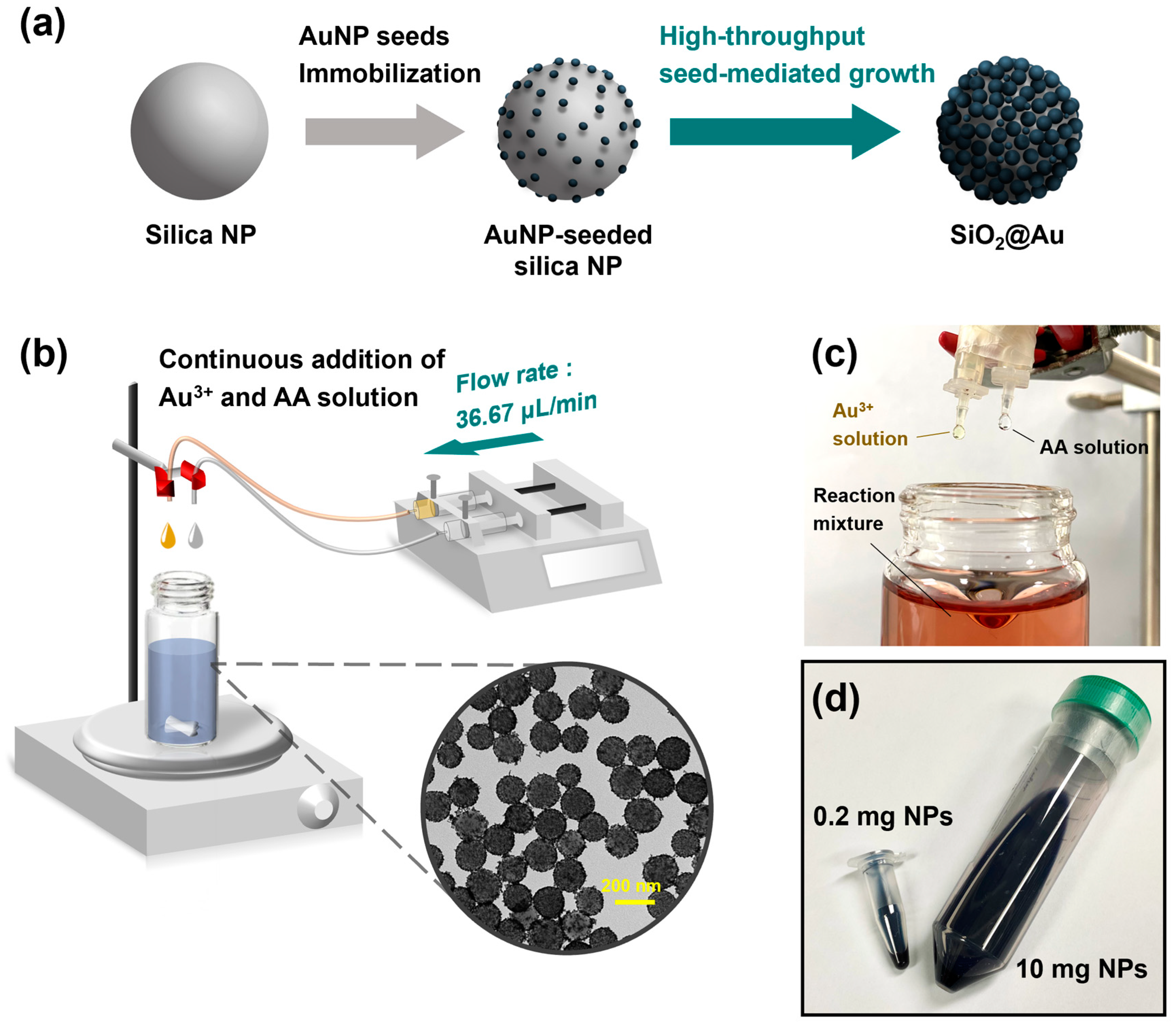
Abstract
:1. Introduction
2. Results and Discussion
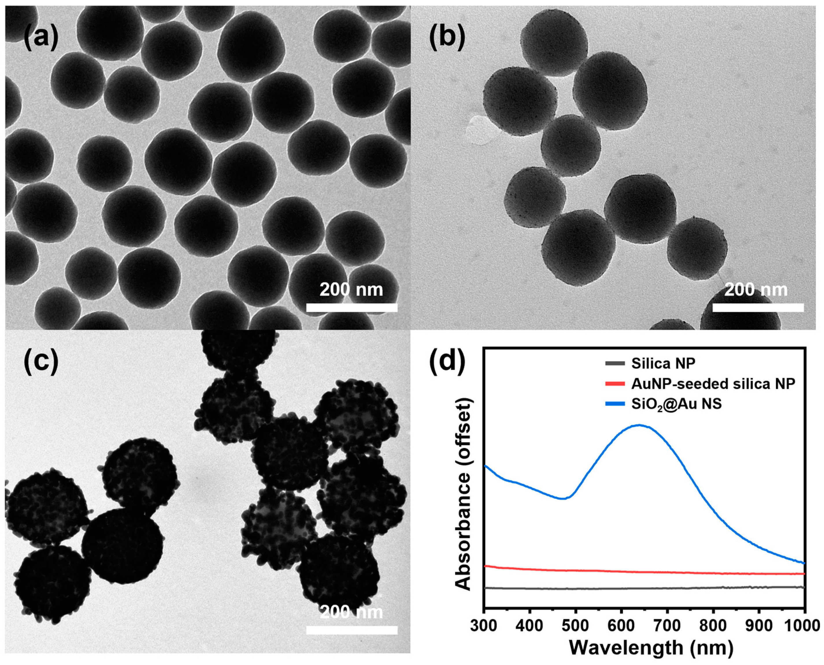
2.1. High-Throughput Synthesis of SiO2@Au NS
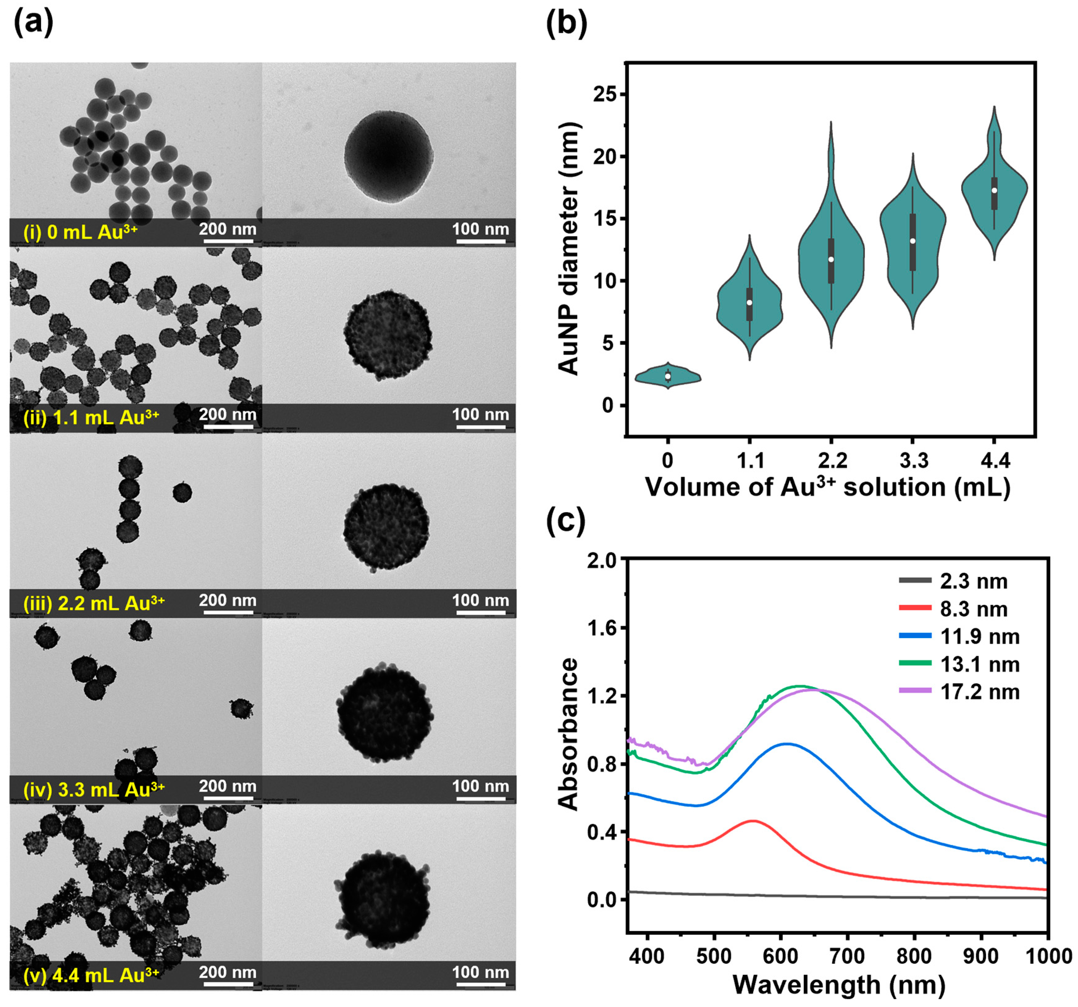
2.2. Control of Gold Shell Thickness
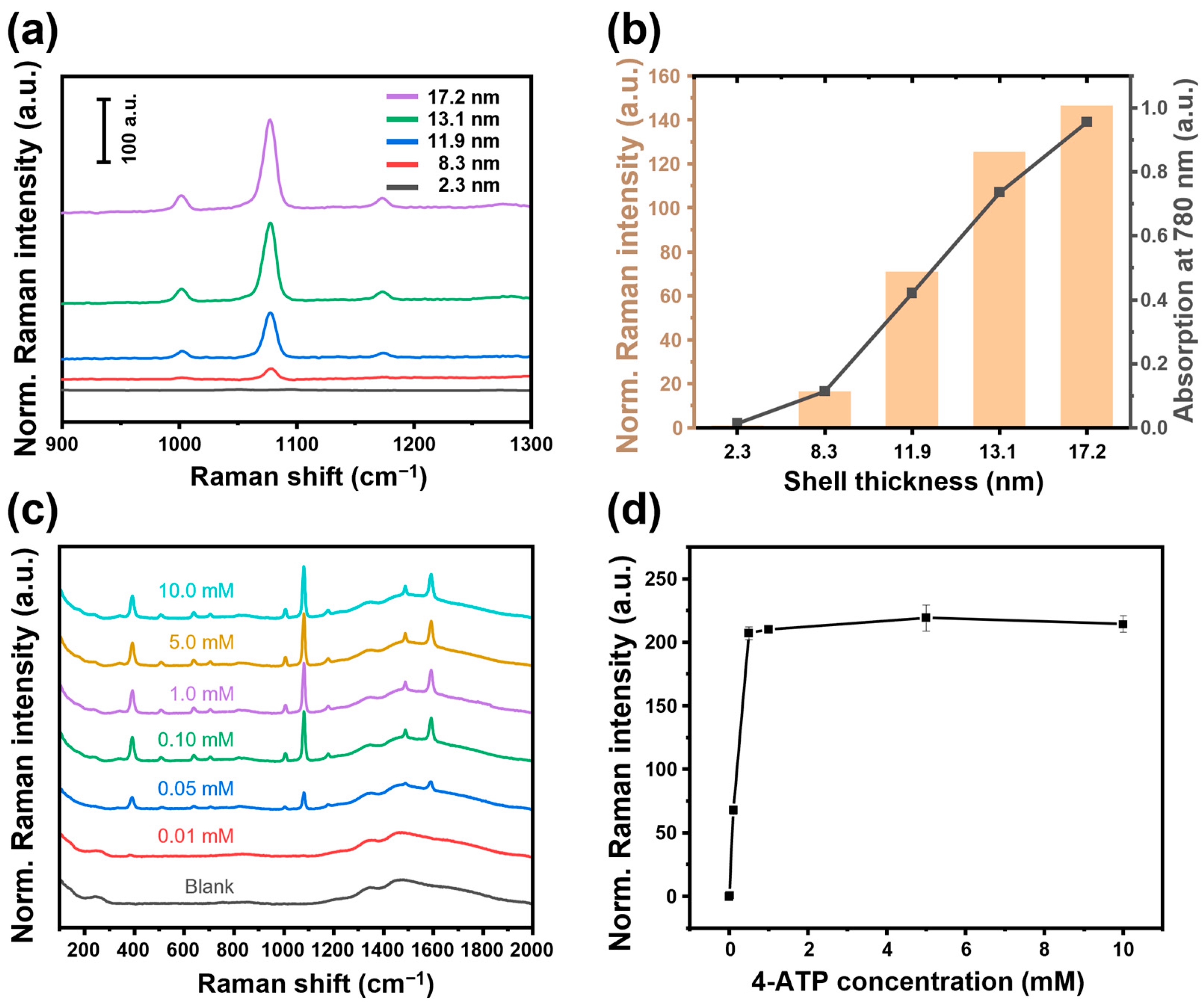
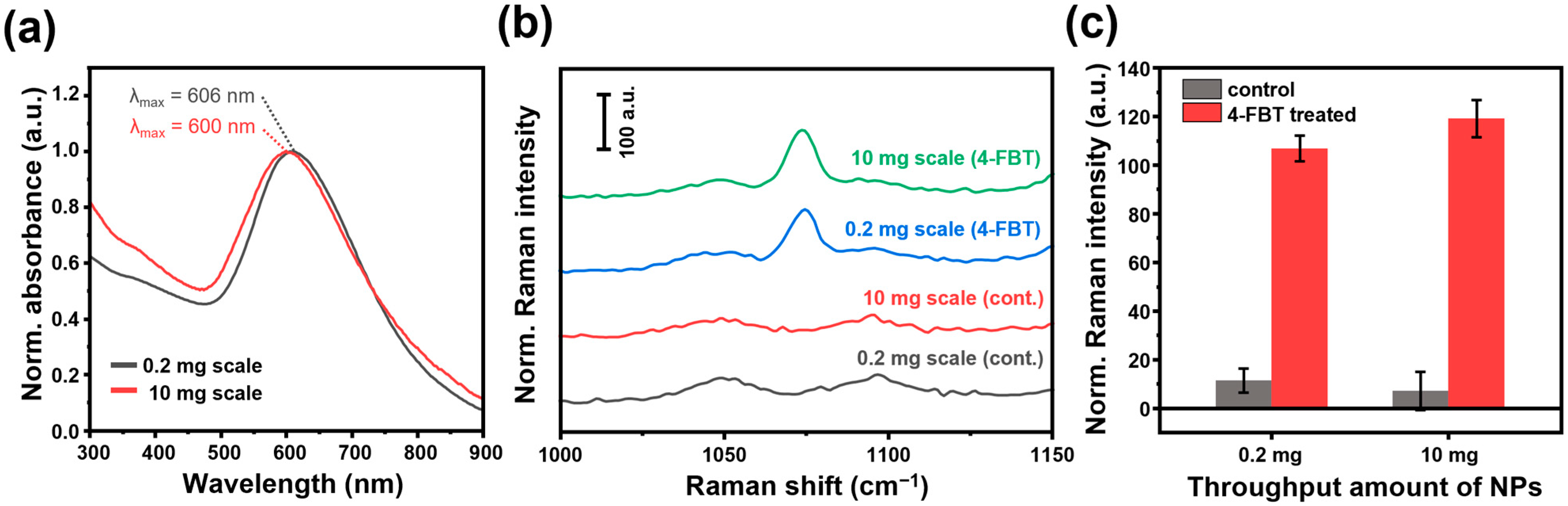
2.3. SiO2@Au NS as SERS Substrate
3. Materials and Methods
3.1. Chemicals
3.2. Characterization
3.3. Synthesis of AuNP Seeds
3.4. High-Throughput Synthesis of SiO2@Au NS
3.5. SERS Measurement
4. Conclusions
Supplementary Materials
Author Contributions
Funding
Institutional Review Board Statement
Data Availability Statement
Conflicts of Interest
References
- Oldenburg, S.J.; Averitt, R.D.; Westcott, S.L.; Halas, N.J. Nanoengineering of Optical Resonances. Chem. Phys. Lett. 1998, 288, 243–247. [Google Scholar] [CrossRef]
- Tam, F.; Goodrich, G.P.; Johnson, B.R.; Halas, N.J. Plasmonic Enhancement of Molecular Fluorescence. Nano Lett. 2007, 7, 496–501. [Google Scholar] [CrossRef] [PubMed]
- Cai, W. Applications of Gold Nanoparticles in Cancer Nanotechnology. Nanotechnol. Sci. Appl. 2008, 1, 17–32. [Google Scholar] [CrossRef] [PubMed]
- Zhao, J.; Wallace, M.; Melancon, M.P. Cancer Theranostics with Gold Nanoshells. Nanomedicine 2014, 9, 2041–2057. [Google Scholar] [CrossRef]
- Prodan, E.; Radloff, C.; Halas, N.J.; Nordlander, P. A Hybridization Model for the Plasmon Response of Complex Nanostructures. Science 2003, 302, 419–422. [Google Scholar] [CrossRef]
- Oldenburg, S.J.; Hale, G.D.; Jackson, J.B.; Halas, N.J. Light Scattering from Dipole and Quadrupole Nanoshell Antennas. Appl. Phys. Lett. 1999, 75, 1063–1065. [Google Scholar] [CrossRef]
- Heck, K.N.; Janesko, B.G.; Scuseria, G.E.; Halas, N.J.; Wong, M.S. Using Catalytic and Surface-Enhanced Raman Spectroscopy-Active Gold Nanoshells to Understand the Role of Basicity in Glycerol Oxidation. ACS Catal. 2013, 3, 2430–2435. [Google Scholar] [CrossRef]
- Huschka, R.; Barhoumi, A.; Liu, Q.; Roth, J.A.; Ji, L.; Halas, N.J. Gene Silencing by Gold Nanoshell-Mediated Delivery and Laser-Triggered Release of Antisense Oligonucleotide and SiRNA. ACS Nano 2012, 6, 7681–7691. [Google Scholar] [CrossRef]
- Wang, Y.; Qian, W.; Tan, Y.; Ding, S. A Label-Free Biosensor Based on Gold Nanoshell Monolayers for Monitoring Biomolecular Interactions in Diluted Whole Blood. Biosens. Bioelectron. 2008, 23, 1166–1170. [Google Scholar] [CrossRef]
- Stiles, P.L.; Dieringer, J.A.; Shah, N.C.; Van Duyne, R.P. Surface-Enhanced Raman Spectroscopy. Annu. Rev. Anal. Chem. 2008, 1, 601–626. [Google Scholar] [CrossRef]
- Bian, K.; Zhang, X.; Yang, M.; Luo, L.; Li, L.; He, Y.; Cong, C.; Li, X.; Zhu, R.; Gao, D. Dual-Template Cascade Synthesis of Highly Multi-Branched Au Nanoshells with Ultrastrong NIR Absorption and Efficient Photothermal Therapeutic Intervention. J. Mater. Chem. B 2019, 7, 598–610. [Google Scholar] [CrossRef] [PubMed]
- Sun, Y.; Xia, Y. Gold and Silver Nanoparticles: A Class of Chromophores with Colors Tunable in the Range from 400 to 750 nm. Analyst 2003, 128, 686–691. [Google Scholar] [CrossRef] [PubMed]
- Vankayala, R.; Lin, C.C.; Kalluru, P.; Chiang, C.S.; Hwang, K.C. Gold Nanoshells-Mediated Bimodal Photodynamic and Photothermal Cancer Treatment Using Ultra-Low Doses of near Infra-Red Light. Biomaterials 2014, 35, 5527–5538. [Google Scholar] [CrossRef]
- Sun, Y.; Xia, Y. Increased Sensitivity of Surface Plasmon Resonance of Gold Nanoshells Compared to That of Gold Solid Colloids in Response to Environmental Changes. Anal. Chem. 2002, 74, 5297–5305. [Google Scholar] [CrossRef]
- Rasch, M.R.; Sokolov, K.V.; Korgel, B.A. Limitations on the Optical Tunability of Small Diameter Gold Nanoshells. Langmuir 2009, 25, 11777–11785. [Google Scholar] [CrossRef] [PubMed]
- Bock, S.; Choi, Y.S.; Kim, M.; Yun, Y.; Pham, X.H.; Kim, J.; Seong, B.; Kim, W.; Jo, A.; Ham, K.M.; et al. Highly Sensitive Near-Infrared SERS Nanoprobes for in Vivo Imaging Using Gold-Assembled Silica Nanoparticles with Controllable Nanogaps. J. Nanobiotechnol. 2022, 20, 130. [Google Scholar] [CrossRef]
- Schlücker, S. Surface-Enhanced Raman Spectroscopy: Concepts and Chemical Applications. Angew. Chem. Int. Ed. 2014, 53, 4756–4795. [Google Scholar] [CrossRef]
- Duraiswamy, S.; Khan, S.A. Plasmonic Nanoshell Synthesis in Microfluidic Composite Foams. Nano Lett. 2010, 10, 3757–3763. [Google Scholar] [CrossRef]
- Watanabe, S.; Asahi, Y.; Omura, H.; Mae, K.; Miyahara, M.T. Flow Microreactor Synthesis of Gold Nanoshells and Patchy Particles. Adv. Powder Technol. 2016, 27, 2335–2341. [Google Scholar] [CrossRef]
- Pham, X.H.; Hahm, E.; Kang, E.; Son, B.S.; Ha, Y.; Kim, H.M.; Jeong, D.H.; Jun, B.H. Control of Silver Coating on Raman Label Incorporated Gold Nanoparticles Assembled Silica Nanoparticles. Int. J. Mol. Sci. 2019, 20, 1258. [Google Scholar] [CrossRef]
- Ayala-Orozco, C.; Urban, C.; Knight, M.W.; Urban, A.S.; Neumann, O.; Bishnoi, S.W.; Mukherjee, S.; Goodman, A.M.; Charron, H.; Mitchell, T.; et al. Au Nanomatryoshkas as Efficient Near-Infrared Photothermal Transducers for Cancer Treatment: Benchmarking against Nanoshells. ACS Nano 2014, 8, 6372–6381. [Google Scholar] [CrossRef] [PubMed]
- Wu, H.; Luo, Y.; Hou, C.; Huo, D.; Zhou, Y.; Zou, S.; Zhao, J.; Lei, Y. Flexible Bipyramid-AuNPs Based SERS Tape Sensing Strategy for Detecting Methyl Parathion on Vegetable and Fruit Surface. Sens. Actuators B Chem. 2019, 285, 123–128. [Google Scholar] [CrossRef]
- Zhao, Y.X.; Zhu, W.W.; Wu, Y.Y.; Chen, Y.Y.; Du, F.K.; Yan, J.; Tan, X.C.; Wang, Q. Sensitive Surface-Enhanced Raman Scattering for the Quantitative Detection of Formaldehyde in Foods Using Gold Nanorod Substrate. Microchem. J. 2021, 160, 105727. [Google Scholar] [CrossRef]
- Duff, D.G.; Baiker, A.; Edwards, P.P. A New Hydrosol of Gold Clusters. J. Chem. Soc. Chem. Commun. 1993, 9, 96–98. [Google Scholar] [CrossRef]
- Hueso, J.L.; Sebastián, V.; Mayoral, Á.; Usón, L.; Arruebo, M.; Santamaría, J. Beyond Gold: Rediscovering Tetrakis-(Hydroxymethyl)-Phosphonium Chloride (THPC) as an Effective Agent for the Synthesis of Ultra-Small Noble Metal Nanoparticles and Pt-Containing Nanoalloys. RSC Adv. 2013, 3, 10427–10433. [Google Scholar] [CrossRef]
- Stober, W.; Fink, A.; Ernst Bohn, D. Controlled Growth of Monodisperse Silica Spheres in the Micron Size Range. J. Colloid Interface Sci. 1968, 26, 62–69. [Google Scholar] [CrossRef]


Disclaimer/Publisher’s Note: The statements, opinions and data contained in all publications are solely those of the individual author(s) and contributor(s) and not of MDPI and/or the editor(s). MDPI and/or the editor(s) disclaim responsibility for any injury to people or property resulting from any ideas, methods, instructions or products referred to in the content. |
© 2024 by the authors. Licensee MDPI, Basel, Switzerland. This article is an open access article distributed under the terms and conditions of the Creative Commons Attribution (CC BY) license (https://creativecommons.org/licenses/by/4.0/).
Share and Cite
Kim, Y.-H.; Cho, H.-S.; Yoo, K.; Ham, K.-M.; Kang, H.; Pham, X.-H.; Jun, B.-H. High-Throughput Synthesis of Nanogap-Rich Gold Nanoshells Using Dual-Channel Infusion System. Int. J. Mol. Sci. 2024, 25, 1649. https://doi.org/10.3390/ijms25031649
Kim Y-H, Cho H-S, Yoo K, Ham K-M, Kang H, Pham X-H, Jun B-H. High-Throughput Synthesis of Nanogap-Rich Gold Nanoshells Using Dual-Channel Infusion System. International Journal of Molecular Sciences. 2024; 25(3):1649. https://doi.org/10.3390/ijms25031649
Chicago/Turabian StyleKim, Yoon-Hee, Hye-Seong Cho, Kwanghee Yoo, Kyeong-Min Ham, Homan Kang, Xuan-Hung Pham, and Bong-Hyun Jun. 2024. "High-Throughput Synthesis of Nanogap-Rich Gold Nanoshells Using Dual-Channel Infusion System" International Journal of Molecular Sciences 25, no. 3: 1649. https://doi.org/10.3390/ijms25031649