Article

Acute Ozone-Induced Transcriptional Changes in Markers of Oxidative Stress and Glucocorticoid Signaling in the Rat Hippocampus and Hypothalamus Are Sex-Specific

by
Matthew C. Valdez
1,2,
Danielle L. Freeborn
1,
Padmaja Vulimiri
1,†,
Joseph M. Valdez
1,2,
Urmila P. Kodavanti
3 and
Prasada Rao S. Kodavanti
1,*
1
Neurological and Endocrine Toxicology Branch, PHITD, CPHEA, ORD, US Environmental Protection Agency, Research Triangle Park, NC 27711, USA
2
Oak Ridge Institute for Science and Education Research Participation Program, U.S. Department of Energy, Oak Ridge, TN 37831, USA
3
Cardiopulmonary and Immunotoxicology Branch, PHITD, CPHEA, ORD, US Environmental Protection Agency, Research Triangle Park, NC 27711, USA
*
Author to whom correspondence should be addressed.
Present address: Biogen, 5000 Davis Drive, Durham, NC 27709, USA.
Int. J. Mol. Sci. 2023, 24(7), 6404; https://doi.org/10.3390/ijms24076404
Submission received: 15 February 2023 / Revised: 17 March 2023 / Accepted: 19 March 2023 / Published: 29 March 2023
(This article belongs to the Special Issue Mitochondria as a Cellular Hub in Neurological Disorders)

Abstract

:
Exposure to a prototypic air pollutant ozone (O3) has been associated with the activation of neuroendocrine stress response along with neural changes in oxidative stress (OS), inflammation, and Alzheimer’s disease-like pathologies in susceptible animal models. We hypothesized that neural oxidative and transcriptional changes induced by O3 in stress responsive regions are sex-dependent. Male and female adult Long–Evans rats were exposed to filtered air or O3 for two consecutive days (0.8 ppm, 4 h/day) and brain regions were flash-frozen. Activities of cerebellar OS parameters and mitochondrial complex I, II, and IV enzymes were assessed to confirm prior findings. We assessed transcriptional changes in hypothalamus (HYP) and hippocampus (HIP) for markers of OS, microglial activity and glucocorticoid signaling using qPCR. Although there were no O3 or sex-related differences in the cerebellar activities of OS and mitochondrial enzymes, the levels of protein carbonyls and complex II activities were higher in females regardless of O3. There were no statistical differences in baseline expression of genes related to OS (Cat, Dhcr24, Foxm1, Gpx1, Gss, Nfe2l2, Sod1) except for lower HYP Sod1 expression in air-exposed females than males, and higher HIP Gss expression in O3-exposed females relative to matched males. Microglial marker Aif1 expression was higher in O3-exposed females relative to males; O3 inhibited Itgam only in males. The expression of Bdnf in HIP and HYP was inhibited by O3 in both sexes. Genes related to glucocorticoid signaling (Fkbp4, Fkbp5, Hsp90aa1, Hspa4, nr3c1, nr3c2) showed sex-specific effects due to O3 exposure. Baseline expression of HIP Fkbp4 was higher in females relative to males. O3 inhibited Nr3c1 in female HIP and male HYP, but Nr3c2 was inhibited in male HYP. Fkbp4 expression was higher in O3-exposed females when compared to matched males, whereas Fkbp5 was expressed at higher levels in both brain regions of males and females. These results indicate that sex-specific brain region responses to O3 might, in part, be caused by OS and regulation of glucocorticoid signaling.

1. Introduction

Sex is a biological variable with significant influence over various physiological functions within all species. Specifically, with regard to lung physiology and immune responses, the factor of sex greatly affects the prognosis of pulmonary morbidities in response to air pollution [1,2,3,4]. Historically, within the literature, causal relationships have been made between air pollution and morbidity and/or mortality by various factors such as genetic susceptibility, environment, and even socioeconomic status. However, little attention has been given to sex differences, especially in animal models. A recent study in Canada was able to analyze nearly three decades of mortality data and hospitalizations along with health outcomes from a geographically diverse cohort representing nearly half of the country as they relate to health effects resulting from air pollution exposure [4]. Shin and colleagues found that, in response to short term exposure to ambient air pollution, there were pollutant season-specific sex differences in circulatory and respiratory hospitalization and mortality where males were at higher risk for warm season, but females were at higher risk than males for cold season [4,5,6]. Given global trends in abnormal climate fluctuations, there is growing concern of human exposure to air pollutants, namely ozone (O3) which is produced at ground level by excessive heat and radiation reacting with volatile organic compounds and nitric oxide (NOX) gases. A recent multinational report has identified pollution as the leading environmental cause of premature death and disease [7]. Of all types of environmental pollutants, this report identified air pollution to be the primary driver of this trend. Therefore, understanding the effect of gender on adverse health outcomes related to air pollution exposure is paramount.
The immunological and physiological consequences of inhaled toxicants, such as air pollution, have been extensively investigated [5,8,9,10,11,12]. The outcomes include changes in body composition, core temperature, cardio-pulmonary effects such as heart rate, pulmonary sensitivity, and neuronal effects such as motor activity deficits. However, those lines of investigation are now converging on a significant role for the aptly named “lung-brain-axis” and its role in extrapulmonary morbidities of air pollution exposure [13,14]. It has become clear that via a yet unknown mechanism (referred to as the lung-brain-axis), an inhaled toxicant such as O3 can generate a physiological stress response through the activation of the hypothalamic-pituitary-adrenal (HPA) axis similar to physical and psychosocial stressors [15,16,17,18,19,20,21,22,23,24]. Sex-dependent differences in HPA axis processing and responses have been previously established [25,26,27]. In this study, we sought to investigate sex-dependent differences in transcriptomic responses in brain regions, specifically the hypothalamus (HYP) and hippocampus (HIP), of animals exposed to O3 to understand the effects on HPA axis. We first present data on oxidative stress (OS) status and mitochondrial complex enzymes across sex in the cerebellum in order to demonstrate consistency with previous reports [5,28,29,30,31,32]. These data are then accompanied by novel gene expression data in two important brain regions for the HPA axis (i.e., HYP and HIP). In these brain regions, we assayed genes related to glucocorticoid signaling, microglia, general stress, and oxidative stress in male and female rats acutely exposed to O3.

2. Results

The effects of O3 exposure on the brain have garnered interest in recent years [20,33,34,35,36,37]. O3 itself is a highly reactive oxidizer, which does not translocate distally in its parent chemical form. However, many of the effects in the brain seem to be mediated by indirect modes of action, such as by sensory reception or other signaling cascades [13,38]. Due to the chemical nature of O3, OS as an adverse outcome is most typically associated with exposure [33,39,40,41].

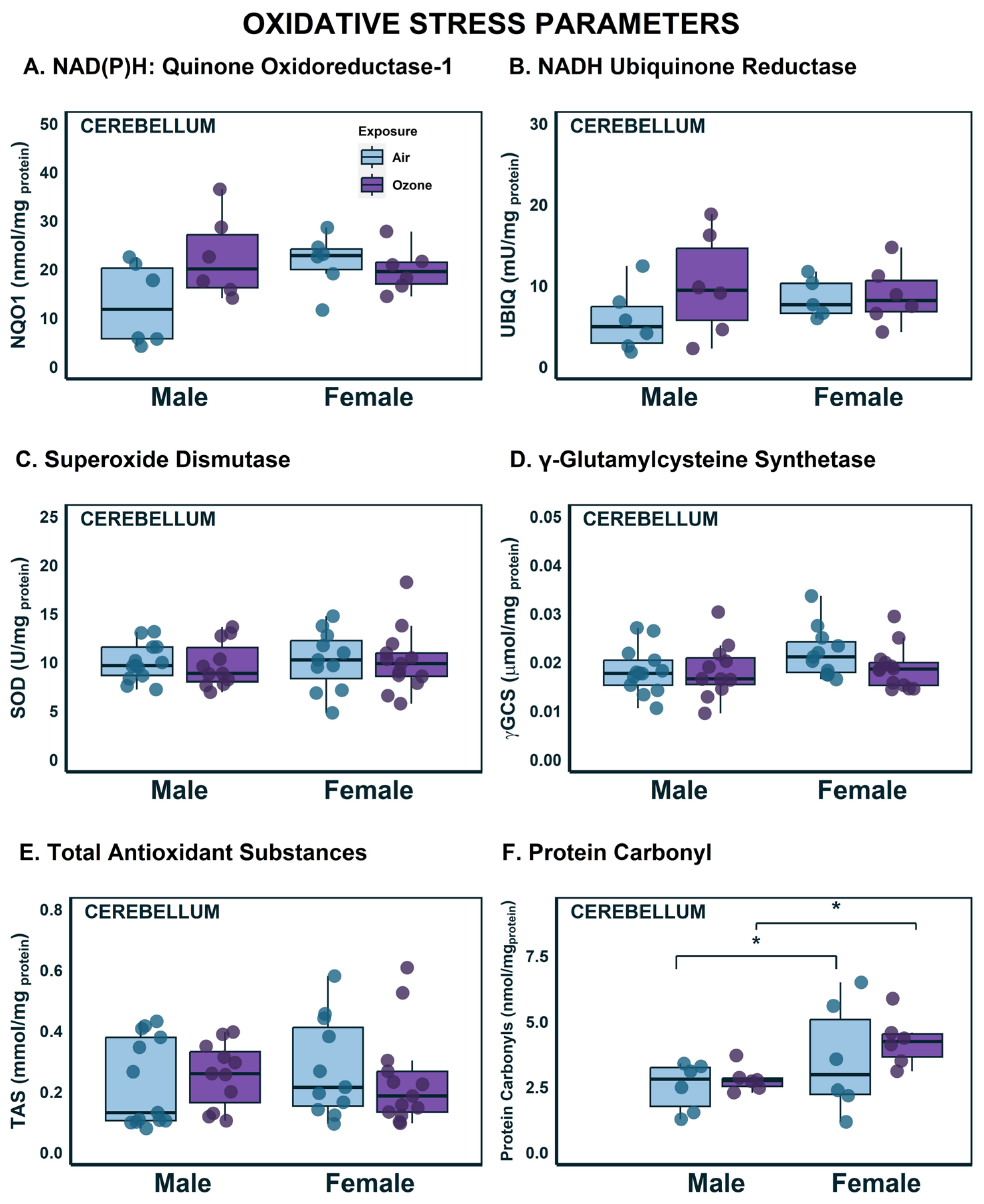
2.1. Effects on OS Parameters in the Cerebellum

We have previously reported O3 effects on OS and mitochondrial complex enzymes in different brain regions of various rat species, different age groups, and in combination with other non-chemical stressors [33,34,36,37]. In association with O3-induced brain genomic effects in this species and at this age, we have conducted studies in these two parameters in the cerebellum to confirm previous observations (Figure 1 and Figure 2). Although the focus of this study is the sex differences within the integrating and regulatory brain regions (HYP and HIP) through genomic changes, we assayed OS parameters and complex enzyme activities in this proxy region of interest because of tissue limitation. Firstly, NAD(P)H: Quinone dehydrogenase 1 (NQO1; Figure 1A) and NADH ubiquinone reductase (UBIQ; Figure 1B), which were used as indicators of ROS production, were upregulated in O3-exposed males when compared to air groups. However, these elevations were minor (43% increase), and a two-way ANOVA failed to indicate a significant difference from air-exposed male animals compared to female animals. For both ROS parameters, the effects were nearly identical in females when exposed to air or O3. Superoxide dismutase (SOD), which is an endogenous antioxidant (Figure 1C), showed similar effects in males and females, indicating no observable differences across sex or exposure. Glutathione is another endogenous antioxidant, and γ-glutamylcysteine synthase (γ-GCS) is a key rate-limiting enzyme necessary to produce glutathione. Similar to the activity patterns of SOD, there were no discernable differences in γ-GSC activity across sex and exposure groups (Figure 1D). Total antioxidant substances (TAS) were measured to ensure a complete picture of the antioxidant capacity of the brain region. Just as with the other ROS measures, there were no differences across sex or exposure groups (Figure 1E). OS can have numerous deleterious effects to surrounding cellular structures [42]. These effects are primarily caused by the oxidation of important biomolecules, such as proteins. Protein carbonyl formation is a common oxidation product due to OS and this results in a loss of protein function [43]. Therefore, we measured protein carbonyls as an indicator of oxidative damage produced by the imbalance between ROS production and cellular antioxidant homeostasis. Despite the lack of differences in ROS measures and antioxidant capacities, there were significant differences attributed to sex in the formation of protein carbonyls (F1,21 = 6.143; p < 0.05) where, compared to their corresponding male exposure groups, females had higher levels of protein carbonyl content (Figure 1F). These results indicate that antioxidant systems that typically respond to ROS production were not upregulated with O3 exposure, but that the oxidized biomolecule landscape is different across sex.

2.2. Effects on Complex Enzyme Activities in the Cerebellum

Mitochondria are the main sources of endogenous OS since they consume oxygen for energy metabolism. Therefore, the key components of mitochondrial energy production, complex enzymes, were assayed. We assayed three of the five complex enzymes (Figure 2) that comprise the electron transport chain (ETC): NADH: Ubiquinone oxidoreductase (Complex Enzyme I), succinate dehydrogenase (Complex Enzyme II), and cytochrome c oxidase (Complex Enzyme IV). Complex I enzyme activity was similar across both sex and exposure groups (Figure 2A). Females had higher activity levels of complex II compared to males (Figure 2B; F1,21 = 24.4; p < 0.001). Both complex I and II are entry points to the ETC and these sex differences could indicate sexually dimorphic energy metabolism strategies. The final energy harvesting enzyme in the ETC is complex IV (Figure 2C). There appeared to be an O3-mediated increase in females, but two-factor ANOVA did not indicate a statistical difference. Taken all together, these results indicate that there may be differences in mitochondrial energy metabolism strategies between males and females, but minimal influence of acute O3 exposure.

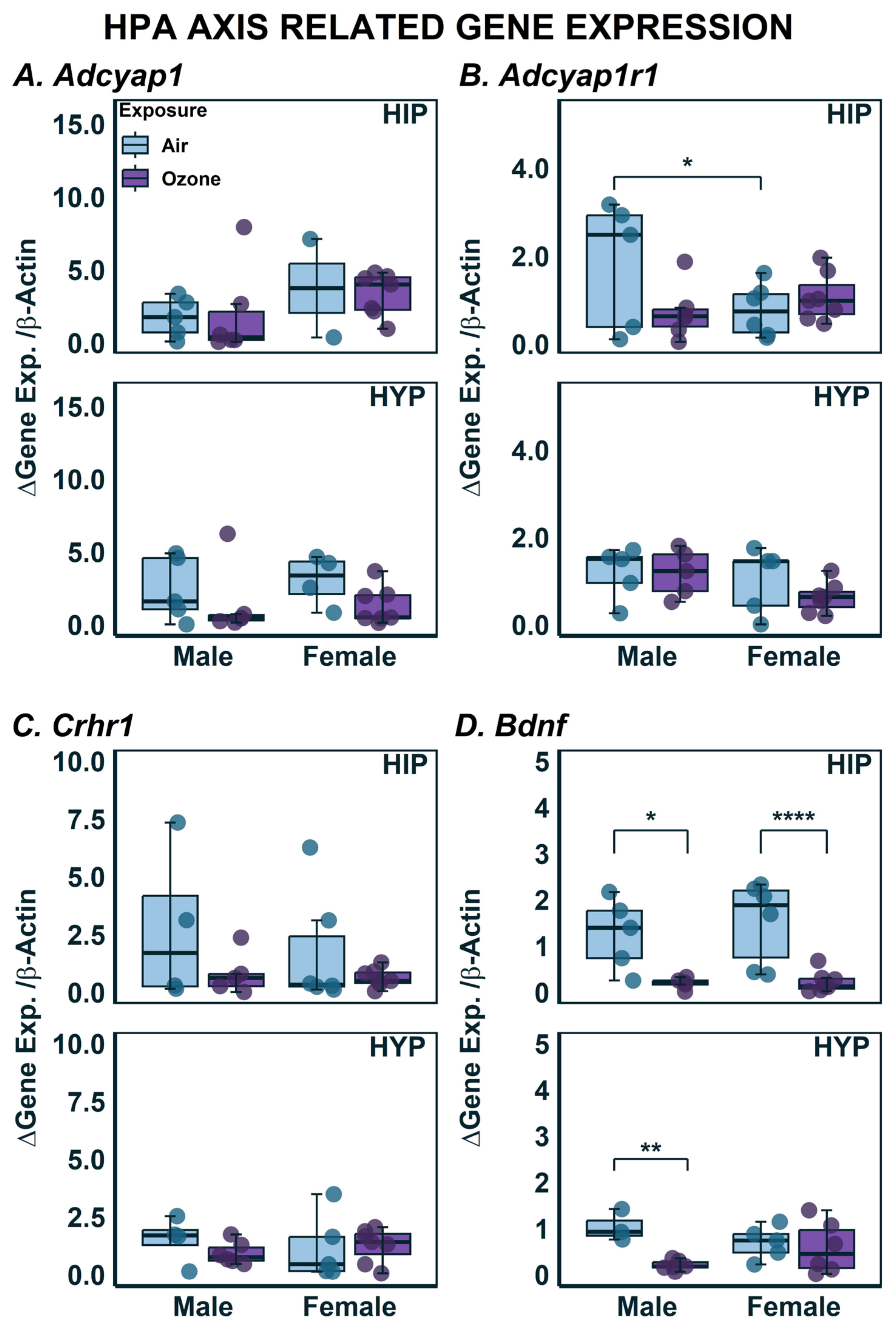
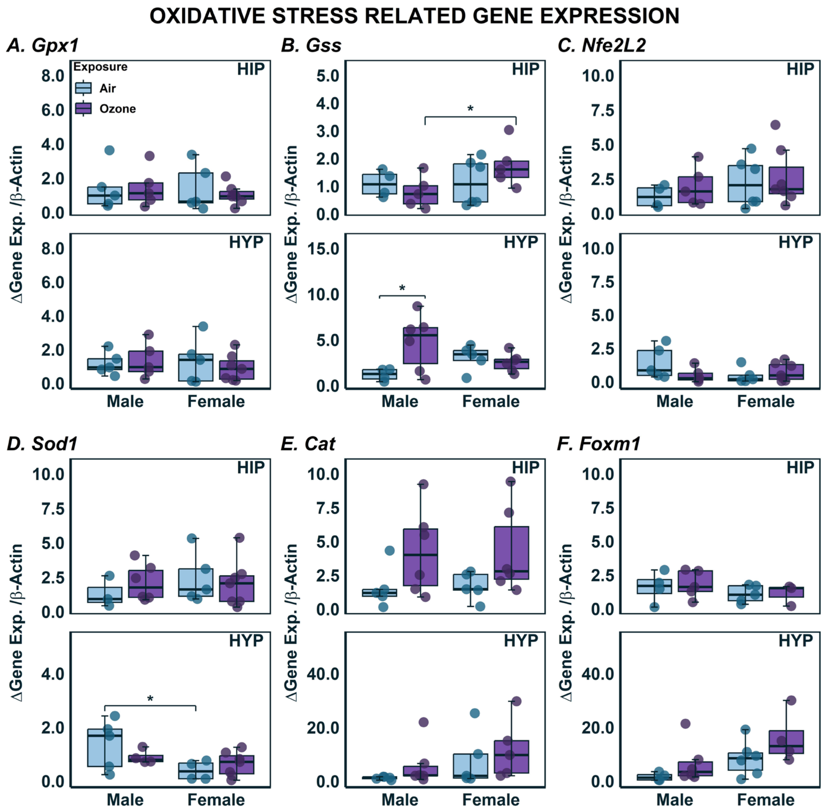
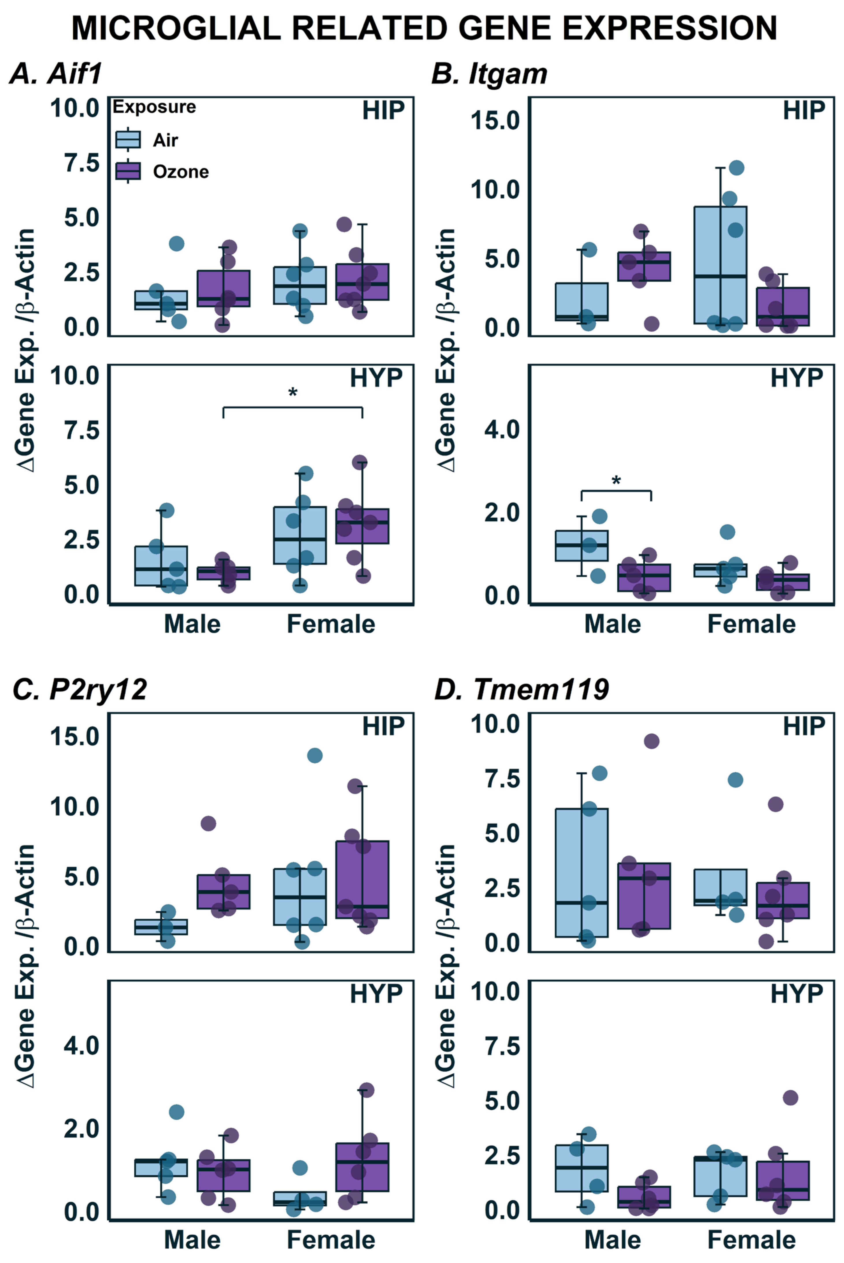
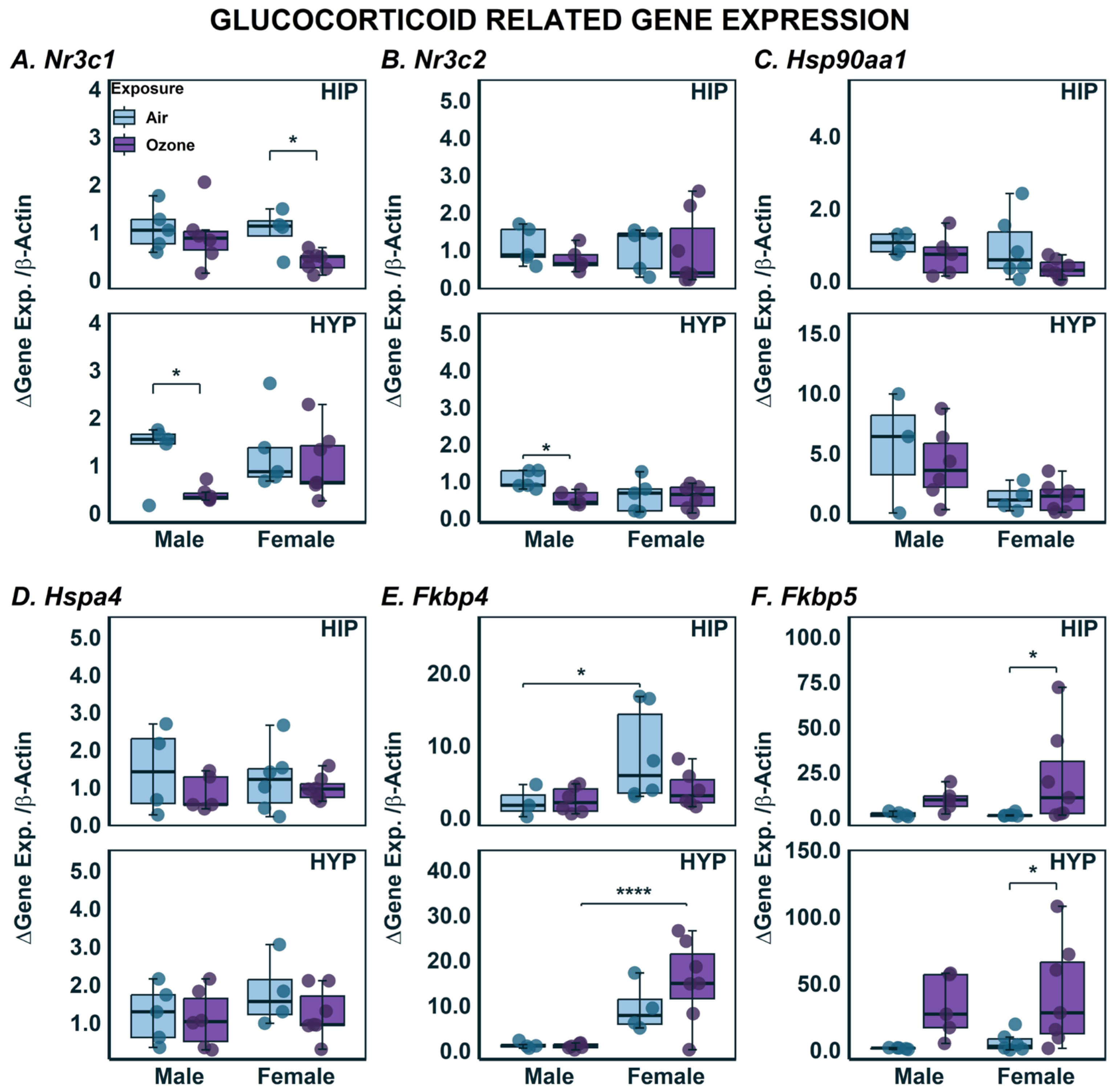
2.3. Effects on Gene Expression in HYP and HIP

The focus of O3 exposure studies has started to shift from analysis of OS and immunological response towards the central control of the responses [20]. Therefore, a panel of neuroendocrine stress, OS-related, microglial, and glucocorticoid gene markers were assayed (Figure 3, Figure 4, Figure 5 and Figure 6). Significantly modified genes showing O3 effects and sex-dependent differences is shown in Table 1. A two-way ANOVA revealed no significant differences between sex or O3 exposure within the HIP and HYP (Figure 3A). The expression of genes which encodes the receptor for the product of Adcyap1, Adcyap1r1, was higher in HIP of air-exposed males when compared to air-exposed females. However, no changes were noted due to O3 exposure in either HIP or HYP (Figure 3B). No significant exposure, brain region or sex-related differences were noted in Crhr1 expression (Figure 3C). Bdnf expression levels were lower in the HIP of male and female rats (Figure 3D; F1,18 = 15.43; p < 0.05). In the HYP, there was also a decrease in Bdnf expression, however, post hoc multiple comparisons only revealed a significant difference in the females.
Genes related to oxidoreductive processes were also investigated. Although there were no significant exposure- or sex-related differences in the expression of Gpx1 in either brain region, there was a significant interaction of sex and exposure on Gss expression where expression seemingly decreased in males with O3 exposure but increased with O3 exposure in females (Figure 4B; F1,14 = 6.235; p < 0.05). In the HYP, there was a significant effect of exposure (F1,14 = 6.324; p < 0.05), where males had higher expression of Gss in response to O3 exposure with no change in females. There were no changes in HIP or HYP Nfe2L2 expression in males or females with or without O3 exposure (Figure 4C). There was significant sex-related difference in expression of Sod1 in the HYP where air-exposed males had higher expression when compared to females (Figure 4D; F1,17 = 5.793; p < 0.05). In the HIP, Cat gene expression tended to be higher in O3-exposed males and females, but these differences did not reach significance (Figure 4E). Lastly, the expression levels of Foxm1 were not affected by sex or O3 exposure in either brain region (Figure 4E).
We have previously reported changes in reactive microglia as a result of O3 exposure [34,36]. In those studies, we used immunohistochemistry targeting Iba1 (encoded by the gene Aif1), to identify microglia in brain sections and then classified them as reactive based on their morphology. In the current study, we assessed gene expression related to microglia function to evaluate if they were transcriptionally regulated and/or showed any sex differences in microglia (Figure 5). Females in general had higher expression of Aif1 in the HYP regardless of air or O3 exposure (Figure 5A; F1,18 = 10.16; p < 0.05). HYP but not HIP Itgam expression was lower in males exposed to O3 when compared to the air group. No sex-related differences were noted either in HIP or HYP (Figure 5B). Two-way ANOVA revealed no significant interaction of sex and exposure on P2ry12 expression (Figure 5C). Tmem119 is a recently discovered gene specific to microglia [44]. Tmem119 expression was neither significantly different between sexes nor exposure in either of the brain regions (Figure 5D).
Acute O3 exposure has been shown to increase glucocorticoids systemically [35], and these levels were linked to O3 adaptation [45]; therefore, we also analyzed genes related to glucocorticoid signaling. The gene that encodes for the glucocorticoid receptor, Nr3c1, showed a significant interaction of both sex and exposure in each brain region (F1,17 = 7.431; p < 0.05) where only females had lower expression in response to O3 exposure in HIP; whereas, in HYP, O3-exposed males had lower expression when compared to air controls (Figure 6A). The gene for the mineralocorticoid receptor, Nr3c2, showed no changes by either sex or O3 exposure in HIP, but in HYP males exposed to O3 had lower expression relative to air controls (Figure 6B). One of the chaperone proteins that is a part of the glucocorticoid receptor heterocomplex is heat shock protein 90, encoded by Hsp90aa1 (Figure 6C). In both brain regions, although there were no apparent effects of O3 exposure on Hsp90aa1 expression, the female rats exhibited lower expression when compared to males in HYP, but these differences did not reach significance. No sex- or exposure-related differences were noted in expression of Hspa4 in either brain region (Figure 6D). Two other members of the glucocorticoid receptor heterocomplex that play roles in the translocation of the complex upon binding of stress hormones are the gene products of Fkbp4 and Fkbp5. In HIP, there was a significant effect of sex (Figure 6E; F1,16 = 7.933; p < 0.05) resulting in overall higher expression levels in females exposed to air compared to males. In the HYP, there was a significant interaction of sex and exposure where females had higher overall expression of Fkbp4, and expression further increased in females with exposure to O3. There were significant effects of O3 exposure in females and in both brain regions on Fkbp5 expression (Figure 6F; HIP: F1,17 = 9.247; p < 0.05; HYP: F1,17 = 16.63; p < 0.05). Fkbp5 expression was also higher in O3-exposed males when compared to air-exposed males, but these differences did not reach significance.

3. Discussion

O3 is a ubiquitous air pollutant and has been extensively studied for its neurological and neuroendocrine impacts from acute and long-term exposures [46,47]. O3 is highly reactive and not likely to translocate to distant organs, such as the brain, upon inhalation; however, numerous studies have shown that O3 exposure induces OS in multiple brain regions [33,34,48], causes transcriptional changes [35], increases inflammatory phenotype [49] and microglial activation [50], and in the long-term promotes Alzheimer’s disease-like pathology in laboratory rodents [47]. More recently exposure to O3 has been shown to activate neuroendocrine stress pathways such as the HPA, leading to increased levels of glucocorticoids in circulation [19,51], however, sex differences and neurological effects are not well-examined in relation to neuroendocrine modulation. In this study, we hypothesized that O3-induced transcriptional changes in OS markers will be associated with markers involved in glucocorticoid signaling and these changes will be sex-dependent.
This study demonstrates that cerebellar protein carbonyl levels and mitochondrial complex II activities were higher in females regardless of O3, but the activities of antioxidants were unchanged between sex or exposure condition. Overall gene expression changes in HIP and HYP of male and female rats indicated inhibition of Bdnf by O3 in HIP of both sexes and only HYP in males. O3 increased Gss expression in female HIP and male HYP, suggesting the potential contribution of glutathione in modulating brain region-specific effects. Microglial marker Aif1 had high baseline expression in HYP of females when compared to males and in the same region the other marker Itgm was inhibited by O3 but only in males. These sex-specific changes in OS, markers were associated with changes in markers of glucocorticoid signaling. We noted that glucocorticoid receptor, Nr3c1 was transcriptionally inhibited by O3 in HIP in females, and in HYP but only in males. The expression of mineralocorticoid receptor, Nr3c2 was also inhibited by O3 (HYP > HIP) but only in males. The expressions of glucocorticoid chaperone proteins were also affected by O3 in sex-specific manner. Baseline expression of Fkbp4 was higher in females and chaperone protein encoded by Fkbp5, critical in regulating glucocorticoid activity, was induced by O3 in both brain regions (females > males), suggesting sex-dependent temporal regulation of HIP and HYP glucocorticoid activity by O3. Collectively, these data show that glucocorticoids known to be increased during O3 exposure [51] might regulate brain region-specific effects on stress dynamic and oxidation reduction processes in sex-dependent manner.
We have recently demonstrated that neural, pulmonary, and systemic effects of O3 were mediated through the activation of sympathetic adrenal medulla (SAM) and HPA, associated with reduction of pituitary hormones regulating gonadal axis in males [35,45,51], and that diminution of circulating adrenal hormones through adrenalectomy resulted in the lack of O3-induced reduction of gonadal hormones. This suggests that adrenal hormones could centrally regulate O3 effects on pituitary gonadal hormones [35]. This also implies that O3 neuroendocrine effects are likely sex-dependent, however, sex differences in O3 neuroendocrine effects are not well examined. Here we wanted to focus on glucocorticoid-mediated neural changes in male and female rats, since this adrenal hormone has been shown to regulate longevity and characteristics of a stress phenotype through brain region and sex-dependent differential regulation of cellular activities [52,53]. Alterations in glucocorticoid mechanisms and their levels are associated with chronic stress-related neurological disorders, known to affect males and females differently [54].
Impacts on oxidoreductive processes such as levels of oxidation byproducts and changes in oxidoreductive enzyme activities after O3 exposure have been demonstrated in various brain regions of rodents [33,34,48]. Although it is not entirely clear if O3 effects are a general oxidative response across all regions, or specific to a given brain region, the available data supports brain region-specific changes [33,36]. Some of these studies have examined sex differences in O3 effects [55]. Here, we wanted to use cerebellar tissues to determine concordance with our prior studies reporting oxidative and mitochondrial bioenergetic changes after O3 exposure in rats [33,34,36] and evaluate sex differences in parallel in the same study. Protein carbonyl contents in 4-month but not 12-month old male Brown Norway rats exposed to 1 ppm O3 for two days was increased in our prior study [33]. Phenotypic oxidative changes were also noted in other brain regions of rats after O3 exposure [33,36], but in this study only the expression of markers linked to OS, mitochondrial activity, and microglia were examined in other brain regions, specifically HIP and HYP. Acute O3 exposure was associated with increased lipid peroxidation in male HIP and cerebellum of rats exposed to O3 [56]. Thus, O3 exposure is associated with oxidative changes in various brain regions of rats, and our data show that females might have greater susceptibility for cerebellar oxidative changes when compared to males.
Transcriptionally regulated genes involved in responding to underlying OS might be differentially expressed between males and females in two stress responsive regions of the brain, likely mediating neural and systemic response to SAM and SAM axes activation by O3 [35,51]. Gss encodes for a critical intracellular antioxidant, glutathione, critical in protecting the brain from oxidative processes [57], and was induced after O3-exposure in female HIP and in male HYP. This suggests sex- and brain region-specific roles in cellular oxidoreductive mechanisms involving transcriptional upregulation, likely in response to increased oxidative cell stress. Interestingly, this was associated with higher baseline Sod1 transcription in HYP of males when compared to females, which suggests that HYP in males might be more susceptible to some stressor-induced changes when compared to females.
The expression of Bdnf involved in neuropsychological disease [58] was inhibited by O3 in HIP of both sexes and in HYP of only males. Bdnf has been suggested to play a role in not only regulating stress mechanisms but also in glucocorticoid signaling [59]. While glucocorticoids are increased in the circulation after O3 exposure, the interactive roles of Bdnf and glucocorticoids have been reported in humans [60]. Lower BDNF protein levels were noted in HIP of rats with compressed dorsal root ganglion and exposure to O3 [61]. O3-induced reduction in circulating BDNF levels was mitigated in rats treated with metyrapone, a glucocorticoid synthesis inhibitor, suggesting a regulatory role of glucocorticoid in mediating transcriptional repression in HIP of male rats exposed to O3 [62]. Interestingly, the effect in HYP of females was smaller compared to males in our study. The overall impact on Bdnf modulation by O3 suggests its involvement in long-term regulation of stress dynamicity.
Microglia in different brain regions play a critical role in maintaining homeostatic processes by responding to stress and inducing appropriate immunological processes [63]. Microglial activation has been reported by a variety of stressors including O3 [14,64,65]. We wanted to assess sex-related differences in microglial activity and potential acute O3-mediated stress effects. Although there were no O3 effects on transcriptional activation of microglial marker Aif1 (also known as Iba1 at transcriptional level), the increased AIF1 immunoreactivity has been noted after O3 exposure in rodents [14]. However, we noted that the baseline levels of Aif1 transcription were higher in female HYP when compared to males. Moreover, we noted that in male but not female HYP, O3 exposure lowered mRNA expression of Itgam (complement receptor 3). Itagm has been shown to be expressed on microglia and plays a critical role in neurodegenerative processes [66]. The significance of transcriptional repression of this gene after O3 exposure is unclear but in chronic exposures might contribute to long-term neurological disorders associated with neuroinflammation.
One of the ancillary objectives of this study was to compare O3 response on markers of glucocorticoid signaling in HIP and HYP of male and female rats based on our prior demonstration of involvement of dynamic glucocorticoids in O3-mediated neural effects, release of pituitary hormones and especially adaptation response noted after repeated O3 exposure [35,45,51]. Brain region- and sex-specific inhibition of glucocorticoid receptor gene, Nr3c1, activated by high levels of circulating glucocorticoids, showing O3-induced inhibition in HIP of females and HYP of males. This coincides with inhibition of Nr3c2, a mineralocorticoid receptor activated by low levels of glucocorticoids in HYP of males, might reflect complex glucocorticoid regulating mechanisms. The differences in receptor density and the glucocorticoid activity regulating mechanisms between these brain regions [67,68] and sex in response to stressors [69] might underlie observed differences in O3-induced transcriptional changes in glucocorticoid receptors [57,70,71]. Nr3c1, a glucocorticoid receptor gene and Nr3c2, a mineralocorticoid receptor gene, are activated by circulating glucocorticoids entering cells in the brain centers and are involved in finetuning the dynamic of a stress response in association with glucocorticoid scaffolding protein encoded by Fkbp5. This fine-tuning of glucocorticoid transcriptional action can be accomplished by relative cell and brain region-specific distribution and transcriptional activity of these receptors and scaffolding proteins, since nearly 20% of the human genome is regulated by these glucocorticoids critical in responding to environmental stressors through changes in physiological processes [70]. The activity of glucocorticoids is also regulated by androgen sex hormones, such as estrogen through its interactive transcriptional regulation [71]. Thus, while the precise mechanisms of how sex hormones might regulate glucocorticoid effects in ozone-induced glucocorticoid receptor transcriptional changes are likely complex and cannot be ascertained through this observational study, here we show that ozone exposure might mediate sex specific changes.
Two FK506-binding immunophilins, encoded by Fkbp4 and Fkbp5 genes comprise an important part of glucocorticoid receptor heterocomplex that plays a role in the regulation of transcriptional activity of glucocorticoids [72,73]. Fkbp5 bound to glucocorticoid receptor is released from the complex upon binding to glucocorticoids, allowing Fkbp4 to bind to the hormone-bound transcriptional complex mediating transcriptional activity through glucocorticoid response elements (GRE) of hundreds of genes including Fkbp5. In turn, Fkbp5 rapidly binds to the cytosolic receptor complex and regulates glucocorticoid activity through a feedback loop [74]. We have shown that O3 exposure leads to HPA-mediated glucocorticoid release in the circulation within minutes of exposure [35,45,51]. We noted that there were sex- and brain region-specific effects of O3, likely mediated by higher circulating glucocorticoids on expression of Fkbp4 and Fkbp5. We noted that females have a high baseline level of mRNA expression of Fkbp4 but not Fkbp5 in both brain regions. The precise role of estrogen in regulating glucocorticoid activity cannot be ascertained, but it has been shown that chaperone proteins encoded by Fkbp4 and Fkbp5 genes can regulate the stability of estrogen receptor alpha [75] and might be influencing the transcriptional activity of these proteins. Transcriptionally regulated Fkbp5 was induced by O3 in both sexes and brain regions, albeit at different levels, suggesting that circulating glucocorticoids modulate O3 stress effects in the brain and might be involved in adaption response through glucocorticoid signaling [45]. Fkbp5 polymorphisms have been associated with a variety of human psychiatric ailments [73]. These data provide the link between air pollution and neural stress-mediated glucocorticoid changes and highlight the importance of neuroendocrine mechanisms in chronic neural diseases that have been recently linked to high air pollution levels [76].
There are a number of limitations in this study. This observational study comparing sex differences in O3-induced OS and glucocorticoid-mediated transcriptional changes neither assessed potential protein levels of markers or functional effects, and rather associated changes to our prior publications related to neural and glucocorticoid changes. The O3 concentrations used are several folds higher than what is encountered environmentally in tropical atmospheres, however, they are comparable to human clinical studies performed during intermittent exercise with O3 concentrations ranging from 0.2 to 0.4 ppm [77,78,79,80]. Humans exposed during intermittent exercise retain 4–5 times the inhaled dose of O3 compared to rodents exposed during rest [81]. These data highlight the sex differences and regulatory roles of neuroendocrine pathways in mediating neural effects of O3, while much of mechanistic sex differences will need to be further examined.

4. Materials and Methods

4.1. Animals

Long–Evans (LE) rat dams were obtained from Charles River Laboratories, Inc. (Raleigh, NC, USA) and maintained in the animal colony. After birth, the litters were standardized on postnatal day (PND) six to four males and four females where possible. The sex was determined by measuring anogenital distance, where it is more for males than females. If four males and four females were not possible, then any combination of eight pups per litter was allowed. At PND 21, they were weaned two males or two females per cage and raised in the animal facility until O3 exposure. The animal rooms are maintained at 21 °C, 55–65%, relative humidity and 12 h light/dark cycle. The EPA animal facility is approved by the Association for Assessment and Accreditation of Laboratory Animal Care. Throughout the acclimation and experimentation period, animals were fed Harlan TD.08806 rat chow (Harlan Teklad Global Diet) and drank tap water, ad libitum. U.S. EPA’s Animal Care and Use Committee approved the experimental protocol and the guidelines of the National Institutes of Health (NIH) for the care and use of rats were followed (NIH Publications No. 8023).

4.2. Ozone Generation & Exposure

When the rats reached an age of ~160 days, they were randomly assigned to a two-day exposure to filtered air or O3 (0.8 ppm for 4 h/day for two consecutive days). Both male and female rats were examined for ozone effects using filtered air or O3 exposures. O3 was generated from oxygen by a silent arc discharge generator (OREC, Phoenix, AZ, USA), and its entry into the Rochester style “Hinners” chambers was controlled by mass flow controllers. The O3 concentrations in the chambers were recorded continuously by photometric O3 analyzers (API Model 400, Teledyne Instruments; San Diego, CA, USA). Air temperature and relative humidity were monitored continuously. The measured levels of O3 in each chamber were within ±0.01 ppm of the targeted concentration. O3 had no effect on environmental variables; air temperature and relative humidity in the chambers were maintained at 23–24 °C and 50%, respectively. The rats were placed in individual stainless-steel wire-mesh exposure cages (27.3 cm long × 14.6 cm wide × 7.75 cm tall) which were part of a 16-cage unit. Animals were exposed to HEPA-filtered room air (0 ppm) or 0.8 ppm O3 for 4 h/day. Exposure began at ~0615 h each day and necropsies were performed after completion of second day exposure. The selected O3 concentration of 0.8 ppm is several folds higher than what has been achieved in the areas of the United States [79]. However, 0.8 ppm O3 exposure in resting rats is comparable to clinical studies using 0.2–0.3 ppm O3 exposure in humans during intermittent exercise based on the airway dose deposition [81]. At these concentrations, O3 is known to induce cardiovascular effects in humans and mild airway inflammation in humans and animals [18]. In metropolitan and tropical areas, O3 concentrations of 0.1 to 0.4 ppm have been reported [77,78]. Using 0.8 ppm concentration in a small subset of rats allowed us to characterize acute effects of O3 in the brain regions and compare sex differences.

4.3. Necropsy & Tissue Isolation

Rats were euthanized within 1–2 h after the second O3 exposure with an intraperitoneal injection of >200 mg/kg sodium pentobarbital (Fatal-Plus diluted 1:1 with saline; Vortech Pharmaceuticals, Ltd., Dearborn, MI, USA). When animals were completely nonresponsive to hind paw pinch after Fetal-Plus injection, abdominal aorta blood samples were collected. Blood was collected directly into vacutainer tubes containing EDTA for complete blood counts and without EDTA for serum preparation. The results from blood samples were published separately [31,32]. Brains were quickly removed, and brain regions (cerebellum, hippocampus, and hypothalamus) were dissected on ice [82], quick frozen on dry ice, and stored at −80 °C until analyzed.

4.4. Tissue Preparation for OS Measures in Cerebellum

The brain cerebellum samples were homogenized with a polytron in 20 mM cold Tris-HCl buffer (pH 7.4) at 50 mg/mL and centrifuged at 8000× g for 20 min at 4 °C. To evaluate ROS production, the activities of NAD(P)H: Quinone oxidoreductase (NQO1) and NADH-Ubiquinone reductase (UBIQ-RD) were measured. NQO1 activity was calculated from the difference in reaction rates of the NADH and menadione-dependent dicumarol-inhibitable reduction of cytochrome C obtained with and without dicumarol. An extinction coefficient of 18.5 mM−1cm−1 was used in calculations of specific activity [83]. The UBIQ-RD activity was assayed by monitoring the oxidation of NADH+, with the ultimate reduction of ubiquinone to ubiquinol [84]. The rate of UBIQ-RD activity was measured as rotenone-sensitive rate of NADH oxidation at 37 °C and 340 nm. Total antioxidant status was measured using a kit from RANDOX Laboratories (Crumlin, Co., Antrim, UK). ABTS® (2,2′-Azino-di-[3-ethylbenzthiazoline sulphonate]) and was incubated with a peroxidase (metmyoglobin) and H2O2 to produce the free radical cation ABTS®+ which has a stable blue-green color, (600 nm). Superoxide dismutase (SOD) activity was measured using a kit from RANSOD (Randox Laboratories, Oceanside, CA, USA). SOD catalyzes the reaction of superoxide radicals to oxygen and hydrogen peroxide. In this assay, xanthine and xanthine oxidase are used to form superoxide radicals, which react with 2-(4iodophenyl)-3-(4-nitrophenol)-5-phenyltetrazolium (INT) to form a red dye. SOD activity measured spectrophotometrically was determined by the degree to which this reaction was inhibited. γ-Glutamyl cysteine synthetase activity was determined from the rate of formation of ADP (assumed to be equal to the rate of oxidation of NADH) as calculated from the change in absorbance at 340 nm. Coomassie Plus Protein Assay Kit (Pierce, Rockford, IL, USA) and BSA standards from Sigma Chemical Co. (St. Louis, MO, USA) were used to determine protein concentration in the cerebellar tissue extract. All these assays were modified and adapted for use on the KONLAB clinical chemistry analyzer (Thermo Clinical Lab Systems, Espoo, Finland).

4.5. Markers of Cellular Damage

Protein carbonyls were assayed using commercial kits from Cayman Chemical Company (Ann Arbor, MI, USA). This assay kit utilizes the 2,4,-dinitrophenylhydrazine (DNPH) reaction to measure the protein carbonyl content in a convenient 96-well format. The amount of protein-hydrazone produced was quantified spectrophotometrically at an absorbance between 360–385 nm.

4.6. Assessment of Cerebellum Mitochondrial Complex I, II, and IV Enzyme Activities

ELISA kits for complex enzymes I, II, and IV (Abcam, Cambridge, MA, USA: #AB109721, #AB109908, and #AB109911, respectively) were used to determine enzyme activities in each brain region. Briefly, Complex I activity was quantified by measuring the oxidation of NADH to NAD+ and simultaneous reduction of dye, which increases absorbance at 450 nm. The mitochondrial Complex II assay kit catalyzes electron transfer from succinate to the electron carrier ubiquinone. The production of ubiquinone is coupled to reduction of dichlorophenolindophenol dye, causing it to become colorless and decrease in absorbance at 600 nm. Mitochondrial Complex IV is quantified by measuring the oxidation of reduced cytochrome c, which yields a decrease in absorbance at 550 nm. Absorbance was determined on 96-well plates read on a SpectraMax M5 spectrophotometer running SoftMax ProV5 software (Molecular Devices, San Jose, CA, USA). The reaction rates (Vmax) were calculated from the linear portion of the output curve. All values were standardized by expressing as activity/mg protein as determined by BCA (Thermo Scientific, Rockford, IL, USA).

4.7. Quantitative Polymerase Chain Reaction

Snap-frozen HIP and HYP were used to isolate RNA via spin column-based isolation kit (Qiagen Rneasy Mini Kit #74104, Germantown, MD, USA). Samples were homogenized with Omni international tissue homogenizer (Omni #59136, Kennesaw, GA, USA) in TriZol (Thermofisher, #15596026, Waltham, MA, USA) then precipitated with chloroform (Sigma, C5312). Seventy percent ethanol was added to the aqueous phase before being processed through the RNeasy spin column. The amount of RNA per sample was quantified using the Qubit 4 Fluorometer (Invitrogen, Q33238, Waltham, MA, USA) and Qubit RNA BR Assay Kit (Invitrogen, Q10211, Waltham, MA, USA) and then diluted to 1 ng/µL with Nuclease free water (Ambion, AM9938, Waltham, MA, USA). The PCR was run using the Bioline SensiFAST™ SYBR® No-ROX One-Step Kit (Bioline, Thomas Scientific, BIO-72005, Waltham, MA, USA). Primers were designed in house and synthesized by IDT (Table 2; Integrated DNA technologies, Coralville, IA, USA). Efficiency curves were performed for all primers and only primer sets with efficiencies of 90–110% were accepted for use in this study. All PCR was performed on the Quant Studio 7 Flex PCR Machine. All PCR reactions underwent melt curve analysis to verify that only single amplicons were amplified. Those that had multiple melt curves, indicating multiple products were synthesized, were removed. For analysis, an R script was written to perform the Pfaffl equation for relative fold gene expression [85]. The Pfaffl equation for differential gene expression takes into consideration the CT values (as in ΔΔCT), and the real-time efficiencies of the primers. Next, we used the Tukey’s Interquartile Range (IQR) outlier test, which uses cutoffs 1.5 IQR as a gating parameter, for each gene within brain regions. Samples that were outliers across more than one gene were removed from the entire dataset, as their values were not reliable. We then used the same Tukey IQR analysis to determine any other potential outliers, which were removed per gene and brain region. It should also be noted that the Fkbp5 gene demonstrated multiple melt curves for all samples. However, given the robust O3 response for Fkbp5, we decided to include this gene in the current data set.

4.8. Statistical Analysis

Statistical analyses were performed using RStudio [86]. Raw data were organized with the dplyr package [87]. Two-way ANOVA was used to analyze difference in gene expression using the factors of O3 exposure and sex. Significant effects and/or interactions were followed with two forms of post-hoc tests looking at main effect differences from O3 exposure and sex. To analyze average effects of sex, a Dunnett’s many-to-one multiple comparison test was performed using the trt.vs.ctrl contrast argument of emmeans package [88]. To analyze the effects of O3 within each sex, we performed pairwise comparisons using the pairwise contrast argument of emmeans. All figures were generated using the ggplot2 [89] and cowplot [90] packages.

5. Conclusions

Supporting the link between exposure to air pollutants and exacerbation of neurological diseases differentially in males and females, here we show that, O3, a prototypic oxidant air pollutant, induced sex-specific alterations in phenotypic markers of OS in the cerebellum, together with transcriptional changes in enzymes regulating OS and microglial activation within HIP and HYP. More importantly, we noted that there were major sex-specific changes in mRNA expression of glucocorticoid and mineralocorticoid receptors that were associated with increases in expression of transcriptionally regulated glucocorticoid scaffolding and chaperone proteins, implying that glucocorticoids might regulate sex-dependent neural effects of O3 in different brain regions. Given the central role of glucocorticoids in physiological and environmental stress and resiliency, their importance in mediating O3 neural effects requires further study.

Author Contributions

M.C.V.: Methodology, Data curation, Formal analysis, Visualization, Project administration, and Writing; P.V.: Methodology, Data collection, Editing; D.L.F.: Methodology, Data collection, and Reviewing; J.M.V.: Data curation, Formal analysis, Visualization, and Writing; U.P.K.: Conceptualization, Validation, Writing, Reviewing and Editing, Supervision, Project administration; P.R.S.K.: Conceptualization, Methodology, Supervision, Resources, Validation, Writing—Reviewing and Editing, Project administration, Funding acquisition. All authors have read and agreed to the published version of the manuscript.

Funding

This research was supported in part by an appointment to the U.S. Environmental Protection Agency (EPA) Research Participation Program administered by the Oak Ridge Institute for Science and Education (ORISE) through an interagency agreement between the U.S. Department of Energy (DOE) and the U.S. Environmental Protection Agency. ORISE is managed by ORAU under DOE contract number DE-SC0014664. All opinions expressed in this paper are the author’s and do not necessarily reflect the policies and views of US EPA, DOE, or ORAU/ORISE.

Institutional Review Board Statement

The research described in this article has been reviewed by the Center for Public Health and Environmental Assessment, US Environmental Protection Agency, and approved for publication. Approval does not signify that the contents necessarily reflect the views and policies of the Agency nor does mention of trade names or commercial products constitute endorsement or recommendation for use.

Informed Consent Statement

Not applicable.

Data Availability Statement

Data will be available upon request.

Acknowledgments

The authors thank Andrew Johnstone from the CPHEA at US EPA and Rubia Martin from the CCTE at US EPA, for their helpful comments on an earlier version of this manuscript.

Conflicts of Interest

No conflict of interest, financial or otherwise, are declared by the authors.

References

  1. Birukova, A.; Cyphert-Daly, J.; Cumming, R.I.; Yu, Y.R.; Gowdy, K.M.; Que, L.G.; Tighe, R.M. Sex Modifies Acute Ozone-Mediated Airway Physiologic Responses. Toxicol. Sci. 2019, 169, 499–510. [Google Scholar] [CrossRef]
  2. Klein, S.L.; Flanagan, K.L. Sex differences in immune responses. Nat. Rev. Immunol. 2016, 16, 626–638. [Google Scholar] [CrossRef] [PubMed]
  3. Shimamoto, A.; Rappeneau, V. Sex-dependent mental illnesses and mitochondria. Schizophr. Res. 2017, 187, 38–46. [Google Scholar] [CrossRef]
  4. Shin, H.H.; Maquiling, A.; Thomson, E.M.; Park, I.W.; Stieb, D.M.; Dehghani, P. Sex-difference in air pollution-related acute circulatory and respiratory mortality and hospitalization. Sci. Total Environ. 2022, 806, 150515. [Google Scholar] [CrossRef]
  5. Gordon, C.J.; Phillips, P.M.; Johnstone, A.F.; Beasley, T.E.; Ledbetter, A.D.; Schladweiler, M.C.; Snow, S.J.; Kodavanti, U.P. Effect of high-fructose and high-fat diets on pulmonary sensitivity, motor activity, and body composition of brown Norway rats exposed to ozone. Inhal. Toxicol. 2016, 28, 203–215. [Google Scholar] [CrossRef] [PubMed]
  6. Snow, S.J.; Broniowska, K.; Karoly, E.D.; Henriquez, A.R.; Phillips, P.M.; Ledbetter, A.D.; Schladweiler, M.C.; Miller, C.N.; Gordon, C.J.; Kodavanti, U.P. Offspring susceptibility to metabolic alterations due to maternal high-fat diet and the impact of inhaled ozone used as a stressor. Sci. Rep. 2020, 10, 16353. [Google Scholar] [CrossRef]
  7. Landrigan, P.J.; Fuller, R.; Acosta, N.J.R.; Adeyi, O.; Arnold, R.; Basu, N.; Baldé, A.B.; Bertollini, R.; Bose-O’Reilly, S.; Boufford, J.I.; et al. The Lancet Commission on pollution and health. Lancet 2018, 391, 462–512. [Google Scholar] [CrossRef] [Green Version]
  8. Gordon, C.J.; Phillips, P.M.; Beasley, T.E.; Ledbetter, A.; Aydin, C.; Snow, S.J.; Kodavanti, U.P.; Johnstone, A.F. Pulmonary sensitivity to ozone exposure in sedentary versus chronically trained, female rats. Inhal. Toxicol. 2016, 28, 293–302. [Google Scholar] [CrossRef]
  9. Koman, P.D.; Mancuso, P. Ozone Exposure, Cardiopulmonary Health, and Obesity: A Substantive Review. Chem. Res. Toxicol. 2017, 30, 1384–1395. [Google Scholar] [CrossRef] [PubMed] [Green Version]
  10. Manisalidis, I.; Stavropoulou, E.; Stavropoulos, A.; Bezirtzoglou, E. Environmental and Health Impacts of Air Pollution: A Review. Front. Public Health 2020, 8, 14. [Google Scholar] [CrossRef] [Green Version]
  11. Paz, C. Some consequences of ozone exposure on health. Arch. Med. Res. 1997, 28, 163–170. [Google Scholar]
  12. Pryor, W.A. How far does ozone penetrate into the pulmonary air/tissue boundary before it reacts? Free. Radic. Biol. Med. 1992, 12, 83–88. [Google Scholar] [CrossRef] [PubMed]
  13. Erickson, M.A.; Jude, J.; Zhao, H.; Rhea, E.M.; Salameh, T.S.; Jester, W.; Pu, S.; Harrowitz, J.; Nguyen, N.; Banks, W.A.; et al. Serum amyloid A: An ozone-induced circulating factor with potentially important functions in the lung-brain axis. FASEB J. 2017, 31, 3950–3965. [Google Scholar] [CrossRef] [PubMed] [Green Version]
  14. Mumaw, C.L.; Levesque, S.; McGraw, C.; Robertson, S.; Lucas, S.; Stafflinger, J.E.; Campen, M.J.; Hall, P.; Norenberg, J.P.; Anderson, T.; et al. Microglial priming through the lung-brain axis: The role of air pollution-induced circulating factors. FASEB J. 2016, 30, 1880–1891. [Google Scholar] [CrossRef] [PubMed] [Green Version]
  15. Gonzalez-Guevara, E.; Martinez-Lazcano, J.C.; Custodio, V.; Hernandez-Ceron, M.; Rubio, C.; Paz, C. Exposure to ozone induces a systemic inflammatory response: Possible source of the neurological alterations induced by this gas. Inhal. Toxicol. 2014, 26, 485–491. [Google Scholar] [CrossRef]
  16. Henriquez, A.; House, J.; Miller, D.B.; Snow, S.J.; Fisher, A.; Ren, H.; Schladweiler, M.C.; Ledbetter, A.D.; Wright, F.; Kodavanti, U.P. Adrenal-derived stress hormones modulate ozone-induced lung injury and inflammation. Toxicol. Appl. Pharmacol. 2017, 329, 249–258. [Google Scholar] [CrossRef]
  17. Henriquez, A.R.; Snow, S.J.; Schladweiler, M.C.; Miller, C.N.; Dye, J.A.; Ledbetter, A.D.; Richards, J.E.; Mauge-Lewis, K.; McGee, M.A.; Kodavanti, U.P. Adrenergic and glucocorticoid receptor antagonists reduce ozone-induced lung injury and inflammation. Toxicol. Appl. Pharmacol. 2018, 339, 161–171. [Google Scholar] [CrossRef]
  18. Kodavanti, U.P. Stretching the stress boundary: Linking air pollution health effects to a neurohormonal stress response. Biochim. Biophys. Acta 2016, 1860, 2880–2890. [Google Scholar] [CrossRef]
  19. Miller, D.B.; Snow, S.J.; Schladweiler, M.C.; Richards, J.E.; Ghio, A.J.; Ledbetter, A.D.; Kodavanti, U.P. Acute Ozone-Induced Pulmonary and Systemic Metabolic Effects Are Diminished in Adrenalectomized Rats. Toxicol. Sci. 2016, 150, 312–322. [Google Scholar] [CrossRef] [Green Version]
  20. Snow, S.J.; Henriquez, A.R.; Costa, D.L.; Kodavanti, U.P. Neuroendocrine Regulation of Air Pollution Health Effects: Emerging Insights. Toxicol. Sci. 2018, 164, 9–20. [Google Scholar] [CrossRef]
  21. Thomas, J.; Guenette, J.; Thomson, E.M. Stress axis variability is associated with differential ozone-induced lung inflammatory signaling and injury biomarker response. Environ. Res. 2018, 167, 751–758. [Google Scholar] [CrossRef]
  22. Thomson, E.M. Air Pollution, Stress, and Allostatic Load: Linking Systemic and Central Nervous System Impacts. J. Alzheimers Dis. 2019, 69, 597–614. [Google Scholar] [CrossRef] [Green Version]
  23. Thomson, E.M.; Pal, S.; Guenette, J.; Wade, M.G.; Atlas, E.; Holloway, A.C.; Williams, A.; Vincent, R. Ozone Inhalation Provokes Glucocorticoid-Dependent and -Independent Effects on Inflammatory and Metabolic Pathways. Toxicol. Sci. 2016, 152, 17–28. [Google Scholar] [CrossRef] [PubMed] [Green Version]
  24. Thomson, E.M.; Pilon, S.; Guenette, J.; Williams, A.; Holloway, A.C. Ozone modifies the metabolic and endocrine response to glucose: Reproduction of effects with the stress hormone corticosterone. Toxicol. Appl. Pharmacol. 2018, 342, 31–38. [Google Scholar] [CrossRef]
  25. Wigger, A.; Neumann, I.D. Periodic Maternal Deprivation Induces Gender-Dependent Alterations in Behavioral and Neuroendocrine Responses to Emotional Stress in Adult Rats. Physiol. Behav. 1999, 66, 293–302. [Google Scholar] [CrossRef]
  26. Viau, V. Functional cross-talk between the hypothalamic-pituitary-gonadal and -adrenal axes. J. Neuroendocrinol. 2002, 14, 506–513. [Google Scholar] [CrossRef] [PubMed] [Green Version]
  27. Kajantie, E.; Phillips, D.I. The effects of sex and hormonal status on the physiological response to acute psychosocial stress. Psychoneuroendocrinology 2006, 31, 151–178. [Google Scholar] [CrossRef]
  28. Gordon, C.J.; Gottipolu, R.R.; Kenyon, E.M.; Thomas, R.; Schladweiler, M.C.; Mack, C.M.; Shannahan, J.H.; Wallenborn, J.G.; Nyska, A.; MacPhail, R.C.; et al. Aging and Susceptibility to Toluene in Rats: A Pharmacokinetic, Biomarker, and Physiological Approach. J. Toxicol. Environ. Health Part A 2009, 73, 301–318. [Google Scholar] [CrossRef]
  29. Gordon, C.J.; Jarema, K.A.; Lehmann, J.R.; Ledbetter, A.D.; Schladweiler, M.C.; Schmid, J.E.; Ward, W.O.; Kodavanti, U.P.; Nyska, A.; MacPhail, R.C. Susceptibility of adult and senescent Brown Norway rats to repeated ozone exposure: An assessment of behavior, serum biochemistry and cardiopulmonary function. Inhal. Toxicol. 2013, 25, 141–159. [Google Scholar] [CrossRef]
  30. Gordon, C.J.; Phillips, P.M.; Johnstone, A.F. Impact of genetic strain on body fat loss, food consumption, metabolism, ventilation, and motor activity in free running female rats. Physiol. Behav. 2016, 153, 56–63. [Google Scholar] [CrossRef] [PubMed]
  31. Gordon, C.J.; Phillips, P.M.; Johnstone, A.F.M.; Schmid, J.; Schladweiler, M.C.; Ledbetter, A.; Snow, S.J.; Kodavanti, U.P. Effects of maternal high-fat diet and sedentary lifestyle on susceptibility of adult offspring to ozone exposure in rats. Inhal. Toxicol. 2017, 29, 239–254. [Google Scholar] [CrossRef] [PubMed]
  32. Gordon, C.J.; Phillips, P.M.; Ledbetter, A.; Snow, S.J.; Schladweiler, M.C.; Johnstone, A.F.; Kodavanti, U.P. Active vs. sedentary lifestyle from weaning to adulthood and susceptibility to ozone in rats. Am. J. Physiol. Lung Cell. Mol. Physiol. 2017, 312, L100–L109. [Google Scholar] [CrossRef] [PubMed] [Green Version]
  33. Kodavanti, P.R.S.; Valdez, M.; Richards, J.E.; Agina-Obu, D.I.; Phillips, P.M.; Jarema, K.A.; Kodavanti, U.P. Ozone-induced changes in oxidative stress parameters in brain regions of adult, middle-age, and senescent Brown Norway rats. Toxicol. Appl. Pharmacol. 2021, 410, 115351. [Google Scholar] [CrossRef]
  34. Valdez, M.; Valdez, J.M.; Freeborn, D.; Johnstone, A.F.M.; Kodavanti, P.R.S. The effects of ozone exposure and sedentary lifestyle on neuronal microglia and mitochondrial bioenergetics of female Long-Evans rats. Toxicol. Appl. Pharmacol. 2020, 408, 115254. [Google Scholar] [CrossRef] [PubMed]
  35. Henriquez, A.R.; House, J.S.; Snow, S.J.; Miller, C.N.; Schladweiler, M.C.; Fisher, A.; Ren, H.; Valdez, M.; Kodavanti, P.R.; Kodavanti, U.P. Ozone-induced dysregulation of neuroendocrine axes requires adrenal-derived stress hormones. Toxicol. Sci. 2019, 172, 38–50. [Google Scholar] [CrossRef] [PubMed]
  36. Valdez, M.C.; Freeborn, D.; Valdez, J.M.; Johnstone, A.F.M.; Snow, S.J.; Tennant, A.H.; Kodavanti, U.P.; Kodavanti, P.R.S. Mitochondrial Bioenergetics in Brain Following Ozone Exposure in Rats Maintained on Coconut, Fish and Olive Oil-Rich Diets. Int. J. Mol. Sci. 2019, 20, 6303. [Google Scholar] [CrossRef] [Green Version]
  37. Valdez, J.M.; Johnstone, A.F.M.; Richards, J.E.; Schmid, J.E.; Royland, J.E.; Kodavanti, P.R.S. Interaction of Diet and Ozone Exposure on Oxidative Stress Parameters within Specific Brain Regions of Male Brown Norway Rats. Int. J. Mol. Sci. 2018, 20, 11. [Google Scholar] [CrossRef] [Green Version]
  38. Gackiere, F.; Saliba, L.; Baude, A.; Bosler, O.; Strube, C. Ozone inhalation activates stress-responsive regions of the CNS. J. Neurochem. 2011, 117, 961–972. [Google Scholar] [CrossRef]
  39. Rivas-Arancibia, S.; Zimbron, L.F.; Rodriguez-Martinez, E.; Maldonado, P.D.; Borgonio Perez, G.; Sepulveda-Parada, M. Oxidative stress-dependent changes in immune responses and cell death in the substantia nigra after ozone exposure in rat. Front. Aging Neurosci. 2015, 7, 65. [Google Scholar] [CrossRef]
  40. Gomez-Crisostomo, N.P.; Rodriguez Martinez, E.; Rivas-Arancibia, S. Oxidative stress activates the transcription factors FoxO 1a and FoxO 3a in the hippocampus of rats exposed to low doses of ozone. Oxid. Med. Cell. Longev. 2014, 2014, 805764. [Google Scholar] [CrossRef] [Green Version]
  41. Kadiiska, M.B.; Basu, S.; Brot, N.; Cooper, C.; Csallany, A.S.; Davies, M.J.; George, M.M.; Murray, D.M.; Roberts, L.J., 2nd; Shigenaga, M.K.; et al. Biomarkers of oxidative stress study V: Ozone exposure of rats and its effect on lipids, proteins, and DNA in plasma and urine. Free Radic. Biol. Med. 2013, 61, 408–415. [Google Scholar] [CrossRef] [Green Version]
  42. Schieber, M.; Chandel, N.S. ROS function in redox signaling and oxidative stress. Curr. Biol. 2014, 24, R453–R462. [Google Scholar] [CrossRef] [Green Version]
  43. Kodavanti, P.R.S. Reactive Oxygen Species and Antioxidants in Neurotoxicology. In Target Organ Toxicity Series (Neurotoxicology); Tilson, H.A., Harry, G.J., Eds.; Taylor and Francis: Abingdon, UK, 1999; pp. 157–178. [Google Scholar]
  44. Mercurio, D.; Fumagalli, S.; Schafer, M.K.; Pedragosa, J.; Ngassam, L.D.C.; Wilhelmi, V.; Winterberg, S.; Planas, A.M.; Weihe, E.; De Simoni, M.G. Protein Expression of the Microglial Marker Tmem119 Decreases in Association with Morphological Changes and Location in a Mouse Model of Traumatic Brain Injury. Front. Cell. Neurosci. 2022, 16, 820127. [Google Scholar] [CrossRef]
  45. Henriquez, A.R.; Snow, S.J.; Dye, J.A.; Schladweiler, M.C.; Alewel, D.I.; Miller, C.N.; Kodavanti, U.P. The contribution of the neuroendocrine system to adaption after repeated daily ozone exposure in rats. Toxicol. Appl. Pharmacol. 2022, 447, 116085. [Google Scholar] [CrossRef]
  46. Kodavanti, U.P. Susceptibility Variations in Air Pollution Health Effects: Incorporating Neuroendocrine Activation. Toxicol. Pathol. 2019, 47, 962–975. [Google Scholar] [CrossRef]
  47. Greve, H.J.; Dunbar, A.L.; Lombo, C.G.; Ahmed, C.; Thang, M.; Messenger, E.J.; Mumaw, C.L.; Johnson, J.A.; Kodavanti, U.P.; Oblak, A.L.; et al. The bidirectional lung brain-axis of amyloid-beta pathology: Ozone dysregulates the peri-plaque microenvironment. Brain 2022, 146, 991–1005. [Google Scholar] [CrossRef]
  48. Rivas-Arancibia, S.; Guevara-Guzman, R.; Lopez-Vidal, Y.; Rodriguez-Martinez, E.; Zanardo-Gomes, M.; Angoa-Perez, M.; Raisman-Vozari, R. Oxidative stress caused by ozone exposure induces loss of brain repair in the hippocampus of adult rats. Toxicol. Sci. 2010, 113, 187–197. [Google Scholar] [CrossRef] [Green Version]
  49. Tyler, C.R.; Noor, S.; Young, T.L.; Rivero, V.; Sanchez, B.; Lucas, S.; Caldwell, K.K.; Milligan, E.D.; Campen, M.J. Aging Exacerbates Neuroinflammatory Outcomes Induced by Acute Ozone Exposure. Toxicol. Sci. 2018, 163, 123–139. [Google Scholar] [CrossRef] [Green Version]
  50. Nery-Flores, S.D.; Ramirez-Herrera, M.A.; Mendoza-Magana, M.L.; Romero-Prado, M.M.J.; Ramirez-Vazquez, J.J.; Banuelos-Pineda, J.; Espinoza-Gutierrez, H.A.; Ramirez-Mendoza, A.A.; Tostado, M.C. Dietary Curcumin Prevented Astrocytosis, Microgliosis, and Apoptosis Caused by Acute and Chronic Exposure to Ozone. Molecules 2019, 24, 2839. [Google Scholar] [CrossRef] [Green Version]
  51. Henriquez, A.R.; Snow, S.J.; Jackson, T.W.; House, J.S.; Motsinger-Reif, A.A.; Ward-Caviness, C.K.; Schladweiler, M.C.; Alewel, D.I.; Miller, C.N.; Farraj, A.K.; et al. Stress Drivers of Glucose Dynamics during Ozone Exposure Measured Using Radiotelemetry in Rats. Environ. Health Perspect. 2022, 130, 127006. [Google Scholar] [CrossRef]
  52. Herman, J.P. The neuroendocrinology of stress: Glucocorticoid signaling mechanisms. Psychoneuroendocrinology 2022, 137, 105641. [Google Scholar] [CrossRef] [PubMed]
  53. Moisan, M.P. Sexual Dimorphism in Glucocorticoid Stress Response. Int. J. Mol. Sci. 2021, 22, 3139. [Google Scholar] [CrossRef] [PubMed]
  54. Sze, Y.; Brunton, P.J. Sex, stress and steroids. Eur. J. Neurosci. 2020, 52, 2487–2515. [Google Scholar] [CrossRef] [PubMed]
  55. Akhter, H.; Ballinger, C.; Liu, N.; van Groen, T.; Postlethwait, E.M.; Liu, R.M. Cyclic Ozone Exposure Induces Gender-Dependent Neuropathology and Memory Decline in an Animal Model of Alzheimer’s Disease. Toxicol. Sci. 2015, 147, 222–234. [Google Scholar] [CrossRef] [Green Version]
  56. Dorado-Martínez, C.; Paredes-carbajal, C.; Mascher, D.; Borgonio-Pérez, G.; Rivas-arancibia, S. Effects of Different Ozone Doses on Memory, Motor Activity and Lipid Peroxidation Levels, in Rats. Int. J. Neurosci. 2001, 108, 149–161. [Google Scholar] [CrossRef]
  57. Aoyama, K. Glutathione in the Brain. Int. J. Mol. Sci. 2021, 22, 5010. [Google Scholar] [CrossRef]
  58. Zheleznyakova, G.Y.; Cao, H.; Schioth, H.B. BDNF DNA methylation changes as a biomarker of psychiatric disorders: Literature review and open access database analysis. Behav. Brain Funct. 2016, 12, 17. [Google Scholar] [CrossRef] [Green Version]
  59. Notaras, M.; van den Buuse, M. Neurobiology of BDNF in fear memory, sensitivity to stress, and stress-related disorders. Mol. Psychiatry 2020, 25, 2251–2274. [Google Scholar] [CrossRef]
  60. Linz, R.; Puhlmann, L.M.C.; Apostolakou, F.; Mantzou, E.; Papassotiriou, I.; Chrousos, G.P.; Engert, V.; Singer, T. Acute psychosocial stress increases serum BDNF levels: An antagonistic relation to cortisol but no group differences after mental training. Neuropsychopharmacology 2019, 44, 1797–1804. [Google Scholar] [CrossRef]
  61. Zhu, L.; Wang, Y.; Lin, X.; Zhao, X.; Fu, Z.J. Effects of Ozone on Hippocampus BDNF and Fos Expressions in Rats with Chronic Compression of Dorsal Root Ganglia. Biomed. Res. Int. 2021, 2021, 5572915. [Google Scholar] [CrossRef]
  62. Rose, M.; Filiatreault, A.; Guenette, J.; Williams, A.; Thomson, E.M. Ozone increases plasma kynurenine-tryptophan ratio and impacts hippocampal serotonin receptor and neurotrophic factor expression: Role of stress hormones. Environ. Res. 2020, 185, 109483. [Google Scholar] [CrossRef]
  63. Woodburn, S.C.; Bollinger, J.L.; Wohleb, E.S. The semantics of microglia activation: Neuroinflammation, homeostasis, and stress. J. Neuroinflamm. 2021, 18, 258. [Google Scholar] [CrossRef]
  64. Torres-Rodriguez, O.; Ortiz-Nazario, E.; Rivera-Escobales, Y.; Velazquez, B.; Colon, M.; Porter, J.T. Sex-dependent effects of microglial reduction on impaired fear extinction induced by single prolonged stress. Front. Behav. Neurosci. 2022, 16, 1014767. [Google Scholar] [CrossRef]
  65. Xu, Y.; Zhang, J.; Fang, Z.; Zhang, H. The effects of social defeat stress on hippocampal glial cells and interleukin-6 in adolescence and adulthood. NeuroReport 2022, 33, 828–834. [Google Scholar] [CrossRef] [PubMed]
  66. Hou, L.; Qu, X.; Qiu, X.; Huang, R.; Zhao, X.; Wang, Q. Integrin CD11b mediates locus coeruleus noradrenergic neurodegeneration in a mouse Parkinson’s disease model. J. Neuroinflamm. 2020, 17, 148. [Google Scholar] [CrossRef]
  67. Koorneef, L.L.; Viho, E.M.G.; Wahl, L.F.; Meijer, O.C. Do Corticosteroid Receptor mRNA Levels Predict the Expression of Their Target Genes? J. Endocr. Soc. 2022, 7, bvac188. [Google Scholar] [CrossRef]
  68. Gray, J.D.; Kogan, J.F.; Marrocco, J.; McEwen, B.S. Genomic and epigenomic mechanisms of glucocorticoids in the brain. Nat. Rev. Endocrinol. 2017, 13, 661–673. [Google Scholar] [CrossRef]
  69. Elakovic, I.; Djordjevic, A.; Adzic, M.; Djordjevic, J.; Radojcic, M.; Matic, G. Gender-specific response of brain corticosteroid receptors to stress and fluoxetine. Brain Res. 2011, 1384, 61–68. [Google Scholar] [CrossRef]
  70. Plieger, T.; Felten, A.; Splittgerber, H.; Duke, E.; Reuter, M. The role of genetic variation in the glucocorticoid receptor (NR3C1) and mineralocorticoid receptor (NR3C2) in the association between cortisol response and cognition under acute stress. Psychoneuroendocrinology 2018, 87, 173–180. [Google Scholar] [CrossRef]
  71. Ayyar, V.S.; DuBois, D.C.; Nakamura, T.; Almon, R.R.; Jusko, W.J. Modeling Corticosteroid Pharmacokinetics and Pharmacodynamics, Part II: Sex Differences in Methylprednisolone Pharmacokinetics and Corticosterone Suppression. J. Pharmacol. Exp. Ther. 2019, 370, 327–336. [Google Scholar] [CrossRef]
  72. Hausl, A.S.; Brix, L.M.; Hartmann, J.; Pohlmann, M.L.; Lopez, J.P.; Menegaz, D.; Brivio, E.; Engelhardt, C.; Roeh, S.; Bajaj, T.; et al. The co-chaperone Fkbp5 shapes the acute stress response in the paraventricular nucleus of the hypothalamus of male mice. Mol. Psychiatry 2021, 26, 3060–3076. [Google Scholar] [CrossRef] [PubMed]
  73. Mendonca, M.S.; Mangiavacchi, P.M.; Rios, A.F.L. Regulatory functions of FKBP5 intronic regions associated with psychiatric disorders. J. Psychiatr. Res. 2021, 143, 1–8. [Google Scholar] [CrossRef] [PubMed]
  74. Davies, T.H.; Ning, Y.M.; Sanchez, E.R. A new first step in activation of steroid receptors: Hormone-induced switching of FKBP51 and FKBP52 immunophilins. J. Biol. Chem. 2002, 277, 4597–4600. [Google Scholar] [CrossRef] [PubMed] [Green Version]
  75. Habara, M.; Sato, Y.; Goshima, T.; Sakurai, M.; Imai, H.; Shimizu, H.; Katayama, Y.; Hanaki, S.; Masaki, T.; Morimoto, M.; et al. FKBP52 and FKBP51 differentially regulate the stability of estrogen receptor in breast cancer. Proc. Natl. Acad. Sci. USA 2022, 119, e2110256119. [Google Scholar] [CrossRef] [PubMed]
  76. Zundel, C.G.; Ryan, P.; Brokamp, C.; Heeter, A.; Huang, Y.; Strawn, J.R.; Marusak, H.A. Air pollution, depressive and anxiety disorders, and brain effects: A systematic review. Neurotoxicology 2022, 93, 272–300. [Google Scholar] [CrossRef] [PubMed]
  77. Calderon-Garciduenas, L.; Mora-Tiscareno, A.; Chung, C.J.; Valencia, G.; Fordham, L.A.; Garcia, R.; Osnaya, N.; Romero, L.; Acuna, H.; Villarreal-Calderon, A.; et al. Exposure to air pollution is associated with lung hyperinflation in healthy children and adolescents in Southwest Mexico City: A pilot study. Inhal. Toxicol. 2000, 12, 537–561. [Google Scholar] [CrossRef]
  78. Xu, J.; Zhang, Y.; Fu, J.S.; Zheng, S.; Wang, W. Process analysis of typical summertime ozone episodes over the Beijing area. Sci. Total Environ. 2008, 399, 147–157. [Google Scholar] [CrossRef]
  79. Perdigones, B.C.; Lee, S.; Cohen, R.C.; Park, J.H.; Min, K.E. Two Decades of Changes in Summertime Ozone Production in California’s South Coast Air Basin. Environ. Sci. Technol. 2022, 56, 10586–10595. [Google Scholar] [CrossRef]
  80. Devlin, R.B.; Duncan, K.E.; Jardim, M.; Schmitt, M.T.; Rappold, A.G.; Diaz-Sanchez, D. Controlled exposure of healthy young volunteers to ozone causes cardiovascular effects. Circulation 2012, 126, 104–111. [Google Scholar] [CrossRef]
  81. Hatch, G.E.; Slade, R.; Harris, L.P.; McDonnell, W.F.; Devlin, R.B.; Koren, H.S.; Costa, D.L.; McKee, J. Ozone dose and effect in humans and rats. A comparison using oxygen-18 labeling and bronchoalveolar lavage. Am. J. Respir. Crit. Care Med. 1994, 150, 676–683. [Google Scholar] [CrossRef]
  82. Glowinski, J.; Iversen, L.L. Regional Studies of Catecholamines in the Rat Brain-I. J. Neurochem. 1966, 13, 655–669. [Google Scholar] [CrossRef]
  83. Lind, C.; Cadenas, E.; Hochstein, P.; Ernster, L. [30] DT-diaphorase: Purification, properties, and function. In Methods in Enzymology; Elsevier: Amsterdam, The Netherlands, 1990; Volume 186, pp. 287–301. [Google Scholar]
  84. Cormier, A.; Morin, C.; Zini, R.; Tillement, J.-P.; Lagrue, G. In vitro effects of nicotine on mitochondrial respiration and superoxide anion generation. Brain Res. 2001, 900, 72–79. [Google Scholar] [CrossRef]
  85. Pfaffl, M.W. A new mathematical model for relative quantification in real-time RT-PCR. Nucleic Acids Res. 2001, 29, e45. [Google Scholar] [CrossRef]
  86. RStudio. RStudio: Integrated Development Environment for R. Available online: http://www.rstudio.com/ (accessed on 31 January 2023).
  87. Wickham, H.; François, R.; Henry, L.; Müller, K. dplyr: A Grammar of Data Manipulation. Available online: https://CRAN.R-project.org/package=dplyr (accessed on 31 January 2023).
  88. Lenth, R.; Singmann, H.; Love, J.; Buerkner, P.; Herve, M. emmeans: Estimated Marginal Means, Aka Least-Squares Means. Available online: https://github.com/rvlenth/emmeans (accessed on 31 January 2023).
  89. Wickham, H. ggplot2: Elegant Graphics for Data Analysis. Available online: https://ggplot2.tidyverse.org (accessed on 31 January 2023).
  90. Wilke, C.O. Cowplot: Streamlined Plot Theme and Plot Annotations for ‘ggplot2’. Available online: https://wilkelab.org/cowplot (accessed on 31 January 2023).
Figure 1. Oxidative stress status in the cerebellum following ozone exposure in male and female rats. (A) NAD(P)H Quinone Oxidoreductase-1 activity (n = 6); (B) NADH Ubiquinone Reductase activity (n = 6); (C) Superoxide dismutase activity (n = 11–12); (D) γ-Glutamyl cysteine Synthase activity (n = 11–12); (E) Total Antioxidant Substances (n = 11–12); (F) Protein Carbonyls (n = 6). * Statistical difference p < 0.05. Data are represented by boxplots with a dot plot overlay to express the distribution of each datapoint.
Ijms 24 06404 g001
Figure 2. Mitochondrial complex enzyme activities in the cerebellum following ozone exposure in male and female rats. (A) Complex Enzyme I activity; (B) Complex Enzyme II activity; (C) Complex Enzyme IV activity. Data are represented by boxplots with a dot plot overlay to express the distribution of each datapoint where n = 6. * Statistical difference p < 0.05.
Ijms 24 06404 g002
Figure 3. Stress Related Gene Expression in the hippocampus (HIP) and hypothalamus (HYP) following ozone exposure in male and female rats. (A) Adcyap1; (B) Adcyap1r1; (C) Crhr1; (D) Bdnf expression in HIP and HYP as determined using qPCR. Data are expressed by boxplots where n = 4–6 after removal of outliers using Tukey’s Interquartile Range (IQR) outlier test. * Represents post-hoc statistical differences where p < 0.05; ** Represents significance at p < 0.01; **** Represents significance at p < 0.0001.
Ijms 24 06404 g003
Figure 4. Gene Expression related to oxidative stress in the hippocampus (HIP) and hypothalamus (HYP) following ozone exposure in male and female rats. (A) Gpx1; (B) Gss; (C) Nfe2l2; (D) Sod1; (E) Cat; (F) Foxm1 expression was assessed using qPCR in HIP and HYP. Data are expressed by boxplots where n = 4–6 after removal of outliers using Tukey’s Interquartile Range (IQR) outlier test. * Represents post-hoc statistical differences where p < 0.05.
Ijms 24 06404 g004
Figure 5. Microglial Associated Gene expression in the hippocampus (HIP) and hypothalamus (HYP) following ozone exposure in male and female rats. (A) Aif1; (B) Itgam; (C) P2ry12; (D) Tmem119 expression was assessed using qPCR in HIP and HYP. Data are expressed by boxplots where n = 4–6 after removal of outliers using Tukey’s Interquartile Range (IQR) outlier test. * Represents post-hoc statistical differences where p < 0.05.
Ijms 24 06404 g005
Figure 6. Glucocorticoid Related Gene Expression in the hippocampus (HIP) and hypothalamus (HYP) following ozone exposure in male and female rats. (A) Nr3c1; (B) Nr3c2; (C) Hsp90aa1; (D) Hspa4; (E) Fkbp4; (F) Fkbp5 expression was assessed using qPCR in HIP and HYP. Data are expressed by boxplots where n = 4–6 after removal of outliers using Tukey’s Interquartile Range (IQR) outlier test. * Represents post-hoc statistical differences where p < 0.05; **** Represents significance at p < 0.0001.
Ijms 24 06404 g006
Table 1. Significantly Modified Genes Showing Ozone Effects and Sex Dependent Changes.
Hippocampus
Male v. FemaleAir v. Ozone
# Males# FemalesSignificanceSignificance
GeneAirOzoneAirOzoneAirOzoneMaleFemale
Glucocorticoid Genes
Fkbp436663.8 × 10−2 ()---
Fkbp55567---2.9 × 10⁻² ()
Nr3c15647---4.1 × 10−2 ()
Nr3c25557----
Hsp90aa14567----
Hspa44567----
HPA Axis Genes
Bdnf5567--1.0 × 10⁻² ()9.0 × 10⁻⁴ ()
Adcyap15627----
Adcyap1r15667--4.3 × 10⁻² ()-
Crhr14567----
Microglial Genes
Aif15667----
Itgam3566----
P2ry123567----
Tmem1195556----
Oxidative Stress Genes
Cat5656----
Foxm14553----
Gpx15557----
Gss4565-4.7 × 10⁻² ()--
Nfe2l24567----
Sod13657----
Hypothalamus
Glucocorticoid Genes
Fkbp44647-4.0 × 10⁻⁴ ()--
Fkbp55567---1.8 × 10⁻² ()
Nr3c15657--2.8 × 10⁻² ()-
Nr3c25556--2.4 × 10⁻² ()-
Hsp90aa13647----
Hspa45647----
HPA Axis Genes
Bdnf3656--7.4 × 10⁻³ ()-
Adcyap15647----
Adcyap1r15557----
Crhr14657----
Microglial Genes
Aif15667-1.5 × 10⁻² ()--
Itgam3556--4.4 × 10⁻² ()-
P2ry125646----
Tmem1194656----
Oxidative Stress Genes
Cat5655----
Foxm14664----
Gpx15557----
Gss4656--1.0 × 10⁻² ()-
Nfe2l25557----
Sod154472.3 × 10−2 ()---
Note: Indicates an upregulation (red) and Indicates a downregulation (blue).
Table 2. List of PCR Primers.
Gene Accession Number Forward Primer Sequence Reverse Primer Sequence Product Length EfficiencyProduct Length
ActbNM_031144.3 GTGTGGATTGGTGGCTCTATC AACGCAGCTCAGTAACAGTC 137 96.184 137
Adcyap1NM_016989.2 GAAGAAGAGGCTTACGATCAGG GTCCAAGACTTTGCGGTAGG 177 97.664 177
Adcyap1r1NM_001270579.1 GGAAGTGAGGTCTTGCTCTATG TCCTGACACTTGCTGCTTAC 127 101.797 127
Aif1NM_017196 ATCGTCATCTCCCCACCTAA GATCATCGAGGAAGTGCTTGT 145 92.694 145
BdnfNM_001270630.1 GGTCGATTAGGTGGCTTCATAG CGGAAACAGAACGAACAGAAAC 160 98.044 160
CatNM_012520 TCCCAACTACTACCCCAACA AAGTGACGTTGTCTTCATTAGCA 121 103.621 121
Crhr1NM_030999 GGTATACACTGACTACATCTACCAG CAGCCTTCCTGTACTGAATGG 143 100.945 143
Fkbp4NM_001191863.1 TCATCAAGAGAGAGGGTACAGG TGGTTGCCACAGCAATATCC 183 103.394 183
Fkbp5NM_001012174 CACCAGTAACAATGAAGAAAACCC CCTCACTAGTCCCCACTCTT 116 108.288 116
Foxm1NM_031633 GGCTTGGAAAGATGAGTTCTGA AACCTTAACCCGATTCTGCTC 101 103.778 101
Gpx1NM_030826.4 ACATCAGGAGAATGGCAAGAATG CATTCACCTCGCACTTCTCAAAC 110 104.432 110
GssNM_012962 GACAGGAAGATCCATGTAATCCG ATCTCGGAAGTAAACCACAGC 120 101.884 120
Hsp90aa1NM_175761.2 AAACAGCACTCCTGTCTTCC GCCTAGTCTACTTCTTCCATGC 199 103.447 199
Hspa4NM_153629.1 ACCACCTCAAGCAAAGAAGG CCGTTCCTTCTCCAGTTTATCC 154 97.097 154
ItgamNM_012711 GATGTTCAAGCAGAATTTCGGT GTATTGCCATCAGCGTCCAT 117 106.195 117
Nfe2l2NM_031789 CAGCATGATGGACTTGGAATTG AGTTGCTCTTGTCTCTCCTTTTC 189 108.400 189
Nr3c1NM_012576.2 CCTTTGTTCTAAGCTAGGGAAGG GTGGATGAGGATGGTTAGAATGG 127 96.072 127
Nr3c2NM_013131.1 GGCAAATCTCAACAACTCAAGG TGAAGTGGCATAGCTGAAGG 142 105.798 142
P2ry12NM_022800 TCCGAGTCAACAGAATAACCAG GATCTTGTAGTCTCTGCTGCAC 113 94.093 113
Sod1NM_017050 GACAATACACAAGGCTGTACCAC TTGCCCAAGTCATCTTGTTTCTC 230 109.360 230
Tmem119NM_001107155 CGAGACAGTTGGACCGAGAC ACAAGTAGCAGCAGAGACAGGAG 141 99.560 141
Disclaimer/Publisher’s Note: The statements, opinions and data contained in all publications are solely those of the individual author(s) and contributor(s) and not of MDPI and/or the editor(s). MDPI and/or the editor(s) disclaim responsibility for any injury to people or property resulting from any ideas, methods, instructions or products referred to in the content.

