Article

Zebrafish HERC7c Acts as an Inhibitor of Fish IFN Response

1
State Key Laboratory of Freshwater Ecology and Biotechnology, Institute of Hydrobiology, Chinese Academy of Sciences, Wuhan 430072, China
2
University of Chinese Academy of Sciences, Beijing 100049, China
3
The Innovation Academy of Seed Design, Chinese Academy of Sciences, Wuhan 430072, China
*
Author to whom correspondence should be addressed.
Int. J. Mol. Sci. 2023, 24(5), 4592; https://doi.org/10.3390/ijms24054592
Submission received: 31 January 2023 / Revised: 14 February 2023 / Accepted: 16 February 2023 / Published: 27 February 2023
(This article belongs to the Collection Feature Papers in Molecular Immunology)

Abstract

:
In humans, four small HERCs (HERC3-6) exhibit differential degrees of antiviral activity toward HIV-1. Recently we revealed a novel member HERC7 of small HERCs exclusively in non-mammalian vertebrates and varied copies of herc7 genes in distinct fish species, raising a question of what is the exact role for a certain fish herc7 gene. Here, a total of four herc7 genes (named HERC7a–d sequentially) are identified in the zebrafish genome. They are transcriptionally induced by a viral infection, and detailed promoter analyses indicate that zebrafish herc7c is a typical interferon (IFN)-stimulated gene. Overexpression of zebrafish HERC7c promotes SVCV (spring viremia of carp virus) replication in fish cells and concomitantly downregulates cellular IFN response. Mechanistically, zebrafish HERC7c targets STING, MAVS, and IRF7 for protein degradation, thus impairing cellular IFN response. Whereas the recently-identified crucian carp HERC7 has an E3 ligase activity for the conjugation of both ubiquitin and ISG15, zebrafish HERC7c only displays the potential to transfer ubiquitin. Considering the necessity for timely regulation of IFN expression during viral infection, these results together suggest that zebrafish HERC7c is a negative regulator of fish IFN antiviral response.

1. Introduction

The HERC family proteins are E3 ubiquitin ligases that contain one C-terminal HECT (homologous to E6AP carboxyl terminus) domain and one or more N-terminal RCC1 (regulator of chromosome condensation 1)-like domains (RLD) [1]. In humans, the HERC family has six members that are traditionally classified into two subgroups: large HERCs (HERC1 and HERC2) and small HERCs (HERC3–6). Compared with large HERC proteins bearing one HECT domain, more than one RLD domain and multiple other conserved regions, small HERC proteins harbor a single HECT and a single RLD domain [2]. Such a structural difference supports the notion that large HERCs and small HERCs arise from convergent evolution of ancestors belonging to two distant families [3].
The E3 ubiquitin ligase activities of HERC proteins are ascribed to their C-terminal HECT domains that are capable of transferring ubiquitin to target proteins, a process of protein translational modification (PTM) termed ubiquitinylation, which has been verified in HERC1, HERC3, and HERC5 [4,5,6]. Apart from ubiquitinylation, human HERC5 and mouse HERC6 are also involved in a second PTM process, termed ISGylation, by utilizing the E3 ligases to catalyze the conjugation of ISG15, a ubiquitin-like protein induced by an antiviral cytokine interferon (IFN) [7,8]. Given the regulatory roles of HERCs-mediated ubiquitinylation and ISGylation in various physiological activities [9,10], four small HERCs exhibit differential potentials to inhibit HIV-1 particle production [1]. Notedly, HERC5 and HERC6 are transcriptionally induced in viral-infected cells [7,8,11]. These results highlight that small HERCs are involved in innate IFN antiviral immunity although the details are largely unknown.
IFN response is believed to begin with a rapid recognition of viral products by host pattern recognition receptors (PRR) including retinoic acid-inducible gene I (RIG-I)-like receptors (RLRs), cGAS (cyclic GMP-AMP synthase), and TLRs (toll-like receptors) [12]. Such recognition initiates distinct signaling cascades through the recruitment of downstream adaptors MAVS (mitochondrial antiviral signaling protein), STING (stimulator of interferon response cGAMP interactor 1, also known as MITA), or TRIF (TIR domain-containing adaptor protein inducing interferon-β), but finally converges on activating the TBK1 (TANK-binding kinase 1)-IRF (IFN regulatory factor) 3/7 signaling axis to turn on cellular IFN expression. The produced IFNs in turn induce the expression of hundreds of ISGs (IFN-stimulated genes), thus constituting the first line of defense against viral replication [12]. Despite the necessity and relevance of cellular IFN response, unregulated IFN expression is pathogenic and often fatal in mammals [13]. Therefore, some ISGs are induced to fine-tune the IFN expression [14,15,16], such as human HERC5 which promotes, as a typical ISG, cellular IFN expression through sustaining IRF3 activation [17].
It is documented that small HERCs originate from a common ancestor by gene duplication and chromosomal rearrangement [1,2,18], as evidenced by the findings that HERC3 and HERC4 generally display a strict 1:1 orthologous relationship across vertebrates, and they both reside in 2 chromosomes bearing the highest homology to each other [1,19]. However, we have recently revealed that only mammalian species have the orthologs of human HERC5 and HERC6, and nonmammalian vertebrates harbor a novel HERC7 subfamily that does not exist in mammals [19]. A herc7 gene from crucian carp Carassius auratus is expressed as an ISG, and in viral-infected cells, it selectively targets three RLR signaling factors to alleviate IFN response by two distinct strategies [19]. Interestingly, varied copies of herc7 genes are present in different fish species, with some unique to fish species [19]. This means that functional characterization of a fish species-specific herc7 gene is of great significance for the delineation of fish species-specific antiviral immunity.
In this study, we found four herc7 homologous genes (named HERC7a–d) in zebrafish Chromosome 1, which were transcriptionally induced in zebrafish tissues following SVCV (spring viremia of carp virus) infection. Phylogenetical analyses supported that the HERC7 subfamily has undergone species-specific expansion during the radiation of teleosts. We focused on zebrafish herc7c which was identified as an IFN-stimulated gene (ISG). Overexpression of zebrafish HERC7c promoted viral replication likely through the downregulation of fish IFN response. Mechanistically, zebrafish HERC7c targeted MAVS, STING, and IRF7 for protein degradation through the proteasomal-dependent pathway. Unlike the recently-identified crucian carp HERC7 showing the potential to conjugate ubiquitin and ISG15 to itself, zebrafish HERC7c has only E3 ubiquitin ligase activity, indicating function diversification of fish HERC7 family members in IFN antiviral response.

2. Results

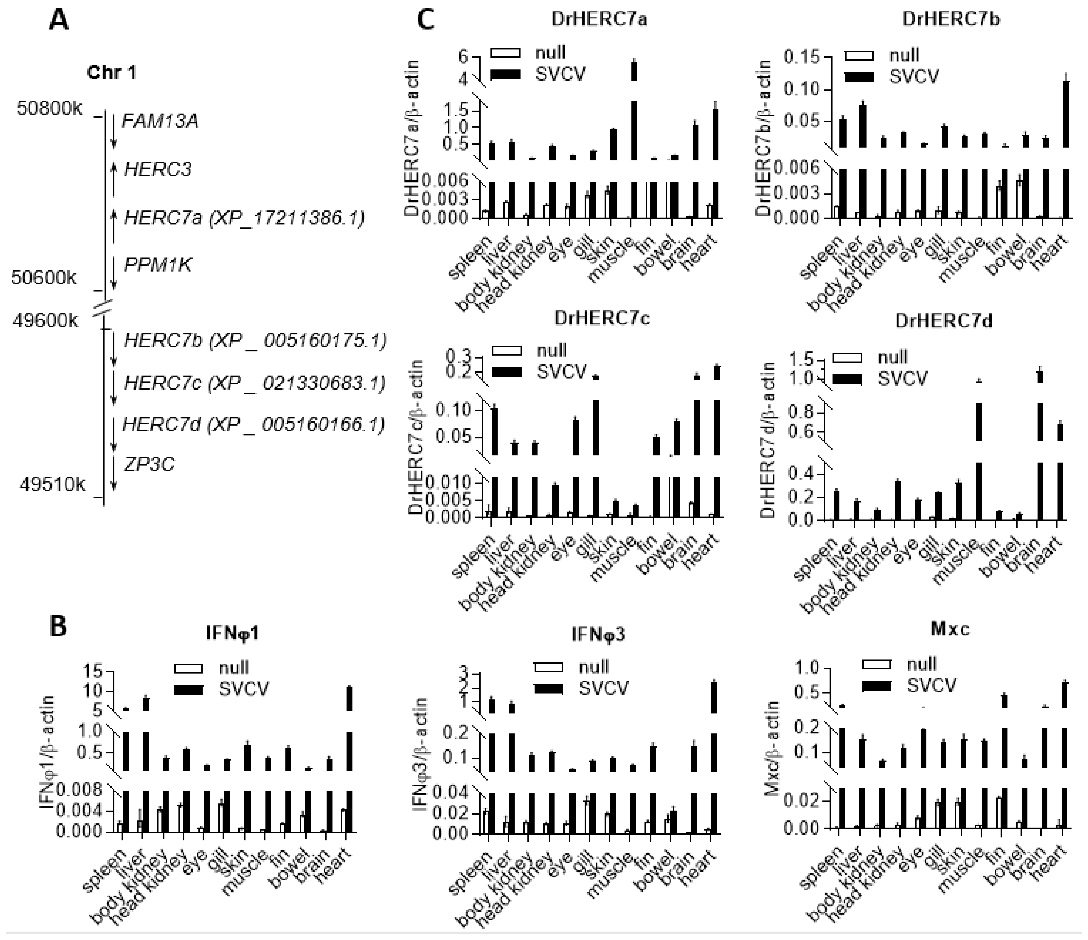
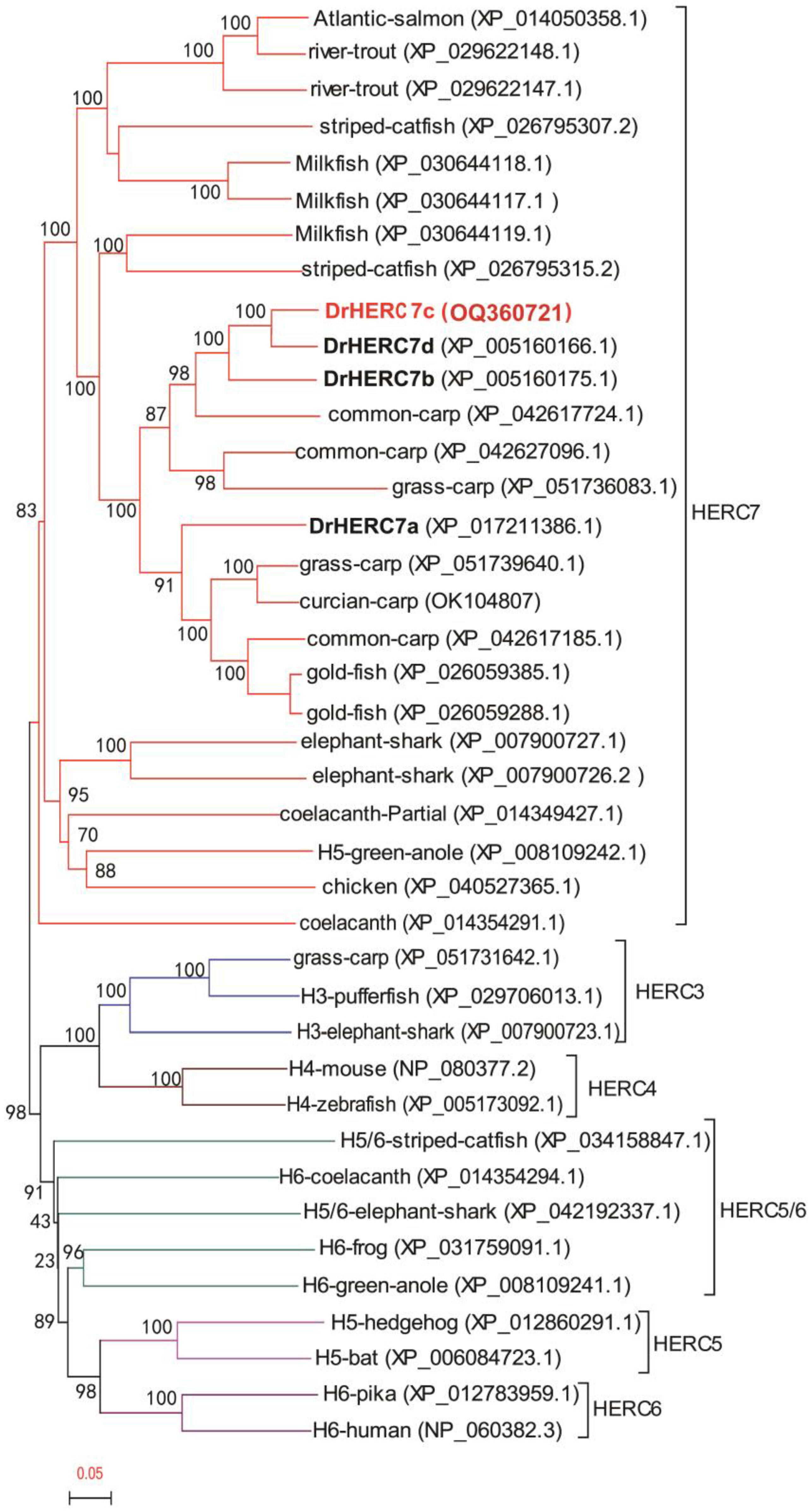
2.1. Zebrafish Has Four herc7 Genes That Are Induced by SVCV Infection

Using crucian carp HERC7 as query, blast searches of zebrafish genome (GRCz11) identified four herc7 homologous genes, sequentially named HERC7a (XP_17211386.1), HERC7b (XP_005160175.1), HERC7c (XP_021330683.1), and HERC7d (XP_005160166.1), which reside near the herc3 gene locus in Chromosome 1 but not the herc4 gene locus in Chromosome 13 (Figure 1A). Considering that the recently identified crucian carp HERC7 is transcriptionally induced by viral infection [19], we determined the expression patterns of four zebrafish herc7 genes in response to SVCV infection. Intraperitoneal injection of SVCV into zebrafish resulted in increased mRNA expressions of zebrafish ifnφ1 and ifnφ3, as well as mxc genes, a typical IFN-stimulated gene [20] (Figure 1B). Consistently, four zebrafish herc7 genes were also transcriptionally induced by SVCV infection (Figure 1C). These results implied that SVCV infection activated a robust IFN response in all zebrafish tissues and also the expression of four zebrafish herc7 genes.

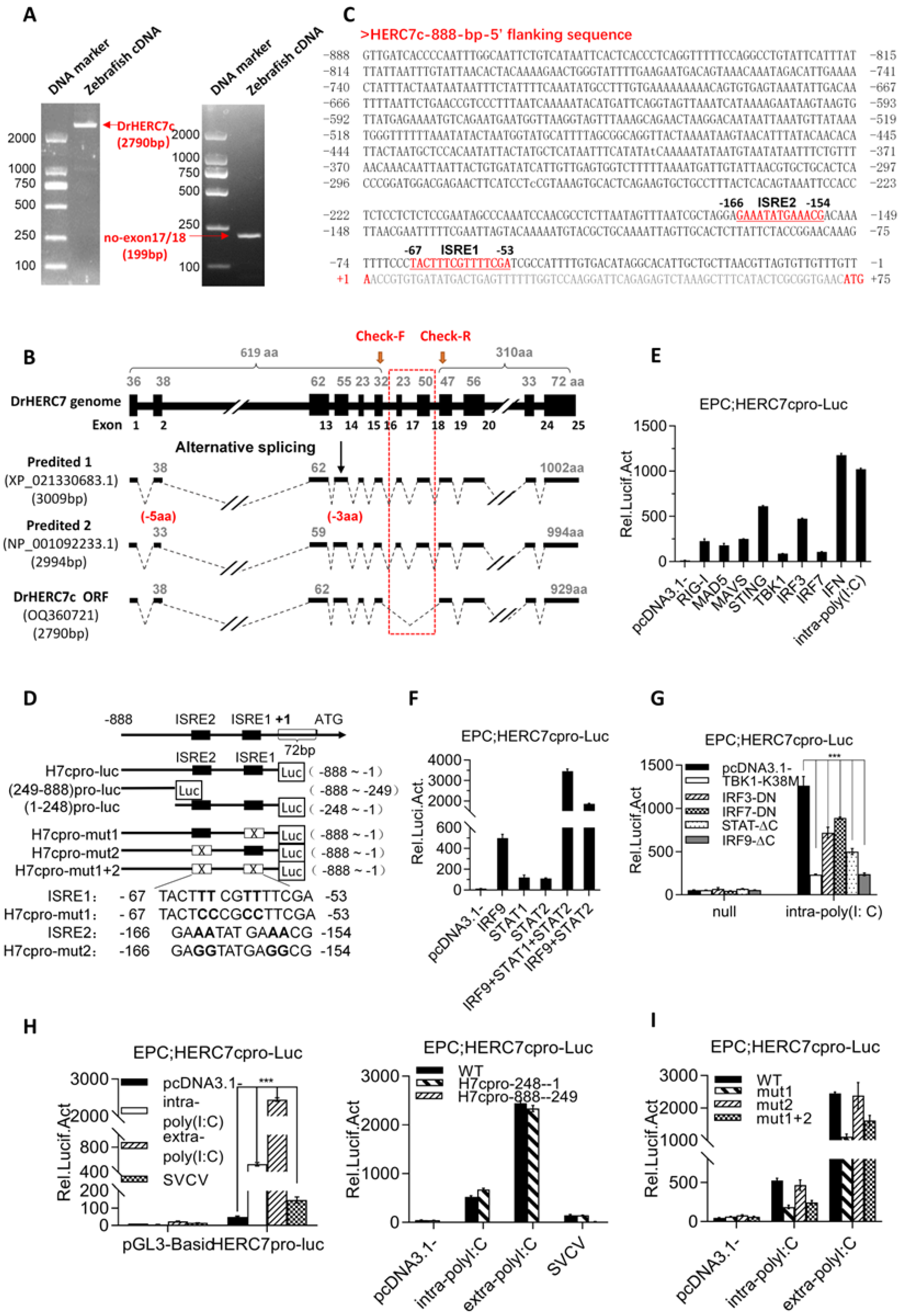
2.2. Zebrafish herc7c Is a Typical IFN-Stimulated Gene

We tried to clone the full-length cDNA sequences of four zebrafish herc7 genes. It was easy to obtain a single PCR band using a pair of primers designed against the 5′ UTR and 3′UTR sequences of the annotated herc7c in zebrafish genome data (left panel in Figure 2A). The cloned cDNA sequence contains the largest ORF of 2790bp, encoding a 929-aa HERC7c protein (Figure 2B). The annotated herc7c gene in zebrafish genome (GRCz11) was identified by automated computational analysis. Interestingly, it was annotated to contain 25 exons and was predicated to generate two transcript variants, encoding a 1002-aa protein and a 994-aa protein, respectively (Figure 2B). Sequence comparison revealed that our cloned herc7c cDNA sequence (OQ360721) does not contain the predicated 17th and 18th exons annotated in zebrafish genome (GRCz11) (Figure 2B). We repeated PCR experiments using different tissues samples; however, the same PCR product rather than both annotated transcript variants was cloned. Further, we designed a second pair of primes against sequences of the 16th and 19th exons (Figure 2B) to verify whether the 2 annotated variants were expressed. RT-PCR assays still obtained a single product with a predicated size of 199 bp representing our cloned herc7c cDNA sequence (OQ360721), without a larger product of 388 bp predicated from the 2 annotated transcription variants (right panel in Figure 2A). Thus, we thought that the cloned cDNA (OQ360721) might be the real transcript of the zebrafish herc7c gene.
To characterize whether zebrafish HERC7c is induced by IFN stimuli, we further cloned an 888-bp-5′ flanking sequence relative to the transcription start site of zebrafish herc7c gene (Figure 2C). This sequence has two putative ISRE/IRF-E sites, and it was next used to construct an herc7c promoter-driven luciferase plasmid (HERC7cpro-luc) and several derived mutants by mutating two predicated ISRE motifs (Figure 2D). As expected, the 888-bp-promoter of herc7c (HERC7cpro-luc) was robustly activated in EPC (Epithelioma papulosum cyprini) cells when transfected with poly(I:C) (polyinosinic-polycytidylic acid) as intracellular poly(I:C), or each of RLR signaling molecules, including RIG-I, MDA5, MAVS, STING, TBK1, IRF3, and IRF7 (Figure 2E). Overexpression of IFN also effectively activated HERC7cpro-luc (Figure 2E), and this activation was seen by individual or collective overexpression of STAT1, STAT2, and IRF9 (Figure 2F), three pivotal molecules involved in the IFN-triggered JAK-STAT signaling pathway [21,22,23].
The RLR pathway-induced IFN response can be triggered in fish cells by poly(I:C) transfection and SVCV infection [24,25]. Subsequent assays showed that intra-poly(I:C)-triggered activation of HERC7cpro-luc was severely decreased by individual overexpression of dominant negative mutants of RLR signaling factors (TBK1-K38M, IRF3-DN, and IRF7-DN) as well as JAK-STAT signaling factors (STAT1-ΔC and IRF9-ΔC) (Figure 2G). Compared with the full-length HERC7c promoter sequence (WT, −888~−1) that could be activated by intra-poly(I:C), extra-poly(I:C), or SVCV, a truncated promoter sequence (−248~−1) containing 2 putative ISRE motifs (ISRE1 and ISRE2) retained the intact promoter activation, but a second truncated promoter sequence devoid of 2 ISRE motifs (−888~−249) failed to respond to 3 stimuli above (Figure 2H). Mutation of ISRE1 but not ISRE2 gave a weakened luciferase activity (Figure 2I), indicating that the proximal ISRE1 motif rather than the distant ISRE2 was responsible for the HERC7c promoter activation. These results together indicated that zebrafish herc7c is a typical ISG.

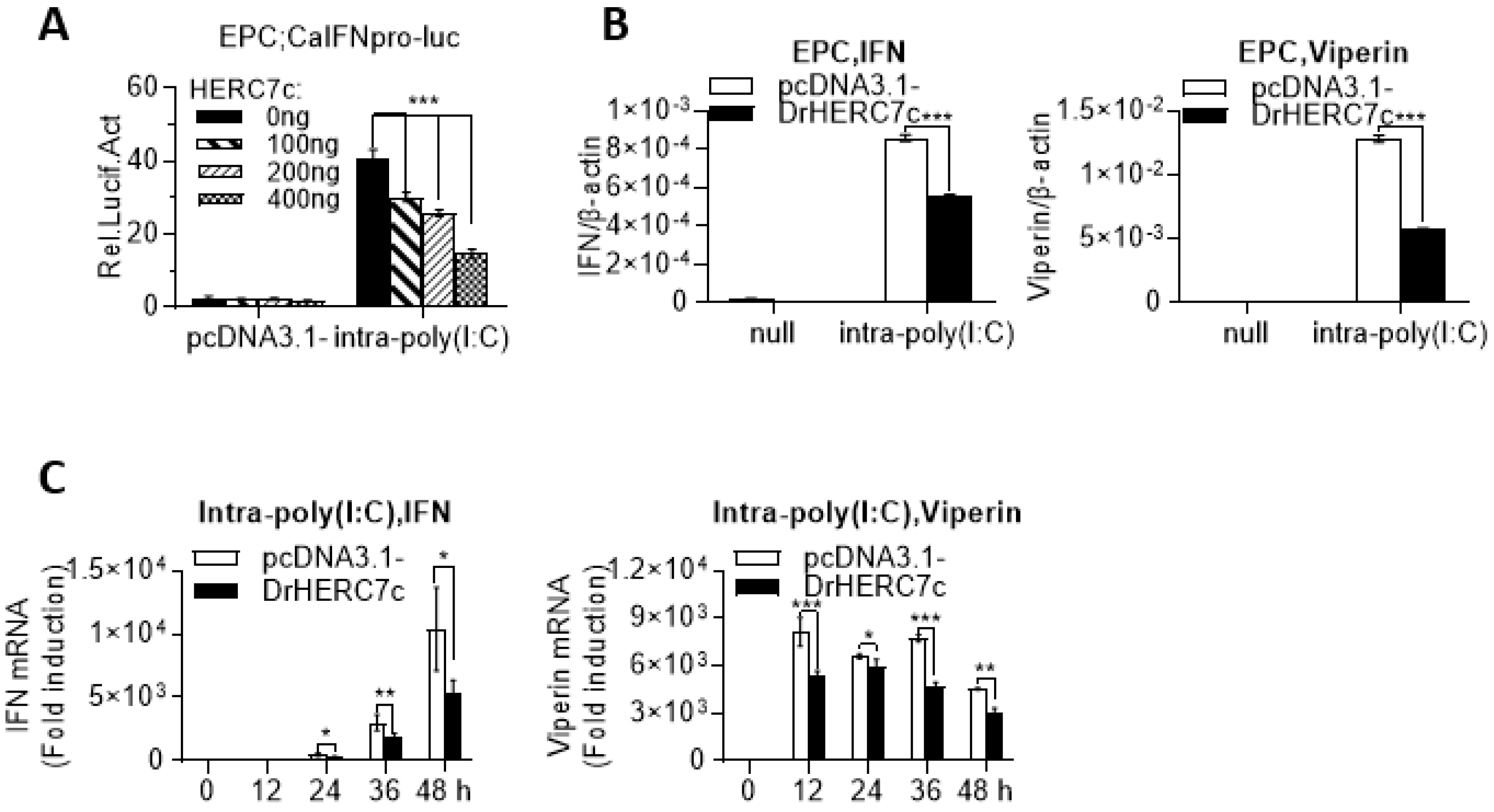
2.3. Zebrafish HERC7c Inhibits Fish IFN Response

Given that the recently identified crucian carp HERC7 is involved in downregulation of the fish IFN response [19], we next investigated whether zebrafish HERC7c played a similar role. Luciferase assays showed that, compared with the control cells transfected with pcDNA3.1, poly(I:C) transfection resulted in robust activation of crucian carp IFN promoter-driven luciferase plasmid (CaIFNpro-luc); however, this activation was severely impeded by overexpression of zebrafish HERC7c in a dose-dependent manner (Figure 3A). Consistently, overexpression of zebrafish HERC7c in EPC cells inhibited poly(I:C)-triggered mRNA expression of ifn and viperin (Figure 3B), which was seen in a time-dependent manner (Figure 3C). These results indicated that zebrafish HERC7c functions as an inhibitor of IFN response in fish.

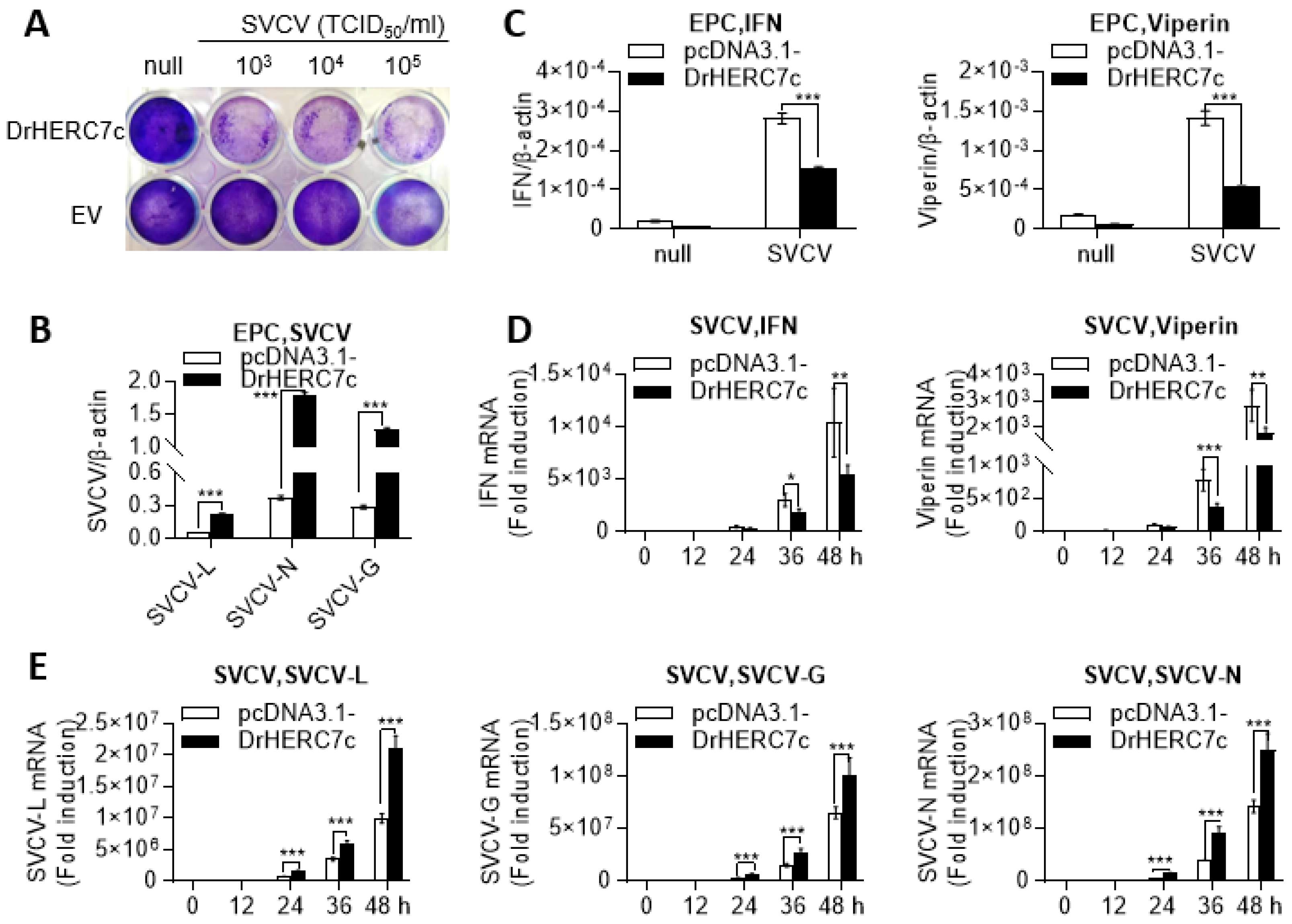
2.4. Zebrafish HERC7c Promotes Virus Replication in Fish Cells

To determine the effect of zebrafish HERC7c on viral replication, EPC cells were transfected with zebrafish HERC7c or pcDNA3.1 as control, followed by infection of SVCV at different titers. SVCV incubation yielded broad CPEs in zebrafish HERC7c-overexpressing cells compared with control cells (Figure 4A). Consistently, a higher transcription level of three SVCV genes (L, N, and G) was detected in HERC7c-overexpressing cells than in control cells (Figure 4B). Under the same conditions, the increase in the expression of cellular ifn and viperin in the presence of HERC7c overexpression was less than in the case of its absence (Figure 4C). Subsequent assays showed that overexpression of zebrafish HERC7c inhibited SVCV-triggered mRNA expression of cellular ifn and viperin over infection time (Figure 4D) and, concomitantly, resulted in a transcription elevation of the three SVCV genes (Figure 4E). These results indicated that zebrafish HERC7c promotes virus replication in fish cells likely through downregulation of the fish IFN response.

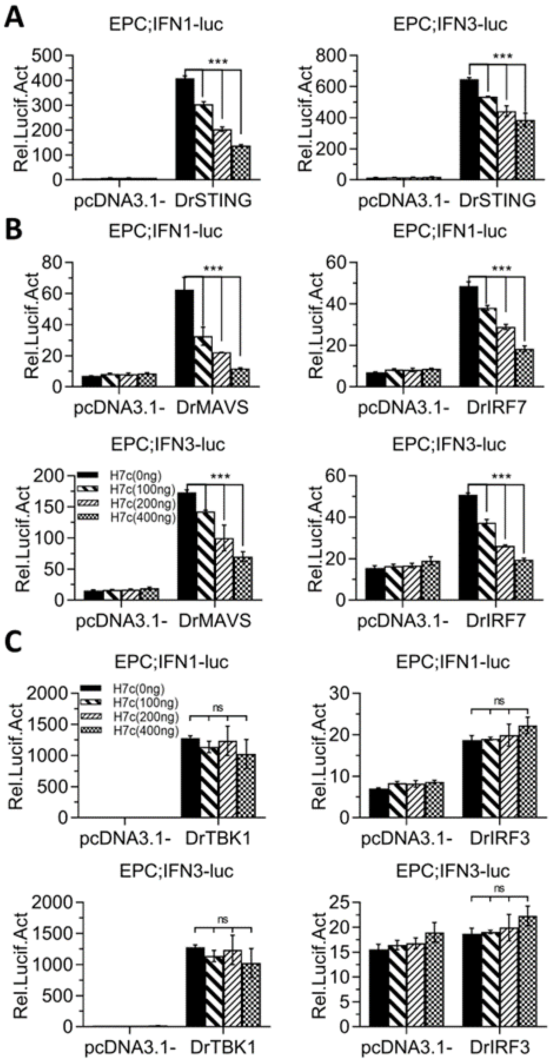
2.5. Zebrafish HERC7c Downregulates Fish IFN Response by Targeting STING, MAVS, and IRF7

Luciferase assays showed that overexpression of STING stimulated the activation of zebrafish IFNφ1 promoter- or IFNφ3 promoter-driven luciferase plasmids (DrIFNφ1pro-luc, DrIFNφ3pro-luc); however, this activation was alleviated in the presence of zebrafish HERC7c (Figure 5A). Zebrafish HERC7c-mediated alleviation was seen when IFN promoter activation was stimulated by MAVS or IRF7 (Figure 5B), but not by TBK1 or IRF3 (Figure 5C). It was noteworthy that overexpression of zebrafish IRF3 just activated zebrafish IFNφ1 promoter but not zebrafish IFNφ3 promoter (Figure 5C), similar to our previous results [24]. These data indicated that zebrafish HERC7c targets STING, MAVS, and IRF7 to downregulate the IFN antiviral response.

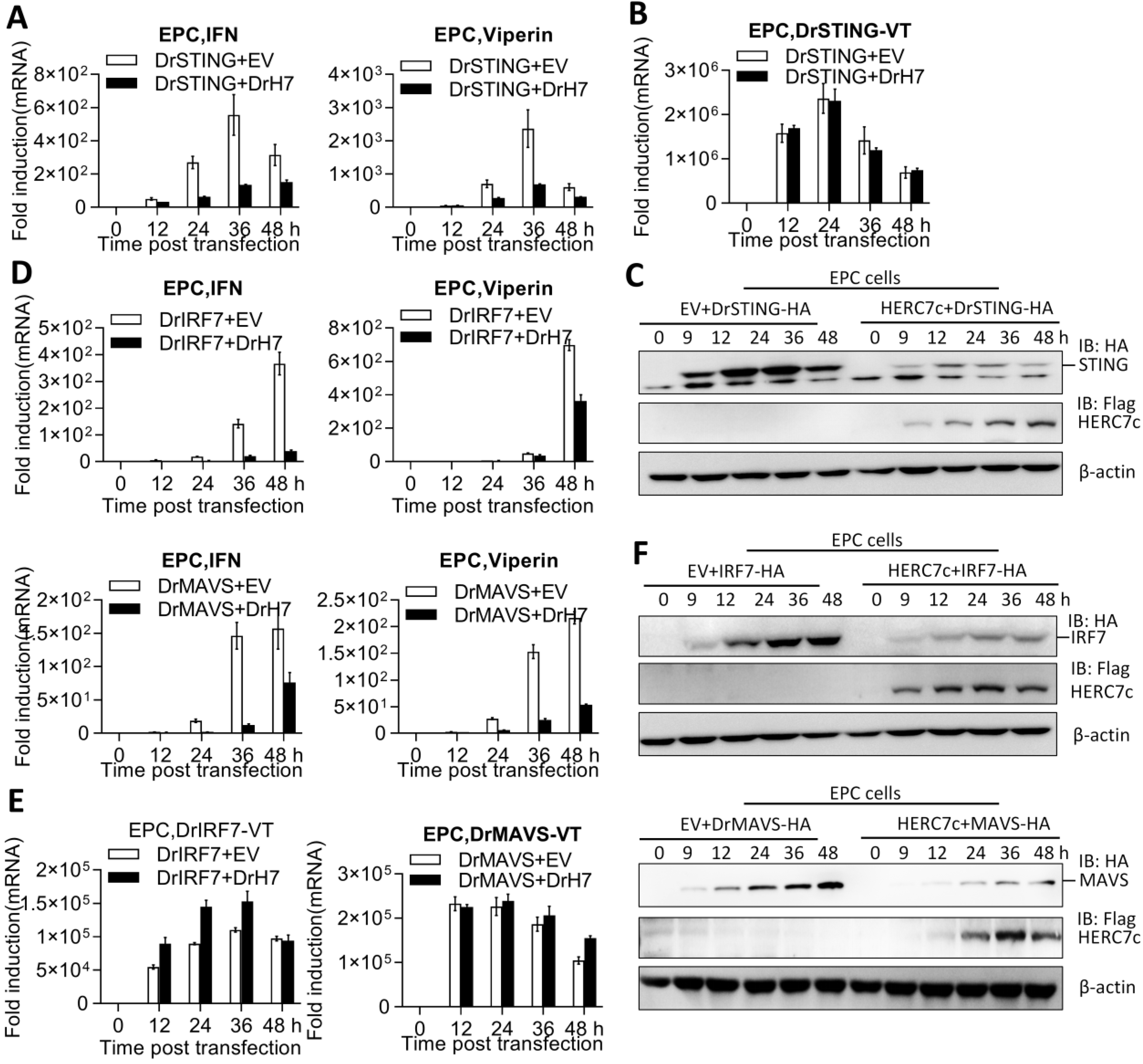
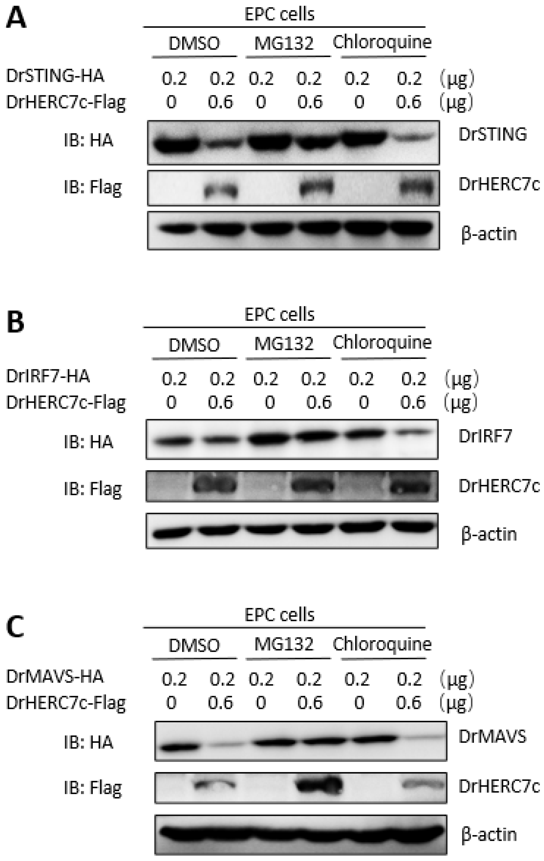
2.6. Zebrafish HERC7c Facilitates STING, MAVS, and IRF7 Protein Degradation to Downregulate IFN Response

To further determine the mechanism of how zebrafish HERC7c downregulated the STING-mediated IFN response, the effect of zebrafish HERC7c on gene transcription of ifn and sting was initially investigated. As anticipated, zebrafish STING-induced transcription of cellular ifn and viperin genes was significantly attenuated by zebrafish HERC7c over infection time (Figure 6A). However, zebrafish HERC7c did not have influences on sting gene transcription because a nearly identical pattern was detected between the cells overexpressing STING alone and the cells overexpressing STING and HERC7c together; the detected pattern was a pair of primers that amplifies mRNAs only from the transfected STING plasmid (Figure 6B). Conversely, Western blots showed that simultaneous transfection of HERC7c and STING resulted in a decrease in STING proteins compared with the single transfection of STING (Figure 6C).
Similar assays showed that overexpression of zebrafish HERC7c time-dependently alleviated mRNA expression of cellular ifn and viperin, which were triggered by MAVS or IRF7 (Figure 6D). Under the same conditions, zebrafish HERC7c did not make a difference on mRNA expression of the transfected mavs or irf7 (Figure 6E) but significantly attenuated their protein expressions (Figure 6F). Zebrafish HERC7c-mediated protein degradation of STING, MAVS, and IRF7 was blocked by the addition of MG132 (an inhibitor of the ubiquitin-proteasomal-dependent degradation pathway) but not of chloroquine (an inhibitor of the autophagy-lysosomal-dependent degradation pathway) (Figure 7A–C). These results indicated that zebrafish HERC7c facilitates STING, MAVS, and IRF7 protein degradation to downregulate the IFN response.

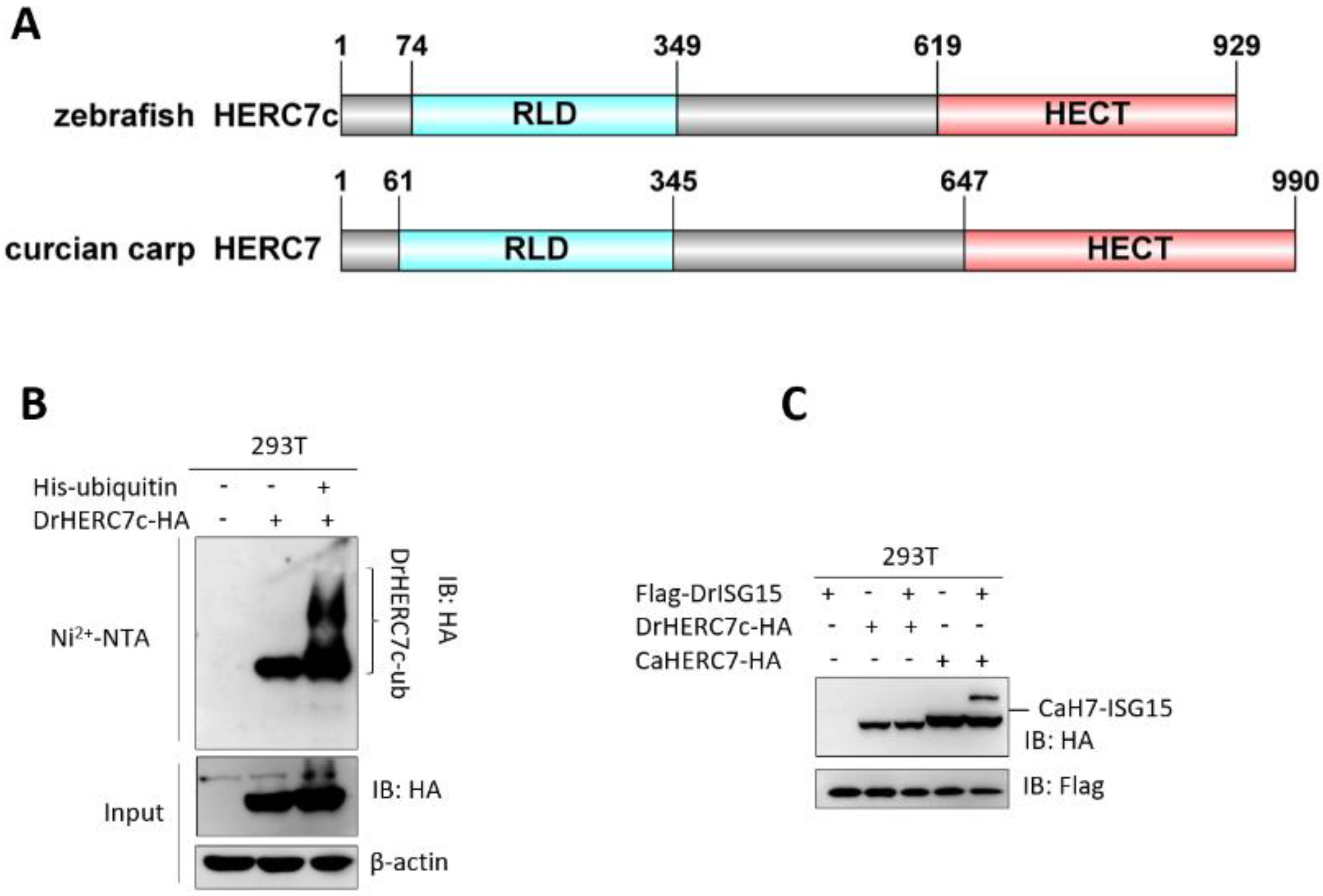
2.7. Zebrafish HERC7c Is an E3 Ligase for the Conjugation of Ubiquitin but Not ISG15

Similar to the recently identified crucian carp HERC7 [19], zebrafish HERC7c contains an N-terminal RLD domain and a C-terminal HERC domain (Figure 8A). It is believed that the HECT domain entitles HERC proteins with the E3 ubiquitin ligase activity, and particularly, human HERC5 and mouse HERC6 are also responsible for protein ISGylation [9]. To this end, we investigated whether zebrafish HERC7c has a potential to transfer ubiquitin or ISG15. Similar to the recently-identified crucian carp HERC7 that can be ubiquitinated by itself [19], transfection of HEK293T cells with His-ubiquitin and HERC7c-Flag followed by affinity purification of His-ubiquitin using Ni2+-NTA resin revealed an enhanced level of ubiquitinated HERC7c proteins compared with overexpression of HERC7c alone (Figure 8B). Similar assays showed that crucian carp HERC7 rather than zebrafish HERC7c could be modified by ISG15 in cells when simultaneously transfected with ISG15 instead of ubiquitin (Figure 8C). These results indicated that zebrafish HERC7c is an E3 ligase responsible for ubiquitinylation but not for ISGylation.

2.8. Zebrafish HERC7c Is a Species-Specific Gene

The HERC7 subfamily exists in non-mammalian vertebrates [19]. To determine the relationship of zebrafish HERC7c in the HERC7 subfamily, we collected a total of 40 small HERC homologs from elephant shark (C. milii) to birds. Apart from four HERC7 members in zebrafish, blast searches of genome data revealed three HERC7 members in grass carp, three in common carp, and two in goldfish. Phylogenetical tree analyses showed that all HERC7 members are clustered into a clad that is distinct from the HERC3, HERC4, HERC5, and HERC6 subfamilies. Four zebrafish HERC7s were divided into two branches and each has no “one to one” orthologs in other fish species (Figure 9). These results indicated the occurrence of the species-specific expansion of the HERC7 subfamily in zebrafish, thus implying that zebrafish HERC7c might be a species-specific gene.

3. Discussion

HERC1 is the founding member of the HERC family characterized in human breast cancer cells [26], followed by the identification of a total of six HERC members in human, which are phylogenetically divided into large HERCs (HERC1 and HERC2) and small HERCs (HERC3-6) [2,10]. By genome-wide search of small HERC homologs from elephant shark to mammals and subsequently comprehensive evolutionary analyses, we provide evidence showing that, whereas HERC3 and HERC4 are conserved across vertebrates, the orthologs of human HERC5 and HERC6 are only present in mammals with a definite orthologous relationship to each other, and importantly, non-mammalian vertebrates have a unique HERC7 subfamily that might have been lost in modern mammals [19].
In this study, we cloned zebrafish herc7c cDNA by nested PCR. Interestingly, the obtained sequence is different from the two “annotated” transcripts in the zebrafish genome (GRCz11) by automated computational analysis. To verify whether our cloned sequence is a novel splicing variant of zebrafish herc7c, we designed a second pair of primers based on our hypothesis that, if there is a splicing variation, two PCR products will be amplified. However, only one band corresponding to the cloned sequence was obtained. Therefore, we think that our cloned cDNA represents the real transcript of the zebrafish herc7c gene.
Subsequently, we identified four herc7 homologs in zebrafish Chromosome 1, showing that they are adjacent to herc3. In mammals, small HERCs generally reside in two gene loci, one containing a single herc4 gene in one chromosome, and the other containing herc3 and other herc genes in a second chromosome [1,19]. The chromosomal location link of zebrafish herc3 and four herc7 genes indicate that they might originate from a common ancestry. In addition, different fish species have varied copies of herc7 genes, most of which seem not to exhibit a “one to one” orthologous relationship to each other. Given that teleost fish have additional whole-genome duplication [27,28,29], we propose that the varied herc7 copies in different fish species might arise from segmental duplication after the additional whole-genome duplication. These results support a notion that the small HERC family has experienced gene duplication, chromosomal rearrangement, and gene loss events during vertebrate evolution [1,2,3,18].
The results in the current study establish that zebrafish HERC7 destabilizes different signaling molecules at the protein level to downregulate the IFN response during viral infection. First, zebrafish HERC7c is transcriptionally expressed along the viral infection. Secondly, zebrafish HERC7c benefits SVCV replication and concomitantly alleviates the IFN response. Thirdly, zebrafish HERC7c markedly abrogates the IFN promoter activation and ifn gene transcription by RLR signaling molecules, including STING, MAVS, and IRF7. Mechanistically, zebrafish HERC7c targets STING, MAVS, and IRF7 for proteasome-dependent protein degradation, as evidenced by the findings that this degradation is blocked by MG132. An interesting question is that zebrafish HERC7c targets IRF7 but not IRF3 to attenuate IFN expression, although IRF3 is most homologous to IRF7 [24,30,31]. This means a possibility that zebrafish HERC7c can specifically select the substrates to exert inhibitory effects. The same is true for the differential function of zebrafish IRF3 and IRF7 because they are responsible for the transcription of the zebrafish ifnφ1 and ifnφ3 genes, respectively [23,24]. Although vertebrate IRF3s exhibit a “one to one” orthologous relationship to each other, fish IRF3 is a virus- and IFN-induced protein, and instead, mammalian IRF3 is constitutively expressed even under viral infection [31,32,33]. These results further suggest that the inhibitory effect of zebrafish HERC7c on the IFN response might be a consequence of selection pressures that are exerted by fish viruses.
Given that zebrafish HERC7c is identified as an inhibitor of the fish IFN response, mammalian HERC5s play a positive role in limiting different virus replications [1]. In this paper, the authors present evidence that a coelacanth protein (XP_014354291.1) also displays a similar inhibitory effect on simian immunodeficiency virus replication [1]. However, subsequently we found that this coelacanth protein (XP_014354291.1) is indeed a HERC7 homolog although erroneously characterized as a coelacanth homolog of human HECR5 by comprehensive phylogenetic analyses [19]. Therefore, coelacanth does not contain an ortholog of human HERC5, and the coelacanth HERC7 protein (XP_014354291.1) acts as an intracellular viral inhibitor, similar to human HERC5 [1]. Moreover, the recently-identified crucian carp HERC7 attenuates the IFN response by utilizing two different mechanisms to degrade STING, MAVS, and IRF7 at the protein and mRNA levels [19]. These results indicate that the HERC7 subfamily members have undergone functional diversification, and thus they might play opposing roles in response to viral infection, such as the coelacanth protein (XP_014354291.1) as a positive regulator, and zebrafish HERC7c and crucian carp HERC7 as a negative regulator.
Small HERCs possess E3 ligase activity as a result of their C-terminal HECT domains [34]. Interestingly, whereas crucian carp HERC7 participates in both ubiquitinylation and ISGylation, zebrafish HERC7c just bears the potential to transfer ubiquitin (ubiquitinylation), further indicating that fish HERC7 subfamily members have experienced function diversification. In the present study, we did not investigate whether the E3 ubiquitin ligase activity of zebrafish HERC7c is tightly related to its inhibitory role in fish antiviral response; however, our previous report has shown that the inherent E3 ligase activity seems to not be required for the crucian carp HERC7 downregulation of the IFN response because a mutant without E3 ligase activity displays a nearly intact ability to alleviate the fish IFN response [19].
Given that four zebrafish herc7 genes are generated by fish species-specific expansion of the HERC7 family, zebrafish herc7c might represent a zebrafish-specific gene, as evidenced by the fact that we failed to find a “one-to-one” ortholog of zebrafish HERC7c by comprehensive analyses of the available fish genome databases. Since zebrafish HERC7c expression is elevated along with viral infection, the data in the present study suggest that zebrafish HERC7c might mediate a fish species-specific regulation of the IFN response to avoid unregulated IFN production during viral infection. Further studies might focus on in vivo function clarification of zebrafish HERC7c. If the function blockade of zebrafish HERC7c were able to effectively improve zebrafish survival against viral infection, it would highlight a relevance of HERC7 subfamily genes, such as zebrafish HERC7c with an inhibitory role, in fish antiviral precision breeding by gene-editing technology [35].

4. Materials and Methods

4.1. Cells, Virus, and Zebrafish

Epithelioma papulosum cyprini cells (EPC) were cultured in medium 199 supplemented with 10% fetal bovine serum (FBS) at 28 °C in a humidified incubator containing 5% CO2. Human embryonic kidney 293T cells (HEK293T, ATCC (CRL-3216)) were cultured in DMEM supplemented with 10% FBS at 37 °C.
Spring viremia of carp virus (SVCV) was propagated in EPC cells and tittered, according to the method of Reed and Muench, by a tissue culture ID50 assay. EPC cells were infected with SVCV at a final concentration of 5 × 10³ TCID50/mL.
Zebrafish adults (2-month-old), with similar sizes and weights (body length: 3 cm; weight: 0.4 g), were raised in a single batch according to standard protocol [36], which was approved by the Animal Care and Use Committee of Institute of Hydrobiology, Chinese Academy of Sciences. For viral infection, zebrafish were intraperitoneally injected with SVCV at 1 × 10⁸ TCID50/mL (25 μL/fish). After injection, they were kept at 28 °C water for 48 h without feeding. The control group received the same dose of 0.9% normal saline. After 48 h, the zebrafish were euthanized by immersing in a mixture of ice water for 20 min, followed by sampling of tissues.

4.2. Gene Cloning, Database Mining, and Sequence Analysis

Four immune tissues (spleen, liver, head kidney, and body kidney), from a zebrafish adult infected for 48 h with SVCV, were mixed to extract total RNA for cDNA synthesis by TRUEscript RT MasterMix (PC5801, Aidlab, Beijing, China). The mixed cDNA was used as a template to clone the ORF of zebrafish herc7c by nested PCR method. Zebrafish genome DNAs were extracted by the Wizard Genomic DNA Purification Kit (Promega) for PCR amplification of the herc7c promoter. The primers were designed against the computational annotated herc7c sequence in the zebrafish genome data (GRCz11) (Table 1).
Using zebrafish HERC7c (GenBank accession no. OQ360721) protein sequence as a query, protein BLAST searches were performed on the genome databases of grass carp (Ctenopharyngodon idella), common carp (Cyprinus carpio), and goldfish (Carassius auratus). Three homologs of zebrafish HERC7 were found in grass carp (XP_051736083.1, XP_051739640.1, and XP_051731642.1) and common carp genomes (XP_042627096.1, XP_042617724.1, and XP_042617185.1), and two in goldfish genome (XP_026059385.1, XP_026059288.1). Meanwhile, 40 sequences of small HERC family members in other species, which were verified by evolutionary analysis in our previous study [19], were collected for subsequent evolutionary tree analysis. They include: HERC3 (pufferfish, elephant shark, and grass carp), HERC4 (mouse and zebrafish), HERC5 (hedgehog and bat), HERC6 (human and pika), HERC5/6 (striped catfish, coelacanth, elephant shark, frog, and green anole), and HERC7 (Atlantic salmon, river trout striped catfish, milkfish, zebrafish, common carp, grass carp, crucian carp, goldfish, elephant shark, coelacanth, green anole, and chicken). Multiple alignments were carried out with ClustalW2 to make a phylogenetic tree by neighbor-joining methods in Geneious. Transcription factor-binding sites were predicated using JASPAR database (http://jaspar.genereg.net/) accessed on 5 June 2022.

4.3. Plasmid Construction

For overexpression, the ORF of DrHERC7c was cloned into EcoR I and BamH I sites of pcDNA3.1/myc-His (-) A (Invitrogen). At the same time, different tags (HA, Flag) were added to the C-terminus of DrHERC7c by reverse amplification primers. For promoter analysis, 3 size-different 5′ flanking sequences of DrHERC7c including HERC7cpro-luc (−888–−1), HERC7cpro-luc (−888–−249), and HERC7cpro-luc (−248–−1) were cloned into Nhe I site of the pGL3-basic plasmid. The same method was carried out for three mutations plasmids (HERC7cpro-mut1, HERC7cpro-mut2, and HERC7cpro-mut1+2). Zebrafish plasmids DrMAVS/DrMAVS-Flag, DrSTING/DrSTING-HA, TBK1/TBK1-HA, IRF3/IRF3-HA, and IRF7/IRF7-HA were described in our previous reports [37,38]. Other plasmids including TBK1-K38M, IRF3-DN, IRF7-DN, IFNφ1pro-luc, IFNφ3pro-luc, STAT1-ΔC, and IRF9-ΔC were described previously [21,22,24,30].

4.4. Transfection and Luciferase Activity Assays

Cell transfection assays were performed with polyethylenimine, Linear (PEI, MW 25,000) (Sigma-Aldrich, Shanghai, China) according to our previous reports [15,36]. Luciferase activity assays were performed by a Junior LB 9509 luminometer (Berthold, Pforzheim, Germany) using Dual-Luciferase Reporter Assay System (Promega), as described previously [15,16,33]. The results were the representative of at least three independent experiments, each performed in triplicate. Luciferase activities were normalized to the amounts of internal control Renilla luciferase activities.

4.5. RNA Extraction, cDNA Synthesis, and Real Time-PCR

Cellular total RNAs were extracted by EASYspin Plus Kit (Aidlab, Beijing, China), followed by DNase treatment to remove residual DNA. First-strand cDNA was synthesized using MonScriptTM RTIII Super Mix with dsDNase Kit (Monad, Suzhou, China) according to the manufacturer’s protocol [36]. Real-time PCR (RT-qPCR) was performed with Hieff qPCR SYBR Green Master Mix (Yeasen, Shanghai, China) on the CFX96 real-time system (Bio-Rad). PCR condition was set by referring to the operation manual of the Hieff qPCR SYBR Green Master Mix. The relative expression was normalized to the expression of β-actin and represented as the fold induction relative to the expression level in the control cells that was set to 1. The primers used in this study are listed in Table 1. The primer designing principle follows a single amplification band and an amplification length between 100–250 bp.

4.6. Ubiquitination Assays and ISGylation Assays

For ubiquitination assays, HEK293T cells seeded in 10 cm2 dishes were transfected with the indicated plasmids. After 30 h, the transfected cells were lysed, and the cell supernatant was incubated with Ni2+-NTA His. Bind Resin (Novagen) at 4 °C overnight, followed by Western blotting using tag-specific Abs [14]. For ISGylation assays, HEK293T cells were transfected with zebrafish HERC7c-HA together with or without 5 μg Flag-ISG15. After 30 h, the cells were collected for Western blotting with tag-specific antibodies.

4.7. Statistical Analysis

Student’s t-test is applied for statistical analysis of the data derived from luciferase assays and RT-PCR assays. All quantitative experiments were performed with at least three independent biological repeats. (* p < 0.05; ** p < 0.01; *** p < 0.001, ns: no significant.)

Author Contributions

Conceptualization, Y.-B.Z. and Y.-L.L.; methodology, Y.-L.L., X.-Y.G., Z.-L.Q. and Y.-B.Z.; validation, Y.-L.L. and Y.-B.Z.; formal analysis, Y.-L.L., X.-Y.G., Z.-L.Q., X.Z., H.-Y.S., L.-L.A., C.D. and Y.-B.Z.; investigation, Y.-L.L.; resources, Y.-B.Z. and J.-F.G.; data curation, Y.-L.L. and Y.-B.Z.; writing—original draft preparation, Y.-B.Z. and Y.-L.L.; writing—review and editing, Y.-B.Z., J.-F.G. and Y.-L.L.; project administration, Y.-B.Z.; funding acquisition, Y.-B.Z. All authors have read and agreed to the published version of the manuscript.

Funding

This work was funded by the Strategic Priority Research Program of the Chinese Academy of Sciences (XDA24010308-2), a grant from the National Key R&D Program of China (2022YFF1000302), and grants from the National Natural Science Foundation (31972826 and 32102838).

Institutional Review Board Statement

The animal study protocol was approved by the Animal Care and Use Committee of the Institute of Hydrobiology, Chinese Academy of Sciences (IHB, CAS, Protocol No. 2016–018).

Data Availability Statement

All of the data referred to in this article are located within the main text. If there are requests for additional unpublished data not referred to in this article, please contact the corresponding author at ybzhang@ihb.ac.cn.

Acknowledgments

We thank the Wuhan regional center of life science for the instrument help.

Conflicts of Interest

The authors declare no conflict of interest.

References

  1. Paparisto, E.; Woods, M.W.; Coleman, M.D.; Moghadasi, S.A.; Kochar, D.S.; Tom, S.K.; Kohio, H.P.; Gibson, R.M.; Rohringer, T.J.; Hunt, N.R.; et al. Evolution-Guided Structural and Functional Analyses of the HERC Family Reveal an Ancient Marine Origin and Determinants of Antiviral Activity. J. Virol. 2018, 92, e00528-18. [Google Scholar] [CrossRef] [PubMed] [Green Version]
  2. Hochrainer, K.; Mayer, H.; Baranyi, U.; Binder, B.; Lipp, J.; Kroismayr, R. The human HERC family of ubiquitin ligases: Novel members, genomic organization, expression profiling, and evolutionary aspects. Genomics 2005, 85, 153–164. [Google Scholar] [CrossRef] [PubMed]
  3. Marin, I. Animal HECT ubiquitin ligases: Evolution and functional implications. BMC Evol. Biol. 2010, 10, 56. [Google Scholar] [CrossRef] [PubMed] [Green Version]
  4. Schwarz, S.E.; Rosa, J.L.; Scheffner, M. Characterization of human hect domain family members and their interaction with UbcH5 and UbcH7. J. Biol. Chem. 1998, 273, 12148–12154. [Google Scholar] [CrossRef] [PubMed] [Green Version]
  5. Cruz, C.; Ventura, F.; Bartrons, R.; Rosa, J.L. HERC3 binding to and regulation by ubiquitin. FEBS Lett. 2001, 488, 74–80. [Google Scholar] [CrossRef]
  6. Kroismayr, R.; Baranyi, U.; Stehlik, C.; Dorfleutner, A.; Binder, B.R.; Lipp, J. HERC5, a HECT E3 ubiquitin ligase tightly regulated in LPS activated endothelial cells. J. Cell. Sci. 2004, 117 Pt 20, 4749–4756. [Google Scholar] [CrossRef] [Green Version]
  7. Dastur, A.; Beaudenon, S.; Kelley, M.; Krug, R.M.; Huibregtse, J.M. Herc5, an interferon-induced HECT E3 enzyme, is required for conjugation of ISG15 in human cells. J. Biol. Chem. 2006, 281, 4334–4338. [Google Scholar] [CrossRef] [PubMed] [Green Version]
  8. Wong, J.J.Y.; Pung, Y.F.; Sze, N.S.K.; Chin, K.C. HERC5 is an IFN-induced HECT-type E3 protein ligase that mediates type I IFN-induced ISGylation of protein targets. Proc. Natl. Acad. Sci. USA 2006, 103, 10735–10740. [Google Scholar] [CrossRef] [Green Version]
  9. Sala-Gaston, J.; Martinez-Martinez, A.; Pedrazza, L.; Lorenzo-Martin, L.F.; Caloto, R.; Bustelo, X.R.; Ventura, F.; Rosa, J.L. HERC Ubiquitin Ligases in Cancer. Cancers 2020, 12, 1653. [Google Scholar] [CrossRef]
  10. Garcia-Gonzalo, F.R.; Rosa, J.L. The HERC proteins: Functional and evolutionary insights. Cell. Mol. Life Sci. 2005, 62, 1826–1838. [Google Scholar] [CrossRef]
  11. Oudshoorn, D.; van Boheemen, S.; Sanchez-Aparicio, M.T.; Rajsbaum, R.; Garcia-Sastre, A.; Versteeg, G.A. HERC6 Is the Main E3 Ligase for Global ISG15 Conjugation in Mouse Cells. PLoS ONE 2012, 7, e29870. [Google Scholar] [CrossRef] [Green Version]
  12. Rehwinkel, J.; Gack, M.U. RIG-I-like receptors: Their regulation and roles in RNA sensing. Nat. Rev. Immunol. 2020, 20, 537–551. [Google Scholar] [CrossRef]
  13. McNab, F.; Mayer-Barber, K.; Sher, A.; Wack, A.; O’Garra, A. Type I interferons in infectious disease. Nat. Rev. Immunol. 2015, 15, 87–103. [Google Scholar] [CrossRef] [PubMed]
  14. Wu, M.; Zhao, X.; Gong, X.Y.; Wang, Y.; Gui, J.F.; Zhang, Y.B. FTRCA1, a Species-Specific Member of finTRIM Family, Negatively Regulates Fish IFN Response through Autophage-Lysosomal Degradation of TBK1. J. Immunol. 2019, 202, 2407–2420. [Google Scholar] [CrossRef] [PubMed]
  15. Qu, Z.L.; Li, Y.L.; Gong, X.Y.; Zhao, X.; Sun, H.Y.; Dan, C.; Gui, J.F.; Zhang, Y.B. A finTRIM Family Protein Acquires RNA-Binding Activity and E3 Ligase Activity to Shape the IFN Response in Fish. J. Immunol. 2022, 209, 1335–1347. [Google Scholar] [CrossRef] [PubMed]
  16. Gong, X.Y.; Zhang, Q.M.; Zhao, X.; Li, Y.L.; Qu, Z.L.; Li, Z.; Dan, C.; Gui, J.F.; Zhang, Y.B. LGP2 is essential for zebrafish survival through dual regulation of IFN antiviral response. iScience 2022, 25, 104821. [Google Scholar] [CrossRef] [PubMed]
  17. Shi, H.X.; Yang, K.; Liu, X.; Liu, X.Y.; Wei, B.; Shan, Y.F.; Zhu, L.H.; Wang, C. Positive regulation of interferon regulatory factor 3 activation by Herc5 via ISG15 modification. Mol. Cell Biol. 2010, 30, 2424–2436. [Google Scholar] [CrossRef] [PubMed] [Green Version]
  18. Jacquet, S.; Pontier, D.; Etienne, L. Rapid Evolution of HERC6 and Duplication of a Chimeric HERC5/6 Gene in Rodents and Bats Suggest an Overlooked Role of HERCs in Mammalian Immunity. Front. Immunol. 2020, 11, 605270. [Google Scholar] [CrossRef] [PubMed]
  19. Li, Y.L.; Gong, X.Y.; Qu, Z.L.; Zhao, X.; Dan, C.; Gui, J.F.; Zhang, Y.B. A Novel Non-Mammalian-Specific HERC7 Negatively Regulates IFN Response through Degrading RLR Signaling Factors. J. Immunol. 2022, 208, 1189–1203. [Google Scholar] [CrossRef]
  20. Zhang, Y.-B.; Li, Q.; Gui, J.-F. Differential expression of two Carassius auratus Mx genes in cultured CAB cells induced by grass carp hemorrhage virus and interferon. Immunogenetics 2004, 56, 68–75. [Google Scholar] [CrossRef]
  21. Yu, F.-F.; Zhang, Y.-B.; Liu, T.-K.; Liu, Y.; Sun, F.; Jiang, J.; Gui, J.-F. Fish virus-induced interferon exerts antiviral function through Stat1 pathway. Mol. Immunol. 2010, 47, 2330–2341. [Google Scholar] [CrossRef] [PubMed]
  22. Shi, J.; Zhang, Y.B.; Liu, T.K.; Sun, F.; Gui, J.F. Subcellular localization and functional characterization of a fish IRF9 from crucian carp Carassius auratus. Fish Shellfish. Immunol. 2012, 33, 258–266. [Google Scholar] [CrossRef] [PubMed]
  23. Zhang, Y.B.; Gui, J.F. Molecular regulation of interferon antiviral response in fish. Dev. Comp. Immunol. 2012, 38, 193–202. [Google Scholar] [CrossRef] [PubMed]
  24. Sun, F.; Zhang, Y.B.; Liu, T.K.; Shi, J.; Wang, B.; Gui, J.F. Fish MITA serves as a mediator for distinct fish IFN gene activation dependent on IRF3 or IRF7. J. Immunol. 2011, 187, 2531–2539. [Google Scholar] [CrossRef] [Green Version]
  25. Gong, X.Y.; Zhang, Q.M.; Gui, J.F.; Zhang, Y.B. SVCV infection triggers fish IFN response through RLR signaling pathway. Fish Shellfish. Immunol. 2019, 86, 1058–1063. [Google Scholar] [CrossRef]
  26. Rosa, J.L.; Casaroli-Marano, R.P.; Buckler, A.J.; Vilaro, S.; Barbacid, M. p619, a giant protein related to the chromosome condensation regulator RCC1, stimulates guanine nucleotide exchange on ARF1 and Rab proteins. EMBO J. 1996, 15, 4262–4273. [Google Scholar] [CrossRef]
  27. Jaillon, O.; Aury, J.-M.; Brunet, F.; Petit, J.-L.; Stange-Thomann, N.; Mauceli, E.; Bouneau, L.; Fischer, C.; Ozouf-Costaz, C.; Bernot, A.; et al. Genome duplication in the teleost fish Tetraodon nigroviridis reveals the early vertebrate proto-karyotype. Nature 2004, 431, 946–957. [Google Scholar] [CrossRef] [Green Version]
  28. Wang, Y.; Li, X.Y.; Xu, W.J.; Wang, K.; Wu, B.; Xu, M.; Chen, Y.; Miao, L.J.; Wang, Z.W.; Li, Z.; et al. Comparative genome anatomy reveals evolutionary insights into a unique amphitriploid fish. Nat. Ecol. Evol. 2022, 6, 1354–1366. [Google Scholar] [CrossRef]
  29. Gui, J.F.; Zhou, L.; Li, X.Y. Rethinking fish biology and biotechnologies in the challenge era for burgeoning genome resources and strengthening food security. Water Biol. Secur. 2022, 1, 100002. [Google Scholar] [CrossRef]
  30. Feng, H.; Zhang, Q.M.; Zhang, Y.B.; Li, Z.; Zhang, J.; Xiong, Y.W.; Wu, M.; Gui, J.F. Zebrafish IRF1, IRF3, and IRF7 Differentially Regulate IFNPhi1 and IFNPhi3 Expression through Assembly of Homo- or Heteroprotein Complexes. J. Immunol. 2016, 197, 1893–1904. [Google Scholar] [CrossRef] [PubMed] [Green Version]
  31. An, L.L.; Zhao, X.; Gong, X.Y.; Li, Y.L.; Qu, Z.L.; Sun, H.Y.; Guo, W.H.; Dan, C.; Gui, J.F.; Zhang, Y.B. Promoter Binding and Nuclear Retention Features of Zebrafish IRF Family Members in IFN Response. Front. Immunol. 2022, 13, 861262. [Google Scholar] [CrossRef]
  32. Au, W.C.; Moore, P.A.; Lowther, W.; Juang, Y.T.; Pitha, P.M. Identification of a Member of the Interferon Regulatory Factor Family That Binds to the Interferon-Stimulated Response Element and Activates Expression of Interferon-Induced Genes. Proc. Natl. Acad. Sci. USA 1995, 92, 11657–11661. [Google Scholar] [CrossRef] [PubMed] [Green Version]
  33. Sun, F.; Zhang, Y.B.; Liu, T.K.; Gan, L.; Yu, F.F.; Liu, Y.; Gui, J.F. Characterization of fish IRF3 as an IFN-inducible protein reveals evolving regulation of IFN response in vertebrates. J. Immunol. 2010, 185, 7573–7582. [Google Scholar] [CrossRef] [PubMed] [Green Version]
  34. Mathieu, N.A.; Paparisto, E.; Barr, S.D.; Spratt, D.E. HERC5 and the ISGylation Pathway: Critical Modulators of the Antiviral Immune Response. Viruses 2021, 13, 1102. [Google Scholar] [CrossRef] [PubMed]
  35. Li, X.Y.; Mei, J.; Ge, C.T.; Liu, X.L.; Gui, J.F. Sex determination mechanisms and sex control approaches in aquaculture animals. Sci. China Life Sci. 2022, 65, 1091–1122. [Google Scholar] [CrossRef]
  36. Gong, X.; Zhang, Y.B. A protocol to characterize zebrafish LGP2 as a dual regulator of IFN response during viral infection. STAR Protoc. 2022, 3, 101844. [Google Scholar] [CrossRef]
  37. Feng, H.; Zhang, Y.B.; Zhang, Q.M.; Li, Z.; Zhang, Q.Y.; Gui, J.F. Zebrafish IRF1 Regulates IFN Antiviral Response through Binding to IFNvarphi1 and IFNvarphi3 Promoters Downstream of MyD88 Signaling. J. Immunol. 2015, 194, 1225–1238. [Google Scholar] [CrossRef] [Green Version]
  38. Zhang, Q.M.; Zhao, X.; Li, Z.; Wu, M.; Gui, J.F.; Zhang, Y.B. Alternative Splicing Transcripts of Zebrafish LGP2 Gene Differentially Contribute to IFN Antiviral Response. J. Immunol. 2018, 200, 688–703. [Google Scholar] [CrossRef] [Green Version]
Figure 1. Zebrafish has four herc7 genes that are induced by SVCV infection. (A) Schematic representation of zebrafish herc3 gene locus containing four herc7 homologous genes. (B,C) Four zebrafish herc7 genes were transcriptionally induced in SVCV-infected zebrafish tissues. Zebrafish adults were intraperitoneally injected with SVCV for 48 h, followed by RT-PCR analyses of ifnφ1, ifnφ3, and mxc (B), as well as herc7a-d (C), in different zebrafish tissues.
Ijms 24 04592 g001
Figure 2. Zebrafish herc7c is a typical ISG. (A) Amplification of full-length cDNA (left) and fragment (right) of the herc7c gene. (B) Exon–intron structure and transcriptional splicing analysis of the zebrafish herc7c genome. The black box represents the exon. The red dotted box indicates the difference area among the possible spliceosomes. The red arrows mark the locations of the check primers. (C) The 5′ flanking sequence of zebrafish herc7c. Two ISRE motifs and the transcription start site are highlighted in red. Their respective positions are numerically marked in the sequence. (D) Schematic diagram of luciferase reporter plasmids under the control of 5′-flanking sequence of zebrafish herc7c gene and the derived truncates and point mutants. (E,F) Zebrafish HERC7c promoter was activated through the RLR signaling pathway and th eJAK-STAT pathway. EPC cells were co-transfected with 200 ng luciferase reporter plasmid (pGL3-Basic or HERC7cpro-Luc), together with 300 ng of each plasmid annotated in the figure. (G) Intracellular poly(I:C) stimulated-activation of zebrafish HERC7c promoter was blocked by overexpression of dominant negative mutants involved in the RLR pathway and the JAK-STAT pathway. (H) Zebrafish HERC7c promoter was activated by intra-poly(I:C), extra-poly(I:C), and SVCV. EPC cells were transfected with 200 ng HERC7cpro-Luc (left panel), or individually with HERC7cpro-Luc and 2 truncated promoter plasmids [(−888~−249) pro-luc, (−248~−1) pro-luc] (right panel). After 12 h, the cells were transfected again with 1 μg/mL poly(I:C) or directly incubated with 50 μg/mL poly(I:C) or infected with SVCV (final titer at 103 TCID50/mL). (I) ISRE motifs were responsible for zebrafish HERC7c promoter activation by intra-poly(I:C). EPC cells were transfected with HERC7cpro-Luc or with 3 ISRE-mutated luciferase plasmids (200 ng each). (*** p < 0.001).
Ijms 24 04592 g002
Figure 3. Zebrafish HERC7c suppresses intracellular poly(I:C)-triggered IFN response by luciferase assays. (A) Overexpression of zebrafish HERC7c inhibited fish IFN promoter activation by intracellular poly(I:C). EPC cells were cotransfected with 100 ng CaIFNpro-luc and increasing doses of zebrafish HERC7c for 24 h, followed by transfection with 1 μg /mL poly(I:C). (B,C) Zebrafish HERC7c inhibited mRNA expression of cellular ifn and viperin in EPC cells by intracellular poly(I:C). EPC cells were transfected with 2 μg of zebrafish HERC7c or pcDNA3.1- as control. After 12 h, the cells were transfected again with 1 μg/mL poly(I:C). At 36 h post infection (B) or at different time points (C), cells were sampled for RT-PCR analyses of cellular ifn and mx transcription. Error bars represent SD obtained by measuring each sample in triplicate. (*** p < 0.001, ** p < 0.01, * p < 0.05).
Ijms 24 04592 g003
Figure 4. Zebrafish HERC7c promotes virus replication in fish cells. (A,B) Overexpression of zebrafish HERC7c promoted viral replication. EPC cells seeded in 24-well plates were transfected for 24 h with 500 ng HERC7c or pcDNA3.1 as control, followed by infection with SVCV at different titers (A) or at 1 × 10³ TCID50/mL (B). After 72 h, the cells were stained with crystal violet for detection of CPE (A), or 24 h later, the cells were collected for RT-PCR analyses of SVCV gene transcription. (CE) SVCV replication was facilitated by overexpression of zebrafish HERC7c. EPC cells seeded in 24-well plates overnight were transfected for 12 h with HERC7c (0.5 µg) or pcDNA3.1(-) as control, followed by infection with SVCV. After 72 h (C), or at different time points (D,E), cells were sampled for RT-PCR detection of cellular ifn and viperin expression (C,D), and SVCV gene expression (E). (* p < 0.05, ** p < 0.01, *** p < 0.001).
Ijms 24 04592 g004
Figure 5. Zebrafish HERC7c suppresses the IFN response by targeting STING, MAVS, and IRF7. EPC cells seeded in 24-well plates were transfected with 100 ng DrIFNφ1pro-luc or DrIFNφ3pro-luc and increasing doses of HERC7c (0 ng, 50 ng, 100 ng, and 400 ng), together with 300 ng of zebrafish STING (A), MAVS or IRF7 (B), and TBK1 or IRF3 (C). After 24 h, the cells were collected for luciferase assays. Error bars represent SD obtained by measuring each sample in triplicate. (*** p < 0.001, ns: no significant).
Ijms 24 04592 g005
Figure 6. Zebrafish HERC7c attenuates STING, MAVS, and IRF7 protein levels to downregulate the IFN response.(A,B) Zebrafish HERC7c attenuated STING-directed ifn gene transcription but not sting gene transcription. EPC cells seeded in 3.5 cm2 dishes were cotransfected with zebrafish STING-HA and zebrafish HERC7c-Flag (1 μg each) for different time points, followed by RT-PCR analysis of cellular ifn and viperin (A) and the transfected herc7c and sting (B). Error bars represent SD obtained by measuring each sample in triplicate. (C) Zebrafish HERC7c attenuated zebrafish STING protein level. EPC cells seeded in 3.5 cm2 dishes were cotransfected with zebrafish HERC7c-Flag and zebrafish STING-HA (1 μg each) for different time points, followed by Western blotting analyses of STING protein. (DF) Zebrafish HERC7c attenuated IRF7- or MAVS-directed ifn gene transcription. EPC cells were cotransfected as in (A,B) by replacing zebrafish STING with zebrafish IRF7 or MAVS, followed by RT-PCR analyses (D,E) or Western blotting (F).
Ijms 24 04592 g006
Figure 7. Zebrafish HERC7c-mediated degradation of STING, MAVS, and IRF7 was blocked by MG132 but not by chloroquine. EPC cells seeded in a 12-well plate were cotransfected with DrHERC7c-Flag and DrSTING-HA (A), DrMAVS-HA (B), or DrIRF7-HA (C), respectively. After 24 h, the cells were treated with MG132 (final concentration of 10 mM) and chloroquine (final concentration of 25 mM) for another 6 h, followed by Western blotting.
Ijms 24 04592 g007
Figure 8. Zebrafish HERC7c is an E3 ligase for the conjugation of ubiquitin rather than ISG15. (A) Schematic diagram of zebrafish HERC7c protein and crucian carp HERC7 protein. (B) Polyubiquitination of zebrafish HERC7c. HEK293T cells plated in 10 cm2 dishes overnight were transfected with 5 μg zebrafish HERC7c-HA together with or without 4 μg His-Ub. After 30 h, cells lysates were incubated with Ni2+-NTA resin, followed by Western blotting. (C) ISGylation of crucian carp HERC7 but not of zebrafish HERC7c. HEK293T cells plated in 10 cm2 dishes were transfected with zebrafish HERC7c-HA or crucian carp HERC7-HA (5 μg each), together with or without 5 μg Flag-ISG15. After 30 h, the cells were collected for Western blotting.
Ijms 24 04592 g008
Figure 9. Phylogenetic evolutionary analysis of HERC7 subfamily containing zebrafish HERC7c. Phylogenetic trees were constructed using the neighbor-joining methods, and the bootstrap sampling was performed with 1000 replicates. The GenBank Accession Numbers of sequences are shown in brackets.
Ijms 24 04592 g009
Table 1. The primers used in this study.
Primer NamePrimer SequenceUsage
DrH7c-F1CCGTGTGATATGACTGAGAmplify
herc7c
ORF
DrH7c-F2TAAAGCTTTCATACTCGCGG
DrH7c-R1GCAAAGTTTAGCAACAAACTTGAC
DrH7c-R2AGGCAAGTTCACCCTCGTCC
Check-FGTGCTTTGGATGGCATCGCGGCCheck
primers
Check-RCCAGTGAACTCTCATGAACTTCC
DrH7c-pro-FGTTGATCACCCCAATTTGGAmplify
5′ flanking
DrH7c-pro-RGCAAAGTTTAGCAACAAACTTGAC
DrH7cpro-luc-FGAGCTCTTACGCGTGGTTGATCACCCCAATTTGGConstruct
luciferase
plasmid
DrH7cpro-luc-RTCGAGCCCGGGCTAGAACAAACAACACTAACGTTAAGC
DrH7cpro-mut1-FTACTCCCGCCTTCGA
DrH7cpro-mut1-RTCGAAGGCGGGAGTA
DrH7cpro-mut2-FGGAGAGGTATGAGGCGAC
DrH7cpro-mut2-RGTCGCCTCATACCTCTCC
3.1-FTGCTGGATATCTGCAGCCACCGCCACCATGConstruct
eukaryotic
expression
plasmids
7c-FLAG-RCGAGCTCGGATCGCTTATCGTCGTCATCCTTGTAATCTCGCCCTCGTCCAAAAACAGC
7c-HA-RCGAGCTCGGATCGAGCGTAGTCTGGGACGTCGTATGGGTATCGCCCTCGTCCAAAAACAG
EPC-actin-Q-FCAGATCATGTTTGAGACCRT-PCR
EPC-actin-Q-RATTGCCAATGGTGATGAC
EPC-Mx-Q-FGGCTGGAGCAGGTGTTGGTATC
EPC-Mx-Q-RTCCACCAGGTCCGGCTTTGTTAA
EPC-IFN-Q-FATGAAAACTCAAATGTGGACGTA
EPC-IFN-Q-RGATAGTTTCCACCCATTTCCTTAA
EPC-Viperin-Q-FAGCGAGGCTTACGACTTCTG
EPC- Viperin-Q-RGCACCAACTCTCCCAGAAAA
VT-pcDNA3.1-Q-FCGACTCACTATAGGGAGACC
VT-DrSTING-Q-RCCTTGAATGGAAGAGCAATTCCTC
VT-DrMAVS-Q-RCCCGATCAGAGATTGTGAGGCA
VT-DrIRF7-Q-RCTCGTTGATCTTGCCGCTGAC
Disclaimer/Publisher’s Note: The statements, opinions and data contained in all publications are solely those of the individual author(s) and contributor(s) and not of MDPI and/or the editor(s). MDPI and/or the editor(s) disclaim responsibility for any injury to people or property resulting from any ideas, methods, instructions or products referred to in the content.

