Review

Molecular Mechanisms of Hyperoxia-Induced Neonatal Intestinal Injury

1
Department of Pediatrics, Shuang Ho Hospital, Taipei Medical University, New Taipei 235, Taiwan
2
Department of Pediatrics, School of Medicine, College of Medicine, Taipei Medical University, Taipei 110, Taiwan
3
Department of Anatomy and Cell Biology, School of Medicine, College of Medicine, Taipei Medical University, Taipei 110, Taiwan
4
Department of Pediatrics, Taipei Medical University Hospital, Taipei 110, Taiwan
*
Author to whom correspondence should be addressed.
Int. J. Mol. Sci. 2023, 24(5), 4366; https://doi.org/10.3390/ijms24054366
Submission received: 27 October 2022 / Revised: 15 February 2023 / Accepted: 20 February 2023 / Published: 22 February 2023

Abstract

:
Oxygen therapy is important for newborns. However, hyperoxia can cause intestinal inflammation and injury. Hyperoxia-induced oxidative stress is mediated by multiple molecular factors and leads to intestinal damage. Histological changes include ileal mucosal thickness, intestinal barrier damage, and fewer Paneth cells, goblet cells, and villi, effects which decrease the protection from pathogens and increase the risk of necrotizing enterocolitis (NEC). It also causes vascular changes with microbiota influence. Hyperoxia-induced intestinal injuries are influenced by several molecular factors, including excessive nitric oxide, the nuclear factor-κB (NF-κB) pathway, reactive oxygen species, toll-like receptor-4, CXC motif ligand-1, and interleukin-6. Nuclear factor erythroid 2-related factor 2 (Nrf2) pathways and some antioxidant cytokines or molecules including interleukin-17D, n-acetylcysteine, arginyl-glutamine, deoxyribonucleic acid, cathelicidin, and health microbiota play a role in preventing cell apoptosis and tissue inflammation from oxidative stress. NF-κB and Nrf2 pathways are essential to maintain the balance of oxidative stress and antioxidants and prevent cell apoptosis and tissue inflammation. Intestinal inflammation can lead to intestinal damage and death of the intestinal tissue, such as in NEC. This review focuses on histologic changes and molecular pathways of hyperoxia-induced intestinal injuries to establish a framework for potential interventions.

1. Introduction

Supplemental oxygen is often used to treat respiratory disorders in newborns; however, high concentrations of oxygen can have both beneficial and adverse effects. In rabbits, liver, and kidney tissue oxygenation increased due to inhalation of a 100% oxygen mixture [1]. High tissue oxygenation increases oxidative stress and leads to tissue injury [2]. Prolonged exposure to normobaric hyperoxia for 2 to 4 days induced lung, kidney, and ileum injury in newborn rats [3]. Previous studies in pediatric medicine have focused on the mechanisms underlying hyperoxia-induced lung injury [3,4,5,6]. In the neonatal intensive care unit (NICU), food intolerance and necrotizing enterocolitis (NEC) are the important problems. The early postnatal stress, physiological inflammatory responses, and microbiota alterations induced by infections or early antimicrobial use all caused impaired development of the gastrointestinal tract [7]. Healthy gut development and microbiome in neonate play indispensable roles in development of brain and immune systems [8,9,10]. This paper reviews the effects of hyperoxia on intestinal development and the mechanisms mediating these effects in newborn animals. A thorough understanding of the molecular mechanisms underlying hyperoxia-induced intestinal injury will help identify novel therapeutic targets in hyperoxia-associated intestinal injury.

2. Embryonic Intestine Development

The primitive gut tube develops through the incorporation of the yolk sac during craniocaudal and lateral folding of the embryo [11,12]. This tube is then divided into three distinct sections: foregut, midgut, and hindgut. The foregut gives rise to the esophagus, stomach, liver, gallbladder, bile ducts, pancreas, and proximal duodenum; the midgut gives rise to the distal duodenum, jejunum, ileum, cecum, appendix, ascending colon, and proximal 2/3 of the transverse colon; and the hindgut gives rise to the distal 1/3 of the transverse colon, descending colon, sigmoid colon, and upper anal canal. The intestinal epithelium, which is the largest surface area of the body, is the major crucial barrier against the outside environment. The intestinal epithelium forms a barrier between the intestinal lumen and the interstitium [13]. Humans have long gestation periods; the gastrointestinal tract is mostly formed during gestation [7,14,15].
In rodents that have short gestation periods, such as rats and mice, the intestine is relatively immature at birth and it becomes mature 2 weeks after birth [16,17]. These features increase the susceptibility of newborn rodents to hyperoxia. Rodents are suitable animal models for studying O2 toxicity-related acute intestinal damage.

3. Intestinal Histological Changes after Hyperoxia

In a mouse model, maternal inflammation during pregnancy altered the gastrointestinal tract of offspring [18]. The gastrointestinal tract plays a key role in innate immunity [19,20]. Intestinal villi and mucosae continue to grow and differentiate after birth [21]. Previous studies of hyperoxia-induced intestinal injury were focused on small intestines and large intestines. Hyperoxia increased ileal mucosal thickness ad induced separation of lamina propria from submucosa [22,23], and fewer Paneth cells, goblet cells, and villi in the intestinal epithelium were observed [18,24,25,26,27]. In neonatal rats reared in an O2-enriched environment, enterocytes were shorter, and the surface of apical cells was flattened [28]. Intestinal secretory components decrease under conditions of hyperoxia. Intestinal secretory components and proteins play vital roles in the mucosal immune system. Secretory components increase the viscosity of mucus, which facilitates mucosal adhesion and the mucosal immunological defense system, thereby preventing pathogen adhesion to host cells and limiting inflammation [29,30]. Secretory components also prevent proteolytic degradation of secretory immunoglobulin A (SIgA), which weakens bacterial translation [31]. Polymeric immunoglobulin receptor (pIgR) transfers soluble dimeric IgA, pentameric IgM, and immune complexes from the basolateral to the apical mucosal epithelial cell surface [32]. PIgR plays a crucial role in intestinal defense against pathogenic microbes [33]. Moderate oxygen induced an increase and hyperoxia induced intestinal secretory components in neonatal rats and this might be brought to increase the intestinal SIgA. Large amounts of secretory components and SIgA may help in maintaining optimal conditions against pathogens [22].
The degrees of vessel dilation, vascularization, inflammation, and fibrosis were significantly increased under conditions of hyperoxia [3]. Misalignment and distension of the basolateral intercellular space in the neonatal rat’s epithelium were also noted [22], and epithelial cell apoptosis and the mortality rate significantly increased [32,34]. The small intestine in neonates is sensitive to excess oxygen. In a rat study, neonatal hyperoxia exposure resulted in injury to the small intestine and disruption of the intestinal barrier during the first 2 postnatal weeks [35]. The densities of Zonula occludens (ZO)-1, occludin, and claudin-4/β-actin were lower in the hyperoxia condition [36], indicating intestinal tight junction dysfunction. Moreover, serum interleukin (IL)-6 levels increased [37], indicating inflammation. This may influence the intestinal barrier and reduce the clearance of bacterial pathogens [18,38]. Paneth cells produce cathelicidins, which are antimicrobial peptides that protect the tight junction from hyperoxia. The number of Paneth cells also decreased in the neonatal rat [22]. Damage to the intestinal barrier leads to the absorption of lipopolysaccharides (LPS) and aggravates bacterial invasion. ZO-1, occludin, cingulin and claudin-4 were significantly down regulated in NEC preterm neonates [24]. Intestinal barrier dysfunction is a major predisposing factor in the development of NEC [28,39,40,41,42].

4. Mechanisms Associated with Hyperoxia-Induced Intestinal Injury (Table 1)

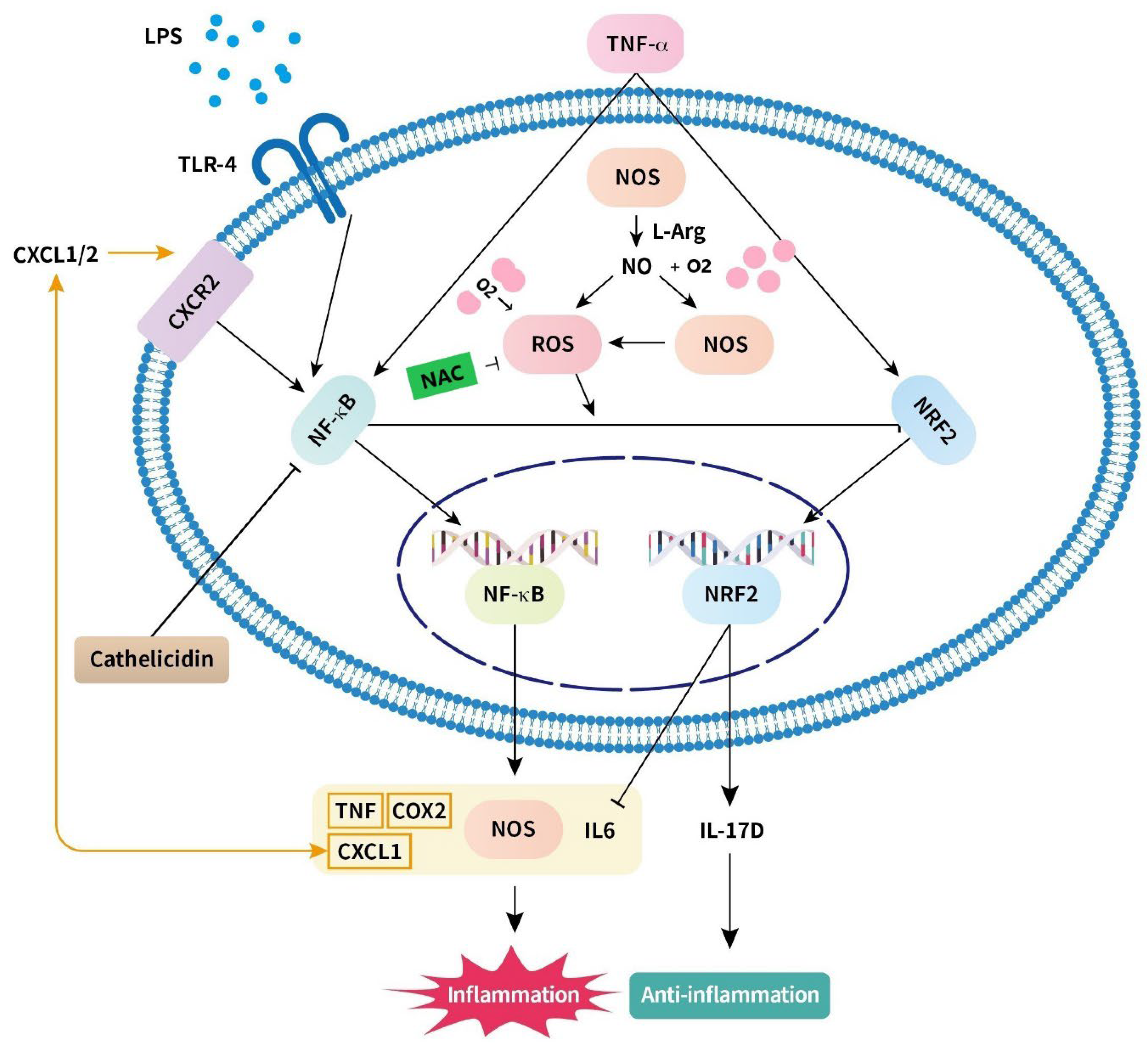
4.1. Nitric Oxide (Figure 1)

Nitric oxide (NO) is a free radical and can be a proinflammatory mediator as a signaling molecule, interacting with oxygen to became oxidized. This reaction is regulated by nitric oxide synthases (NOS), inhibits platelet adhesion, prevents mast cell activation, and functions as an antioxidant [43,44]. High NO levels disrupt actin cytoskeletons, inhibit ATP formation, dilate cellular tight junction, and increase intestinal permeability [45]. Under hyperoxic conditions, NO dysregulation occurs and NOS II protein concentrations in the villus, crypts, submucosa, and muscularis are increased [23]. Excessive NO production leads to mucosal injury and may cause NEC [43,46,47].

4.2. Nuclear Factor-κB (Figure 1)

Nuclear factor-κB (NF-κB) is a protein complex that controls DNA transcription and regulates the expression of cytokines, inducible NOS, and cyclooxygenase (COX)-2 [48]. NF-κB is activated in response to many external stimuli, including cytokines, free radicals, and bacterial or viral antigens. NF-κB is a transcription factor and plays a crucial role in immune response and inflammation [48,49]. Under hyperoxic conditions, NF-κB transcription increases, which induces the production of proinflammatory mediators, such as tumor necrosis factor (TNF)-α and interferon (IFN)-γ [30,50,51]. Excessive NF-κB expression may be an important inflammatory mechanism of NEC [52] and may be related to inflammatory bowel disease [45,53]. NF-κB can directly repress Nrf2 signaling (the protective mechanism, it will be discussed in part 5.1) to increase inflammation [54].

4.3. Reactive Oxygen Species (Figure 1)

Reactive oxygen species (ROS) are a chemically defined group of reactive molecules derived from molecular oxygen and ROS are produced by mitochondria. Low levels of ROS are related to cell proliferation and differentiation. Hyperoxia is associated with increased production of ROS, and excessive ROS induces apoptosis, cell autophagy, and DNA oxidative damage. A redox imbalance under hyperoxic conditions causes inflammation via the NF-κB and TNF pathways [32,44], which leads to an intestinal inflammation cascade and, ultimately, mucosal damage [36]. Mucosal damage induces the production of proinflammatory cytokines including IFN-g and IL-1. ROS also can modify cellular structure and function through covalent changes of NO and cause NO dysregulation [55]. Under conditions of hyperoxia, ROS promotes inflammatory cascades in the gut and may relate to inflammatory bowel disease [24,56].

4.4. Toll-like Receptor (Figure 1)

Toll-like receptors (TLRs) are membrane-spanning proteins and TLR4 is activated by bacterial LPS. TLRs are pathogen recognition receptors and play a critical role in the early innate immune response to invading pathogens [57,58]. Thus, they initiate innate immune and inflammatory responses. TLR4 also mediate NEC [40,59]. Hyperoxia increases the expression of TLR4 and NF-κB, thereby inducing inflammatory reactions and leading to intestinal injury via the NF-κB pathway [52]. The ROS levels also increase. A cascade of proinflammatory cytokines and interferons then begins, which induces inflammation [28]. Gut-derived endotoxemia may contribute to lung injury via the TLR4 pathway under conditions of hyperoxia [24]. Inhibition of the TLR pathway may prevent NEC [60].

4.5. Chemokine (CXC Motif) Ligand (Figure 1)

CXC motif ligand (CXCL) 1 is a member of the CXC chemokine family. It plays a vital role in the development of many inflammatory diseases [61]. It activates the CXC receptors (CXCR) 1 and 2. CXCL1 transcription involves the NF-κB pathway and cytokines such as IFN-γ, IL-1β, IL-17, transforming growth factor-β, and TNF-α [62,63].
The level of CXCL1 increases under inflammatory conditions and induces angiogenesis and the recruitment of neutrophils. Gut neutrophilia may induce gut injury under conditions of hyperoxia [24].

4.6. IL-6 (Figure 1)

IL-6 is a potent cytokine that modulates the innate immune system. Fetal exposure to maternal inflammation significantly increases the susceptibility and severity of subsequent intestinal injury with goblet cell loss via IL-6 [18,24]. Hyperoxia increases IL-6 levels in intestinal epithelial cells and aggravates inflammatory responses [47]. The hallmark of vascular NF-κB activation is the production of IL-6, which may play a role in vascular inflammation [64]. IL-6 is associated with intestinal barrier dysfunction and intestinal injury [37].
Table 1. Evidence of the roles of various molecules in the intestine under hyperoxic conditions.
Injury Mechanism
CandidateMechanismModelOutcomeReference
Excessive Nitric oxide (NO)
  • Signaling molecule
  • Proinflammatory mediator
Rat; Piglet
  • Disrupts actin cytoskeletons
  • Inhibits ATP formation
  • Dilates tight junction
  • May cause NEC
[23,45,47]
Nuclear factor-κB
(NF-κB)
  • Protein complex
  • Transcription factor
Human HT-29 cell; Rat
  • Regulates the expression of NOS and COX2 cytokines
  • Produces proinflammatory mediators, such as TNF-α and IFN-γ
  • Repress Nrf2 signaling
  • May be related to NEC and IBD
[30,52]
Reactive oxygen species
(ROS)
  • Signaling molecule
  • Proinflammatory mediator
Rat; Human Caco-2 cell; Human NCM460 cell
  • Induces apoptosis, cell autophagy, and DNA oxidative damage
  • Increases intestinal inflammation and mucosal damage
  • Increases levels of TNF-α and IL-1
  • May be related to IBD
[24,32,36,56]
Toll-like receptor-4
(TLR4)
  • Transmembrane protein
  • Trigger of signaling cascades of proinflammatory cytokines
Rat; Human cell
  • Initiates innate immune response and inflammatory reaction
  • Increases ROS level
[40,52,59]
CXC motif ligand-1
(CXCL1)
  • Member of the CXC chemokine family
Rat
  • Transcription with NF-κB pathway and inflammatory cytokines
  • Induces angiogenesis and recruitment of neutrophils
[24]
Interleukin-6
(IL-6)
  • Cytokine that modulates the innate immune system
Rat; Piglet
  • Aggravates inflammatory response
  • Causes intestinal injury with goblet cell loss
[18,24,47,64]
Protective Mechanism
CandidateMechanismModelOutcomeReference
Nuclear factor erythroid 2-related factor 2
(Nrf2)
  • Transcription factor
Rat; Piglet; Human NCM460 cell; Human F244 cell; Murine embryonic fibroblasts cell
  • Regulates the expression of multiple antioxidant genes
  • ROS detoxification and scavenging
  • Decreases cellular oxidative damage
  • Maintains the balance of intracellular redox status
  • Interferes with IL-6 induction
  • Promotes IL-17D production
[54,65,66]
Interleukin-17D
(IL-17D)
  • Cytokine that modulates inflammation and host defense
  • Anti-inflammatory cytokine
Rat; Human F244 cell; Murine embryonic fibroblasts cell
  • Induces CXCL2 expression
  • Causes recruitment of NK cells
  • Inhibits the bacterial phagocytic ability of macrophages
  • Inhibits and regulate TNF-α and other proinflammatory cytokines
  • Delays the intestinal inflammatory response and protects the GI tract from damage
[65,66]
N-acetylcysteine
(NAC)
  • Powerful antioxidant
  • Scavenger of hydroxyl radicals
Rat
  • Anti-inflammatory activities
  • Inhibits ROS
  • Reduces hyperoxia-induced ZO-1, occludin, and claudin-4 damage
  • Improves intestinal barrier
[36]
Arginyl-glutamine
Arg-Gln
  • Dipeptide
Rat; preterm neonate
  • Preserves the actin cytoskeleton to maintain the function of the intestinal barrier and intercellular junction
  • Anti-inflammatory
  • Improves mucosal integrity and gut healing
  • Reduces the risk of NEC
[67,68]
Deoxyribonucleic acid
(DHA)
  • Omega-3 long-chain fatty acid
Rat
  • Reduces inflammation
  • Blocks platelet-activating factor-induced apoptosis in intestinal epithelial cells
  • Reduces the risk of NEC
[67,69]
Cathelicidin
  • Antimicrobial peptide
Rat; Piglet; Porcine epithelial cell line J2
  • Antibacterial, antiviral, and antifungal
  • Inhibits hyperoxia-induced NF-κB pathway reaction
  • Enhances the phagocytosis of immune cells
  • Suppresses intestinal inflammation
  • Reduces LPS-induced disruption of intestinal barrier
[22,70,71]
Health gut microbiome
  • Community of microorganisms in the gut
  • Regulates the immune function and immune homeostasis
Rat; Human infant
  • Modulates NF-κB pathway reaction
  • Prevents bacterial infection
  • Reduces the risk of NEC
  • Reduces lung inflammation
  • May be related to neurodevelopmental disorders, neurodegenerative disorders, and metabolic syndromes
[24,38,72,73,74,75,76]
Abbreviations: ATP: adenosine triphosphate; COX2: cyclooxygenase 2; DNA: deoxyribonucleic acid; GI: gastrointestinal; IBD: inflammatory bowel disease; IFN-γ: interferon-γ; LPS: lipopolysaccharide; NAC: N-acetylcysteine; NEC: necrotizing enterocolitis; NK cell: natural killer cell; NOS: nitric oxide synthases; TNF-α: tumor necrosis factor-α; ZO-1: zonula occludens-1.
Figure 1. Schematic of the mechanism of hyperoxia-induced intestinal inflammation and anti-inflammatory response. Hyperoxia increases NOS and causes NO dysregulation and NO interacts with oxygen to became oxidated. Excessive NO induces the production of ROS. Furthermore, hyperoxia increases production of ROS. ROS causes inflammation through the NF-κB pathway and inhibits the Nrf2 pathway, which plays a crucial role in anti-inflammatory response. Production of inflammatory cytokines, including TNF, COX2, CXCL1, and IL-6, enhances inflammatory injury. CXCL1 activates CXCR1/2 and induces the NF-κB pathway. Under hyperoxic conditions, levels of TLR4 increase and bacterial LPS signal to enhance inflammation via the NF-κB pathway. Nrf2 inhibits the production of IL-6 and promotes the production of IL-17D, which may play a role in the production of anti-inflammatory responses. L-Arg inhibits apoptosis via increased NO production and antioxidant capacity and via inhibition of inflammation mediated by the NF-κB pathway. IL-10 inhibits TNF-α, and other proinflammatory cytokines delay intestinal inflammatory responses. NAC inhibits the ROS system to protect intestinal epithelial cells from hyperoxia. Abbreviations: Arg: arginine; COX2: cyclooxygenase 2; CXCL-1: chemokine (C-X-C motif) ligand-1; CXCR: C-X-C motif chemokine receptor; IL: interleukin; LPS: lipopolysaccharide; NAC: N-acetylcysteine; NF-κB: nuclear factor-κB; NO: nitric oxide; NOS: nitric oxide synthases; Nrf2: nuclear factor erythroid 2-related factor 2; ROS: reactive oxygen species; TLR-4: toll-like receptor-4; TNF: tumor necrosis factor.
Ijms 24 04366 g001

5. Protective Mechanism under Hyperoxic Conditions (Table 1)

5.1. Nuclear Factor Erythroid 2-Related Factor 2 and IL-17D (Figure 1)

Nuclear factor erythroid 2-related factor 2 (Nrf2) initiates the transcription of downstream regulatory antioxidant proteins and regulates the expression of multiple antioxidant genes [77]. It is involved in ROS detoxification and scavenging, reduces cellular oxidative damage, and maintains the balance of intracellular redox [65].
IL-17D is a cytokine of the IL-17 family and is involved in inflammation and the host defense system. IL-17D is produced by intestinal lymphocytes and epithelial cells of the small intestinal villi. It induces CXCL2 expression and recruits natural killer cells, thereby initiating an antitumor or antiviral immune response. IL-17D directly inhibits the bacterial phagocytic ability of macrophages and aggravates sepsis via the NF-κB pathway [78]. IL-17D is a critical cytokine during intracellular bacterial and viral infection [65,79].
Nrf2 promotes IL-17D expression; hyperoxia promotes the nuclear translocation of Nrf2 and increases Nrf2 with IL-17D in intestinal epithelial cells [66]. Nrf2 also interferes with IL6 induction and inflammatory phenotypes in vivo [80]. Nrf2 and NF-κB are key pathways regulating the balance of cellular redox status and responses to stress and inflammation. NF-κB can directly repress Nrf2 signaling to increase inflammation [54].

5.2. N-Acetylcysteine (Figure 1)

N-acetylcysteine (NAC) is a powerful antioxidant and a scavenger of hydroxyl radicals. NAC has anti-inflammatory properties. NAC protects intestinal epithelial cells under conditions of hyperoxia through the inhibition of ROS. Moreover, NAC can reverse the decreases in ZO-1, occludin, and claudin-4 induced by hyperoxia, indicating that NAC can improve the intestinal barrier status [36].

5.3. Arginyl–Glutamine (Figure 1)

Arginyl–glutamine (Arg–Gln) dipeptide is an aqueous stable source of glutamine. Glutamine (Gln) maintains the function of the intestinal barrier and intercellular junction by preserving the actin cytoskeleton. It also reduces the levels of proinflammatory cytokines and prevents cytokine-related apoptosis of intestinal epithelial cells, thereby reducing the risk of developing NEC in premature animals [78]. Arginine (Arg) has anti-inflammatory properties and improves mucosal integrity and gut healing. L-Arg inhibits apoptosis via increased NO production and antioxidant capacity and inhibition of inflammation mediated by the NF-κB pathway [81]. In hyperoxia-induced intestinal injury neonatal mice, the Arg-Gln supplementation group experienced less intestinal injury than the room air group [67]. Low Arg levels are associated with NEC, and therefore, Arg supplementation may reduce the risk of NEC in premature animals and neonates [67,68,82].

5.4. Docosahexaenoic Acid (Figure 1)

Omega-3 long-chain fatty acids such as docosahexaenoic acid (DHA) decrease inflammation. DHA blocks platelet-activating factor-induced apoptosis in intestinal epithelial cells [67]. Insufficient levels of DHA may predispose neonates to acute inflammatory conditions [83]. In hyperoxia-induced intestinal injury neonatal mice, the DHA supplementation group had less intestinal injury as the room air group [67]. Thus, DHA supplementation may reduce the incidence of NEC in neonate rats [69].

5.5. Cathelicidin (Figure 1)

Cathelicidin is an antimicrobial peptide with antibacterial, antiviral, and antifungal properties. It acts as a multifunctional effector molecule in innate immunity [84]. Cathelicidin enhances hyperoxia-induced phagocytosis in weaning piglets through inhibition of the hyperoxia-induced NF-κB pathway [70], suppresses intestinal inflammation, and prevents intestinal barrier dysfunction. It also reduces LPS-induced disruption of the intestinal barrier in rats [70,71]. Therefore, cathelicidin treatment can ameliorate intestinal injury by protecting the tight junction from hyperoxia [22].

5.6. Health Gut Microbiome

Gut microbiomes contribute to gut mucosal development and integrity through innate immunity. Microbiota play a crucial role in human health and disease. [85,86]. Gut microbiota and the microbial metabolites may affect immune function and immune homeostasis. Microbiota may be associated with NEC [87]. The composition of gut microbiota under conditions of hyperoxia may influence the levels of lung cytokines and is associated with lung inflammation [46]. Interactions between the brain and gut microbiota may also be associated with neurodevelopmental disorders, neurodegenerative disorders, and metabolic syndromes [72].
When mice were exposed to hyperoxia in the first week of life, their gut microbial composition changed. Alpha diversity and taxa of microbiota decreased until adolescence, and Proteobacteria abundance increased [38]. Hyperoxia-induced gut injury was time- and dose-dependent in a mouse model. Hyperoxia not only diminished obligate anaerobes but also enriched facultative anaerobes, such as Escherichia-Shigella, Enterobacteriaceae, Gammaproteobacteria, and Proteobacteria [24]. Moreover, in a human study, maternal chorioamnionitis was demonstrated to increase the incidence of late-onset sepsis and death among preterm infants and shifted the fecal microbiome of preterm infants [73]. Prenatal LPS exposure also induced significant changes in the intestinal microbiome of the offspring, with a significant increase in the abundance of Proteobacteria (Escherichia-Shigella) and a decrease in Firmicutes at 7 days after birth [74].
The microbiome is a promising target for prevention and treatment with probiotics. Saccharomyces boulardii was shown to modulate NEC in neonatal mice via the NF-κB pathway [75]. The incidence of NEC was decreased in preterm infants given probiotics [76].

6. Conclusions

In this study, we reviewed the mechanism underlying neonatal intestinal damage caused by hyperoxia at a histological and molecular level. Under conditions of hyperoxia, protective mechanisms are activated to prevent intestinal inflammation and injury. The NF-κB and Nrf2 pathways play key roles in intestinal inflammation. Under conditions of hyperoxia, the redox status becomes imbalanced, resulting in a cascade of intestinal damage. We discussed the importance of the microbiome in hyperoxia-induced intestinal damage. Supplemental oxygen therapy is essential for preterm neonates with respiratory distress syndrome and other pulmonary conditions. The optimal target range of oxygen saturation is 91–95% in infants less than 28 weeks gestation [88]. There was increased mortality and NEC in the low oxygen saturation target group (85–89%) compared to the high SpO2 group (91–95%) [89]. However, high concentrations of oxygen can have adverse effects on newborns. In conclusion, oxygen therapy is important for newborns and hyperoxia can cause intestinal histological changes including mucosal damage and vascular changes. Thus, caution in its use is required. Further investigation is warranted to clarify the effects of hyperoxia on neonatal intestines and to identify methods of reducing hyperoxia-induced intestinal damage.

Author Contributions

H.-C.W., H.-C.C. and C.-M.C. wrote the manuscript. All authors have read and agreed to the published version of the manuscript.

Funding

The study was supported by a grant from the Ministry of Science and Technology in Taiwan (MOST 110-2622-B-038-006).

Institutional Review Board Statement

Not applicable.

Informed Consent Statement

Not applicable.

Data Availability Statement

The data presented in this study are available on request from the corresponding author.

Conflicts of Interest

The authors declare no conflict of interest.

References

  1. Cheng, H.L. Effect of hyperoxia and hypercapnia on tissue oxygen and perfusion response in the normal liver and kidney. PLoS ONE 2012, 7, e40485. [Google Scholar] [CrossRef] [PubMed] [Green Version]
  2. Mathias, M.; Chang, J.; Perez, M.; Saugstad, O. Supplemental Oxygen in the Newborn: Historical Perspective and Current Trends. Antioxidants 2021, 10, 1879. [Google Scholar] [CrossRef] [PubMed]
  3. Torbati, D.; Tan, G.H.; Smith, S.; Frazier, K.S.; Gelvez, J.; Fakioglu, H.; Totapally, B.R. Multiple-organ effect of normobaric hyperoxia in neonatal rats. J. Crit. Care 2006, 21, 85–93, discussion 93–84. [Google Scholar] [CrossRef]
  4. Giusto, K.; Wanczyk, H.; Jensen, T.; Finck, C. Hyperoxia-induced bronchopulmonary dysplasia: Better models for better therapies. Dis. Model. Mech. 2021, 14, dmm047753. [Google Scholar] [CrossRef] [PubMed]
  5. Zhu, X.; Lei, X.; Wang, J.; Dong, W. Protective effects of resveratrol on hyperoxia-induced lung injury in neonatal rats by alleviating apoptosis and ROS production. J. Matern. Fetal Neonatal Med. 2020, 33, 4150–4158. [Google Scholar] [CrossRef] [PubMed]
  6. Bhandari, V. Hyperoxia-derived lung damage in preterm infants. Semin. Fetal. Neonatal. Med. 2010, 15, 223–229. [Google Scholar] [CrossRef] [PubMed] [Green Version]
  7. Indrio, F.; Neu, J.; Pettoello-Mantovani, M.; Marchese, F.; Martini, S.; Salatto, A.; Aceti, A. Development of the Gastrointestinal Tract in Newborns as a Challenge for an Appropriate Nutrition: A Narrative Review. Nutrients 2022, 14, 1405. [Google Scholar] [CrossRef]
  8. Sanidad, K.Z.; Zeng, M.Y. Neonatal gut microbiome and immunity. Curr. Opin. Microbiol. 2020, 56, 30–37. [Google Scholar] [CrossRef]
  9. Yu, J.C.; Khodadadi, H.; Malik, A.; Davidson, B.; Salles, É.D.S.L.; Bhatia, J.; Hale, V.L.; Baban, B. Innate Immunity of Neonates and Infants. Front. Immunol. 2018, 9, 1759. [Google Scholar] [CrossRef] [Green Version]
  10. Jena, A.; Montoya, C.A.; Mullaney, J.A.; Dilger, R.N.; Young, W.; McNabb, W.C.; Roy, N.C. Gut-Brain Axis in the Early Postnatal Years of Life: A Developmental Perspective. Front. Integr. Neurosci. 2020, 14, 44. [Google Scholar] [CrossRef]
  11. Esrefoglu, M.; Cetin, A. Development of Small and Large Intestine. Bezmialem. Sci. 2016, 5, 36–40. [Google Scholar] [CrossRef]
  12. Chin, A.M.; Hill, D.R.; Aurora, M.; Spence, J.R. Morphogenesis and maturation of the embryonic and postnatal intestine. Semin. Cell Dev. Biol. 2017, 66, 81–93. [Google Scholar] [CrossRef] [PubMed]
  13. Clayburgh, D.R.; Shen, L.; Turner, J.R. A porous defense: The leaky epithelial barrier in intestinal disease. Lab. Investig. A J. Tech. Methods Pathol. 2004, 84, 282–291. [Google Scholar] [CrossRef] [PubMed] [Green Version]
  14. De Zwart, L.L.; Haenen, H.E.; Versantvoort, C.H.; Wolterink, G.; van Engelen, J.G.; Sips, A.J. Role of biokinetics in risk assessment of drugs and chemicals in children. Regul. Toxicol. Pharmacol. 2004, 39, 282–309. [Google Scholar] [CrossRef] [PubMed]
  15. Pácha, J. Development of intestinal transport function in mammals. Physiol. Rev. 2000, 80, 1633–1667. [Google Scholar] [CrossRef]
  16. Henning, S.J. Postnatal development: Coordination of feeding, digestion, and metabolism. Am. J. Physiol. 1981, 241, G199–G214. [Google Scholar] [CrossRef]
  17. Dotinga, B.M.; Mintzer, J.P.; Moore, J.E.; Hulscher, J.B.F.; Bos, A.F.; Kooi, E.M.W. Maturation of Intestinal Oxygenation: A Review of Mechanisms and Clinical Implications for Preterm Neonates. Front. Pediatr. 2020, 8, 354. [Google Scholar] [CrossRef]
  18. Elgin, T.G.; Fricke, E.M.; Gong, H.; Reese, J.; Mills, D.A.; Kalantera, K.M.; Underwood, M.A.; McElroy, S.J. Fetal exposure to maternal inflammation interrupts murine intestinal development and increases susceptibility to neonatal intestinal injury. Dis. Model. Mech. 2019, 12, dmm040808. [Google Scholar] [CrossRef] [PubMed] [Green Version]
  19. Yuan, Q.; Walker, W.A. Innate immunity of the gut: Mucosal defense in health and disease. J. Pediatr. Gastroenterol. Nutr. 2004, 38, 463–473. [Google Scholar] [CrossRef] [Green Version]
  20. Montalban-Arques, A.; Chaparro, M.; Gisbert, J.P.; Bernardo, D. The Innate Immune System in the Gastrointestinal Tract: Role of Intraepithelial Lymphocytes and Lamina Propria Innate Lymphoid Cells in Intestinal Inflammation. Inflamm. Bowel Dis. 2018, 24, 1649–1659. [Google Scholar] [CrossRef] [Green Version]
  21. Lenfestey, M.W.; Neu, J. Gastrointestinal Development: Implications for Management of Preterm and Term Infants. Gastroenterol. Clin. N. Am. 2018, 47, 773–791. [Google Scholar] [CrossRef] [PubMed]
  22. Chou, H.C.; Chen, C.M. Cathelicidin attenuates hyperoxia-induced intestinal injury through inhibition of NF-kappaB activity in newborn rats. Exp. Mol. Pathol. 2020, 113, 104269. [Google Scholar] [CrossRef] [PubMed]
  23. Giannone, P.J.; Bauer, J.A.; Schanbacher, B.L.; Reber, K.M. Effects of hyperoxia on postnatal intestinal development. Biotech. Histochem. 2007, 82, 17–22. [Google Scholar] [CrossRef] [PubMed]
  24. Li, Y.; Tao, Y.; Xu, J.; He, Y.; Zhang, W.; Jiang, Z.; He, Y.; Liu, H.; Chen, M.; Zhang, W.; et al. Hyperoxia Provokes Time- and Dose-Dependent Gut Injury and Endotoxemia and Alters Gut Microbiome and Transcriptome in Mice. Front. Med. 2021, 8, 732039. [Google Scholar] [CrossRef]
  25. Sherman, M.P.; Bennett, S.H.; Hwang, F.F.; Sherman, J.; Bevins, C.L. Paneth cells and antibacterial host defense in neonatal small intestine. Infect. Immun. 2005, 73, 6143–6146. [Google Scholar] [CrossRef] [Green Version]
  26. Vaishnava, S.; Behrendt, C.L.; Ismail, A.S.; Eckmann, L.; Hooper, L.V. Paneth cells directly sense gut commensals and maintain homeostasis at the intestinal host-microbial interface. Proc. Natl. Acad. Sci. USA 2008, 105, 20858–20863. [Google Scholar] [CrossRef] [Green Version]
  27. Lueschow, S.R.; McElroy, S.J. The Paneth Cell: The Curator and Defender of the Immature Small Intestine. Front. Immunol. 2020, 11, 587. [Google Scholar] [CrossRef] [Green Version]
  28. Chou, H.C.; Chen, C.M. Neonatal hyperoxia disrupts the intestinal barrier and impairs intestinal function in rats. Exp. Mol. Pathol. 2017, 102, 415–421. [Google Scholar] [CrossRef]
  29. Liu, D.Y.; Li, J.J. Effect of hyperoxia on the intestinal IgA secretory component in neonatal rats and on intestinal epithelial cells in vitro. Braz. J. Med. Biol. Res. 2010, 43, 1034–1041. [Google Scholar] [CrossRef] [Green Version]
  30. Bruno, M.E.; Frantz, A.L.; Rogier, E.W.; Johansen, F.E.; Kaetzel, C.S. Regulation of the polymeric immunoglobulin receptor by the classical and alternative NF-κB pathways in intestinal epithelial cells. Mucosal. Immunol. 2011, 4, 468–478. [Google Scholar] [CrossRef] [Green Version]
  31. Liu, D.Y.; Jiang, T.; Wang, S.; Cao, X. Effect of hyperoxia on pulmonary SIgA and its components, IgA and SC. J. Clin. Immunol. 2013, 33, 1009–1017. [Google Scholar] [CrossRef] [PubMed] [Green Version]
  32. Zhao, M.; Tang, S.; Xin, J.; Liu, D. Influence of reactive oxygen species on secretory component in the intestinal epithelium during hyperoxia. Exp. Med. 2017, 14, 4033–4040. [Google Scholar] [CrossRef] [PubMed] [Green Version]
  33. Davids, B.J.; Palm, J.E.; Housley, M.P.; Smith, J.R.; Andersen, Y.S.; Martin, M.G.; Hendrickson, B.A.; Johansen, F.E.; Svärd, S.G.; Gillin, F.D.; et al. Polymeric immunoglobulin receptor in intestinal immune defense against the lumen-dwelling protozoan parasite Giardia. J. Immunol. 2006, 177, 6281–6290. [Google Scholar] [CrossRef] [PubMed] [Green Version]
  34. Li, T.M.; Liu, D.Y. Mechanism of Neonatal Intestinal Injury Induced by Hyperoxia Therapy. J. Immunol. Res. 2022, 2022, 2316368. [Google Scholar] [CrossRef]
  35. Chen, C.M.; Chou, H.C. Hyperoxia disrupts the intestinal barrier in newborn rats. Exp. Mol. Pathol. 2016, 101, 44–49. [Google Scholar] [CrossRef]
  36. Liu, D.Y.; Lou, W.J.; Zhang, D.Y.; Sun, S.Y. ROS Plays a Role in the Neonatal Rat Intestinal Barrier Damages Induced by Hyperoxia. Biomed. Res. Int. 2020, 2020, 8819195. [Google Scholar] [CrossRef]
  37. Bölke, E.; Jehle, P.M.; Orth, K.; Steinbach, G.; Hannekum, A.; Storck, M. Changes of gut barrier function during anesthesia and cardiac surgery. Angiology 2001, 52, 477–482. [Google Scholar] [CrossRef]
  38. Lo, Y.C.; Chen, K.Y.; Chou, H.C.; Lin, I.H.; Chen, C.M. Neonatal hyperoxia induces gut dysbiosis and behavioral changes in adolescent mice. J. Chin. Med. Assoc. 2021, 84, 290–298. [Google Scholar] [CrossRef]
  39. Berg, R.D. Bacterial translocation from the gastrointestinal tract. Adv. Exp. Med. Biol. 1999, 473, 11–30. [Google Scholar] [CrossRef]
  40. Hackam, D.J.; Good, M.; Sodhi, C.P. Mechanisms of gut barrier failure in the pathogenesis of necrotizing enterocolitis: Toll-like receptors throw the switch. Semin. Pediatr. Surg. 2013, 22, 76–82. [Google Scholar] [CrossRef] [Green Version]
  41. Bein, A.; Eventov-Friedman, S.; Arbell, D.; Schwartz, B. Intestinal tight junctions are severely altered in NEC preterm neonates. Pediatr. Neonatol. 2018, 59, 464–473. [Google Scholar] [CrossRef] [PubMed] [Green Version]
  42. Managlia, E.; Yan, X.; De Plaen, I.G. Intestinal Epithelial Barrier Function and Necrotizing Enterocolitis. Newborn 2022, 1, 32–43. [Google Scholar] [CrossRef] [PubMed]
  43. Kubes, P.; Suzuki, M.; Granger, D.N. Nitric oxide: An endogenous modulator of leukocyte adhesion. Proc. Natl. Acad. Sci. USA 1991, 88, 4651–4655. [Google Scholar] [CrossRef] [PubMed] [Green Version]
  44. Gaboury, J.; Woodman, R.C.; Granger, D.N.; Reinhardt, P.; Kubes, P. Nitric oxide prevents leukocyte adherence: Role of superoxide. Am. J. Physiol. 1993, 265, H862–H867. [Google Scholar] [CrossRef] [PubMed]
  45. Hsu, C.M.; Liu, C.H.; Chen, L.W. Nitric oxide synthase inhibitor ameliorates oral total parenteral nutrition-induced barrier dysfunction. Shock 2000, 13, 135–139. [Google Scholar] [CrossRef] [Green Version]
  46. Chen, C.M.; Chou, H.C.; Yang, Y.S.H.; Su, E.C.; Liu, Y.R. Predicting Hyperoxia-Induced Lung Injury from Associated Intestinal and Lung Dysbiosis in Neonatal Mice. Neonatology 2021, 118, 163–173. [Google Scholar] [CrossRef]
  47. Di Lorenzo, M.; Krantis, A. Altered nitric oxide production in the premature gut may increase susceptibility to intestinal damage in necrotizing enterocolitis. J. Pediatr. Surg. 2001, 36, 700–705. [Google Scholar] [CrossRef]
  48. Morgan, M.J.; Liu, Z.G. Crosstalk of reactive oxygen species and NF-κB signaling. Cell Res. 2011, 21, 103–115. [Google Scholar] [CrossRef] [Green Version]
  49. Lawrence, T. The nuclear factor NF-kappaB pathway in inflammation. Cold Spring Harb. Perspect. Biol. 2009, 1, a001651. [Google Scholar] [CrossRef] [Green Version]
  50. Shea, L.M.; Beehler, C.; Schwartz, M.; Shenkar, R.; Tuder, R.; Abraham, E. Hyperoxia activates NF-kappaB and increases TNF-alpha and IFN-gamma gene expression in mouse pulmonary lymphocytes. J. Immunol. 1996, 157, 3902–3908. [Google Scholar] [CrossRef]
  51. Ye, Y.; Lin, P.; Zhang, W.; Tan, S.; Zhou, X.; Li, R.; Pu, Q.; Koff, J.L.; Dhasarathy, A.; Ma, F.; et al. DNA Repair Interacts with Autophagy To Regulate Inflammatory Responses to Pulmonary Hyperoxia. J. Immunol. 2017, 198, 2844–2853. [Google Scholar] [CrossRef] [PubMed] [Green Version]
  52. Yin, Y.; Liu, F.; Li, Y.; Tang, R.; Wang, J. mRNA expression of TLR4, TLR9 and NF-κB in a neonatal murine model of necrotizing enterocolitis. Mol. Med. Rep. 2016, 14, 1953–1956. [Google Scholar] [CrossRef] [PubMed] [Green Version]
  53. Babu, D.; Lee, J.S.; Park, S.Y.; Thapa, D.; Choi, M.K.; Kim, A.R.; Park, Y.J.; Kim, J.A. Involvement of NF-kappaB in the inhibitory actions of Platycarya strobilacea on the TNF-alpha-induced monocyte adhesion to colon epithelial cells and chemokine expression. Arch. Pharmacal. Res. 2008, 31, 727–735. [Google Scholar] [CrossRef] [PubMed]
  54. Liu, X.; Li, T.; Liu, Y.; Sun, S.; Liu, D. Nuclear factor erythroid 2-related factor 2 potentiates the generation of inflammatory cytokines by intestinal epithelial cells during hyperoxia by inducing the expression of interleukin 17D. Toxicology 2021, 457, 152820. [Google Scholar] [CrossRef] [PubMed]
  55. Andreadou, I.; Schulz, R.; Papapetropoulos, A.; Turan, B.; Ytrehus, K.; Ferdinandy, P.; Daiber, A.; Di Lisa, F. The role of mitochondrial reactive oxygen species, NO and H2S in ischaemia/reperfusion injury and cardioprotection. J. Cell. Mol. Med. 2020, 24, 6510–6522. [Google Scholar] [CrossRef] [PubMed]
  56. Zhao, M.; Tang, S.; Xin, J.; Wei, Y.; Liu, D. Reactive oxygen species induce injury of the intestinal epithelium during hyperoxia. Int. J. Mol. Med. 2018, 41, 322–330. [Google Scholar] [CrossRef] [PubMed] [Green Version]
  57. Ozinsky, A.; Underhill, D.M.; Fontenot, J.D.; Hajjar, A.M.; Smith, K.D.; Wilson, C.B.; Schroeder, L.; Aderem, A. The repertoire for pattern recognition of pathogens by the innate immune system is defined by cooperation between toll-like receptors. Proc. Natl. Acad. Sci. USA 2000, 97, 13766–13771. [Google Scholar] [CrossRef] [Green Version]
  58. Fitzgerald, K.A.; Kagan, J.C. Toll-like Receptors and the Control of Immunity. Cell 2020, 180, 1044–1066. [Google Scholar] [CrossRef]
  59. Egan, C.E.; Sodhi, C.P.; Good, M.; Lin, J.; Jia, H.; Yamaguchi, Y.; Lu, P.; Ma, C.; Branca, M.F.; Weyandt, S.; et al. Toll-like receptor 4-mediated lymphocyte influx induces neonatal necrotizing enterocolitis. J. Clin. Investig. 2016, 126, 495–508. [Google Scholar] [CrossRef] [Green Version]
  60. Hou, Y.; Lu, X.; Zhang, Y. IRAK Inhibitor Protects the Intestinal Tract of Necrotizing Enterocolitis by Inhibiting the Toll-Like Receptor (TLR) Inflammatory Signaling Pathway in Rats. Med. Sci. Monit. Int. Med. J. Exp. Clin. Res. 2018, 24, 3366–3373. [Google Scholar] [CrossRef]
  61. Baggiolini, M. Chemotactic and inflammatory cytokines—CXC and CC proteins. Adv. Exp. Med. Biol. 1993, 351, 1–11. [Google Scholar] [CrossRef] [PubMed]
  62. Cao, Z.; Fu, B.; Deng, B.; Zeng, Y.; Wan, X.; Qu, L. Overexpression of Chemokine (C-X-C) ligand 1 (CXCL1) associated with tumor progression and poor prognosis in hepatocellular carcinoma. Cancer Cell Int. 2014, 14, 86. [Google Scholar] [CrossRef] [PubMed] [Green Version]
  63. Miyake, M.; Goodison, S.; Urquidi, V.; Gomes Giacoia, E.; Rosser, C.J. Expression of CXCL1 in human endothelial cells induces angiogenesis through the CXCR2 receptor and the ERK1/2 and EGF pathways. Lab. Investig. A J. Tech. Methods Pathol. 2013, 93, 768–778. [Google Scholar] [CrossRef] [PubMed] [Green Version]
  64. Brasier, A.R. The nuclear factor-kappaB-interleukin-6 signalling pathway mediating vascular inflammation. Cardiovasc. Res. 2010, 86, 211–218. [Google Scholar] [CrossRef] [PubMed] [Green Version]
  65. Saddawi-Konefka, R.; Seelige, R.; Gross, E.T.; Levy, E.; Searles, S.C.; Washington, A., Jr.; Santosa, E.K.; Liu, B.; O’Sullivan, T.E.; Harismendy, O.; et al. Nrf2 Induces IL-17D to Mediate Tumor and Virus Surveillance. Cell Rep. 2016, 16, 2348–2358. [Google Scholar] [CrossRef] [Green Version]
  66. Liu, X.; Zhang, D.; Cai, Q.; Liu, D.; Sun, S. Involvement of nuclear factor erythroid 2related factor 2 in neonatal intestinal interleukin17D expression in hyperoxia. Int. J. Mol. Med. 2020, 46, 1423–1432. [Google Scholar] [CrossRef]
  67. Li, N.; Ma, L.; Liu, X.; Shaw, L.; Li Calzi, S.; Grant, M.B.; Neu, J. Arginyl-glutamine dipeptide or docosahexaenoic acid attenuates hyperoxia-induced small intestinal injury in neonatal mice. J. Pediatr. Gastroenterol. Nutr. 2012, 54, 499–504. [Google Scholar] [CrossRef] [Green Version]
  68. Polycarpou, E.; Zachaki, S.; Tsolia, M.; Papaevangelou, V.; Polycarpou, N.; Briana, D.D.; Gavrili, S.; Kostalos, C.; Kafetzis, D. Enteral L-arginine supplementation for prevention of necrotizing enterocolitis in very low birth weight neonates: A double-blind randomized pilot study of efficacy and safety. JPEN J. Parenter. Enter. Nutr. 2013, 37, 617–622. [Google Scholar] [CrossRef]
  69. Lu, J.; Jilling, T.; Li, D.; Caplan, M.S. Polyunsaturated fatty acid supplementation alters proinflammatory gene expression and reduces the incidence of necrotizing enterocolitis in a neonatal rat model. Pediatr. Res. 2007, 61, 427–432. [Google Scholar] [CrossRef] [Green Version]
  70. Yi, H.; Yu, C.; Zhang, H.; Song, D.; Jiang, D.; Du, H.; Wang, Y. Cathelicidin-BF suppresses intestinal inflammation by inhibiting the nuclear factor-κB signaling pathway and enhancing the phagocytosis of immune cells via STAT-1 in weanling piglets. Int. Immunopharmacol. 2015, 28, 61–69. [Google Scholar] [CrossRef]
  71. Han, F.; Lu, Z.; Liu, Y.; Xia, X.; Zhang, H.; Wang, X.; Wang, Y. Cathelicidin-BF ameliorates lipopolysaccharide-induced intestinal epithelial barrier disruption in rat. Life Sci. 2016, 152, 199–209. [Google Scholar] [CrossRef]
  72. Morais, L.H.; Schreiber, H.L.t.; Mazmanian, S.K. The gut microbiota-brain axis in behaviour and brain disorders. Nat. Rev. Microbiol. 2021, 19, 241–255. [Google Scholar] [CrossRef] [PubMed]
  73. Puri, K.; Taft, D.H.; Ambalavanan, N.; Schibler, K.R.; Morrow, A.L.; Kallapur, S.G. Association of Chorioamnionitis with Aberrant Neonatal Gut Colonization and Adverse Clinical Outcomes. PLoS ONE 2016, 11, e0162734. [Google Scholar] [CrossRef] [PubMed] [Green Version]
  74. Huang, Q.; Lu, S.; Zhu, Y.; Wei, B.; Chen, Y.; Bai, F. Bacterial endotoxin-induced maternal inflammation leads to fetal intestinal injury and affects microbial colonization in the neonatal period. J. Matern. Fetal Neonatal Med. 2021, 35, 6917–6927. [Google Scholar] [CrossRef] [PubMed]
  75. Zhang, K.; Zhang, X.; Lv, A.; Fan, S.; Zhang, J. Saccharomyces boulardii modulates necrotizing enterocolitis in neonatal mice by regulating the sirtuin 1/NF-κB pathway and the intestinal microbiota. Mol. Med. Rep. 2020, 22, 671–680. [Google Scholar] [CrossRef] [PubMed]
  76. Patel, R.M.; Underwood, M.A. Probiotics and necrotizing enterocolitis. Semin. Pediatr. Surg. 2018, 27, 39–46. [Google Scholar] [CrossRef] [PubMed]
  77. Huang, Y.; Li, W.; Su, Z.Y.; Kong, A.N. The complexity of the Nrf2 pathway: Beyond the antioxidant response. J. Nutr. Biochem. 2015, 26, 1401–1413. [Google Scholar] [CrossRef] [PubMed]
  78. Yan, X.; Tu, H.; Liu, Y.; Chen, T.; Cao, J. Interleukin-17D Aggravates Sepsis by Inhibiting Macrophage Phagocytosis. Crit. Care Med. J. 2020, 48, e58–e65. [Google Scholar] [CrossRef]
  79. Lee, Y.; Clinton, J.; Yao, C.; Chang, S.H. Interleukin-17D Promotes Pathogenicity During Infection by Suppressing CD8 T Cell Activity. Front. Immunol. 2019, 10, 1172. [Google Scholar] [CrossRef] [Green Version]
  80. Kobayashi, E.H.; Suzuki, T.; Funayama, R.; Nagashima, T.; Hayashi, M.; Sekine, H.; Tanaka, N.; Moriguchi, T.; Motohashi, H.; Nakayama, K.; et al. Nrf2 suppresses macrophage inflammatory response by blocking proinflammatory cytokine transcription. Nat. Commun. 2016, 7, 11624. [Google Scholar] [CrossRef] [Green Version]
  81. Zheng, H.; Guo, Q.; Duan, X.; Xu, Z.; Wang, Q. l-arginine inhibited apoptosis of fish leukocytes via regulation of NF-κB-mediated inflammation, NO synthesis, and anti-oxidant capacity. Biochimie 2019, 158, 62–72. [Google Scholar] [CrossRef] [PubMed]
  82. Shah, P.S.; Shah, V.S.; Kelly, L.E. Arginine supplementation for prevention of necrotising enterocolitis in preterm infants. Cochrane Database Syst. Rev. 2017, 4, CD004339. [Google Scholar] [CrossRef] [PubMed]
  83. Lapillonne, A.; Jensen, C.L. Reevaluation of the DHA requirement for the premature infant. Prostaglandins Leukot. Essent. Fat. Acids 2009, 81, 143–150. [Google Scholar] [CrossRef] [PubMed]
  84. Doss, M.; White, M.R.; Tecle, T.; Hartshorn, K.L. Human defensins and LL-37 in mucosal immunity. J. Leukoc. Biol. 2010, 87, 79–92. [Google Scholar] [CrossRef]
  85. Kåhrström, C.T.; Pariente, N.; Weiss, U. Intestinal microbiota in health and disease. Nature 2016, 535, 47. [Google Scholar] [CrossRef] [Green Version]
  86. Dickson, R.P. The microbiome and critical illness. Lancet. Respir. Med. 2016, 4, 59–72. [Google Scholar] [CrossRef] [Green Version]
  87. Humberg, A.; Fortmann, I.; Siller, B.; Kopp, M.V.; Herting, E.; Gopel, W.; Hartel, C.; German Neonatal Network, German Center for Lung Research and Priming Immunity at the beginning of life (PRIMAL) Consortium. Preterm birth and sustained inflammation: Consequences for the neonate. Semin. Immunopathol. 2020, 42, 451–468. [Google Scholar] [CrossRef]
  88. Ali, S.K.M.; Mohammed, N.; Qureshi, N.; Gupta, S. Oxygen therapy in preterm infants: Recommendations for practice. Paediatr. Child Health 2021, 31, 1–6. [Google Scholar] [CrossRef]
  89. Saugstad, O.D. Oxygenation of the Immature Infant: A Commentary and Recommendations for Oxygen Saturation Targets and Alarm Limits. Neonatology 2018, 114, 69–75. [Google Scholar] [CrossRef]
Disclaimer/Publisher’s Note: The statements, opinions and data contained in all publications are solely those of the individual author(s) and contributor(s) and not of MDPI and/or the editor(s). MDPI and/or the editor(s) disclaim responsibility for any injury to people or property resulting from any ideas, methods, instructions or products referred to in the content.

