Article

Stable Expression of dmiR-283 in the Brain Promises Positive Effects in Endurance Exercise on Sleep–Wake Behavior in Aging Drosophila

1
Key Laboratory of Physical Fitness and Exercise Rehabilitation of Hunan Province, Hunan Normal University, Changsha 410012, China
2
The Center for Heart Development, State Key Laboratory of Development Biology of Freshwater Fish, College of Life Sciences, Hunan Normal University, Changsha 410081, China
*
Author to whom correspondence should be addressed.
These authors contributed equally to this work.
Int. J. Mol. Sci. 2023, 24(4), 4180; https://doi.org/10.3390/ijms24044180
Submission received: 14 December 2022 / Revised: 12 February 2023 / Accepted: 15 February 2023 / Published: 20 February 2023

Abstract

:
Sleep–wake stability is imbalanced with natural aging, and microRNAs (miRNAs) play important roles in cell proliferation, apoptosis, and aging; however, the biological functions of miRNAs in regulating aging-related sleep–wake behavior remain unexplored. This study varied the expression pattern of dmiR-283 in Drosophila and the result showed that the aging decline in sleep–wake behavior was caused by the accumulation of brain dmiR-283 expression, whereas the core clock genes cwo and Notch signaling pathway might be suppressed, which regulate the aging process. In addition, to identify exercise intervention programs of Drosophila that promote healthy aging, mir-283SP/+ and Pdf > mir-283SP flies were driven to perform endurance exercise for a duration of 3 weeks starting at 10 and 30 days, respectively. The results showed that exercise starting in youth leads to an enhanced amplitude of sleep–wake rhythms, stable periods, increased activity frequency upon awakening, and the suppression of aging brain dmiR-283 expression in mir-283SP/+ middle-aged flies. Conversely, exercise performed when the brain dmiR-283 reached a certain accumulation level showed ineffective or negative effects. In conclusion, the accumulation of dmiR-283 expression in the brain induced an age-dependent decline in sleep–wake behavior. Endurance exercise commencing in youth counteracts the increase in dmiR-283 in the aging brain, which ameliorates the deterioration of sleep–wake behavior during aging.

1. Introduction

Disturbances in circadian rhythms and disruptions in the sleep–wake cycle that occur during aging are associated with age-dependent functional deterioration of the nervous system and are important determinants of longevity and a healthy lifespan [1,2]. There is evidence that circadian rhythm disturbances and sleep disorders can lead to premature aging and age-related diseases [3,4,5]. Decreased sleep quality in the elderly is thought to be the result of a reduced ability to initiate and/or maintain sleep, which may be associated with damage to the prefrontal cortex of the brain [2,6] and alterations of sleep–wake regulatory mechanisms in the pons and hypothalamus [2]. However, the molecular mechanisms underlying age-related decreases in sleep–wake behavior are not fully clear.
MicroRNAs (miRNA) are non-coding RNAs involved in post-transcriptional regulation of gene expression, which regulate a variety of biological functions by degrading mRNA expression and/or preventing its translation into proteins. Several studies have demonstrated that miRNAs are regulators of healthy aging and longevity in the organism [7,8,9,10], and play an important role in regulation of the biological clock [11,12]. Further studies have shown that there are significant differences in the expression levels of miRNAs between sleep and wakefulness [13,14], and in patients with sleep disorders [15], although the mechanisms of regulation remain to be fully investigated.
Over the past few decades, Drosophila melanogaster has become an excellent model organism for studying the complex interactions between aging and circadian rhythms. Drosophila’s circadian rhythm regulation mechanisms and sleep regulation principles are highly consistent with mammals [16] and show common features such as sleep fragmentation, low sleep efficiency, and reduced wakefulness thresholds in older populations [17,18]. Exercise as a beneficial lifestyle has a positive contribution to healthy aging and longevity. In terms of sleep and circadian rhythm, moderate age-matched exercise can improve several aging characteristics [2] and reset the circadian biological clock [19]. Therefore, we performed a study on the miRNA-regulated mechanism of aging decline in circadian rhythms relying on the Drosophila model and further explored the ameliorating effect of exercise initiation in early and mid-life on the improvement of sleep–wake behavioral decline.

2. Results

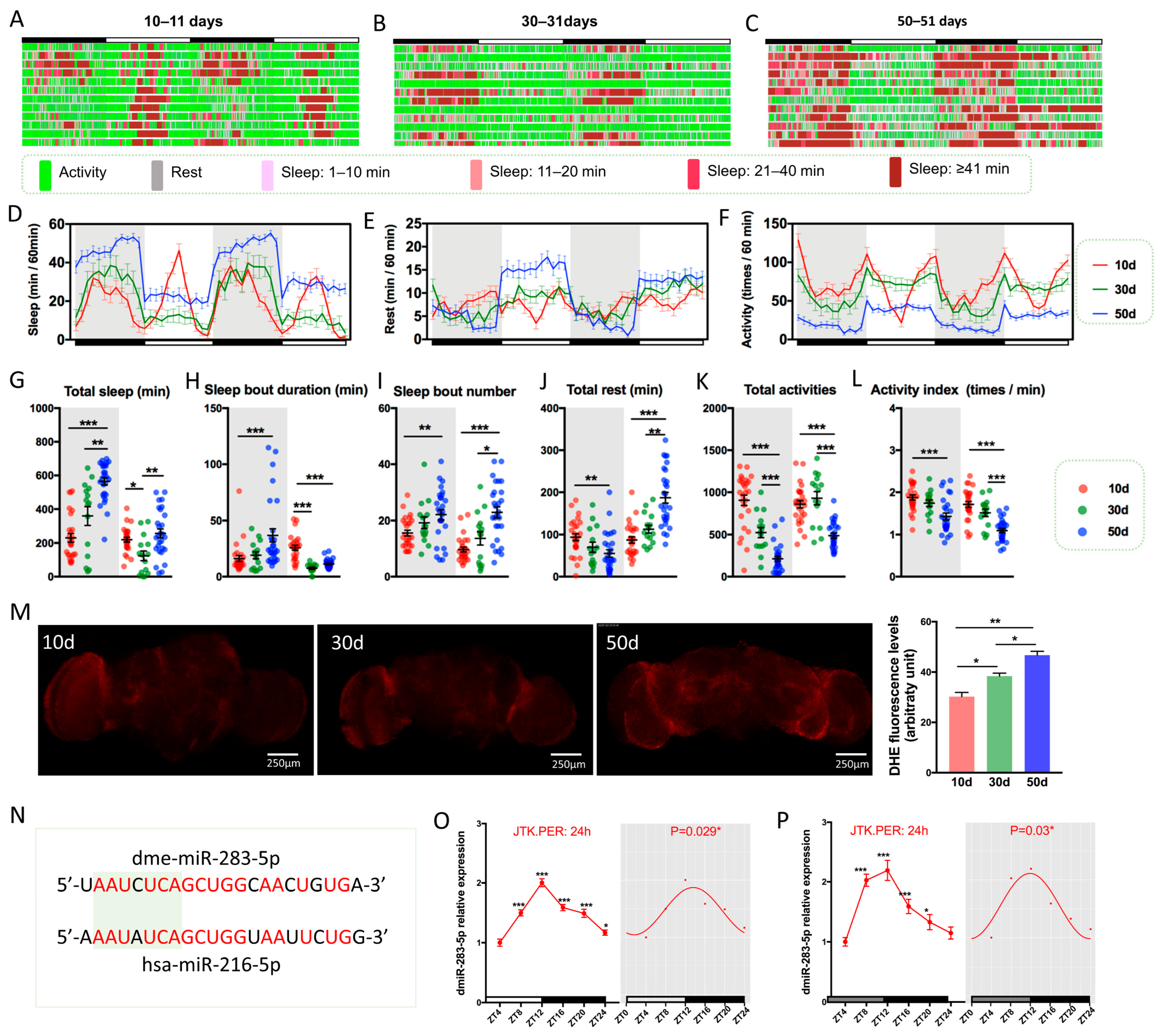
2.1. Drosophila Exhibits Age-Dependent Decline in Sleep–Wake Behavior during Aging

Loss of sleep stability is one of the main features of aging, as evidenced by decreased sleep continuity, more fragmented sleep, and increased night-time wakefulness and daytime sleep [20]. In addition, excessive sleep (≥10 h) in the elderly was considered to be an underlying disease or poor health condition [21]. Therefore, we analyzed the sleep–wake pattern and found that 10 days (10d) flies gradually entered the sleep state after the lights were turned off, woke up spontaneously approximately 2 h before the lights were turned on, and “napped” in the middle of the day; their activities were mainly concentrated before and after the lights were turned on/off (Figure 1A), and the sleep–wake cycle was maintained at 12 h (Figure 1D–F, Table S1). With increasing age, 30 days (30d) and 50 days (50d) flies developed increased nocturnal sleep, disappearance of daytime “naps”, increased fragmented sleep (Figure 1B,C,G,H), and a tendency for nocturnal sleep and daytime wakefulness to be light-induced (Figure 1B,C), suggesting a gradual decline in endogenous circadian rhythms in aging Drosophila. Circadian rhythm analysis revealed that the sleep–wake cycle shifted to 24 h in 30d and 50d flies (Table S1), and the amplitude and mesor values of sleep and activity showed an age-dependent decrease (Figure 1E,F, Table S1). Additionally, 50d flies noticeably had significantly more resting time and significantly lower autonomous activity behavior during daytime awakenings (Figure 1J,K), indicating an increased “sedentary and less active” state in older flies and a reduced frequency of activity during awakening (Figure 1L), which further suggests that 50d flies are in a disease state or are otherwise dysfunctional [22,23].
We measured aging brain ROS by dye assays and found that ROS levels increased with age (Figure 1M), which is consistent with previously reported oxidative damage [24], indicating that aging Drosophila brains accumulate higher levels of superoxide anions. Vaccaro et al. also found significantly increased levels of ROS in ClkAR mutant Drosophila brains [24], further associating an aging brain with the circadian biological clock.

2.2. dmiR-283 Is Rhythmically Expressed in Drosophila Brains

Precursor miRNA and mature miRNA have been reported to display circadian expression patterns and are conserved from plants to mammals [25,26]. The results of in situ hybridization in Drosophila embryos suggested that dmiR-283 was expressed in the nervous system and the silencing of dmiR-283 induced neurological defects [27], whereas the expression and functions of dmiR-283 in adult Drosophila remain unreported. Drosophila dmiR-283-5p has a high homology with human hsa-miR-216a-5p (Figure 1N, 77.27%), and the nucleotides of the seed sequence are also highly congruent (Figure 1N, 85.71%), demonstrating that dmiR-283 has similar regulatory roles to mammalian miR-216a. It has been demonstrated that miR-216a was involved in regulating the progression of Alzheimer’s disease and Parkinson’s disease, which indicates that miR-216a plays an important role in the process of neurodegeneration and provides a new perspective for the study of dmiR-283. To investigate whether there is a circadian rhythm in the expression pattern of dmiR-283, we collected 10d brains at 4 h intervals, and 12 h: 12 h light-dark (LD) entrainment as well as a dark–dark (DD) environment were included. For the mir-283 expression assay, the 5′ end of mature mir-283 was detected, representing the expression of mir-283. The qPCR results showed a significant 24 h expression rhythm of dmiR-283 in both LD and DD situations (p < 0.05), with the highest expression at zeitgeber time 12 (ZT12) and reduced expression levels before and after lights on (ZT24) (Figure 1O,P), which indicated that the circadian rhythm changes of sleep–wake behavior in Drosophila may be related to the rhythmic expression of dmiR-283.

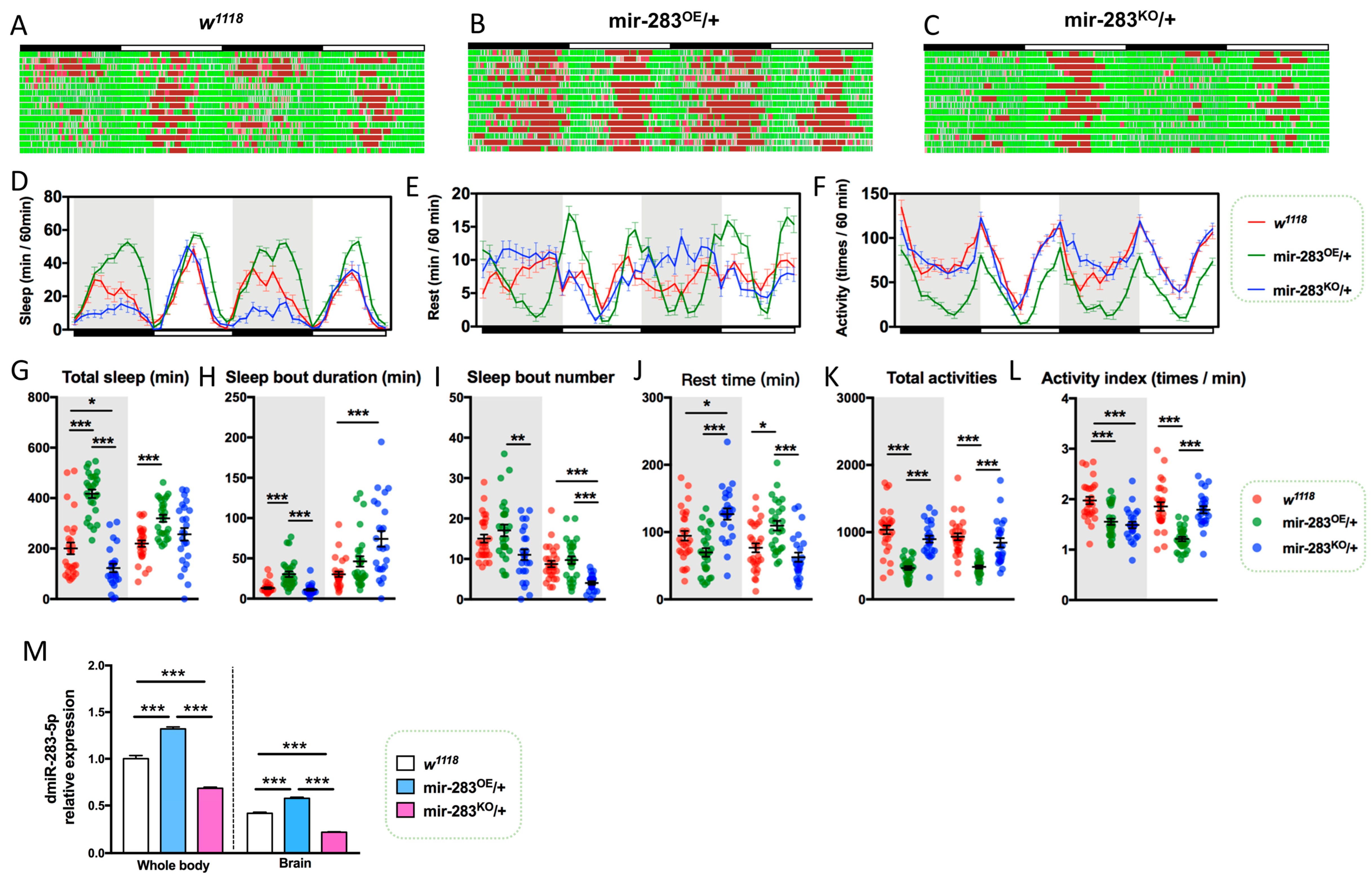
2.3. LNvs-Specific Knockdown of dmiR-283 Induces the Aging Sleep–Wake Behavior

To investigate the association of dmiR-283 with sleep–wake behavior, we analyzed the autonomic activity behavior of 10d Drosophila after the overexpression and knockdown of dmiR-283. The results showed that mir-283OE/+ flies exhibited increased sleep behavior (Figure 2B,D,G,H), decreased mobility (Figure 2B,F,K), and increased daytime rest time (Figure 2J), which was consistent with the sleep–wake behavior of wild-type 50d flies (Figure 1). In contrast, mir-283KO/+ resulted in reduced sleep time and increased wakefulness at night (Figure 2G,J), and a sleep rebound that may be the reason for the prolonged daytime sleep duration (Figure 2H). In addition, both the overexpression and knockdown of dmiR-283 in Drosophila reduced activity frequency (Figure 2L), suggesting that the stable expression of dmiR-283 is necessary to maintain normal sleep–wake behavior. CircaCompare analysis revealed that mir-283OE/+ Drosophila had delayed sleep and activity phases (Table S2) and their resting rhythms exhibited abnormal peaks and troughs (Figure 2E), which indicated that the overexpression of dmiR-283 disrupts the stable expression of endogenous circadian rhythms. The mir-283OE/+ and mir-283KO/+ brains showed significantly increased and decreased dmiR-283 expression (Figure 2M), further suggesting that disrupted regular dmiR-283 dynamic expression in the brain would lead to abnormal sleep–wake behavior.
Approximately 150 circadian neurons constitute the biological clock network of the Drosophila brain [28], where the PDF is secreted by Drosophila LNvs, similar in function to the mammalian suprachiasmatic nucleus (SCN), and primarily regulates circadian pacing, sleep, and light-mediated arousal [29]. We knocked down the expression of dmiR-283 by driving the dmir-283 sponge through Pdf-Gal4 specifically expressed in LNvs. The mir-283 sponge was fluorescently labeled with mCherry (Figure 3A), which similarly localized the expression of PDF protein while highlighting the LNvs. However, the expression levels of dmiR-283 were significantly increased in whole brains with the PDF-specific knockdown of dmiR-283 (Figure 3B), implying that the LNvs-specific knockdown of dmiR-283 might lead to a compensatory increase in other parts of the brain. To verify this conjecture, we expressed dmir-283 sponge in the heart with Hand-Gal4 and detected no significant change in the expression level of brain dmiR-283 (Figure 3B).
Lifespan is considered to be a comprehensive indicator for detecting physiological health in Drosophila, and miRNAs have been demonstrated to be involved in regulating neuronal activity and lifespan [30,31,32,33]. Several studies have proven that disruption of the biological clock function, both in insects and rodents, has many detrimental effects on healthy lifespan, including oxidative damage, increased accumulation of protein glycosylation, disruption of metabolic homeostasis, premature aging, and age-related diseases [3,34,35,36,37,38]. Therefore, we counted the survival rate of knockdown dmiR-283 in Drosophila LNvs to further clarify whether brain dmiR-283 is involved in lifespan regulation. Statistics on survival displayed an earlier mortality and shorter average lifespan in Pdf > mir-283SP flies (Figure 3C), indicating that the stable expression of brain dmiR-283 is also important for maintaining a normal lifespan.
Thus, does the LNvs-specific knockdown of dmiR-283 induce a circadian phenotype of aging? Sleep–wake profiling revealed that Pdf > mir-283SP flies had increased daytime and night-time sleep (Figure 3D–F), prolonged sleep duration and total sleep time (Figure 3J,K), and reduced resting time (Figure 3M) and activity behavior (Figure 3N) at 10d, 30d, and 50d compared with the age-matched control flies, which resembled the age-dependent increased sleep phenotype. Furthermore, Pdf > mir-283SP flies had significantly lower circadian rhythm amplitude and increased mesor for sleep rhythms, and decreased mesor for rest and activity rhythms (Figure 3G–I, Table S3), indicating that the LNvs-specific knockdown of dmiR-283 induces a senescent circadian rhythm phenotype. In conclusion, our findings suggest that brain dmiR-283 is an important miRNA regulating sleep–wake actions.

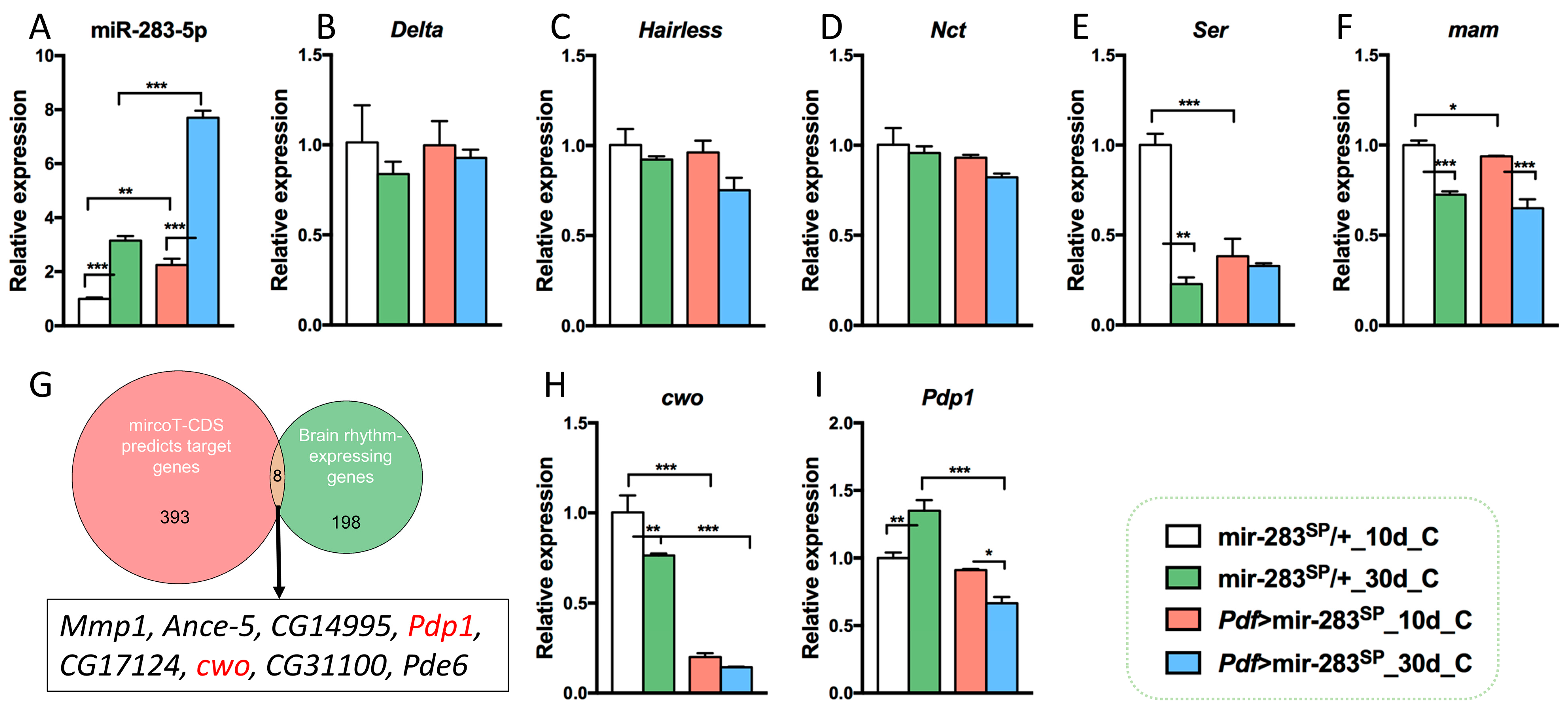
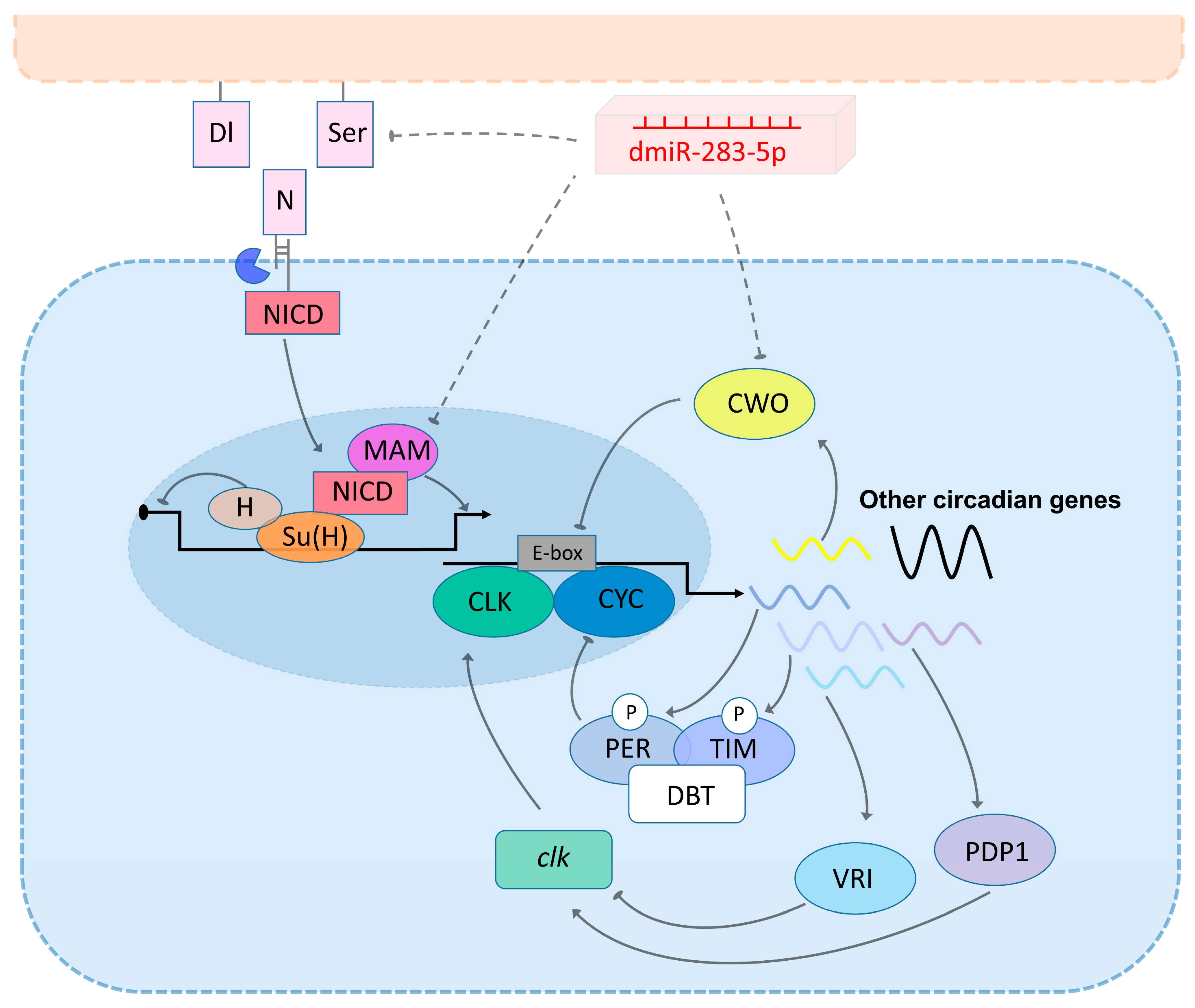
2.4. Prospective Mechanisms of dmiR-283 to Regulate age-Dependent Circadian Rhythm Decline and Sleep Prolongation

miRNAs perform post-transcriptional regulation by target binding mRNAs; typically, a single miRNA can combine hundreds of target mRNAs [39]. The 401 potential target mRNAs for dmiR-283 were predicted by the DIANA microT-CDS online tool and mainly enriched in the Notch signaling pathway (Delta, Hairless, Nct, ser, and mam, p = 0.0055). Previous studies have shown that the Notch signaling pathway is evolutionarily conserved and plays a regulatory role in the developing and post-developmental nervous system [40], whose over-activation or inhibition leads to drowsiness behavior in C. elegans [41]. Therefore, we examined the expression levels of related genes in the brain and found that both aging and LNvs knockdown of dmiR-283 resulted in the significant upregulation of dmiR-283 expression throughout the brain (Figure 4A), while Ser and mam were downregulated (Figure 4F). This implied that Ser and mam, the predicted target genes of dmiR-283 and key genes of the Notch pathway, were suppressed due to the accumulation of dmiR-283 expression. Thus, we predict that the age-dependent increase in sleep is attributed to the accumulation of brain dmiR-283, which suppresses Ser and mam, key genes of the Notch pathway, leading to a disorder of sleep behavior.
To further investigate the mechanisms underlying the reduced amplitude of sleep and activity in Pdf > mir-283SP Drosophila, we intersected the predicted target genes of mircoT-CDS with 206 rhythmically expressed genes obtained from Drosophila brain transcriptome sequencing and found eight predicted genes for rhythmic expression (Figure 4G), of which Pdp1 and cwo were the predominant clock genes [42]. It has been established that CWO, as a repressor of CLK-activated transcription, is an important transcription inhibitor of circadian rhythm amplitude [43], whereas the activator PDP1 regulates Clk transcription in conjunction with the repressor VRI [44,45]. We examined the brain levels of Pdp1 and cwo in aging as well as LNvs knockdown dmiR-283, and found a significant age-dependent reduction in cwo expression as well as a dmiR-283-knockdown-induced reduction (Figure 4H), suggesting that a weakened rhythmic amplitude in aging or Pdf > mir-283SP Drosophila may be associated with a decrease in cwo. In contrast, Pdp1 expression increased and decreased in senescent mir-283SP/+ and Pdf > mir-283SP Drosophila brains, respectively, which is inconsistent with the expected result of accumulation of dmiR-283-induced target gene degradation in senescent brains; therefore, we hypothesized that the regulation of circadian rhythm by Pdp1 is not directly affected by dmiR-283 alterations.

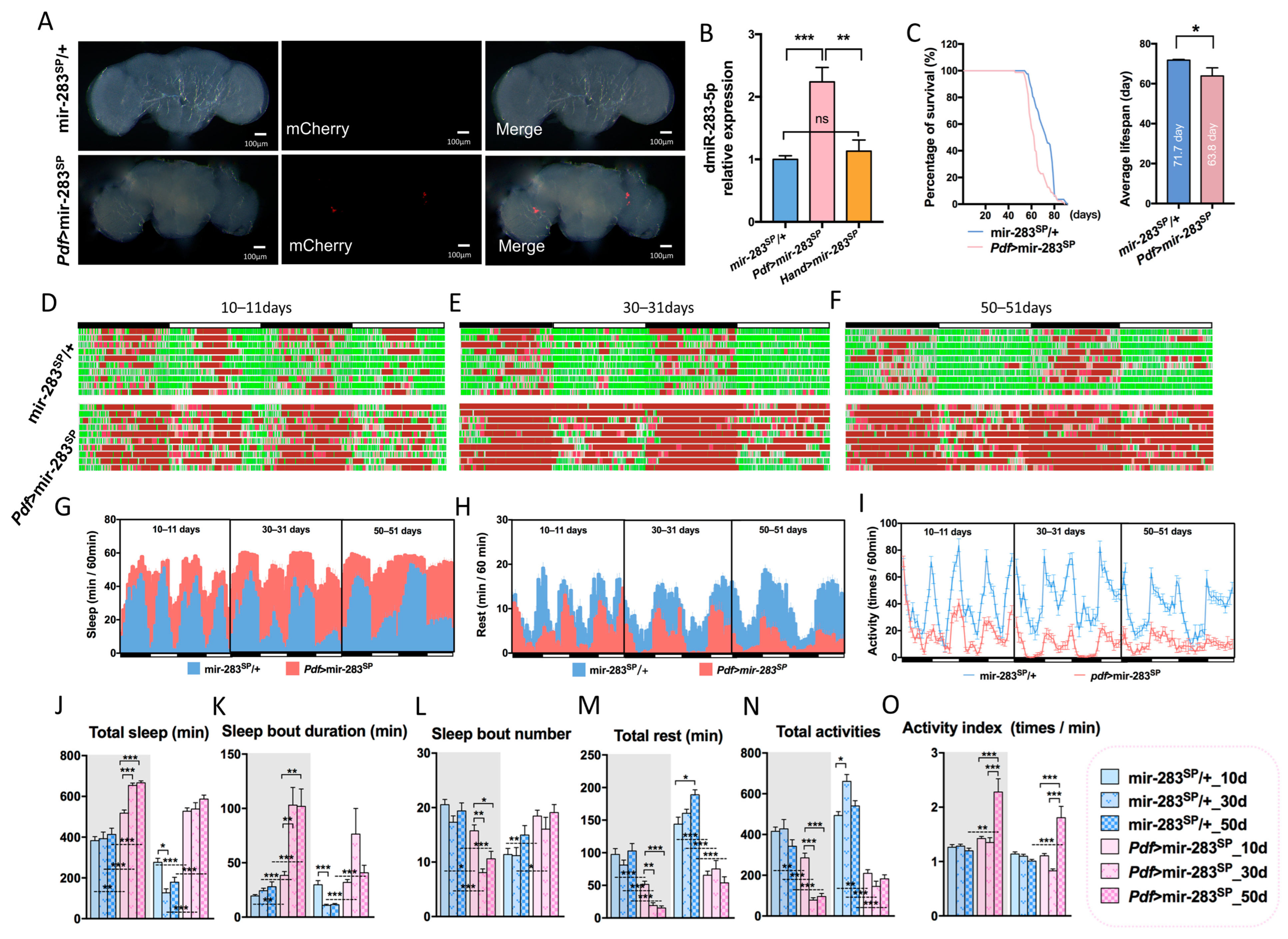
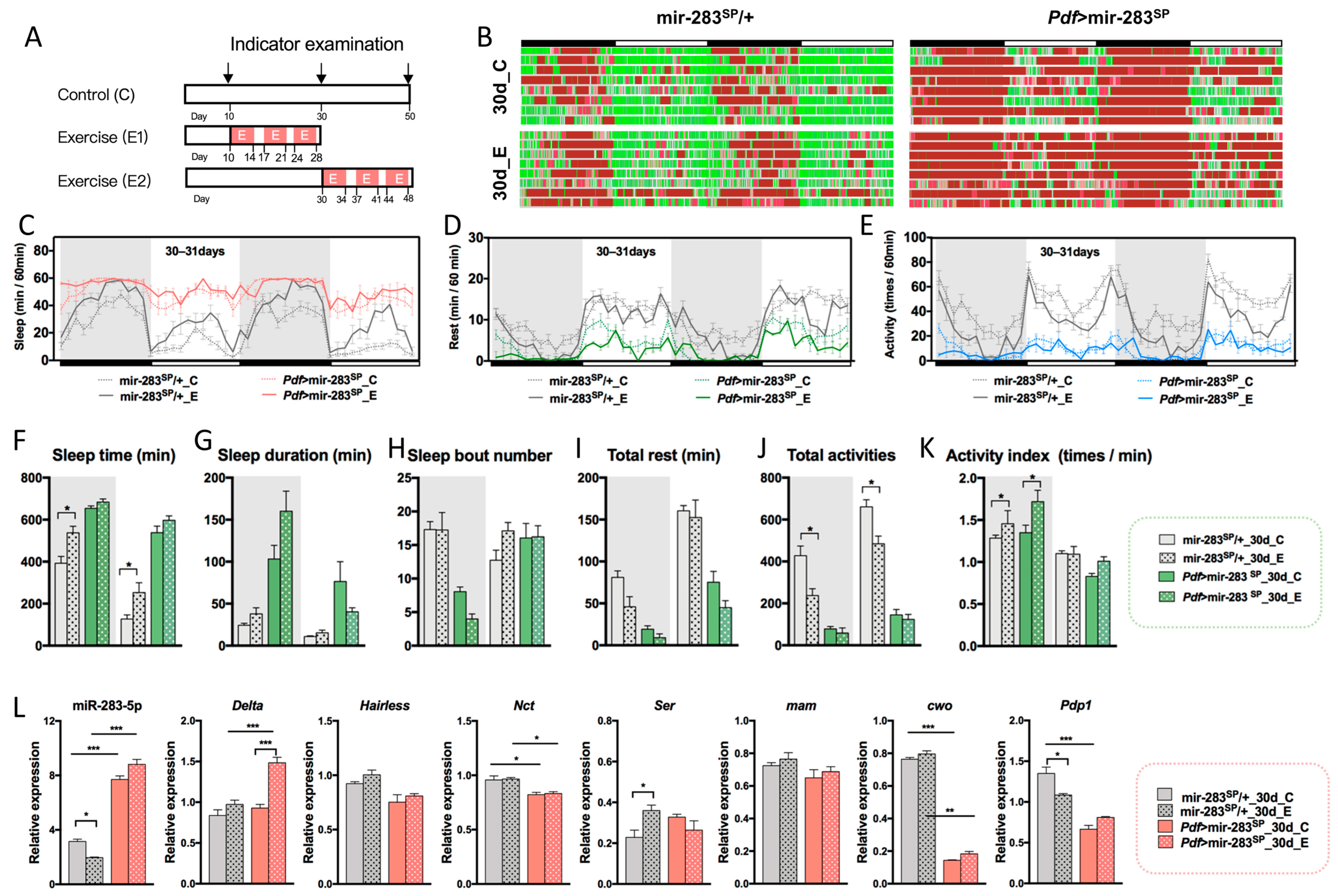
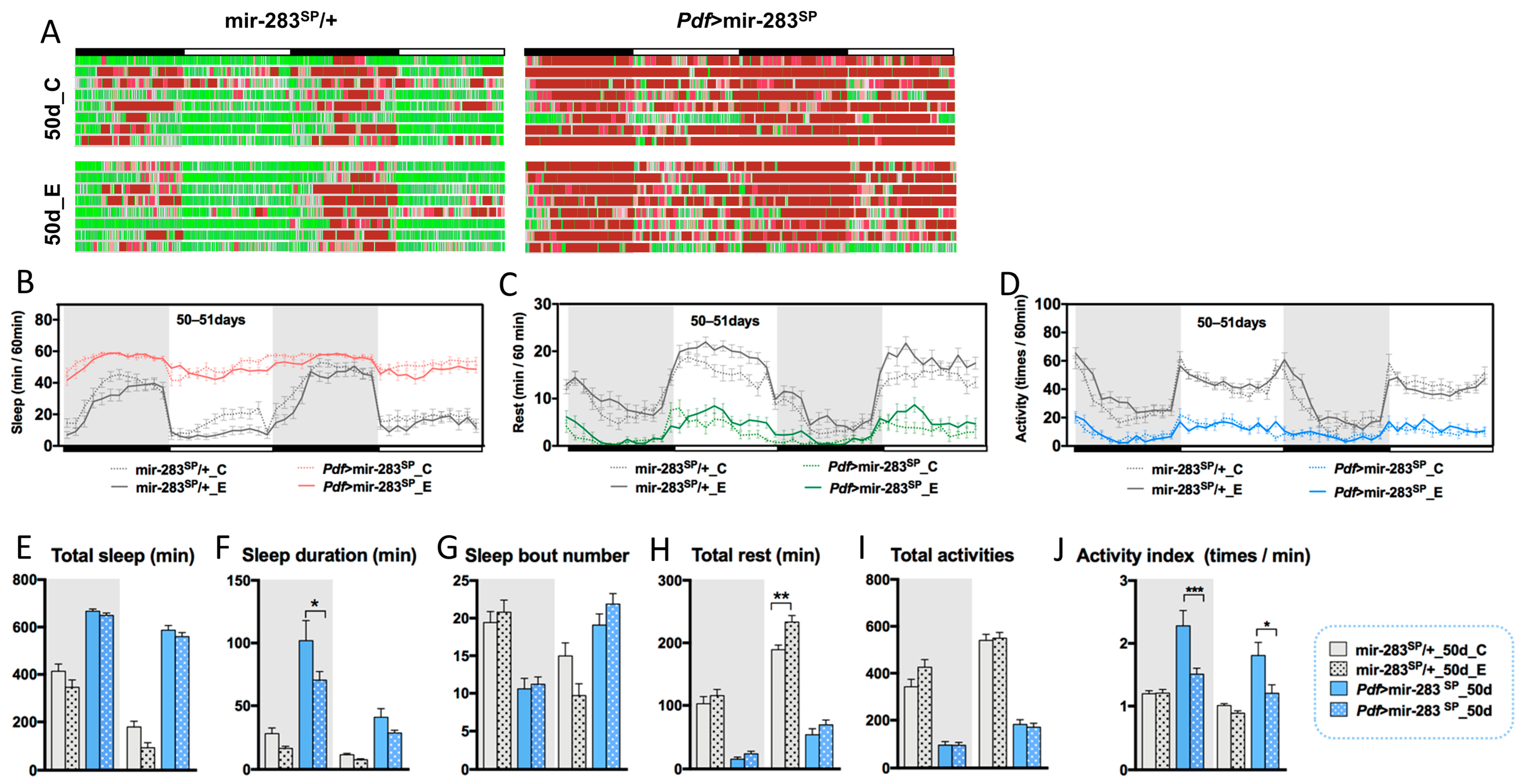
2.5. Expression Levels of dmiR-283 in the Brain Play a Key Role in Response to Exercise Effects

Our previous studies demonstrated that prolonged fatigue exercise increases sleep stability and improves sleep quality in aging Drosophila [46,47,48], but the regulatory mechanisms remain unclear. Based on the aging change characteristics of sleep–wake behavior, two exercise protocols were designed in this study: exercise starting from young adulthood to middle age (E1), and exercise starting from middle age to old age (E2) (Figure 5A), which aimed to investigate the effect of endurance exercise starting at different ages on age-dependent sleep–wake behavior decline.
In the E1 exercise protocol, we found that endurance exercise increased sleep time in mir-283SP/+ flies (Figure 5C,F). Although activity behavior decreased (Figure 5E,J), the activity frequency increased upon awakening (Figure 5K), as the activity index (the number of activities per minute) was a better indicator of health in the flies [22,23,48], indicating that exercise improved health in middle-aged Drosophila. JTK_CYCLE and CircaCompare analyses indicated that endurance exercise ameliorated age-dependent sleep cycle lengthening, allowing mir-283SP/+ Drosophila to still maintain a 12 h circadian oscillation with an increased rhythmic amplitude of sleep, activity, and rest (Figure 5C–E, Table S3), suggesting that endurance exercise initiated in youth maintains a relatively stable sleep–wake rhythm output. The expression of dmiR-283 was down-regulated after exercise in mir-283SP/+ brains, with a trend toward the increased expression of its predicted target genes Ser and mam (Figure 5L). Although the change in cwo was not significant, we found that exercise significantly reduced the accumulation of Pdp1 expression in aging brains (Figure 5L), predicting that the improvement of circadian rhythm by endurance exercise may be related to exercise, suppressing the overexpression of Pdp1 in the aging brain.
Pdf > mir-283SP flies appeared to be insensitive to exercise, with no significant changes in circadian rhythm amplitude and phase, except for a significant increase and decrease in the mesor of sleep and rest rhythms after exercise (Table S3). In addition, endurance exercise, while increasing the frequency of night-time activity in Pdf > mir-283SP Drosophila, had no significant effect on most sleep–wake behaviors and related gene expression (Figure 5F–L), indicating that the overexpression of brain dmiR-283 may affect the ameliorative effect of exercise on the aging-related sleep–wake behavioral decline. The speculation was further validated by endurance exercise starting at mid-age (E2). Brain dmiR-283 expression was significantly higher in mir-283SP/+ Drosophila at 30d (Figure 4A), and the onset of endurance exercise at midlife was only effective for the maintenance of activity rhythms (Table S3), with no significant changes in other sleep–wake behaviors. The expression of dmiR-283 in the brain was increased 7.7-fold in Pdf > mir-283SP middle-aged Drosophila compared with young mir-283SP/+ flies (Figure 4A), and sleep–wake behavior after endurance exercise (E2) showed negative effects such as decreased nocturnal sleep stability (Figure 6F) and reduced day/night activity frequency (Figure 6I,J), which further suggests that overexpression of dmiR-283 in the brain causes exercise to play a negative role.

3. Discussion

In recent years, an expanding number of studies have found that miRNA-mediated post-transcriptional regulation plays an important role in the generation and maintenance of biological clocks in Drosophila and mammals, but whether aging-related circadian rhythm disorders are associated with age-dependent alterations in miRNA expression remains unreported. In our study, we found that flies developed diminished sleep–wake rhythm amplitudes and decreased mesor in middle age, which further intensified in old age, and also displayed age-dependent changes in sleep, activity, and rest behaviors, which may be associated with the elevated expression of dmiR-283 in the brain. Increased dmiR-283 levels consistent with aging were detected in both mir-283OE/+ and Pdf > mir-283SP young Drosophila brains, and sleep–wake behavior tended to change with aging, which evidenced that brain dmiR-283 is a key miRNA regulating sleep–wake behavior during aging.
Notably, we observed longer daytime sleep (comparable to nighttime sleep duration) in the 10d flies of w1118, as well as differences in sleep structure (sleep time, number of sleep episodes, and sleep latency) between w1118 and Canton S strains as observed by Vienne et al. [17]. This could be due to the white mutation that exacerbates progressive retinal degeneration in Drosophila [49], further affecting sleep structures. Therefore, whether the age-dependent changes we observed in w1118 are applicable to other Drosophila strains or species remains to be further investigated.
Bioinformatic predictions revealed that dmiR-283 is mainly enriched in the Notch pathway, a ligand–receptor signaling pathway for the transmembrane transmission of information between cells, which is highly conserved evolutionarily [50,51]. In Drosophila, the Notch signaling pathway is activated by binding of the transmembrane receptor Notch (N) to the transmembrane ligand Delta (Dl) or Serrate (Ser) on adjacent cells, which hydrolyzes and releases the Notch intracellular structural domain (NICD). NICD translocates into the nucleus to interact with Su (H) and mam to form a transcriptional complex that activates downstream target gene transcription [51,52], and the repressor Hairless (H), which binds competitively to Su (H) and antagonizes the activation of the Notch signaling pathway [53] (Figure 7). Several studies have evidenced that Notch signaling plays an important role in sleep regulation in C. elegans and Drosophila [41,54,55]. Notch and Delta in the adult Drosophila brain can mediate interneuron–glial signaling through intercellular connections, and Notch signaling activation (overexpression of Delta and NICD, and the functional acquisition of Notch alleles) prevents the onset of sleep rebound after sleep deprivation and maintains sleep stability [54]. Our study reveals that dmiR-283 accumulates in aging and LNvs knockdown brains with a decreased expression of transmembrane ligand Ser and coactivator mam in its predicted target genes, predicting inhibition of the Notch signaling pathway, which may contribute to increased sleep. In mammals, Notch-related gene expression was downregulated in the hippocampus of sleep-deprived rats [56]; genome-wide association analysis has also shown a potential relationship between Notch signaling and insomnia [57], although the specific regulatory mechanisms between sleep/sleep-like behavior and Notch signaling need to be further explored.
Our study is the first to reveal that the expression of dmiR-283 exhibits 24 h circadian oscillations in young Drosophila brains and is predicted to be involved in the regulation of circadian rhythm amplitude as well. Of the predicted target genes, cwo and Pdp1 constitute the core biological clock regulatory network. Expressed in rhythmically paced neurons, cwo serves as an important transcriptional repressor that regulates circadian rhythm amplitude, and its functional deficiency leads to a prolonged and weak autonomic activity rhythm in Drosophila [43,58,59], similar to the CLK–CYC transcriptional repression that prolongs or eliminates the circadian rhythm period [60]. Therefore, we hypothesized that the reduced circadian rhythm amplitude and prolonged cycles in aging and LNvs knockdown of dmiR-283 flies may be attributable to the degradation of cwo by the hyper-expression of dmiR-283 in the brain. Molecular studies have shown that the transcriptional activation of CWO is regulated by CLK–CYC, which, in addition to forming a self-regulatory negative feedback loop [61], synergistically represses the transcriptional activation of CLK target genes (per, tim, vri, and Pdp1) with PER [59] (Figure 7). A recent study also revealed that CWO promotes the transcriptional activation of CLK–CYC by repressing the negative regulator Clock interacting protein circadian [62]. In conclusion, the mechanisms of CWO in circadian rhythm regulation are becoming better defined, and our study confirmed that age-dependent declines in sleep–wake rhythm are associated with an accumulation of dmiR-283 in the brain. Additionally, a further suggestion is that dmiR-283 regulates the sleep–wake rhythm of aging or LNvs dmiR-283 knockdown Drosophila by targeting cwo for degradation.
A previous study proposed that the activation of vascular endothelial growth factor expression by the biological clock gene Bmal1 crosstalks mechanistically with the Notch signaling pathway, regulating vascular sprouting and function [63]. Therefore, we hypothesized that the behavior of prolonged sleep and reduced rhythm of autonomous activity during aging in Drosophila is associated with an inhibited Notch signaling pathway and reduced expression of the core rhythm gene cwo in the brain, which are co-regulated negatively by the aging accumulation of dmiR-283 (Figure 7).
Exercise as a beneficial lifestyle has been identified as a positive contributor to healthy aging and longevity [2,64], not only by improving potential sleep disturbances in the young and old [65,66], but also by resetting the circadian biological clock [67,68,69]. Aerobic exercise, especially, has been shown to play a positive role in promoting brain health and healthy sleep patterns; thus, exercise has been proposed as a non-pharmacological intervention for patients with sleep disorders [70]. A study of young, healthy male subjects showed that 1 h of high-intensity exercise improved sleep quality by increasing slow-wave sleep [71]. Similarly, we found that endurance exercise starting in youth showed positive effects in healthy mir-283SP/+ flies, in terms of enhanced sleep–wake rhythm amplitude, stable sleep cycles, and increased activity frequency during waking. We propose that endurance exercise down-regulates dmiR-283 accumulated in the brain, thereby releasing the inhibition of the Notch signaling pathway by aging. Although the level of cwo did not change notably after exercise, we found that exercise significantly reduced the accumulation of Pdp1 expression in aging brains, predicting that the improvement in circadian rhythm may be related to exercise suppressing the overexpression of Pdp1 in the aging brain. Pdp1, a key activator of Clk, results in reduced CLK and PER expression and circadian rhythm dysfunctional behavior in the presence of either Pdp1 mutations or a dysregulated Pdp1/vri ratio [44,72]. Our study further explored the exercise effects in the presence of high brain dmiR-283 expression (aging and LNvs knockdown of dmiR-283) and showed ineffective or negative sleep–wake behaviors, indicating a negative correlation between dmiR-283 expression levels and exercise benefits.
Aging is a major risk factor for most chronic diseases and functional disorders. Taking advantage of the short life span and the homologous molecular regulatory mechanisms of Drosophila, we found that increased sleep-like behaviors and reduced circadian rhythms during aging are associated with the accumulation of brain dmiR-283. Additionally, we reasonably speculate that the increased sleep behaviors probably occurred by suppressing the Notch-related pathway and weakening the circadian rhythm by depressing the expression of the core clock gene cwo. Thus, dmiR-283 may act as a co-regulator of the Notch signaling pathway and biological clock core genes to promote the aging process in the brain. Endurance exercise initiated in youth reduces brain dmiR-283 levels, maintaining a solid circadian output in aging Drosophila. In contrast, exercise performed at a certain accumulation level of brain dmiR-283 displayed ineffective or negative outcomes, indicating that the affirmative results of exercise are dependent on dmiR-283 stable-expression.

4. Materials and Methods

4.1. Drosophila Strains, Crosses, and Rearing

Wild-type strains (w1118, BS3605), driver strains of pigment dispersing factor (Pdf)-expressing ventral lateral neurons (LNvs) in the brain (Pdf-Gal4, BS41286), mir-283 knockdown strains (mir-283SP, BS61415, w [*]; P{y [+t7.7] w [+mC] = UAS-mCherry.mir-283.sponge.V2}attP40; P{y [+t7.7] w [+mC] = UAS-mCherry.mir-283.sponge.V2}attP2), and mir-283 knockout strains (mir-283KO, BS58912, w [*] TI{TI}mir-283 [KO]) were purchased from the United States Bloomington Drosophila Stock Center. The overexpression strain of mir-283 (mir-283OE, BCF132#, the attP40 landing site (Chr.2 25C6) stock used for the fly embryo injection and transformation was y [1] M{vas-int.Dm}ZH-2A w [*]; P{CaryP}attP40) was purchased from the Core Facility of Drosophila Resource and Technology, CEMCS, CAS. The cardiac-specific driver strain (Hand-Gal4) was presented by the Center for Heart Development of Hunan Normal University. Virgin flies of the mir-283SP strain that fledged within 8 h were collected and crossed with male strains w1118 and Pdf-Gal4; correspondingly, the F1 generation virgin flies contained control group (mir-283SP/+) and knockdown group (Pdf > mir-283SP). For reducing the effect of genetic background on Drosophila, virgin flies of pure mir-283OE and mir-283KO strains, which fledged within 8 h, were collected and crossed with male strain w1118, and F1 generation virgin flies were overexpressed (mir-283OE/+) or mutated (mir-283KO/+) heterozygous flies, corresponding to the wild-type (+) allele, and w1118 virgin flies of the same age served as a control. All flies were reared in standard yeast flour/cornmeal/soybean flour/sucrose/maltose/agar medium under conditions of 25 ± 1 °C, 60% humidity, and a 12 h light and 12 h dark environment. For the analysis under constant darkness (DD), newly collected virgin flies were first entrained in light–dark conditions for 3 days and then reared in a DD environment for 3 days for testing. The remaining Drosophila were reared under standard conditions of 12 h light:12 h dark.
We conducted experimental comparisons between different sexes at the beginning of the experiment, and the results showed that male flies slept more at night and during the day, showing stronger rhythmicity than females. However, in the induced climbing movement experiment, males showed poor performance in negative tending ground climbing ability and endurance, which may reduce the effect of induced movement. Therefore, we chose females with higher locomotor ability as the experimental subjects. In addition, flies were housed in groups during rearing and hybridization, and in single housing in the activity monitoring experiment.

4.2. Exercise Training Program

Based on the anti-gravity climbing characteristics of Drosophila, we developed a next-generation Drosophila climbing exercise device (Patent No. CN202021948877.0), which induced flies to climb upward along the test tube wall by setting an arbitrary flipping angle and dwell time. In this study, the motor speed was set to 0.45 r/s, and a 180° vertical flip was performed and then left for 5 s, during which most flies were able to actively crawl upward to the top of the test tube.
According to the optimal exercise protocol determined in a previous study [47], flies were induced to perform 2.5 h of climbing exercise per day for 5 days followed by 2 days of rest, and the exercise program lasted for a total of 3 weeks. There is a distinct sleep–wake rhythm in young Drosophila, with sleep time concentrated in the middle of the day/night period and high activity before and after lights on/off (Figure 1A); therefore, the exercise protocol was performed during the peak activity period before lights off (ZT9~ZT12), to avoid inducing sleep deprivation. The 10th (10d), 30th (30d), and 50th (50d) days of age in Drosophila correspond to the young, middle-aged, and old stages [73]. To investigate the effect of exercise initiated at different periods on sleep–wake behavior in Drosophila, two exercise protocols were designed, as shown in Figure 5A.

4.3. Life Expectancy Statistics

The number of dead flies was recorded from the first day after fledging and counted during ZT14-ZT15 until all flies were dead. Flies were transferred to fresh medium every 3 to 4 days during the lifespan count. Based on the total number of flies and the number of survivors, survival curves were plotted and the average lifespan was calculated.

4.4. Sleep–Wake Behavior Analysis

During ZT12~ZT15, 2 days before data collection, age-appropriate flies (8d, 28d, and 48d) were transferred individually into a 5 mm × 65 mm polycarbonate tube with standard culture medium at one end and a sponge plug at the other end. Under standard experimental conditions, activity data were monitored for 3 consecutive days using a Drosophila activity monitoring system (DAMS, TriKinetics, Waltham, MA, USA), where an infrared beam is emitted at the center of each glass activity tube and the number of passes through the infrared beam is automatically recorded as the flies move back and forth through the monitoring tube, with data collection intervals of 1 min. Data from the first day were discarded to avoid the effect of changes in the survival environment [74,75]. Data from days 2 to 3 (9~11d, 29~31d, and 49~51d) were used for sleep–wake behavior and circadian rhythm analysis.
Behavioral and electrophysiological studies showed that the continuous 5 min inactivity of Drosophila resembles the mammalian sleep state [76,77,78]. Based on this feature, we analyzed DAMS data using a custom JavaScript script and visualized sleep–wake behavior with ECharts, in which sleep behavior was shown in red with darker colors indicating higher sleep persistence. To further study the arousal characteristics of Drosophila, we divided arousal into two states, active and resting. Activity means the time to cross the infrared beam, shown in green; resting means the flies did not cross the infrared beam for 1–4 min and, at this time, the flies are not active, but also not asleep, shown in gray. Both resting and active behaviors belong to the awakening state (Drosophila Sleep-Wake Behavior Analysis System V1.0, Computer Software Copyright Registration No.: 2022SR1402794).

4.5. Circadian Rhythm Analysis

Cycle analysis and circadian rhythm significance analysis were performed using JTK_CYCLE in the metacycle package for hourly sleep time, hourly rest time, hourly activity, and continuous 24 h gene expression levels in the brain. The circadian rhythm amplitude (half of the difference between the peak and trough), phase (time corresponding to peak), and mesor (response rhythm-adjusted average) were analyzed differentially according to the CircaCompare program developed by Parsons et al. [79] to plot circadian rhythm fitting curves.

4.6. Tissue Sample Collection, Total RNA Extraction, and RT-qPCR

Adult Drosophila brains were obtained with reference to the method described by Williamson and Hiesinger [80]. To investigate the circadian expression levels of genes, Drosophila brains were collected at 4 h intervals from ZT4 to ZT24, and the other brain tissue sampling was performed from ZT8 to ZT12. According to the instruction manual, total RNA was extracted using RNAfast200 (Shanghai Feijie Biotech). The Mir-X miRNA First-Strand Synthesis Kit and PrimeScript™ RT reagent Kit with gDNA Eraser (Takara Biomedical Technology (Beijing) Co., Ltd., Beijing, China) were used for miRNA and mRNA reverse transcription reactions, respectively. Quantitative RT-PCR assays were performed using TB Green® Premix Ex Taq™ II (Takara Biomedical Technology (Beijing) Co., Ltd., Beijing, China) on a CFX ConnectTM fluorescent quantitative PCR instrument (Bio-Rad Laboratories, Berkeley, CA, USA). The primer sequences are shown in Table 1.

4.7. Reactive Oxygen (ROS) Testing

Brains were stripped in oxygen-enriched hemolymph solution, and the fluorescent probe of Dihydroethidium (DHE, Beyotime Biotechnology Co., Shanghai, China) was diluted to 1:1000 with an artificial hemolymph according to the superoxide anion fluorescent probe instructions. After incubation for 30 min at room temperature in a shaker, the brains were washed with hemolymph solution to adequately remove DHE that had not entered the cells, and were imaged immediately using a fluorescent microscope; all images were collected based on the same parameters. Relative ROS levels were measured by quantification of the dye fluorescence using ImageJ 1.53a software.

4.8. Fluorescence Imaging

Brain DHE fluorescence was photographed using a Leica stereomicroscope (Leica, DVM6) to detect ROS levels. In addition, the mir-283SP strain of Drosophila carries a red fluorescent marker gene that expresses the red fluorescent protein when specifically knocking down the LNvs dmiR-283 after crossing with Pdf-Gal4. The expression of the red fluorescent protein in the Drosophila brain was photographed using a Leica stereomicroscope to determine if the strain was successfully constructed.

4.9. Bioinformatics Analysis

The DIANA microT-CDS online tool (http://diana.imis.athena-innovation.gr/DianaTools/ accessed on 1 August 2022) was used to predict target genes for dme-miR-283-5p, whereas the DAVID Bioinformatics Resource (https://david.ncifcrf.gov/ accessed on 1 August 2022) was utilized to perform pathway enrichment analysis of the predicted target genes. Moreover, the predicted target genes were intersected with the rhythmically expressed genes obtained by transcriptome sequencing of the brain of w1118 strain wild-type Drosophila [42] to identify potential transcription factors of dmiR-283 regulating the circadian rhythm.

4.10. Data Analysis and Processing

SPSS 22.0 was used for statistical analysis of the data. For data obeying normal distributions with uniform variance, an independent-samples t-test or one-way ANOVA was performed, followed by Tukey’s multiple comparisons to assess significance. Otherwise, independent-samples nonparametric tests with Kruskal–Wallis multiple comparisons were used. Graphs were generated in GraphPad Prism 7 and data were expressed as mean ± standard error (M ± SEM) with a significance level of p < 0.05.

5. Conclusions

In summary, the accumulation of dmiR-283 expression in the brain induced an age-dependent decline in sleep–wake behavior, and the core clock gene cwo and Notch signaling pathway may co-mediate this regulation. Furthermore, commencing endurance exercise in youth counteracts the increase in dmiR-283 in the aging brain, which ameliorates the deterioration of sleep–wake behavior during aging. Meanwhile, the stable expression of dmiR-283 is a guarantee that endurance exercise delays brain aging.

6. Patents

This study produced a Computer Software Copyright Registration entitled “Drosophila Sleep-Wake Behavior Analysis System V1.0” (No.: 2022SR1402794).

Supplementary Materials

The following supporting information can be downloaded at: https://www.mdpi.com/article/10.3390/ijms24044180/s1.

Author Contributions

Conceptualization, Q.L. and L.Z.; methodology, Q.L.; software, L.W.; validation, Y.C., X.W. and C.T.; formal analysis, Q.L. and L.W.; data curation, Q.L., L.W. and Y.C.; writing—original draft preparation, Q.L. and L.W.; writing—review and editing, Q.L., L.W. and L.Z.; visualization, Q.L.; supervision, L.Z.; project administration, L.Z.; funding acquisition, Q.L., L.W. and L.Z. All authors have read and agreed to the published version of the manuscript.

Funding

This research was funded by the National Natural Science Foundation of China (grant number 32071175) and the Postgraduate Scientific Research Innovation Project of Hunan Province (grant numbers CX20210480 and CX20220518).

Institutional Review Board Statement

Not applicable.

Informed Consent Statement

Not applicable.

Data Availability Statement

Not applicable.

Acknowledgments

We thank the Heart Development Center (College of Life Sciences, Hunan Normal University) for supporting the Hand-Gal4 strain of Drosophila.

Conflicts of Interest

The authors declare no conflict of interest.

References

  1. Musiek, E.S.; Holtzman, D.M. Mechanisms linking circadian clocks, sleep, and neurodegeneration. Science 2016, 354, 1004–1008. [Google Scholar] [CrossRef] [PubMed] [Green Version]
  2. Panagiotou, M.; Michel, S.; Meijer, J.H.; Deboer, T. The aging brain: Sleep, the circadian clock and exercise. Biochem. Pharmacol. 2021, 191, 114563. [Google Scholar] [CrossRef] [PubMed]
  3. Kondratov, R.V.; Kondratova, A.A.; Gorbacheva, V.Y.; Vykhovanets, O.V.; Antoch, M.P. Early aging and age-related pathologies in mice deficient in BMAL1, the core componentof the circadian clock. Genes Dev. 2006, 20, 1868–1873. [Google Scholar] [CrossRef] [PubMed] [Green Version]
  4. Sterniczuk, R.; Theou, O.; Rusak, B.; Rockwood, K. Sleep Disturbance is Associated with Incident Dementia and Mortality. Curr. Alzheimer Res. 2013, 10, 767–775. [Google Scholar] [CrossRef]
  5. Lim, A.S.; Kowgier, M.; Yu, L.; Buchman, A.S.; Bennett, D.A. Sleep Fragmentation and the Risk of Incident Alzheimer’s Disease and Cognitive Decline in Older Persons. Sleep 2013, 36, 1027–1032. [Google Scholar] [CrossRef] [Green Version]
  6. Sowell, E.R.; Peterson, B.S.; Thompson, P.M.; Welcome, S.E.; Henkenius, A.L.; Toga, A.W. Mapping cortical change across the human life span. Nat. Neurosci. 2003, 6, 309–315. [Google Scholar] [CrossRef]
  7. Kinser, H.E.; Pincus, Z. MicroRNAs as modulators of longevity and the aging process. Hum. Genet. 2020, 139, 291–308. [Google Scholar] [CrossRef]
  8. Garg, D.; Cohen, S.M. miRNAs and aging: A genetic perspective. Ageing Res. Rev. 2014, 17, 3–8. [Google Scholar] [CrossRef]
  9. Kumar, S.; Vijayan, M.; Bhatti, J.S.; Reddy, P.H. MicroRNAs as Peripheral Biomarkers in Aging and Age-Related Diseases. Prog. Mol. Biol. Transl. 2017, 146, 47–94. [Google Scholar] [CrossRef]
  10. Wagner, K.H.; Cameron-Smith, D.; Wessner, B.; Franzke, B. Biomarkers of Aging: From Function to Molecular Biology. Nutrients 2016, 8, 338. [Google Scholar] [CrossRef] [Green Version]
  11. Chen, X.; Rosbash, M. MicroRNA-92a is a circadian modulator of neuronal excitability in Drosophila. Nat. Commun. 2017, 8, 14707. [Google Scholar] [CrossRef] [Green Version]
  12. Sha, N.; Wang, H.W.; Sun, B.; Gong, M.; Miao, P.; Jiang, X.L.; Yang, X.F.; Li, M.; Xu, L.X.; Feng, C.X.; et al. The role of pineal microRNA-325 in regulating circadian rhythms after neonatal hypoxic-ischemic brain damage. Neural Regen. Res. 2021, 16, 2071. [Google Scholar] [CrossRef]
  13. Davis, C.J.; Bohnet, S.G.; Meyerson, J.M.; Krueger, J.M. Sleep loss changes microRNA levels in the brain: A possible mechanism for state-dependent translational regulation. Neurosci. Lett. 2007, 422, 68–73. [Google Scholar] [CrossRef] [Green Version]
  14. Davis, C.J.; Clinton, J.M.; Krueger, J.M. MicroRNA 138, let-7b, and 125a inhibitors differentially alter sleep and EEG delta-wave activity in rats. J. Appl. Physiol. 2012, 113, 1756–1762. [Google Scholar] [CrossRef] [Green Version]
  15. Holm, A.; Bang-Berthelsen, C.H.; Knudsen, S.; Kornum, B.R.; Modvig, S.; Jennum, P.; Gammeltoft, S. miRNA Profiles in Plasma from Patients with Sleep Disorders Reveal Dysregulation of miRNAs in Narcolepsy and Other Central Hypersomnias. Sleep 2014, 37, 1525–1533. [Google Scholar] [CrossRef] [Green Version]
  16. Shafer, O.T.; Keene, A.C. The Regulation of Drosophila Sleep. Curr. Biol. 2021, 31, R38–R49. [Google Scholar] [CrossRef]
  17. Vienne, J.; Spann, R.; Guo, F.; Rosbash, M. Age-Related Reduction of Recovery Sleep and Arousal Threshold in Drosophila. Sleep 2016, 39, 1613–1624. [Google Scholar] [CrossRef] [Green Version]
  18. De Nobrega, A.K.; Lyons, L.C. Aging and the clock: Perspective from flies to humans. Eur. J. Neurosci. 2020, 51, 454–481. [Google Scholar] [CrossRef] [Green Version]
  19. Gabriel, B.M.; Zierath, J.R. Circadian rhythms and exercise—Re-setting the clock in metabolic disease. Nat. Rev. Endocrinol. 2019, 15, 197–206. [Google Scholar] [CrossRef]
  20. Koh, K.; Evans, J.M.; Hendricks, J.C.; Sehgal, A. A Drosophila model for age-associated changes in sleep:wake cycles. Proc. Natl. Acad. Sci. USA 2006, 103, 13843–13847. [Google Scholar] [CrossRef] [Green Version]
  21. Grandner, M.A.; Drummond, S.P.A. Who are the long sleepers? Towards an understanding of the mortality relationship. Sleep Med. Rev. 2007, 11, 341–360. [Google Scholar] [CrossRef] [PubMed] [Green Version]
  22. Andretic, R.; Shaw, P.J. Essentials of sleep recordings in Drosophila: Moving beyond sleep time. Method Enzymol. 2005, 393, 759–772. [Google Scholar] [CrossRef]
  23. Chiu, J.C.; Low, K.H.; Pike, D.H.; Yildirim, E.; Edery, I. Assaying locomotor activity to study circadian rhythms and sleep parameters in Drosophila. J. Vis. Exp. 2010, 43, e2157. [Google Scholar] [CrossRef] [Green Version]
  24. Vaccaro, A.; Issa, A.R.; Seugnet, L.; Birman, S.; Kiarsfeid, A. Drosophila Clock Is Required in Brain Pacemaker Neurons to Prevent Premature Locomotor Aging Independently of Its Circadian Function. PLoS Genet. 2017, 13, e1006507. [Google Scholar] [CrossRef] [Green Version]
  25. Mendoza-Viveros, L.; Bouchard-Cannon, P.; Hegazi, S.; Cheng, A.H.; Pastore, S.; Cheng, H.M. Molecular modulators of the circadian clock: Lessons from flies and mice. Cell. Mol. Life Sci. 2017, 74, 1035–1059. [Google Scholar] [CrossRef]
  26. Mehta, N.; Cheng, H.Y. Micro-managing the circadian clock: The role of microRNAs in biological timekeeping. J. Mol. Biol. 2013, 425, 3609–3624. [Google Scholar] [CrossRef]
  27. Aboobaker, A.A.; Tomancak, P.; Patel, N.; Rubin, G.M.; Lai, E.C. Drosophila microRNAs exhibit diverse spatial expression patterns during embryonic development. Proc. Natl. Acad. Sci. USA 2005, 102, 18017–18022. [Google Scholar] [CrossRef] [Green Version]
  28. Xue, Y.B.; Zhang, Y. Emerging roles for microRNA in the regulation of Drosophila circadian clock. BMC Neurosci. 2018, 19. [Google Scholar] [CrossRef] [Green Version]
  29. Kula-Eversole, E.; Nagoshi, E.; Shang, Y.; Rodriguez, J.; Allada, R.; Rosbash, M. Surprising gene expression patterns within and between PDF-containing circadian neurons in Drosophila. Proc. Natl. Acad. Sci. USA 2010, 107, 13497–13502. [Google Scholar] [CrossRef] [Green Version]
  30. Klarsfeld, A.; Rouyer, F. Effects of circadian mutations and LD periodicity on the life span of Drosophila melanogaster. J. Biol. Rhythms 1998, 13, 471–478. [Google Scholar] [CrossRef]
  31. Fernandes, J.; Varghese, J. Sexually dimorphic microRNA miR-190 regulates lifespan in male Drosophila. RNA Biol. 2022, 19, 1085–1093. [Google Scholar] [CrossRef]
  32. Esslinger, S.M.; Schwalb, B.; Helfer, S.; Michalik, K.M.; Witte, H.; Maier, K.C.; Martin, D.; Michalke, B.; Tresch, A.; Cramer, P.; et al. Drosophila miR-277 controls branched-chain amino acid catabolism and affects lifespan. RNA Biol. 2013, 10, 1042–1056. [Google Scholar] [CrossRef] [Green Version]
  33. Pandey, M.; Bansal, S.; Bar, S.; Yadav, A.K.; Sokol, N.S.; Tennessen, J.M.; Kapahi, P.; Chawla, G. miR-125-chinmo pathway regulates dietary restriction-dependent enhancement of lifespan in Drosophila. Elife 2021, 10, e62621. [Google Scholar] [CrossRef]
  34. Pittendrigh, C.S.; Minis, D.H. Circadian systems: Longevity as a function of circadian resonance in Drosophila melanogaster. Proc. Natl. Acad. Sci. USA 1972, 69, 1537–1539. [Google Scholar] [CrossRef] [Green Version]
  35. Hendricks, J.C.; Lu, S.; Kume, K.; Yin, J.C.; Yang, Z.; Sehgal, A. Gender dimorphism in the role of cycle (BMAL1) in rest, rest regulation, and longevity in Drosophila melanogaster. J. Biol. Rhythms 2003, 18, 12–25. [Google Scholar] [CrossRef]
  36. Krishnan, N.; Kretzschmar, D.; Rakshit, K.; Chow, E.; Giebultowicz, J.M. The circadian clock gene period extends healthspan in aging Drosophila melanogaster. Aging 2009, 1, 937–948. [Google Scholar] [CrossRef] [Green Version]
  37. Krishnan, N.; Rakshit, K.; Chow, E.S.; Wentzell, J.S.; Kretzschmar, D.; Giebultowicz, J.M. Loss of circadian clock accelerates aging in neurodegeneration-prone mutants. Neurobiol. Dis. 2012, 45, 1129–1135. [Google Scholar] [CrossRef] [Green Version]
  38. Katewa, S.D.; Akagi, K.; Bose, N.; Rakshit, K.; Camarella, T.; Zheng, X.; Hall, D.; Davis, S.; Nelson, C.S.; Brem, R.B.; et al. Peripheral Circadian Clocks Mediate Dietary Restriction-Dependent Changes in Lifespan and Fat Metabolism in Drosophila. Cell. Metab. 2016, 23, 143–154. [Google Scholar] [CrossRef] [Green Version]
  39. Menzel, P.; McCorkindale, A.L.; Stefanov, S.R.; Zinzen, R.P.; Meyer, I.M. Transcriptional dynamics of microRNAs and their targets during Drosophila neurogenesis. RNA Biol. 2019, 16, 69–81. [Google Scholar] [CrossRef] [Green Version]
  40. Salazar, J.L.; Yang, S.A.; Yamamoto, S. Post-Developmental Roles of Notch Signaling in the Nervous System. Biomolecules 2020, 10, 985. [Google Scholar] [CrossRef]
  41. Huang, H.; Zhu, C.T.; Skuja, L.L.; Hayden, D.J.; Hart, A.C. Genome-Wide Screen for Genes Involved in Caenorhabditis elegans Developmentally Timed Sleep. G3 Genes Genom. Genet. 2017, 7, 2907–2917. [Google Scholar] [CrossRef] [Green Version]
  42. Litovchenko, M.; Meireles-Filho, A.C.A.; Frochaux, M.V.; Bevers, R.P.J.; Prunotto, A.; Anduaga, A.M.; Hollis, B.; Gardeux, V.; Braman, V.S.; Russeil, J.M.C.; et al. Extensive tissue-specific expression variation and novel regulators underlying circadian behavior. Sci. Adv. 2021, 7, eabc3781. [Google Scholar] [CrossRef] [PubMed]
  43. Lim, C.; Chung, B.Y.; Pitman, J.L.; McGill, J.J.; Pradhan, S.; Lee, J.; Keegan, K.P.; Choe, J.; Allada, R. Clockwork orange encodes a transcriptional repressor important for circadian-clock amplitude in Drosophila. Curr. Biol. 2007, 17, 1082–1089. [Google Scholar] [CrossRef] [Green Version]
  44. Cyran, S.A.; Buchsbaum, A.M.; Reddy, K.L.; Lin, M.C.; Glossop, N.R.; Hardin, P.E.; Young, M.W.; Storti, R.V.; Blau, J. vrille, Pdp1, and dClock form a second feedback loop in the Drosophila circadian clock. Cell 2003, 112, 329–341. [Google Scholar] [CrossRef] [PubMed]
  45. Benito, J.; Zheng, H.; Hardin, P.E. PDP1epsilon functions downstream of the circadian oscillator to mediate behavioral rhythms. J. Neurosci. 2007, 27, 2539–2547. [Google Scholar] [CrossRef] [Green Version]
  46. Zheng, L.; Li, Q.F.; Ni, L.; Wang, H.; Ruan, X.C.; Wu, X.S. Lifetime regular exercise affects the incident of different arrhythmias and improves organismal health in aging female Drosophila melanogaster. Biogerontology 2017, 18, 97–108. [Google Scholar] [CrossRef]
  47. Zheng, L.; Feng, Y.; Wen, D.T.; Wang, H.; Wu, X.S. Fatiguing exercise initiated later in life reduces incidence of fibrillation and improves sleep quality in Drosophila. Age 2015, 37, 77. [Google Scholar] [CrossRef] [Green Version]
  48. Li, Q.F.; Wang, H.; Zheng, L.; Yang, F.; Li, H.Z.; Li, J.X.; Cheng, D.; Lu, K.; Liu, Y. Effects of Modest Hypoxia and Exercise on Cardiac Function, Sleep-Activity, Negative Geotaxis Behavior of Aged Female Drosophila. Front. Physiol. 2019, 10, 1610. [Google Scholar] [CrossRef] [Green Version]
  49. Ferreiro, M.J.; Perez, C.; Marchesano, M.; Ruiz, S.; Caputi, A.; Aguilera, P.; Barrio, R.; Cantera, R. Drosophila melanogaster White Mutant w(1118) Undergo Retinal Degeneration. Front. Neurosci. 2017, 11, 732. [Google Scholar] [CrossRef] [Green Version]
  50. Majumder, S.; Crabtree, J.S.; Golde, T.E.; Minter, L.M.; Osborne, B.A.; Miele, L. Targeting Notch in oncology: The path forward. Nat. Rev. Drug Discov. 2021, 20, 125–144. [Google Scholar] [CrossRef]
  51. Yamamoto, S.; Schulze, K.L.; Bellen, H.J. Introduction to Notch signaling. Methods Mol. Biol. 2014, 1187, 1–14. [Google Scholar] [CrossRef]
  52. Yang, S.A.; Deng, W.M. Serrate/Notch Signaling Regulates the Size of the Progenitor Cell Pool in Drosophila Imaginal Rings. Genetics 2018, 209, 829–843. [Google Scholar] [CrossRef] [Green Version]
  53. Maier, D. Hairless: The ignored antagonist of the Notch signalling pathway. Hereditas 2006, 143, 212–221. [Google Scholar] [CrossRef]
  54. Seugnet, L.; Suzuki, Y.; Merlin, G.; Gottschalk, L.; Duntley, S.P.; Shaw, P.J. Notch signaling modulates sleep homeostasis and learning after sleep deprivation in Drosophila. Curr. Biol. 2011, 21, 835–840. [Google Scholar] [CrossRef] [Green Version]
  55. Bennett, H.L.; Khoruzhik, Y.; Hayden, D.; Huang, H.; Sanders, J.; Walsh, M.B.; Biron, D.; Hart, A.C. Normal sleep bouts are not essential for C. elegans survival and FoxO is important for compensatory changes in sleep. BMC Neurosci. 2018, 19, 10. [Google Scholar] [CrossRef] [Green Version]
  56. Wadhwa, M.; Prabhakar, A.; Anand, J.P.; Ray, K.; Prasad, D.; Kumar, B.; Panjwani, U. Complement activation sustains neuroinflammation and deteriorates adult neurogenesis and spatial memory impairment in rat hippocampus following sleep deprivation. Brain Behav. Immun. 2019, 82, 129–144. [Google Scholar] [CrossRef]
  57. Ding, M.; Li, P.; Wen, Y.; Zhao, Y.; Cheng, B.; Zhang, L.; Ma, M.; Cheng, S.; Liu, L.; Du, Y.; et al. Integrative analysis of genome-wide association study and brain region related enhancer maps identifies biological pathways for insomnia. Prog. Neuropsychopharmacol. Biol. Psychiatry 2018, 86, 180–185. [Google Scholar] [CrossRef]
  58. Fathallah-Shaykh, H.M.; Bona, J.L.; Kadener, S. Mathematical model of the Drosophila circadian clock: Loop regulation and transcriptional integration. Biophys. J. 2009, 97, 2399–2408. [Google Scholar] [CrossRef] [Green Version]
  59. Kadener, S.; Stoleru, D.; McDonald, M.; Nawathean, P.; Rosbash, M. Clockwork Orange is a transcriptional repressor and a new Drosophila circadian pacemaker component. Gene Dev. 2007, 21, 1675–1686. [Google Scholar] [CrossRef] [Green Version]
  60. Zhao, J.; Kilman, V.L.; Keegan, K.P.; Peng, Y.; Emery, P.; Rosbash, M.; Allada, R. Drosophila clock can generate ectopic circadian clocks. Cell 2003, 113, 755–766. [Google Scholar] [CrossRef] [Green Version]
  61. Matsumoto, A.; Ukai-Tadenuma, M.; Yamada, R.G.; Houl, J.; Uno, K.D.; Kasukawa, T.; Dauwalder, B.; Itoh, T.Q.; Takahashi, K.; Ueda, R.; et al. A functional genomics strategy reveals clockwork orange as a transcriptional regulator in the Drosophila circadian clock. Genes Dev. 2007, 21, 1687–1700. [Google Scholar] [CrossRef] [PubMed] [Green Version]
  62. Rivas, G.B.S.; Zhou, J.; Merlin, C.; Hardin, P.E. Clockwork orange promotes CLOCK-CYCLE activation via the putative Drosophila ortholog of clock interacting protein circadian. Curr. Biol. 2021, 31, 4207–4218. [Google Scholar] [CrossRef] [PubMed]
  63. Jensen, L.D.; Cao, Z.; Nakamura, M.; Yang, Y.; Brautigam, L.; Andersson, P.; Zhang, Y.; Wahlberg, E.; Lanne, T.; Hosaka, K.; et al. Opposing effects of circadian clock genes bmal1 and period2 in regulation of VEGF-dependent angiogenesis in developing zebrafish. Cell. Rep. 2012, 2, 231–241. [Google Scholar] [CrossRef] [PubMed] [Green Version]
  64. Hillman, C.H.; Erickson, K.I.; Kramer, A.F. Be smart, exercise your heart: Exercise effects on brain and cognition. Nat. Rev. Neurosci. 2008, 9, 58–65. [Google Scholar] [CrossRef] [PubMed]
  65. Yang, P.Y.; Ho, K.H.; Chen, H.C.; Chien, M.Y. Exercise training improves sleep quality in middle-aged and older adults with sleep problems: A systematic review. J. Physiother. 2012, 58, 157–163. [Google Scholar] [CrossRef] [Green Version]
  66. Kredlow, M.A.; Capozzoli, M.C.; Hearon, B.A.; Calkins, A.W.; Otto, M.W. The effects of physical activity on sleep: A meta-analytic review. J. Behav. Med. 2015, 38, 427–449. [Google Scholar] [CrossRef]
  67. Aschoff, J. Exogenous and endogenous components in circadian rhythms. Cold Spring Harb. Symp. Quant. Biol. 1960, 25, 11–28. [Google Scholar] [CrossRef]
  68. Van Reeth, O.; Sturis, J.; Byrne, M.M.; Blackman, J.D.; L’Hermite-Baleriaux, M.; Leproult, R.; Oliner, C.; Refetoff, S.; Turek, F.W.; Van Cauter, E. Nocturnal exercise phase delays circadian rhythms of melatonin and thyrotropin secretion in normal men. Am. J. Physiol. 1994, 266, E964–E974. [Google Scholar] [CrossRef]
  69. Buxton, O.M.; Frank, S.A.; L’Hermite-Baleriaux, M.; Leproult, R.; Turek, F.W.; Van Cauter, E. Roles of intensity and duration of nocturnal exercise in causing phase delays of human circadian rhythms. Am. J. Physiol. 1997, 273, E536–E542. [Google Scholar] [CrossRef]
  70. Cassim, T.Z.; McGregor, K.M.; Nocera, J.R.; Garcia, V.V.; Sinon, C.G.; Kreuzer, M.; Garcia, P.S. Effects of exercise on the sleep microarchitecture in the aging brain: A study on a sedentary sample. Front. Syst. Neurosci. 2022, 16, 855107. [Google Scholar] [CrossRef]
  71. Park, I.; Diaz, J.; Matsumoto, S.; Iwayama, K.; Nabekura, Y.; Ogata, H.; Kayaba, M.; Aoyagi, A.; Yajima, K.; Satoh, M.; et al. Exercise improves the quality of slow-wave sleep by increasing slow-wave stability. Sci. Rep. 2021, 11, 4410. [Google Scholar] [CrossRef]
  72. Zheng, X.; Koh, K.; Sowcik, M.; Smith, C.J.; Chen, D.; Wu, M.N.; Sehgal, A. An isoform-specific mutant reveals a role of PDP1 epsilon in the circadian oscillator. J. Neurosci. 2009, 29, 10920–10927. [Google Scholar] [CrossRef] [Green Version]
  73. Weigelt, C.M.; Sehgal, R.; Tain, L.S.; Cheng, J.; Esser, J.; Pahl, A.; Dieterich, C.; Gronke, S.; Partridge, L. An Insulin-Sensitive Circular RNA that Regulates Lifespan in Drosophila. Mol. Cell. 2020, 79, 268–279. [Google Scholar] [CrossRef]
  74. Linford, N.J.; Chan, T.P.; Pletcher, S.D. Re-patterning sleep architecture in Drosophila through gustatory perception and nutritional quality. PLoS Genet. 2012, 8, e1002668. [Google Scholar] [CrossRef] [Green Version]
  75. Chung, B.Y.; Ro, J.; Hutter, S.A.; Miller, K.M.; Guduguntla, L.S.; Kondo, S.; Pletcher, S.D. Drosophila Neuropeptide F Signaling Independently Regulates Feeding and Sleep-Wake Behavior. Cell. Rep. 2017, 19, 2441–2450. [Google Scholar] [CrossRef] [Green Version]
  76. Hendricks, J.C.; Finn, S.M.; Panckeri, K.A.; Chavkin, J.; Williams, J.A.; Sehgal, A.; Pack, A.I. Rest in Drosophila is a sleep-like state. Neuron 2000, 25, 129–138. [Google Scholar] [CrossRef] [Green Version]
  77. Shaw, P.J.; Cirelli, C.; Greenspan, R.J.; Tononi, G. Correlates of sleep and waking in Drosophila melanogaster. Science 2000, 287, 1834–1837. [Google Scholar] [CrossRef] [Green Version]
  78. van Alphen, B.; Yap, M.H.; Kirszenblat, L.; Kottler, B.; van Swinderen, B. A dynamic deep sleep stage in Drosophila. J. Neurosci. 2013, 33, 6917–6927. [Google Scholar] [CrossRef] [Green Version]
  79. Parsons, R.; Parsons, R.; Garner, N.; Oster, H.; Rawashdeh, O. CircaCompare: A method to estimate and statistically support differences in mesor, amplitude and phase, between circadian rhythms. Bioinformatics 2020, 36, 1208–1212. [Google Scholar] [CrossRef]
  80. Williamson, W.R.; Hiesinger, P.R. Preparation of developing and adult Drosophila brains and retinae for live imaging. J. Vis. Exp. 2010, 37, e1936. [Google Scholar] [CrossRef] [Green Version]
Figure 1. Age-dependent changes in sleep–wake behavior and brain ROS levels in Drosophila. (AC) Sketches of sleep–wake behavior of young (10–11 days old), middle-aged (30–31 days old), and old (50–51 days old) Drosophila for 2 consecutive days. Black bars indicate night-time and white bars indicate daytime. n = 25–32. (DF) Sleep time per hour, rest time per hour, and activity per hour for young, middle-aged, and old flies for 2 consecutive days, respectively. (GL) The sleep–wake changes in young, middle-aged, and old flies at night and during the day, respectively, with the gray part indicating night-time and the white part indicating daytime (Kruskal–Wallis test). Of these, (G) indicates total sleep time at night/day, (H) indicates sleep duration at night/day, (I) indicates the number of sleep segments at night/day, (J) indicates total rest time at night/day, (K) indicates total activity numbers at night/day, and (L) indicates the number of activities per minute during wakefulness at night/day. (M) Representative images of ROS levels in young, middle-aged, and old Drosophila brains, which were presented with the fluorescent probe of Dihydroethidium (DHE), with 5–10 brains imaged for each condition, the average fluorescence intensity at each age assessed using ImageJ, and entire brain regions labeled as regions of interest (one-way ANOVA test). (N) Homology comparison of Drosophila dme-miR-283-5p and human hsa-miR-216-5p, with the seed sequence shaded in green. (O,P) The rhythmic expression of dmiR-283 in young Drosophila brains. (O) indicates the expression of dmiR-283 in a 10-day-old Drosophila brain under 12h:12h LD conditions. (P) indicates that after 3 days of LD entrainment after fledge, 3 days of DD rearing was performed and brain dmiR-283 expression was detected at day 7 in age. Total RNA of approximately 30 brains was obtained at 4 h intervals for detection, with the reference gene U6, and differential analysis was performed with reference to the expression level of zeitgeber time 4 (ZT4). JTK.PER was used for gene expression period analysis and CircaCompare was used for circadian rhythm significance analysis. ZT0 indicates that lights were turned on, and ZT12 indicates that lights were turned off. * p < 0.05, ** p < 0.01, *** p < 0.001.
Ijms 24 04180 g001
Figure 2. Changes in sleep–wake behavior in dmiR-283 overexpression and knockdown in Drosophila. (AC) Sketches of sleep–wake behavior of wild-type (w1118), dmiR-283-overexpressing (mir-283OE/+), and dmiR-283 knockdown (mir-283KO/+) young flies (10-11 days old) for 2 consecutive days, n = 28–32. (DF) Sketches of sleep–wake behavior of w1118, mir-283OE/+, and mir-283KO/+ young flies for 2 consecutive days of sleep time per hour, rest time per hour, and activity per hour. (GL) Total sleep time at night/day (Kruskal–Wallis test) (G), sleep duration at night/day (H), the number of sleep segments at night/day (I), total rest time at night/day (J), total activity numbers at night/day (K), and the number of activities per minute during wakefulness at night/day (L) for w1118, mir-283OE/+, and mir-283KO/+ young flies. (M) Relative expression levels of dmiR-283-5p in the whole body and brain of w1118, mir-283OE/+, and mir-283KO/+ young flies (ZT8), with the reference gene U6 (one-way ANOVA test). * p < 0.05, ** p < 0.01, *** p < 0.001.
Ijms 24 04180 g002
Figure 3. Effects of the LNvs-specific knockdown of dmiR-283 on lifespan and sleep–wake behavior. (A) Fluorescence acquisition images of 3-day-old Drosophila brains from control (mir-283SP/+) and LNvs-specific expression of mir-283 sponge (Pdf > mir-283SP) groups, with mCherry fluorescence labeled where mir-283 sponge was driven; from left to right, bright field, mCherry filter, merged; n = 8–10. (B) Expression levels of dmiR-283-5p relative to U6 in 10-day-old Drosophila brains of mir-283SP/+, Pdf > mir-283SP, and Hand > mir-283SP (one-way ANOVA test). (C) Survival and mean lifespan of mir-283SP/+ and Pdf > mir-283SP flies, n = 80–100 (t-test). (DF) Sketches of sleep–wake behavior of mir-283SP/+ and Pdf > mir-283SP flies on 2 consecutive days at youth, middle age, and old age, n = 20–32. (GI) Sleep time per hour, rest time per hour, and activities per hour for mir-283SP/+ and Pdf > mir-283SP flies on 2 consecutive days in youth, middle age, and old age, respectively. (JO) Total sleep time at night/day (Kruskal–Wallis test) (J), sleep duration at night/day (K), the number of sleep segments at night/day (L), total rest time at night/day (M), total activity numbers at night/day (N), and the number of activities per minute during wakefulness at night/day (O) in mir-283SP/+ and Pdf > mir-283SP flies at youth, middle age, and old age. * p < 0.05, ** p < 0.01, *** p < 0.001.
Ijms 24 04180 g003
Figure 4. Expression of dmiR-283 and of its predicted target genes in mir-283SP/+ and Pdf > mir-283SP in young and middle-aged Drosophila brains. (A) Expression levels of dmiR-283-5p relative to U6. (BF) dmiR-283-5p predicts target gene enrichment in the Notch signaling pathway, and related gene mRNA expression levels with reference to rp49. (GI) Intersection analysis of dmiR-283-5p predicted target genes with brain-rhythm-expressing genes (G), and relative expression levels of core rhythm genes cwo (H) and Pdp1 (I) with reference to rp49. * p < 0.05, ** p < 0.01, *** p < 0.001 (one-way ANOVA test).
Ijms 24 04180 g004
Figure 5. Effects of exercise initiated in youth on sleep–wake behavior and related gene expression in middle-aged Drosophila. (A) A simple diagram of the exercise program, with E1 indicating the start of exercise in youth to be tested in middle age and E2 indicating the start of exercise in middle age to be tested in old age. (B) Sleep–wake diagrams of mir-283SP/+ and Pdf > mir-283SP in middle age for the non-exercise and E1 groups on 2 consecutive days, n = 25–32. (CE) Sleep time per hour, rest time per hour, and the number of activities per hour for 2 consecutive days for age-matched non-exercise and E1 groups of mir-283SP/+ and Pdf > mir-283SP, respectively. (FK) Total sleep time at night/day (Kruskal–Wallis test) (F), sleep duration at night/day (G), the number of sleep segments at night/day (H), total rest time at night/day (I), total activity numbers at night/day (J), and the number of activities per minute during wakefulness at night/day (K) of mir-283SP/+ and Pdf > mir-283SP Drosophila in non-exercise and after E1 intervention. (L) The expression levels of dmiR-283-5p and its predicted target genes in the brain of Drosophila mir-283SP/+ and Pdf > mir-283SP in non-exercise and after E1 intervention (one-way ANOVA test). * p < 0.05, ** p < 0.01, *** p < 0.001.
Ijms 24 04180 g005
Figure 6. Effects of exercise initiated in midlife on sleep–wake behavior in aged Drosophila. (A) Sleep–wake diagrams for the non-exercise and E2 groups of elderly mir-283SP/+ and Pdf > mir-283SP for 2 consecutive days, n = 22–32. (BD) Sleep time per hour, rest time per hour, and the number of activities per hour for 2 consecutive days for non-exercise and E2 groups of mir-283SP/+ and Pdf > mir-283SP elderly Drosophila. (EJ) Total sleep time at night/day (Kruskal–Wallis test) (E), sleep duration at night/day (F), the number of sleep segments at night/day (G), total rest time at night/day (H), total activity numbers at night/day (I), and the number of activities per minute during wakefulness at night/day (J) in non-exercise and E2 intervention of mir-283SP/+ and Pdf > mir-283SP Drosophila. * p < 0.05, ** p < 0.01, *** p < 0.001.
Ijms 24 04180 g006
Figure 7. Potential mechanisms of dmiR-283-5p regulation of the Notch signaling pathway and circadian rhythm. The solid line with arrows indicates the facilitatory action; the solid line with ellipses indicates the inhibitory role; the dashed line with ellipses indicates the potential inhibitory effect of dmiR-283-5p on the Notch signaling pathway and circadian clock regulation.
Ijms 24 04180 g007
Table 1. Primer sequence.
GeneForward PrimerBackward Primer
U6 snRNA5′-TGGCCCCTGCGCAAGGATG-3′——
dmiR-283-5p5′-TCTCGAATAGCGTTGTGACTGA-3′——
rp495′-CTAAGCTGTCGCACAAATGG-3′5′-AACTTCTTGAATCCGGTGGG-3′
cwo5′-GCCGTATCGAGAAGACGGAG-3′5′-TCCATGTAGCCACTCCGGTA-3′
pdp15′-CGTCCCCAACACTGATCGAA-3′5′-TGTGTTACCTTGAGGTCGGC-3′
Delta5′-GGGTACCTTCTCGCTGATCG-3′5′-GTCCAAATGAATCGTCGCGG-3′
Hairless5′-TTGGTGGCGGTCTAAGTCAC-3′5′-GACTCCGTTTTCCTCCAGCA-3′
Nct5′-GAAAGTGCGCAACGTTTCCT-3′5′-GGCCTTAAAGAGTGGGCAGT-3′
Ser5′-CACAGCCACCGCGATTATTG-3′5′-GCAATCGCGACCCTTGAATC-3′
mam5′-CGATCTCGGCTCATTGGACA-3′5′-AAGCCATCGAGGAAACTGGG-3′
Disclaimer/Publisher’s Note: The statements, opinions and data contained in all publications are solely those of the individual author(s) and contributor(s) and not of MDPI and/or the editor(s). MDPI and/or the editor(s) disclaim responsibility for any injury to people or property resulting from any ideas, methods, instructions or products referred to in the content.

