1. Introduction
Nanomedicine is a new generation of medicine that involves the application of nanoscaled materials. Among them, graphene is an emerging material due to the variety of its properties. In most research, graphene has been studied in electrochemical applications as an electrode material for constructing supercapacitors, photovoltaic cells, and metal–air batteries [
1,
2,
3,
4,
5,
6]. Additionally, the co-authors of this paper contributed to this research trend [
7,
8,
9,
10]. Electrochemical tests showed the great potential of the obtained materials for the above-mentioned applications. Despite the progress regarding the electrochemical applications of graphene, some areas of potential applicability are still underestimated. The high chemical stability and relatively low reactivity of graphene let us hope for applications requiring such features [
11]. In particular, the properties of graphene may be useful in cases when a chemically and biologically fragile matter may be deteriorated by contacting a reactive phase. This particularly applies to bio-originated matter and substances, which are commonly regarded as unstable and susceptible to damages of different origins.
By definition, graphene is a paternal 2D material; however, its 3D structuring is possible and may deliver a new sort of material comprising graphene’s unique properties (chemical stability, high electric conductivity, and mechanical strength) with ones typically attributed to standard 3D materials (porosity and enhanced surface area) [
12,
13,
14].
The 2D nature of pristine graphene hinders its applicability to the immobilization of large molecules (drugs, enzymes, etc.) and microscopic objects such as cells or bacteria [
15,
16]. Such applications, in plenty of cases, rely on the presence of adequately developed pore structures due to increased adsorption potential inside the pores. Thus, any method which restructures the pristine 2D graphene flakes into a 3D porous structure is in favor of such applications. Three-dimensional graphene may form an environment appropriate for cell attachment, proliferation, and differentiation to its unique properties [
17]. Theoretically, 3D graphene is a porous structure that provides good mechanical support for growing cells and tissue. It allows for rapid mass and electron transport necessary for the cell differentiation process [
18]. Graphene application in nanomedicine provides promising solutions to present problems, such as the low mechanical properties of scaffolds, swelling behavior, which inhibits mass and electron transport, as well as high surface free energy, which limits cell attachment. Even if successful from the structural point of view, 3D structuring of graphene flakes should deliver a porous matrix of high compatibility to bio-originated matter such as blood cells, enzymes, etc. The current report tries to answer if the already discovered 3D structuring methods of commercial graphene may yield a porous matrix being somehow “friendly” to blood cells. This effect might be crucial for the application of 3D graphene matrixes as scaffolds for direct drug delivery to human blood.
Graphene has also been studied as an additive to polymeric matrixes. Such hybrid materials have shown improved mechanical properties [
19]. Graphene oxide has been used as an additive for electrospun nanocomposite polycaprolactone (PCL)-based scaffolds. It improves osteogenic differentiation, which may enhance bone regeneration [
20]. Graphene’s effectiveness is related to its ability to form different interactions (electrostatic, hydrogen binding, etc.) with biomolecules that induce cell differentiation. Additionally, it stimulates mechanosensing pathways, electrical activity, and cytoskeletal rearrangement [
21]. Graphene-based nanosystems have been studied for anticancer therapy in breast cancer treatment. This provides promising graphene systems to replace traditional chemotherapy and radiotherapy [
22,
23]. There is a need to improve graphene properties using the wet chemical exfoliation method [
14].
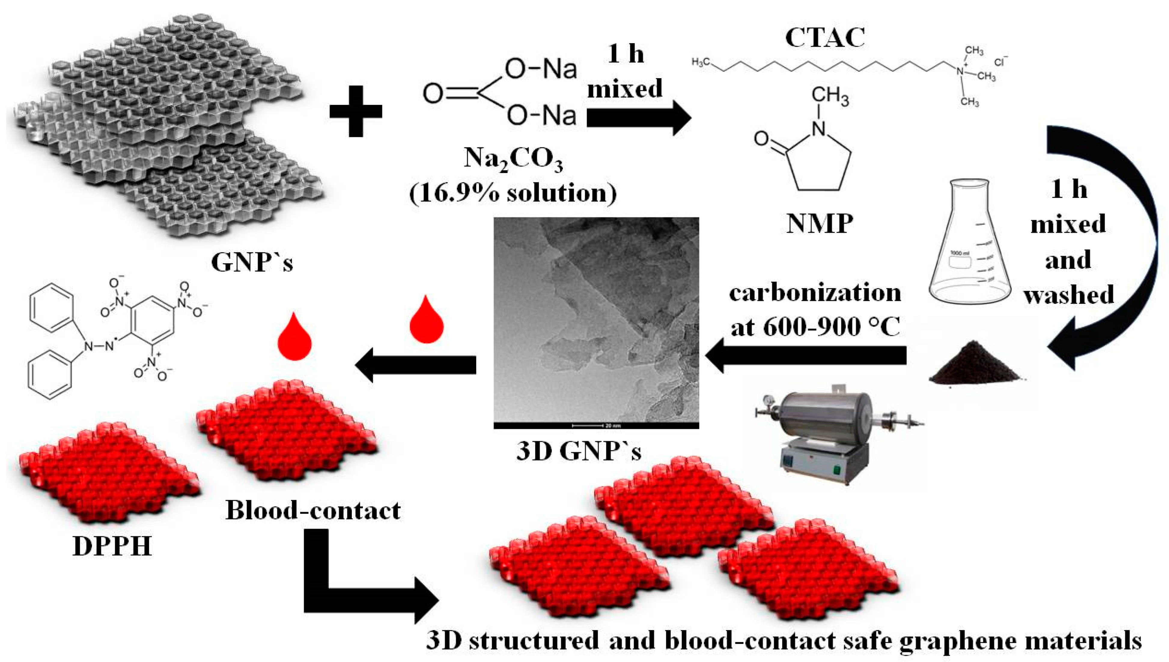
However, all modifications should be defined as safe and non-toxic. The aim of the proposed study was to determine graphene properties in terms of its applicability as a material for living-cell-bearing supports of advanced 3D architecture. The scheme of the method used by the authors is shown in
Figure 1.
In such a case, non-destructive activity towards basic body fluids such as blood and lymph is absolutely essential since such cell-containing structures will stay in permanent contact with fluids. Moreover, the antioxidant properties of graphene-based materials should be tested, since this feature is a part of widely understood blood compatibility. In this work, the so-far-achieved goals in the 3D structuring of graphene flakes will be exploited to receive 3D graphene derivatives, the blood compatibility of which is still unknown. We assume that blood compatibility should be investigated since our 3D structuring protocol involves the addition of a hard template, a cationic surfactant, and other chemical reagents, which have to be used to achieve advanced pore structure. The up-to-date collected knowledge and skill by authors in the area of graphene exfoliation and subsequent 3D structuring has been assumed as a starting point for the design of 3D graphene derivatives that are safe in contact with blood.
2. Results and Discussion
Graphene and other carbon materials need to be characterized by a set of widely approved experimental methods, as in this study. Such features as surface area, pore structure, and chemical composition are crucial for any carbon-based material apart from biological activity.
The elemental composition and surface parameters for the obtained samples of porous 3D graphene are given in
Table 1. The carbon content in all investigated samples is very high (higher than 90 wt.%). High carbon content is, among others, a measure of the oxidation of graphene after the performed 3D structuring. Potentially, chemical manipulations may lead to the oxidation of graphene and its conversion to graphene oxide. The aim of our study is to investigate the biocompatibility of graphene and not of graphene oxide. Thus, the carbon content above 90% is characteristic of materials accounted to the graphene category. This statement applies to all samples under investigation. There is a noticeable relationship between the degree of graphitization and the carbonization temperature range. Elemental analysis revealed that carbon content increases with increasing carbonization temperature in obtained materials. The results obtained in the case of the wet chemical exfoliation method indicated that Na
2CO
3 nanocrystals precipitate among exfoliated graphene flakes upon drying the reaction mixture. Thus, the template particles were removed by HCl etching and washing with distilled water. N
2 sorption analysis showed that the specific surface area decreased (330–391 m
2/g) below the value determined for the used paternal graphene nanoplatelets (750 m
2/g). In parallel, changes of the pore structure became visible. The total pore volume decreased in all samples from 0.999 (GF-750) to 0.220 cm
3/g (GF_Na_600). Surprisingly, the contribution of the mesopore volume V
me to the total pore volume V
t increased from 36% to 79%. It is notable that for three of four investigated samples the share of mesopores is close to 50% or much higher. The presence of mesopores (average pore diameter from 2 to 50 nm) is significant for applications involving a liquid environment where adsorption/desorption proceeds (for example, drug delivery).
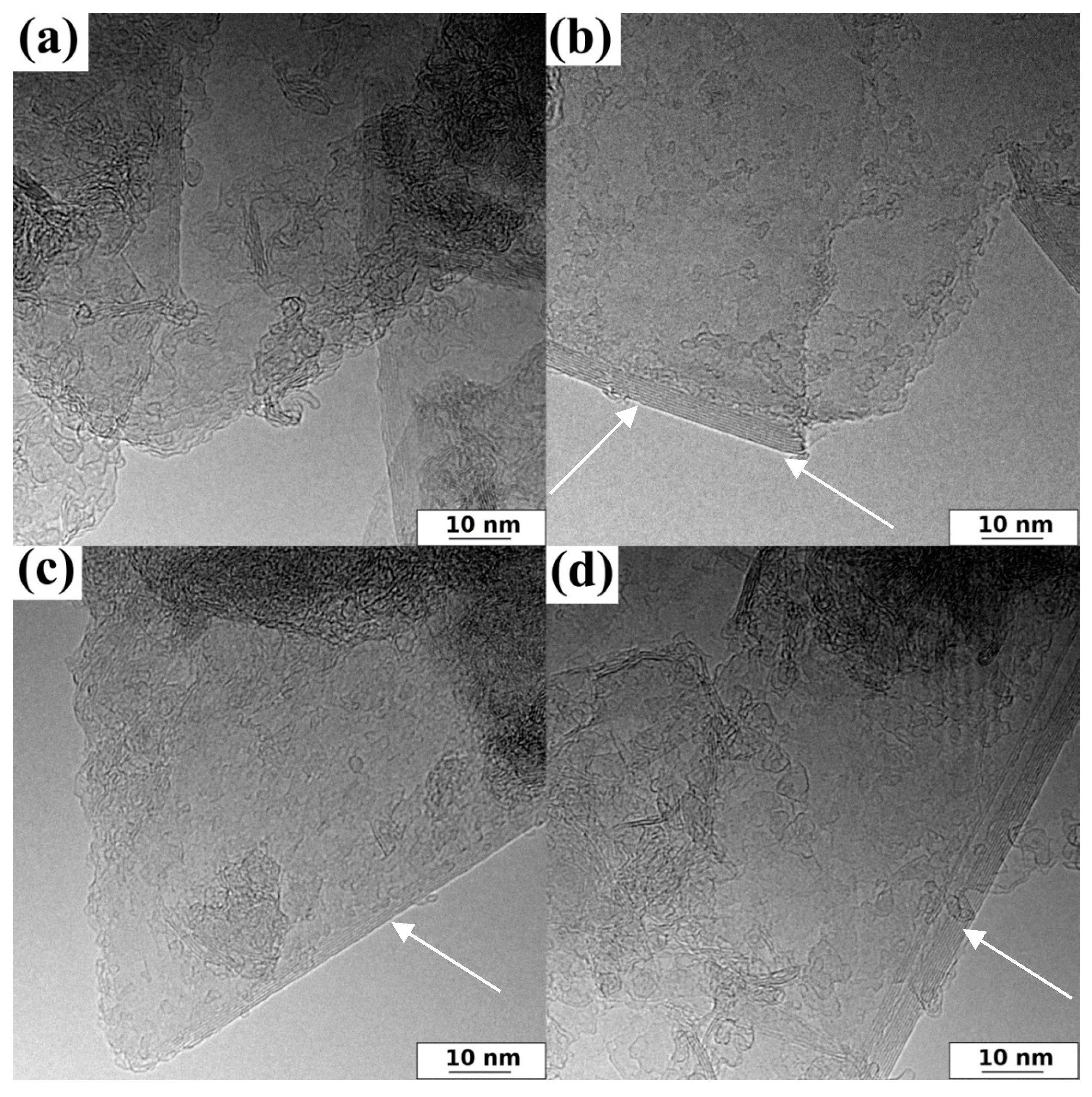
Additionally, the morphology details of representative samples are shown in the HRTEM images (
Figure 2).
Figure 2 shows that all investigated 3D graphene samples had similar irregular surface characteristics due to randomly oriented graphene layers. Mainly, HRTEM allowed thin graphene flakes to be observed. It has to be stated that, in general, SEM morphology is not informative and therefore, HRTEM images need to be acquired. Two carbon phases are visible on the HRTEM images (
Figure 2): exfoliated graphene sheets and bundle-looking domains of amorphous carbon, which is typical of a carbon phase originating from the thermal decomposition of polyfurfuryl alcohol applied for a durable stacking of graphene flakes.
In the case of carbon-based materials, the surface elemental composition may substantially differ from the bulk elemental content. In general, carbon is a reactive element, and surface atoms may come into reaction with gases present in the atmosphere. In particular, oxygen is supposed to yield diversified surface-oxygen-based species. Therefore, two methods need to be applied to determine elemental composition: elemental combustion analysis (bulk analysis) and XPS (surface analysis).
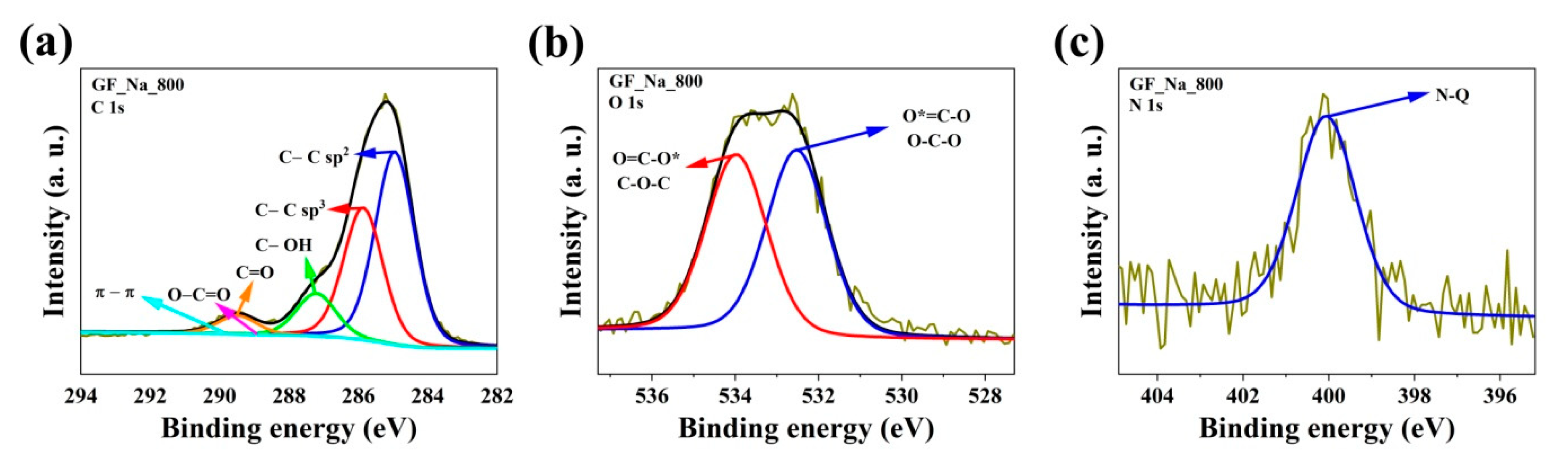
The results of combustion elemental analysis only allowed the bulk content of three elements to be determined: C, N, and H, while the specific chemical environment of the heteroatoms embedded in the carbon matrix remains unknown. Therefore, obtained samples were additionally supported by X-ray photoelectron spectroscopy (XPS) investigations. A sample with the most promising biological properties was selected for the XPS study based on DPPH and blood compatibility. The XPS spectra of the representative GF_Na_800 sample were determined and are demonstrated in
Figure 3 and
Table 2.
The XPS elemental content of carbon was high (88.6 at.%) and very close to the values obtained by combustion analysis. However, XPS C content is slightly lower, which is a natural effect of spontaneous oxidation of the carbon surface by atmospheric oxygen. Carbon atoms were mostly bonded as sp
3 hybridized atoms (band C 1s at binding energy 285.0 eV), which is characteristic of graphene materials. The C 1s spectra of the GF_Na_800 sample are composed of four peaks corresponding to C-C bond (sp
3) peak at 285 eV [
24]; C-O-C or C-OH or C-NH bond peak at 286.3 eV [
25]; C=O or O-C-O or N-C-O bond peak at 287.7 eV [
26]; and O-C=O peak at 288.6 eV [
26]. The total amount of oxygen is in a range from 9.6 at.%. The peak at 532.0 eV signifies the presence of a O-C-N or C=O bond and the peak at 533.3 eV is characteristic of a O*=C-O or O-C-O bond [
24,
26,
27]. Moreover, the presence of nitrogen is noticeable (1.8 at.%). Nitrogen was used only as an additive surfactant and during the carbonization process as an inert gas flow. The high-resolution N 1s spectra can be deconvoluted into one peak, located at 400.5 eV, which is attributed to quaternary (N-Q) groups [
28].
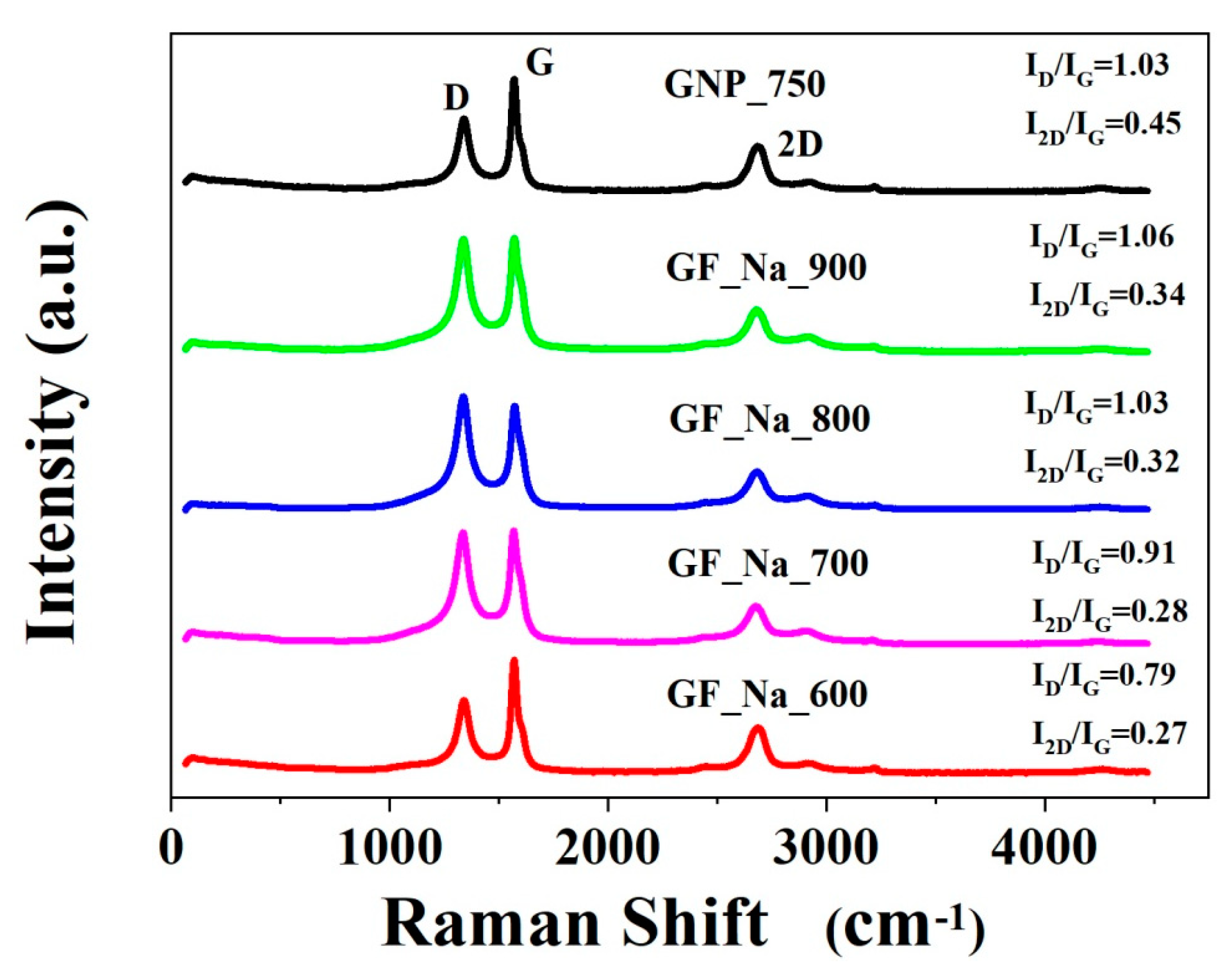
In turn, the Raman studies confirm exfoliation to FLG as a building block of the final structure [
9,
29].
Figure 4 and
Table 3 contain information on the D, G, and 2D band placement and intensity.
The intensity ratios I2D/IG and ID/IG prove that the investigated samples contain mainly multilayered graphene. The carbonization temperature of the presented samples affects the position of the G and 2D bands. The GF_Na_900 sample had more vacancies and disorders since it had the highest ID/IG ratio and moreover the highest carbon at.%.
The activity against free radicals of graphene samples before and after thermal modification was tested using the DPPH method (
Table 4). The RSA of the material increased after graphene modification, which suggests that antioxidant properties are improved. Graphene with the 750 m
2/g area showed higher RSA than 300 m
2/g. Additionally, with the increase in the modification temperature, the RSA increases. The maximum RSA = 89% was noticed after graphene treatment at 800 °C. When 900 °C temperature was applied, RSA decreased rapidly.
The material compatibility with blood is an important factor that classifies its potential medical application. Materials that are implanted in the body may not show hemolysis above 5% [
30]. As it is listed in
Table 5, all tested graphene samples caused hemolysis in the range of 0.28–0.64%. The increase in the temperature in the preparation process results in an increase in the hemolysis rate.
The authors have performed multidirectional and extended studies on the synthesis of 3D graphene as a porous material in recent years. Synthesis methodology has been described in several papers, among which some of the most meaningful may be cited [
8,
9,
14]. The aims of nearly all performed studies were exclusively focused on energy applications, including graphene-based electrode materials for batteries, supercapacitors, and photovoltaic cells. However, the well-developed pore structure and high carbon purity of such materials have encouraged authors to find radically new applications as a porous platform for bioactive species and/or in permanent contact with constituents of living organisms. Several investigations were performed to define some bioapplicability limitations of 3D structured graphene platforms with respect to the mentioned target.
Antioxidants protect cells against injury by free radicals. The antioxidant activity of materials introduced to the human body is important to protect cells during medical treatment [
31]. Graphene-based materials have been characterized as antioxidants by Qiu et al. [
32]. Radical scavenging activity was inversely proportional to their total surface area. However, our modification method results in an increase in surface area as well as antioxidant activity. The modification method is a crucial method for safe and effective graphene-based material preparation. Hemocompatibility is one of the crucial parameters to consider with materials for potential biomedical applications. Erythrocytes are sensitive to hemolysis due to shear stress. Insufficient hemocompatibility has been found to impair safety through the activation of blood coagulation [
33]. We compared our results with ASTM F756-00 standard materials where the hemolytic index 0–2% is considered as non-hemolytic, 2–5% is slightly hemolytic, and <5% is classified as hemolytic [
34]. We observed that all tested graphene samples might be classified as non-hemolytic. Thereby, they are allowed to be proposed for biomedical application as they have antioxidant activity as well as low rates of hemolysis.


 ,
, 


