Review

Targeting and Monitoring Acute Myeloid Leukaemia with Nucleophosmin-1 (NPM1) Mutation

Department of Medicine, School of Clinical Medicine, The University of Hong Kong, Hong Kong, China
*
Author to whom correspondence should be addressed.
Int. J. Mol. Sci. 2023, 24(4), 3161; https://doi.org/10.3390/ijms24043161
Submission received: 31 December 2022 / Revised: 1 February 2023 / Accepted: 2 February 2023 / Published: 5 February 2023
(This article belongs to the Special Issue Molecular Mechanism of Leukemia)

Abstract

:
Mutations in NPM1, also known as nucleophosmin-1, B23, NO38, or numatrin, are seen in approximately one-third of patients with acute myeloid leukaemia (AML). A plethora of treatment strategies have been studied to determine the best possible approach to curing NPM1-mutated AML. Here, we introduce the structure and function of NPM1 and describe the application of minimal residual disease (MRD) monitoring using molecular methods by means of quantitative polymerase chain reaction (qPCR), droplet digital PCR (ddPCR), next-generation sequencing (NGS), and cytometry by time of flight (CyTOF) to target NPM1-mutated AML. Current drugs, now regarded as the standard of care for AML, as well as potential drugs still under development, will also be explored. This review will focus on the role of targeting aberrant NPM1 pathways such as BCL-2 and SYK; as well as epigenetic regulators (RNA polymerase), DNA intercalators (topoisomerase II), menin inhibitors, and hypomethylating agents. Aside from medication, the effects of stress on AML presentation have been reported, and some possible mechanisms outlined. Moreover, targeted strategies will be briefly discussed, not only for the prevention of abnormal trafficking and localisation of cytoplasmic NPM1 but also for the elimination of mutant NPM1 proteins. Lastly, the advancement of immunotherapy such as targeting CD33, CD123, and PD-1 will be mentioned.

Graphical Abstract

1. Introduction

Due to the large proportion of AML patients exhibiting NPM1-mutations, targeting NPM1 is a constant and ongoing area of research, and the wide variety of drugs available are dependent on the types of co-mutations present in NPM1-mutants. Despite various clinical trials and research of new agents targeting NPM1-mutants, about 50% of patients either relapse or unfortunately die due to the progression of the disease, and this is often the case in patients who are unable to withstand the clinically aggressive nature of AML, nor the harshness of intensive chemotherapy required for disease remission. Seeing as various cellular pathways are affected by NPM1 mutations, various targeted approaches are being investigated to tackle the challenges in treating relapsed and refractory AML patients. Some examples of NPM1-targeting approaches include DNA modulation, ribosome biogenesis, protein deregulation, mitochondrial-mediated cell death, and antibody-mediated treatments.
With the current understanding of NPM1-mutant detection, haematologists and medical governing bodies are implementing the use of MRD, also known as a measurable residual disease, for AML patients. The use of MRD can act as a prognostic tool for patients’ treatment, especially during the post-transplantation stage, allowing for a better understanding of the disease progression and prediction of survival outcomes.
Within this review, an overview of the structural changes influencing the functionality of cells is given, followed by NPM1-mutated AML risk stratification, monitoring of NPM1 mutants by MRD, and finally targeting NPM1-mutations for treatment of AML patients.

2. Functional Role of NPM1

2.1. Genetic Characteristics

NPM1 is localised to chromosome 5q35 and consists of 12 exons. Specifically, NPM1 contains the N-terminal core domain, acidic domain, and nuclear localisation signal domain are found to be highly conserved among each protein of the NPM family, indicating that all three proteins have a similar functional role. However, the less conserved C-terminus, on the other hand, suggests an alternative role for NPM1. It is highly conserved between metazoans and when mutated can result in leukaemic-initiating activity [1]. The NPM1-mutated gene is a dominant negative, haploid insufficient mutation that is generally limited to exon 12 [2,3], although rare occurrences have been reported in other regions such as exon 5, 9, and 11 [4,5]. NPM1-mutated AML is mostly categorized into type A, B, or D according to a 4 base-pair insertion sequence at the 288th amino acid position (rs587776806) [6]. Type A (sequence: TCTG) is the most frequent, comprising of approximately 72% of NPM1-mutated AML cases. Types B (sequence: CATG) representing ~12% and D (sequence: CCTG) with ~4% are less frequent but more so than all other types which represent less than 1% of the NPM1 mutated population (Seen in Figure 1) [6,7]. The frameshift resulting from NPM1 mutant insertions causes a change in the C-terminal domain (CTD) amino acid sequence from DLWQWRKSL to DLC(L/M)AVEEVSLRK, consequently replacing one or both tryptophan residues that are required for nucleolar localisation signaling with a nuclear export signal (NES) motif, i.e., LXXX(V/L/F/C/M)XXVXL, where X stands for any residue [2,3,6,7]. Since NPM1 proteins interact with a shuttling receptor XPO1, also known as Exportin, the mutant protein is exported at rates exceeding that at which it is imported, ultimately leading to the delocalisation of mutant NPM1 to the cytoplasm [3]. Wild type (WT) NPM1 is also shuttled out of the nucleus in the process, as NPM1-mutant and NPM1-WT form heterodimers to be co-exported [8].
Modulation of the NPM1 structure is governed mainly by post-translational modifications including phosphorylation, SUMOylation, and ubiquitination. NPM1 proteins are comprised of three different regions: the N-terminal domain (NTD), the central region, and the CTD, each with a distinct functional role (illustrated in Figure 2) [9]. The NTD is highly conserved and acts as a core domain due to its structural integrity and NES region. Post-translational phosphorylation of the NTD region of NPM1 results in instability and the induction of apoptosis but also limits the ability of cells to prevent DNA damage [10]. The oligomerisation of NPM1, on the other hand, causes cellular proliferation and localisation out of the nucleus to the cytoplasm by two weak NES regions [10]. NES interacts with a shuttling receptor XPO1, also known as Exportin 1 or CRM1 export protein (Chromosomal Maintenance 1), which subsequently causes XPO1 association with NES-containing proteins that result in cytoplasmic relocation of NPM1; this deviation gives rise to cytoplasmic NPM1 (cNPM1) [11,12]. In cases of NPM1 mutants, the additional NES at the CTD alongside two weak NES at the NTD results in strong associations with XPO1 and as a consequence, nuclear export to the cytoplasm dominates [3]. Studies have shown that cNPM1 impedes protein-protein interaction and subsequent nuclear interactions for example CCCTC-binding factor (CTCF) [13]. Through cNPM1 interactions, CTCF is relocalised to the cytoplasm, and consequently, gene regulation, RNA splicing, and DNA looping are reduced; abolishment of this interaction resolves CTCF DNA binding and nuclear concentrations [13].
The central region of NPM1 contains highly acidic amino acids (aspartic and glutamic acids) with their negative charge properties allowing mimicry of nucleic acids and thereby influencing the nuclear localisation signal (NLS). NLS regions interact with importin alpha and beta causing nuclear translocation of the NPM1 protein. The acidic central region of NPM1 facilitates histone binding to both core and linker histones for the formation of perinuclear heterochromatin [14]. Additionally, the central domain contains basic regions, with both influencing the ribonuclease activity of NPM1 and ribosome biogenesis activity [15]. Ribosomal RNA (rRNA) processing via NPM1’s endoribonuclease activity converts pre-rRNA to mature 28S rRNA.
The CTD contains predominantly basic and positively charged amino acids influencing its role in binding to nucleic acids. At the CTD, the nucleolar localisation signal (NoLS) domain allows the localisation of NPM1 to the nucleolus with aromatic residues (tryptophan 288 and 290) forming a three-helix structure and stabilises NoLS due to its hydrophobic core. The lack of aromatic residues is common in NPM1 mutant proteins and results in the unfolding of its helical structure which inhibits nuclear import and nucleolus import of NPM1. Aberrant localisation of NPM1 is typical amongst AML patients.

2.2. Functions of NPM1

2.2.1. Ribosome Biogenesis

NPM1 is a multifunctional protein influencing processes involved in ribosome biogenesis, mRNA transcription, embryonic development, DNA repair, chromatin remodelling, and apoptosis. The most well-studied function of NPM1 is its involvement in directing ribosome biogenesis [16].
A mechanism known as liquid-liquid phase separation (LPS), has been proposed to explain the effects of NPM1 localisation in the nucleolus on ribosome biogenesis. Modulation of LPS occurs via homotypic and heterotypic interactions of the three key regions (NTD, central region, and CTD) of the NPM1 protein [17]. Three types of interactions are interplayed for the LPS mechanism to occur: (1) homotypic oligomerisation, (2) heterotypic interaction with rRNA, and (3) heterotypic interaction of R-motif-rich proteins.
The nucleolus subcomponent—the fibrillar centre (FC), is responsible for rRNA transcription. In the initiation of rRNA gene transcription, two components of the FC are formed: the dense fibrillar component (DFC) and the granular component (GC) [18]. The GC is dominated by homotypic interactions, which refer to NPM1-NPM1 interactions via electrostatic charges that result in the formation of a pentamer. Associations occur primarily through the NTDs and the intrinsically disordered regions (IDR) of NPM1 [19]. Heterotypic interactions with rRNA predominate at the DFC. The physicochemical properties of the nucleoplasm are altered, affecting surface tension and translocation out of the nucleolus [17,20]. Ribosomal proteins and pre-rRNA components (e.g., CDK1) interact with nucleolar scaffolds to allow for NPM1 sequestration and the formation of pre-ribosomal particles to be transported from the nucleolus to the nucleoplasm [21]. The most well-studied NPM1 interactions are the heterogenic interactions amongst R-motif-containing proteins. Direct interaction of ribosomal protein L5 (rpL5) with NPM1 allows shuttling and colocalisation of 5S rRNA, premature 60S rRNA, 80S ribosomes, and polysomes. Co-immunoprecipitation of NPM1 potential partner proteins such as RPL23, RPS7, RPL35, and RPS20 indicate its role as a scaffold in ribosome biogenesis within the nucleolus. NPM1 concentrations are particularly high in proliferating cells and were essential for ribosome maturation in the nucleolus [22].

2.2.2. RNA Processing

DNA damage causes defects in mRNA processing, which leads to potential translation errors downstream; however, this is often remedied by translesion synthesis which is employed as a DNA repair mechanism by Polymerase Eta (Poly eta), a low-fidelity polymerase [23,24]. NPM1 is a key regulator of Poly eta by protecting it from degradation, as evidenced by the increased rate of Poly eta degradation in cells with higher mutant cytoplasmic NPM1 copies [24]. Therefore, translesion maintenance is important to consider when determining the prognosis of patients with NPM1-mutated AML.
A recent study also noted the impact of long noncoding RNA (lncRNA) increasing NPM1 mutant samples. Analysis using genome-wide RNA sequencing concluded that the introduction of lncRNA led to an increase in cell differentiation proteins; THSB1, MAFB, and ASB2. In addition, in vivo experimentation on mice confirmed decreased survival and accelerated AML pathogenesis in the presence of lncRNA. Decreased levels of lncRNA increases sensitivity to the chemotherapy drug, cytarabine, thus affecting treatment outcome [25]. Overall, an increase in cytoplasmic NPM1 mutants causes an increase in nuclear retention of lncRNA, ultimately contributing to the AML disease course [26].

2.2.3. Chromatin Remodelling

Centrosome duplication influences the activation of chromatin and thus microtubule distribution. Correct chromatin remodelling allows a smooth transition of the cell cycle from interphase to metaphase [27]. The formation of CRM1/Exportin 1 contributes to the translocation of centrosomes and ribosomes with NES-bearing proteins. NPM1 contains two weak NES regions and interacts with the Ran-Crm1 complex. The formation of bipolar spindles requires regulation by the Ran-Crm1 network to ensure centrosome duplication [28,29]. Therefore, centrosome localisation to NPM1 is promoted by Ran-Crm1 complexes; the mutation T95D at the NES region of NPM1 inhibits centrosome duplication [30]. Histone chaperone activity is similarly affected by NPM1 mutations. Assembly of the histone octamers, removal of DNA from histones, and transfer of histones from other chaperone proteins or enzymes are all roles of histone chaperones that are associated with NPM1 [31]. Collectively, the acidic and basic characteristics of the structural components of NPM1 allow the binding of nucleic acids and histones [32,33].

2.2.4. Regulation of Apoptosis and Cellular Growth

Within healthy cells, NPM1 has the capacity to trigger either cell proliferation or apoptosis. However, a shift in homeostasis can affect NPM1 translocation to the cytoplasm, which in turn affects protein interactions in the cytoplasm profoundly. NPM1-induced activation of p53 stress-response mechanisms leads to a mitochondrial-associated release of extrinsic factors such as caspase and cytochrome c [34]. Mouse double minute 2 homolog (MDM2) is a negative regulator of p53, thus preventing its role as a tumour suppressor [35,36]. NPM1 directly binds to MDM2, hindering its inhibitory role on p53. Notably, this NPM1 mechanism indirectly affects p53 activity as indicated by studies on NPM1-negative mice and embryo cell lines that still express p53. The cysteine 275 to serine NPM1 mutant is incapable of undergoing S-glutathionylation, resulting in the release of nucleolar nucleic acids from NPM1. Consequently, nucleolar stress prevents translocation of NPM1 to the cytoplasm and binding to MDM2; this leads to the limited activation of p53 which is suggestive of nucleolar oxidation. The release of p14ARF by NPM1 allows binding to MDM2 via rpL5. ARF-binding partners, such as MDM2 and NPM1, associate with ribosomal proteins, which implicates its role in p53 homeostasis [37]. Studies have also implicated direct interactions of NPM1 with p53 [38]. Therefore, NPM1 protein activity in apoptotic initiating pathways such as p53 are involved in both its roles in nucleolar stress and ribosome production [39]. Recent studies have also suggested that p53 mutated AML patients display distinct molecular signatures compared to p53 wild-type plus NPM1 mutation-positive AML patients [40]. Therefore, the data strongly suggests that specific molecular pathways are involved between NPM1 and p53.

2.2.5. Genome Instability

DNA repair response mechanisms are key to maintaining proper cell differentiation and genome stability. Post-translational modification of NPM1 regulates cell cycle proteins and promotes stability. DNA double-stranded lesions caused by UV results in the initiation of the nucleotide excision repair pathway (NER) which is overexpressed in NPM1-mutated AML, and this increases its activity as a chromatin-binding factor by upregulating proliferating cell nuclear antigen (PCNA), a NER protein. Subsequently, PCNA forms a homo trimer, interacts with cell cycle regulator p21 in the nucleus, and promotes DNA repair [32,41]. Release of retinoblastoma tumour suppressor (pRB) is also initiated by UV, causing dephosphorylation of NPM1 at Threonine 199, 234, and 237 by protein phosphatase 1 β (PP1β). Subsequently, pRB causes activation of E2F1-dependent DNA repair mechanisms [42]. NPM1-mutants, therefore, prevent fine tuning of DNA repair pathways by direct protein-protein interaction such as with apurinic/apyrimidinic endonuclease 1 (APE1) protein. Consequently, a break in equilibrium leads to an increase in DNA repair proteins translocation to the nucleus thus, cell proliferation is halted and normal ribosome biogenesis is impaired [43]. As mentioned above, the ARF-binding proteins also influence the endonuclease activity of NPM1, which in turn modulates the apoptotic and genome stability of cells [44,45].

3. NPM1 Mutations in AML and Other Malignancies

According to the current WHO classification of hematopoietic malignancies, NPM1-mutated AML has been acknowledged as a distinct entity [46]. Nonetheless, it is usually accompanied by concomitant mutations, including but not limited to FLT3-ITD (FMS-like receptor tyrosine kinase-3 internal tandem duplication), DNMT3A (DNA methyltransferase 3 alpha), IDH1/2 (isocitrate dehydrogenase ½), and TET2 (Tet Methylcytosine Dioxygenase), raising speculation of synergistic interactions that promote AML transformation and progression [3,47,48,49]. Indeed, mutated NPM1 is a driver mutation and often a secondary event to pre-existing mutations generated by clonal hematopoiesis of indeterminate potential (CHIP) [2,7,47,50]. The most recognized risk stratification system for AML is the European LeukemiaNet (ELN) genetic risk classification at diagnosis. The ELN risk classification states that cytogenetically normal NPM1-mutated AML, without FLT3-ITD, belongs to a favorable risk group. Cytogenetically normal NPM1-mutated AML with FLT3-ITD belongs to an intermediate risk group exhibiting adverse outcomes, especially in the presence of the aforementioned co-mutations [51]. It must be noted that 85% of NPM1-mutated AML cases exhibit normal karyotypes, with the remaining cases showing various cytogenetic abnormalities e.g., del (9q), +21, +8, and +4 in AML [52].
NPM1 insertional mutations have also been found in myeloid neoplasms including myelodysplastic syndrome (MDS), myeloproliferative neoplasm (MPN), and chronic myelomonocytic leukaemia (CMML). In lymphoid neoplasms, translocation events are reported. Accounting for about 2% of NPM1-mutant-related diseases, NPM1-mutation in MDS has been associated with a higher risk of secondary AML and is most commonly found in MDS with an excess blast (MDS-EB) [53,54]. Molecular diagnosis of MPN occurs from the presence of mutant JAK2-V617F and associated diseases include essential thrombocythemia (ET) and myelofibrosis (MF) of which NPM1-mutations have been found on a rare occasion [55,56]. This shows the high sensitivity of NPM1-mutant cells seen in MPN and possible regimens without the need for stem cell transplantation. Contributing to 3% of NPM1 mutant cases, CMML with NPM1 mutation shows chemosensitivity to anthracyclines and cytarabine (7 + 3 induction chemotherapy), and hypomethylating drugs have promising effects in maintaining remission [55,57]. On the other hand, for translocation events of NPM1, treatment has been associated with the elimination of the fusion protein directly. This includes anaplastic large-cell lymphoma with NPM1-ALK (anaplastic lymphoma kinase) and T-cell lymphoma with NPM1-TYK2 (tyrosine kinase 2) [58,59]. It must be noted that only a small proportion of patients who exhibit NPM1 mutations outside of AML can be found.

4. Measurable-residual monitoring in NPM1-mutated AML

With the advent of modern technology and improved molecular techniques, MRD has become indispensable for AML patient management and monitoring of mutant NPM1 before, after, and during the course of treatment [3,60,61,62,63]. MRD positivity has been defined by the ELN as either the conversion of CRMRD− to CRMRD+, or the increase of PB/BM-MRD by ≥1log10 between two positive samples [60]. For better harmonization of NPM1-mutated AML data, log reduction of MRD should be the preferred choice for reporting, as multiple studies have shown that MRD kinetics are reliable prognostic indicators of the outcome, but not absolute MRD levels [62,64]. Technical aspects and sample collection for MRD monitoring are extensively stated in the ELN guideline [51].

4.1. Conventional Technologies for MRD

Conventional technologies for MRD detection include flow cytometry and molecular MRD assessments by qPCR and ddPCR. In AML patients receiving hematopoietic stem cell transplantation (HSCT), MRD positivity at pre-HSCT by flow cytometry is strongly correlated with worsened overall survival and leukemia-free survival (LFS) than MRD-negative patients. Pre-HSCT MRD negativity is also associated with a reduced incidence of relapse supporting the predictive significance of MRD monitoring on survival and post-HSCT relapse [65]. Similarly, in high-risk AML patients with normal karyotype, flow cytometric MRD positivity is strongly associated with relapse-free survival in these patients [66].
When evaluating MRD using multiparametric flow cytometry specifically in NPM1-mutated patients, a unique immunophenotype of AML patients was identified [67]. Leukaemic cells in patients with type A NPM1 mutation strongly expressed myeloperoxidase (MPO) and CD33 with dim expression of other myelomonocytic antigens commonly expressed in AML including CD13, CD65, CD15, and CD14. Its utility in the MRD monitoring of NPM1-muated AML in general is yet to be confirmed. Therefore, molecular MRD strategies targeting these specific mutations are crucial for more specific analyses of MRD kinetics and clonality.
As per the latest MRD guidelines from the ELN, molecularly defined groups such as NPM1-mutated AML should be monitored by qPCR and ddPCR; the implication of both approaches have been extensively published and reviewed [2,6,60,61,68]. Patients that fail to achieve a 4-log reduction of MRD have a significantly higher risk of relapse as well as shorter survival [64]. NPM1 mutations are commonly monitored using RNA normalized to ABL transcripts, a technique that is used to monitor other fusion genes in a similar fashion. Meanwhile, recent studies and in-house data (data not published) have shown that quantification of NPM1 mutation allele frequency at the DNA level is a good alternative to RNA-based assay. In spite of lower sensitivity than RNA-based assays, clinical correlation with DNA mutation allele frequencies are also observed. This permits flexibility of the MRD protocol and alleviates the need to normalize to ABL1 which has a significantly slower expression rate than NPM1 [68,69].

4.2. Advanced Technologies for NPM1 MRD Monitoring

Application of NGS for MRD monitoring can be conducted at bulk tumour level and single-cell level, although none of them are routinely applied. Bulk tumour sequencing is most commonly adopted at the early stages of AML as screening tools that allow simultaneous screening of a vast number of mutations. Whole exome sequencing (WES) can detect NPM1 mutations months prior to the onset of de novo AML even though coverage is generally lower than gene panel sequencing [70]. NGS also permits the sub-classification of NPM1 mutations as non-type-A mutations that are reported to confer shorter survivals and inferior clinical outcomes than wild-type or type A mutations [71,72,73]. Despite the demanding nature of NGS bioinformatic analyse and interpretation, its application in MRD detection is beneficial especially when most PCR-based assays detect NPM1 type A mutants but not other NPM1 mutation subtypes.
Similarly, the application of single-cell technology is continuously under development for MRD monitoring for improved resolution. Both DNA and conventional RNA single-cell sequencing have been employed in MRD monitoring in AML but are not routinely used due to cost and the high requirement for sample preparation [74,75]. With the inclusion of surface marker staining, single-cell multi-omic analysis further increases the resolution of the MRD in NPM1 mutated AML patients [76]. In these studies, clonal heterogeneity of the NPM1 mutant clone has been revealed, and serial monitoring has shown that the population changes dynamically with the acquisition of NPM1 and FLT3 mutations during the intermediate stages of the disease. Clonal switch of NPM1 co-mutation with PTPN11 to WT1 is also resolvable at relapse, demonstrating the benefits of high resolution NGS. The ultra-sensitivity of single-cell technology is demonstrated with <1% of mutant cells carrying NPM1 or other SNPs out of as few as 7000 cells being sequenced per time point [74]. When single-cell analyses are coupled with mass cytometry and further supplemented by bulk tumour sequencing, dynamic monitoring of AML responses to chemotherapy can be achieved [77]. In a cohort of 32 AML patients, 36-plex mass cytometry has been used to monitor 24 h responses to standard 7+3 induction chemotherapy. The employment of machine learning analysis of the cell population has further identified high p-ERK1/2 in blast cells at 24 h being a predictive marker for poorer MRD and five year survival. While the majority of the p-ERK1/2low patient achieved CR, the MRD positivity observed in a few p-ERK1/2low patients is due to the presence of NPM1 mutations. This provides a novel method that deciphers the effect of intracellular signalling on MRD dynamics [77]. This also envisions the significance of early evaluation of MRD adopting a multi-omic approach.

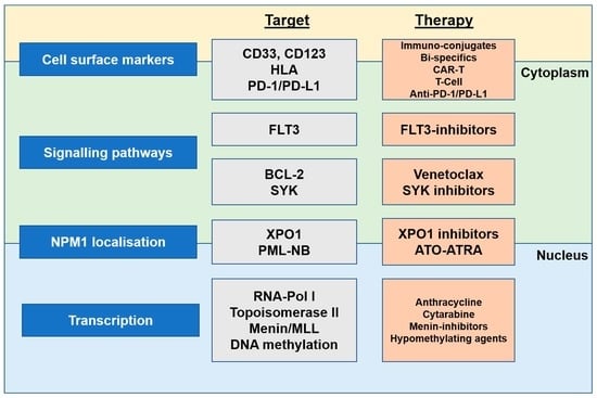
5. Targeting NPM1-Mutated AML

At least one-third of AML cases are NPM1-mutated, so developing treatment approaches for this population is becoming crucial. Upon initiation of treatment, a risk assessment is recommended by the ELN for diagnosis and management [51]. Primary determinants of the therapeutic approach are age, comorbidities, and diagnostic molecular profile [78]. Standard treatment protocol for young and fit NPM1-mutated AML patients involves induction with intensive chemotherapy followed by consolidation chemotherapy. Co-occurrence of FLT3-ITD and/or DNMT3A mutation is found in approximately 66% of NPM1-mutated AML, which portends an inferior prognosis [79,80,81,82,83], constituting an indication for allogeneic haematopoietic stem cell transplantation (HSCT) in eligible patients. The persistence of NPM1 mutant transcripts following induction or consolidation chemotherapy is associated with high relapse rates [64,84]. Patients with the persistence of NPM1-mutant transcripts in the peripheral blood after the second cycle of chemotherapy were associated with a higher risk of relapse compared to those with absent NPM1 mutant transcripts (82% versus 30%) after three years of follow-up [85]. In the United Kingdom National Cancer Research Institute (NCRI), an AML17 study that involved 107 patients with NPM1-mutated AML receiving allogeneic HSCT, the estimated two year overall survival was 83%, 63%, and 13% in patients with negative, low (<200 copies per 105 ABL in the peripheral blood and <1000 copies in the bone marrow aspirate), and high levels of MRD respectively after a median follow-up of 4.9 years [86]. In patients who were FLT3-ITD positive with low-level MRD pre-HSCT, the two year overall survival was only 17% [86]. In NPM1-mutated AML patients undergoing allogeneic HSCT, positive measurable residual disease (MRD) prior to transplantation was associated with high risk of post-HSCT relapse [86]. The one year relapse-free survival post HSCT in patients with positive MRD pre-HSCT is 45% [86]. In another study, the two year cumulative risk of relapse was 65% in patients who were MRD-positive before allogeneic HSCT [87]. On the other end of the spectrum, patients who are unfit or over 75 years of age are given Venotoclax with either HMA or low dose cytarabine (LDAC). As a result of the variety of treatments available, the current review will outline current and novel agents, as well as the mechanisms involved in targeting NPM1-mutated AML (Figure 3).

5.1. Current Agents or Standard Treatment

5.1.1. Anthracycline

NPM1-mutated AML patients are commonly treated with the “7 + 3” regimen which is a combination of cytarabine for 7 days and anthracycline for 3 days. The favourable response of “7 + 3” has enabled it to become the standard of care for AML due to its CR rate of 60–80% in younger patients and 40–60% in older adults (>60 years) [88,89]. Anthracyclines are a group of potent chemotherapeutics that lack selectivity due to their aromatic ring and attached amino-sugar, thus may warrant unwanted side effects such as cardiac abnormalities in the elderly AML population [90,91]. The chemical structure allows for the transfer of electrons between structures, causing an increase in free radical formation that results in the peroxidation of lipids within the cell [92]. The most clinically used anthracyclines include Daunorubicin (daunomycin), Doxorubicin, Epirubicin, and Idarubicin. Various mechanisms have been suggested, one being a DNA intercalator. The minor groove of DNA has been found to interact directly with Daunorubicin at sequence CGTACG to form a “daunorubicin-d(CGTACG)” complex [93]. A stable structure determined by X-ray crystallography shows the complex involves water molecules, anthracyclines, and DNA forming hydrogen bonds [93]. Next, doxorubicin has been shown to cause double-strand breaks (DSB) by the inhibition of DNA topoisomerases II [94,95]. During replication, normal DNA topoisomerase II prevents DSB by performing DNA catenation and knotting. Anthracyclines prevent the formation of the intermediate DNA-Topoisomerase II cleavage complex (Top2cc), resulting in apoptosis due to a DSB in the DNA [96]. However, it should be emphasised that due to the high potency of anthracyclines, cardiotoxicity is frequently observed and likely attribute to chromatin damage by histone removal [95], which is believed to cause apoptosis or necrotic events.
Another mechanism of anthracyclines is via the immunostimulatory pathway STING, which is a common immune response pathway upon pathogen entry [97]. STING modulates the release of dead-cell antigens contributing to the cascade of immunogenic cell death [98]. Caspases and ATP are released allowing stress-like signals to initiate an immune response. Innate immunity is manifested in cells through the activation of interferon regulatory factors (IRFs), with a focus on IRF1 genes and IRF3 phosphorylation [99]. IRF3 has been shown to contribute to the transcription of type I interferons and is linked via the activation of toll-like receptor (TLR)-3 and TLR-4 signalling [100]. Thus, the activation of the STING pathway, which causes IFN upregulation, is a key mechanism of anthracycline that activates immune responses [101,102].

5.1.2. Cytarabine

Cytarabine is used with anthracycline (7 + 3 regimen) during the remission induction. High-dose cytarabine (HDAC) is used in the consolidation of AML. LDAC is prescribed for patients unfit for intensive induction chemotherapy. Cytarabine (arabinosylcytosine, ARA-C) is a pyrimidine analogue that competes with the base pair cytidine in both DNA and RNA. Therefore, by limiting DNA polymerase beta and base excision repair, inhibition of DNA replication occurs, particularly at the S phase [103,104]. In 2014, CPX-351, a new encapsulated drug with a 5:1 ratio of cytarabine and daunorubicin was run in parallel with the standard treatment protocol (7 + 3 regimen) in newly diagnosed elderly NPM1-mutated AML individuals. Notably, older adults showed longer remission rates in the CPX-351 arm and had a similar safety profile as the intensive standard chemotherapy arm [105]. This suggests the significance of cytarabine and the appropriate isoform of anthracycline to be administered to both the elderly and the young. In NPM1-mutated AML patients, cytarabine efficacy is comparable to anthracycline efficacy due to cytarabine’s role in inhibiting DNA replication and the capacity of NPM1 to regulate transcriptional processes. As a result, it is highly effective against rapidly diving cells regardless of the subtype of mutated NPM1 [106,107].

5.1.3. FLT3 Inhibitors

As a large proportion (about 25%) of NPM1-mutated patients carry FLT3 mutations, initial screening of patients with FLT3-ITD or FLT3-TKD (tyrosine kinase domain with missense point mutation) benefit greatly from FLT3 inhibitors in combination with various chemotherapy [78]. Under normal circumstances, FLT3 receptor activation occurs upon binding of FLT3 ligand and the subsequent rotational change of amino acids—aspartate, phenylalanine, and glycine into an active conformation, which causes dimerization and tyrosine autophosphorylation [108]. Subsequently, TKD is activated and initiates a signalling cascade of pathways—JAK/STAT, RAS/MAPK, and PI3/AKT/mTOR signalling [109,110]. The most commonly used and researched FLT3 inhibitor, first-generation Midostaurin, has the capability of targeting a wide range of receptor tyrosine kinases (RTK), including FLT3 [111,112].
FLT3-ITD mutants exhibit persistent activation in the absence of ligand binding, resulting in the formation of its dimeric form and downstream cell proliferation [113]. Midostaurin acts by binding on the ATP binding pocket of FLT3, preventing activation and autophosphorylation [114]. Second-generation FLT3 inhibitors, Gilteritinib and quizartinib, are commonly used for relapsed/refractory FLT3-ITD positive AML cases [115]. Collected from in vitro analysis, Midostaurin shows possible off-target inhibition on platelet-derived growth factor receptors (PDGFRs), cyclin-dependent kinase 1, spleen tyrosine kinase, and vascular endothelial growth factor receptor [116]. Second-generation FLT3 inhibitors have greater specificity for FLT3 but lack inhibition of other RTK mechanisms, which may help to amplify anti-leukemic effects. However, because of the lack of specificity, first-generation FLT-3 inhibitors are both more potent and toxic, thus unsuitable for the elderly and unfit. In addition, it must be noted, recently the acknowledgment of FLT3-ITD-positive MRD pre-transplant has been associated with higher rates of relapse. A recent study has shown better overall survival within double positive FLT3-ITD and NPM1 mutations in both younger and older patients, signifying NPM1’s influence on using FLT3-ITD inhibitors [117]. Therefore, studies relating the efficacy of FLT3 inhibitors still require further investigation of the potential risk of how FLT3-ITD mutants play in NPM1-mutated AML patients. Some studies also show combination with venetoclax show molecular clearance of FLT3 mutations [118].

5.1.4. Gemtuzumab Ozogamicin

The CD33 antibody-drug conjugate (ADC)—gemtuzumab ozogamicin (GO (CMA-676); Wyeth Laboratories, Mylotarg®, Philadelphia, PA, USA) is the next FDA approved drug for AML. According to the national comprehensive cancer network (NCCN), GO administration should be given to either CD33-positive patients as post-remission therapy or patients placed in the favourable and intermediate cytogenetic risk groups [119]. In combination with intensive induction chemotherapy, GO has been shown effective in NPM1-mutated AML. In terms of NPM1 influence on GO treatment effect, CD33 cells are abundant among NPM1-mutated AML patients [120]. Several trials involving the antibody conjugate approach conclude that standard intensive chemotherapy compared with the addition of GO leads to a reduction in relapse and a better benefit/risk ratio [119,121,122]. In addition, a prospective study (AMLSG 09-09 Phase III trial) showed benefits in patients less than 70 years old with NPM1-mutated/FLT3-WT genotype [123].
GO is useful due to its conjugated cytotoxic antibiotics, calicheamicin. Calicheamicin (N-acetyl-γ-calicheamicin 1,2-dimethyl hydrazine dichloride) inflicts double-stranded cleavages on DNA which result in apoptosis [124]. The CD33 IgG4 antibody prompts action against CD33 antigen-presenting leukemic myeloblasts due to its specificity. The use of IgG4 is critical because (1) its chemical structure allows a longer half-life amongst its isoforms, and (2) a poor Fc region limits the initiation of antibody-mediated cytotoxicity and thus improves the safety profile as an antibody-drug conjugate [125]. CD33 expression occurs at all stages of myeloid differentiation but is particularly abundant in NPM1-mutated AML [120]. Calicheamicin is internalized when IgG4 binds to CD33, causing endocytosis and lysosome entry into the endosome. As a result of lysosome C release, the reduction of pH from 7.4 to 4 causes hydrolysis of covalently AcBut linker-bound calicheamicin to be released [126]. Next, glutathione reduction produces active forms of the organic compound enediyne, allowing it to directly bind to the minor groove in DNA, causing double-strand breaks [124].

5.1.5. Venetoclax

The standard treatment protocol for newly diagnosed NPM1-mutated AML patients who are unfit and/or elderly (>60 years), is a combination of venetoclax and HMA or LDAC. The small FDA approved molecule, venetoclax, targets the NPM1 apoptotic pathway by inhibiting B-cell leukaemia/lymphoma-2 (BCL-2). BCL-2 is a key anti-apoptotic protein that sequesters BAX/BAK, thereby regulating mitochondrial outer membrane permeabilization (MOMP) [127]. BAX/BAK are key regulators of apoptosis which shuttle out of the cytosol in response to stress, initiating MOMP and causing cell death. Release of BAX/BAK occurs via binding of BCL-2 homology 3 (BH3) containing proteins (such as apoptotic BIM, PUMA, and BID) to BCL-2 [128,129]. Venetoclax inhibits BCL-2’s ability to bind to BAK/BAX as a “BH3-mimetic” because of its structural similarity to a BH3-containing protein, resulting in competitive inhibition [130]. Studies on the human monocytic leukaemia cell line THP-1 with NPM1 mutation reveal an increase in sensitivity to chemotherapy drugs through suppressing NF-κB activity and BCL-2/BAX expression, which is pertinent to the efficacy of venetoclax seen in NPM1-mutated AML [131]. Predictive markers for venetoclax sensitivity show that relapsed/refractory AML or untreated AML patients exhibiting high BCL2 protein concentrations show better outcomes [132]. However, this is not the case in all patients. A possible reason is the presence of FLT3-ITD or loss of TP53 causing a mitochondrial defect in NPM1-mutated AML patients. Additionally, IDH1/2 mutations occur in NPM1-mutated AML with increasing age influencing the efficacy of venetoclax-based therapy [133].

5.1.6. Hypomethylating Drugs

The use of HMA drugs in standard protocols is due to their proven efficacy and effect in MDS patients, but it must be noted that AML contains specific clinical characteristics, making it distinct from MDS. Mechanistically, the influence of NPM1 on DNA methylation was studied in mouse models with mutant DNA methyltransferase (DNMT3A) and NPM1. DNMT3A mutants in hematopoietic stem cells (HSC) and commonly associated as a concomitant gene for NPM1-mutated AML, and precursors for de novo adult AML cases. Cooperation between the two genes shows the activation of promoters influencing preleukaemic mechanisms such as PI3K/Akt/mTOR signalling, stem cell pathways, and HSC cycling. Thus, showing the significance of using demethylating drugs such as azacytidine and decitabine in treating NPM1-mutated AML [134]. Azacitidine prevents abnormal growth of cells by targeting DNMT1 causing a reorganisation in the chromatin structure. It can be easily transported due to its similarities to nucleotide cytidine and can be easily incorporated into DNA both RNA. Classified as an antimetabolite and a demethylating agent, trials developed till phase 3, QUAZAR AML-001 trial (NCT01757535), show good tolerability and outcome in AML, therefore suggesting a good alternative for elderly patients who are unfit for intensive chemotherapy [135]. HMA’s have also been used in relapsed/refractory patients due to their ability to sensitise leukemic cells for further chemotherapy and its utility in maintenance therapy.

5.1.7. Actinomycin D

Another potential target is inducing nucleolar stress on NPM1-mutated cells. Actinomycin D (or dactinomycin) is a low-cost chemotherapy drug that induces nucleolar stress by targeting RNA polymerase I to inhibit transcription elongation. The structure of Actinomycin D resembles the ring structure of nitrogenous bases in DNA, therefore enabling its intercalation into DNA. At low doses, actinomycin D induces stress responses in the cell by increasing ROS through the activation of cyclic GMP-AMP synthase signalling and the release of mitochondrial DNA [136]. Mechanistically, NPM1-mutants are involved in altering the normal activity of mitochondrial biogenesis, and the lack of PML-NB in NPM1-mutant AML prevents TP53 activation. [136]. Thereby, a change in NPM1-mutants will alter ROS production via the mitochondria. Thus, actinomycin D is an effective treatment for the elderly in modulating defective mitochondrial function and has been used in combination with venetoclax to potentiate mitochondrial-induced apoptosis [137].

5.2. Novel Agents and Future Directions

5.2.1. Arsenic Trioxide (ATO) and All-Trans Retinoic Acid (ATRA)

ATO and ATRA are popular molecular targeted treatment strategies that inhibit downstream phosphorylation pathways and have been implicated in acute promyelocytic leukaemia (APL) treatment. The myeloid leukaemia cell differentiation protein (Mcl-1) inhibits apoptosis by promoting the growth of AML cells. Studies have suggested ATO inhibits AKT and the extracellular signal-regulated kinase 1/2 (ERK1/2) cascade thereby upregulating Mcl-1. Usually, Mcl-1 is downregulated via the AKT (protein kinase B) pathway by GSK-3β inhibiting Mcl-1 phosphorylated at Ser159 [138]. In addition, ERK1/2 inhibits Mcl-1’s phosphorylation at Thr163. The phosphorylation at both regions on Mcl-1 triggers the release of more caspases and starts the destruction of AML cells [138]. With the use of venetoclax to downregulate Mcl-1, clinical trials show the synergistic effects of ATO to promote venetoclax activity [138].
As a curative frontline treatment for APL, ATO-ATRA molecularly targets the oncogenic fusion protein; promyelocytic leukaemia–retinoic acid receptor α (PML::RARA) [139]. By applying the knowledge gained from APL treatment which relies on the degradation of oncogenic PML-RARA, targeting the mutated NPM1 protein is an obvious choice of treatment. In APL, ATO targets proteasomal degradation via PML-NB formation and SUMO-1 association; these results were similar to those observed when using ATO on NPM1-mutated AML cells [140]. ATO possesses oxidative abilities via the ROS-mediated p53 pathway activation of NPM1, to which NPM1-mutated AML cells with cysteine 288 are highly sensitive [141]. Arsenic acts as a pro-oxidant drug that induces apoptosis and anti-leukemic effects. With regards to ATRA on NPM1-mutated AML, a possible explanation for its efficacy is its synergy with ATO as seen in both APL and AML cases. Notably, daunorubicin sensitivity increases NPM1-mutated AML cells’ post-treatment sensitivity to ATO-ATRA, showing promise as possible sensitisers for leukemic cells before chemotherapy [141].

5.2.2. XPO1 Inhibitors

XPO1 is a key protein that interacts with the NES regions of NPM1, resulting in an increase of cytoplasmic NPM1 proteins due to increased NES signalling. Therefore, targeting chaperone proteins of NPM1 will limit its activity in the cytoplasm, thereby sequestering it to the nucleolus/nucleus. Examples of XPO1 inhibitors include leptomycin, selinexor, and eltanexor. Leptomycin B has shown severe toxicity in vitro as it binds irreversibly to XPO1, therefore selinexor monotherapy was implemented in NPM1-mutated AML patients; however, selinexor has shown only temporary and insignificant clinical benefits due to its high toxicity [142]. The knowledge gained on the mechanisms of both leptomycin B and selinexor led to the development of second-generation eltanexor. It has significantly longer-lasting beneficial effects with prolonged survival and terminal AML differentiation due to a downregulation of HOX as seen in leukemic mice. Mechanistically, XPO1 inhibition decreases cNPM1, allowing an increase in NPM1 proteins within the nucleolus for its roles in maintaining ribosome biogenesis [142]. An advantage of eltanexor is its better tolerability amongst patients, allowing an increased dosing schedule to induce a greater anti-leukemic effect [143].

5.2.3. Menin Inhibitors

Menin is a scaffold protein involved in many protein-protein interactions and most notably, it can act as an oncogenic cofactor when bound to KMT2A (histone-lysine-N-methlytransferase 2A (MLL1)) complexes, resulting in gene transactivation [144]. KMT2A is an important transcription factor that facilitates histone H3K4 trimethylation [145]. The involvement of NPM1 mutants in histone modification occurs by indirect interaction of the MLL1-Menin complex which directly binds to chromatin targets [146,147]. In NPM1-mutant AML cases, overexpression of HOXA and HOXB genes and cofactor MEIS1 are often observed. Inhibition of MLL-Menin complex binding results in the downregulation of HOX cluster genes, specifically HOXA9 (leukemogenic homeobox A9) and MEIS1 (myeloid stem progenitor homeobox transcription factor) [145]. This HOX/MEIS1 complex allows the recruitment of MLL3/MLL4 and CEBPα to further promote leukemogenesis. Recent studies show NPM1-CRM1 complexes are guided by distinct binding motifs but vary between NPM1 subsets and cell types (OCI-AML3 and 293T). Suggesting multiple pathways influencing the transcriptional effect of NPM1 on histone binding and contributing to specificity towards active chromatin binding [146]. As proven, one pathway involves HOXA and HOXB cluster genes, thus implying menin inhibitors have a potential indirect influence on histone binding.
Currently, a few drugs are undergoing early-phase clinical trials on relapsed/refractory AML patients with KMT2Ar/NPM1 to evaluate efficacy as either an MLL-Menin complex inhibitor (KO-539 (NCT04067336), SNDX-5613 (NCT04065399), and JNJ-75276617 (NCT04811560)) or menin inhibitor (DS-1594b (NCT04752163) and BMF-219 (NCT05153330)). However, due to the targeted nature of therapy, the careful identification of biomarkers indicating patient suitability for the use of menin inhibitors would be beneficial and a key strategy in overcoming unwanted leukemic activity. The clinical trial, SNDX-5613 (NCT05326516), is currently underway to evaluate the potential of using menin inhibitors with chemotherapy drug cocktails [148].

5.2.4. HLA-Dependent T-Cell Immunotherapy

Another targeted-therapy approach on the rise is cancer immunotherapy treatments. As mentioned previously, NPM1 is a highly specific and stable mutation with relapses usually detected by increasing MRD [149]. Due to the nature of NPM1-mutated AML, with the driving factor being an oncoprotein, immunotherapy is an ideal approach that focuses on either targeting mutant NPM1-derived neoantigens or genetically engineering T cells specific for NPM1-mutated peptides. Various studies have attempted to determine ideal NPM1-derived immunogenic peptides suitable for HLA- (Human leukocyte antigen) induced T cell response. Efficient HLA presentation has been seen targeting 11 residues of the NPM1-mutated C-terminus due to the distinct amino acid sequence not normally found in human tissues. Specifically, HLA class I ligandomes: AIQDLCLAV, AIQDLCVAV, AVEEVSLRK 9-mer, CLAVEEVSL, LAVEEVSLR, and CLAVEEVSLRK have been studied [149,150,151,152]. Single-cell RNA sequencing has exhibited upregulation of immune responses and major metabolic pathways caused by increased cytokine-induced memory-like NK cells; the pathways activated relate to mitochondrial maintenance and show similar activity upon IL-15 activation of NK cells. However, it must be noted that peptide-HLA complex recognition is successful only in cells that express the target neoantigen. Occasionally, immuno-editing can occur, whereby the immune system constrains and promotes cancer growth in an attempt to control immune escape mechanisms; this is more common in cells with a higher mutational load.

5.2.5. CAR-T Therapy

Another immunotherapy approach targeting NPM1 is the use of chimeric-antigen receptor T cells (CAR-T) and TCR-engineered T cell therapies. The most successful antibody therapy is the monoclonal antibody, GO, which shows promise in targeting the myeloid-specific transmembrane CD33. From this knowledge, bispecific CAR-T cells were designed that targeted CD33 as well as CD123, an IL3 receptor found overexpressed in leukemic neoplasms for example AML. Dual CAR targeting the CD33 and CD123 allows specificity to AML leukemic cells; this strategy is effective as a single expression of CD33 or CD123 by healthy cells will result in no or attenuated CAR-T cell response. The anti-CD123 first-generation CAR is attached to a CD3ζ domain, whilst anti-CD33 contains only CD28 and 41BB costimulatory domains [153].

5.2.6. Immune Checkpoint Inhibition

Another T-cell immune response strategy involves targeting the programmed death 1 antibody (PD-1). PD-1/PD-L1-directed immunotherapy shows potential due to its expression in NPM1-mutated patients and absence in NPM1-WT. The ligand PD-L1, which inhibits the activation of T cells via PD-1 receptors downstream, is commonly overexpressed in tumour cells and is frequently associated with poor clinical outcomes [154]. Nivolumab, an anti-PD-1 antibody, is one example. It inhibits the immune checkpoint PD-1, which stimulates leukaemia-associated antigen (LAA) activity in both mutant and WT cytotoxic T lymphocytes but only in the presence of the NPM1 mutant peptide proliferation [155]. Nivolumab, in particular, targets leukemic progenitor/stem cells (LPC/LSC), a population thought to be responsible for AML relapse [155]. Studies show mixed conclusions, some suggest that PD-L1 expression does not directly influence T cell activation, while others show that inhibiting PD-1 signalling causes Treg expansion to be delayed. Anti-PD-L1 antibody limits Treg cell expansion in murine AML models, limiting disease progression [154]. According to the research, AML patients with higher levels of PD-1+ Treg cells have a better prognosis when undergoing anti-PD-1 medication. Therefore, due to the heterogeneity of AML patients, this new selective targeted immunotherapy may be advantageous for the prevention of relapse in PD-1+/NPM1mut patient subgroups.

5.2.7. SYK Inhibitor

Another target of interest is non-receptor spleen tyrosine kinase (SYK). Under normal circumstances, Lyn (tyrosine kinase protein) auto-phosphorylates Syk at Tyr518 and generates binding sites for the BCR signalling pathway regulators CBL, VAV1, and phospholipase C-gamma [156]. SYK inhibitors for NPM1-mutated AML are currently undergoing clinical trials, and these include entospletinib (GS-9973) and mivavotinib (TAK-659). The second-generation SYK inhibitor, entospletinib, is a reversible competitive small molecule with higher sensitivity to JAK-2, c-KIT, FLT 3, and RET [157]. Clinical trials (NCT05020665) on entospletinib for NPM1-mutated AML patients are still ongoing. Mivavotinib, a heteroaromatic SYK inhibitor, inhibits both SYK and FLT3 by competitively binding at the adenosine triphosphate-binding site. To note, AML patients with HOXA9/MEIS1 overexpression show improved survival with entospletinib, suggesting its involvement with menin-MLL complex inhibition [158]. In vitro AML cell studies have shown that SYK inhibition perturbated AML growth via pathways similar to FLT3 pathway inhibition. Demonstrated in vivo, SYK interacts with and activates FLT3 directly, making FLT3-ITD more sensitive to SYK inhibition, further supporting the screening of FLT3-ITD as an important determinant of prognosis in AML patients [159]. Thus, clinical trials conclude that combination therapy with standard chemotherapy drugs (i.e., cytarabine and daunorubicin) show a better therapeutic response for NPM1-mutant AML with a better-predicted response rate for FLT3-ITD positive patients [158].

5.2.8. Stress Inducing AML

Recent studies show the significance of stress in the promotion of AML due to the accumulation of IL-1β. One possible mechanism is via the activation of the NLR family pyrin domain containing 3 (NLRP3) inflammasomes leading to the progression of AML cells. Studies conducted on chronically stressed mice showed upregulation of NLRP3 mRNA and further downstream analysis on AML cell lines saw the presence of high mobility group box 1 (HMGB1) protein. This suggests the influence of stress on the HMGB1-NLRP3 axis and is a potential target for preventing AML progression [160]. In addition, as previously noted, the cooperation between mutated NPM1 and DNMT3A shows an increased expression of HMGB1, similar to what is hypothesised on mice exhibiting chronic stress. Thus, showing an overlap between the various suggested concepts. Another suggested mechanism for targeted therapy involves diverting immune escape by preventing the release of the immune suppressor protein galectin-9, which limits the release of cytokines. The stress-related hormone cortisol triggers the upregulation of neuronal receptor latrophilin 1 (LPHN1), and the upregulation of fibronectin leucine-rich transmembrane protein 3 (FLRT3). Subsequently, the release of galectin-9 causes immune escape [161]. With regards to stress management of AML, new studies show targeting stress-induced inflammation signalling pathways can further sustain current cancer treatment strategies. This includes pharmacological and psychosocial interventions preventing the upregulation of noradrenaline, adrenaline, and glucocorticoids showing potential in decreasing the overall survival of cancers such as non-small cell lung cancer [162,163]. Though further studies still need to be conducted on the effects of stress on haematological malignancies.
Altogether, it is evident that the genetic diversity of NPM1-mutated AML should be examined in terms of its mutational landscape and clonal evolution for better patient-specific treatment needs. Likewise understanding the immunophenotypic markers (e.g., CD4, CD34, and CD56) of patients can better understand immune resistance and escape mechanisms in AML [164,165]. One possible non-conventional strategy is meditation, which has been linked to 68 genes modulating interferon signalling [166]. With continued understanding of the clinical implications of stress and the immune system, modulation could support current AML treatment options to bolster LFS and overall survival.

6. Conclusions

Targeted therapy has become an important treatment strategy in targeting NPM1-mutated AML. Research focusing on understanding the functional biology of mutated NPM1 showed numerous mechanisms contributing to disease progression. However, the underlying molecular pathways driving NPM1-mutated AML pathogenesis have yet to be elucidated. Therefore, conducting further research into the molecular drivers of NPM1-mutated AML pathogenesis can lead to new targeted strategies. As recommended, mutational screening of FLT3-ITD, DNMT3A, IDH1/2, and TET2 could help guide clinicians in selecting the appropriate targeted treatment strategies for patients.
Currently, there are many targeted therapies, but due to the heterogeneity of NPM1 mutants, treatment options will differ. Although the standard of care currently for AML is the “7 + 3” regimen, consideration is needed for NPM1-mutated AML co-mutated with; FLT3-ITD, for which FLT3 inhibitors are used or SYK inhibitors may also be used as in vivo studies have inferred SYK involvement in FLT3 activation. Other current treatment strategies include venetoclax and hypomethylating agents, Actinomycin D, and GO. Novel treatment strategies worth investigating is the use of daunorubicin, a popular anthracycline, in tandem with ATO and ATRA, menin inhibitors (due to the commonly seen overexpression of HOX cluster gene and XPO1 inhibitors linked to prolonged survival in mice studies. Lastly, immunotherapeutic approaches such as HLA-dependent T cell, CAR-T, and anti-PD-1 immunotherapy, should also be explored.
In addition, due to the wide array of suggested treatment strategies potential unwarranted side effects can occur. Therefore, further research on MRD monitoring strategies such as single-cell NGS and CyTOF on top of the typical qPCR and ddPCR may benefit in overall survival of patients and treatment management.

Author Contributions

Conceptualization, H.G.; writing—original draft preparation, L.C. and C.Y.G.W.; writing—review and editing, L.C., C.Y.G.W. and H.G.; supervision, H.G. All authors have read and agreed to the published version of the manuscript.

Funding

This research received no external funding.

Institutional Review Board Statement

Not applicable.

Informed Consent Statement

Not applicable.

Data Availability Statement

Not applicable.

Conflicts of Interest

The authors declare no conflict of interest.

References

  1. Umekawa, H.; Chang, J.H.; Correia, J.J.; Wang, D.; Wingfield, P.T.; Olson, M.O. Nucleolar protein B23: Bacterial expression, purification, oligomerization and secondary structures of two isoforms. Cell. Mol. Biol. Res. 1993, 39, 635–645. [Google Scholar]
  2. Heath, E.M.; Chan, S.M.; Minden, M.D.; Murphy, T.; Shlush, L.I.; Schimmer, A.D. Biological and clinical consequences of NPM1 mutations in AML. Leukemia 2017, 31, 798–807. [Google Scholar] [CrossRef]
  3. Falini, B.; Brunetti, L.; Sportoletti, P.; Martelli, M.P. NPM1-mutated acute myeloid leukemia: From bench to bedside. Blood 2020, 136, 1707–1721. [Google Scholar] [CrossRef]
  4. Martelli, M.P.; Rossi, R.; Venanzi, A.; Meggendorfer, M.; Perriello, V.M.; Martino, G.; Spinelli, O.; Ciurnelli, R.; Varasano, E.; Brunetti, L.; et al. Novel NPM1 exon 5 mutations and gene fusions leading to aberrant cytoplasmic nucleophosmin in AML. Blood 2021, 138, 2696–2701. [Google Scholar] [CrossRef]
  5. Duployez, N.; Chebrek, L.; Helevaut, N.; Fournier, E.; Bemba, M.; Caillault, A.; Geffroy, S.; Preudhomme, C. A novel type of NPM1 mutation characterized by multiple internal tandem repeats in a case of cytogenetically normal acute myeloid leukemia. Haematologica 2018, 103, e575–e577. [Google Scholar] [CrossRef]
  6. Kelemen, K. The Role of Nucleophosmin 1 (NPM1) Mutation in the Diagnosis and Management of Myeloid Neoplasms. Life 2022, 12, 109. [Google Scholar] [CrossRef]
  7. Hindley, A.; Catherwood, M.A.; McMullin, M.F.; Mills, K.I. Significance of NPM1 Gene Mutations in AML. Int. J. Mol. Sci. 2021, 22, 10400. [Google Scholar] [CrossRef]
  8. Ranieri, R.; Pianigiani, G.; Sciabolacci, S.; Perriello, V.M.; Marra, A.; Cardinali, V.; Pierangeli, S.; Milano, F.; Gionfriddo, I.; Brunetti, L.; et al. Current status and future perspectives in targeted therapy of NPM1-mutated AML. Leukemia 2022, 36, 2351–2367. [Google Scholar] [CrossRef]
  9. Hingorani, K.; Szebeni, A.; Olson, M.O. Mapping the functional domains of nucleolar protein B23. J. Biol. Chem. 2000, 275, 24451–24457. [Google Scholar] [CrossRef]
  10. Mitrea, D.M.; Grace, C.R.; Buljan, M.; Yun, M.K.; Pytel, N.J.; Satumba, J.; Nourse, A.; Park, C.G.; Madan Babu, M.; White, S.W.; et al. Structural polymorphism in the N-terminal oligomerization domain of NPM1. Proc. Natl. Acad. Sci. USA 2014, 111, 4466–4471. [Google Scholar] [CrossRef]
  11. McBride, K.M.; McDonald, C.; Reich, N.C. Nuclear export signal located within theDNA-binding domain of the STAT1transcription factor. Embo. J. 2000, 19, 6196–6206. [Google Scholar] [CrossRef]
  12. Stade, K.; Ford, C.S.; Guthrie, C.; Weis, K. Exportin 1 (Crm1p) is an essential nuclear export factor. Cell 1997, 90, 1041–1050. [Google Scholar] [CrossRef]
  13. Wang, A.J.; Han, Y.; Jia, N.; Chen, P.; Minden, M.D. NPM1c impedes CTCF functions through cytoplasmic mislocalization in acute myeloid leukemia. Leukemia 2020, 34, 1278–1290. [Google Scholar] [CrossRef]
  14. Olausson, K.H.; Nistér, M.; Lindström, M.S. Loss of nucleolar histone chaperone NPM1 triggers rearrangement of heterochromatin and synergizes with a deficiency in DNA methyltransferase DNMT3A to drive ribosomal DNA transcription. J. Biol. Chem. 2014, 289, 34601–34619. [Google Scholar] [CrossRef]
  15. Cela, I.; Di Matteo, A.; Federici, L. Nucleophosmin in its interaction with ligands. Int. J. Mol. Sci. 2020, 21, 4885. [Google Scholar] [CrossRef]
  16. Chan, W.Y.; Liu, Q.R.; Borjigin, J.; Busch, H.; Rennert, O.M.; Tease, L.A.; Chan, P.K. Characterization of the cDNA encoding human nucleophosmin and studies of its role in normal and abnormal growth. Biochemistry 1989, 28, 1033–1039. [Google Scholar] [CrossRef]
  17. Mitrea, D.M.; Cika, J.A.; Stanley, C.B.; Nourse, A.; Onuchic, P.L.; Banerjee, P.R.; Phillips, A.H.; Park, C.G.; Deniz, A.A.; Kriwacki, R.W. Self-interaction of NPM1 modulates multiple mechanisms of liquid-liquid phase separation. Nat. Commun. 2018, 9, 842. [Google Scholar] [CrossRef]
  18. Smetana, K. Structural features of nucleoli in blood, leukemic, lymphoma and myeloma cells. Eur. J. Histochem. 2002, 46, 125–132. [Google Scholar] [CrossRef]
  19. Hisaoka, M.; Nagata, K.; Okuwaki, M. Intrinsically disordered regions of nucleophosmin/B23 regulate its RNA binding activity through their inter- and intra-molecular association. Nucleic Acids Res. 2014, 42, 1180–1195. [Google Scholar] [CrossRef]
  20. Feric, M.; Vaidya, N.; Harmon, T.S.; Mitrea, D.M.; Zhu, L.; Richardson, T.M.; Kriwacki, R.W.; Pappu, R.V.; Brangwynne, C.P. Coexisting Liquid Phases Underlie Nucleolar Subcompartments. Cell 2016, 165, 1686–1697. [Google Scholar] [CrossRef]
  21. Mitrea, D.M.; Cika, J.A.; Guy, C.S.; Ban, D.; Banerjee, P.R.; Stanley, C.B.; Nourse, A.; Deniz, A.A.; Kriwacki, R.W. Nucleophosmin integrates within the nucleolus via multi-modal interactions with proteins displaying R-rich linear motifs and rRNA. eLife 2016, 5, e13571. [Google Scholar] [CrossRef]
  22. Okuwaki, M.; Saito, S.; Hirawake-Mogi, H.; Nagata, K. The interaction between nucleophosmin/NPM1 and the large ribosomal subunit precursors contribute to maintaining the nucleolar structure. Biochim. Et Biophys. Acta (BBA)-Mol. Cell Res. 2021, 1868, 118879. [Google Scholar] [CrossRef]
  23. Twayana, S.; Bacolla, A.; Barreto-Galvez, A.; De-Paula, R.B.; Drosopoulos, W.C.; Kosiyatrakul, S.T.; Bouhassira, E.E.; Tainer, J.A.; Madireddy, A.; Schildkraut, C.L. Translesion polymerase eta both facilitates DNA replication and promotes increased human genetic variation at common fragile sites. Proc. Natl. Acad. Sci. USA 2021, 118, e2106477118. [Google Scholar] [CrossRef]
  24. Ziv, O.; Zeisel, A.; Mirlas-Neisberg, N.; Swain, U.; Nevo, R.; Ben-Chetrit, N.; Martelli, M.P.; Rossi, R.; Schiesser, S.; Canman, C.E.; et al. Identification of novel DNA-damage tolerance genes reveals regulation of translesion DNA synthesis by nucleophosmin. Nat. Commun. 2014, 5, 5437. [Google Scholar] [CrossRef]
  25. Chen, L.; Hu, N.; Wang, C.; Zhao, H. HOTAIRM1 knockdown enhances cytarabine-induced cytotoxicity by suppression of glycolysis through the Wnt/β-catenin/PFKP pathway in acute myeloid leukemia cells. Arch. Biochem. Biophys. 2020, 680, 108244. [Google Scholar] [CrossRef]
  26. Gourvest, M.; De Clara, E.; Wu, H.-C.; Touriol, C.; Meggetto, F.; De Thé, H.; Pyronnet, S.; Brousset, P.; Bousquet, M. A novel leukemic route of mutant NPM1 through nuclear import of the overexpressed long noncoding RNA LONA. Leukemia 2021, 35, 2784–2798. [Google Scholar] [CrossRef]
  27. Karsenti, E.; Newport, J.; Kirschner, M. Respective roles of centrosomes and chromatin in the conversion of microtubule arrays from interphase to metaphase. J. Cell. Biol. 1984, 99, 47s–54s. [Google Scholar] [CrossRef]
  28. Keryer, G.; Di Fiore, B.; Celati, C.; Lechtreck, K.F.; Mogensen, M.; Delouvée, A.; Lavia, P.; Bornens, M.; Tassin, A.-M. Part of Ran is associated with AKAP450 at the centrosome: Involvement in microtubule-organizing activity. Mol. Biol. Cell 2003, 14, 4260–4271. [Google Scholar] [CrossRef]
  29. Forgues, M.; Difilippantonio, M.J.; Linke, S.P.; Ried, T.; Nagashima, K.; Feden, J.; Valerie, K.; Fukasawa, K.; Wang, X.W. Involvement of Crm1 in hepatitis B virus X protein-induced aberrant centriole replication and abnormal mitotic spindles. Mol. Cell. Biol. 2003, 23, 5282–5292. [Google Scholar] [CrossRef]
  30. Wang, X.W.; Wang, W.; Budhu, A.; Forgues, M. Temporal and spatial control of nucleophosmin by the Ran-Crm1 complex in centrosome duplication. Nat. Cell Biol. 2005, 7, 823–830. [Google Scholar] [CrossRef]
  31. Groth, A.; Rocha, W.; Verreault, A.; Almouzni, G. Chromatin challenges during DNA replication and repair. Cell 2007, 128, 721–733. [Google Scholar] [CrossRef] [Green Version]
  32. Box, J.K.; Paquet, N.; Adams, M.N.; Boucher, D.; Bolderson, E.; O’Byrne, K.J.; Richard, D.J. Nucleophosmin: From structure and function to disease development. BMC Mol. Biol. 2016, 17, 19. [Google Scholar] [CrossRef]
  33. Okuwaki, M. The structure and functions of NPM1/Nucleophsmin/B23, a multifunctional nucleolar acidic protein. J. Biochem. 2008, 143, 441–448. [Google Scholar] [CrossRef]
  34. Yang, K.; Wang, M.; Zhao, Y.; Sun, X.; Yang, Y.; Li, X.; Zhou, A.; Chu, H.; Zhou, H.; Xu, J.; et al. A redox mechanism underlying nucleolar stress sensing by nucleophosmin. Nat. Commun. 2016, 7, 13599. [Google Scholar] [CrossRef]
  35. Kurki, S.; Peltonen, K.; Latonen, L.; Kiviharju, T.M.; Ojala, P.M.; Meek, D.; Laiho, M. Nucleolar protein NPM interacts with HDM2 and protects tumor suppressor protein p53 from HDM2-mediated degradation. Cancer Cell 2004, 5, 465–475. [Google Scholar] [CrossRef]
  36. Jin, A.; Itahana, K.; O’Keefe, K.; Zhang, Y. Inhibition of HDM2 and activation of p53 by ribosomal protein L23. Mol. Cell. Biol. 2004, 24, 7669–7680. [Google Scholar] [CrossRef]
  37. Sloan, K.E.; Bohnsack, K.E.; Watkins, N.J. The 5S RNP Couples p53 Homeostasis to Ribosome Biogenesis and Nucleolar Stress. Cell Rep. 2013, 5, 237–247. [Google Scholar] [CrossRef]
  38. Colombo, E.; Marine, J.-C.; Danovi, D.; Falini, B.; Pelicci, P.G. Nucleophosmin regulates the stability and transcriptional activity of p53. Nat. Cell Biol. 2002, 4, 529–533. [Google Scholar] [CrossRef]
  39. Lee, S.B.; Xuan Nguyen, T.L.; Choi, J.W.; Lee, K.H.; Cho, S.W.; Liu, Z.; Ye, K.; Bae, S.S.; Ahn, J.Y. Nuclear Akt interacts with B23/NPM and protects it from proteolytic cleavage, enhancing cell survival. Proc. Natl. Acad. Sci. USA 2008, 105, 16584–16589. [Google Scholar] [CrossRef]
  40. Scarpa, F.J.; Paul, M.; Daringer, R.; Wolfson, W.A.; Lopez Diaz, F.; Agersborg, S.; Funari, V.A.; Weiss, L.M.; Blocker, F. TP53/NPM1-mutated acute myeloid leukemia as a molecularly distinct disease entity. J. Clin. Oncol. 2021, 39, 7030. [Google Scholar] [CrossRef]
  41. Li, R.; Hannon, G.J.; Beach, D.; Stillman, B. Subcellular distribution of p21 and PCNA in normal and repair-deficient cells following DNA damage. Curr. Biol. 1996, 6, 189–199. [Google Scholar] [CrossRef] [Green Version]
  42. Lin, C.Y.; Tan, B.C.; Liu, H.; Shih, C.J.; Chien, K.Y.; Lin, C.L.; Yung, B.Y. Dephosphorylation of nucleophosmin by PP1β facilitates pRB binding and consequent E2F1-dependent DNA repair. Mol. Biol. Cell 2010, 21, 4409–4417. [Google Scholar] [CrossRef]
  43. Poletto, M.; Lirussi, L.; Wilson, D.M., 3rd; Tell, G. Nucleophosmin modulates stability, activity, and nucleolar accumulation of base excision repair proteins. Mol. Biol. Cell 2014, 25, 1641–1652. [Google Scholar] [CrossRef]
  44. Gibbs, E.; Perrone, B.; Hassan, A.; Kümmerle, R.; Kriwacki, R. NPM1 exhibits structural and dynamic heterogeneity upon phase separation with the p14ARF tumor suppressor. J. Magn. Reson. 2020, 310, 106646. [Google Scholar] [CrossRef]
  45. Itahana, K.; Bhat, K.P.; Jin, A.; Itahana, Y.; Hawke, D.; Kobayashi, R.; Zhang, Y. Tumor Suppressor ARF Degrades B23, a Nucleolar Protein Involved in Ribosome Biogenesis and Cell Proliferation. Mol. Cell 2003, 12, 1151–1164. [Google Scholar] [CrossRef]
  46. Hwang, S.M. Classification of acute myeloid leukemia. Blood Res. 2020, 55, S1–S4. [Google Scholar] [CrossRef]
  47. Cappelli, L.V.; Meggendorfer, M.; Baer, C.; Nadarajah, N.; Hutter, S.; Jeromin, S.; Dicker, F.; Kern, W.; Haferlach, T.; Haferlach, C.; et al. Indeterminate and oncogenic potential: CHIP vs CHOP mutations in AML with NPM1 alteration. Leukemia 2022, 36, 394–402. [Google Scholar] [CrossRef]
  48. SanMiguel, J.M.; Eudy, E.; Loberg, M.A.; Miles, L.A.; Stearns, T.; Mistry, J.J.; Rauh, M.J.; Levine, R.L.; Trowbridge, J.J. Cell origin-dependent cooperativity of mutant Dnmt3a and Npm1 in clonal hematopoiesis and myeloid malignancy. Blood Adv. 2022, 6, 3666–3677. [Google Scholar] [CrossRef]
  49. Onate, G.; Bataller, A.; Garrido, A.; Hoyos, M.; Arnan, M.; Vives, S.; Coll, R.; Tormo, M.; Sampol, A.; Escoda, L.; et al. Prognostic impact of DNMT3A mutation in acute myeloid leukemia with mutated NPM1. Blood Adv. 2022, 6, 882–890. [Google Scholar] [CrossRef]
  50. Cocciardi, S.; Dolnik, A.; Kapp-Schwoerer, S.; Rucker, F.G.; Lux, S.; Blatte, T.J.; Skambraks, S.; Kronke, J.; Heidel, F.H.; Schnoder, T.M.; et al. Clonal evolution patterns in acute myeloid leukemia with NPM1 mutation. Nat. Commun. 2019, 10, 2031. [Google Scholar] [CrossRef]
  51. Döhner, H.; Wei, A.H.; Appelbaum, F.R.; Craddock, C.; DiNardo, C.D.; Dombret, H.; Ebert, B.L.; Fenaux, P.; Godley, L.A.; Hasserjian, R.P.; et al. Diagnosis and management of AML in adults: 2022 recommendations from an international expert panel on behalf of the ELN. Blood 2022, 140, 1345–1377. [Google Scholar] [CrossRef]
  52. Haferlach, C.; Mecucci, C.; Schnittger, S.; Kohlmann, A.; Mancini, M.; Cuneo, A.; Testoni, N.; Rege-Cambrin, G.; Santucci, A.; Vignetti, M.; et al. AML with mutated NPM1 carrying a normal or aberrant karyotype show overlapping biologic, pathologic, immunophenotypic, and prognostic features. Blood 2009, 114, 3024–3032. [Google Scholar] [CrossRef] [PubMed]
  53. Schnittger, S.; Bacher, U.; Haferlach, C.; Alpermann, T.; Dicker, F.; Sundermann, J.; Kern, W.; Haferlach, T. Characterization of NPM1-mutated AML with a history of myelodysplastic syndromes or myeloproliferative neoplasms. Leukemia 2011, 25, 615–621. [Google Scholar] [CrossRef] [PubMed]
  54. Forghieri, F.; Nasillo, V.; Paolini, A.; Bettelli, F.; Pioli, V.; Giusti, D.; Gilioli, A.; Colasante, C.; Acquaviva, G.; Riva, G.; et al. NPM1-Mutated Myeloid Neoplasms with <20% Blasts: A Really Distinct Clinico-Pathologic Entity? Int. J. Mol. Sci. 2020, 21, 8975. [Google Scholar] [CrossRef]
  55. Itzykson, R.; Fenaux, P.; Bowen, D.; Cross, N.C.P.; Cortes, J.; De Witte, T.; Germing, U.; Onida, F.; Padron, E.; Platzbecker, U.; et al. Diagnosis and Treatment of Chronic Myelomonocytic Leukemias in Adults: Recommendations From the European Hematology Association and the European LeukemiaNet. Hemasphere 2018, 2, e150. [Google Scholar] [CrossRef]
  56. Pasqualucci, L.; Li, S.; Meloni, G.; Schnittger, S.; Gattenlohner, S.; Liso, A.; Di Ianni, M.; Martelli, M.P.; Pescarmona, E.; Foa, R.; et al. NPM1-mutated acute myeloid leukaemia occurring in JAK2-V617F+ primary myelofibrosis: De-novo origin? Leukemia 2008, 22, 1459–1463. [Google Scholar] [CrossRef] [PubMed]
  57. Liapis, K.; Kotsianidis, I. Approaching First-Line Treatment in Patients With Advanced CMML: Hypomethylating Agents or Cytotoxic Treatment? Front. Oncol. 2021, 11, 801524. [Google Scholar] [CrossRef]
  58. Andraos, E.; Dignac, J.; Meggetto, F. NPM-ALK: A Driver of Lymphoma Pathogenesis and a Therapeutic Target. Cancers 2021, 13, 144. [Google Scholar] [CrossRef]
  59. Kuravi, S.; Baker, R.W.; Mushtaq, M.U.; Saadi, I.; Lin, T.L.; Vivian, C.J.; Valluripalli, A.; Abhyankar, S.; Ganguly, S.; Cui, W.; et al. Functional characterization of NPM1–TYK2 fusion oncogene. NPJ Precis. Oncol. 2022, 6, 3. [Google Scholar] [CrossRef]
  60. Heuser, M.; Freeman, S.D.; Ossenkoppele, G.J.; Buccisano, F.; Hourigan, C.S.; Ngai, L.L.; Tettero, J.M.; Bachas, C.; Baer, C.; Bene, M.C.; et al. 2021 Update on MRD in acute myeloid leukemia: A consensus document from the European LeukemiaNet MRD Working Party. Blood 2021, 138, 2753–2767. [Google Scholar] [CrossRef]
  61. Falini, B.; Brunetti, L.; Martelli, M.P. How I diagnose and treat NPM1-mutated AML. Blood 2021, 137, 589–599. [Google Scholar] [CrossRef]
  62. Moors, I.; Vandepoele, K.; Philippe, J.; Deeren, D.; Selleslag, D.; Breems, D.; Straetmans, N.; Kerre, T.; Denys, B. Clinical implications of measurable residual disease in AML: Review of current evidence. Crit. Rev. Oncol. Hematol. 2019, 133, 142–148. [Google Scholar] [CrossRef] [PubMed]
  63. Dillon, R.; Potter, N.; Freeman, S.; Russell, N. How we use molecular minimal residual disease (MRD) testing in acute myeloid leukaemia (AML). Br. J. Haematol. 2021, 193, 231–244. [Google Scholar] [CrossRef] [PubMed]
  64. Balsat, M.; Renneville, A.; Thomas, X.; de Botton, S.; Caillot, D.; Marceau, A.; Lemasle, E.; Marolleau, J.P.; Nibourel, O.; Berthon, C.; et al. Postinduction Minimal Residual Disease Predicts Outcome and Benefit From Allogeneic Stem Cell Transplantation in Acute Myeloid Leukemia With NPM1 Mutation: A Study by the Acute Leukemia French Association Group. J. Clin. Oncol. 2017, 35, 185–193. [Google Scholar] [CrossRef]
  65. Meur, G.L.; Plesa, A.; Larcher, M.V.; Fossard, G.; Barraco, F.; Loron, S.; Balsat, M.; Ducastelle-Lepretre, S.; Gilis, L.; Thomas, X.; et al. Impact on Outcome of Minimal Residual Disease after Hematopoietic Stem Cell Transplantation with Fludarabine, Amsacrine, and Cytosine Arabinoside-Busulfan Conditioning: A Retrospective Monocentric Study. Transplant. Cell Ther. 2023, 29, 38.e31–38.e39. [Google Scholar] [CrossRef] [PubMed]
  66. Singh, N.; Gupta, A.; Kumar, S.; Mawalankar, G.; Gupta, B.; Dhole, N.; Kori, R.; Singh, A. Flow cytometric measurable residual disease in adult acute myeloid leukemia: A preliminary report from Eastern India. J. Hematop. 2023. Online ahead of print. [Google Scholar] [CrossRef]
  67. Kern, W.; Haferlach, C.; Bacher, U.; Haferlach, T.; Schnittger, S. Flow cytometric identification of acute myeloid leukemia with limited differentiation and NPM1 type A mutation: A new biologically defined entity. Leukemia 2009, 23, 1361–1364. [Google Scholar] [CrossRef]
  68. Pettersson, L.; Johansson Alm, S.; Almstedt, A.; Chen, Y.; Orrsjo, G.; Shah-Barkhordar, G.; Zhou, L.; Kotarsky, H.; Vidovic, K.; Asp, J.; et al. Comparison of RNA- and DNA-based methods for measurable residual disease analysis in NPM1-mutated acute myeloid leukemia. Int. J. Lab. Hematol. 2021, 43, 664–674. [Google Scholar] [CrossRef]
  69. Jennings, L.J. Normalization of NPM1 mutant transcript to the wild-type transcript. eJHaem 2022, 3, 1343–1345. [Google Scholar] [CrossRef]
  70. Quiros, P.M.; Gu, M.; Barcena, C.; Iyer, V.; Vassiliou, G.S. NPM1 gene mutations can be confidently identified in blood DNA months before de novo AML onset. Blood Adv. 2022, 6, 2409–2413. [Google Scholar] [CrossRef]
  71. Lesieur, A.; Thomas, X.; Nibourel, O.; Boissel, N.; Fenwarth, L.; De Botton, S.; Fournier, E.; Celli-Lebras, K.; Raffoux, E.; Recher, C.; et al. Minimal residual disease monitoring in acute myeloid leukemia with non-A/B/D-NPM1 mutations by digital polymerase chain reaction: Feasibility and clinical use. Haematologica 2021, 106, 1767–1769. [Google Scholar] [CrossRef]
  72. Koh, Y.; Park, J.; Bae, E.K.; Ahn, K.S.; Kim, I.; Bang, S.M.; Lee, J.H.; Yoon, S.S.; Lee, D.S.; Lee, Y.Y.; et al. Non-A type nucleophosmin 1 gene mutation predicts poor clinical outcome in de novo adult acute myeloid leukemia: Differential clinical importance of NPM1 mutation according to subtype. Int. J. Hematol. 2009, 90, 1–5. [Google Scholar] [CrossRef] [PubMed]
  73. Alpermann, T.; Schnittger, S.; Eder, C.; Dicker, F.; Meggendorfer, M.; Kern, W.; Schmid, C.; Aul, C.; Staib, P.; Wendtner, C.M.; et al. Molecular subtypes of NPM1 mutations have different clinical profiles, specific patterns of accompanying molecular mutations and varying outcomes in intermediate risk acute myeloid leukemia. Haematologica 2016, 101, E55–E58. [Google Scholar] [CrossRef]
  74. Ediriwickrema, A.; Aleshin, A.; Reiter, J.G.; Corces, M.R.; Kohnke, T.; Stafford, M.; Liedtke, M.; Medeiros, B.C.; Majeti, R. Single-cell mutational profiling enhances the clinical evaluation of AML MRD. Blood Adv. 2020, 4, 943–952. [Google Scholar] [CrossRef]
  75. Li, K.; Du, Y.; Cai, Y.; Liu, W.; Lv, Y.; Huang, B.; Zhang, L.; Wang, Z.; Liu, P.; Sun, Q.; et al. Single-cell analysis reveals the chemotherapy-induced cellular reprogramming and novel therapeutic targets in relapsed/refractory acute myeloid leukemia. Leukemia 2022. Online ahead of print. [Google Scholar] [CrossRef] [PubMed]
  76. Duchmann, M.; Joudinaud, R.; Boudry, A.; Pasanisi, J.; Di Feo, G.; Kim, R.; Bucci, M.; Chauvel, C.; Chat, L.; Larcher, L.; et al. Hematopoietic differentiation at single-cell resolution in NPM1-mutated AML. Blood Cancer J. 2022, 12, 136. [Google Scholar] [CrossRef] [PubMed]
  77. Tislevoll, B.S.; Hellesoy, M.; Fagerholt, O.H.E.; Gullaksen, S.E.; Srivastava, A.; Birkeland, E.; Kleftogiannis, D.; Ayuda-Duran, P.; Piechaczyk, L.; Tadele, D.S.; et al. Early response evaluation by single cell signaling profiling in acute myeloid leukemia. Nat. Commun. 2023, 14, 115. [Google Scholar] [CrossRef] [PubMed]
  78. Khoury, J.D.; Solary, E.; Abla, O.; Akkari, Y.; Alaggio, R.; Apperley, J.F.; Bejar, R.; Berti, E.; Busque, L.; Chan, J.K.C.; et al. The 5th edition of the World Health Organization Classification of Haematolymphoid Tumours: Myeloid and Histiocytic/Dendritic Neoplasms. Leukemia 2022, 36, 1703–1719. [Google Scholar] [CrossRef] [PubMed]
  79. Schlenk, R.F.; Dohner, K.; Krauter, J.; Frohling, S.; Corbacioglu, A.; Bullinger, L.; Habdank, M.; Spath, D.; Morgan, M.; Benner, A.; et al. Mutations and treatment outcome in cytogenetically normal acute myeloid leukemia. N. Engl. J. Med. 2008, 358, 1909–1918. [Google Scholar] [CrossRef]
  80. Ley, T.J.; Ding, L.; Walter, M.J.; McLellan, M.D.; Lamprecht, T.; Larson, D.E.; Kandoth, C.; Payton, J.E.; Baty, J.; Welch, J.; et al. DNMT3A mutations in acute myeloid leukemia. N. Engl. J. Med. 2010, 363, 2424–2433. [Google Scholar] [CrossRef] [PubMed]
  81. Metzeler, K.H.; Walker, A.; Geyer, S.; Garzon, R.; Klisovic, R.B.; Bloomfield, C.D.; Blum, W.; Marcucci, G. DNMT3A mutations and response to the hypomethylating agent decitabine in acute myeloid leukemia. Leukemia 2012, 26, 1106–1107. [Google Scholar] [CrossRef] [Green Version]
  82. Gale, R.E.; Lamb, K.; Allen, C.; El-Sharkawi, D.; Stowe, C.; Jenkinson, S.; Tinsley, S.; Dickson, G.; Burnett, A.K.; Hills, R.K.; et al. Simpson’s Paradox and the Impact of Different DNMT3A Mutations on Outcome in Younger Adults With Acute Myeloid Leukemia. J. Clin. Oncol. 2015, 33, 2072–2083. [Google Scholar] [CrossRef] [PubMed]
  83. Peterlin, P.; Renneville, A.; Ben Abdelali, R.; Nibourel, O.; Thomas, X.; Pautas, C.; de Botton, S.; Raffoux, E.; Cayuela, J.M.; Boissel, N.; et al. Impact of additional genetic alterations on the outcome of patients with NPM1-mutated cytogenetically normal acute myeloid leukemia. Haematologica 2015, 100, e196–e199. [Google Scholar] [CrossRef] [PubMed]
  84. Ivey, A.; Hills, R.K.; Simpson, M.A.; Jovanovic, J.V.; Gilkes, A.; Grech, A.; Patel, Y.; Bhudia, N.; Farah, H.; Mason, J.; et al. Assessment of Minimal Residual Disease in Standard-Risk AML. N. Engl. J. Med. 2016, 374, 422–433. [Google Scholar] [CrossRef] [PubMed]
  85. Hills, R.K.; Ivey, A.; Grimwade, D.; Group, U.K.N.C.R.I.A.W. Assessment of Minimal Residual Disease in Standard-Risk AML. N. Engl. J. Med. 2016, 375, e9. [Google Scholar] [CrossRef] [PubMed]
  86. Dillon, R.; Hills, R.; Freeman, S.; Potter, N.; Jovanovic, J.; Ivey, A.; Kanda, A.S.; Runglall, M.; Foot, N.; Valganon, M.; et al. Molecular MRD status and outcome after transplantation in NPM1-mutated AML. Blood 2020, 135, 680–688. [Google Scholar] [CrossRef]
  87. Bill, M.; Grimm, J.; Jentzsch, M.; Kloss, L.; Goldmann, K.; Schulz, J.; Beinicke, S.; Hantschel, J.; Cross, M.; Vucinic, V.; et al. Digital droplet PCR-based absolute quantification of pre-transplant NPM1 mutation burden predicts relapse in acute myeloid leukemia patients. Ann. Hematol. 2018, 97, 1757–1765. [Google Scholar] [CrossRef]
  88. Döhner, H.; Estey, E.; Grimwade, D.; Amadori, S.; Appelbaum, F.R.; Büchner, T.; Dombret, H.; Ebert, B.L.; Fenaux, P.; Larson, R.A.; et al. Diagnosis and management of AML in adults: 2017 ELN recommendations from an international expert panel. Blood 2017, 129, 424–447. [Google Scholar] [CrossRef]
  89. Lancet, J.E.; Uy, G.L.; Newell, L.F.; Lin, T.L.; Ritchie, E.K.; Stuart, R.K.; Strickland, S.A.; Hogge, D.; Solomon, S.R.; Bixby, D.L.; et al. CPX-351 versus 7+3 cytarabine and daunorubicin chemotherapy in older adults with newly diagnosed high-risk or secondary acute myeloid leukaemia: 5-year results of a randomised, open-label, multicentre, phase 3 trial. Lancet Haematol. 2021, 8, e481–e491. [Google Scholar] [CrossRef]
  90. Nadas, J.; Sun, D. Anthracyclines as effective anticancer drugs. Expert Opin. Drug Discov. 2006, 1, 549–568. [Google Scholar] [CrossRef]
  91. Neuendorff, N.R.; Loh, K.P.; Mims, A.S.; Christofyllakis, K.; Soo, W.K.; Bölükbasi, B.; Oñoro-Algar, C.; Hundley, W.G.; Klepin, H.D. Anthracycline-related cardiotoxicity in older patients with acute myeloid leukemia: A Young SIOG review paper. Blood Adv. 2020, 4, 762–775. [Google Scholar] [CrossRef] [PubMed] [Green Version]
  92. Beretta, G.L.; Zunino, F. Molecular Mechanisms of Anthracycline Activity. In Anthracycline Chemistry and Biology II: Mode of Action, Clinical Aspects and New Drugs; Krohn, K., Ed.; Springer Berlin Heidelberg: Berlin/Heidelberg, Germany, 2008; pp. 1–19. [Google Scholar]
  93. Wang, A.H.; Ughetto, G.; Quigley, G.J.; Rich, A. Interactions between an anthracycline antibiotic and DNA: Molecular structure of daunomycin complexed to d(CpGpTpApCpG) at 1.2-A resolution. Biochemistry 1987, 26, 1152–1163. [Google Scholar] [CrossRef] [PubMed]
  94. Marinello, J.; Delcuratolo, M.; Capranico, G. Anthracyclines as Topoisomerase II Poisons: From Early Studies to New Perspectives. Int. J. Mol. Sci. 2018, 19, 3480. [Google Scholar] [CrossRef] [PubMed]
  95. van der Zanden, S.Y.; Qiao, X.; Neefjes, J. New insights into the activities and toxicities of the old anticancer drug doxorubicin. FEBS J. 2021, 288, 6095–6111. [Google Scholar] [CrossRef]
  96. Morimoto, S.; Tsuda, M.; Bunch, H.; Sasanuma, H.; Austin, C.; Takeda, S. Type II DNA Topoisomerases Cause Spontaneous Double-Strand Breaks in Genomic DNA. Genes 2019, 10, 868. [Google Scholar] [CrossRef]
  97. Burdette, D.L.; Vance, R.E. STING and the innate immune response to nucleic acids in the cytosol. Nat. Immunol. 2013, 14, 19–26. [Google Scholar] [CrossRef]
  98. Zhu, Y.; An, X.; Zhang, X.; Qiao, Y.; Zheng, T.; Li, X. STING: A master regulator in the cancer-immunity cycle. Mol. Cancer 2019, 18, 152. [Google Scholar] [CrossRef]
  99. Tamura, T.; Ishihara, M.; Lamphier, M.S.; Tanaka, N.; Oishi, I.; Aizawa, S.; Matsuyama, T.; Mak, T.W.; Taki, S.; Taniguchi, T. An IRF-1-dependent pathway of DNA damage-induced apoptosis in mitogen-activated T lymphocytes. Nature 1995, 376, 596–599. [Google Scholar] [CrossRef]
  100. Doyle, S.E.; Vaidya, S.A.; O’Connell, R.; Dadgostar, H.; Dempsey, P.W.; Wu, T.-T.; Rao, G.; Sun, R.; Haberland, M.E.; Modlin, R.L.; et al. IRF3 Mediates a TLR3/TLR4-Specific Antiviral Gene Program. Immunity 2002, 17, 251–263. [Google Scholar] [CrossRef]
  101. Grisendi, S.; Mecucci, C.; Falini, B.; Pandolfi, P.P. Nucleophosmin and cancer. Nat. Rev. Cancer 2006, 6, 493–505. [Google Scholar] [CrossRef]
  102. Malfatti, M.C.; Gerratana, L.; Dalla, E.; Isola, M.; Damante, G.; Di Loreto, C.; Puglisi, F.; Tell, G. APE1 and NPM1 protect cancer cells from platinum compounds cytotoxicity and their expression pattern has a prognostic value in TNBC. J. Exp. Clin. Cancer Res. 2019, 38, 309. [Google Scholar] [CrossRef] [PubMed] [Green Version]
  103. Rechkoblit, O.; Johnson, R.E.; Buku, A.; Prakash, L.; Prakash, S.; Aggarwal, A.K. Structural insights into mutagenicity of anticancer nucleoside analog cytarabine during replication by DNA polymerase η. Sci. Rep. 2019, 9, 16400. [Google Scholar] [CrossRef] [PubMed]
  104. Prakasha Gowda, A.S.; Polizzi, J.M.; Eckert, K.A.; Spratt, T.E. Incorporation of gemcitabine and cytarabine into DNA by DNA polymerase beta and ligase III/XRCC1. Biochemistry 2010, 49, 4833–4840. [Google Scholar] [CrossRef] [PubMed]
  105. Lancet, J.E.; Uy, G.L.; Cortes, J.E.; Newell, L.F.; Lin, T.L.; Ritchie, E.K.; Stuart, R.K.; Strickland, S.A.; Hogge, D.; Solomon, S.R.; et al. CPX-351 (cytarabine and daunorubicin) Liposome for Injection Versus Conventional Cytarabine Plus Daunorubicin in Older Patients With Newly Diagnosed Secondary Acute Myeloid Leukemia. J. Clin. Oncol. 2018, 36, 2684–2692. [Google Scholar] [CrossRef]
  106. Döhner, K.; Schlenk, R.F.; Habdank, M.; Scholl, C.; Rücker, F.G.; Corbacioglu, A.; Bullinger, L.; Fröhling, S.; Döhner, H. Mutant nucleophosmin (NPM1) predicts favorable prognosis in younger adults with acute myeloid leukemia and normal cytogenetics: Interaction with other gene mutations. Blood 2005, 106, 3740–3746. [Google Scholar] [CrossRef]
  107. Pastore, F.; Greif, P.A.; Schneider, S.; Ksienzyk, B.; Mellert, G.; Zellmeier, E.; Braess, J.; Sauerland, C.M.; Heinecke, A.; Krug, U.; et al. The NPM1 mutation type has no impact on survival in cytogenetically normal AML. PLoS ONE 2014, 9, e109759. [Google Scholar] [CrossRef]
  108. Grafone, T.; Palmisano, M.; Nicci, C.; Storti, S. An overview on the role of FLT3-tyrosine kinase receptor in acute myeloid leukemia: Biology and treatment. Oncol. Rev. 2012, 6, e8. [Google Scholar] [CrossRef] [PubMed]
  109. Gilliland, D.G.; Griffin, J.D. The roles of FLT3 in hematopoiesis and leukemia. Blood 2002, 100, 1532–1542. [Google Scholar] [CrossRef] [PubMed]
  110. Nakao, M.; Yokota, S.; Iwai, T.; Kaneko, H.; Horiike, S.; Kashima, K.; Sonoda, Y.; Fujimoto, T.; Misawa, S. Internal tandem duplication of the flt3 gene found in acute myeloid leukemia. Leukemia 1996, 10, 1911–1918. [Google Scholar]
  111. Propper, D.J.; McDonald, A.C.; Man, A.; Thavasu, P.; Balkwill, F.; Braybrooke, J.P.; Caponigro, F.; Graf, P.; Dutreix, C.; Blackie, R.; et al. Phase I and pharmacokinetic study of PKC412, an inhibitor of protein kinase C. J. Clin. Oncol. 2001, 19, 1485–1492. [Google Scholar] [CrossRef]
  112. Stölzel, F.; Steudel, C.; Oelschlägel, U.; Mohr, B.; Koch, S.; Ehninger, G.; Thiede, C. Mechanisms of resistance against PKC412 in resistant FLT3-ITD positive human acute myeloid leukemia cells. Ann. Hematol. 2010, 89, 653–662. [Google Scholar] [CrossRef] [PubMed]
  113. Mizuki, M.; Fenski, R.; Halfter, H.; Matsumura, I.; Schmidt, R.; Müller, C.; Grüning, W.; Kratz-Albers, K.; Serve, S.; Steur, C.; et al. Flt3 mutations from patients with acute myeloid leukemia induce transformation of 32D cells mediated by the Ras and STAT5 pathways. Blood 2000, 96, 3907–3914. [Google Scholar] [CrossRef] [PubMed]
  114. Gallogly, M.M.; Lazarus, H.M.; Cooper, B.W. Midostaurin: A novel therapeutic agent for patients with FLT3-mutated acute myeloid leukemia and systemic mastocytosis. Ther. Adv. Hematol. 2017, 8, 245–261. [Google Scholar] [CrossRef] [PubMed]
  115. Larrosa-Garcia, M.; Baer, M.R. FLT3 Inhibitors in Acute Myeloid Leukemia: Current Status and Future Directions. Mol. Cancer Ther. 2017, 16, 991–1001. [Google Scholar] [CrossRef]
  116. Gallogly, M.M.; Lazarus, H.M. Midostaurin: An emerging treatment for acute myeloid leukemia patients. J. Blood Med. 2016, 7, 73–83. [Google Scholar] [CrossRef]
  117. Juliusson, G.; Jädersten, M.; Deneberg, S.; Lehmann, S.; Möllgård, L.; Wennström, L.; Antunovic, P.; Cammenga, J.; Lorenz, F.; Ölander, E.; et al. The prognostic impact of FLT3-ITD and NPM1 mutation in adult AML is age-dependent in the population-based setting. Blood Adv. 2020, 4, 1094–1101. [Google Scholar] [CrossRef]
  118. Daver, N.; Perl, A.E.; Maly, J.; Levis, M.; Ritchie, E.; Litzow, M.R.; McCloskey, J.; Smith, C.C.; Schiller, G.J.; Bradley, T.; et al. Venetoclax in Combination with Gilteritinib Demonstrates Molecular Clearance of FLT3 mutation in Relapsed/Refractory FLT3-Mutated Acute Myeloid Leukemia. Blood 2021, 138, 691. [Google Scholar] [CrossRef]
  119. Burnett, A.K.; Hills, R.K.; Russell, N. Twenty five years of UK trials in acute myeloid leukaemia: What have we learned? Br. J. Haematol. 2020, 188, 86–100. [Google Scholar] [CrossRef]
  120. De Propris, M.S.; Raponi, S.; Diverio, D.; Milani, M.L.; Meloni, G.; Falini, B.; Foà, R.; Guarini, A. High CD33 expression levels in acute myeloid leukemia cells carrying the nucleophosmin (NPM1) mutation. Haematologica 2011, 96, 1548–1551. [Google Scholar] [CrossRef]
  121. Castaigne, S.; Pautas, C.; Terré, C.; Raffoux, E.; Bordessoule, D.; Bastie, J.-N.; Legrand, O.; Thomas, X.; Turlure, P.; Reman, O.; et al. Effect of gemtuzumab ozogamicin on survival of adult patients with de-novo acute myeloid leukaemia (ALFA-0701): A randomised, open-label, phase 3 study. Lancet 2012, 379, 1508–1516. [Google Scholar] [CrossRef]
  122. Burnett, A.K.; Hills, R.K.; Milligan, D.; Kjeldsen, L.; Kell, J.; Russell, N.H.; Yin, J.A.L.; Hunter, A.; Goldstone, A.H.; Wheatley, K. Identification of Patients With Acute Myeloblastic Leukemia Who Benefit From the Addition of Gemtuzumab Ozogamicin: Results of the MRC AML15 Trial. J. Clin. Oncol. 2011, 29, 369–377. [Google Scholar] [CrossRef] [PubMed]
  123. Schlenk, R.F.; Paschka, P.; Krzykalla, J.; Weber, D.; Kapp-Schwoerer, S.; Gaidzik, V.I.; Leis, C.; Fiedler, W.; Kindler, T.; Schroeder, T.; et al. Gemtuzumab Ozogamicin in NPM1-Mutated Acute Myeloid Leukemia: Early Results From the Prospective Randomized AMLSG 09-09 Phase III Study. J. Clin. Oncol. 2020, 38, 623–632. [Google Scholar] [CrossRef] [PubMed]
  124. Ricart, A.D. Antibody-Drug Conjugates of Calicheamicin Derivative: Gemtuzumab Ozogamicin and Inotuzumab Ozogamicin. Clin. Cancer Res. 2011, 17, 6417–6427. [Google Scholar] [CrossRef] [PubMed]
  125. Herbener, P.; Schönfeld, K.; König, M.; Germer, M.; Przyborski, J.M.; Bernöster, K.; Schüttrumpf, J. Functional relevance of in vivo half antibody exchange of an IgG4 therapeutic antibody-drug conjugate. PLoS ONE 2018, 13, e0195823. [Google Scholar] [CrossRef]
  126. Hamann, P.R.; Hinman, L.M.; Hollander, I.; Beyer, C.F.; Lindh, D.; Holcomb, R.; Hallett, W.; Tsou, H.R.; Upeslacis, J.; Shochat, D.; et al. Gemtuzumab ozogamicin, a potent and selective anti-CD33 antibody-calicheamicin conjugate for treatment of acute myeloid leukemia. Bioconjugate Chem. 2002, 13, 47–58. [Google Scholar] [CrossRef]
  127. Lama, D.; Sankararamakrishnan, R. Identification of core structural residues in the sequentially diverse and structurally homologous Bcl-2 family of proteins. Biochemistry 2010, 49, 2574–2584. [Google Scholar] [CrossRef]
  128. Du, H.; Wolf, J.; Schafer, B.; Moldoveanu, T.; Chipuk, J.E.; Kuwana, T. BH3 domains other than Bim and Bid can directly activate Bax/Bak. J. Biol. Chem. 2011, 286, 491–501. [Google Scholar] [CrossRef]
  129. Shimizu, S.; Narita, M.; Tsujimoto, Y. Bcl-2 family proteins regulate the release of apoptogenic cytochrome c by the mitochondrial channel VDAC. Nature 1999, 399, 483–487. [Google Scholar] [CrossRef]
  130. Cheng, E.H.; Wei, M.C.; Weiler, S.; Flavell, R.A.; Mak, T.W.; Lindsten, T.; Korsmeyer, S.J. BCL-2, BCL-X(L) sequester BH3 domain-only molecules preventing BAX- and BAK-mediated mitochondrial apoptosis. Mol. Cell 2001, 8, 705–711. [Google Scholar] [CrossRef]
  131. Zhang, S.; Qin, F.; Yang, L.; Xian, J.; Zou, Q.; Jin, H.; Wang, L.; Zhang, L. Nucleophosmin Mutations Induce Chemosensitivity in THP-1 Leukemia Cells by Suppressing NF-κB Activity and Regulating Bax/Bcl-2 Expression. J. Cancer 2016, 7, 2270–2279. [Google Scholar] [CrossRef]
  132. Roberts, A.W.; Davids, M.S.; Pagel, J.M.; Kahl, B.S.; Puvvada, S.D.; Gerecitano, J.F.; Kipps, T.J.; Anderson, M.A.; Brown, J.R.; Gressick, L.; et al. Targeting BCL2 with Venetoclax in Relapsed Chronic Lymphocytic Leukemia. N. Engl. J. Med. 2016, 374, 311–322. [Google Scholar] [CrossRef] [PubMed]
  133. Lachowiez, C.A.; Reville, P.K.; Kantarjian, H.; Jabbour, E.; Borthakur, G.; Daver, N.; Issa, G.; Furudate, K.; Tanaka, T.; Pierce, S.; et al. Contemporary outcomes in IDH-mutated acute myeloid leukemia: The impact of co-occurring NPM1 mutations and venetoclax-based treatment. Am. J. Hematol. 2022, 97, 1443–1452. [Google Scholar] [CrossRef] [PubMed]
  134. Kon Kim, T.; Gore, S.D.; Zeidan, A.M. Epigenetic Therapy in Acute Myeloid Leukemia: Current and Future Directions. Semin. Hematol. 2015, 52, 172–183. [Google Scholar] [CrossRef] [PubMed]
  135. Ravandi, F.; Roboz, G.J.; Wei, A.H.; Döhner, H.; Pocock, C.; Selleslag, D.; Montesinos, P.; Sayar, H.; Musso, M.; Figuera-Alvarez, A.; et al. Management of adverse events in patients with acute myeloid leukemia in remission receiving oral azacitidine: Experience from the phase 3 randomized QUAZAR AML-001 trial. J. Hematol. Oncol. 2021, 14, 133. [Google Scholar] [CrossRef]
  136. Wu, H.C.; Rérolle, D.; Berthier, C.; Hleihel, R.; Sakamoto, T.; Quentin, S.; Benhenda, S.; Morganti, C.; Wu, C.; Conte, L.; et al. Actinomycin D Targets NPM1c-Primed Mitochondria to Restore PML-Driven Senescence in AML Therapy. Cancer Discov. 2021, 11, 3198–3213. [Google Scholar] [CrossRef]
  137. Gionfriddo, I.; Brunetti, L.; Mezzasoma, F.; Milano, F.; Cardinali, V.; Ranieri, R.; Venanzi, A.; Pierangeli, S.; Vetro, C.; Spinozzi, G.; et al. Dactinomycin induces complete remission associated with nucleolar stress response in relapsed/refractory NPM1-mutated AML. Leukemia 2021, 35, 2552–2562. [Google Scholar] [CrossRef]
  138. Cho, H.; Jang, J.E.; Eom, J.-I.; Jeung, H.-K.; Chung, H.; Kim, J.S.; Cheong, J.-W.; Min, Y.H. Arsenic trioxide synergistically promotes the antileukaemic activity of venetoclax by downregulating Mcl-1 in acute myeloid leukaemia cells. Exp. Hematol. Oncol. 2021, 10, 28. [Google Scholar] [CrossRef]
  139. Chin, L.; Kumana, C.; Kwong, Y.-L.; Gill, H. The Development and Clinical Applications of Oral Arsenic Trioxide for Acute Promyelocytic Leukaemia and Other Diseases. Pharmaceutics 2022, 14, 1945. [Google Scholar] [CrossRef]
  140. El-Hajj, H.; Dassouki, Z.; Berthier, C.; Raffoux, E.; Ades, L.; Legrand, O.; Hleihel, R.; Sahin, U.; Tawil, N.; Salameh, A.; et al. Retinoic acid and arsenic trioxide trigger degradation of mutated NPM-1 resulting in apoptosis of AML cells. Blood 2015, 125, 3447–3454. [Google Scholar] [CrossRef]
  141. Huang, M.; Thomas, D.; Li, M.; Feng, W.; Chan, S.; Majeti, R.; Mitchell, B. Role of cysteine 288 in nucleophosmin cytoplasmic mutations: Sensitization to toxicity induced by arsenic trioxide and bortezomib. Leukemia 2013, 27, 1970–1980. [Google Scholar] [CrossRef]
  142. Pianigiani, G.; Gagliardi, A.; Mezzasoma, F.; Rocchio, F.; Tini, V.; Bigerna, B.; Sportoletti, P.; Caruso, S.; Marra, A.; Peruzzi, S.; et al. Prolonged XPO1 inhibition is essential for optimal antileukemic activity in NPM1-mutated AML. Blood Adv. 2022, 6, 5938–5949. [Google Scholar] [CrossRef] [PubMed]
  143. Fischer, M.A.; Friedlander, S.Y.; Arrate, M.P.; Chang, H.; Gorska, A.E.; Fuller, L.D.; Ramsey, H.E.; Kashyap, T.; Argueta, C.; Debler, S.; et al. Venetoclax response is enhanced by selective inhibitor of nuclear export compounds in hematologic malignancies. Blood Adv. 2020, 4, 586–598. [Google Scholar] [CrossRef] [PubMed] [Green Version]
  144. Cierpicki, T.; Grembecka, J. Challenges and opportunities in targeting the menin-MLL interaction. Future Med. Chem. 2014, 6, 447–462. [Google Scholar] [CrossRef]
  145. Fiskus, W.; Boettcher, S.; Daver, N.; Mill, C.P.; Sasaki, K.; Birdwell, C.E.; Davis, J.A.; Takahashi, K.; Kadia, T.M.; DiNardo, C.D.; et al. Effective Menin inhibitor-based combinations against AML with MLL rearrangement or NPM1 mutation (NPM1c). Blood Cancer J. 2022, 12, 5. [Google Scholar] [CrossRef]
  146. Uckelmann, H.J.; Haarer, E.L.; Takeda, R.; Wong, E.M.; Hatton, C.; Marinaccio, C.; Perner, F.; Rajput, M.; Antonissen, N.J.C.; Wen, Y.; et al. Mutant NPM1 Directly Regulates Oncogenic Transcription in Acute Myeloid Leukemia. Cancer Discov. 2023. Online ahead of print. [Google Scholar] [CrossRef]
  147. Ghasemi, R.; Struthers, H.; Wilson, E.R.; Spencer, D.H. Contribution of CTCF binding to transcriptional activity at the HOXA locus in NPM1-mutant AML cells. Leukemia 2021, 35, 404–416. [Google Scholar] [CrossRef]
  148. Swaminathan, M.; Bourgeois, W.; Armstrong, S.A.; Wang, E.S. Menin Inhibitors in Acute Myeloid Leukemia-What Does the Future Hold? Cancer J. 2022, 28, 62–66. [Google Scholar] [CrossRef] [PubMed]
  149. Forghieri, F.; Riva, G.; Lagreca, I.; Barozzi, P.; Bettelli, F.; Paolini, A.; Nasillo, V.; Lusenti, B.; Pioli, V.; Giusti, D.; et al. Neoantigen-Specific T-Cell Immune Responses: The Paradigm of NPM1-Mutated Acute Myeloid Leukemia. Int. J. Mol. Sci. 2021, 22, 9159. [Google Scholar] [CrossRef]
  150. Kuzelova, K.; Brodska, B.; Schetelig, J.; Röllig, C.; Ráčil, Z.; Stickel, J.; Helbig, G.; Fuchs, O.; Vraná, M.; Pecherkova, P.; et al. Association of HLA class I type with prevalence and outcome of patients with acute myeloid leukemia and mutated nucleophosmin. PLoS ONE 2018, 13, e0204290. [Google Scholar] [CrossRef]
  151. Dong, H.; Ham, J.D.; Hu, G.; Xie, G.; Vergara, J.; Liang, Y.; Ali, A.; Tarannum, M.; Donner, H.; Baginska, J.; et al. Memory-like NK cells armed with a neoepitope-specific CAR exhibit potent activity against NPM1 mutated acute myeloid leukemia. Proc. Natl. Acad. Sci. USA 2022, 119, e2122379119. [Google Scholar] [CrossRef]
  152. van der Lee, D.I.; Reijmers, R.M.; Honders, M.W.; Hagedoorn, R.S.; de Jong, R.C.; Kester, M.G.; van der Steen, D.M.; de Ru, A.H.; Kweekel, C.; Bijen, H.M.; et al. Mutated nucleophosmin 1 as immunotherapy target in acute myeloid leukemia. J. Clin. Investig. 2019, 129, 774–785. [Google Scholar] [CrossRef]
  153. Perriello, V.M.; Rotiroti, M.C.; Pisani, I.; Alberti, G.; Pianigiani, G.; Rossi, R.; Ciaurro, V.; Serafini, M.; Martelli, M.P.; Falini, B. CD123 and CD33 Co-Targeting By Balanced Signaling on CAR-CIK Cells Reduces Potential Off-Target Toxicity While Preserving the Anti-Leukemic Activity of Acute Myeloid Leukemia. Blood 2021, 138, 1699. [Google Scholar] [CrossRef]
  154. Sun, C.; Mezzadra, R.; Schumacher, T.N. Regulation and Function of the PD-L1 Checkpoint. Immunity 2018, 48, 434–452. [Google Scholar] [CrossRef] [PubMed]
  155. Greiner, J.M.; Schneider, V.; Schrezenmeier, H.; Hofmann, S.; Götz, M. Enhanced Stimulation of Antigen-Specific Immune Responses Against NPM1-Mutated AML. Blood 2021, 138, 1292. [Google Scholar] [CrossRef]
  156. Liu, D.; Mamorska-Dyga, A. Syk inhibitors in clinical development for hematological malignancies. J. Hematol. Oncol. 2017, 10, 145. [Google Scholar] [CrossRef]
  157. Currie, K.S.; Kropf, J.E.; Lee, T.; Blomgren, P.; Xu, J.; Zhao, Z.; Gallion, S.; Whitney, J.A.; Maclin, D.; Lansdon, E.B.; et al. Discovery of GS-9973, a selective and orally efficacious inhibitor of spleen tyrosine kinase. J. Med. Chem. 2014, 57, 3856–3873. [Google Scholar] [CrossRef]
  158. Walker, A.R.; Byrd, J.C.; Blachly, J.S.; Bhatnagar, B.; Mims, A.S.; Orwick, S.; Lin, T.L.; Crosswell, H.E.; Zhang, D.; Minden, M.D.; et al. Entospletinib in Combination with Induction Chemotherapy in Previously Untreated Acute Myeloid Leukemia: Response and Predictive Significance of HOXA9 and MEIS1 Expression. Clin. Cancer Res. 2020, 26, 5852–5859. [Google Scholar] [CrossRef]
  159. Puissant, A.; Fenouille, N.; Alexe, G.; Pikman, Y.; Bassil, C.F.; Mehta, S.; Du, J.; Kazi, J.U.; Luciano, F.; Rönnstrand, L.; et al. SYK Is a Critical Regulator of FLT3 in Acute Myeloid Leukemia. Cancer Cell 2014, 25, 226–242. [Google Scholar] [CrossRef]
  160. Liu, N.; Wu, Y.; Wen, X.; Li, P.; Lu, F.; Shang, H. Chronic stress promotes acute myeloid leukemia progression through HMGB1/NLRP3/IL-1β signaling pathway. J. Mol. Med. 2021, 99, 403–414. [Google Scholar] [CrossRef]
  161. Sakhnevych, S.S.; Yasinska, I.M.; Bratt, A.M.; Benlaouer, O.; Gonçalves Silva, I.; Hussain, R.; Siligardi, G.; Fiedler, W.; Wellbrock, J.; Gibbs, B.F.; et al. Cortisol facilitates the immune escape of human acute myeloid leukemia cells by inducing latrophilin 1 expression. Cell. Mol. Immunol. 2018, 15, 994–997. [Google Scholar] [CrossRef]
  162. Eckerling, A.; Ricon-Becker, I.; Sorski, L.; Sandbank, E.; Ben-Eliyahu, S. Stress and cancer: Mechanisms, significance and future directions. Nat. Rev. Cancer 2021, 21, 767–785. [Google Scholar] [CrossRef]
  163. Fucà, G.; Galli, G.; Poggi, M.; Lo Russo, G.; Proto, C.; Imbimbo, M.; Ferrara, R.; Zilembo, N.; Ganzinelli, M.; Sica, A.; et al. Modulation of peripheral blood immune cells by early use of steroids and its association with clinical outcomes in patients with metastatic non-small cell lung cancer treated with immune checkpoint inhibitors. ESMO Open 2019, 4, e000457. [Google Scholar] [CrossRef] [PubMed] [Green Version]
  164. Guo, R.J.; Atenafu, E.G.; Schimmer, A.D.; Minden, M.D.; Chang, H. Expression of CD4 is correlated with an unfavorable prognosis in wild-type NPM1, FLT3-ITD-negative cytogenetically normal adult acute myeloid leukemia. Int. J. Lab. Hematol. 2017, 39, 429–437. [Google Scholar] [CrossRef] [PubMed]
  165. Jiang, A.; Jiang, H.; Brandwein, J.; Kamel-Reid, S.; Chang, H. Prognostic factors in normal karyotype acute myeloid leukemia in the absence of the FLT3-ITD mutation. Leuk. Res. 2011, 35, 492–498. [Google Scholar] [CrossRef] [PubMed]
  166. Chandran, V.; Bermúdez, M.-L.; Koka, M.; Chandran, B.; Pawale, D.; Vishnubhotla, R.; Alankar, S.; Maturi, R.; Subramaniam, B.; Sadhasivam, S. Large-scale genomic study reveals robust activation of the immune system following advanced Inner Engineering meditation retreat. Proc. Natl. Acad. Sci. USA 2021, 118, e2110455118. [Google Scholar] [CrossRef]
Figure 1. Genetic and functional characteristics of wild-type NPM1 protein. NES: Nuclear export signals, NLS: nuclear localisation signal, NoLS: Nucleolar localisation signal. Type A, B and D NPM1 mutations (denoted as NPM1-mut A, B, and D) with corresponding frequencies found in patients with NPM1-mutated AML: CDS mutation (the change that has occurred in the nucleotide sequence) leads to a frameshift, therefore, abolishing tryptophan (W) W288 and W298 at the c-terminal domain (usually present in wild-type NPM1 protein). AA mutation: amino acid mutation of W288Cfs*12 (tryptophan 288 to cysteine frameshift at exon 12).
Ijms 24 03161 g001
Figure 2. NPM1 wild-type protein, and NPM1-mutant protein structural influences in nucleo-cytoplasmic relocation. Acidic regions influence binding to histone proteins, NLS: nuclear localisation signal binds to importin alpha and beta causing nuclear localisation, NoLS: Nucleolar localisation signals transport NPM1 to the nucleolus, W288, W298: Tryptophan stabilises NoLS and form a C-term helix structure, NES: Nuclear export signals interact with XPO1/CRM1 (chromosomal maintenance 1) protein resulting in an increase in cytoplasmic NPM1.
Ijms 24 03161 g002
Figure 3. Targeted therapy for NPM1-mutated AML. (1) Anthracycline, (2) Cytarabine, (3) FLT3 inhibitors, (4) Gemtuzumab ozogamicin (GO), (5) Venetoclax, (6) Hypomethylating agents (Azacitidine), (7) Actinomycin D/Dactinomycin, (8) Arsenic trioxide and ATRA, (9) XPO1 inhibitors (Leptomycin) with cytoplasmic NPM1 (cNPM1), (10) Menin inhibitors disruption of Menin with histone methyltransferase MLL1 (KMT2A), Immunotherapy located on cell membrane: (11) HLA-dependent T cell immunotherapy, (12) CAR-T immunotherapy, (13) Anti-PD-1 immunotherapy, and (14) SYK inhibitor.
Ijms 24 03161 g003
Disclaimer/Publisher’s Note: The statements, opinions and data contained in all publications are solely those of the individual author(s) and contributor(s) and not of MDPI and/or the editor(s). MDPI and/or the editor(s) disclaim responsibility for any injury to people or property resulting from any ideas, methods, instructions or products referred to in the content.

