Article

A Comparative Study on the Effects of the Lysine Reagent Pyridoxal 5-Phosphate and Some Thiol Reagents in Opening the Tl+-Induced Mitochondrial Permeability Transition Pore

by
Sergey M. Korotkov
* and
Artemy V. Novozhilov
Sechenov Institute of Evolutionary Physiology and Biochemistry, Russian Academy of Sciences, Thorez pr. 44, 194223 St. Petersburg, Russia
*
Author to whom correspondence should be addressed.
Int. J. Mol. Sci. 2023, 24(3), 2460; https://doi.org/10.3390/ijms24032460
Submission received: 27 December 2022 / Revised: 24 January 2023 / Accepted: 24 January 2023 / Published: 27 January 2023
(This article belongs to the Special Issue Mitochondrial Function in Health and Disease 3.0)

Abstract

:
Lysine residues are essential in regulating enzymatic activity and the spatial structure maintenance of mitochondrial proteins and functional complexes. The most important parts of the mitochondrial permeability transition pore are F1F0 ATPase, the adenine nucleotide translocase (ANT), and the inorganic phosphate cotransporter. The ANT conformation play a significant role in the Tl+-induced MPTP opening in the inner membrane of calcium-loaded rat liver mitochondria. The present study tests the effects of a lysine reagent, pyridoxal 5-phosphate (PLP), and thiol reagents (phenylarsine oxide, tert-butylhydroperoxide, eosin-5-maleimide, and mersalyl) to induce the MPTP opening that was accompanied by increased swelling, membrane potential decline, and decreased respiration in 3 and 3UDNP (2,4-dinitrophenol uncoupled) states. This pore opening was more noticeable in increasing the concentration of PLP and thiol reagents. However, more significant concentrations of PLP were required to induce the above effects comparable to those of these thiol reagents. This study suggests that the Tl+-induced MPTP opening can be associated not only with the state of functionally active cysteines of the pore parts, but may be due to a change in the state of the corresponding lysines forming the pore structure.

Graphical Abstract

1. Introduction

Recent studies have found that the main structural parts of the mitochondrial permeability transition pore (MPTP) are very likely to be the Ca2+-modified ANT and F1F0-ATPase [1,2,3]. We previously showed that the interaction of Tl+ ions with calcium-loaded rat liver and rat heart mitochondria resulted in Tl+-induced mitochondrial permeability pore (MPTP) opening in the inner membrane [4,5]. This pore opening was accompanied by massive mitochondrial swelling, a marked decrease in mitochondrial respiration in states 3 and 3UDNP, and the inner membrane potential (ΔΨmito) decline. We showed that the Tl+-induced MPTP opening became more palpable in experiments with thiol reagents (phenylarsine oxide (PAO), 4,4’-diisothiocyanostilbene-2, 2’-disulfonate (DIDS), high mersalyl (MSL), high n-ethylmaleimide (NEM)) and thiol oxidants (tert-butylhydroperoxide (tBHP), diamide (Diam)) [6,7,8]. On the contrary, the decrease in this opening has occurred in the presence of eosin-5-maleimide (EMA) and low MSL, and it was especially noticeable in experiments with bongkrekic acid (BKA), ADP, and low NEM [6,9].
It was previously shown that the MPTP opening becomes more probable if ANT is in the c-conformation, and, conversely, it visibly decreases when this exchanger is stabilized in the m-conformation [1,3,10,11,12]. So, our results above [5,6,7,8,9] are because the former reagents (PAO, DIDS, high MSL, high NEM, tBHP, and Diam) stabilize the ANT c-conformation, and the latter ones (ADP, EMA, BKA, low NEM, and low MSL) favor this exchanger in the m-conformation [10]. It is well known that the ANT cysteines play an essential role in maintaining this exchanger conformation. One of the most critical cysteines is Cys159 which is faced toward the matrix side [10]. If the former reagents interact with the ANT Cys159, the translocase c-conformation is fixed [10,12,13,14]. As a result, the affinity of calcium-binding sites to Ca2+ increases, but it falls to ADP [10,13,14,15]. EMA weakly penetrating IMM reacts with the ANT Cys159 with a markedly lower affinity [10,11,14,16]. If the reaction has a place with Cys47 in experiments with low NEM and low EMA, then stabilization of the m-conformation is observed [10,14,15]. Moreover, the histidine protonation and chemical modification of arginines are known to cause the closure of the mitochondrial permeability transition pore [17,18].
Lysine and cysteine residues are essential for maintaining many mitochondrial enzymes’ catalytic activity and optimal structure. Acetylated lysine residues contain 20% of mitochondrial proteins, including the F1 ATP synthase α, β, subunits that participate in the MPTP formation [1,19]. The covalent modification of lysine residues possibly affects the MPTP open probability [20]. Many mitochondrial enzymes (ligases, aminotransferases, synthases, hydroxymethyltransferases, and desulfurases) use PLP as their cofactor [21,22]. The reaction of PLP with protein is accompanied by the forming of a Schiff base [23]. In this regard, investigations often use PLP, which is known to be a lysine-selective reagent and universal mitochondrial transporter inhibitor [24,25,26,27,28,29]. Experiments with isolated mitochondria and omnifarious proteoliposomes showed that PLP inhibited or notably affected oxoglutarate/fumarate and malate/phosphate exchange, aspartate/glutamate, glutamine, and citrate (tricarboxylate) carriers as well as the transport of oxoglutarate, malonate, coenzyme A, inorganic phosphate, uridine-5’-diphospho-N-acetylglucosamine, and ascorbate [25,28,29,30,31,32,33,34,35,36,37,38,39,40,41]. On the other hand, PLP inhibited the proton-translocating NAD(P)+ transhydrogenase and pyruvate and malate dehydrogenases [42,43,44,45]. Both lysine and cysteine residues play an essential role in maintaining the ANT optimal structure [24]. If the ANT is in the m-state, then the matrix faced Lys42 and Lys48 are more susceptible to PLP than with the c-state [24]. MPTP opening was previously shown to have been stimulated by the ANT inhibitors (carboxyatractyloside, palmitoyl coenzyme A, and membrane-impermeant PLP) [2,46]. The ANT c-conformation was supposed to be stabilized due to the oxidation of sulfhydryl groups associated with the oxidation of mitochondrial pyridine nucleotides [47]. Currently, there are no studies on the role of the functional state of mitochondrial lysine residues in Tl+ toxic effects on mitochondria. Our goal was to conduct comparative studies on the combined impact of Tl+ with PLP or thiol reagents on the Tl+-induced MPTP opening in the inner membrane of Ca2+-loaded rat liver mitochondria. We investigated oxygen consumption rates in 4, 3, and 3UDNP (2,4-dinitrophenol uncoupled) states, as well as swelling and ΔΨmito decline in medium containing TlNO3, KNO3, succinate, rotenone, and MPTP inhibitors (ADP, cyclosporine A (CsA), NEM) as well as Ca2+, PLP, and thiol reagents (where indicated).

2. Results

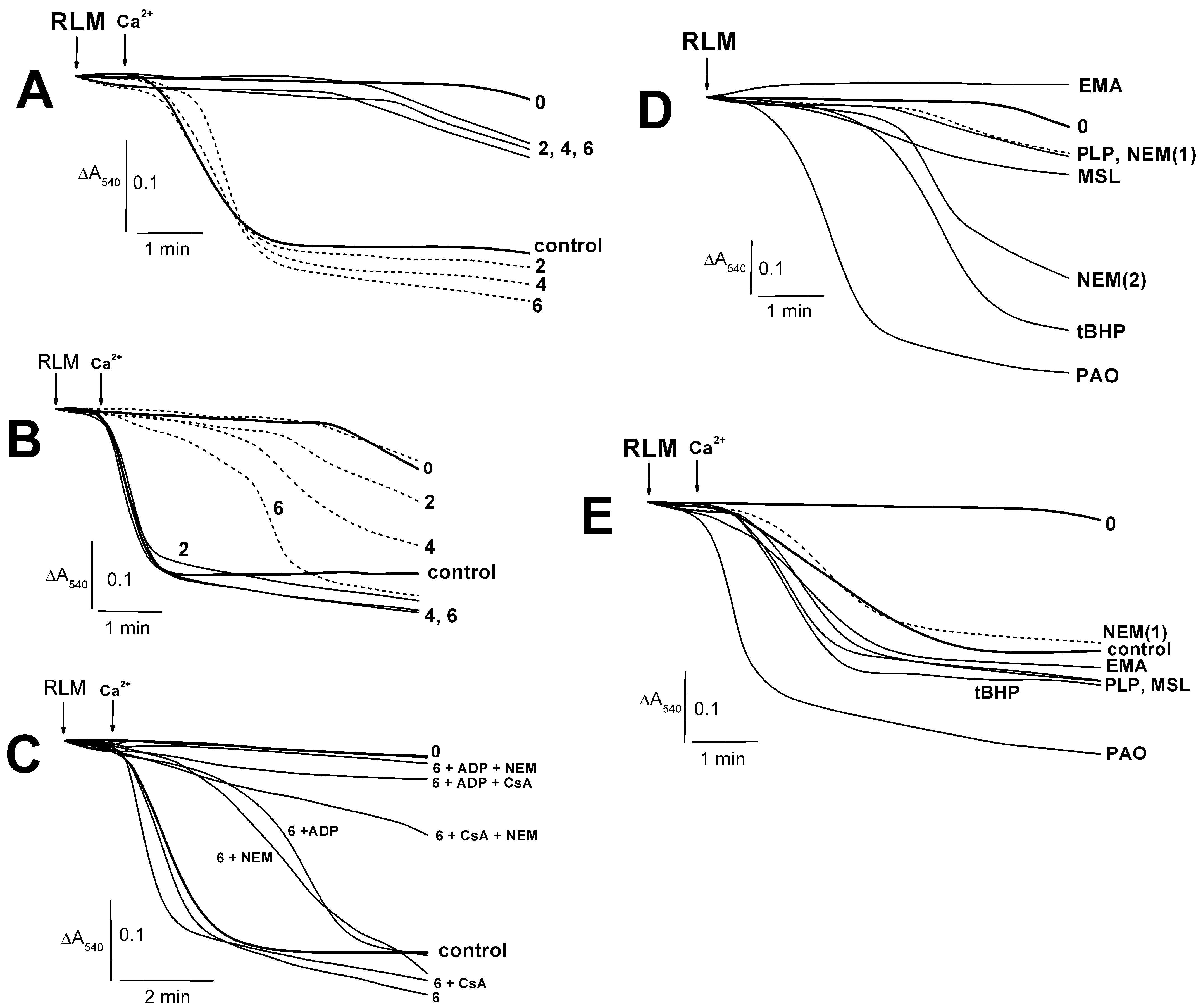
2.1. Effects of PLP and Thiol Reagents on the Tl+–Induced Swelling of Succinate-Energized Rat Liver Mitochondria

The swelling of succinate-energized mitochondria was slightly increased in the presence of 2–6 mM PLP (Figure 1A). At once, calcium-loaded rat liver mitochondria swelled a little more with 2–6 mM PLP in comparison to the control experiments (Figure 1A). ADP inhibited this swelling with minimal effect at 6 mM PLP (Figure 1B). The MPTP inhibitors significantly prevented the swelling of calcium-loaded RLM in the presence of 6 mM PLP (Figure 1C). This swelling decreased in the series CsA or PLP alone > control > ADP or NEM > CsA + NEM > ADP + CsA or ADP + NEM > “0” (free of Ca2+ and additions) (Figure 1C). CsA is interested in has not inhibited this swelling even in similar experiments with 2 mM PLP (not shown here). The swelling of succinate-energized mitochondria in experiments with thiol reagents and PLP increased in the series EMA < “0” (free of additions) < PLP or NEM(1) < NEM(2) < high MSL < tBHP < PAO (Figure 1D). A minor swelling increase (Figure 1E) was found in similar experiments with calcium-loaded mitochondria in the presence of EMA, PLP, MSL, and tBHP. Herewith, the similar effect of PAO was maximal.

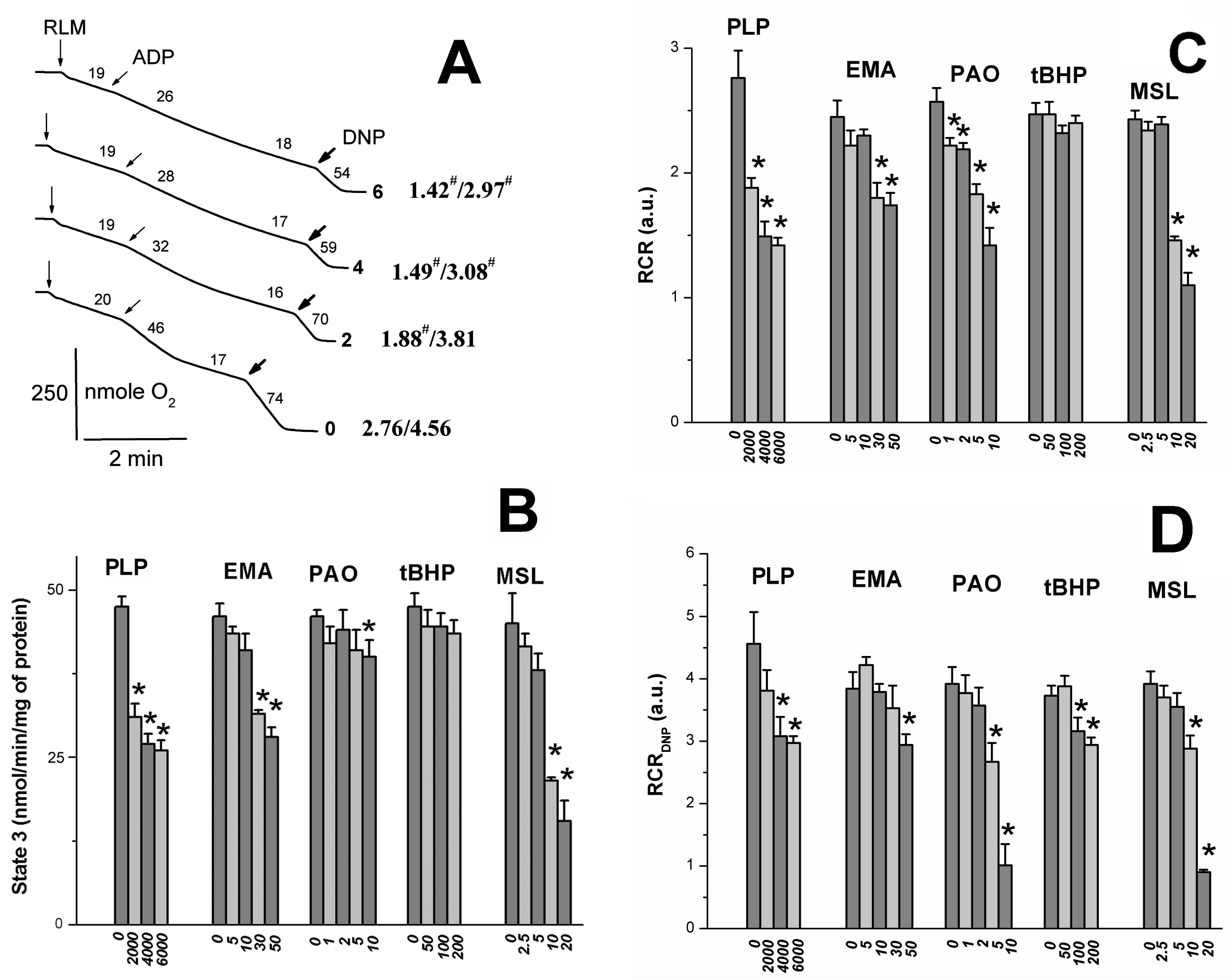
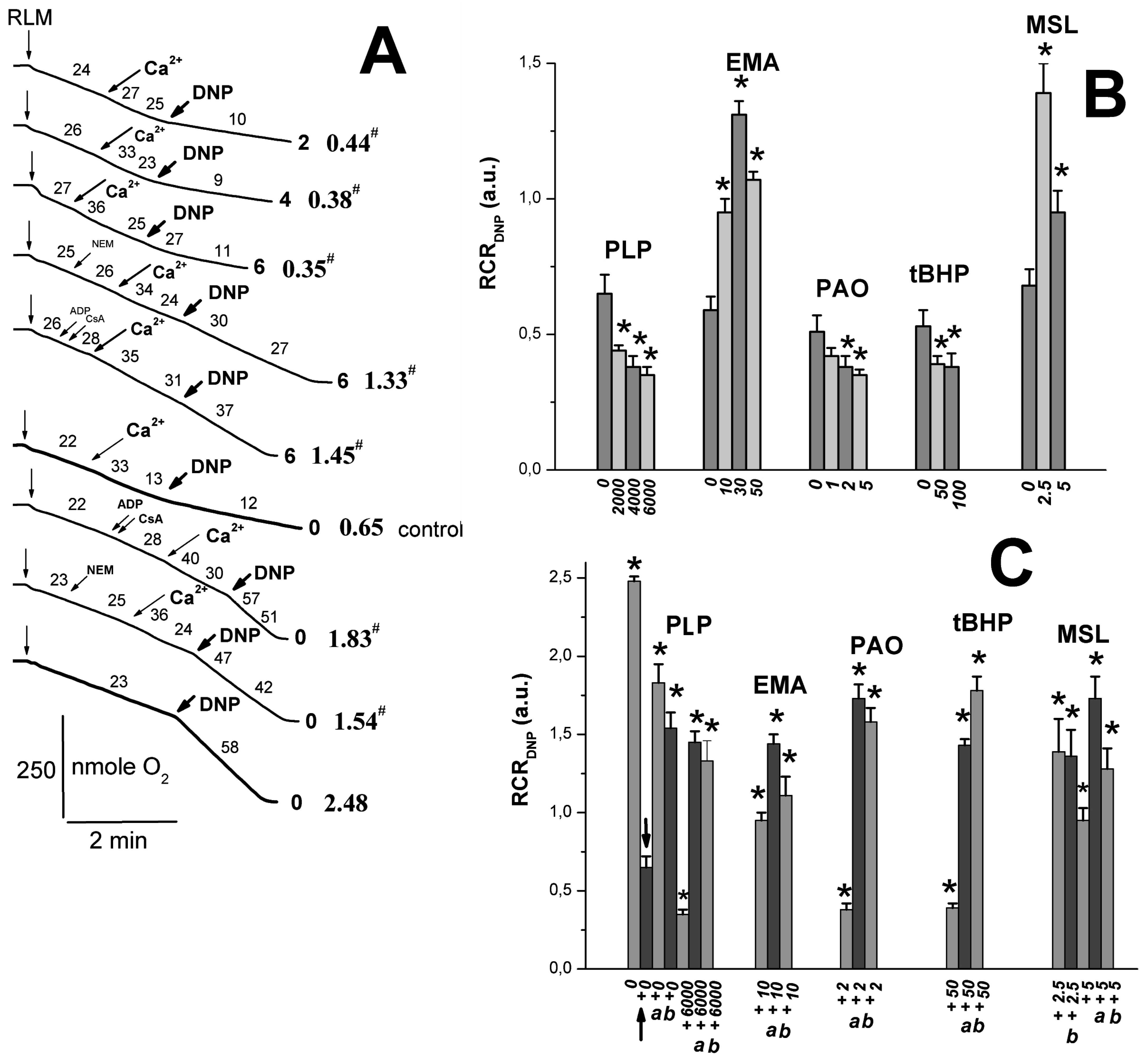
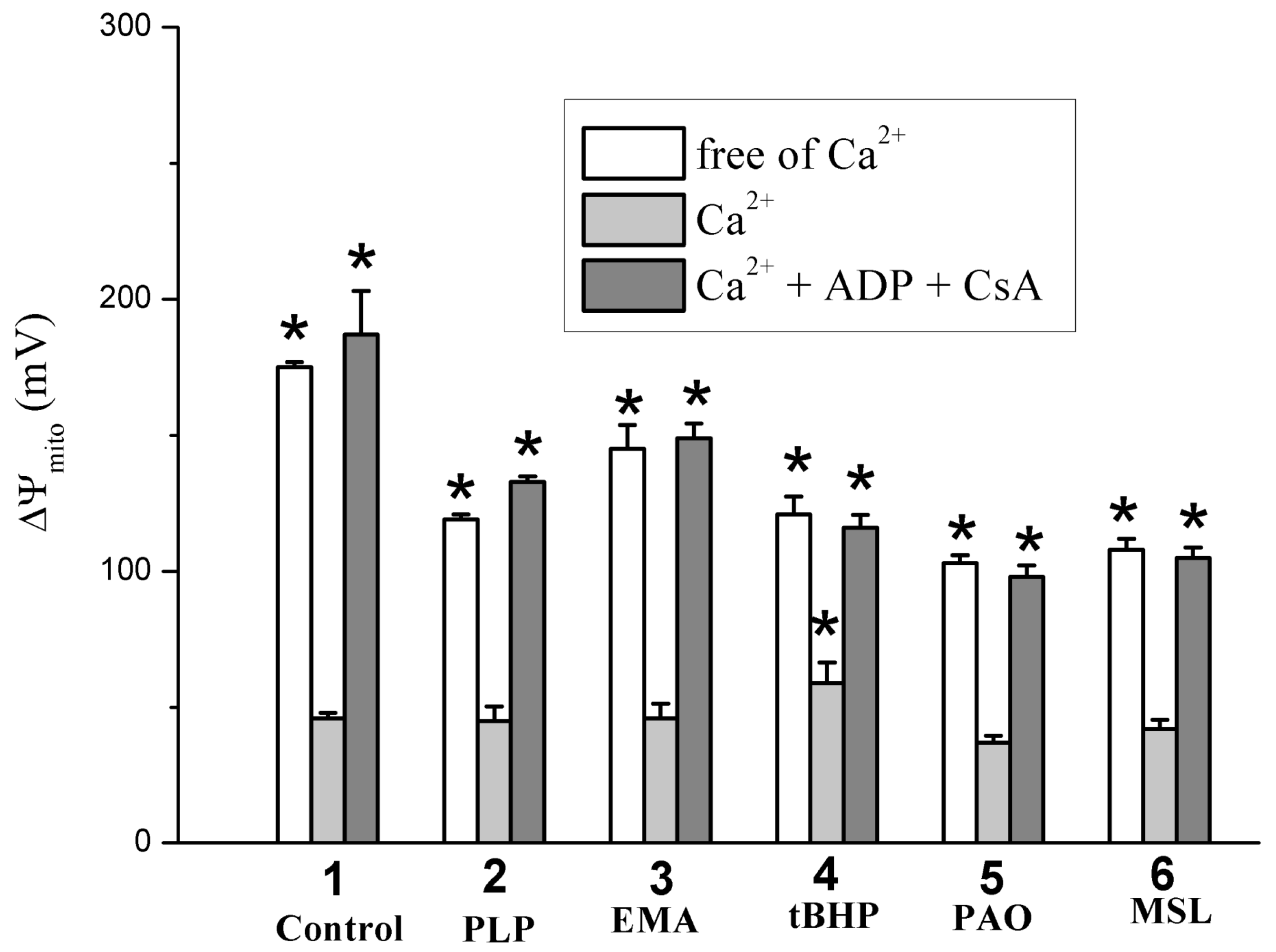
2.2. Effects of Tl+ and Thiol-Modifying Agents on Respiration and ΔΨmito of Succinate-Energized Rat Liver Mitochondria
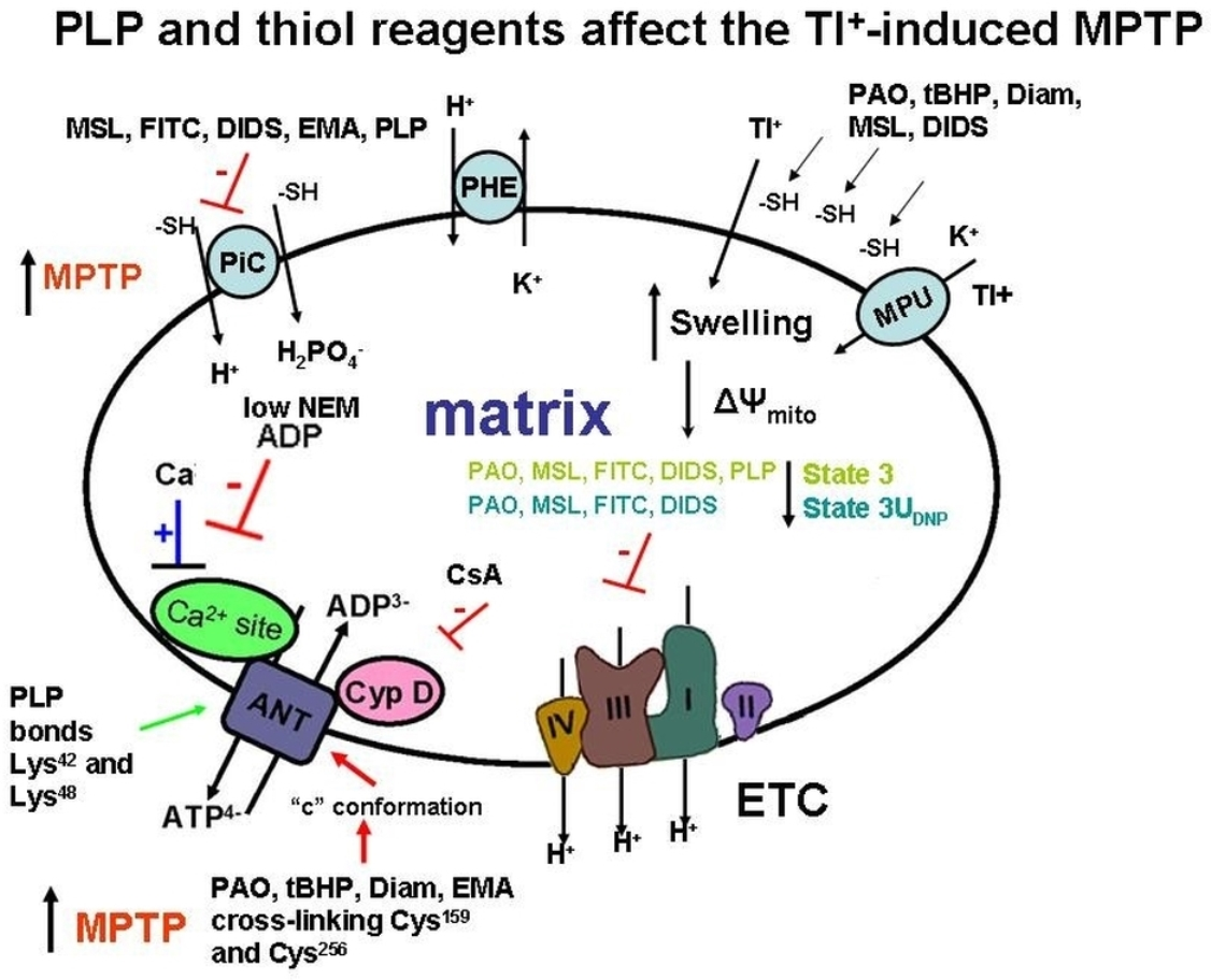
One can see in Figure 2A that PLP partly inhibited states 3 and 3UDNP respiration; however, the PLP effect on DNP-uncoupled respiration was not so pronounced. As for other thiol reagents, their inhibitory effect on RCR (Figure 2B) and state 3 respiration (Figure 2C) [6,9] increased in the series tBHP < EMA < PLP or MSL < PAO. DNP-uncoupled respiration [6,7,9] and RCRDNP (Figure 2D) decreased similarly in experiments with these reagents. However, the inhibitory effect of PAO on RCRDNP (Figure 2D) was more visible due to the PAO-induced state 4 increase [6]. The decrease in state 3UDNP respiration and RCRDNP in calcium-loaded RLM (control) was visibly greater in increasing PLP concentration in the medium containing TlNO3 and 2–6 mM PLP (Figs. 3A and B). The mitochondrial response to DNP added into the medium was preserved in this case. Thiol reagents (PAO and tBHP), similar to PLP and unlike EMA or MSL, contributed to a further decrease in both 3UDNP respiration and RCRDNP in experiments with calcium-loaded mitochondria (Figure 3A,B) [6,7,9]. The MPTP inhibitors (ADP with CsA or NEM alone) noticeably prevented a Ca2+-induced decrease in state 3UDNP respiration or RCRDNP in these experiments with/without PLP (Figure 3A,C). The Ca2+-induced respiration decrease was markedly prevented by the MPTP inhibitors in the medium containing both PLP and tBHP or PAO (Figure 3B,C) [6,7,9]. On the other hand, the above mitochondrial parameters were affected by EMA or MSL, similar to the MPTP inhibitors used. The additive effect of MSL with EMA was observed in this case. The fluorescent dye, safranin O, was used to evaluate the inner mitochondrial membrane potential (ΔΨmito). The addition of succinate into the medium led to the uptake of dye by succinate-energized mitochondria due to the inner membrane potential appearance. As a result, a decrease in the mitochondrial suspension fluorescence was observed. Calcium visibly decreased the membrane potential. An even greater decrease in the potential (Figure 4) was observed after adding calcium to the medium containing thiol reagents (PLP, EMA, MSL, tBHP, and PAO) [6,7,9]. However, this decrease was completely eliminated in the medium containing ADP and CsA.

3. Discussion

PLP was shown to penetrate slowly and passively into the mitochondrial matrix along a concentration gradient regardless of the presence of inhibitors and oxidative phosphorylation uncouplers [21,48]. The manganese transporter (Mtm1) transfers PLP into the matrix with micromolar affinity [49]. PLP and PAO were found to have produced increased swelling, decreased respiration in 3 and 3UDNP states, and ΔΨmito decline in the RLM experiments (Figure 1, Figure 2 and Figure 4) [6]. However, the above PLP effects occurred at a much lower rate since PLP concentrations of three orders of magnitude more than PAO concentrations are required to produce an impact of similar magnitude. Other thiol reagents (tBHP, Diam, DIDS, MSL, and high MSL) similarly affected swelling and respiration in 3 and 3UDNP states in experiments with calcium-free media (Figure 1, Figure 2 and Figure 4) [50]. The similarity of these effects may be because PLP can react not only with the ANT lysine residues but PLP can also interact with the ANT cysteine residues, similar to the reaction of PAO and above reagents with the cystein ones [10,11,16,24,28,29,50]. Low concentrations of PLP and thiol reagents were insufficient to inhibit visible RCR and state 3 respiration (Figure 2A–C) [6,7,8,9,50]. Wherein, state 3, not state 4, was markedly inhibited by these reagents at high concentrations. The state 3 inhibition increased in the series tBHP, Diam < PAO < EMA < PLP, FITC < high MSL, DIDS, high NEM (Figure 2C) [6,50]. RCR showed similar series, but PAO-induced RCR decrease was maximal due to the state 3 decline and state 4 increase [6].
State 3 respiration is known to depend on the activities of F1F0-ATP synthase, the adenine nucleotide translocase (ANT), and the mitochondrial phosphate cotransporter (PiC), which are necessary for the ATP synthesis carried out by mitochondria. PLP and DIDS can inhibit F1F0-ATP synthase, gastric H+/K+-ATPase, and tonoplast ATPase [6,51,52,53,54]. The ANT inhibition was observed in the presence of PLP [24,55], EMA [30], and FITC (discussed in [9]) as well as PAO, tBHP, and Diam (discussed in [6]). This inhibition resulted in the reaction of these reagents with the ANT cysteine residues. However, FITC does not interact with ANT cysteines, but inhibits ADP transport in mitochondria and reacts with the PiC cysteines [9,30]. The mitochondrial H+/Pi cotransporter activity was notably decreased in experiments with PLP [16,41], low MSL [7], EMA [56], and FITC [9]. State 3 respiration decrease may be due to inhibiting the enzymes involved in the oxidative phosphorylation processes and blocking the respiratory substrate transport into mitochondria. PLP and some thiol reagents (PAO, DIDS, FITC, and high MSL) inhibit a succinate transport into the matrix [6,7,9]. At the same time, state 3UDNP respiration decrease [6,7,9] was insignificant in experiments with mitochondria, energized substrates of the first respiratory complex (glutamate with malate) in the presence of reagents (DIDS, MSL, EMA, and FITC). Thus, the succinate transport inhibition may explain the marked decrease in state 3UDNP respiration and RCRDNP values (Figure 2A,D) in experiments with the latter reagents. By comparing the effects of other reagents (PLP, tBHP, Diam, and EMA) on states 3 and 3UDNP (Figure 2), it can be concluded that they have a weak impact on succinate transport.
Some used reagents (PAO, MSL, DIDS, tBHP, and Diam) showed increased swelling in experiments with Ca2+-loaded mitochondria (Figure 1) [9,50]. A similar effect of PLP was minor (Figure 1), whereas EMA and DIDS partially prevented this increase [6,9]. The Ca2+-induced decrease in state 3UDNP respiration was more visible in experiments with reagents (PAO, tBHP, Diam, FITC, and PLP) in comparison to DIDS, MSL, and EMA that slowed down these effects (Figure 3) [9,50]. Similar results are associated with the involvement of ANT in the MPTP opening under the action of thiol reagents (PAO, MSL, and DIDS) and stress inducers (tBHP and Diam) on mitochondria due to the stabilization of this exchanger c-conformation because of their reaction with the ANT cysteines [6,50]. Opposite, DIDS and low MSL decreased the MPTP opening due to ANT conformational changes, altering the thiol group reactivity [6,7]. In addition, MSL or DIDS presence discovered that the state 3UDNP respiration decrease was not so pronounced in calcium-loaded mitochondria injected in the medium with TlNO3 (Figure 3B) [50]. These results suggest that the interaction of these reagents with the PiC, not ANT thiol groups, plays a primary role. EMA, compared to NEM, showed a more significant affinity to ANT cysteine thiol groups, but EMA weak penetrates across the inner mitochondrial membrane [9,10,11,14,16]. EMA inhibited beef heart mitochondria swelling due to this reagent interacting with the cytoplasm-faced PiC essential thiol groups [9,56]. If the reaction has a place with Cys47 in experiments with low NEM and low EMA, then the m-conformation stabilization is observed [10,13,14]. The EMA block of the state 3UDNP respiration decrease (Figure 3B) may be due to the EMA reaction with the ANT Cys47 that followed this exchanger m-conformation stabilization [10,14,15]. The PLP effects on calcium-loaded mitochondria are most likely associated with the interaction of PLP with the ANT cysteine residues [24].
The MPTP inhibitors (ADP, EMA, and low NEM) are known to fix ANT in the m-conformation and to prevent the increased swelling and ΔΨmito decline in experiments with Ca2+-loaded mitochondria in the presence of thiol reagents (PAO, tBHP, Diam, EMA, arsenite, or menadione) that react with the ANT Cys159 and Cys256 [10,11,13,14]. The ADP inhibition of Tl+-induced MPTP opening was less pronounced in experiments with PLP, PAO, and DIDS than with MSL, EMA, and FITC (Figure 1, Figure 3 and Figure 4) [6,7,9,51]. Such a difference in effects may result from the fact that the former actively interacts with ANT cysteines, while the latter did not reveal such a critical effect on the ANT structure [6,7,9,11,24,56]. Thus, this circumstance ultimately allowed ADP to inhibit the MPTP opening. Low NEM (50 µM) prevented the increase in swelling and the decrease in RCRDNP and state 3UDNP respiration in experiments with the used thiol reagents (Figure 1C and Figure 3C) [6,7,8,9,50]. The Tl+-induced MPTP inhibition was due to the NEM interaction with the above ANT cysteines in experiments with PAO, Diam, EMA, tBHP, and PLP [10,11,13,14]. However, FITC- and MSL-induced MPTP opening in TlNO3 media (Figure 1C and Figure 3C) was inhibited by low NEM due to the reaction of NEM with cytoplasm-faced PiC thiols [7,9,50]. CsA visibly inhibited the Tl+-induced MPTP opening in experiments with tBHP, EMA, FITC, and MSL, but not PLP (Figure 1C and Figure 3C) [6,7,9,50]. This result suggests that CyP-D is not involved in the MPTP opening in experiments with PLP. In this case, the increased swelling decreased respiration, and ΔΨmito decline in experiments with used thiol and lysine (PLP) reagents were noticeably leveled or completely disappeared in simultaneous presencing NEM with ADP or NEM with CsA in the experiments with the TlNO3 medium. The inhibitory effect of ADP with CsA was maximal (Figure 1C, Figure 3C and Figure 4) [6,7,9,50].

4. Materials and Methods

4.1. Animals

Male Wistar rats (250–300 g) were kept at 20–23 °C under a 12 h light/dark cycle with free access to water ad libitum and the standard rat diet. All treatment procedures of animals were performed according to the Animal Welfare act and the Institute Guide for Care and Use of Laboratory Animals (Protocol # 6/5/2022).

4.2. Chemicals

Sucrose, CaCl2, Mg(NO3)2, H3PO4, KNO3, TlNO3, and 2,4-dinitrophenol (DNP) were of analytical grade from Nevareactiv (St. Petersburg, Russia). Rotenone, oligomycin, pyridoxal-5-phosphate (PLP), PAO, tBHP, NEM, safranin O, tris-OH, ethylene glycol tetraacetic acid EGTA, ADP, CsA, and succinic acid were from Sigma (St. Louis, MO, USA). Sucrose as a 1 M solution was refined from cation traces on a column filled with a KU-2-8 resin from Azot (Kemerovo, Russia).

4.3. Mitochondrial Isolation

Rat liver mitochondria were isolated according to [9] in a buffer containing 250 mM sucrose, 3 mM Tris-HCl (pH 7.3), and 0.5 mM EGTA; subsequent mitochondrial sediment was washed out twice by resuspension-centrifugation in a medium containing 250 mM sucrose and 3 mM Tris-HCl (pH 7.3) and finally suspended in 1 mL of the last buffer. According to Bradford, the mitochondrial protein content assayed was within 50–60 mg/mL.

4.4. Swelling of Mitochondria

Mitochondrial swelling (Figure 1) was tested as a decrease in A540 at 20 °C using an SF-46 spectrophotometer (LOMO, St. Petersburg, Russia). Mitochondria (1.5 mg of protein/mL) were injected into a 1-cm cuvette with 1.5 mL of 400 mOsm medium containing 75 mM TlNO3, 125 mM KNO3, 5 mM Tris-NO3 (pH 7.3), 2 μM rotenone, and 1 μg/mL of oligomycin. PLP, PAO, tBHP, NEM, Ca2+, succinate, NEM, ADP, and CsA were added into the medium before or after mitochondria (see Figure 1 legend). The swelling, oxygen consumption rates, and ΔΨmito dissipation were investigated in 400 mOsm media to verify the comparability and consistency between data in different experiments.

4.5. Oxygen Consumption Assay

Mitochondrial respiration (oxygen consumption rate) was measured polarographically using an Expert-001 analyzer (Econix-Expert Ltd., Moscow, Russia) in a 1.3 mL closed thermostatic chamber with magnetic stirring at 26 °C. Mitochondria (1.5 mg of protein/mL) were added to 400 mOsm medium containing 25 mM TlNO3, 100 mM sucrose, 3 mM Mg(NO3)2, and 3 mM Tris-Pi (Figure 2) or 75 mM TlNO3 and 1 μg/mL of oligomycin (Figure 3) as well as 125 mM KNO3, 5 mM Tris-NO3 (pH 7.3), 5 mM succinate, and 2 μM rotenone. PLP, PAO, tBHP, EMA, MSL, Ca2+, NEM, ADP, and CsA were added in the medium before or after mitochondria (see Figure 2 and Figure 3 legend). ADP of 130 μM (Figure 2) and 30 μM DNP (Figure 2 and Figure 3) were correspondingly added into the medium after 2 min recording of state 4 to induce state 3 and 3UDNP respiration. The respiratory control ratio (RCRADP) was calculated as a ratio of state 3 to state 4 (Figure 2). The RCRDNP was accordingly quantified as a ratio of DNP-uncoupled respiration to state 4 (Figure 2) or state 40 respiration (Figure 3).

4.6. Mitochondrial Membrane Potential

The inner membrane potential (ΔΨmito) induced by injection of 5 mM succinate into a medium was tested according to Waldmeier et al. [57]. The safranin O fluorescence intensity (arbitrary units) in the mitochondrial suspension was tested at 20 °C (Figure 4) using the microplate reader (CLARIOstar® Plus, BMG LABTECH, Ortenberg, Germany) at 485/590 nm wavelength (excitation/emission). The mitochondria (0.5 mg of protein/mL) were added into cells containing 300 μL of the medium with 20 mM TlNO3, 125 mM KNO3, 110 mM sucrose, 5 mM Tris-NO3 (pH 7.3), 1 mM Tris-Pi, 2 μM rotenone, 3 μM safranin O, and 1 μg/mL of oligomycin. ADP, CsA, PLP, EMA, PAO, tBHP, and MSL were injected into the medium before mitochondria (see Figure 4 legend). 5 mM succinate, 75 μM Ca2+, and 30 μM DNP was administrated into the medium after the mitochondria. The safranin O fluorescence change after the succinate injection was taken as 100% in control experiments free of the above additions (PLP, thiol reagents, ADP, CsA, and Ca2+). The other cases’ fluorescence values were calculated relative to this control. A parallel fourfold measurement for each 300 μl aliquot was made from three independent preparations.

4.7. Statistical Analysis

The statistical differences in results and corresponding p-values were evaluated using two population t-tests (Microcal Origin, Version 6.0, Microcal Software). These differences are presented as a percent of the average (p < 0.05) from one of three independent experiments (Figure 1, Figure 2, Figure 3 and Figure 4). A more detailed statistical analysis is in supplementary materialssupplementary materials, Figures S1–S4 and Table S1.

5. Conclusions

Mitochondrial proteins contain up to 20% acetylated lysine residues, including F1F0-ATP synthase structural parts [19]. The covalent modification of lysine residues is believed to affect the MPTP open probability [13,20]. The MPTP opening resulted in a fluorescamine reaction with the ANT lysine residues that induced efflux of accumulated Ca2+, ΔΨmito decline, and mitochondrial swelling [58]. Experiments with BKA- and CAT-treated mitochondria showed that one or more lysine residues could be involved in CAT and BKA binding with the ANT [24,55]. Lys401 of bovine heart mitochondrial F1F0-ATP synthase was modified explicitly with 4-chloro-7-nitrobenzofurazan [59]. Pyridoxal 5’-diphospho-5’-adenosine was shown to bind to the isolated alpha-subunit from E. coli F1-ATP synthase [60]. FITC reacts with cysteine and the PiC lysine residues. It quickly penetrates the mitochondrial matrix, binds to α and γ subunits of the F1-ATP synthase, and localizes in the inner membrane lipid part [9,30]. FITC binds the PiC Lys residue more efficiently than PLP [30]. It can be seen that lysine residues are essential for the functioning of the MPTPs most important parts, namely F1F0-ATP synthase, ANT, and Pi cotransporter. An analysis of the study results allows us to conclude that the Tl+-induced MPTP opening is associated not only with functionally active cysteines of the latter three parts, but may also be related to activities of the corresponding lysines that form the pore structure parts.

Supplementary Materials

The following supporting information can be downloaded at: https://www.mdpi.com/article/10.3390/ijms24032460/s1.

Author Contributions

S.M.K. performed the experiments, analyzed and discussed the data and results, wrote the manuscript, supervised the research, and conceptualized the manuscript. A.V.N. performed the mitochondrial membrane potential experiments. All authors have read and agreed to the published version of the manuscript.

Funding

The work is supported by the IEPhB Research Program 075-00967-23-00 and the IEPhB Research Resource Center.

Institutional Review Board Statement

The data supporting this study’s findings are available from the corresponding authors, [S.M.K.], upon reasonable request.

Informed Consent Statement

Not applicable.

Data Availability Statement

The data that support the findings of this study are available from the corresponding authors, [S.M.K.], upon reasonable request.

Acknowledgments

The authors are grateful to Irina V. Brailovskaya for help in isolating mitochondria and the oxygen consumption rates in rat liver mitochondrial suspensions.

Conflicts of Interest

The authors declare no conflict of interest.

Abbreviations

3UDNP state2,4-Dinitrophenol uncoupled state
ΔΨmitoInner mitochondrial membrane potential
ANTAdenine nucleotide translocase
CATCarboxyatractyloside
CsACyclosporine A
CyP-DCyclophilin D
DiamDiamide
DIDS4,4′-Diisothiocyanostilbene-2,2′-disulfonate
DNP2,4-Dinitrophenol
EGTAEthylene glycol-bis(β-aminoethyl ether) N,N,N′,N′-tetraacetic acid
EMAEosin-5-maleimide
FITCFluorescein isothiocyanate
IMMInner mitochondrial membrane
MPTPMitochondrial permeability transition pore
MSLMersalyl
NEMn-Ethylmaleimide
PAOPhenylarsine oxide
PiCMitochondrial phosphate carrier
PiInorganic phosphate
PLPPyridoxal 5-phosphate
RCRRespiratory control ratio
RLMReactive oxygen species
tBHPTert-butyl hydroperoxide

References

  1. Bernardi, P.; Carraro, M.; Lippe, G. The mitochondrial permeability transition: Recent progress and open questions. FEBS Lett. 2021, 289, 7051–7074. [Google Scholar] [CrossRef]
  2. Brustovetsky, N. The role of adenine nucleotide translocase in the mitochondrial permeability transition. Cells 2020, 9, 2686. [Google Scholar] [CrossRef] [PubMed]
  3. Halestrap, A.P. The c ring of the F1Fo ATP synthase forms the mitochondrial permeability transition pore: A critical appraisal. Front. Oncol. 2014, 4, 234. [Google Scholar] [CrossRef] [PubMed] [Green Version]
  4. Korotkov, S.M.; Saris, N.E. Influence of Tl+ on mitochondrial permeability transition pore in Ca2+-loaded rat liver mitochondria. J. Bioenerg. Biomembr. 2011, 43, 149–162. [Google Scholar] [CrossRef] [PubMed]
  5. Korotkov, S.M. Mitochondria as a Key Intracellular Target of Thallium Toxicity; Elsevier: Amsterdam, The Netherlands; Academic Press: Amsterdam, The Netherlands, 2022; pp. 1–264. [Google Scholar]
  6. Korotkov, S.M.; Konovalova, S.A.; Brailovskaya, I.V.; Saris, N.E. To involvement the conformation of the adenine nucleotide translocase in opening the Tl+-induced permeability transition pore in Ca2+-loaded rat liver mitochondria. Toxicol. Vitr. 2016, 32, 320–332. [Google Scholar] [CrossRef] [Green Version]
  7. Korotkov, S.M.; Konovalova, S.A.; Nesterov, V.P.; Brailovskaya, I.V. Mersalyl prevents the Tl+-induced permeability transition pore opening in the inner membrane of Ca2+-loaded rat liver mitochondria. Biochem. Biophys. Res. Commun. 2018, 495, 1716–1721. [Google Scholar] [CrossRef]
  8. Korotkov, S.M.; Konovalova, S.A.; Brailovskaya, I.V. Diamide accelerates opening of the Tl+-induced permeability transition pore in Ca2+-loaded rat liver mitochondria. Biochim. Biophys. Res. Commun. 2015, 468, 360–364. [Google Scholar] [CrossRef]
  9. Korotkov, S.M.; Novozhilov, A.V. The joint influence of Tl+ and thiol-modifying agents on rat liver mitochondrial parameters in vitro. Int. J. Mol. Sci. 2022, 23, 8964. [Google Scholar] [CrossRef]
  10. McStay, G.P.; Clarke, S.J.; Halestrap, A.P. Role of critical thiol groups on the matrix surface of the adenine nucleotide translocase in the mechanism of the mitochondrial permeability transition pore. Biochem. J. 2002, 367, 541–548. [Google Scholar] [CrossRef] [PubMed] [Green Version]
  11. Majima, E.; Koike, H.; Hong, Y.M.; Shinohara, Y.; Terada, H. Characterization of cysteine residues of mitochondrial ADP/ATP carrier with the SH-reagents eosin 5-maleimide and N-ethylmaleimide. J. Biol. Chem. 1993, 268, 22181–22187. [Google Scholar] [CrossRef]
  12. Halestrap, A.P. A pore way to die: The role of mitochondria in reperfusion injury and cardioprotection. Biochem. Soc. Trans. 2010, 38, 841–860. [Google Scholar] [CrossRef] [PubMed] [Green Version]
  13. Petronilli, V.; Costantini, P.; Scorrano, L.; Colonna, R.; Passamonti, S.; Bernardi, P. The voltage sensor of the mitochondrial permeability transition pore is tuned by the oxidation-reduction state of vicinal thiols. Increase of the gating potential by oxidants and its reversal by reducing agents. J. Biol. Chem. 1994, 269, 16638–16642. [Google Scholar] [CrossRef]
  14. Halestrap, A.P.; Brenner, C. The adenine nucleotide translocase: A central part of the mitochondrial permeability transition pore and key player in cell death. Curr. Med. Chem. 2003, 10, 1507–1525. [Google Scholar] [CrossRef] [PubMed] [Green Version]
  15. Nantes, I.L.; Rodrigues, T.; Caires, A.C.; Cunha, R.L.; Pessoto, F.S.; Yokomizo, C.H.; Araujo-Chaves, J.C.; Faria, P.A.; Santana, D.P.; dos Santos, C.G. Specific effects of reactive thiol drugs on mitochondrial bioenergetics. J. Bioenerg. Biomembr. 2011, 43, 11–18. [Google Scholar] [CrossRef] [PubMed]
  16. Majima, E.; Shinohara, Y.; Yamaguchi, N.; Hong, Y.M.; Terada, H. Importance of loops of mitochondrial ADP/ATP carrier for its transport activity deduced from reactivities of its cysteine residues with the sulfhydryl reagent eosin-5-maleimide. Biochemistry 1994, 33, 9530–9536. [Google Scholar] [CrossRef] [PubMed]
  17. Nicolli, A.; Petronilli, V.; Bernardi, P. Modulation of the mitochondrial cyclosporin A-sensitive permeability transition pore by matrix pH. Evidence that the pore open-closed probability is regulated by reversible histidine protonation. Biochemistry 1993, 32, 4461–4465. [Google Scholar] [CrossRef]
  18. Eriksson, O.; Fontaine, E.; Bernardi, P. Chemical modification of arginines by 2,3-butanedione and phenylglyoxal causes closure of the mitochondrial permeability transition pore. J. Biol. Chem. 1998, 273, 12669–12674. [Google Scholar] [CrossRef] [Green Version]
  19. Antoniel, M.; Giorgio, V.; Fogolari, F.; Glick, G.D.; Bernardi, P.; Lippe, G. The oligomycin-sensitivity conferring protein of mitochondrial ATP synthase: Emerging new roles in mitochondrial pathophysiology. Int. J. Mol. Sci. 2014, 15, 7513–7536. [Google Scholar] [CrossRef] [Green Version]
  20. Eriksson, O.; Fontaine, E.; Petronilli, V.; Bernardi, P. Inhibition of the mitochondrial cyclosporin A-sensitive permeability transition pore by the arginine reagent phenylglyoxal. FEBS Lett. 1997, 409, 361–364. [Google Scholar] [CrossRef] [Green Version]
  21. Whittaker, J.W. Intracellular trafficking of the pyridoxal cofactor. Implications for health and metabolic disease. Arch. Biochem. Biophys. 2015, 592, 20–26. [Google Scholar] [CrossRef]
  22. Tragni, V.; Primiano, G.; Tummolo, A.; Beltrame, L.C.; La Piana, G.; Sgobba, M.N.; Cavalluzzi, M.M.; Paterno, G.; Gorgoglione, R.; Volpicella, M.; et al. Personalized medicine in mitochondrial health and disease: Molecular basis of therapeutic approaches based on nutritional supplements and their analogs. Molecules 2022, 27, 3494. [Google Scholar] [CrossRef] [PubMed]
  23. Means, G.E.; Feeney, R.E. Affinity labeling of pancreatic ribonuclease. J. Biol. Chem. 1971, 246, 5532–5533. [Google Scholar] [CrossRef] [PubMed]
  24. Bogner, W.; Aquila, H.; Klingenberg, M. The transmembrane arrangement of the ADP/ATP carrier as elucidated by the lysine reagent pyridoxal 5-phosphate. Eur. J. Biochem. 1986, 161, 611–620. [Google Scholar] [CrossRef] [PubMed]
  25. Banerjee, P.S.; Ma, J.; Hart, G.W. Diabetes-associated dysregulation of O-GlcNAcylation in rat cardiac mitochondria. Proc. Natl. Acad. Sci. USA 2015, 112, 6050–6055. [Google Scholar] [CrossRef] [Green Version]
  26. Kunk, C.; Kruger, J.; Mendoza, G.; Markitan, J.; Bias, T.; Mann, A.; Nath, A.; Geldenhuys, W.J.; Menze, M.A.; Konkle, M.E. MitoNEET’s reactivity of Lys55 toward pyridoxal phosphate demonstrates its activity as a transaminase enzyme. ACS Chem. Biol. 2022, 17, 2716–2722. [Google Scholar] [CrossRef]
  27. Stipani, I.; Mangiullo, G.; Stipani, V.; Daddabbo, L.; Natuzzi, D.; Palmieri, F. Inhibition of the reconstituted mitochondrial oxoglutarate carrier by arginine-specific reagents. Arch. Biochem. Biophys. 1996, 331, 48–54. [Google Scholar] [CrossRef]
  28. Gremse, D.A.; Dean, B.; Kaplan, R.S. Effect of pyridoxal 5′-phosphate on the function of the purified mitochondrial tricarboxylate transport protein. Arch. Biochem. Biophys. 1995, 316, 215–219. [Google Scholar] [CrossRef]
  29. Tahiliani, A.G.; Keene, T.; Kaplan, R.S. Characterization of the inhibitor sensitivity of the coenzyme A transport system in isolated rat heart mitochondria. J. Bioenerg. Biomembr. 1992, 24, 635–640. [Google Scholar] [CrossRef]
  30. Majima, E.; Ishida, M.; Miki, S.; Shinohara, Y.; Terada, H. Specific labeling of the bovine heart mitochondrial phosphate carrier with fluorescein 5-isothiocyanate: Roles of Lys185 and putative adenine nucleotide recognition site in phosphate transport. J. Biol. Chem. 2001, 276, 9792–9799. [Google Scholar] [CrossRef] [Green Version]
  31. Palmieri, L.; Lasorsa, F.M.; De Palma, A.; Palmieri, F.; Runswick, M.J.; JWalker, E. Identification of the yeast ACR1 gene product as a succinate-fumarate transporter essential for growth on ethanol or acetate. FEBS Lett. 1997, 417, 114–118. [Google Scholar] [CrossRef]
  32. Palmieri, L.; Vozza, A.; Agrimi, G.; De Marco, V.; Runswick, M.J.; Palmieri, F.; Walker, J.E. Identification of the yeast mitochondrial transporter for oxaloacetate and sulfate. J. Biol. Chem. 1999, 274, 22184–22190. [Google Scholar] [CrossRef] [PubMed] [Green Version]
  33. Palmieri, L.; Agrimi, G.; Runswick, M.J.; Fearnley, I.M.; Palmieri, F.; Walker, J.E. Identification in Saccharomyces cerevisiae of two isoforms of a novel mitochondrial transporter for 2-oxoadipate and 2-oxoglutarate. J. Biol. Chem. 2001, 276, 1916–1922. [Google Scholar] [CrossRef] [PubMed] [Green Version]
  34. Fiermonte, G.; Palmieri, L.; Dolce, V.; Lasorsa, F.M.; Palmieri, F.; Runswick, M.J.; Walker, J.E. The sequence, bacterial expression, and functional reconstitution of the rat mitochondrial dicarboxylate transporter cloned via distant homologs in yeast and Caenorhabditis elegans. J. Biol. Chem. 1998, 273, 24754–24759. [Google Scholar] [CrossRef] [PubMed] [Green Version]
  35. Gutiérrez-Aguilar, M.; Baines, C.P. Physiological and pathological roles of mitochondrial SLC25 carriers. Biochem. J. 2013, 45, 371–386. [Google Scholar] [CrossRef] [Green Version]
  36. Scalera, V.; Giangregorio, N.; De Leonardis, S.; Console, L.; Carulli, E.S.; Tonazzi, A. Characterization of a novel mitochondrial ascorbate transporter from rat liver and potato mitochondria. Front. Mol. Biosci. 2018, 5, 58. [Google Scholar] [CrossRef] [Green Version]
  37. Natuzzi, D.; Daddabbo, L.; Stipani, V.; Cappello, A.R.; Miniero, D.V.; Capobianco, L.; Stipani, I. Inactivation of the reconstituted oxoglutarate carrier from bovine heart mitochondria by pyridoxal 5′-phosphate. J. Bioenerg. Biomembr. 1999, 31, 535–541. [Google Scholar] [CrossRef]
  38. Indiveri, C.; Abruzzo, G.; Stipani, I.; Palmieri, F. Identification and purification of the reconstitutively active glutamine carrier from rat kidney mitochondria. Biochem. J. 1998, 333, 285–290. [Google Scholar] [CrossRef] [Green Version]
  39. Stappen, R.; Dierks, T.; Bröer, A.; Krämer, R. Probing the active site of the reconstituted aspartate/glutamate carrier from mitochondria. Structure/function relationship involving one lysine and two cysteine residues. Eur. J. Biochem. 1992, 210, 269–277. [Google Scholar] [CrossRef]
  40. Dierks, T.; Krämer, R. Asymmetric orientation of the reconstituted aspartate/glutamate carrier from mitochondria. Biochim. Biophys. Acta 1988, 937, 112–126. [Google Scholar] [CrossRef]
  41. Stappen, R.; Krämer, R. Functional properties of the reconstituted phosphate carrier from bovine heart mitochondria: Evidence for asymmetric orientation and characterization of three different transport modes. Biochim. Biophys. Acta 1993, 1149, 40–48. [Google Scholar] [CrossRef]
  42. Stepp, L.R.; Reed, L.J. Active-site modification of mammalian pyruvate dehydrogenase by pyridoxal 5′-phosphate. Biochemistry 1985, 24, 7187–7191. [Google Scholar] [CrossRef] [PubMed]
  43. Yamaguchi, M.; Hatefi, Y. Mitochondrial nicotinamide nucleotide transhydrogenase: Inhibition by ethoxyformic anhydride, dansyl chloride, and pyridoxal phosphate. Arch. Biochem. Biophys. 1985, 243, 20–27. [Google Scholar] [CrossRef] [PubMed]
  44. Wimmer, M.J.; Mo, T.; Sawyers, D.L.; Harrison, J.H. Biphasic inactivation of procine heart mitochondrial malate dehydrogenase by pyridoxal 5′-phosphate. J. Biol. Chem. 1975, 250, 710–715. [Google Scholar] [CrossRef]
  45. Chen, S.S.; Engel, P.C. Reversible modification of pig heart mitochondrial malate dehydrogenase by pyridoxal 5′-phosphate. Biochem. J. 1975, 151, 297–303. [Google Scholar] [CrossRef] [Green Version]
  46. Le-Quoc, K.; Le-Quoc, D. Involvement of the ADP/ATP carrier in calcium-induced perturbations of the mitochondrial inner membrane permeability: Importance of the orientation of the nucleotide binding site. Arch. Biochem. Biophys. 1988, 265, 249–257. [Google Scholar] [CrossRef] [PubMed]
  47. Le-Quoc, D.; Le-Quoc, K. Relationships between the NAD(P) redox state, fatty acid oxidation, and inner membrane permeability in rat liver mitochondria. Arch. Biochem. Biophys. 1989, 273, 466–478. [Google Scholar] [CrossRef] [PubMed]
  48. Lui, A.; Lumeng, L.; Li, T.K. Transport of pyridoxine and pyridoxal 5′-phosphate in isolated rat liver mitochondria. J. Biol. Chem. 1982, 257, 14903–14906. [Google Scholar] [CrossRef]
  49. Whittaker, M.M.; Penmatsa, A.; Whittaker, J.W. The Mtm1p carrier and pyridoxal 5′-phosphate cofactor trafficking in yeast mitochondria. Arch. Biochem. Biophys. 2015, 568, 64–70. [Google Scholar] [CrossRef] [Green Version]
  50. Korotkov, S.M. Effects of Tl+ on the inner membrane thiol groups, respiration, and swelling in succinate-energized rat liver mitochondria were modified by thiol reagents. Biometals 2021, 34, 987–1006. [Google Scholar] [CrossRef] [PubMed]
  51. Moriyama, Y.; Takano, T.; Ohkuma, S. Similarity of lysosomal H+-ATPase to mitochondrial F0F1-ATPase in sensitivity to anions and drugs as revealed by solubilization and reconstitution. Biochim. Biophys. Acta 1986, 854, 102–108. [Google Scholar] [CrossRef] [PubMed]
  52. Maeda, M.; Tagaya, M.; Futai, M. Modification of gastric (H+ + K+)-ATPase with pyridoxal 5′-phosphate. J. Biol. Chem. 1988, 263, 3652–3656. [Google Scholar] [CrossRef] [PubMed]
  53. Komatsu-Takaki, M. Effects of energization and substrates on the reactivities of lysine residues of the chloroplast ATP synthase beta subunit. Eur. J. Biochem. 1995, 228, 265–270. [Google Scholar] [CrossRef]
  54. Tzeng, C.M.; Hsu, L.H.; Pan, R.L. Inhibition of tonoplast ATPase from etiolated mung bean seedlings by fluorescein 5′-isothiocyanate. Biochem. J. 1992, 285, 737–743. [Google Scholar] [CrossRef] [Green Version]
  55. Bogner, W.; Aquila, H. ADP/ATP carrier: Analysis of transmembrane folding using pyridoxal phosphate. Methods Enzymol. 1986, 125, 650–658. [Google Scholar] [CrossRef] [PubMed]
  56. Houstĕk, J.; Pedersen, P.L. Adenine nucleotide and phosphate transport systems of mitochondria. Relative location of sulfhydryl groups based on the use of the novel fluorescent probe eosin-5-maleimide. J. Biol. Chem. 1985, 260, 6288–6295. [Google Scholar] [CrossRef] [PubMed]
  57. Waldmeier, P.C.; Feldtrauer, J.J.; Qian, T.; Lemasters, J.J. Inhibition of the mitochondrial permeability transition by the nonimmunosuppressive cyclosporine derivative NIM811. Mol. Pharmacol. 2002, 62, 22–29. [Google Scholar] [CrossRef] [PubMed] [Green Version]
  58. Buelna-Chontal, M.; Pavón, N.; Correa, F.; Hernández-Esquivel, L.; Chávez, E. Titration of lysine residues on adenine nucleotide translocase by fluorescamine induces permeability transition. Cell Biol. Int. 2014, 38, 287–295. [Google Scholar] [CrossRef] [PubMed]
  59. Sutton, R.; Ferguson, S.J. Identification of an essential beta chain lysine residue from bovine heart mitochondrial ATPase specifically modified with nitrobenzofurazan. FEBS Lett. 1985, 179, 283–288. [Google Scholar] [CrossRef] [Green Version]
  60. Rao, R.; Cunningham, D.; Cross, R.L.; Senior, A.E. Pyridoxal 5′-diphospho-5′-adenosine binds at a single site on isolated alpha-subunit from Escherichia coli F1-ATPase and specifically reacts with lysine 201. J. Biol. Chem. 1988, 263, 5640–5645. [Google Scholar] [CrossRef]
Figure 1. Effects of Ca2+, pyridoxal 5-phosphate, and thiol reagents on the Tl+-induced swelling of succinate-energized rat liver mitochondria. Mitochondria (1.5 mg of protein per mL) were injected into the medium containing 75 mM TlNO3, 125 mM KNO3, 100 μM Ca2+ (where indicated), and 5 mM Tris-succinate (pH 7.3). In addition, the medium was supplemented with 500 μM ADP ((B) short dash traces). The following reagents were added into the medium before mitochondria ((CE), indicated to the right of traces) 500 μM ADP (ADP), 1 μM CsA (CsA), 50 μM NEM (NEM(1)), 500 μM NEM (NEM(2)), 10 μM EMA (EMA), 20 μM MSL (MSL), 6 mM PLP (PLP), 100 μM tBHP (tBHP), and 5 μM PAO (PAO). Additions of mitochondria (RLM) and 100 μM Ca2+ (Ca2+) are shown by arrows. Experiments free of Ca2+ (solid traces) or ones with 100 μM Ca2+ (short dash traces) showed on panel A. The numbers to the right of the traces (AC) show concentrations (μM) of PLP, which was added into the medium before mitochondria. The bold traces show experiments free of Ca2+ and PLP (0) or ones with Ca2+ alone (control). Representative traces from one of three independent experiments are presented.
Ijms 24 02460 g001
Figure 2. Effects of pyridoxal 5-phosphate and thiol reagents on oxygen consumption rates of rat liver mitochondria. Mitochondria (1.5 mg/mL of protein) were added into a medium containing 25 mM TlNO3, 125 mM KNO3, 100 mM sucrose, 5 mM Tris-NO3 (pH 7.3), 3 mM Tris-Pi, 3 mM Mg(NO3)2, 5 mM succinate, and 2 μM rotenone. Additions of mitochondria (RLM), 130 μM ADP (ADP), and 30 μM DNP (DNP) are correspondingly shown by vertical arrows, inclined arrows, and bold arrows. Oxygen consumption rates (nmole O2 min/mg of protein) are presented as numbers placed above experimental traces. The numbers to the right of the traces (A) show PLP (mM) concentrations. Numbers (A) in bold with the slash between them to the right of the traces, respectively, show values of the RCRADP and the RCRDNP (see the Section 4). The hash to the right of the latter numbers indicates that the difference between appropriate values of the RCRADP and the RCRDNP is statistically insignificant to the values found in experiments free of PLP. Representative traces from one of three independent experiments are presented. The ordinate (B) marks oxygen consumption rates (nmole O2 min/mg of protein) in state 3. The ordinates (C,D) show RCR and RCRDNP values. Numbers below the abscissa (BD) indicate the concentration (μM) of thiol reagents (PLP, EMA, MSL, PAO, and tBHP). * shows significant differences from the control (“0” under the abscissa) experiments free of thiol reagents (p < 0.05).
Ijms 24 02460 g002
Figure 3. Effects of Ca2+, pyridoxal 5-phosphate, and thiol reagents on oxygen consumption rates of rat liver mitochondria. Mitochondria (1.5 mg/mL of protein) were added into a medium containing 75 mM TlNO3, 125 mM KNO3, 5 mM Tris-NO3 (pH 7.3), 5 mM succinate, 2 μM rotenone, and 1 μg/mL of oligomycin. Additions of mitochondria (RLM), 100 μM Ca2+ (Ca2+), and 30 μM DNP (DNP) are correspondingly shown by the vertical arrows, inclined long arrows, inclined long bold arrows, and bold arrows. Inclined arrows indicate additions of 500 μM ADP (ADP), 1 μM CsA (CsA), and 50 μM NEM (NEM). The numbers (A) to the right of the traces show concentrations of PLP (mM). Oxygen consumption rates (nmole O2 min/mg of protein) are presented as numbers placed above experimental traces. Numbers (A) to the right of the traces in bold show the value of the RCRDNP. Experiments free of additions (A) are indicated to the right of the traces: free of Ca2+ and PLP (0) and 100 μM Ca2+ alone (control). Representative traces from one of three independent experiments are presented. The ordinates (B,C) show RCRDNP values. Designations (with axes and concentration reagents) on panels (B) and (C) are the same as in Figure 2 (panels (C,D)). The plus sign (C) indicates experiments with 100 μM Ca2+, and Latin letters (a,b) indicate experiments correspondingly with 500 μM ADP and 1 μM CsA (a) or 50 μM NEM alone (b), added after mitochondria as in panel (A). * shows significant differences (p < 0.05) from the control (“0” under the abscissa) experiments free of thiol reagents (B) or Ca2+ alone (C, column enclosed between arrows).
Ijms 24 02460 g003
Figure 4. The influence of Ca2+, pyridoxal 5-phosphate, and thiol reagents on the inner membrane potential (ΔΨmito). Mitochondria (0.5 mg/mL of protein) were injected into the medium containing 20 mM TlNO3, 125 mM KNO3, 110 mM sucrose, 5 mM Tris-NO3 (pH 7.3), 1 mM Tris-Pi, 2 μM rotenone, 3 μM safranin O, and 1 μg/mL of oligomycin. The ordinate shows ΔΨmito change (mV) calculated from the safranine O fluorescence intensity (see Section 4). Numerals on the abscissa indicate the used reagents: control experiments (1), 6 mM PLP (2), 10 μM EMA (3), 100 μM tBHP (4), and 2 μM PAO (5). Besides, 500 μM ADP and 1 μM CsA (where indicated) were correspondingly added into the medium before mitochondria and Ca2+. Next, 75 μM Ca2+ was injected into the medium after mitochondria. * shows significant differences from the control experiments with Ca2+ and free of the reagents (p < 0.05).
Ijms 24 02460 g004
Disclaimer/Publisher’s Note: The statements, opinions and data contained in all publications are solely those of the individual author(s) and contributor(s) and not of MDPI and/or the editor(s). MDPI and/or the editor(s) disclaim responsibility for any injury to people or property resulting from any ideas, methods, instructions or products referred to in the content.

