
Shedding Light on the Pathogenesis of Splanchnic Vein Thrombosis
 , ,
, , 
Abstract
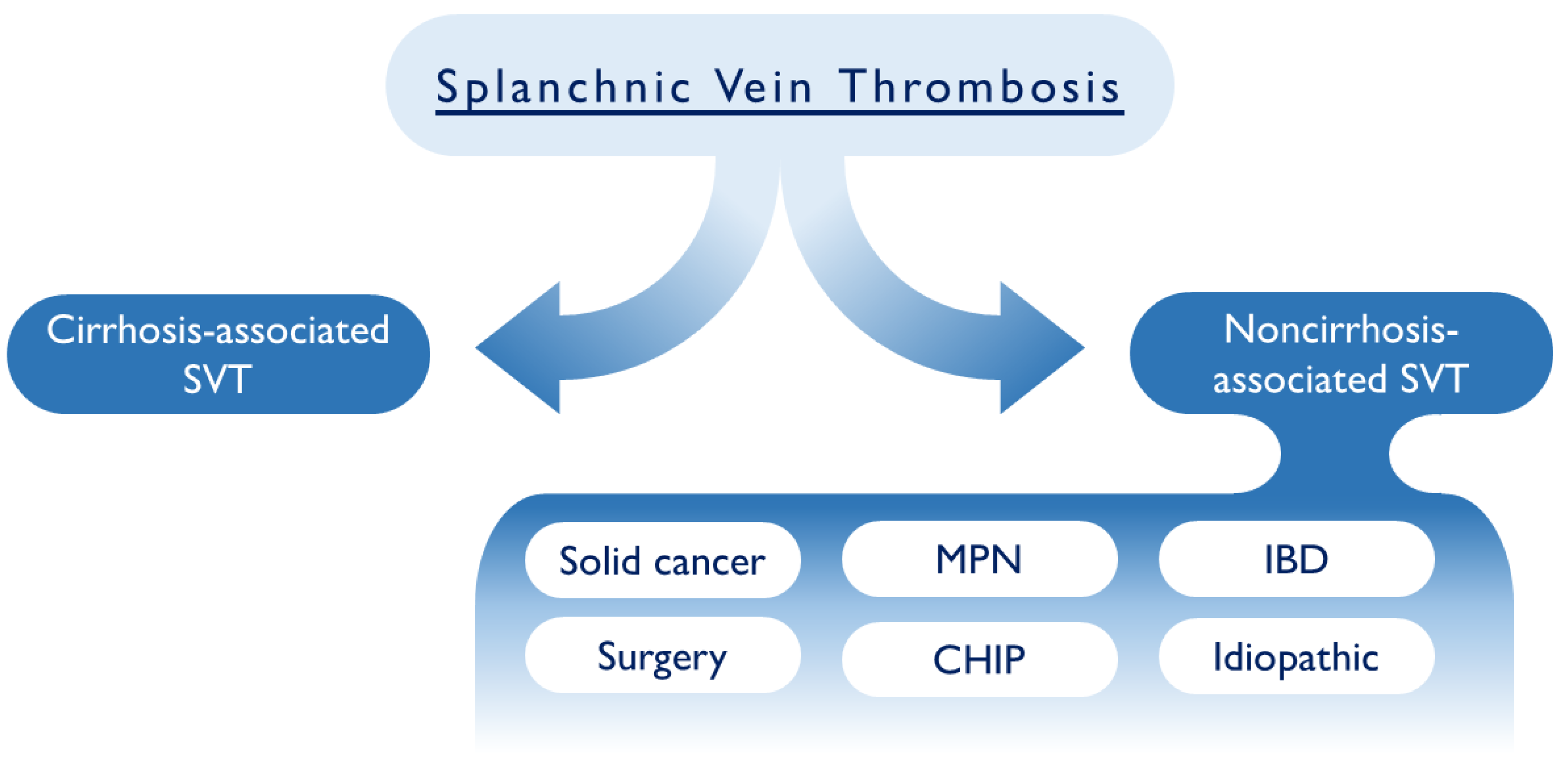
:1. Introduction
2. Results
2.1. Epidemiology
2.2. Risks Factors and Correlations
2.3. Cirrhosis
2.4. Thrombophilia
2.5. Autoimmune Disorders
2.6. Cancer-Associated SVT
2.7. Myeloproliferative Neoplasms- Associated SVT
2.8. COVID-19 Infection
3. Clinical Manifestations of SVT
Clinical Evolution of SVT
4. Diagnostic Considerations
4.1. Imaging Tests for the Diagnosis of SVT
4.2. The Role of Blood d-Dimers Test in the Diagnosis of SVT
5. Therapeutic Implications
5.1. A Role for Direct Oral Anticoagulants (DOACs)
5.2. Cytoreductive Therapy in MPN-Associated SVTs
5.3. New Insights in SVT: A Role for Clonal Hematopoiesis?
6. Conclusions and Future Directions
Author Contributions
Funding
Informed Consent Statement
Data Availability Statement
Acknowledgments
Conflicts of Interest
References
- Tan, R.; Daneshmand, A.; Parys, S.; Watanabe, Y.; Sieunarine, K. Splanchnic Venous Thrombosis: Aetiologies and a Review of the Literature. ANZ J. Surg. 2022, 92, 2224–2228. [Google Scholar] [CrossRef] [PubMed]
- Donadini, M.P.; Dentali, F.; Ageno, W. Splanchnic Vein Thrombosis: New Risk Factors and Management. Thromb. Res. 2012, 129 (Suppl. S1), S93–S96. [Google Scholar] [CrossRef] [PubMed]
- Valeriani, E.; Riva, N.; Di Nisio, M.; Ageno, W. Splanchnic Vein Thrombosis: Current Perspectives. Vasc. Health Risk Manag. 2019, 15, 449–461. [Google Scholar] [CrossRef] [Green Version]
- Ageno, W.; Dentali, F.; Pomero, F.; Fenoglio, L.; Squizzato, A.; Pagani, G.; Re, R.; Bonzini, M. Incidence Rates and Case Fatality Rates of Portal Vein Thrombosis and Budd-Chiari Syndrome. Thromb. Haemost. 2017, 117, 794–800. [Google Scholar] [CrossRef] [PubMed] [Green Version]
- Acosta, S.; Alhadad, A.; Svensson, P.; Ekberg, O. Epidemiology, Risk and Prognostic Factors in Mesenteric Venous Thrombosis. Br. J. Surg. 2008, 95, 1245–1251. [Google Scholar] [CrossRef] [PubMed]
- Rajani, R.; Melin, T.; Björnsson, E.; Broomé, U.; Sangfelt, P.; Danielsson, A.; Gustavsson, A.; Grip, O.; Svensson, H.; Lööf, L.; et al. Budd-Chiari Syndrome in Sweden: Epidemiology, Clinical Characteristics and Survival—An 18-Year Experience. Liver Int. 2009, 29, 253–259. [Google Scholar] [CrossRef]
- Thatipelli, M.R.; McBane, R.D.; Hodge, D.O.; Wysokinski, W.E. Survival and Recurrence in Patients with Splanchnic Vein Thromboses. Clin. Gastroenterol. Hepatol. 2010, 8, 200–205. [Google Scholar] [CrossRef]
- Ageno, W.; Riva, N.; Schulman, S.; Beyer-Westendorf, J.; Bang, S.M.; Senzolo, M.; Grandone, E.; Pasca, S.; Di Minno, M.N.D.; Duce, R.; et al. Long-Term Clinical Outcomes of Splanchnic Vein Thrombosis: Results of an International Registry. JAMA Intern. Med. 2015, 175, 1474–1480. [Google Scholar] [CrossRef]
- Schulman, S. Splanchnic Vein Thrombosis: What Are the Long-Term Risks? Lancet Haematol. 2018, 5, e431–e432. [Google Scholar] [CrossRef]
- Verbeek, T.A.; Stine, J.G.; Saner, F.H.; Bezinover, D. Hypercoagulability in End-Stage Liver Disease: Review of Epidemiology, Etiology, and Management. Transplant. Direct. 2018, 4, e403. [Google Scholar] [CrossRef]
- Ginès, P.; Krag, A.; Abraldes, J.G.; Solà, E.; Fabrellas, N.; Kamath, P.S. Liver Cirrhosis. Lancet 2021, 398, 1359–1376. [Google Scholar] [CrossRef] [PubMed]
- Zhou, W.-C.; Zhang, Q.-B.; Qiao, L. Pathogenesis of Liver Cirrhosis. World J. Gastroenterol. 2014, 20, 7312–7324. [Google Scholar] [CrossRef] [PubMed]
- Ramachandran, P.; Dobie, R.; Wilson-Kanamori, J.R.; Dora, E.F.; Henderson, B.E.P.; Luu, N.T.; Portman, J.R.; Matchett, K.P.; Brice, M.; Marwick, J.A.; et al. Resolving the Fibrotic Niche of Human Liver Cirrhosis at Single-Cell Level. Nature 2019, 575, 512–518. [Google Scholar] [CrossRef] [PubMed]
- Mangia, A.; Villani, M.R.; Cappucci, G.; Santoro, R.; Ricciardi, R.; Facciorusso, D.; Leandro, G.; Caruso, N.; Andriulli, A. Causes of Portal Venous Thrombosis in Cirrhotic Patients: The Role of Genetic and Acquired Factors. Eur. J. Gastroenterol. Hepatol. 2005, 17, 745–751. [Google Scholar] [CrossRef]
- La Mura, V.; Tripodi, A.; Tosetti, G.; Cavallaro, F.; Chantarangkul, V.; Colombo, M.; Primignani, M. Resistance to Thrombomodulin Is Associated with de Novo Portal Vein Thrombosis and Low Survival in Patients with Cirrhosis. Liver Int. 2016, 36, 1322–1330. [Google Scholar] [CrossRef] [PubMed]
- Zocco, M.A.; Di Stasio, E.; De Cristofaro, R.; Novi, M.; Ainora, M.E.; Ponziani, F.; Riccardi, L.; Lancellotti, S.; Santoliquido, A.; Flore, R.; et al. Thrombotic Risk Factors in Patients with Liver Cirrhosis: Correlation with MELD Scoring System and Portal Vein Thrombosis Development. J. Hepatol. 2009, 51, 682–689. [Google Scholar] [CrossRef] [PubMed]
- Rodríguez-Castro, K.I.; Porte, R.J.; Nadal, E.; Germani, G.; Burra, P.; Senzolo, M. Management of Nonneoplastic Portal Vein Thrombosis in the Setting of Liver Transplantation: A Systematic Review. Transplantation 2012, 94, 1145–1153. [Google Scholar] [CrossRef]
- Senzolo, M.; Riva, N.; Dentali, F.; Rodriguez-Castro, K.; Sartori, M.T.; Bang, S.-M.; Martinelli, I.; Schulman, S.; Alatri, A.; Beyer-Westendorf, J.; et al. Long-Term Outcome of Splanchnic Vein Thrombosis in Cirrhosis. Clin. Transl. Gastroenterol. 2018, 9, 176. [Google Scholar] [CrossRef]
- Caiano, L.M.; Riva, N.; Carrier, M.; Gatt, A.; Ageno, W. Treatment of Portal Vein Thrombosis: An Updated Narrative Review. Minerva Med 2021, 112, 713–725. [Google Scholar] [CrossRef]
- Shukla, A.; Giri, S. Portal Vein Thrombosis in Cirrhosis. J. Clin. Exp. Hepatol. 2022, 12, 965–979. [Google Scholar] [CrossRef]
- Galante, A.; De Gottardi, A. Portal Vein Thrombosis: An Overview of Current Treatment Options. Acta Gastroenterol. Belg. 2021, 84, 327–332. [Google Scholar] [CrossRef] [PubMed]
- E, S.; Rd, M.; Aj, T.; K, S.; De, G.; Jp, S.; We, W. Thrombophilia Differences in Splanchnic Vein Thrombosis and Lower Extremity Deep Venous Thrombosis in North America. J. Gastroenterol. 2013, 48, 1111–1118. [Google Scholar] [CrossRef]
- Qi, X.; Ren, W.; De Stefano, V.; Fan, D. Associations of Coagulation Factor V Leiden and Prothrombin G20210A Mutations with Budd-Chiari Syndrome and Portal Vein Thrombosis: A Systematic Review and Meta-Analysis. Clin. Gastroenterol. Hepatol. 2014, 12, 1801–1812.e7. [Google Scholar] [CrossRef] [PubMed]
- Aggarwal, R.; Ravishankar, B.; Misra, R.; Aggarwal, A.; Dwivedi, S.; Naik, S.R. Significance of Elevated IgG Anticardiolipin Antibody Levels in Patients with Budd-Chiari Syndrome. Am. J. Gastroenterol. 1998, 93, 954–957. [Google Scholar] [CrossRef] [PubMed]
- Qi, X.; De Stefano, V.; Su, C.; Bai, M.; Guo, X.; Fan, D. Associations of Antiphospholipid Antibodies with Splanchnic Vein Thrombosis: A Systematic Review with Meta-Analysis. Medicine 2015, 94, e496. [Google Scholar] [CrossRef] [PubMed]
- Tektonidou, M.G.; Andreoli, L.; Limper, M.; Amoura, Z.; Cervera, R.; Costedoat-Chalumeau, N.; Cuadrado, M.J.; Dörner, T.; Ferrer-Oliveras, R.; Hambly, K.; et al. EULAR Recommendations for the Management of Antiphospholipid Syndrome in Adults. Ann. Rheum. Dis. 2019, 78, 1296–1304. [Google Scholar] [CrossRef] [PubMed]
- Bayraktar, Y.; Balkanci, F.; Bayraktar, M.; Calguneri, M. Budd-Chiari Syndrome: A Common Complication of Behçet’s Disease. Am. J. Gastroenterol. 1997, 92, 858–862. [Google Scholar] [PubMed]
- Naymagon, L.; Tremblay, D.; Zubizarreta, N.; Moshier, E.; Naymagon, S.; Mascarenhas, J.; Schiano, T. The Natural History, Treatments, and Outcomes of Portal Vein Thrombosis in Patients With Inflammatory Bowel Disease. Inflamm. Bowel. Dis. 2021, 27, 215–223. [Google Scholar] [CrossRef] [PubMed]
- Di Fabio, F.; Lykoudis, P.; Gordon, P.H. Thromboembolism in Inflammatory Bowel Disease: An Insidious Association Requiring a High Degree of Vigilance. Semin. Thromb. Hemost. 2011, 37, 220–225. [Google Scholar] [CrossRef]
- Prandoni, P.; Lensing, A.W.; Büller, H.R.; Cogo, A.; Prins, M.H.; Cattelan, A.M.; Cuppini, S.; Noventa, F.; ten Cate, J.W. Deep-Vein Thrombosis and the Incidence of Subsequent Symptomatic Cancer. N. Engl. J. Med. 1992, 327, 1128–1133. [Google Scholar] [CrossRef]
- Timp, J.F.; Braekkan, S.K.; Versteeg, H.H.; Cannegieter, S.C. Epidemiology of Cancer-Associated Venous Thrombosis. Blood 2013, 122, 1712–1723. [Google Scholar] [CrossRef] [PubMed] [Green Version]
- Sørensen, H.T.; Mellemkjaer, L.; Olsen, J.H.; Baron, J.A. Prognosis of Cancers Associated with Venous Thromboembolism. N. Engl. J. Med. 2000, 343, 1846–1850. [Google Scholar] [CrossRef] [PubMed]
- Handa, S.; Gupta, K.; Sterpi, M.; Khan, A.; Hoskote, A.; Kasi, A. Trends and In-Hospital Outcomes of Splanchnic Vein Thrombosis Associated with Gastrointestinal Malignancies: A Nationwide Analysis. Gastrointest Tumors 2021, 8, 71–80. [Google Scholar] [CrossRef] [PubMed]
- Søgaard, K.K.; Farkas, D.K.; Pedersen, L.; Sørensen, H.T. Splanchnic Venous Thrombosis Is a Marker of Cancer and a Prognostic Factor for Cancer Survival. Blood 2015, 126, 957–963. [Google Scholar] [CrossRef] [Green Version]
- Afzal, A.; Suhong, L.; Gage, B.F.; Schoen, M.W.; Carson, K.; Thomas, T.; Sanfilippo, K. Splanchnic Vein Thrombosis Predicts Worse Survival in Patients with Advanced Pancreatic Cancer. Thromb. Res. 2020, 185, 125–131. [Google Scholar] [CrossRef] [Green Version]
- Connolly, G.C.; Chen, R.; Hyrien, O.; Mantry, P.; Bozorgzadeh, A.; Abt, P.; Khorana, A.A. Incidence, Risk Factors and Consequences of Portal Vein and Systemic Thromboses in Hepatocellular Carcinoma. Thromb. Res. 2008, 122, 299–306. [Google Scholar] [CrossRef] [PubMed] [Green Version]
- Manzano-Robleda, M.D.C.; Barranco-Fragoso, B.; Uribe, M.; Méndez-Sánchez, N. Portal Vein Thrombosis: What Is New? Ann. Hepatol. 2015, 14, 20–27. [Google Scholar] [CrossRef]
- Tripodi, A.; Mannucci, P.M. The Coagulopathy of Chronic Liver Disease. N. Engl. J. Med. 2011, 365, 147–156. [Google Scholar] [CrossRef] [PubMed] [Green Version]
- Zanetto, A.; Campello, E.; Spiezia, L.; Burra, P.; Simioni, P.; Russo, F.P. Cancer-Associated Thrombosis in Cirrhotic Patients with Hepatocellular Carcinoma. Cancers 2018, 10, 450. [Google Scholar] [CrossRef] [Green Version]
- van der Windt, D.J.; Sud, V.; Zhang, H.; Varley, P.R.; Goswami, J.; Yazdani, H.O.; Tohme, S.; Loughran, P.; O’Doherty, R.M.; Minervini, M.I.; et al. Neutrophil Extracellular Traps Promote Inflammation and Development of Hepatocellular Carcinoma in Nonalcoholic Steatohepatitis. Hepatology 2018, 68, 1347–1360. [Google Scholar] [CrossRef] [Green Version]
- Taleb, R.S.Z.; Moez, P.; Younan, D.; Eisenacher, M.; Tenbusch, M.; Sitek, B.; Bracht, T. Quantitative Proteome Analysis of Plasma Microparticles for the Characterization of HCV-Induced Hepatic Cirrhosis and Hepatocellular Carcinoma. Proteomics Clin. Appl. 2017, 11, 1700014. [Google Scholar] [CrossRef]
- Flood, E.C.; Hajjar, K.A. The Annexin A2 System and Vascular Homeostasis. Vascul. Pharmacol. 2011, 54, 59–67. [Google Scholar] [CrossRef] [PubMed] [Green Version]
- Khorana, A.A.; Fine, R.L. Pancreatic Cancer and Thromboembolic Disease. Lancet Oncol. 2004, 5, 655–663. [Google Scholar] [CrossRef]
- Kang, M.; Suh, K.J.; Kim, J.-W.; Byun, J.M.; Kim, J.W.; Lee, J.Y.; Lee, J.-O.; Bang, S.-M.; Kim, Y.J.; Kim, S.H.; et al. Clinical Characteristics and Disease Course of Splanchnic Vein Thrombosis in Gastrointestinal Cancers: A Prospective Cohort Study. PLoS ONE 2022, 17, e0261671. [Google Scholar] [CrossRef]
- Hultcrantz, M.; Björkholm, M.; Dickman, P.W.; Landgren, O.; Derolf, Å.R.; Kristinsson, S.Y.; Andersson, T.M.L. Risk for Arterial and Venous Thrombosis in Patients With Myeloproliferative Neoplasms: A Population-Based Cohort Study. Ann. Intern. Med. 2018, 168, 317–325. [Google Scholar] [CrossRef] [PubMed]
- Tremblay, D.; Winters, A.; Beckman, J.D.; Naymagon, L.; Patel, R.; Mascarenhas, J.; Schiano, T.D. Splanchnic Vein Thrombosis Associated with Myeloproliferative Neoplasms. Thromb. Res. 2022, 218, 8–16. [Google Scholar] [CrossRef] [PubMed]
- Smalberg, J.H.; Arends, L.R.; Valla, D.C.; Kiladjian, J.-J.; Janssen, H.L.A.; Leebeek, F.W.G. Myeloproliferative Neoplasms in Budd-Chiari Syndrome and Portal Vein Thrombosis: A Meta-Analysis. Blood 2012, 120, 4921–4928. [Google Scholar] [CrossRef] [PubMed]
- How, J.; Trinkaus, K.M.; Oh, S.T. Distinct Clinical, Laboratory and Molecular Features of Myeloproliferative Neoplasm Patients with Splanchnic Vein Thrombosis. Br. J. Haematol. 2018, 183, 310–313. [Google Scholar] [CrossRef] [Green Version]
- De Stefano, V.; Fiorini, A.; Rossi, E.; Za, T.; Farina, G.; Chiusolo, P.; Sica, S.; Leone, G. Incidence of the JAK2 V617F Mutation among Patients with Splanchnic or Cerebral Venous Thrombosis and without Overt Chronic Myeloproliferative Disorders. J. Thromb. Haemost. 2007, 5, 708–714. [Google Scholar] [CrossRef]
- Dentali, F.; Squizzato, A.; Brivio, L.; Appio, L.; Campiotti, L.; Crowther, M.; Grandi, A.M.; Ageno, W. JAK2V617F Mutation for the Early Diagnosis of Ph- Myeloproliferative Neoplasms in Patients with Venous Thromboembolism: A Meta-Analysis. Blood 2009, 113, 5617–5623. [Google Scholar] [CrossRef]
- Magaz, M.; Alvarez-Larrán, A.; Colomer, D.; López-Guerra, M.; García-Criado, M.Á.; Mezzano, G.; Belmonte, E.; Olivas, P.; Soy, G.; Cervantes, F.; et al. Next-Generation Sequencing in the Diagnosis of Non-Cirrhotic Splanchnic Vein Thrombosis. J. Hepatol. 2021, 74, 89–95. [Google Scholar] [CrossRef] [PubMed]
- Wu, Z.; McGoogan, J.M. Characteristics of and Important Lessons From the Coronavirus Disease 2019 (COVID-19) Outbreak in China: Summary of a Report of 72 314 Cases From the Chinese Center for Disease Control and Prevention. JAMA 2020, 323, 1239–1242. [Google Scholar] [CrossRef] [PubMed]
- Kaur, P.; Qaqa, F.; Ramahi, A.; Shamoon, Y.; Singhal, M.; Shamoon, F.; Maroules, M.; Singh, B. Acute Upper Limb Ischemia in a Patient with COVID-19. Hematol. Oncol. Stem Cell Ther. 2021, 14, 348–350. [Google Scholar] [CrossRef] [PubMed]
- Singh, B.; Kaur, P.; Maroules, M. Splanchnic Vein Thrombosis in COVID-19: A Review of Literature. Dig. Liver Dis. 2020, 52, 1407–1409. [Google Scholar] [CrossRef] [PubMed]
- Roncon, L.; Zuin, M.; Barco, S.; Valerio, L.; Zuliani, G.; Zonzin, P.; Konstantinides, S.V. Incidence of Acute Pulmonary Embolism in COVID-19 Patients: Systematic Review and Meta-Analysis. Eur. J. Intern. Med. 2020, 82, 29–37. [Google Scholar] [CrossRef] [PubMed]
- Tripolino, C.; Pizzini, A.M.; Zaccaroni, S.; Cicognani, C.; Dapporto, S.; Cipollini, M.L.; Giannone, C.; Cavoli, C.; Silingardi, M. Is SARS-CoV-2 Infection an Emerging Risk Factor for Splanchnic Venous Thrombosis? Clin. Hemorheol. Microcirc. 2021, 79, 347–355. [Google Scholar] [CrossRef]
- Parry, A.H.; Wani, A.H.; Yaseen, M. Acute Mesenteric Ischemia in Severe Coronavirus-19 (COVID-19): Possible Mechanisms and Diagnostic Pathway. Acad. Radiol. 2020, 27, 1190. [Google Scholar] [CrossRef]
- Moschonas, I.C.; Tselepis, A.D. SARS-CoV-2 Infection and Thrombotic Complications: A Narrative Review. J. Thromb. Thrombolysis 2021, 52, 111–123. [Google Scholar] [CrossRef]
- Jeilani, M.; Hill, R.; Riad, M.; Abdulaal, Y. Superior Mesenteric Vein and Portal Vein Thrombosis in a Patient with COVID-19: A Rare Case. BMJ Case Rep. 2021, 14, e244049. [Google Scholar] [CrossRef]
- Stefely, J.A.; Christensen, B.B.; Gogakos, T.; Cone Sullivan, J.K.; Montgomery, G.G.; Barranco, J.P.; Van Cott, E.M. Marked Factor V Activity Elevation in Severe COVID-19 Is Associated with Venous Thromboembolism. Am. J. Hematol. 2020, 95, 1522–1530. [Google Scholar] [CrossRef]
- Barale, C.; Melchionda, E.; Morotti, A.; Russo, I. Prothrombotic Phenotype in COVID-19: Focus on Platelets. Int. J. Mol. Sci. 2021, 22, 13638. [Google Scholar] [CrossRef] [PubMed]
- Hussein, M.H.; Alabdaljabar, M.S.; Alfagyh, N.; Badran, M.; Alamiri, K. Splanchnic Venous Thrombosis in a Nephrotic Patient Following COVID-19 Infection: A Case Report. BMC Nephrol. 2021, 22, 420. [Google Scholar] [CrossRef] [PubMed]
- Muir, K.-L.; Kallam, A.; Koepsell, S.A.; Gundabolu, K. Thrombotic Thrombocytopenia after Ad26.COV2.S Vaccination. N. Engl. J. Med. 2021, 384, 1964–1965. [Google Scholar] [CrossRef]
- Kawata, E.; Siew, D.-A.; Payne, J.G.; Louzada, M.; Kovacs, M.J.; Lazo-Langner, A. Splanchnic Vein Thrombosis: Clinical Manifestations, Risk Factors, Management, and Outcomes. Thromb. Res. 2021, 202, 90–95. [Google Scholar] [CrossRef]
- De Stefano, V.; Martinelli, I. Splanchnic Vein Thrombosis: Clinical Presentation, Risk Factors and Treatment. Intern. Emerg. Med. 2010, 5, 487–494. [Google Scholar] [CrossRef]
- Wang, J.-T.; Zhao, H.-Y.; Liu, Y.-L. Portal Vein Thrombosis. Hepatobiliary Pancreat. Dis. Int. 2005, 4, 515–518. [Google Scholar]
- Martens, P.; Nevens, F. Budd-Chiari Syndrome. United Eur. Gastroenterol. J. 2015, 3, 489–500. [Google Scholar] [CrossRef]
- Søgaard, K.K.; Darvalics, B.; Horváth-Puhó, E.; Sørensen, H.T. Survival after Splanchnic Vein Thrombosis: A 20-Year Nationwide Cohort Study. Thromb. Res. 2016, 141, 1–7. [Google Scholar] [CrossRef]
- Wells, P.S.; Theberge, I.; Bowdridge, J.; Kelly, E.; Kielar, A.; Forgie, M.A.; John, S.; van Walraven, C. Association of Splanchnic Vein Thrombosis on Survival: 15-Year Institutional Experience With 1561 Cases. J. Am. Heart Assoc. 2020, 9, e016600. [Google Scholar] [CrossRef]
- Søgaard, K.K.; Adelborg, K.; Darvalics, B.; Horváth-Puhó, E.; Beyer-Westendorf, J.; Ageno, W.; Sørensen, H.T. Risk of Bleeding and Arterial Cardiovascular Events in Patients with Splanchnic Vein Thrombosis in Denmark: A Population-Based Cohort Study. Lancet Haematol. 2018, 5, e441–e449. [Google Scholar] [CrossRef]
- Ansell, J. The Subtle Benefit of Anticoagulant Therapy for Splanchnic Vein Thrombosis. JAMA Intern. Med. 2015, 175, 1481–1482. [Google Scholar] [CrossRef]
- Riva, N.; Ageno, W. Clinical Manifestations and Imaging Tools in the Diagnosis of Splanchnic and Cerebral Vein Thromboses. Thromb. Res. 2018, 163, 252–259. [Google Scholar] [CrossRef]
- European Association for the Study of the Liver. EASL Clinical Practice Guidelines: Vascular Diseases of the Liver. J. Hepatol. 2016, 64, 179–202. [Google Scholar] [CrossRef]
- DeLeve, L.D.; Valla, D.-C.; Garcia-Tsao, G. American Association for the Study Liver Diseases Vascular Disorders of the Liver. Hepatology 2009, 49, 1729–1764. [Google Scholar] [CrossRef]
- Weber, S.M.; Rikkers, L.F. Splenic Vein Thrombosis and Gastrointestinal Bleeding in Chronic Pancreatitis. World J. Surg. 2003, 27, 1271–1274. [Google Scholar] [CrossRef] [PubMed]
- Björck, M.; Koelemay, M.; Acosta, S.; Bastos Goncalves, F.; Kölbel, T.; Kolkman, J.J.; Lees, T.; Lefevre, J.H.; Menyhei, G.; Oderich, G.; et al. Editor’s Choice—Management of the Diseases of Mesenteric Arteries and Veins: Clinical Practice Guidelines of the European Society of Vascular Surgery (ESVS). Eur. J. Vasc. Endovasc. Surg. 2017, 53, 460–510. [Google Scholar] [CrossRef] [Green Version]
- Riva, N.; Attard, L.M.; Vella, K.; Squizzato, A.; Gatt, A.; Calleja-Agius, J. Diagnostic Accuracy of D-Dimer in Patients at High-Risk for Splanchnic Vein Thrombosis: A Systematic Review and Meta-Analysis. Thromb. Res. 2021, 207, 102–112. [Google Scholar] [CrossRef]
- Riva, N.; Ageno, W. Cerebral and Splanchnic Vein Thrombosis: Advances, Challenges, and Unanswered Questions. J. Clin. Med. 2020, 9, 743. [Google Scholar] [CrossRef] [PubMed] [Green Version]
- Dai, J.; Qi, X.; Peng, Y.; Hou, Y.; Chen, J.; Li, H.; Guo, X. Association between D-Dimer Level and Portal Venous System Thrombosis in Liver Cirrhosis: A Retrospective Observational Study. Int. J. Clin. Exp. Med. 2015, 8, 15296–15301. [Google Scholar]
- Ageno, W.; Dentali, F.; Squizzato, A. How I Treat Splanchnic Vein Thrombosis. Blood 2014, 124, 3685–3691. [Google Scholar] [CrossRef] [Green Version]
- Valeriani, E.; Di Nisio, M.; Riva, N.; Cohen, O.; Garcia-Pagan, J.-C.; Magaz, M.; Porreca, E.; Ageno, W. Anticoagulant Therapy for Splanchnic Vein Thrombosis: A Systematic Review and Meta-Analysis. Blood 2021, 137, 1233–1240. [Google Scholar] [CrossRef] [PubMed]
- Kearon, C.; Akl, E.A.; Comerota, A.J.; Prandoni, P.; Bounameaux, H.; Goldhaber, S.Z.; Nelson, M.E.; Wells, P.S.; Gould, M.K.; Dentali, F.; et al. Antithrombotic Therapy for VTE Disease: Antithrombotic Therapy and Prevention of Thrombosis, 9th Ed: American College of Chest Physicians Evidence-Based Clinical Practice Guidelines. Chest 2012, 141, e419S–e496S. [Google Scholar] [CrossRef] [Green Version]
- Tufano, A.; Ageno, W.; Di Micco, P.; Niglio, A.; Rosa, V.; Ballaz, A.; Braester, A.; Rubio, C.M.; Isern, V.; Imbalzano, E.; et al. Outcomes during Anticoagulation in Patients with Symptomatic vs. Incidental Splanchnic Vein Thrombosis. Thromb. Res. 2018, 164, 69–74. [Google Scholar] [CrossRef] [PubMed]
- Condat, B.; Pessione, F.; Hillaire, S.; Denninger, M.H.; Guillin, M.C.; Poliquin, M.; Hadengue, A.; Erlinger, S.; Valla, D. Current Outcome of Portal Vein Thrombosis in Adults: Risk and Benefit of Anticoagulant Therapy. Gastroenterology 2001, 120, 490–497. [Google Scholar] [CrossRef] [PubMed] [Green Version]
- Di Nisio, M.; Valeriani, E.; Riva, N.; Schulman, S.; Beyer-Westendorf, J.; Ageno, W. Anticoagulant Therapy for Splanchnic Vein Thrombosis: ISTH SSC Subcommittee Control of Anticoagulation. J. Thromb. Haemost. 2020, 18, 1562–1568. [Google Scholar] [CrossRef] [Green Version]
- Cheng, D.-L.; Xu, H.; Li, C.-L.; Lv, W.-F.; Li, C.-T.; Mukhiya, G.; Fang, W.-W. Interventional Treatment Strategy for Primary Budd-Chiari Syndrome with Both Inferior Vena Cava and Hepatic Vein Involvement: Patients from Two Centers in China. Cardiovasc. Interv. Radiol. 2019, 42, 1311–1321. [Google Scholar] [CrossRef]
- Mathew, C.; Zumberg, M. Evidence-Based Minireview: Should Warfarin or a Direct Oral Anticoagulant Be Used in Patients Presenting with Thrombosis in the Splanchnic or Cerebral Veins? Hematology Am. Soc. Hematol. Educ. Program. 2021, 2021, 100–105. [Google Scholar] [CrossRef]
- Einstein Investigators. Oral Rivaroxaban for Symptomatic Venous Thromboembolism. N. Engl. J. Med. 2010, 363, 2499–2510. [Google Scholar] [CrossRef] [Green Version]
- Einstein–PE Investigators. Oral Rivaroxaban for the Treatment of Symptomatic Pulmonary Embolism. N. Engl. J. Med. 2012, 366, 1287–1297. [Google Scholar] [CrossRef] [Green Version]
- Agnelli, G.; Buller, H.R.; Cohen, A.; Curto, M.; Gallus, A.S.; Johnson, M.; Masiukiewicz, U.; Pak, R.; Thompson, J.; Raskob, G.E.; et al. Oral Apixaban for the Treatment of Acute Venous Thromboembolism. N. Engl. J. Med. 2013, 369, 799–808. [Google Scholar] [CrossRef] [Green Version]
- Naymagon, L.; Tremblay, D.; Zubizarreta, N.; Moshier, E.; Troy, K.; Schiano, T.; Mascarenhas, J. The Efficacy and Safety of Direct Oral Anticoagulants in Noncirrhotic Portal Vein Thrombosis. Blood Adv. 2020, 4, 655–666. [Google Scholar] [CrossRef] [Green Version]
- Ageno, W.; Beyer Westendorf, J.; Contino, L.; Bucherini, E.; Sartori, M.T.; Senzolo, M.; Grandone, E.; Santoro, R.; Carrier, M.; Delluc, A.; et al. Rivaroxaban for the Treatment of Noncirrhotic Splanchnic Vein Thrombosis: An Interventional Prospective Cohort Study. Blood Adv. 2022, 6, 3569–3578. [Google Scholar] [CrossRef]
- Serrao, A.; Merli, M.; Lucani, B.; Aprile, F.; Fiori, L.; Gioia, S.; Breccia, M.; Riggio, O.; Chistolini, A. Outcomes of Long-Term Anticoagulant Treatment for the Secondary Prophylaxis of Splanchnic Venous Thrombosis. Eur. J. Clin. Investig. 2021, 51, e13356. [Google Scholar] [CrossRef]
- Barbui, T.; Tefferi, A.; Vannucchi, A.M.; Passamonti, F.; Silver, R.T.; Hoffman, R.; Verstovsek, S.; Mesa, R.; Kiladjian, J.-J.; Hehlmann, R.; et al. Philadelphia Chromosome-Negative Classical Myeloproliferative Neoplasms: Revised Management Recommendations from European LeukemiaNet. Leukemia 2018, 32, 1057–1069. [Google Scholar] [CrossRef] [Green Version]
- De Stefano, V.; Rossi, E.; Carobbio, A.; Ghirardi, A.; Betti, S.; Finazzi, G.; Vannucchi, A.M.; Barbui, T. Hydroxyurea Prevents Arterial and Late Venous Thrombotic Recurrences in Patients with Myeloproliferative Neoplasms but Fails in the Splanchnic Venous District. Pooled Analysis of 1500 Cases. Blood Cancer J. 2018, 8, 112. [Google Scholar] [CrossRef] [Green Version]
- Carrà, G.; Giugliano, E.; Camerlo, S.; Rosati, G.; Branca, E.; Maffeo, B.; Russo, I.; Piazza, R.; Cilloni, D.; Morotti, A. Clonal Hematopoiesis by DNMT3A Mutations as a Common Finding in Idiopathic Splanchnic Vein Thrombosis. Haematologica 2022. [Google Scholar] [CrossRef]
- Marnell, C.S.; Bick, A.; Natarajan, P. Clonal Hematopoiesis of Indeterminate Potential (CHIP): Linking Somatic Mutations, Hematopoiesis, Chronic Inflammation and Cardiovascular Disease. J. Mol. Cell Cardiol. 2021, 161, 98–105. [Google Scholar] [CrossRef] [PubMed]
- Jaiswal, S.; Fontanillas, P.; Flannick, J.; Manning, A.; Grauman, P.V.; Mar, B.G.; Lindsley, R.C.; Mermel, C.H.; Burtt, N.; Chavez, A.; et al. Age-Related Clonal Hematopoiesis Associated with Adverse Outcomes. N. Engl. J. Med. 2014, 371, 2488–2498. [Google Scholar] [CrossRef] [PubMed] [Green Version]
- Jaiswal, S.; Natarajan, P.; Silver, A.J.; Gibson, C.J.; Bick, A.G.; Shvartz, E.; McConkey, M.; Gupta, N.; Gabriel, S.; Ardissino, D.; et al. Clonal Hematopoiesis and Risk of Atherosclerotic Cardiovascular Disease. N. Engl. J. Med. 2017, 377, 111–121. [Google Scholar] [CrossRef]
- Soudet, S.; Jedraszak, G.; Evrard, O.; Marolleau, J.P.; Garcon, L.; Pietri, M.A.S. Is Hematopoietic Clonality of Indetermined Potential a Risk Factor for Pulmonary Embolism? TH Open 2021, 5, e338–e342. [Google Scholar] [CrossRef] [PubMed]
- Segura-Díaz, A.; Stuckey, R.; Florido, Y.; González-Martín, J.M.; López-Rodríguez, J.F.; Sánchez-Sosa, S.; González-Pérez, E.; Sáez Perdomo, M.N.; Perera, M.D.M.; de la Iglesia, S.; et al. Thrombotic Risk Detection in Patients with Polycythemia Vera: The Predictive Role of DNMT3A/TET2/ASXL1 Mutations. Cancers 2020, 12, 934. [Google Scholar] [CrossRef] [PubMed]

Disclaimer/Publisher’s Note: The statements, opinions and data contained in all publications are solely those of the individual author(s) and contributor(s) and not of MDPI and/or the editor(s). MDPI and/or the editor(s) disclaim responsibility for any injury to people or property resulting from any ideas, methods, instructions or products referred to in the content. |
© 2023 by the authors. Licensee MDPI, Basel, Switzerland. This article is an open access article distributed under the terms and conditions of the Creative Commons Attribution (CC BY) license (https://creativecommons.org/licenses/by/4.0/).
Share and Cite
Camerlo, S.; Ligato, J.; Rosati, G.; Carrà, G.; Russo, I.; De Gobbi, M.; Morotti, A. Shedding Light on the Pathogenesis of Splanchnic Vein Thrombosis. Int. J. Mol. Sci. 2023, 24, 2262. https://doi.org/10.3390/ijms24032262
Camerlo S, Ligato J, Rosati G, Carrà G, Russo I, De Gobbi M, Morotti A. Shedding Light on the Pathogenesis of Splanchnic Vein Thrombosis. International Journal of Molecular Sciences. 2023; 24(3):2262. https://doi.org/10.3390/ijms24032262
Chicago/Turabian StyleCamerlo, Sofia, Jacopo Ligato, Giorgio Rosati, Giovanna Carrà, Isabella Russo, Marco De Gobbi, and Alessandro Morotti. 2023. "Shedding Light on the Pathogenesis of Splanchnic Vein Thrombosis" International Journal of Molecular Sciences 24, no. 3: 2262. https://doi.org/10.3390/ijms24032262