
Tailored Supramolecular Cage for Efficient Bio-Labeling
 and
and 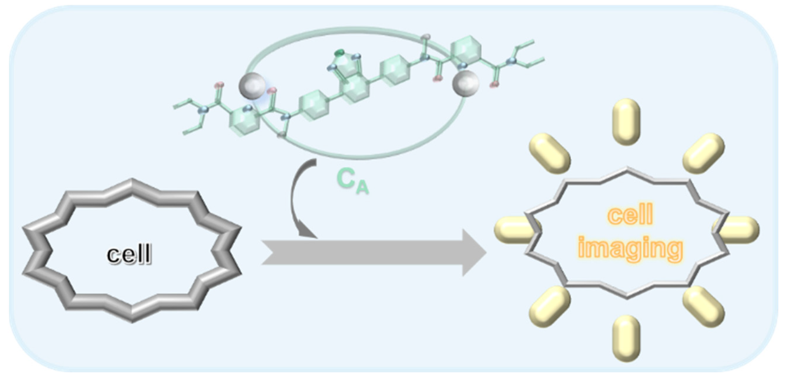
Abstract
:1. Introduction
2. Results and Discussion
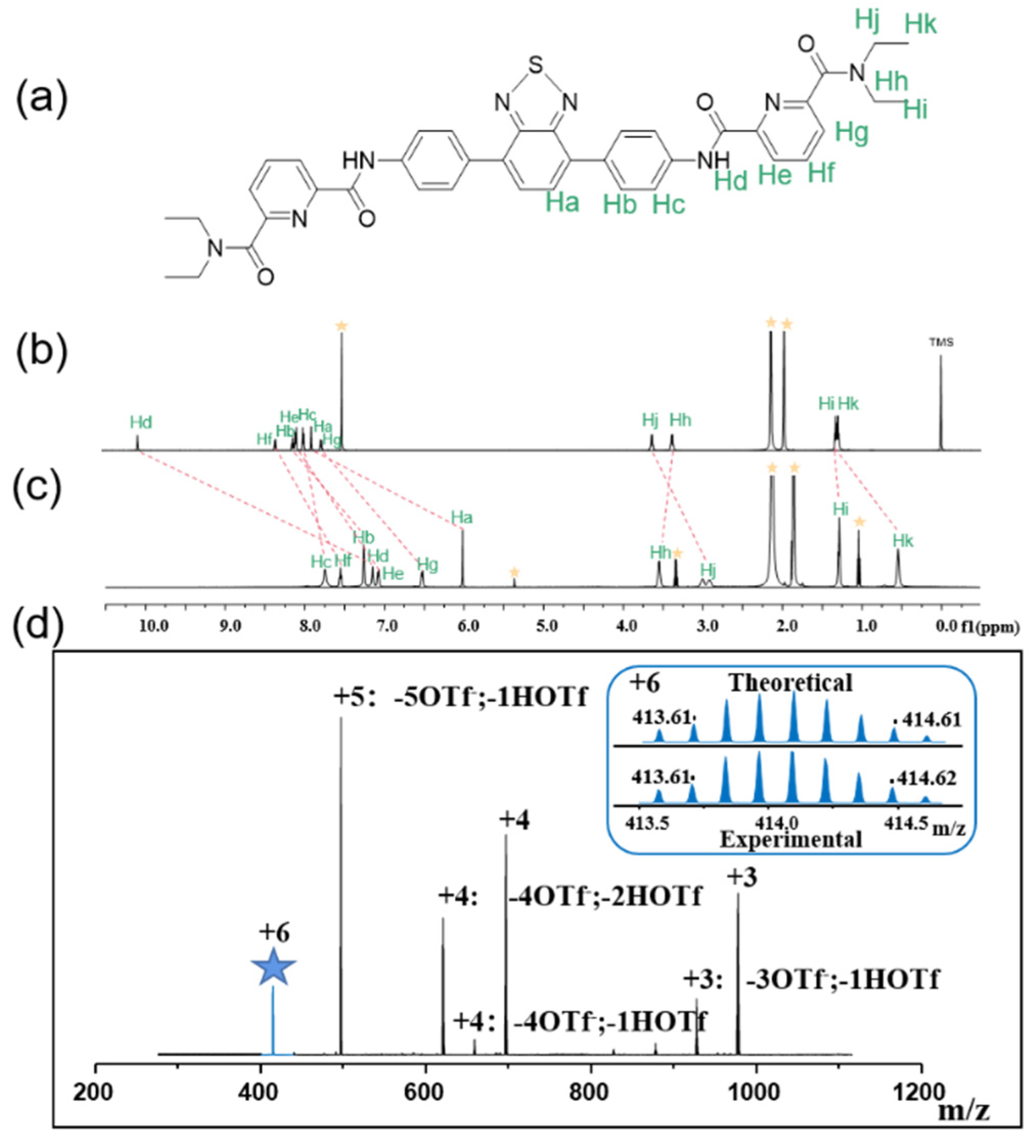
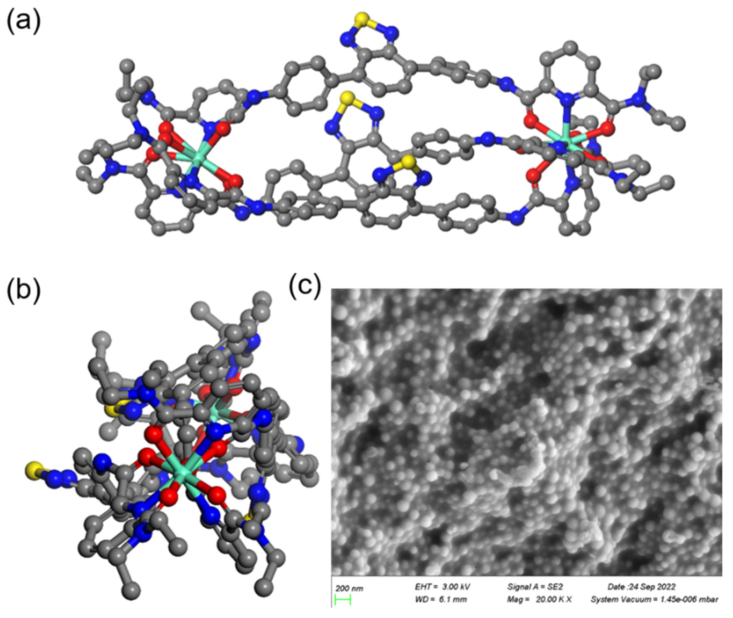
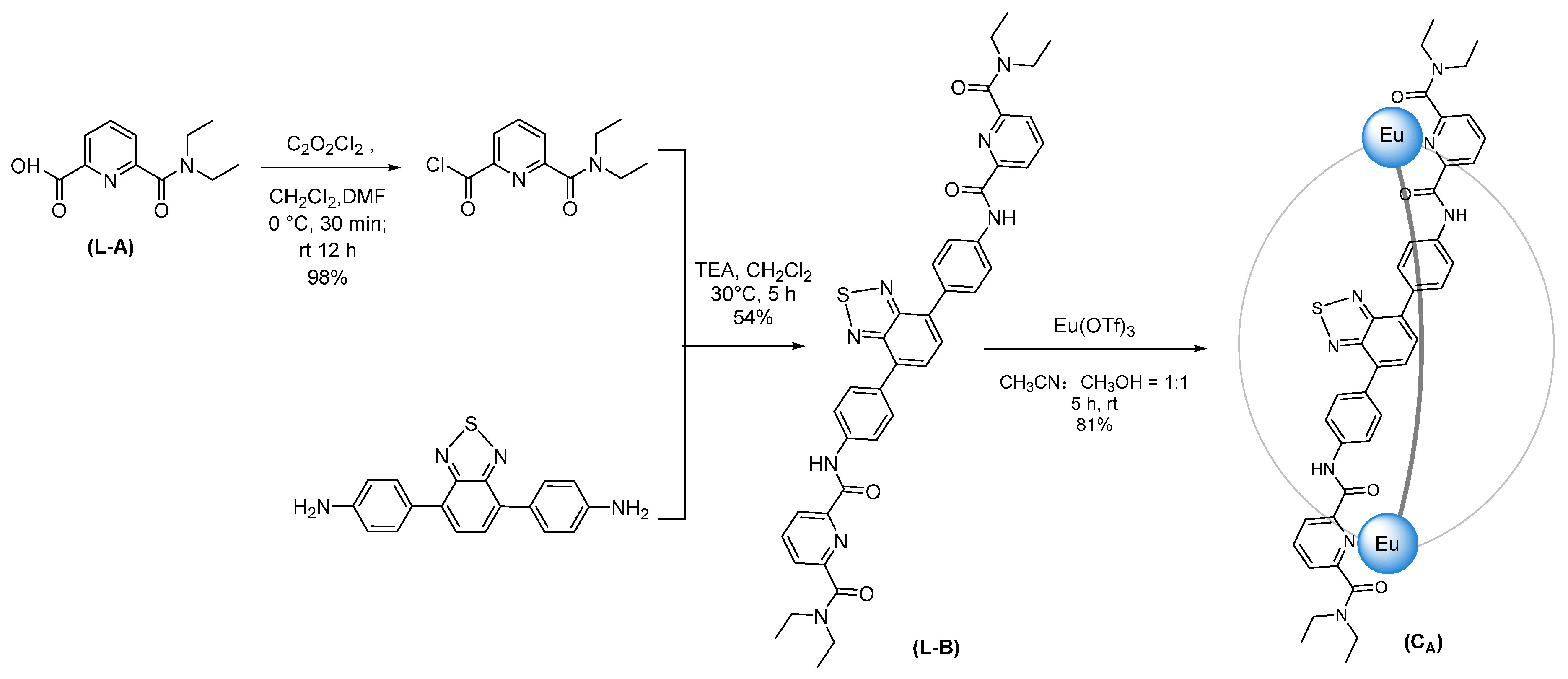
2.1. The Preparation and Characterization of CA
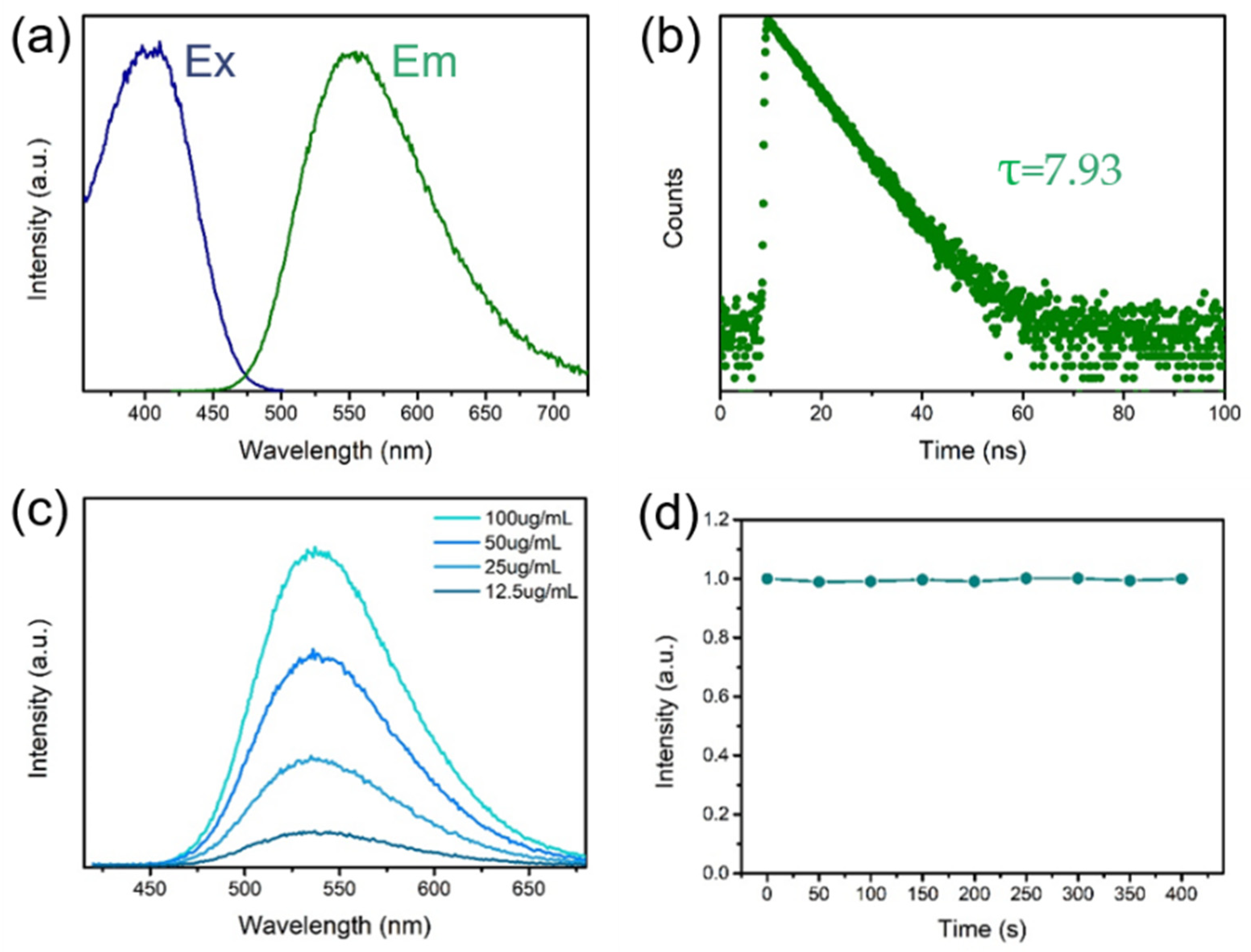
2.2. Optical Properties
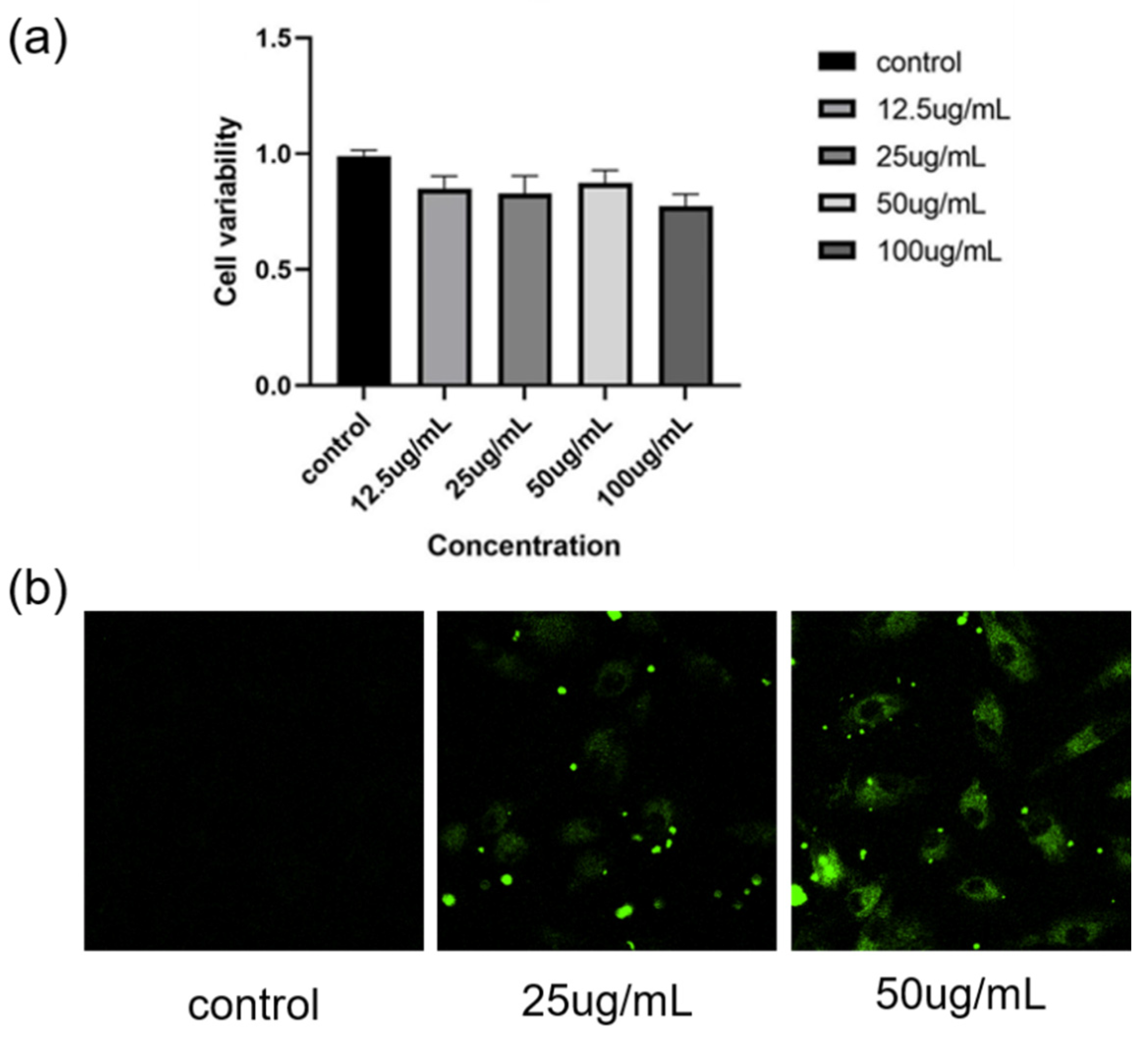
2.3. In Vitro Cytotoxicity Profiles and CLSM Images
3. Materials and Methods
3.1. Materials
3.2. Measurements
3.3. Materials Synthesis
3.3.1. Preparation of 2-Ethoxycarbonyl-carboxypyridine
3.3.2. Preparation of L-A
3.3.3. Preparation of L-B
3.3.4. Preparation of CA
3.4. Fluorescence Lifetime
3.5. Fluorescence Quantum Yield
3.6. Cell Variability Assay
3.7. Live Cell Imaging
4. Conclusions
Supplementary Materials
Author Contributions
Funding
Institutional Review Board Statement
Informed Consent Statement
Data Availability Statement
Conflicts of Interest
References
- Cook, T.R.; Stang, P.J. Recent Developments in the Preparation and Chemistry of Metallacycles and Metallacages via Coordination. Chem. Rev. 2015, 115, 7001–7045. [Google Scholar] [CrossRef] [PubMed]
- Whitesides, M.G.; Grzybowsk, B. Self-Assembly at All Scales. Science 2002, 295, 2418–2421. [Google Scholar] [CrossRef] [PubMed] [Green Version]
- Leininger, S.; Olenyuk, B.; Stang, P.J. Self-Assembly of Discrete Cyclic Nanostructures Mediated by Transition Metals. Chem. Rev. 2000, 100, 853–908. [Google Scholar] [CrossRef] [PubMed]
- Chakrabarty, R.; Mukherjee, P.S.; Stang, P.J. Supramolecular Coordination: Self-Assembly of Finite Two- and Three-Dimensional Ensembles. Chem. Rev. 2011, 111, 6810–6918. [Google Scholar] [CrossRef] [Green Version]
- Cakmak, Y.; Erbas-Cakmak, S.; Leigh, D.A. Asymmetric Catalysis with a Mechanically Point-Chiral Rotaxane. J. Am. Chem. Soc. 2016, 138, 1749–1751. [Google Scholar] [CrossRef] [Green Version]
- Cullen, W.; Takezawa, H.; Fujita, M. Demethylenation of Cyclopropanes via Photoinduced Guest-to-Host Electron Transfer in an M6L4 Cage. Angew. Chem. Int. Ed. 2019, 131, 9269–9271. [Google Scholar] [CrossRef]
- Cullen, W.; Misuraca, M.C.; Hunter, C.A.; Williams, N.H.; Ward, M.D. Highly Efficient Catalysis of the Kemp Elimination in the Cavity of a Cubic Coordination Cage. Nat. Chem. 2016, 8, 231–236. [Google Scholar] [CrossRef]
- Zhang, L.; Wei, Z.; Thanneeru, S.; Meng, M.; Kruzyk, M.; Ung, G.; Liu, B.; He, J. A Polymer Solution To Prevent Nanoclustering and Improve the Selectivity of Metal Nanoparticles for Electrocatalytic CO2 Reduction. Angew. Chem. Int. Ed. 2019, 131, 15981–15987. [Google Scholar] [CrossRef]
- Oldacre, A.N.; Friedman, A.E.; Cook, T.R. A Self-Assembled Cofacial Cobalt Porphyrin Prism for Oxygen Reduction Catalysis. J. Am. Chem. Soc. 2017, 139, 1424–1427. [Google Scholar] [CrossRef]
- Holloway, L.R.; Bogie, P.M.; Lyon, Y.; Ngai, C.; Miller, T.F.; Julian, R.R.; Hooley, R.J. Tandem Reactivity of a Self-Assembled Cage Catalyst with Endohedral Acid Groups. J. Am. Chem. Soc. 2018, 140, 8078–8081. [Google Scholar] [CrossRef]
- Lei, Y.; Chen, Q.; Liu, P.; Wang, L.; Wang, H.; Li, B.; Lu, X.; Chen, Z.; Pan, Y.; Huang, F.; et al. Molecular Cages Self-Assembled by Imine Condensation in Water. Angew. Chem. Int. Ed. 2021, 133, 4755–4761. [Google Scholar] [CrossRef]
- Burke, M.J.; Nichol, G.S.; Lusby, P.J. Orthogonal Selection and Fixing of Coordination Self-Assembly Pathways for Robust Metallo-organic Ensemble Construction. J. Am. Chem. Soc. 2016, 138, 9308–9315. [Google Scholar] [CrossRef] [PubMed] [Green Version]
- Jansze, S.M.; Cecot, G.; Severin, K. Reversible Disassembly of Metallasupramolecular Structures Mediated by A Metastable-state Photoacid. Chem. Sci. 2018, 9, 4253–4257. [Google Scholar] [CrossRef] [PubMed] [Green Version]
- Oldknow, S.; Martir, D.R.; Pritchard, V.E.; Blitz, M.A.C.; Zysman-Colman, E.; Hardie, M.J. Structure-switching M3L2 Ir(iii) Coordination Cages with Photo-isomerising Azo-aromatic Linkers. Chem. Sci. 2018, 9, 8150–8159. [Google Scholar] [CrossRef] [PubMed] [Green Version]
- Samanta, D.; Mukherjee, P.S. Sunlight-Induced Covalent Marriage of Two Triply Interlocked Pd6 Cages and Their Facile Thermal Separation. J. Am. Chem. Soc. 2014, 136, 17006–17009. [Google Scholar] [CrossRef]
- Kurihara, K.; Yazaki, K.; Akita, M.; Yoshizawa, M. A Switchable Open/closed Polyaromatic Macrocycle that Shows Reversible Binding of Long Hydrophilic Molecules. Angew. Chem. Int. Ed. 2017, 129, 11518–11522. [Google Scholar] [CrossRef]
- Regeni, I.; Chen, B.; Frank, M.; Baksi, A.; Holstein, J.J.; Clever, G.H. Teerfarben-basierte Koordinationskäfige und-helikate. Angew. Chem. Int. Ed. 2021, 133, 5736–5741. [Google Scholar] [CrossRef]
- Yang, G.; Zheng, W.; Tao, G.; Wu, L.; Zhou, Q.-F.; Kochovski, Z.; Ji, T.; Chen, H.; Li, X.; Lu, Y.; et al. Diversiform and Transformable Glyco-Nanostructures Constructed from Amphiphilic Supramolecular Metallocarbohydrates through Hierarchical Self-Assembly: The Balance between Metallacycles and Saccharides. ACS Nano 2019, 13, 13474–13485. [Google Scholar] [CrossRef]
- Wiester, M.J.; Ulmann, P.A.; Mirkin, C.A. Enzymnachbildungen Auf Der Basis Supramolekularer Koordinationschemie. Angew. Chem. Int. Ed. 2011, 123, 118–142. [Google Scholar] [CrossRef]
- Clever, P.G.H. Imidazole-modified G-quadruplex DNA as Metal-triggered Peroxidase. Chem. Sci. 2019, 10, 2513–2518. [Google Scholar] [CrossRef]
- Zamora-Olivares, D.; Kaoud, T.S.; Dalby, K.N.; Anslyn, E.V. In-Situ Generation of Differential Sensors that Fingerprint Kinases and the Cellular Response to Their Expression. J. Am. Chem. Soc. 2013, 135, 14814–14820. [Google Scholar] [CrossRef] [PubMed] [Green Version]
- Ulrich, S.; Dumy, P. Probing Secondary Interactions in Biomolecular Recognition by Dynamic Combinatorial Chemistry. Chem. Commun. 2014, 50, 5810. [Google Scholar] [CrossRef] [PubMed]
- Howlader, P.; Mondal, S.; Ahmed, S.; Mukherjee, P.S. Guest-Induced Enantioselective Self-Assembly of a Pd6 Homochiral Octahedral Cage with a C3-Symmetric Pyridyl Donor. J. Am. Chem. Soc. 2020, 142, 20968–20972. [Google Scholar] [CrossRef] [PubMed]
- Wu, K.; Zhang, B.; Drechsler, C.; Holstein, J.J.; Clever, G.H. Rückgrat-verknüpfte Liganden Erhöhen Die Vielfalt in Heteroleptischen Koordinationskäfigen. Angew. Chem. Int. Ed. 2021, 133, 6473–6478. [Google Scholar] [CrossRef]
- García-Simón, C.; Garcia-Borràs, M.; Gómez, L.; Parella, T.; Osuna, S.; Juanhuix, J.; Imaz, I.; Maspoch, D.; Costas, M.; Ribas, X. Sponge-like Molecular Cage for Purification of Fullerenes. Nat. Commun. 2014, 5, 5557. [Google Scholar] [CrossRef] [Green Version]
- Sawada, T.; Fujita, M. A Single Watson-Crick G·C Base Pair in Water: Aqueous Hydrogen Bonds in Hydrophobic Cavities. J. Am. Chem. Soc. 2010, 132, 7194–7201. [Google Scholar] [CrossRef]
- Samanta, S.K.; Quigley, J.; Vinciguerra, B.; Briken, V.; Isaacs, L. Cucurbit[7]uril Enables Multi-Stimuli-Responsive Release from the Self-Assembled Hydrophobic Phase of a Metal Organic Polyhedron. J. Am. Chem. Soc. 2017, 139, 9066–9074. [Google Scholar] [CrossRef]
- Zheng, Y.-R.; Suntharalingam, K.; Johnstone, T.C.; Lippard, S.J. Encapsulation of Pt(iv) Prodrugs within a Pt(ii) Cage for Drug Delivery. Chem. Sci. 2015, 6, 1189–1193. [Google Scholar] [CrossRef] [Green Version]
- Samanta, S.K.; Moncelet, D.; Briken, V.; Isaacs, L. Metal-Organic Polyhedron Capped with Cucurbit[8]uril Delivers Doxorubicin to Cancer Cells. J. Am. Chem. Soc. 2016, 138, 14488–14496. [Google Scholar] [CrossRef] [Green Version]
- Haynes, C.J.E.; Zhu, J.; Chimerel, C.; Hernández-Ainsa, S.; Riddell, I.A.; Ronson, T.K.; Keyser, U.F.; Nitschke, J.R. Blockable Zn10L15 Ion Channels through Subcomponent Self-Assembly. Angew. Chem. Int. Ed. 2017, 129, 15590–15594. [Google Scholar] [CrossRef]
- Kawano, R.; Horike, N.; Hijikata, Y.; Kondo, M.; Carné-Sánchez, A.; Larpent, P.; Ikemura, S.; Osaki, T.; Kamiya, K.; Kitagawa, S.; et al. Metal-Organic Cuboctahedra for Synthetic Ion Channels with Multiple Conductance States. Chem 2017, 2, 393–403. [Google Scholar] [CrossRef] [Green Version]
- Zhou, J.; Li, J.; Du, X.; Xu, B. Supramolecular Biofunctional Materials. Biomaterials 2017, 129, 1–27. [Google Scholar] [CrossRef] [PubMed] [Green Version]
- Xie, B.; Ding, Y.-F.; Shui, M.; Yue, L.; Gao, C.; Wyman, I.W.; Wang, R. Supramolecular Biomaterials for Bio-imaging and Imaging-guided Therapy. Eur. J. Nucl. Med. Mol. Imaging 2022, 49, 1200–1210. [Google Scholar] [CrossRef] [PubMed]
- Bünzli, J.-C.G.; Piguet, C. Lanthanide-Containing Molecular and Supramolecular Polymetallic Functional Assemblies. Chem. Rev. 2002, 102, 1897–1928. [Google Scholar] [CrossRef] [Green Version]
- Barry, D.E.; Caffrey, D.F.; Gunnlaugsson, T. Lanthanide-directed Synthesis of Luminescent Self-assembly Supramolecular Structures and Mechanically Bonded Systems from Acyclic Coordinating Organic Ligands. Chem. Soc. Rev. 2016, 45, 3244–3274. [Google Scholar] [CrossRef]
- Yeung, C.-T.; Yim, K.-H.; Wong, H.-Y.; Pal, R.; Lo, W.-S.; Yan, S.-C.; Yee-Man Wong, M.; Yufit, D.; Smiles, D.E.; McCormick, L.J.; et al. Chiral transcription in Self-assembled Tetrahedral Eu4L6 Chiral Cages Displaying Sizable Circularly Polarized Luminescence. Nat. Commun. 2017, 8, 1128. [Google Scholar] [CrossRef] [Green Version]
- Albrecht, M.; Osetska, O.; Fröhlich, R.; Bünzli, J.-C.G.; Aebischer, A.; Gumy, F.; Hamacek, J. Highly Efficient Near-IR Emitting Yb/Yb and Yb/Al Helicates. J. Am. Chem. Soc. 2007, 129, 14178–14179. [Google Scholar] [CrossRef]
- Zhou, Y.; Li, H.; Zhu, T.; Gao, T.; Yan, P. A Highly Luminescent Chiral Tetrahedral Eu4L4 (L’)4 Cage: Chirality Induction, Chirality Memory, and Circularly Polarized Luminescence. J. Am. Chem. Soc. 2019, 141, 19634–19643. [Google Scholar] [CrossRef]
- Yang, X.; Lin, X.; Zhao, Y.; Zhao, Y.S.; Yan, D. Lanthanide Metal-Organic Framework Microrods: Colored Optical Waveguides and Chiral Polarized Emission. Angew. Chem. Int. Ed. 2017, 56, 7853–7857. [Google Scholar] [CrossRef]
- Liu, C.-L.; Zhang, R.-L.; Lin, C.-S.; Zhou, L.-P.; Cai, L.-X.; Kong, J.-T.; Yang, S.-Q.; Han, K.-L.; Sun, Q.-F. Intraligand Charge Transfer Sensitization on Self-Assembled Europium Tetrahedral Cage Leads to Dual-Selective Luminescent Sensing toward Anion and Cation. J. Am. Chem. Soc. 2017, 139, 12474–12479. [Google Scholar] [CrossRef]
- dos Santos, C.M.G.; Harte, A.J.; Quinn, S.J.; Gunnlaugsson, T. Recent Developments in the Field of Supramolecular Lanthanide Luminescent Sensors and Self-assemblies. Nat. Commun. 2008, 252, 2512–2527. [Google Scholar] [CrossRef]
- Hess, B.A.; Kȩdziorski, A.; Smentek, L.; Bornhop, D.J. Role of the Antenna in Tissue Selective Probes Built of Lanthanide-Organic Chelates. J. Phys. Chem. A 2008, 112, 2397–2407. [Google Scholar] [CrossRef] [PubMed]
- Song, B.; Sivagnanam, V.; Vandevyver, C.D.B.; Hemmilä, I.; Lehr, H.-A.; Gijs, M.A.M.; Bünzli, J.-C.G. Time-resolved Lanthanide Luminescence for Lab-on-a-chip Detection of Biomarkers on Cancerous Tissues. Analyst 2009, 134, 1991. [Google Scholar] [CrossRef] [PubMed]
- Kaczmarek, M.T.; Zabiszak, M.; Nowak, M.; Jastrzab, R. Lanthanides: Schiff base Complexes, Applications in Cancer Diagnosis, Therapy, and Antibacterial Activity. Coord. Chem. Rev. 2018, 370, 42–54. [Google Scholar] [CrossRef]
- Caulder, D.L.; Brückner, C.; Powers, R.E.; König, S.; Parac, T.N.; Leary, J.A.; Raymond, K.N. Design, Formation and Properties of Tetrahedral M4L4 and M4L6 Supramolecular Clusters1. J. Am. Chem. Soc. 2001, 123, 8923–8938. [Google Scholar] [CrossRef] [PubMed]
- Dalla Favera, N.; Guénée, L.; Bernardinelli, G.; Piguet, C. In Search for Tuneable Intramolecular Intermetallic Interactions in Polynuclear Lanthanide Complexes. Dalton Trans. 2009, 7625–7638. [Google Scholar] [CrossRef] [PubMed]
- Garin, A.B.; Rakarić, D.; Andrić, E.K.; Kosanović, M.M.; Balić, T.; Perdih, F. Synthesis of Monosubstituted Dipicolinic Acid Hydrazide Derivative and Structural Characterization of Novel Co(III) and Cr(III) Complexes. Polyhedron 2019, 166, 226–232. [Google Scholar] [CrossRef]


Disclaimer/Publisher’s Note: The statements, opinions and data contained in all publications are solely those of the individual author(s) and contributor(s) and not of MDPI and/or the editor(s). MDPI and/or the editor(s) disclaim responsibility for any injury to people or property resulting from any ideas, methods, instructions or products referred to in the content. |
© 2023 by the authors. Licensee MDPI, Basel, Switzerland. This article is an open access article distributed under the terms and conditions of the Creative Commons Attribution (CC BY) license (https://creativecommons.org/licenses/by/4.0/).
Share and Cite
An, D.; Shi, L.; Li, T.; Zhang, H.-Y.; Chen, Y.; Hao, X.-Q.; Song, M.-P. Tailored Supramolecular Cage for Efficient Bio-Labeling. Int. J. Mol. Sci. 2023, 24, 2147. https://doi.org/10.3390/ijms24032147
An D, Shi L, Li T, Zhang H-Y, Chen Y, Hao X-Q, Song M-P. Tailored Supramolecular Cage for Efficient Bio-Labeling. International Journal of Molecular Sciences. 2023; 24(3):2147. https://doi.org/10.3390/ijms24032147
Chicago/Turabian StyleAn, Dongdong, Linlin Shi, Tianyu Li, Hong-Yu Zhang, Yahong Chen, Xin-Qi Hao, and Mao-Ping Song. 2023. "Tailored Supramolecular Cage for Efficient Bio-Labeling" International Journal of Molecular Sciences 24, no. 3: 2147. https://doi.org/10.3390/ijms24032147