Article

Genome-Wide Analysis of ATP Binding Cassette (ABC) Transporters in Peach (Prunus persica) and Identification of a Gene PpABCC1 Involved in Anthocyanin Accumulation

1
CAS Key Laboratory of Plant Germplasm Enhancement and Specialty Agriculture, Wuhan Botanical Garden, The Innovative Academy of Seed Design, Chinese Academy of Sciences, Wuhan 430074, China
2
Hubei Hongshan Laboratory, Wuhan 430070, China
3
University of Chinese Academy of Sciences, Beijing 100049, China
4
Sino-African Joint Research Center, Chinese Academy of Sciences, Wuhan 430074, China
*
Authors to whom correspondence should be addressed.
Int. J. Mol. Sci. 2023, 24(3), 1931; https://doi.org/10.3390/ijms24031931
Submission received: 27 December 2022 / Revised: 13 January 2023 / Accepted: 16 January 2023 / Published: 18 January 2023
(This article belongs to the Special Issue Advances in Molecular Plant Sciences)

Abstract

:
The ATP-binding cassette (ABC) transporter family is a large and diverse protein superfamily that plays various roles in plant growth and development. Although the ABC transporters are known to aid in the transport of a wide range of substrates across biological membranes, their role in anthocyanin transport remains elusive. In this study, we identified a total of 132 putative ABC genes in the peach genome, and they were phylogenetically classified into eight subfamilies. Variations in spatial and temporal gene expression levels resulted in differential expression patterns of PpABC family members in various tissues of peach. PpABCC1 was identified as the most likely candidate gene essential for anthocyanin accumulation in peach. Transient overexpression of PpABCC1 caused a significant increase in anthocyanin accumulation in tobacco leaves and peach fruit, whereas virus-induced gene silencing of PpABCC1 in the blood-fleshed peach resulted in a significant decrease in anthocyanin accumulation. The PpABCC1 promoter contained an MYB binding cis-element, and it could be activated by anthocyanin-activator PpMYB10.1 based on yeast one-hybrid and dual luciferase assays. Thus, it seems that PpABCC1 plays a crucial role in anthocyanin accumulation in peach. Our results provide a new insight into the vacuolar transport of anthocyanins in peach.

1. Introduction

ATP binding cassette (ABC) proteins constitute one of the largest diverse superfamilies of transporters in cells of all species [1,2]. These transporters function in the translocation of a wide range of substrates across membranes through ATP binding and hydrolysis and can be characterized as either importers or exporters depending on the direction of substrate transport [3]. ABC transporters consist of four structural domains: two transmembrane domains (TMDs) and two cytoplasmic nucleotide-binding domains (NBDs) [4,5]. TMDs are composed of several membrane-spanning α-helices that regulate substrate recognition and specificity [6]. NBDs consist of highly conserved motifs, including Walker A, Walker B, the ABC signature, the H loop and the Q loop [3,7].
Plant ABC transporter proteins are classified into three types based on their domain structure: full-sized transporters composed of two TMD and two NBD domains, half-sized transporters containing a single TMD and NBD domain, and incomplete or divergent transporters characterized by two NBD domains but lack of TMD [3,8,9]. ABC transporters in plants are clustered into eight subfamilies, ABCA-ABCG and ABCI, based on domain structural organization and functional characterization. The ABCH subfamily is exclusively present in the animal genome [3,10].
ABCA subfamily comprises one full-sized transporter and several half-sized transporters (Table S1). The transmembrane transporter is involved in different functions in animals, including lipid metabolism, cholesterol modulation and lipoprotein transport. However, its physiological role in plants still remains unclear [11,12]. ABCB subfamily is classified into full-sized transporters known as multidrug resistance protein (MDR) or P-glycoprotein (PGP) and half-sized transporters that are categorized into three groups, transporter associated with antigen processing (TAP), lipid A-like exporters, putative (LLP) and ABC transporters of the mitochondria (ATP). These proteins are mainly involved in the transport of organic compounds across the plasma membrane [13,14]. ABCD subfamily, also known as peroxisomal membrane proteins (PMPs), consists of both full and half-sized transporters and functions in the peroxisomal transport of fatty acids across the plant membrane [15,16,17]. Soluble plant proteins are located in the ABCE, ABCF and ABCI subfamilies and comprise two NBDs but lack the TMD domain. These proteins perform different functions in plants, such as RNA interference (RNAi), gene expression regulation and plastid lipid synthesis [10,18]. ABCG constitutes the largest subfamily with a reverse domain organization (NBD-TMD). It comprises both full- and half-sized transporters that are known as pleiotropic drug resistance (PDR) and white-brown complex (WBC), respectively [3]. The ABCG-type transporters are localized in the plasma membrane and function in the transport of phytohormones and pathogen defense [10,19].
ABCC subfamily is full-sized transporters that are also known as multidrug resistance-associated proteins (MRP) due to their role in vacuolar sequestration of glutathione (GSH) and glucuronide-conjugates [3]. ABCC subfamily has a similar domain organization to MDR proteins, but they differ in the N-terminal region where a hydrophobic N-terminal extension (NTE or TMD0) is present in the MRP proteins but absent in the MDR proteins [20]. Additionally, the ABCC subfamily has been shown to function in stomatal regulation, phytate transport, vacuolar folate transport, chlorophyll catabolite transport, heavy metal and cadmium stress tolerance [21].
Recent studies suggest that the ABCC subfamily plays an important role in the accumulation of anthocyanin in plants. In cereals, the maize ZmMrp3 gene and the rice OsMRP15 gene have both been reported to transport anthocyanins and flavonoids into the vacuole [22,23]. In grapes, the VvABCC1 gene acts as an anthocyanidin 3-O-glucosides transporter and is GSH-dependent without the formation of anthocyanin-GSH conjugates [24]. Transcriptome analysis of black and white spine grapes (Vitis davidii) revealed that both ABCC1 and ABCC2 were essential for anthocyanin accumulation, with higher expression observed in black berry grapes [25]. In Arabidopsis thaliana, the AtABCC2 gene has been reported to be involved in the transport of anthocyanins and various flavonoid compounds in the vegetative tissues [26]. AtABCC1 and AtABCC14 have recently been reported to aid in the uptake of acylated anthocyanins in vitro and are not dependent on glutathione conjugation as previously seen with other ABCC transporters for anthocyanidin 3-O-monoglucosides [27].
Despite recent advances in genome-wide analysis of the ABC transporters in various plant species such as Arabidopsis [16], rice [18], maize [11], pineapple [28], pepper [29], soybean [30], rapeseed [31,32], tomato [1], strawberry [8] and grape [24], little is known about the ABC transporters in peach. Peach (Prunus persica L. (Batsch)) belongs to the Rosaceae family and is an important economic fruit tree cultivated worldwide. Due to its relatively small genome size (~230 Mb), the peach is deemed a model fruit species for comparative and functional genomics in deciduous woody perennial trees [33]. Anthocyanin pigmentation is an essential factor determining peach fruit quality. Previous studies have reported that multidrug and toxic extrusion (MATE), glutathione S-transferases (GSTs) and ABC proteins mediate vacuolar sequestration of anthocyanins in perennial fruit crops [22,34,35,36,37]. Due to the significance of anthocyanin accumulation in determining fruit quality in peaches, we investigated here the role of ABC transporters in anthocyanin accumulation. In this work, we identified the ABC members in peach and found a candidate ABC transporter involved in anthocyanin vacuolar sequestration in peach fruit. Our results will provide valuable information that contributes to a better understanding of the ABC family and the molecular mechanisms underlying anthocyanin accumulation in peach fruit.

2. Results

2.1. Identification of ABC Transporters in Peach

A total of 132 ABC genes were identified in the peach genome. Detailed information on the chemical characterization, including the length, Mw, theoretical pI, total number of negatively/positively charged amino acids, GRAVY, aliphatic and instability index of the identified 132 ABC gene family, were analyzed (Table S2). The protein length of the 132 PpABC proteins ranged from 71 (Prupe.1G250300) amino acids (aa) to 2524 (Prupe.1G103900) aa with an average of 949.61 aa. The predicted Mws of the ABC proteins ranged from 8188.53 (Prupe.1G250300) to 271552.51 (Prupe.1G103900) kDA with an average of 105449.96 kDA, while the pIs ranged from 5.05 (Prupe.I000500) to 10.48 (Prupe.5G155100) with an average of 7.94. The number of negatively/positively charged amino acids ranged from 6 (Prupe.5G155100) to 256 (Prupe.1G103900) and from 8 (Prupe.1G250300) to 242 (Prupe.1G103900), respectively. A total of 44 PpABC proteins were predicted to be hydrophilic proteins due to their relatively low hydropathy score of GRAVY (value < 0). The aliphatic index of the PpABC proteins ranged from 69.96 (Prupe.3G046700) to 118 (Prupe.1G148500). Most of the PpABC proteins (89) were deemed stable due to their instability index being less than 40 (Table S2). Subcellular localization predictions showed that the majority of the PpABC proteins (103) were located in the plasma membrane, while 11, 8, 5 and 4 were located in the cytoplasmic membrane, nucleus, mitochondria and chloroplast, respectively. Only one ABC (Prupe.5G155200) was predicted to be located in the extracellular space. Several PpABC proteins (11) were found located in multiple positions (Table S3; Figure S1C).

2.2. Phylogenetic Analysis of PpABCs

To explore the evolutionary relationship of the ABC transporters, a phylogenetic tree was constructed using the amino acid sequences of PpABCs (Figure 1). The 132 PpABC genes were divided into eight subfamilies (A–G and I). In our analysis, ABCG had the largest number of members in all subfamilies. ABCB was the second largest subgroup comprising 29 members, followed by the ABCC, which had 26 members. Subfamily ABCI had 13 members, while subfamilies ABCA, ABCE, ABCF and ABCD had 9, 5, 3 and 2 members, respectively (Table S2, Figure S1A).
The evolutionary history and relationship of the ABC gene family in plants were analyzed by comparing the ABC distribution in twelve different species (Figure S1B, Table S4). The number of each subfamily showed great variations among plant species. In general, we discovered that ABCA, ABCB, ABCC, ABCG and ABCI appeared to have expanded more than the other groups, while ABCD, ABCE and ABCF constituted the least number of members in all the studied species. In peach, the numbers of the ABCB, ABCC, ABCG and ABCI subfamilies were much higher than those of the other subfamilies.

2.3. Gene Structure and Conserved Motifs of PpABC Gene Family

Since structural analysis is important in understanding the functional and evolutionary relationships between homologous genes, we examined the exon–intron structure of the PpABC gene family (Figure S2). The highest number of exons (forty) was observed in the ABCA gene Prupe.7G058100. By contrast, the least number of exons was identified in five ABCG genes, Prupe.4G168100, Prupe.4G272400, Prupe.4G266200, Prupe.2G303100 and Prupe.2G224800, and one ABCC gene Prupe.7G259900, all of which had only one exon and lacked introns.
The structural diversity of PpABC genes was further investigated using the conserved motif analysis (Figures S2 and S3). A total of fifteen conserved motifs were identified in the PpABC proteins. Closely related ABC members showed similarity in motif alignment and position, suggesting that ABC proteins clustered together in the same group might perform similar biological functions. Motif 9 was found to be unique to the ABCG group, while motifs 2 and 3 were present in most of the eight subfamilies displaying the structural divergence of the PpABCs proteins.

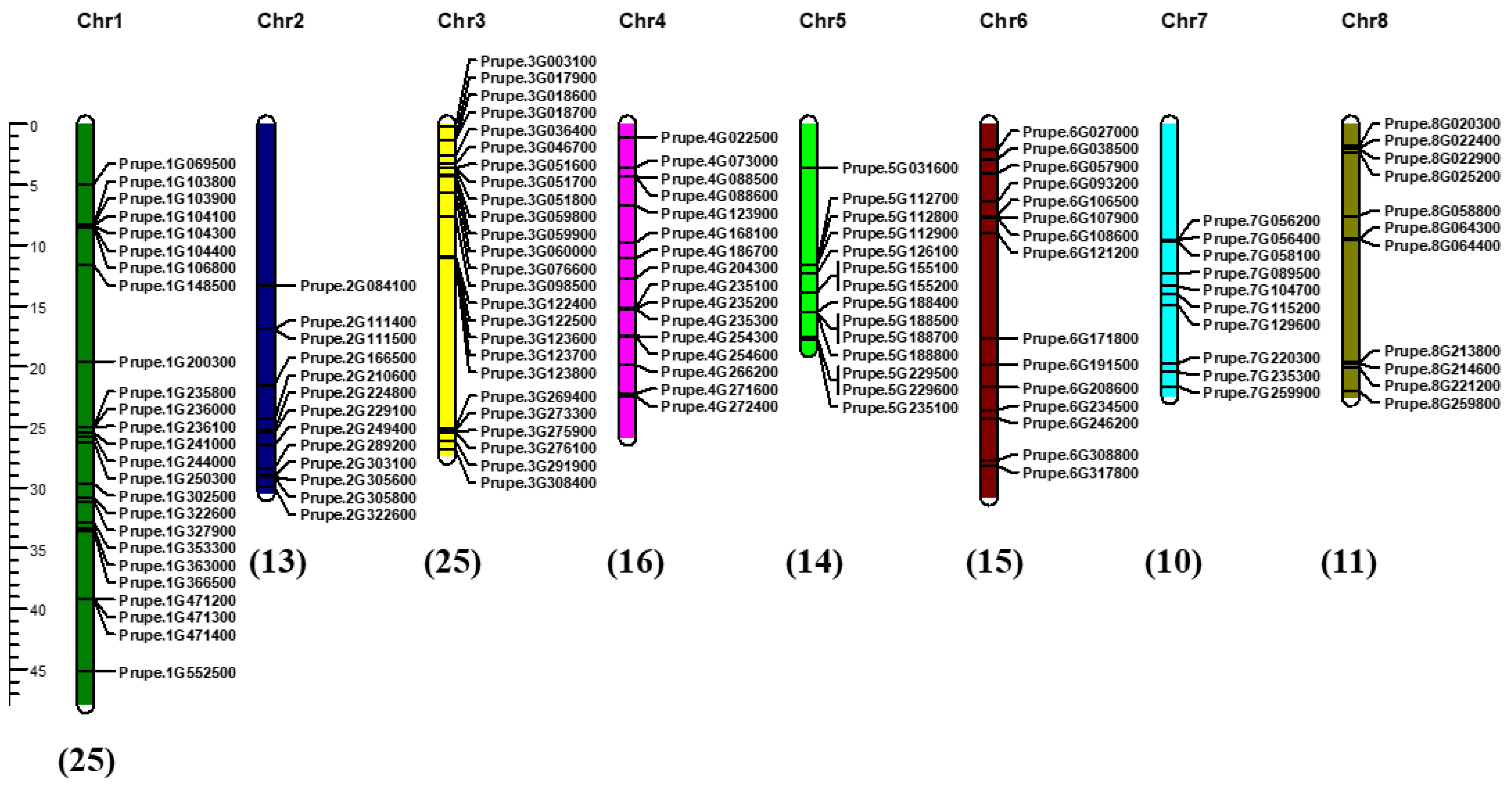
2.4. Chromosomal Distribution and Collinearity Analysis of PpABC Genes

Chromosomal distribution analysis indicated the uneven distribution of 132 ABC genes on eight chromosomes in the peach genome, with three PpABC genes (Prupe.I000500, Prupe.I000600 and Prupe.I000700) mapped under unassembled scaffolds (Figure 2, Table S5). In the peach genome, chromosomes (Chr) 1 and Chr3 contained the most ABC genes (25 members each), followed by 16 genes located on Chr4 and 15 genes located on Chr6. The hierarchical chromosomal distribution of the PpABC genes in different subfamilies was observed. The PpABC genes of group G, with the most members (43), were randomly distributed across the eight chromosomes. Groups A (9 members), B (29 members), C (26 members), D (2 members), E (5 members), F (3 members) and I (13 members) were located on 3, 7, 7, 2, 2, 3 and 6 chromosomes, respectively. To further understand the evolutionary and collinearity relationship of the PpABC gene family, we performed the syntenic collinearity analysis of the ABC family in peach. A total of 3, 4 and 2 collinear gene pairs were identified in ABCC, ABCG and ABCI subgroups, respectively (Figure S4). These results suggested that segmental duplications contributed to the expansion of PpABC genes.

2.5. Expression Pattern of PpABC Genes in Different Tissues of Peach

In order to further explore the expression pattern of PpABC genes in peach, transcription levels of PpABC genes in different tissues such as leaf, stem, root and callus were analyzed. A total of 119, 109, 105 and 100 PpABC genes were expressed in the leaf, stem, root and callus, respectively (Figure 3, Table S6). A total of 123 PpABC genes (accounting for 93.2% of total PpABCs) were expressed in all analyzed tissues. Among these, sixteen PpABC genes showed high expression levels (FPKM > 15) in all tested tissues. In addition, some PpABC genes presented tissues-specific expression, with 42 PpABC genes (34% of 123 expressed PpABC genes), 29 PpABC genes (24%), 27 PpABC genes (22%) and 22 PpABC genes (18%) specifically expressed in leaf, callus, root and stem, respectively. The expression of nine PpABC genes was not detected in these tissues. Overall, these results provide a baseline for the identification of various functional genes in peach.
The role of PpABC genes in regulating fruit development was also investigated. The expression profiles of PpABC genes during fruit development were estimated using fruit samples of two peach cultivars, ‘Dahongpao’ and ‘Jinxiang’, which were collected at the green stage (S1) and color break stage (S2). A heatmap representing the transcript expressions of the detected PpABC genes was constructed (Figure 4, Table S7). A total of 102 PpABC genes were expressed in fruit, and 67 had an expression level over 1 (FPKM) at both developmental stages. A total of 23 PpABC genes all showed higher expression (FPKM > 15) in the S2 stage of both cultivars. Among the 23 PpABC genes, 2, 4, 6, 1, 3, 3, 1 and 3 belonged to subgroups A, B, C, D, E, F, G and I, respectively. Six PpABCC members had higher expression profiles in fruits of ‘Dahongpao’ at the S2 stage. Given that the ABCC members are reported to be related to flavonoid biosynthesis [38], these six PpABCC genes may play critical roles in anthocyanin accumulation.

2.6. Analysis of Cis-Elements in the PpABCC Promoter

The putative cis-acting elements in the promoter region were identified for all the PpABC genes except Prupe.I000500 due to the lack of its promoter sequence in the database. The cis-acting regulatory elements of PpABCC subfamily members were classified into three groups based on their functional annotation: plant growth and development, phytohormone responsive, and abiotic and biotic stress (Figure 5). The majority of the elements (27) in the PpABCC subgroup members were related to abiotic and biotic stress, suggesting their important roles in stress response. However, the number of stress-related regulatory elements varied between the PpABCC subgroup members. The cis-elements related to abiotic and biotic stress were abundant, including light-responsiveness elements (G-Box) and low-temperature-responsive elements (LTR). The second group is the phytohormone response group, which consisted of twelve elements, with ABRE, ERE, TGACG, and CGTCA-motifs being widely distributed among PpABCC subfamily members. They are involved in abscisic acid (ABA), ethylene, and methyl jasmonate (MeJA) responsiveness in plants, respectively. The third group was related to plant growth and development and had nine elements, such as the GCN4-motif, O2 site, and MBSI, involved in endosperm expression, zein metabolism, and flavonoid biosynthetic gene regulation, respectively. In addition, an MYB binding site element (MBSI) was found in the promoter region of PpABCC1 (Prupe.4G088500). These results suggested that ABCC genes play important roles in a variety of developmental and physiological processes, including flavonoid biosynthesis in peach.

2.7. Identification of PpABCC Genes Regulating Anthocyanin Accumulation in Peach

To identify the ABCC genes involved in anthocyanin accumulation in peach, a phylogenetic tree was constructed based on Arabidopsis C-subgroup and PpABCC members (Figure S5). Among the 26 candidates ABCC members in peach, PpABCC1 was found to be clustered in the same group with the Arabidopsis gene AtABCC2, which was responsible for flavonoid transport in Arabidopsis [26]. The PpABCC1 gene was highly expressed in fruits of blood peach ‘Dahongpao’ at the S2 stage when anthocyanin accumulation occurred but had very weak expression in fruits of yellow peach ‘Jinxiang’ at both S1 and S2 stages (Figure 4). This suggested that the PpABCC1 gene could be involved in fruit anthocyanin accumulation in peach.
To verify this hypothesis, we carried out a correlation analysis between anthocyanin content and the PpABCC1 transcript abundance in mature fruits of twenty peach cultivars (Figure 6A, Table S8). Both the PpABCC1 expression and anthocyanin content varied among cultivars, with the highest expression of PpABCC1 observed in the ‘97-8-1′ cultivar that contained the highest content of anthocyanins. Linear regression analysis showed a strong positive correlation of the PpABCC1 expression with anthocyanin content among different cultivars (r = 0.974, p < 0.01). In addition, the subcellular localization assay showed that PpABCC1 was localized in the nuclei and the tonoplast (Figure 6B). Taken together, these results suggested that PpABCC1 is the most likely candidate gene involved in anthocyanin accumulation in peach.

2.8. Functional Analysis of PpABCC1 Using Transient Transformation Assay

In order to validate the role of PpABCC1 in anthocyanin coloration in vivo, its transient overexpression was performed in tobacco leaves and peach fruits. Tobacco leaves were infiltrated with the combination of PpMYB10.1, PpbHLH3 and PpABCC1, and the infiltration of PpMYB10.1 + PpbHLH3 was used as the control. Five days after injection, deep purple color patches were observed around the sites infiltrated with either PpMYB10.1/PpbHLH3/PpABCC1 or PpMYB10.1/PpbHLH3 (Figure 7A). However, leaf tissues around the site infiltrated with PpMYB10.1/PpbHLH3/PpABCC1 had a stronger pigmentation compared to those infiltrated with PpMYB10.1/PpbHLH3 which only showed a slight change in coloration. Anthocyanin content in leave tissues infiltrated with PpMYB10.1/PpbHLH3/PpABCC1 was 1.6-fold higher than that in leave tissues infiltrated with PpMYB10.1/PpbHLH3 (Figure 7A). PpABCC1-overexpression significantly increased the expression of NtANS by 1.8-fold, which was well consistent with the increased anthocyanin accumulation in the tobacco leaves (Figure 7A).
Similarly, intense red color patches appeared one week after infiltration in both the flesh and peel infiltrated with PpMYB10.1/PpbHLH3/PpABCC1 compared to these infiltrated with PpMYB10.1/PpbHLH3 (Figure 7B,C). Anthocyanin content in the flesh around the sites infiltrated with PpMYB10.1/PpbHLH3/PpABCC1 showed 1.5-fold higher as compared to that in the flesh around the sites infiltrated with PpMYB10.1/PpbHLH3. PpABCC1-overexpression significantly increased the expression of anthocyanin biosynthetic genes, PpANS and PpUFGT, by 2.9-fold and 1.9-fold higher, respectively, which correlated well with the color change in the flesh. For transient overexpression assay in the peel, anthocyanin content around the site infiltrated with PpMYB10.1/PpbHLH3/PpABCC1 showed a 1.5-fold increase compared to that around the site infiltrated with PpMYB10.1/PpbHLH3. The expression of PpANS in the peel around the site infiltrated with PpMYB10.1/PpbHLH3/PpABCC1 was 1.7-fold higher than that in the peel around the site infiltrated with PpMYB10.1/PpbHLH3, but no significant difference was observed in the expression of PpUFGT (Figure 7C). These results suggested the role of PpABCC1 in promoting anthocyanin accumulation in peach fruit.
The VIGS system was used to further validate the role of PpABCC1 in anthocyanin accumulation in peach fruit. Agrobacterium cultures containing pTRV1/pTRV2 or pTRV1/pTRV2-PpABCC1 constructs were transiently infiltrated into the fruits of blood-fleshed peach ‘Dahongpao’ at the color break stage. After infiltration for a week, a decreased pigmentation was observed in the flesh tissues around the site infiltrated with pTRV1/pTRV2-PpABCC1 as compared to the flesh tissues around the site infiltrated with pTRV1/pTRV2 (Figure 8A). Consistently, anthocyanin content (0.039 mg/100 g FW) in the flesh around the site infiltrated with pTRV1/pTRV2-PpABCC1 was significantly lower than that in the flesh around the site infiltrated with pTRV1/pTRV2 (0.064 mg/100 g FW). Silencing of the PpABCC1 resulted in a significant decrease in the expression of PpANS (Figure 8A). These results demonstrated that silencing of PpABCC1 reduced anthocyanin accumulation in peach fruit.

2.9. Transcriptional Activation of PpABCC1 by PpMYB10.1

To verify whether the anthocyanin-related TFs had the ability to regulate the promoter of PpABCC1, Y1H and dual luciferase assays were carried out. Since PpMYB10.1 has been previously reported to be the master regulator of anthocyanin accumulation in peach fruits [38,39], we investigated whether it could activate the transcription of PpABCC1. Y1H assay indicated that yeast cells harboring the PpMYB10.1 gene and the PpABCC1 promoter could grow on SD/Ura-Leu medium containing 50 ng/mL AbA (Figure 8B). Dual luciferase assay further showed that PpMYB10.1 had a strong activation activity on the promoter of PpABCC1 (Figure 8C). These results suggested that PpMYB10.1 could activate the transcription of PpABCC1.

3. Discussion

ABC proteins constitute one of the largest and most diverse transporter families ubiquitously present in different species. ABCs are involved in the transport of a wide range of substrates, such as secondary metabolites, phytohormone transport across membranes and heavy metal detoxification [1,2,3]. In this study, we conducted a comprehensive genome-wide and functional analysis of the ABC transporter gene family in order to determine its role in anthocyanin accumulation in peach fruit. A total of 132 PpABC genes were identified in the peach genome, which was divided into eight subfamilies and unevenly located on eight chromosomes.
Gene diversification and evolution caused variations in the chemical characterization of the PpABC transporter family. Sequence analysis revealed that the PpABC transporter family had notable differences in length, Mw, theoretical pI, total number of negatively/positively charged amino acids, GRAVY, aliphatic and instability index. Subcellular localization showed a majority of PpABC (103) genes were localized in the plasma membrane. The exon–intron structural diversity is essential in the evolution of gene families and supports the phylogenetic classification of genes in an organism [40,41]. In this study, closely related members showed similarities in structure and alignment. A typical full-size ABC protein has over 1200 amino acids [9]. The size of the 132 PpABC proteins ranged from 71 to 2524 amino acids. The number of exons in PpABC genes ranged from one to forty, which was largely consistent with the gene structure in Arabidopsis [16] and strawberry [8]. Motif structural analysis revealed a total of fifteen conserved motifs among the different subfamilies. A similar motif sequence pattern was also observed in closely related members indicating that these ABC proteins might perform similar functions.
Whole genome duplication, segmental duplication and tandem duplication mechanisms are the major causes of rapid gene family expansion and evolution in plants [42]. Previous studies have identified duplication events in the ABC genes in different lineages indicating the unending process of gene evolution [43]. Gene duplication events caused a rapid expansion of ABC genes in different species [44,45]. Here, our results showed that ABCB, ABCC, ABCG and ABCI expanded more than other subgroups in peach, thus confirming the diverse nature of the ABC transporters genes and their potential for rapid gene expansion and evolution from their progenitors. To further understand the evolutionary and collinearity relationship of the PpABC gene family, we performed the syntenic collinearity analysis of the ABC family in peach. The results suggested that ABC syntenic gene pairs mainly occurred in the ABCG subgroup, indicating a possible evolutionary relationship among the subgroup members.
Expression pattern analysis of PpABC family genes was conducted in different tissues and fruits to investigate the function of the ABC genes in peach. Expression profiles in different tissues help to improve our understanding of the tissue-specific and dynamic molecular function of genes. The preferential expression patterns of PpABC genes suggested specificities for certain tissues. The majority of the PpABC genes displayed high expression levels in the leaf, and low expression levels were detected in the callus (Figure 3). The majority of the ABC genes, including PpABCC1, were expressed in all four tissues tested, suggesting their roles in the development of the vegetative and reproductive tissues in peach. Of the PpABC genes, 42, 29, 27 and 22 exhibited tissue-specific expression patterns in leaf, callus, root and stem, respectively, suggesting functional divergence of the PpABC gene family in peach. The analysis of the fruit expression pattern revealed a high transcript expression level of PpABC genes in the ‘Dahongpao’ cultivar. The highest expression levels of six PpABCC gene members were observed at the S2 fruit stage, implying that these genes are potentially related to fruit ripening. However, functional redundancies between members are to be expected due to the presence of several PpABCC genes and their differential expression levels during fruit development. These results provide a baseline for the identification of various functional genes in peach.
Anthocyanin is synthesized in the cytosolic surface of the endoplasmic reticulum and then transported into the vacuole for storage. Although intracellular transport is also an important rate-limiting step for anthocyanin accumulation, the related mechanism remains poorly understood. Over the last few decades, different types of tonoplast-localized transporters, such as MATE and ABCC, have been unraveled to participate in anthocyanin accumulation. The involvement of ABCC members in trafficking anthocyanins was found in several plants. In this study, a peach ABCC gene (PpABCC1), the ortholog of Arabidopsis AtABCC2, a well-known gene for flavonoid transport [26], was functionally characterized to participate in the vacuolar accumulation of anthocyanins. PpABCC1 was differentially expressed in ‘Dahongpao’ and ‘Jinxiang’ at developmental stages. Additionally, the expression of PpABCC1 showed a significant correlation with anthocyanin content in ripe fruit of different peach cultivars, indicating that it might be the major ABC transporter in anthocyanin accumulation. Transient silencing of the PpABCC1 gene caused a significant decrease in anthocyanin accumulation in peach fruit, while its transient overexpression resulted in a deeper coloration in peach fruit. Taken together, these results undoubtedly indicated the essential role of PpABCC1 in the anthocyanin pigmentation of fruits in peach. It is worth noting that the PpABCC1 gene was expressed in all four tissues examined in this study. Thus, it is reasonable to speculate that the PpABCC1 gene plays an important role in anthocyanin accumulation in both vegetative and reproductive tissues.
Numerous discoveries have revealed that anthocyanin biosynthesis is regulated by the MYB-bHLH-WDR (MBW) transcriptional complex, which is highly conserved in a variety of plant species. In peach, PpMYB10.1 plays a critical role in the regulation of anthocyanin accumulation. PpMYB10.1 has been shown to interact with PpbHLH3 to induce anthocyanin biosynthesis in blood-fleshed peach [38,46]. Analysis of cis-elements indicated the presence of an MYB binding site element (MBSI) in the promoter region of PpABCC1. Therefore, we speculated that PpABCC1 might be regulated by the master regulator PpMYB10.1, such as anthocyanin biosynthetic genes. The Y1H and dual luciferase assay indicated the transcriptional activity of PpABCC1 was up-regulated by PpMYB10.1. Our results found that PpMYB10.1 could trans-activate the expression of PpABCC1, suggesting that PpMYB10.1 was a key transcription factor in anthocyanin accumulation due to its dual role in anthocyanin biosynthesis and transport. Moreover, light-responsive elements, low temperature-responsive elements and hormone-responsive elements (MeJA-responsive elements, gibberellin-responsive elements, auxin-responsive elements) were found in the promoter of PpABCC1 (Figure 5). Thus, the expression of PpABCC1 may be regulated by various external environments and physiological factors such as light, temperature and hormones. Given that anthocyanin accumulation is influenced by various abiotic and biotic stresses, it is possible that PpABCC1 implicated in anthocyanin transport is sensitive to internal and external stimuli.
Anthocyanin accumulation is a complex process that is determined by synthesis, transport and degradation. Efforts have been made in recent years to understand the mechanism underlying the intracellular transport of anthocyanin in fruit. In a previous study, loss-of-function mutation of a GST gene has been shown to affect anthocyanin accumulation in peach [47]. However, how anthocyanins are transported into the vacuolar lumen by membrane transporters is still not clear. In this study, an ABC membrane transporter was found to participate in anthocyanin accumulation in peach fruit. Our results provide valuable information for further research into the molecular mechanisms underlying anthocyanin transport in fruit.

4. Materials and Methods

4.1. Plant Materials

All peach accessions used in this study are maintained at the Wuhan Botanical Garden of the Chinese Academy of Sciences, Wuhan, China. Two peach cultivars, a blood-fleshed peach ‘Dahongpao’ and yellow-fleshed peach ‘Jinxiang’, were selected for gene expression profiling and RNA-seq analysis. Fruit samples were collected at two developmental stages, green stage (S1) at 65 days after full blossom (DAFB) and color break stage (S2) at 85 DAFB. Flesh samples were cut into small pieces, immediately frozen in liquid nitrogen and stored at −80 °C until use. For each sample, three biological replicates were conducted, with at least four fruits in each replicate. Additionally, peach fruits used for transient overexpression and virus-induced gene silencing (VIGS) assays were collected at the color break stage.

4.2. Identification and Sequence Analysis of the ABC Gene Family in Peach

Peach ABCs were identified by running a basic local alignment search tool (BLAST) search against the protein sequence database of peach (https://www.rosaceae.org/; accessed on 4 March 2022) using Arabidopsis ABC protein sequences as query sequences. The length, molecular weight (Mw), theoretical pI, total number of negatively/positively charged amino acids, grand average of hydropathicity (GRAVY), and aliphatic and instability index of the peach ABC family were calculated by ExPASy (https://web.expasy.org/protparam/; accessed on 10 May 2022). Subcellular localization was predicted by CELLO (http://cello.life.nctu.edu.tw/; accessed on 16 May 2022). Chromosomal distribution of the ABC transporter genes was visualized using MapChart [48]. Advanced Circos from TBtools software [49] was used to perform a collinearity analysis on the ABC genes to identify the duplicated genes. All parameters were set to the software’s default values.

4.3. Phylogenetic, Conserved Motifs and Gene Structure Analysis

Sequence alignment of the ABC protein sequences in peach was performed using ClustalX (1.81). The phylogenetic tree was constructed using the neighbor-joining algorithm in MEGA 6.0 with 1000 bootstrap replicates [50]. The conserved motifs of the putative peach ABC proteins were determined using the online MEME suite (http://meme-suite.org/tools/meme; accessed on 28 May 2022). The MEME results were visualized using TBtools. Gene structural analysis of the exon–intron organization was identified using the online Gene Structure Display Server (GSDS 2.0; http://gsds.cbi.pku.edu.cn; accessed on 2 June 2022) program [51]. All parameters were set as the default value of the software.

4.4. Cis-Acting Regulatory Element Analysis

To predict putative cis-acting elements in the ABC family genes, the promoter sequences 1.5 kb upstream of the ATG start codon were extracted from peach genome. The cis-acting elements were then predicted using the online Plant Cis-acting Regulatory DNA Elements (PlantCARE) database (http://bioinformatics.psb.ugent.be/webtools/plantcare/html/; accessed on 20 May 2022).

4.5. RNA Extraction and Quantitative Real-Time PCR

Total RNA was extracted from peach fruit samples using the Total RNA Rapid Extraction Kit (Magen, Guangzhou, China). Removal of genomic DNA and first strand cDNA synthesis were carried out using PrimeScriptTM RT reagent Kit with gDNA Eraser (Takara, Dalian, China). Quantitative real-time PCR (qRT-PCR) was conducted using SYBR® Premix Ex Taq™ II (Takara, Dalian, China), with the following amplification program: one cycle of 30 s at 95 °C, followed by 40 cycles of 5 s at 95 °C and 30 s at 60 °C. The peach PpTEF2 gene encoding a translation elongation factor was used as an internal control to normalize the expression of all target genes [52]. All analyses were repeated using three biological replicates. The quantification of gene expression level was calculated using the 2−ΔΔCT method [53]. Primer sequences used for gene expression are listed in Table S9.

4.6. Transcriptome Data Analysis

The transcriptome data of two cultivars, ‘Dahongpao’ and ‘Jinxiang’, were used to investigate expression profiles of the PpABC genes at two developmental stages. The RNA-seq data were extracted from our previous study [38]. Gene expression levels were estimated based on the expected number of fragments per kilobase of transcript sequence per millions of base pairs sequenced (FPKM). DESEQ2 was used to identify differentially expressed genes (DEGs) [54], with the following parameters: log2 fold change (FC) cut off of ±1 and a false discovery rate (FDR) ≤ 0.05. In addition, expression patterns of the PpABC genes in the leaf, callus (induced from cotyledon), root and stem tissues were investigated using the transcriptome data of the peach cultivar ‘Zaoyoutao’. Gene expression levels were estimated based on the FPKM values, which were used to generate a heatmap using TBtools.

4.7. Extraction and Quantification of Anthocyanin Content

Extraction and quantification of anthocyanin content were carried out using the Plant Anthocyanin Assay Kit (Solarbio, Beijing, China). The absorbance of the samples was determined at 530 and 700 nm using an Infinite M200 luminometer (Tecan, Mannerdorf, Switzerland). Total anthocyanin (TA) content was calculated according to a previous report [55]. Three biological replicates were performed for each sample.

4.8. Yeast One-Hybrid Assay (Y1H)

The yeast one-hybrid assay (Y1H) was conducted using the Matchmaker® Gold Yeast One-Hybrid Library Screening System User Manual (Clontech, Palo Alto, CA, USA) kit. The promoter sequence of PpABCC1, 2 kb upstream of the start codon, was amplified and inserted into the reporter vector pAbAi to generate pBait-pAbAis constructs. The full-length coding sequence of the PpMYB10.1 was inserted into the effector plasmid of pGADT7 to generate the pGADT7-PpMYB10.1 construct. Positive yeast cells were used to determine the activity of the pGADT7-TF with pBait-pAbAis on SD/-Ura/AbA* medium (*, the minimal inhibitory concentration of AbA). Primer sequences used for Y1H are listed in Table S10.

4.9. Dual Luciferase Reporter Assay

Dual-luciferase reporter assay in Nicotiana benthamiana leaves was conducted according to a previously described protocol [39]. Promoter sequences 2 kb upstream of the ATG start codon were inserted into the pGreen II 0800-LUC vector. The constructs were individually transformed into the Agrobacterium strain GV3101 containing the pSoup helper plasmid. Agrobacterium cultures carrying the constructs were then resuspended in infiltration buffer (10 mM MES, 10 mM MgCl2, 200 μM acetosyringone, pH 5.7) to an optimal density (A600 = 0.75) and incubated at 25 °C without shaking for 2 h before infiltration. The mixture of Agrobacterium containing the transcription factor (1 mL) and the promoter (100 μL) was injected into N. benthamiana leaves. After three days of infiltration, the ratio of firefly luciferase (LUC) to renilla luciferase (REN) activity was measured using the Dual-Glo® Luciferase Assay System (Promega, MI, USA). Four biological replicates were performed for each treatment. Primer sequences used for vector construction are listed in Table S10.

4.10. Subcellular Localization Assay

Subcellular localization was examined by transiently expressing the PpABCC1-YFP in N. benthamiana leaves using Agrobacterium (GV3101), similar to the protocol described for the dual-luciferase assay above. Tonoplast marker and transgenic N. benthamiana plants containing a red fluorescent nuclear marker (Nucleus-RFP) were used for the identification of the subcellular structures [56]. Fluorescence was detected three days after infiltration using the confocal microscope (TCS SP8, Leica, Microsystems, Wetzlar, Germany). Primer sequences used for vector construction are listed in Table S10.

4.11. Transient Overexpression and Virus-Induced Gene Silencing (VIGS) in Tobacco and Peach Fruit

Transient overexpression was carried out by inserting full-length coding sequences of PpABCC1 into the pSAK277 vector, which was then transformed into the Agrobacterium strain GV3101. Agrobacterium cultures were then resuspended in an infiltration buffer using the same protocol as described above for the dual-luciferase assay. The infiltrated tobacco (Nicotiana tabacum) leaves and peach fruits of ‘Huangjinmi’ were placed in a growth chamber (25 °C under 16 h light and 8 h dark photoperiod). Five days after infiltration, digital photographs were taken. For the VIGS system, Agrobacterium cultures containing pTRV1 and pTRV2 or pTRV2-PpABCC1 were mixed in a ratio of 1:1 and infiltrated into the blood-fleshed peach fruit. The injected fruits were placed in a growth chamber (25 °C under 16 h light and 8 h dark photoperiod). Digital photographs were taken one week after infiltration. Each treatment was performed with three biological replicates with at least ten fruits per replicate. Primer sequences used for vector construction are listed in Table S10.

Supplementary Materials

The following supporting information can be downloaded at: https://www.mdpi.com/article/10.3390/ijms24031931/s1.

Author Contributions

Y.H. and Y.Z. (Yun Zhao) planned and designed the experiments; C.S. performed the experiments with help from J.S., Y.Z. (Yuanqiang Zhang), C.N. and C.O.; C.S. wrote the manuscript; Y.H. revised the manuscript. All authors have read and agreed to the published version of the manuscript.

Funding

This project was financially supported by the National Natural Science Foundation of China (32272687 and 32202460), the China Agriculture Research System (CARS-30) and Hubei Hongshan Laboratory (2021hszd017).

Institutional Review Board Statement

Not applicable.

Informed Consent Statement

Not applicable.

Data Availability Statement

Not applicable.

Conflicts of Interest

The authors declare no conflict of interest.

References

  1. Ofori, P.A.; Mizuno, A.; Suzuki, M.; Martinoia, E.; Reuscher, S.; Aoki, K.; Shibata, D.; Otagaki, S.; Matsumoto, S.; Shiratake, K. Genome-wide analysis of ATP binding cassette (ABC) transporters in tomato. PLoS ONE 2018, 13, e0200854. [Google Scholar]
  2. Trigui-Lahiani, H.; Abdeljalil, S.; Hadj Sassi, A.; Skouri-Gargouri, H.; Gargouri, A. Molecular characterization and modeling study of the Podr1 gene and genome-scale identification of whole ATP-binding cassette (ABC) transporters in Penicillium occitanis. Genomics 2021, 113, 795–811. [Google Scholar]
  3. Verrier, P.J.; Bird, D.; Burla, B.; Dassa, E.; Forestier, C.; Geisler, M.; Klein, M.; Kolukisaoglu, U.; Lee, Y.; Martinoia, E.; et al. Plant ABC proteins-a nomenclature and updated inventory. Trends Plant Sci. 2008, 13, 151–159. [Google Scholar] [CrossRef] [Green Version]
  4. Rees, D.C.; Johnson, E.; Lewinson, O. ABC transporters: The power to change. Nat. Rev. Mol. Cell Biol. 2009, 10, 218–227. [Google Scholar]
  5. Hollenstein, K.; Frei, D.C.; Locher, K.P. Structure of an ABC transporter in complex with its binding protein. Nature 2007, 446, 213–216. [Google Scholar] [CrossRef]
  6. Higgins, C.F.; Linton, K.J. The ATP switch model for ABC transporters. Nat. Struct. Mol. Biol. 2004, 11, 918–926. [Google Scholar]
  7. Rea, P.A. Plant ATP-binding cassette transporters. Annu. Rev. Plant Biol. 2007, 58, 347–375. [Google Scholar] [CrossRef]
  8. Shi, M.Y.; Wang, S.S.; Zhang, Y.; Wang, S.; Zhao, J.; Feng, H.; Sun, P.P.; Fang, C.B.; Xie, X.B. Genome-wide characterization and expression analysis of ATP-binding cassette (ABC) transporters in strawberry reveal the role of FvABCC11 in cadmium tolerance. Sci. Hortic. 2020, 271, 109464. [Google Scholar]
  9. Martinoia, E.; Klein, M.; Geisler, M.; Bovet, L.; Forestier, C.; Kolukisaoglu, U.; Muller-Rober, B.; Schulz, B. Multifunctionality of plant ABC transporters-more than just detoxifiers. Planta 2002, 214, 345–355. [Google Scholar] [CrossRef]
  10. Kretzschmar, T.; Burla, B.; Lee, Y.; Martinoia, E.; Nagy, R. Functions of ABC transporters in plants. Essays Biochem. 2011, 50, 145–160. [Google Scholar]
  11. Pang, K.; Li, Y.; Liu, M.; Meng, Z.; Yu, Y. Inventory and general analysis of the ATP-binding cassette (ABC) gene superfamily in maize (Zea mays L.). Gene 2013, 526, 411–428. [Google Scholar] [PubMed]
  12. Do, T.; Martinoia, E.; Lee, Y.; Hwang, J.U. 2021 update on ATP-binding cassette (ABC) transporters: How they meet the needs of plants. Plant Physiol. 2021, 187, 1876–1892. [Google Scholar] [PubMed]
  13. Noh, B.; Murphy, A.S.; Spalding, E.P. Multidrug resistance-like genes of Arabidopsis required for auxin transport and auxin-mediated development. Plant Cell 2001, 13, 2441–2454. [Google Scholar]
  14. Sasaki, T.; Ezaki, B.; Matsumoto, H. A gene encoding multidrug resistance (MDR)-Like protein is induced by aluminum andinhibitors of calcium flux in Wheat. Plant Cell Physiol. 2002, 43, 177–185. [Google Scholar] [CrossRef] [Green Version]
  15. Hayashi, M.; Nito, K.; Takei-Hoshi, R.; Yagi, M.; Kondo, M.; Suenaga, A.; Yamaya, T.; Nishimura, M. Ped3p is a peroxisomal ATP-binding cassette transporter that might supply substrates for fatty acid beta-oxidation. Plant Cell Physiol. 2002, 43, 1–11. [Google Scholar] [CrossRef] [Green Version]
  16. Sanchez-Fernandez, R.; Davies, T.G.; Coleman, J.O.; Rea, P.A. The Arabidopsis thaliana ABC protein superfamily, a complete inventory. J. Biol. Chem. 2001, 276, 30231–30244. [Google Scholar]
  17. Zolman, B.K.; Silva, I.D.; Bartel, B. The Arabidopsis pxa1 mutant is defective in an ATP-binding cassette transporter-like protein required for peroxisomal fatty acid β-Oxidation. Plant Physiol. 2001, 127, 1266–1278. [Google Scholar]
  18. Garcia, O.; Bouige, P.; Forestier, C.; Dassa, E. Inventory and comparative analysis of rice and Arabidopsis ATP-binding cassette (ABC) systems. J. Mol. Biol. 2004, 343, 249–265. [Google Scholar]
  19. Borghi, L.; Kang, J.; Ko, D.; Lee, Y.; Martinoia, E. The role of ABCG-type ABC transporters in phytohormone transport. Biochem. Soc. Trans. 2015, 43, 924–930. [Google Scholar]
  20. Klein, M.; Burla, B.; Martinoia, E. The multidrug resistance-associated protein (MRP/ABCC) subfamily of ATP-binding cassette transporters in plants. FEBS Lett. 2006, 580, 1112–1122. [Google Scholar]
  21. Wanke, D.; Kolukisaoglu, H.U. An update on the ABCC transporter family in plants: Many genes, many proteins, but how many functions? Plant Biol. 2010, 12, 15–25. [Google Scholar] [PubMed]
  22. Goodman, C.D.; Casati, P.; Walbot, V. A multidrug resistance-associated protein involved in anthocyanin transport in Zea mays. Plant Cell 2004, 16, 1812–1826. [Google Scholar] [CrossRef] [PubMed]
  23. Zhu, Q.; Xie, X.; Zhang, J.; Xiang, G.; Li, Y.; Wu, H.B. In silico analysis of a MRP transporter gene reveals its possible role in anthocyanins or flavonoids transport in Oryze sativa. Am. J. Plant Sci. 2013, 4, 555–560. [Google Scholar] [CrossRef] [Green Version]
  24. Francisco, R.M.; Regalado, A.; Ageorges, A.; Burla, B.J.; Bassin, B.; Eisenach, C.; Zarrouk, O.; Vialet, S.; Marlin, T.; Chaves, M.M.; et al. ABCC1, an ATP binding cassette protein from grape berry, transports anthocyanidin 3-O-Glucosides. Plant Cell 2013, 25, 1840–1854. [Google Scholar] [CrossRef] [PubMed] [Green Version]
  25. Sun, L.; Fan, X.; Zhang, Y.; Jiang, J.; Sun, H.; Liu, C. Transcriptome analysis of genes involved in anthocyanins biosynthesis and transport in berries of black and white spine grapes (Vitis davidii). Hereditas 2016, 153, 17. [Google Scholar]
  26. Behrens, C.E.; Smith, K.E.; Iancu, C.V.; Choe, J.Y.; Dean, J.V. Transport of anthocyanins and other flavonoids by the Arabidopsis ATP-binding cassette transporter AtABCC2. Sci. Rep. 2019, 9, 437. [Google Scholar] [PubMed] [Green Version]
  27. Dean, J.V.; Willis, M.; Shaban, L. Transport of acylated anthocyanins by the Arabidopsis ATP-binding cassette transporters AtABCC1, AtABCC2, and AtABCC14. Physiol. Plant. 2022, 174, e13780. [Google Scholar] [CrossRef]
  28. Chen, P.; Li, Y.; Zhao, L.; Hou, Z.; Yan, M.; Hu, B.; Liu, Y.; Azam, S.M.; Zhang, Z.; Rahman, Z.U.; et al. Genome-wide identification and expression profiling of ATP-binding cassette (ABC) transporter gene family in Pineapple (Ananas comosus (L.) Merr.) reveal the role of AcABCG38 in pollen development. Front. Plant Sci. 2017, 8, 2150. [Google Scholar] [CrossRef]
  29. Lopez-Ortiz, C.; Dutta, S.K.; Natarajan, P.; Pena-Garcia, Y.; Abburi, V.; Saminathan, T.; Nimmakayala, P.; Reddy, U.K. Genome-wide identification and gene expression pattern of ABC transporter gene family in Capsicum spp. PLoS ONE 2019, 14, e0215901. [Google Scholar]
  30. Huang, J.; Li, X.; Chen, X.; Guo, Y.; Liang, W.; Wang, H. Genome-wide identification of Soybean ABC transporters relate to aluminum toxicity. Int. J. Mol. Sci. 2021, 22, 6556. [Google Scholar] [CrossRef]
  31. Zhang, X.D.; Zhao, K.X.; Yang, Z.M. Identification of genomic ATP binding cassette (ABC) transporter genes and Cd-responsive ABCs in Brassica napus. Gene 2018, 664, 139–151. [Google Scholar] [CrossRef]
  32. Yan, C.; Duan, W.; Lyu, S.; Li, Y.; Hou, X. Genome-wide identification, evolution, and expression analysis of the ATP-Binding cassette transporter gene family in Brassica rapa. Front. Plant Sci. 2017, 8, 349. [Google Scholar] [CrossRef] [PubMed] [Green Version]
  33. Verde, I.; Abbott, A.G.; Scalabrin, S.; Jung, S.; Shu, S.; Marroni, F.; Zhebentyayeva, T.; Dettori, M.T.; Grimwood, J.; Cattonaro, F.; et al. The high-quality draft genome of peach (Prunus persica) identifies unique patterns of genetic diversity, domestication and genome evolution. Nat. Genet. 2013, 45, 487–494. [Google Scholar] [CrossRef] [Green Version]
  34. Marinova, K.; Pourcel, L.; Weder, B.; Schwarz, M.; Barron, D.; Routaboul, J.M.; Debeaujon, I.; Klein, M. The Arabidopsis MATE transporter TT12 acts as a vacuolar flavonoid/H+-antiporter active in proanthocyanidin-accumulating cells of the seed coat. Plant Cell 2007, 19, 2023–2038. [Google Scholar] [CrossRef] [PubMed]
  35. Gomez, C.; Terrier, N.; Torregrosa, L.; Vialet, S.; Fournier-Level, A.; Verries, C.; Souquet, J.M.; Mazauric, J.P.; Klein, M.; Cheynier, V.; et al. Grapevine MATE-type proteins act as vacuolar H+-dependent acylated anthocyanin transporters. Plant Physiol. 2009, 150, 402–415. [Google Scholar] [CrossRef] [PubMed] [Green Version]
  36. Zhao, J.; Dixon, R.A. MATE transporters facilitate vacuolar uptake of epicatechin 3′-O-glucoside for proanthocyanidin biosynthesis in Medicago truncatula and Arabidopsis. Plant Cell 2009, 21, 2323–2340. [Google Scholar] [CrossRef] [PubMed] [Green Version]
  37. Zhao, Y.; Dong, W.; Zhu, Y.; Allan, A.C.; Lin-Wang, K.; Xu, C. PpGST1, an anthocyanin-related glutathione S-transferase gene, is essential for fruit coloration in peach. Plant Biotechnol. J. 2020, 18, 1284–1295. [Google Scholar] [CrossRef] [PubMed] [Green Version]
  38. Zhou, H.; Lin-Wang, K.; Wang, H.; Gu, C.; Dare, A.P.; Espley, R.V.; He, H.; Allan, A.C.; Han, Y. Molecular genetics of blood-fleshed peach reveals activation of anthocyanin biosynthesis by NAC transcription factors. Plant J. 2015, 82, 105–121. [Google Scholar] [CrossRef] [PubMed]
  39. Zhou, H.; Lin-Wang, K.; Wang, F.; Espley, R.V.; Ren, F.; Zhao, J.; Ogutu, C.; He, H.; Jiang, Q.; Allan, A.C.; et al. Activator-type R2R3-MYB genes induce a repressor-type R2R3-MYB gene to balance anthocyanin and proanthocyanidin accumulation. New Phytol. 2019, 221, 1919–1934. [Google Scholar] [CrossRef] [Green Version]
  40. Shiu, S.H.; Bleecker, A.B. Expansion of the receptor-like kinase/pelle gene family and receptor-like proteins in Arabidopsis. Plant Physiol. 2003, 132, 530–543. [Google Scholar] [CrossRef] [Green Version]
  41. Wang, J.; Marowsky, N.C.; Fan, C. Divergence of gene body DNA methylation and evolution of plant duplicate genes. PLoS ONE 2014, 9, e110357. [Google Scholar] [CrossRef] [PubMed] [Green Version]
  42. Cannon, S.B.; Mitra, A.; Baumgarten, A.; Young, N.D.; May, G. The roles of segmental and tandem gene duplication in the evolution of large gene families in Arabidopsis thaliana. BMC Plant Biol. 2004, 4, 10. [Google Scholar] [CrossRef] [PubMed] [Green Version]
  43. Rensing, S.A. Gene duplication as a driver of plant morphogenetic evolution. Curr. Opin. Plant Biol. 2014, 17, 43–48. [Google Scholar] [CrossRef] [PubMed]
  44. Annilo, T.; Chen, Z.Q.; Shulenin, S.; Costantino, J.; Thomas, L.; Lou, H.; Stefanov, S.; Dean, M. Evolution of the vertebrate ABC gene family: Analysis of gene birth and death. Genomics 2006, 88, 1–11. [Google Scholar] [CrossRef] [PubMed]
  45. Moitra, K.; Dean, M. Evolution of ABC transporters by gene duplication and their role in human disease. Biol. Chem. 2011, 392, 29–37. [Google Scholar] [CrossRef] [PubMed]
  46. Tuan, P.A.; Bai, S.; Yaegaki, H.; Tamura, T.; Hihara, S.; Moriguchi, T.; Oda, K. The crucial role of PpMYB10.1 in anthocyanin accumulation in peach and relationships between its allelic type and skin color phenotype. BMC Plant Biol. 2015, 15, 280. [Google Scholar] [CrossRef] [Green Version]
  47. Cheng, J.; Liao, L.; Zhou, H.; Gu, C.; Wang, L.; Han, Y. A small indel mutation in an anthocyanin transporter causes variegated colouration of peach flowers. J. Exp. Bot. 2015, 66, 7227–7239. [Google Scholar] [CrossRef] [Green Version]
  48. Voorrips, R.E. MapChart: Software for the graphical presentation of linkage maps and QTLs. J. Hered. 2002, 93, 77–78. [Google Scholar] [CrossRef] [Green Version]
  49. Chen, C.; Chen, H.; Zhang, Y.; Thomas, H.R.; Frank, M.H.; He, Y.; Xia, R. TBtools: An integrative toolkit developed for interactive analyses of big biological data. Mol. Plant 2020, 13, 1194–1202. [Google Scholar] [CrossRef]
  50. Tamura, K.; Stecher, G.; Peterson, D.; Filipski, A.; Kumar, S. MEGA6: Molecular evolutionary genetics analysis version 6.0. Mol. Biol. Evol. 2013, 30, 2725–2729. [Google Scholar] [CrossRef] [Green Version]
  51. Hu, B.; Jin, J.; Guo, A.Y.; Zhang, H.; Luo, J.; Gao, G. GSDS 2.0: An upgraded gene feature visualization server. Bioinformatics 2015, 31, 1296–1297. [Google Scholar] [CrossRef] [PubMed] [Green Version]
  52. Tong, Z.; Gao, Z.; Wang, F.; Zhou, J.; Zhang, Z. Selection of reliable reference genes for gene expression studies in peach using real-time PCR. BMC Mol. Biol. 2009, 10, 71. [Google Scholar] [CrossRef] [PubMed] [Green Version]
  53. Livak, K.J.; Schmittgen, T.D. Analysis of relative gene expression data using real-time quantitative PCR and the 2(-Delta Delta C(T)) Method. Methods 2001, 25, 402–408. [Google Scholar] [CrossRef] [PubMed]
  54. Love, M.I.; Huber, W.; Anders, S. Moderated estimation of fold change and dispersion for RNA-seq data with DESeq2. Genome Biol. 2014, 15, 550. [Google Scholar] [CrossRef]
  55. Li, Y.; Cui, W.; Qi, X.; Lin, M.; Qiao, C.; Zhong, Y.; Hu, C.; Fang, J. MicroRNA858 negatively regulates anthocyanin biosynthesis by repressing AaMYBC1 expression in kiwifruit (Actinidia arguta). Plant Sci. 2020, 296, 110476. [Google Scholar] [CrossRef]
  56. Nelson, B.K.; Cai, X.; Nebenfuhr, A. A multicolored set of in vivo organelle markers for co-localization studies in Arabidopsis and other plants. Plant J. 2007, 51, 1126–1136. [Google Scholar] [CrossRef]
Figure 1. Phylogenetic analysis of ABC proteins from peach. The phylogenetic tree was constructed using the neighbor-joining method in MEGA 6.0 with 1000 bootstrap replicates. Different colors highlighted the eight subfamilies.
Ijms 24 01931 g001
Figure 2. Chromosomal distribution of PpABC genes. The chromosomes were visualized using MapChart program. The chromosome number is indicated at the top of each chromosome, and the number of PpABC genes in each chromosome is indicated at the bottom of each chromosome in brackets. The scale bar described the relative lengths of the chromosomes in megabases (Mb).
Ijms 24 01931 g002
Figure 3. Expression profile of PpABC genes in different tissues of peach. The FPKM values were used to generate a heatmap with hierarchical clustering analysis. The color scale displaying the expression levels (−1.5 to 1.5) was indicated.
Ijms 24 01931 g003
Figure 4. Expression profile of PpABC genes during fruit development of peach. Transcript expression levels were determined during two developmental stages, S1 (green stage at 65 DAFB) and S2 (color break stage at 85 DAFB). The FPKM values were used to generate a heatmap with hierarchical clustering analysis. A–G and I indicated the eight subfamilies of PpABC genes. The color scale displaying the expression levels (−1.5 to 1.5) was indicated.
Ijms 24 01931 g004
Figure 5. Cis-elements in the 1.5-kb upstream region of PpABCC genes. The bars on the top represent the total number of cis-elements in each gene promoter region. Different colors indicate different types of cis-elements. The color intensity and number in the cells indicated the number of cis-element in these PpABCC genes.
Ijms 24 01931 g005
Figure 6. Expression and subcellular location for the PpABCC1 gene: (A) expression of PpABCC1 and anthocyanin content in ripe fruits of 20 peach cultivars and the correlation analysis between the PpABCC1 expression and anthocyanin content. Data represent means (±SD) of three biological replicates; (B) subcellular localization assay of PpABCC1 in tobacco leaves. Scale bars indicated 50 μm.
Ijms 24 01931 g006
Figure 7. Transient overexpression of PpMYB10.1 + PpbHLH3 with or without PpABCC1 in tobacco leaves (A) and peach (cv. ‘Huangjinmi’) fruit (B,C). Relative expression of PpABCC1 and anthocyanin biosynthetic genes, changes in anthocyanin contents in tobacco leaves (A) and peach fruit (B,C). Data were means (±SD) from three biological replicates. *, Significant difference at p < 0.05 level.
Ijms 24 01931 g007
Figure 8. Functional analysis of the PpABCC1 gene: (A) Transient silencing of PpABCC1 in the fruit of blood peach ‘Dahongpao’ at S2 using virus-induced gene silencing (VIGS) system. Expression of PpABCC1 and anthocyanin biosynthetic genes and anthocyanin content were measured in the flesh tissues around the infiltration sites. (B) Assay of the interaction between PpMYB10.1 and the promoter of PpABCC1 using yeast one-hybrid (Y1H). (C) Assay of the activation activity of PpMYB10.1 on the promoter of PpABCC1 using dual luciferase reporter system. Data represented means (±SD) of three independent biological replicates. *, Significant difference at p < 0.05 level.
Ijms 24 01931 g008
Disclaimer/Publisher’s Note: The statements, opinions and data contained in all publications are solely those of the individual author(s) and contributor(s) and not of MDPI and/or the editor(s). MDPI and/or the editor(s) disclaim responsibility for any injury to people or property resulting from any ideas, methods, instructions or products referred to in the content.

