Article

Inhibiting Intracellular α2C-Adrenoceptor Surface Translocation Using Decoy Peptides: Identification of an Essential Role of the C-Terminus in Receptor Trafficking

1
Dr. Panjwani Center for Molecular Medicine and Drug Research (PCMD), International Center for Chemical and Biological Sciences, University of Karachi, Karachi 75270, Pakistan
2
Husein Ebrahim Jamal (H.E.J.) Research Institute of Chemistry, International Center for Chemical and Biological Sciences, University of Karachi, Karachi 75270, Pakistan
*
Author to whom correspondence should be addressed.
Int. J. Mol. Sci. 2023, 24(24), 17558; https://doi.org/10.3390/ijms242417558
Submission received: 12 November 2023 / Revised: 7 December 2023 / Accepted: 10 December 2023 / Published: 16 December 2023
(This article belongs to the Special Issue Advances in Protein-Protein Interactions 2.0)

Abstract

:
The G protein-coupled α2-adrenoceptor subtype C (abbreviated α2C-AR) has been implicated in peripheral vascular conditions and diseases such as cold feet–hands, Raynaud’s phenomenon, and scleroderma, contributing to morbidity and mortality. Microvascular α2C-adrenoceptors are expressed in specialized smooth muscle cells and mediate constriction under physiological conditions and the occlusion of blood supply involving vasospastic episodes and tissue damage under pathological conditions. A crucial step for receptor biological activity is the cell surface trafficking of intracellular receptors, triggered by cAMP-Epac-Rap1A GTPase signaling, which involves protein–protein association with the actin-binding protein filamin-2, mediated by critical amino acid residues in the last 14 amino acids of the receptor carboxyl (C)-terminus. This study assessed the role of the C-terminus in Rap1A GTPase coupled receptor trafficking by domain-swapping studies using recombinant tagged receptors in transient co-transfections and compared with wild-type receptors using immunofluorescence microscopy. We further tested the biological relevance of the α2C-AR C-terminus, when introduced as competitor peptides, to selectively inhibit intracellular α2C-AR surface translocation in transfected as well as in microvascular smooth muscle cells expressing endogenous receptors. These studies contribute to establishing proof of principle to target intracellular α2C-adrenoceptors to reduce biological activity, which in clinical conditions can be a target for therapy.

1. Introduction

Raynaud’s syndrome (RS) is a vasospastic disorder of the microvasculature triggered by cold or emotional stress [1,2]. RS is categorized as idiopathic or primary Raynaud’s syndrome (PRS) when no underlying reason for this impairment is identified, and secondary Raynaud’s syndrome (SRS) when it is associated with a pre-existing or multiple diseased condition [3]. About 5% of the general population is affected by RS [4], and this number increases in areas where the climate is comparatively colder [5,6,7]. RS has a very high prevalence rate in patients with scleroderma and systemic sclerosis [3]. The onset of RS physically manifests with a triphasic coloration of the digits [8,9], followed by swelling, numbness, and lesions [10].
RS is known to be a multifactorial disease and can be caused by defective endothelial vasodilation caused by reduced levels of calcitonin gene-related peptide (CGRP) [11] resulting in a drop in the production of vasodilator nitric oxide [12]. One of the sole causes of RS seems to be a default in the vascular thermoregulatory system [13,14], especially in the digits where lower temperatures can significantly reduce blood flow in not only in the arteriovenous anastomosis but also in the nutritional flow in the capillaries due to the increased sympathetic activation of α2-adrenoceptors in vascular smooth muscle cells [15].
The α2-adrenoceptors belong to the family of G protein-coupled receptors (GPCRs), and pharmacologically, three subtypes of α2-adrenoceptors have been identified; α2A, α2B, and α2C [16]. Initially, studies conducted in the intact cardiovascular system of mice showed that for α2-adrenoceptors, only subtype α2A and α2B possessed physiological roles [17,18], whereas α2C was found unresponsive or silent [19]. However, Chotani and colleagues further investigated and disclosed that at 37 °C, the α2C-adrenoceptor remained non-functional as found in earlier studies, but at 28 °C, the α2C-adrenoceptor became activated and could generate a cold-induced vasospastic episode in RS [20].
Studies have found that α2C-adrenoceptors are primarily localized in the ER or Golgi compartment of the cell [21,22,23] at 37 °C and translocate to the cell surface when the temperature is lowered to 28 °C [20,24]. The α2C-adrenoceptors have been found to contain an arginine-rich region (RRRRR) in the carboxyl (C)-terminus, known to be a retention signal for the ER [25], which may explain its perinuclear localization in the cell. Several signaling pathways mediate the translocation of α2C-adrenoceptors toward the cell surface. Upon the selective pharmacological inhibition of Rho kinase by fasudil at 28 °C and of COX-2 by celecoxib, a decrease in the expression and translocation of these receptors has been observed [23,26,27]. cAMP has a dual effect on the regulation of α2C-adrenoceptors, augmenting its transcription through the JNK-AP1 pathway [28] and redistributing it to the cell surface through RhoA-Rho-kinase using the cytoskeletal actin-binding protein filamin-2 through Rap1A GTPase intracellular signaling [29]. Filamin-2 possesses a conserved serine whose phosphorylation has been reported to be a key player in the reconstruction of the cytoskeleton [30,31]. The amino acids in the C-terminus of α2C-adrenoceptors interact with a 78 amino acid sequence motif of filamin-2 containing the conserved serine, to translocate to the cell surface.
In this study, we have assessed the role of the α2C-adrenoceptor C-terminus in receptor cell surface translocation and have utilized the α2-adrenoceptor subtype A (α2A-adrenoceptor), which does not associate with filamin-2, for comparison. These studies provide a unique insight into the role of the C-termini of both α2-adrenoceptor subtypes in regulating receptor cellular localization and allow to experimentally test the application of using the C-terminal domain to target receptor trafficking.

2. Results

2.1. Transient Co-Transfections

The α2C-adrenoceptors have a unique mechanism of regulation; intracellular receptors translocate to the cell surface in response to extracellular stimuli, including signaling which increases intracellular levels of cAMP, which was established in our previous studies utilizing primary cultures of microvascular smooth muscle cells (microVSM) derived either from murine tail artery or human dermal arterioles [29]. For example, see Figure 1 for the effect of the adenylyl cyclase activator forskolin (which activates intracellular cAMP-Epac-Rap1A signaling) on endogenous α2C-adrenoceptor translocation when added to quiescent primary murine microVSM, similarly seen in human-derived microVSM [29]. These cell-surface-associated α2C-adrenoceptors were shown to be functional in both murine- and human-derived microVSM [29,32].
We initially tested recombinant DNA expressing α2C-adrenoceptor fused to green fluorescent protein (α2C-AR-GFP) in primary cultures of murine microVSM. For example, see Figure 2 for the localization of α2C-AR-GFP in quiescent, unstimulated microVSM, which is similar to endogenous α2C-adrenoceptors under quiescent conditions (compared with localization in DMSO-solvent control cells, Figure 1), which illustrates the feasibility of using GFP-fused or other HA-tagged constructs for transient transfection studies. However, due to the extremely low transfection efficiency in these primary cultures (<1%, assessed by determining the number of GFP-positive cells), further studies on recombinant DNA constructs could not be conducted in microVSM. Alternatively, we utilized and optimized a mouse-embryo-derived fibroblast (NIH/3T3) cell line for transient transfections/co-transfections. NIH/3T3 cells were relatively fast growing and showed better transfection efficiency and were therefore utilized for further studies.
Transient co-transfections were performed in NIH/3T3 cells utilizing recombinant DNA tagged constructs along with a constitutively active version of the small GTPase Rap1A (Rap1A-63E), compared with the control backbone expression vector pcDNA. Indeed, the results showed the recapitulation of intracellular signaling associated with α2C-adrenoceptors in NIH/3T3 cells compared with the Rap1A-α2C-adrenoceptor coupled signaling and cell surface receptor trafficking in human and murine microVSM [29], for receptors tagged either with HA (Figure 3) or with GFP (Figure 4). In these transfections, in comparison, we noted differences in signal fluorescence intensities between cells labeled with primary and secondary antibodies (higher fluorescence) versus the direct detection of fluorescence in cells expressing green fluorescent protein (relatively lower fluorescence). Further studies were therefore carried out in NIH/3T3 cells to test the role of the C-termini in receptor trafficking coupled to Rap1A GTPase intracellular signaling, utilizing the receptor chimeras described in Section 4.

2.2. Domain-Swapping Studies

To test the biological relevance of C-termini in Rap1A GTPase coupled trafficking, receptor chimeras were generated for comparison with wild-type receptors. These are described in detail in Section 4.

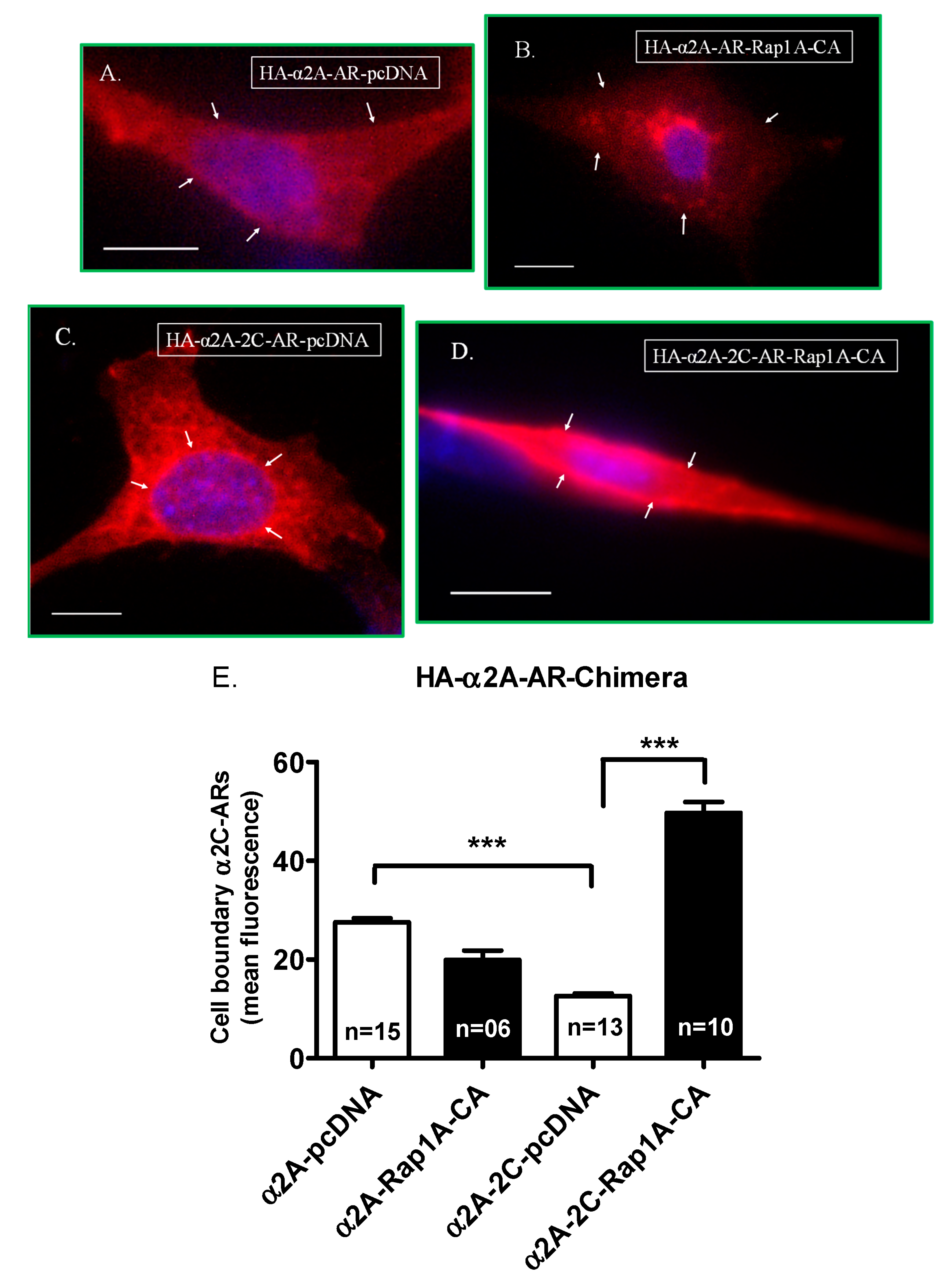
2.2.1. α2A-Adrenoceptor Chimera Studies

Transient co-transfection studies showed the wild-type α2A-adrenoceptors to be present primarily on the cell surface in cells with the control expression vector pcDNA. However, the localization of α2A-adrenoceptors changed significantly (p < 0.0001) in response to Rap1A GTPase signaling, with a reduction in cell surface receptors (Figure 5A,B,E).
This profile changed dramatically when the α2C-adrenoceptor C-terminus was swapped with the wild-type α2A-adrenoceptor C-terminus region. The chimera α2A-2C-AR was primarily intracellular, similar to α2C-adrenoceptor wild-type receptors, when co-transfected with the control expression vector pcDNA. However, in cells co-transfected with activated Rap1A, the α2A-2C-AR chimera showed a significant association with the cell surface (p < 0.0001), similar to wild-type α2C-adrenoceptors (Figure 5C–E).

2.2.2. α2C-Adrenoceptor Chimera Studies

Transient co-transfection studies showed the wild-type α2C-adrenoceptor to be primarily intracellular with the control expression vector pcDNA. However, the localization of α2C-adrenoceptors changed significantly (p < 0.0001) in response to Rap1A GTPase signaling, with an increase in cell surface receptors (Figure 6A,B,E).
This profile of wild-type α2C-adrenoceptors changed dramatically when the α2A-adrenoceptor C-terminus was swapped with the wild type α2C-adrenoceptor C-terminus region. The chimera α2C-2A-AR was primarily on the cell surface, similar to α2A-adrenoceptor wild-type receptors, when co-transfected with pcDNA. However, in cells co-transfected with activated Rap1A, the α2C-2A-AR chimera was significantly reduced on the cell surface (p < 0.0001), similar to wild-type α2A-adrenoceptors (Figure 6C,D,E).

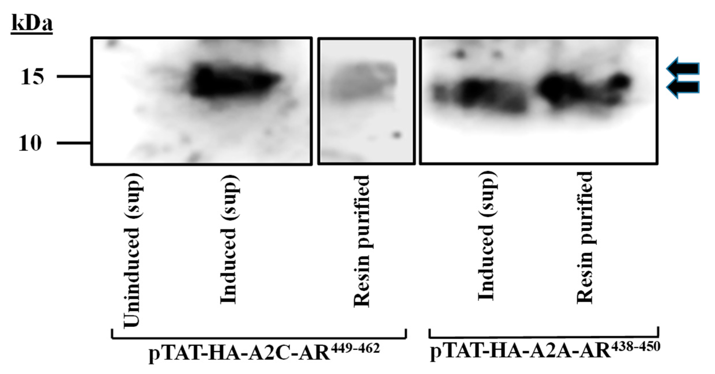
2.3. Peptide Expression, Purification, and Detection by Western Blotting

The peptides were expressed in a BL21 (DE3)-STAR bacteria strain and purified as described in Section 4.
The peptides contain the HA-tag, which was used for the confirmation of expression and the purification in the Western blot analysis (Figure 7).

2.3.1. Peptide Delivery and Localization in NIH/3T3 Cells

Intracellular delivery and the specificity of peptides (encoding α2C-AR449–462 target sequence and control sequence for α2-adrenoceptor subtype A, α2A-AR438–450, 25 µL each of purified peptide) were tested in transient co-transfection studies utilizing α2C-AR-GFP/Rap1A-CA and anti-HA antibody for peptide detection using immunofluorescence microscopy.
The effect on GFP-positive cells with peptide uptake was assessed. Our observations showed that the peptide staining was distinct from the background signal, which was assessed for cells without peptides and with or without primary anti-HA antibody (Figure 8A–D). Using this approach, we detected the cellular uptake of both peptides, which showed localization in the cytosol or were perinuclear, Figure 8E–G for pTAT–HA–A2A–AR438–450 (Peptide A) and Figure 8H–J for pTAT–HA–A2C–AR449–462 (Peptide C).

2.3.2. Peptide Specificity to Inhibit Receptor Translocation in NIH/3T3 Cells

Based on the outcomes of the domain-swapping studies, we hypothesized that the peptide encoding α2C-AR449–462 should selectively inhibit α2C-adrenoceptor trafficking.
Indeed, upon quantification of cell-surface-associated α2C-AR-GFP, Rap1A stimulation significantly increased receptor translocation compared with control expression vector pcDNA (p < 0.0001), which was not significantly affected in the presence of intracellular Peptide A (p = ns). However, in the presence of intracellular Peptide C, the α2C-AR-GFP receptor translocation was selectively and significantly (p < 0.0001) reduced (Figure 9).

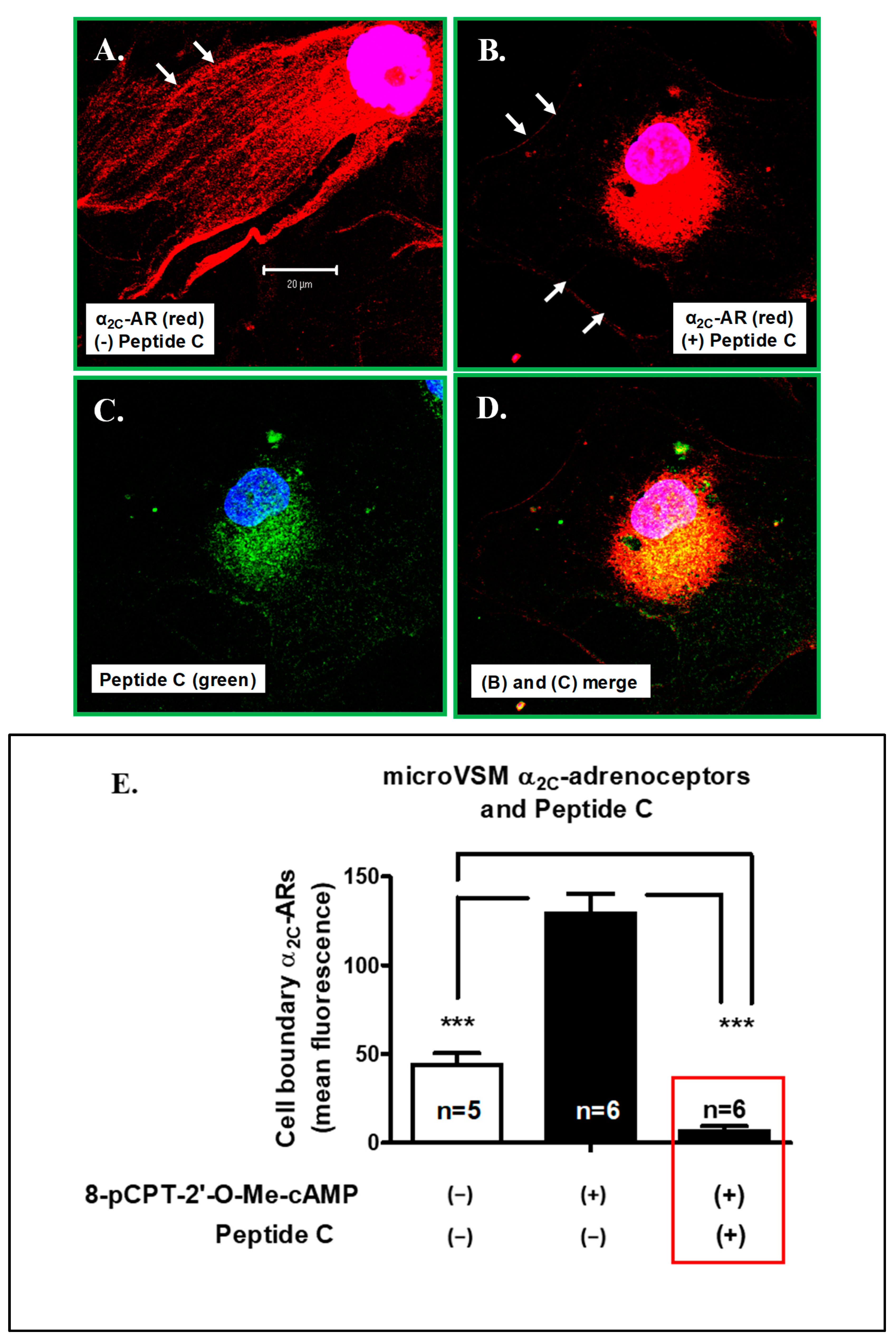
2.3.3. Peptide C Effect on Endogenous microVSM α2C-Adrenoceptor Translocation and Function

Based on the outcomes of transient transfection studies, Peptide C was further tested in human-arteriolar-derived microVSM which express α2C-adrenoceptors [27]. In these cells, adenylyl cyclase activation by forskolin activates cAMP-Epac-Rap1A signaling which is coupled to the increased expression and translocation of α2C-adrenoceptors through the RhoA-Rho kinase pathway and receptor trafficking via an association with the actin-binding protein filamin-2 [29,33]. Alternatively, the Epac-Rap1A signaling can also be activated by the cAMP analog 8-pCPT-2′-O-Me-cAMP [29,33]. In these microVSM, the cAMP analog 8-pCPT-2′-O-Me-cAMP increased surface α2C-adrenoceptors compared with the solvent control cells (Figure 10A,E, p < 0.0001). However, in the presence of Peptide C, the cell surface localization of the receptor was significantly reduced (p < 0.0001) and was below the baseline level in unstimulated cells (Figure 10B–E, p < 0.0001). The receptor and Peptide C co-localized in the perinuclear region of microVSM (Figure 10C,D). These observations were further validated by assessing microVSM receptor function by measuring the intracellular levels of cAMP in 8-pCPT-2′-O-Me-cAMP (100 µM, 14 h) stimulated cells in the absence or presence of Peptide C. Since the α2-adrenoceptors are Gi-coupled, activation by the agonist UK, 14,304 inhibits adenylyl cyclase and intracellular cAMP levels [24,29,33]. Indeed, these studies supported the loss of cell surface receptor functional activity (Figure 10F). In the absence of this inhibition, intracellular levels of cAMP increased noticeably.

3. Discussion

There has been tremendous interest in understanding the physiological mechanisms involved in the extreme temperature sensing and avoidance ability in humans pertaining to survival and protection from tissue damage. This includes the recent identification of cold sensor receptors in the sensory nerves of the skin, which has added to a better understanding of the mechanisms involved in thermoregulation [34]. Particularly, interest in inhibiting the biological activity of vascular α2C-adrenoceptors (which play a role as thermo-effectors) has grown over the years and includes the recent elucidation of crystal structure to identify molecular interactions during ligand binding which can aid in designing selective α2C-adrenoceptor antagonists [35]. Indeed, clinical trials have been performed using α2C-adrenoceptor antagonists targeting cell surface receptors and include compounds OPC-28326, ORM-12471, and BAY1193397 [36,37]. However, since this receptor is also expressed on the cell surface in non-VSM cells, other tissues, and organs, this approach, although promising in short-term trials, also showed an effect on inhibiting pre-synaptic α2C-adrenoceptors and in augmenting sympathetic activity over long-term use, which affects heart function [38]. There are therefore impediments to clinical progress as there is no targeted therapy for vasospasms involving α2C-adrenoceptors, for example, in Raynaud’s phenomenon.
Consequently, therapies are focused on improving blood circulation and include the use of vasodilators, including calcium channel blockers, angiotensin-converting enzyme (ACE) inhibitors to reduce the number, duration, and severity of attacks and to reduce the inflammation using cyclooxygenase-2 (COX-2) inhibitors [39,40,41,42]. For severe Raynaud’s non-responsive to medication, digital sympathectomy, which involves cutting the sympathetic nerves going to the blood vessel in the fingers, is a surgical option [7]. Therefore, new approaches selectively targeting α2C-adrenoceptor translocation and surface expression in early stages of the disease are necessary for better clinical outcomes.
The work performed by Chotani and colleagues utilizing human arteriolar smooth muscle cells identified a novel molecular mechanism of α2C-adrenoceptor cell surface translocation at a physiological temperature of 37 °C, which is coupled to the intracellular second messenger cyclic AMP (cAMP). cAMP, through the guanine exchange factor Epac, activates the Ras superfamily small GTP-binding protein Rap1A which mobilizes functional α2C-adrenoceptors through Rho-Rho kinase ROCK and filamentous (F) actin to the cell surface [29]. Using experimental yeast two-hybrid genetic screen and in silico approaches, it was determined that the carboxyl terminus’ last 14 amino acids (amino acids 449–462 in the full length protein) are crucial for protein–protein interactions with the actin cross-linker protein filamin-2 amino acids [33,43]. Critical residues were identified in the α2C-adrenoceptor involved in filamin-2 interaction, which include amino acids 449, 454, 456, and 461 [43]. Research has suggested that targeting the specific interaction of α2C-adrenoceptors with the actin cytoskeleton can be of high therapeutic importance in order to treat people suffering from Raynaud’s syndrome [44].
In the current study, we have built on these findings and have further validated the role of the carboxyl terminus using independent approaches, including receptor chimeras and competitor/decoy peptides. The outcomes of these studies show that the carboxyl termini of α2-adrenoceptor subtypes A and C play a major role in receptor localization and trafficking. Specifically, the subtypes showed different localization under basal, unstimulated conditions, with the α2A-adrenoceptor subtype predominantly on the cell surface, and the α2C-adrenoceptor subtype predominantly intracellular. However, upon Rap1A stimulation, cell surface α2A-adrenoceptors were reduced, whereas α2C-adrenoceptors were increased. Rap1A signaling, therefore, appears to have opposing effects on α2-adrenoceptor subtype localization and cell trafficking, increasing the cell surface α2C-adrenoceptor subtype, and at the same time, reducing the cell surface α2A-adrenoceptor subtype.
In our previous studies, we have observed the cell surface association of endogenous α2A-adrenoceptors in quiescent arteriolar microVSM. However, the expression (mRNA and protein) of these α2A-adrenoceptors was inhibited by serum. For example, quiescent microVSM showed increased α2A-adrenoceptor biological activity which was significantly reduced in cells stimulated with 10% FBS. In contrast, serum significantly increased α2C-adrenoceptor expression (mRNA and protein) and cell-surface-associated biological activity [27]. The current study, therefore, supports the differential regulation of α2-adrenoceptors A and C, specifically coupled to Rap1A GTPase signaling, which agrees with previous studies which showed the activation of Rap1 GTPase by serum [45]. The current study further shows that this differential regulation is mediated by the carboxyl-termini of these receptor subtypes.
Similarly, the differential regulation of α2-adrenoceptors and function were observed in cutaneous blood vessels during cooling. Specifically, α2A-adrenoceptor activity predominated at warm temperatures (37 °C) in mouse cutaneous arteries, whereas the cold-induced (28 °C) vasoconstriction was mediated by α2C-adrenoceptors [20]. The transient transfection studies showed similar cell surface localization and functional responses of α2A-adrenoceptors at 37 °C and 28 °C and, therefore, supported the distinct role of α2C-adrenoceptors in modulating vessel tone [24].
Previous studies have reported α2A-adrenoceptor single nucleotide polymorphism which contributes to increased receptor expression affecting insulin secretion and increasing type 2 diabetes risk [46]. Similarly, two recent genome-wide association studies (GWAS) have identified α2-adrenoceptor, particularly the α2A-adrenoceptor subtype variant rs7090046 (which affects expression in microvascular smooth muscle cells in blood vessels), as a major risk factor in primary Raynaud’s in a subset of patients having the clinical manifestation of Raynaud’s phenomenon, suggesting that the α2A-adrenoceptor modulates the susceptibility to Raynaud’s [47,48]. The combined effect of α2A-adrenoceptor variant rs7090046 and α2C-adrenoceptors may therefore contribute to disease severity and clinical symptoms.
Additionally, Tervi et al. also identified a variant rs3918226 of nitric oxide 3 (NOS3), which potentially affects gene transcription and is associated with essential hypertension [47,49]. NOS3 is predominantly expressed in endothelial cells which line blood vessels and regulates the production of vasodilator nitric oxide (NO).
NO is known to increase cyclic guanosine monophosphate—cGMP-dependent protein kinase (cGMP-cGK) in VSM and inhibit RhoA [50,51] and, therefore, can potentially inhibit α2C-adrenoceptor translocation. Indeed, preliminary studies in our laboratory using cGMP analogs support this possibility (MA Chotani, unpublished observations). Further, NOS substrate L-arginine and phosphodiesterase-5 inhibitors (that increase cGMP) have been reported to improve the dilation of microcirculation and have been suggested treatments for stage-dependent Raynaud’s [52,53,54,55,56,57]. Altogether, these studies allow the formulation of a conceptual model summarized in Figure 11.
In ongoing parallel studies, we have further explored the findings of the protein–protein docking of the α2C-adrenoceptor C-terminus and filamin-2, which identified critical amino acid residues of α2C-adrenoceptor interacting with filamin-2 residues. These include three arginine residues at amino acids 454, 456, and 461 which interact with negatively charged residues in filamin-2 (E2004, E2059, and D2060), respectively. Also, lysine K449 in α2C-adrenoceptor is stabilized by aspartic acid D2032 in filamin-2 [43]. The virtual screening of small molecule libraries for docking to the hotspot regions of the α2C-adrenoceptor-filamin-2 complex, which could potentially disrupt these interactions, was performed. These studies have identified five lead compounds that could potentially interfere with receptor protein interactions. We have successfully tested one lead compound in established primary vascular smooth muscle cultures which express endogenous α2C-adrenoceptors, which were examined using immunofluorescence microscopy and functional assays [32]. It is anticipated that the combination of these approaches will provide alternative and new approaches to target intracellular α2C-adrenoceptors.

4. Materials and Methods

4.1. Recombinant DNA Used in the Study

4.1.1. Plasmid Constructs

For Domain-Swapping Studies

The α2A-adrenoceptors used in previous studies also harbor the FXXXFXXXF motif in the C-terminus similar to the α2C-adrenoceptors but not the arginine-rich region. Further, the α2A-adrenoceptor C-terminal region did not show a specific interaction with filamin-2 [33]. Also, the α2C-adrenoceptor arginine region when mutated to alanines (R454–458→A454–458) abolished the retention signal and diminished the intracellular retention of α2C-adrenoceptors. This mutation did not interact with filamin-2 and behaved like the α2A-adrenoceptors and was expressed on the cell surface [33]. In this scenario, the ER export motif may mediate cell surface delivery. Domain-swapping experiments were performed to further test this model. Full-length amino termini HA-tagged chimeras of murine α2-adrenoceptors (α2A- and α2C-adrenoceptors) were generated, which are summarized in Figure 12. The murine and human receptors are highly homologous (particularly, the regions of interest are identical) and show similar physiological responses to cooling and to cAMP-EPAC-RAP1A signaling [24,27].
A BamHI restriction enzyme site was introduced in the coding region of α2A- or α2C-AR in HA-tagged vectors using site-directed mutagenesis with forward and reverse oligonucleotide primers (QuickChange XL site-directed mutagenesis kit, Stratagene, La Jolla, CA). This allowed the release of a DNA fragment harboring the carboxyl terminus regions, which were agarose gel purified and ligated to BamHI linearized expression plasmids for α2A- or α2C-adrenoceptors, transformed in bacteria, and colonies screened utilizing miniprep DNA. DNA that showed the successful ligation of carboxyl-terminus fragments was further screened by DNA sequencing for the confirmation and selection of the correct construct. Finally, the BamHI restriction site in each construct was removed, and the wild-type DNA sequence was restored using site-directed mutagenesis.
The constructs (referred to as Chimera 1, HA-α2A-2C and Chimera 2, HA-α2C-2A) generated by this approach were examined for any gross deletions by restriction enzyme digestions and agarose gel analyses and were finally confirmed by DNA sequencing.

Hemagglutinin-Tagged Receptors

The DNA constructs, including murine amino-terminal hemagglutinin (HA)-tagged full-length wild-type α2A- and α2C-adrenoceptors in pcDNA3 expression vector have been described in previous studies [21,24]. These constructs are referred to as HA-α2A-AR and HA-α2C-AR.

Green Fluorescent Protein (GFP)-Tagged α2C-Adrenoceptor

Green fluorescent protein-tagged human wild-type α2C-adrenoceptor has been described previously [33]. The α2C-adrenoceptor coding region was fused in frame with enhanced green fluorescent protein (EGFP), with six amino acids of spacer region between the two genes. This construct is referred to as α2C-AR-GFP.

Rap1A Expression Construct

Rap1A-63E (constitutively active; Rap1A-CA) plasmid DNA and the backbone plasmid (pcDNA) were used in α2-AR studies described previously [33,45].

For Peptide Expression and Uptake Studies

These constructs were generated by PCR amplification using primers with restriction enzyme overhangs, utilizing wild-type receptors as a template (Figure 13), and were ligated to compatible restriction enzyme sites in a bacterial expression pTAT-HA plasmid. This plasmid is a T7 RNA polymerase-based expression vector for use with an isopropyl β-D-1-thiogalactopyranoside (IPTG)-inducible expression system and confers ampicillin resistance to bacteria for the selection of transformed cells [58]. The peptides of interest can be expressed using this plasmid and include the in-frame fusion of 6X-Histidine (HHHHHH) for nickel column purification, TAT protein transduction domain (YGRKKRRQRRR) for cellular delivery, and hemagglutinin (HA, YPYDVPDYA) tag for the detection of peptides by anti-HA antibody in Western blotting and immunofluorescence microscopy. The delivery of TAT-conjugated peptides has been previously reported to occur in a cell-type independent manner [59], through micropinocytosis [60].
The correct constructs were identified through restriction enzyme digestions of miniprep DNA, and the sequence was confirmed through DNA sequencing. The constructs were designated as pTAT–HA–α2A–AR438–450 and pTAT–HA–α2C–AR449–462.
The specific amino acid sequences of the peptides along with the predicted molecular mass are summarized in Figure 14. The ProtParam tool was used to estimate the molecular mass of each peptide (https://web.expasy.org/protparam; accessed on 15 February 2022).

4.2. Peptide Expression

For the bacterial expression of peptides, the purified constructs were transformed in bacterial strain BL21(DE3) derivative STAR (ThermoFisher Scientific, Waltham, MA, USA) and conditions were optimized for enhanced peptide expression.
The BL21(DE3)-STAR strain is a genetically modified strain which lacks the ability to degrade mRNA. This strain contains a mutated rne gene (rne131) which is responsible for the production of truncated RNAse E enzyme which allows the mRNAs to be stable for elongated periods of time in the cell, thus facilitating protein expression.
In order to optimize the expression conditions of the peptides for successful purifications, multiple expression conditions were tested, which included varying IPTG concentrations, incubation temperatures, and the duration of incubation. The optimized conditions included inductions at an IPTG concentration of 0.5–0.7 mM, at 18 °C for 16 h.
After induction, cultures were centrifuged and pellets were frozen for SDS-PAGE and Western blot analysis by transferring to polyvinylidine fluoride (PVDF, 0.2 µm, Bio-Rad, Hercules, CA, USA) membrane for blotting utilizing anti-HA primary antibody (monoclonal 2-2.2.14, ThermoFisher Scientific, Waltham, MA, USA) against the peptide tag and secondary goat anti-mouse HRP (ThermoFisher Scientific, Waltham, MA, USA). The signal was visualized by chemiluminescence (WesternSure Reagent, LI-COR Biosciences, Lincoln, NE, USA) and detected using digital imaging (C-DiGit® Blot Scanner, LI-COR Biosciences, Lincoln, NE, USA).

Peptide Purification with Immobilized Metal Ion Chromatography

Peptides were purified through immobilized metal ion chromatography (IMAC), manually using nickel resins, as well as through a pre-packed column. For manual purification, HisPur Ni-NTA resins were used (Thermo-Fisher Scientific, Waltham, MA, USA), and for column purification, a HisTrap column (Cytiva, Global Life Sciences, Marlborough, MA, USA) was used following the manufacturer’s recommendations. The purified proteins were analyzed on SDS-PAGE followed by Western blotting.

4.3. Mammalian Cell Culture

4.3.1. MicroVSM

Human microVSM were derived from dermal arterioles and have been characterized in our previous studies on α2-adrenoceptors [27,28,29,33,45]. Murine microVSM were derived from C57BL/6 wild-type mouse tail artery explants, which have been characterized in our previous studies for α2C-adrenoceptor trafficking and function [29,32,33]. These cells were derived from the mid-distal portion of the tail artery which shows α2C-adrenoceptor expression and α2C-AR-mediated vasoconstriction responses [20,29].

4.3.2. NIH/3T3

NIH/3T3 mouse embryo fibroblast cell line (American Type Culture Collection (ATCC) No. CRL-1658TM) was available at the institute (PCMD) and kindly provided by the PCMD Bio-Bank Facility. NIH/3T3 cells were grown in ATCC-recommended medium, Dulbecco’s Modified Eagle Medium DMEM (high glucose/L-glutamine/pyridoxine HCl, 110 mg/L sodium pyruvate, 1.5 g/L sodium bicarbonate, supplemented with donor bovine serum fortified with iron) (Gibco, Grand Island, NY, USA).

4.4. Transient Transfections and Co-Transfection Studies

Transfections of NIH/3T3 cells were performed by plating 50,000 cells on glass coverslips which were placed in 24-well culture plates. Cells were transfected 24 h after plating. Cells were transfected using LipofectamineTM 3000 transfection reagent (Thermo Scientific, Waltham, MA, USA) using Opti-MEMTM reduced serum medium (Thermo Scientific, Waltham, MA, USA), following the manufacturer’s recommendations. LipofectamineTM 3000 (0.9–1 µL) was used with 1 µg of total DNA, with a 24 h time period of transfections in Opti-MEMTM reduced serum medium, after which the medium was washed once with 0.5% DMEM, and cells were allowed to recover for 48 h in 0.5% DMEM.

Transient Co-Transfections and Peptide Studies

After a 24 h period of transfections in Opti-MEMTM, the medium was washed once with 0.5% DMEM, and cells were allowed to recover for 24 h in 0.5% DMEM. Subsequently, 25 µL of purified peptides were added to the medium, and cells were incubated for another 24 h. The cells were fixed for 20 min at room temperature in 4% paraformaldehyde and stored at 4 °C.

4.5. Immunostaining and Imaging

Fixed cells were permeabilized (0.1% Triton X–100, 15 min), placed in a blocking solution (ReadyProbesTM 2.5% normal goat serum, Thermo Scientific, Waltham, MA, USA) for 90 min at 37 °C, followed by incubations with the primary antibody (anti-HA monoclonal 2-2.2.14, used at 1:250 dilution, 90 min, at 37 °C), secondary antibody (Alexa Fluor 568 goat anti-mouse, used at 1:200 dilution, 90 min, at 37 °C), and finally, with DAPI nuclear stain (Thermo Scientific, Waltham, MA, USA, 1 µg/mL, 30 min, room temperature). The coverslips were mounted on glass slides with anti-fade mounting media (ProLongTM Antifade Mountant, Thermo Scientific, Waltham, MA, USA). For endogenous α2C-adrenoceptors, a 1:80 dilution of the primary antibody (monoclonal anti-α2C-adrenoceptor, clone S330A-80, 1 μg/μL stock, Invitrogen, Carlsbad, CA, USA) was used.

Imaging and Receptor Quantification

Cells were visualized using fluorescence microscopy (Nikon Eclipse Ti2 fluorescence microscope with DS-Ri2 camera, Nikon Corporation, Tokyo, Japan), and the mean fluorescence intensity of the surface HA- or GFP-tagged or endogenous α2-adrenoceptors at the cell boundary was quantitated by Nikon NIS Elements software (version 5.10) and the region of interest (ROI) tool at 4–6 random ROI on the cell boundary per cell. The fluorescence was compared between various transfected or non-transfected cells by using the identical power level and fluorescence channel camera settings.

4.6. Assessing the Effect of Peptide C on Receptor Function

This was achieved by cAMP assays, established in our previous studies [29,32]. Briefly, microVSM (100,000 cells/well) were plated in a 6-well plate in 10% growth medium and incubated at 37 °C for 24 h in a 5% CO2 humidified incubator. Subsequently, cells were placed in 0.5% quiescent media for 5 days, after which Peptide C was added to the medium for 4–6 h (for uptake), the medium removed, and cells washed with 0.5% quiescent medium, followed by the addition of solvent (H2O, control) or 8-pCPT-2′-O-Me-cAMP (100 µM, 14 h). Cells were pre-treated with the phosphodiesterase inhibitor IBMX (225 µM) for 30 min at 37 °C, followed by treatment with the α2-adrenoceptor agonist UK-14,304 (10 nM) for 1 min, then with forskolin (10 µM, 5 min). After one ice-cold PBS wash, cells were lysed in 0.1M HCl and stored at −20 °C. The frozen cell lysates were thawed and centrifuged at 600× g for 10 min, and the supernatant was used to assess intracellular cAMP in the presence or absence of Peptide C using a competitive ELISA kit (Enzo Life Sciences, Farmingdale, NY, USA), following the manufacturer’s guidelines.

4.7. Statistical Analysis

The significance of relative difference was assessed by statistical analysis using Student’s t-test. The graphs were created using GraphPad Prism 5 for Windows (GraphPad, Boston, MA, USA) and the mean values were expressed as the ±SEM. Differences were considered as statistically significant when p ≤ 0.05.

5. Conclusions

In this study we have tested the role of the carboxyl-terminus of α2-adrenoceptor subtype C in receptor trafficking from intracellular store to the cell surface via protein–protein interactions with the actin-binding protein filamin-2. By using domain-swapping and decoy peptide approaches to interfere with protein interactions, we have established the proof of principle to target intracellular α2C-adrenoceptors to modulate biological activity.

Supplementary Materials

The supporting information can be downloaded at: https://www.mdpi.com/article/10.3390/ijms242417558/s1.

Author Contributions

A.R.: methodology, investigation, data curation, formal analysis, visualization, writing—original draft preparation; S.M.: resources, investigation, methodology; F.S.: resources, investigation, visualization; S.A.A.: resources, investigation, methodology, co-supervision, review and editing; M.A.C.: conceptualization, funding acquisition, project administration, supervision, investigation, methodology, resources, formal analysis, data curation, validation, writing—review and editing. All authors have read and agreed to the published version of the manuscript.

Funding

The research in the MAC laboratory is supported by institutional funds to MAC and research project support by the Higher Education Commission, Pakistan, to MAC (NRPU No. 5742 and 7440). The studies were completed in partial fulfilment of a Master of Philosophy (M. Phil.) degree for AR.

Institutional Review Board Statement

Not applicable.

Informed Consent Statement

Not applicable.

Data Availability Statement

The data presented in this study (Figure 5, Figure 6, Figure 9, Figure 10) are available in the Supplementary Materials.

Acknowledgments

We would like to thank all personnel (past and present) in the Chotani Laboratory for their technical support and all personnel associated with institutional administrative support, including the purchasing of equipment and reagents used in the study.

Conflicts of Interest

The authors declare no conflict of interest. The funders had no role in the design of the study; in the collection, analyses, or interpretation of data; in the writing of the manuscript; or in the decision to publish the results.

References

  1. Musa, R.; Qurie, A. Raynaud Disease (Raynaud Phenomenon, Raynaud Syndrome). Available online: https://scholar.google.com.hk/scholar?hl=en&as_sdt=0%2C5&q=Musa%2C+R.%3B+Qurie%2C+A.++Raynaud+Disease+%28Raynaud+Phenomenon%2C+Raynaud+Syndrome%29.+&btnG= (accessed on 25 September 2023).
  2. Belch, J.J. The Phenomenon, Syndrome and Disease of Maurice Raynaud; Oxford University Press: Oxford, UK, 1990; Volume 29, pp. 162–165. [Google Scholar]
  3. Hughes, M.; Allanore, Y.; Chung, L.; Pauling, J.D.; Denton, C.P.; Matucci-Cerinic, M. Raynaud phenomenon and digital ulcers in systemic sclerosis. Nat. Rev. Rheumatol. 2020, 16, 208–221. [Google Scholar] [CrossRef] [PubMed]
  4. Maundrell, A.; Proudman, S.M. Epidemiology of Raynaud’s phenomenon. In Raynaud’s Phenomenon: A Guide to Pathogenesis and Treatment; Springer: Berlin/Heidelberg, Germany, 2014; pp. 21–35. [Google Scholar]
  5. Pistorius, M.; Pottier, P.; Aymard, B.; Planchon, B. Cold climate could be an etiologic factor involved in Raynaud’s phenomenon physiopathology. Epidemiological investigation from 954 consultations in general practic. Int. Angiol. A J. Int. Union Angiol. 2014, 34, 467–474. [Google Scholar]
  6. Maricq, H.; Carpentier, P.; Weinrich, M.; Keil, J.; Palesch, Y.; Biro, C.; Vionnet-Fuasset, M.; Jiguet, M.; Valter, I. Geographic variation in the prevalence of Raynaud’s phenomenon: A 5 region comparison. J. Rheumatol. 1997, 24, 879–889. [Google Scholar] [PubMed]
  7. Block, J.A.; Sequeira, W. Raynaud’s phenomenon. Lancet 2001, 357, 2042–2048. [Google Scholar] [CrossRef] [PubMed]
  8. Temprano, K.K. A review of Raynaud’s disease. Mo. Med. 2016, 113, 123. [Google Scholar] [PubMed]
  9. Choi, E.; Henkin, S. Raynaud’s phenomenon and related vasospastic disorders. Vasc. Med. 2021, 26, 56–70. [Google Scholar] [CrossRef]
  10. Cooke, J.P.; Marshall, J.M. Mechanisms of Raynaud’s disease. Vasc. Med. 2005, 10, 293–307. [Google Scholar] [CrossRef]
  11. Bunker, C.; Dowd, P.; Terenghi, G.; Springall, D.; Polak, J. Deficiency of calcitonin gene-related peptide in Raynaud’s phenomenon. Lancet 1990, 336, 1530–1533. [Google Scholar] [CrossRef]
  12. Caterina, M.J.; Schumacher, M.A.; Tominaga, M.; Rosen, T.A.; Levine, J.D.; Julius, D. The capsaicin receptor: A heat-activated ion channel in the pain pathway. Nature 1997, 389, 816–824. [Google Scholar] [CrossRef]
  13. Herrick, A.L.; Wigley, F.M. Raynaud’s phenomenon. Best Pract. Res. Clin. Rheumatol. 2020, 34, 101474. [Google Scholar] [CrossRef]
  14. Nawaz, I.; Nawaz, Y.; Nawaz, E.; Manan, M.R.; Mahmood, A. Raynaud’s phenomenon: Reviewing the pathophysiology and management strategies. Cureus 2022, 14, e21681. [Google Scholar] [CrossRef] [PubMed]
  15. Flavahan, N.A. Pathophysiological regulation of the cutaneous vascular system in Raynaud’s phenomenon. In Raynaud’s Phenomenon: A Guide to Pathogenesis and Treatment; Springer: Berlin/Heidelberg, Germany, 2014; pp. 57–79. [Google Scholar]
  16. Langer, S.Z. Presynaptic regulation of catecholamine release. Biochem. Pharmacol. 1974, 23, 1793–1800. [Google Scholar] [CrossRef]
  17. Link, R.E.; Desai, K.; Hein, L.; Stevens, M.E.; Chruscinski, A.; Bernstein, D.; Barsh, G.S.; Kobilka, B.K. Cardiovascular regulation in mice lacking α2-adrenergic receptor subtypes b and c. Science 1996, 273, 803–805. [Google Scholar] [CrossRef]
  18. MacMillan, L.B.; Hein, L.; Smith, M.S.; Piascik, M.T.; Limbird, L.E. Central hypotensive effects of the α2A-adrenergic receptor subtype. Science 1996, 273, 801–803. [Google Scholar] [CrossRef] [PubMed]
  19. MacDonald, E.; Kobilka, B.K.; Scheinin, M. Gene targeting—Homing in on α2-adrenoceptor-subtype function. Trends Pharmacol. Sci. 1997, 18, 211–219. [Google Scholar] [CrossRef] [PubMed]
  20. Flavahan, N.A. A vascular mechanistic approach to understanding Raynaud Phenomenon. Nat. Rev. Rheumatol. 2015, 11, 146–158. [Google Scholar] [CrossRef]
  21. Daunt, D.A.; Hurt, C.; Hein, L.; Kallio, J.; Feng, F.; Kobilka, B.K. Subtype-specific intracellular trafficking of α2-adrenergic receptors. Mol. Pharmacol. 1997, 51, 711–720. [Google Scholar] [CrossRef]
  22. Angelotti, T.; Daunt, D.; Shcherbakova, O.G.; Kobilka, B.; Hurt, C.M. Regulation of G-Protein Coupled Receptor Traffic by an Evolutionary Conserved Hydrophobic Signal. Traffic 2010, 11, 560–578. [Google Scholar] [CrossRef]
  23. Bailey, S.; Eid, A.; Mitra, S.; Flavahan, S.; Flavahan, N.A. Rho kinase mediates cold-induced constriction of cutaneous arteries: Role of α2C-adrenoceptor translocation. Circ. Res. 2004, 94, 1367–1374. [Google Scholar] [CrossRef]
  24. Fardoun, M.M.; Nassif, J.; Issa, K.; Baydoun, E.; Eid, A.H. Raynaud’s Phenomenon: A brief review of the underlying mechanisms. Front. Pharmacol. 2016, 7, 438. [Google Scholar] [CrossRef]
  25. Ma, D.; Zerangue, N.; Lin, Y.-F.; Collins, A.; Yu, M.; Jan, Y.N.; Jan, L.Y. Role of ER export signals in controlling surface potassium channel numbers. Science 2001, 291, 316–319. [Google Scholar] [CrossRef] [PubMed]
  26. Thompson-Torgerson, C.S.; Holowatz, L.A.; Flavahan, N.A.; Kenney, W.L. Cold-induced cutaneous vasoconstriction is mediated by Rho kinase in vivo in human skin. Am. J. Physiol. Heart Circ. Physiol. 2007, 292, H1700–H1705. [Google Scholar] [CrossRef]
  27. Chotani, M.A.; Mitra, S.; Su, B.Y.; Flavahan, S.; Eid, A.H.; Clark, K.R.; Montague, C.R.; Paris, H.; Handy, D.E.; Flavahan, N.A. Regulation of α2-adrenoceptors in human vascular smooth muscle cells. Am. J. Physiol. Heart Circ. Physiol. 2004, 286, H59–H67. [Google Scholar] [CrossRef] [PubMed]
  28. Eid, A.H.; Chotani, M.A.; Mitra, S.; Miller, T.J.; Flavahan, N.A. Cyclic AMP acts through Rap1 and JNK signaling to increase expression of cutaneous smooth muscle α2C-adrenoceptors. Am. J. Physiol. Heart Circ. Physiol. 2008, 295, H266–H272. [Google Scholar] [CrossRef] [PubMed]
  29. Jeyaraj, S.C.; Unger, N.T.; Eid, A.H.; Mitra, S.; Paul El-Dahdah, N.; Quilliam, L.A.; Flavahan, N.A.; Chotani, M.A. Cyclic AMP-Rap1A signaling activates RhoA to induce α2c-adrenoceptor translocation to the cell surface of microvascular smooth muscle cells. Am. J. Physiol. Cell Physiol. 2012, 303, C499–C511. [Google Scholar] [CrossRef] [PubMed]
  30. Vadlamudi, R.K.; Li, F.; Adam, L.; Nguyen, D.; Ohta, Y.; Stossel, T.P.; Kumar, R. Filamin is essential in actin cytoskeletal assembly mediated by p21-activated kinase 1. Nat. Cell Biol. 2002, 4, 681–690. [Google Scholar] [CrossRef] [PubMed]
  31. Woo, M.S.; Ohta, Y.; Rabinovitz, I.; Stossel, T.P.; Blenis, J. Ribosomal S6 kinase (RSK) regulates phosphorylation of filamin A on an important regulatory site. Mol. Cell. Biol. 2004, 24, 3025–3035. [Google Scholar] [CrossRef]
  32. Shahwar, D.; Saeed, M.; Hazrat, H.; Javaid, S.; Mohsin, S.; Ul-Haq, Z.; Chotani, M.A. The nucleoside adenosine inhibits intracellular microvascular α2C-adrenoceptor surface trafficking. J. Mol. Struct. 2022, 1267, 133637. [Google Scholar] [CrossRef]
  33. Motawea, H.K.; Jeyaraj, S.C.; Eid, A.H.; Mitra, S.; Unger, N.T.; Ahmed, A.A.; Flavahan, N.A.; Chotani, M.A. Cyclic AMP-Rap1A signaling mediates cell surface translocation of microvascular smooth muscle α2C-adrenoceptors through the actin-binding protein filamin-2. Am. J. Physiol. Cell Physiol. 2013, 305, C829–C845. [Google Scholar] [CrossRef]
  34. Gong, J.; Liu, J.; Ronan, E.A.; He, F.; Cai, W.; Fatima, M.; Zhang, W.; Lee, H.; Li, Z.; Kim, G.-H. A cold-sensing receptor encoded by a glutamate receptor gene. Cell 2019, 178, 1375–1386.e1311. [Google Scholar] [CrossRef]
  35. Chen, X.; Xu, Y.; Qu, L.; Wu, L.; Han, G.W.; Guo, Y.; Wu, Y.; Zhou, Q.; Sun, Q.; Chu, C. Molecular mechanism for ligand recognition and subtype selectivity of α2C adrenergic receptor. Cell Rep. 2019, 29, 2936–2943.e2934. [Google Scholar] [CrossRef] [PubMed]
  36. Wise, R.A.; Wigley, F.M.; White, B.; Leatherman, G.; Zhong, J.; Krasa, H.; Kambayashi, J.i.; Orlandi, C.; Czerwiec, F.S. Efficacy and tolerability of a selective α2C-adrenergic receptor blocker in recovery from cold-induced vasospasm in scleroderma patients: A single-center, double-blind, placebo-controlled, randomized crossover study. Arthritis Rheum. 2004, 50, 3994–4001. [Google Scholar] [CrossRef] [PubMed]
  37. Kapsa, S.; Thuss, U.; Boxnick, S.; Schaumann, F.; Schultz, A.; Unger, S.; Otto, C. Pharmacokinetics, Safety, and Tolerability of the α2C-Adrenoreceptor Antagonist BAY 1193397 in Healthy Male Subjects. Clin. Pharmacol. Drug Dev. 2022, 11, 296–308. [Google Scholar] [CrossRef] [PubMed]
  38. Chotani, M.A.; Flavahan, N.A. Intracellular α2C-adrenoceptors: Storage depot, stunted development or signaling domain? Biochim. Et Biophys. Acta (BBA) Mol. Cell Res. 2011, 1813, 1495–1503. [Google Scholar] [CrossRef] [PubMed]
  39. Hummers, L.K.; Wigley, F.M. Management of Raynaud’s phenomenon and digital ischemic lesions in scleroderma. Rheum. Dis. Clin. 2003, 29, 293–313. [Google Scholar] [CrossRef]
  40. Boin, F.; Wigley, F.M. Understanding, assessing and treating Raynaud’s phenomenon. Curr. Opin. Rheumatol. 2005, 17, 752–760. [Google Scholar] [PubMed]
  41. Henness, S.; Wigley, F.M. Current drug therapy for scleroderma and secondary Raynaud’s phenomenon: Evidence-based review. Curr. Opin. Rheumatol. 2007, 19, 611–618. [Google Scholar] [CrossRef]
  42. Riemekasten, G.; Sunderkötter, C. Vasoactive therapies in systemic sclerosis. Rheumatology 2006, 45, iii49–iii51. [Google Scholar] [CrossRef]
  43. Pawlowski, M.; Saraswathi, S.; Motawea, H.K.; Chotani, M.A.; Kloczkowski, A. In silico modeling of human α2C-adrenoreceptor interaction with filamin-2. PLoS ONE 2014, 9, e103099. [Google Scholar] [CrossRef]
  44. Philippe, G.J.; Craik, D.J.; Henriques, S.T. Converting peptides into drugs targeting intracellular protein–protein interactions. Drug Discov. Today 2021, 26, 1521–1531. [Google Scholar] [CrossRef]
  45. Chotani, M.A.; Mitra, S.; Eid, A.H.; Han, S.A.; Flavahan, N.A. Distinct cAMP signaling pathways differentially regulate α2C-adrenoceptor expression: Role in serum induction in human arteriolar smooth muscle cells. Am. J. Physiol. Heart Circ. Physiol. 2005, 288, H69–H76. [Google Scholar] [CrossRef] [PubMed]
  46. Rosengren, A.H.; Jokubka, R.; Tojjar, D.; Granhall, C.; Hansson, O.; Li, D.Q.; Nagaraj, V.; Reinbothe, T.M.; Tuncel, J.; Eliasson, L.; et al. Overexpression of alpha2A-adrenergic receptors contributes to type 2 diabetes. Science 2010, 327, 217–220. [Google Scholar] [CrossRef] [PubMed]
  47. Tervi, A.R.M.; Abner, E.; Cheng, P.; Lane, J.M.; Maher, M.; Lammi, V.; Strausz, S.; Nguyen, T.; Docampo, M.L.; Gu, W.; et al. The α2A-adrenergic receptor (ADRA2A) modulates 3 susceptibility to Raynaud’s syndrome. Medrxiv 2023. [Google Scholar] [CrossRef]
  48. Hartmann, S.; Yasmeen, S.; Jacobs, B.M.; Denaxas, S.; Pirmohamed, M.; Gamazon, E.R.; Caulfield, M.J.; Genes & Health Research Team; Hemingway, H.; Pietzner, M.; et al. ADRA2A and IRX1 are putative risk genes for Raynaud’s phenomenon. Nat. Commun. 2023, 14, 6156. [Google Scholar] [CrossRef]
  49. Salvi, E.; Kutalik, Z.; Glorioso, N.; Benaglio, P.; Frau, F.; Kuznetsova, T.; Arima, H.; Hoggart, C.; Tichet, J.; Nikitin, Y.P.; et al. Genomewide association study using a high-density single nucleotide polymorphism array and case-control design identifies a novel essential hypertension susceptibility locus in the promoter region of endothelial NO synthase. Hypertension 2012, 59, 248–255. [Google Scholar] [CrossRef]
  50. Sauzeau, V.; Le Jeune, H.; Cario-Toumaniantz, C.; Smolenski, A.; Lohmann, S.M.; Bertoglio, J.; Chardin, P.; Pacaud, P.; Loirand, G. Cyclic GMP-dependent protein kinase signaling pathway inhibits RhoA-induced Ca2+ sensitization of contraction in vascular smooth muscle. J. Biol. Chem. 2000, 275, 21722–21729. [Google Scholar] [CrossRef]
  51. Sauzeau, V.; Sevilla, M.A.; Montero, M.J.; Bustelo, X.R. The Rho/Rac exchange factor Vav2 controls nitric oxide-dependent responses in mouse vascular smooth muscle cells. J. Clin. Investig. 2010, 120, 315–330. [Google Scholar] [CrossRef]
  52. Freedman, R.R.; Girgis, R.; Mayes, M.D. Acute effect of nitric oxide on Raynaud’s phenomenon in scleroderma. Lancet 1999, 354, 739. [Google Scholar] [CrossRef]
  53. Rembold, C.M.; Ayers, C.R. Oral L-arginine can reverse digital necrosis in Raynaud’s phenomenon. Mol. Cell. Biochem. 2003, 244, 139–141. [Google Scholar] [CrossRef]
  54. Levien, T.L. Phosphodiesterase inhibitors in Raynaud’s phenomenon. Ann. Pharmacother. 2006, 40, 1388–1393. [Google Scholar] [CrossRef]
  55. Matucci Cerinic, M.; Kahaleh, M.B. Beauty and the beast. The nitric oxide paradox in systemic sclerosis. Rheumatology 2002, 41, 843–847. [Google Scholar] [CrossRef] [PubMed]
  56. Kamata, Y.; Kamimura, T.; Iwamoto, M.; Minota, S. Comparable effects of sildenafil citrate and alprostadil on severe Raynaud’s phenomenon in a patient with systemic sclerosis. Clin. Exp. Dermatol. 2005, 30, 451. [Google Scholar] [CrossRef] [PubMed]
  57. Fries, R.; Shariat, K.; von Wilmowsky, H.; Bohm, M. Sildenafil in the treatment of Raynaud’s phenomenon resistant to vasodilatory therapy. Circulation 2005, 112, 2980–2985. [Google Scholar] [CrossRef] [PubMed]
  58. Nagahara, H.; Vocero-Akbani, A.M.; Snyder, E.L.; Ho, A.; Latham, D.G.; Lissy, N.A.; Becker-Hapak, M.; Ezhevsky, S.A.; Dowdy, S.F. Transduction of full-length TAT fusion proteins into mammalian cells: TAT-p27Kip1 induces cell migration. Nat. Med. 1998, 4, 1449–1452. [Google Scholar] [CrossRef]
  59. Wadia, J.S.; Dowdy, S.F. Transmembrane delivery of protein and peptide drugs by TAT-mediated transduction in the treatment of cancer. Adv. Drug Deliv. Rev. 2005, 57, 579–596. [Google Scholar] [CrossRef]
  60. Gump, J.M.; Dowdy, S.F. TAT transduction: The molecular mechanism and therapeutic prospects. Trends Mol. Med. 2007, 13, 443–448. [Google Scholar] [CrossRef]
Figure 1. Endogenous α2C-adrenoceptor translocation in murine microVSM. The effect of solvent DMSO (16 h, (A)) compared with the effect of the adenylyl cyclase activator forskolin (16 h, (B)) on endogenous α2C-adrenoceptor translocation when added to quiescent primary murine microVSM, determined by immunofluorescence (Alexa Fluor 568 red, α2C-adrenoceptors; DAPI blue, nucleus). A similar response is seen in human-derived microVSM [29]. The arrows in (A) point to intracellular receptors which are localized in the perinuclear region, whereas the arrows in (B) point to receptors on the cell surface (referred to as the cell boundary). Scale bar = 50 µm, 60× oil objective.
Ijms 24 17558 g001
Figure 2. Transient transfection of α2C-AR-GFP in murine microVSM. Recombinant α2C-adrenoceptor tagged with green fluorescent protein (GFP) transiently transfected in murine tail artery explanted vascular smooth muscle cells. The intracellular localization of the receptor can be seen in the perinuclear region under quiescent, unstimulated conditions, similar to endogenous α2C-adrenoceptors in Figure 1A. Scale bar = 50 µm, 40× objective.
Ijms 24 17558 g002
Figure 3. Transient co-transfections in NIH/3T3 cells using HA-α2C-adrenoceptors (HA-α2C-AR). Cells were co-transfected with HA-α2C-AR along with an empty expression vector (pcDNA, (A)) or with constitutively active Rap1A (Rap1A-CA, (B)). The arrows in (A) point to intracellular receptors which are localized in the perinuclear region, whereas the arrows in (B) point to receptors on the cell surface (referred to as the cell boundary), similar to the endogenous receptors in Figure 1. Scale bar = 10 µm, 60× oil objective.
Ijms 24 17558 g003
Figure 4. Transient co-transfections in NIH/3T3 cells using α2C-AR-GFP. Cells were co-transfected with α2C-AR-GFP along with an empty expression vector (pcDNA, (A)) or with constitutively active Rap1A (Rap1A-CA, (B)). The arrows in (A) point to intracellular receptors which are localized in the perinuclear region, whereas the arrows in (B) point to receptors on the cell surface (referred to as the cell boundary), similar to the endogenous receptors and recombinant HA-tagged receptors in Figure 1 and Figure 3, respectively. Scale bar = 10 µm, 60× oil objective.
Ijms 24 17558 g004
Figure 5. Transient co-transfections of wild-type α2A-adrenoceptors and α2A-2C-AR chimera. Cells were co-transfected with α2A-adrenoceptor and the control expression vector pcDNA (A) or with constitutively active Rap1A (Rap1A-CA) (B). Similarly, the α2A-2C-AR chimera was co-transfected with pcDNA (C) or with Rap1A-CA (D). The receptors at the cell boundary were assessed by quantitating the mean fluorescence intensity at six random regions of interest (ROI) on the cell boundary per cell. The data from the (n) number of cells analyzed for each construct are shown; *** p < 0.0001 (E). The arrows point to receptor localization under the tested conditions. The scale bar = 10 µm, 40× objective. The data presented are available in the Supplementary Materials.
Ijms 24 17558 g005
Figure 6. Transient co-transfections of wild-type α2C-adrenoceptor and α2C-2A-AR chimera. Cells were co-transfected with α2C-adrenoceptor and the control expression vector pcDNA (A), or with constitutively active Rap1A (Rap1A-CA) (B). Similarly, the α2C-2A-AR chimera was co-transfected with pcDNA (C) or with Rap1A-CA (D). The receptors at the cell boundary were assessed by quantitating the mean fluorescence intensity at six random regions of interest (ROI) on the cell boundary per cell. The data from the (n) number of cells analyzed for each construct are shown; *** p < 0.0001 (E). The arrows point to receptor localization under the tested conditions. The scale bar = 10 µm, 60× oil objective. The data presented are available in the Supplementary Materials.
Ijms 24 17558 g006aIjms 24 17558 g006b
Figure 7. Western blotting and the detection of expressed peptides. SDS-PAGE peptide separation of pTAT–HA–A2A–AR438–450 and pTAT–HA–A2C–AR449–462 expressed in BL21 (DE3)-STAR on 16% resolving gel. The peptides were detected using an anti-HA monoclonal antibody (clone 2-2.2.14), targeting the HA-tag on the peptides. Supernatant (sup).
Ijms 24 17558 g007
Figure 8. Peptide delivery and localization. The peptide staining was distinct from the background signal, which was assessed for α2C-AR-GFP/Rap1A-CA co-transfected cells without peptides and without or with primary anti-HA antibody (AD). The cellular uptake of both peptides was observed, with localization in the cytosol and/or the perinuclear region for pTAT–HA–A2A–AR438–450 (Peptide A) (EG) and pTAT–HA–A2C–AR449–462 (Peptide C) (HJ). The arrows point to α2C-AR-GFP localization. The scale bar = 10 µm, 60× oil objective.
Ijms 24 17558 g008aIjms 24 17558 g008b
Figure 9. Peptide specificity to inhibit receptor translocation. Transient co-transfection experiments were performed using an α2C-AR-GFP reporter along with the control expression vector pcDNA or Rap1A-CA in the absence or presence of pTAT–HA–A2A–AR438–450 (Peptide A) or pTAT–HA–A2C–AR449–462 (Peptide C). The receptors at the cell boundary were assessed by quantitating the mean fluorescence intensity at six random regions of interest (ROI) on the cell boundary per cell. The data from the (n) number of cells analyzed for each set of experiments are shown; (*** p < 0.0001; ns, not significant). The data presented are available in the Supplementary Materials.
Ijms 24 17558 g009
Figure 10. Human microvascular smooth muscle cell Peptide C delivery and effect on endogenous α2C-adrenoceptors. (AD) Delivery of HA-TAT-α2C-AR (Peptide C) to human microVSM and effect on receptor translocation examined by immunofluorescence microscopy (α2C-adrenoceptors, Alexa Fluor 568, red; Peptide C, AlexaFluor 488, green; nucleus, blue). Cells were treated with the cAMP analog and Epac-Rap1A activator 8-pCPT-2′-O-Me-cAMP (100 µM, 16 h). The arrows point to cell boundary. Scale bar = 20 µm. The α2C-adrenoceptors are perinuclear in the presence of the peptide. (E) Quantification of cell boundary α2C-adrenoceptors. The receptors at the cell boundary were assessed by quantitating the mean fluorescence intensity at four random regions of interest (ROI) on the cell boundary per cell. The data from the (n) number of cells analyzed for each set of experiments are shown; (*** p < 0.0001). In the presence of Peptide C, the cell surface localization of the receptor was below the baseline level in unstimulated cells (red box). (F) Assessing microVSM receptor function by measuring intracellular levels of cAMP in 8-pCPT-2′-O-Me-cAMP (100 µM, 14 h) stimulated cells in the absence or presence of Peptide C. The data are corrected for baseline cAMP level and shown as the percent response to forskolin alone, expressed as mean ± SEM for four independent replicates (see Section 4 for details of the assay; * p < 0.05). The α2-adrenoceptors are Gi-coupled, and activation by the agonist UK, 14,304 inhibits adenylyl cyclase and intracellular cAMP levels. The data presented are available in the Supplementary Materials.
Ijms 24 17558 g010aIjms 24 17558 g010b
Figure 11. Impact of increased intracellular α2-adrenoceptors on VSM constriction at warm and cold temperatures. In the early stage, vascular stress (for example, injury due to cycles of vasospastic attacks or mechanical, followed by inflammation) elevates cyclooxygenase-2 (COX-2), intracellular cyclic AMP (cAMP), transcriptionally increasing α2C-adrenoceptors and the intracellular pool of receptors (warm). Cooling mobilizes α2C-adrenoceptors to the cell surface, causing vasoconstriction (cold). Vascular injury contributes to the impaired dilator (nitric oxide (NO)—cyclic guanosine monophosphate, cGMP) function of endothelium and increases vasoconstriction via α2C-adrenoceptors during the disease process. The cyclic AMP-Rap1A signaling predominates. There is, therefore, a loss of balance between vasodilation and vasoconstriction, tipping in favor of vasoconstriction. Individuals with the variant α2A-adrenoceptor (α2A-AR) rs7090046 and NOS3 variant rs3918226 have an increased expression of α2A-adrenoceptors and reduced vasodilator NO, contributing to a severe clinical condition versus the mild condition in individuals without these variants. The colors denote α2A-adrenoceptors (2A, green), and α2C-adrenoceptors (2C, red).
Ijms 24 17558 g011
Figure 12. Plasmid constructs for domain-swapping studies. (A) Full-length amino terminus HA-tagged wild-type receptor α2A- (green) and α2C-adrenoceptor (red) and (B) chimeras generated for the studies. Carboxyl-termini and part of transmembrane 7 (TMVII) are shown. Putative regulatory regions are indicated, including NPXXY, FXXXFXXXF, and a non-conserved α2C-adrenoceptor arginine-rich region (R-454–458). Single letter amino acid codes are shown.
Ijms 24 17558 g012
Figure 13. DNA fragments with restriction enzyme overhangs harboring α2-adrenoceptor subtypes A and C regions of interest. These fragments were used to generate DNA constructs pTAT–HA–A2A–AR438–450 (WT) and pTAT–HA–A2C–AR449–462 (WT) for peptide bacterial expression.
Ijms 24 17558 g013
Figure 14. The amino acid sequence of the peptides used in the study. The peptides, including (A) pTAT–HA–A2A–AR438–450 (WT) and (B) pTAT–HA–A2C–AR449–462 (WT), were expressed using the bacterial expression vector pTAT-HA. The peptides include the in-frame fusion of 6X-Histidine (HHHHHH) for nickel column purification, TAT protein transduction domain (YGRKKRRQRRR) for cellular delivery, and hemagglutinin (HA, YPYDVPDYA) tag for the detection of peptides by anti-HA antibody in Western blotting and immunofluorescence microscopy.
Ijms 24 17558 g014
Disclaimer/Publisher’s Note: The statements, opinions and data contained in all publications are solely those of the individual author(s) and contributor(s) and not of MDPI and/or the editor(s). MDPI and/or the editor(s) disclaim responsibility for any injury to people or property resulting from any ideas, methods, instructions or products referred to in the content.

