
Biomedical Approach of Nanotechnology and Biological Risks: A Mini-Review
 , , , , and
Abstract
:1. Introduction
2. Nanomaterials and Tissue Engineering
2.1. Nanoparticles and Nanowires
2.2. Nanofibers
2.3. Nanoemulsions
2.4. Nanotubes
3. Nanomaterials in Biosensing and Monitoring
Applications of Nano-Biosensors in Healthcare
4. Delivery Drug
4.1. Lipid Nanoparticles
4.1.1. Liposomes
4.1.2. Exosomes
4.1.3. Nanostructured Lipid Carriers
4.2. Polymeric Nanoparticles
Polymeric Nanocapsules
4.3. Metallic Nanoparticles
5. Nanomaterials for Imaging and Diagnosis
6. Biological Risks and Biocompatibility of Nanomaterials
7. Conclusions
Author Contributions
Funding
Institutional Review Board Statement
Data Availability Statement
Conflicts of Interest
References
- Han, L.; Ren, Q.; Lian, J.; Luo, L.; Liu, H.; Ma, T.; Li, X.; Deng, X.; Liu, X. Numerical Analysis of the Hemodynamics of Rat Aorta Based on Magnetic Resonance Imaging and Fluid–Structure Interaction. Int. J. Numer. Method Biomed. Eng. 2021, 37, e3457. [Google Scholar] [CrossRef]
- Zuluaga-Gomez, J.; Al Masry, Z.; Benaggoune, K.; Meraghni, S.; Zerhouni, N. A CNN-Based Methodology for Breast Cancer Diagnosis Using Thermal Images. Comput. Methods Biomech. Biomed. Eng. Imaging Vis. 2021, 9, 131–145. [Google Scholar] [CrossRef]
- Al-Omri, M.M.M.; Alduais, N.A.M.; Adon, M.N.; Saad, A.-M.H.Y.; Abdul-Qawy, A.S.H.; Sutikno, T. Internet of Things-Based Telemonitoring Rehabilitation System for Knee Injuries. Bull. Electr. Eng. Inform. 2020, 9, 2658–2666. [Google Scholar] [CrossRef]
- Guimarães, D.; Noro, J.; Silva, C.; Cavaco-Paulo, A.; Nogueira, E. Protective Effect of Saccharides on Freeze-Dried Liposomes Encapsulating Drugs. Front. Bioeng. Biotechnol. 2019, 7, 424. [Google Scholar] [CrossRef]
- Bin, D.-S.; Xu, Y.-S.; Guo, S.-J.; Sun, Y.-G.; Cao, A.-M.; Wan, L.-J. Manipulating Particle Chemistry for Hollow Carbon-Based Nanospheres: Synthesis Strategies, Mechanistic Insights, and Electrochemical Applications. Acc. Chem. Res. 2020, 54, 221–231. [Google Scholar] [CrossRef]
- Cheng, W.; Zeng, X.; Chen, H.; Li, Z.; Zeng, W.; Mei, L.; Zhao, Y. Versatile Polydopamine Platforms: Synthesis and Promising Applications for Surface Modification and Advanced Nanomedicine. ACS Nano 2019, 13, 8537–8565. [Google Scholar] [CrossRef] [PubMed]
- Peh, J.; Loh, X.J. Improving Drug Biological Effects by Encapsulation into Polymeric Nanocapsules. In Polymer Capsules; Jenny Stanford Publishing: New York, NY, USA, 2019; pp. 172–201. ISBN 0429428731. [Google Scholar]
- Pradeep, T. Nano: The Essentials: Understanding Nanoscience and Nanotechnology; McGraw-Hill Education: New York, NY, USA, 2007; ISBN 0071548297. [Google Scholar]
- Perumal, G.; Sivakumar, P.M.; Nandkumar, A.M.; Doble, M. Synthesis of Magnesium Phosphate Nanoflakes and Its PCL Composite Electrospun Nanofiber Scaffolds for Bone Tissue Regeneration. Mater. Sci. Eng. C 2020, 109, 110527. [Google Scholar] [CrossRef]
- Chimene, D.; Miller, L.; Cross, L.M.; Jaiswal, M.K.; Singh, I.; Gaharwar, A.K. Nanoengineered Osteoinductive Bioink for 3D Bioprinting Bone Tissue. ACS Appl. Mater. Interfaces 2020, 12, 15976–15988. [Google Scholar] [CrossRef]
- Niu, Y.; Liu, G.; Fu, M.; Chen, C.; Fu, W.; Zhang, Z.; Xia, H.; Stadler, F.J. Designing a Multifaceted Bio-Interface Nanofiber Tissue-Engineered Tubular Scaffold Graft to Promote Neo-Vascularization for Urethral Regeneration. J. Mater. Chem. B 2020, 8, 1748–1758. [Google Scholar] [CrossRef] [PubMed]
- Kalva, S.N.; Augustine, R.; Al Mamun, A.; Dalvi, Y.B.; Vijay, N.; Hasan, A. Active Agents Loaded Extracellular Matrix Mimetic Electrospun Membranes for Wound Healing Applications. J. Drug Deliv. Sci. Technol. 2021, 63, 102500. [Google Scholar] [CrossRef]
- Zajda, J.; Wróblewska, A.; Ruzik, L.; Matczuk, M. Methodology for Characterization of Platinum-Based Drug’s Targeted Delivery Nanosystems. J. Control. Release 2021, 335, 178–190. [Google Scholar] [CrossRef] [PubMed]
- Abu-Huwaij, R.; Al-Assaf, S.F.; Hamed, R. Recent Exploration of Nanoemulsions for Drugs and Cosmeceuticals Delivery. J. Cosmet. Dermatol. 2022, 21, 3729–3740. [Google Scholar] [CrossRef] [PubMed]
- Raj, S.; Khurana, S.; Choudhari, R.; Kesari, K.K.; Kamal, M.A.; Garg, N.; Ruokolainen, J.; Das, B.C.; Kumar, D. Specific Targeting Cancer Cells with Nanoparticles and Drug Delivery in Cancer Therapy. In Proceedings of the Seminars in Cancer Biology; Elsevier: Amsterdam, The Netherlands, 2021; Volume 69, pp. 166–177. [Google Scholar]
- Jiang, P.; Zhang, Y.; Hu, R.; Shi, B.; Zhang, L.; Huang, Q.; Yang, Y.; Tang, P.; Lin, C. Advanced Surface Engineering of Titanium Materials for Biomedical Applications: From Static Modification to Dynamic Responsive Regulation. Bioact. Mater. 2023, 27, 15–57. [Google Scholar] [CrossRef] [PubMed]
- Song, C.; Sun, W.; Xiao, Y.; Shi, X. Ultrasmall Iron Oxide Nanoparticles: Synthesis, Surface Modification, Assembly, and Biomedical Applications. Drug Discov. Today 2019, 24, 835–844. [Google Scholar] [CrossRef]
- Sun, W.; Liu, W.; Wu, Z.; Chen, H. Chemical Surface Modification of Polymeric Biomaterials for Biomedical Applications. Macromol. Rapid Commun. 2020, 41, 1900430. [Google Scholar] [CrossRef]
- Vishnu, J.; Manivasagam, V.K.; Gopal, V.; Garcia, C.B.; Hameed, P.; Manivasagam, G.; Webster, T.J. Hydrothermal Treatment of Etched Titanium: A Potential Surface Nano-Modification Technique for Enhanced Biocompatibility. Nanomedicine 2019, 20, 102016. [Google Scholar] [CrossRef]
- Yang, Y.; Gao, P.; Wang, J.; Tu, Q.; Bai, L.; Xiong, K.; Qiu, H.; Zhao, X.; Maitz, M.F.; Wang, H. Endothelium-Mimicking Multifunctional Coating Modified Cardiovascular Stents via a Stepwise Metal-Catechol-(Amine) Surface Engineering Strategy. Research 2020, 2020, 9203906. [Google Scholar] [CrossRef]
- Stanford, C.M. Surface Modification of Biomedical and Dental Implants and the Processes of Inflammation, Wound Healing and Bone Formation. Int. J. Mol. Sci. 2010, 11, 354–369. [Google Scholar] [CrossRef]
- Chen, X.-J.; Zhang, X.-Q.; Liu, Q.; Zhang, J.; Zhou, G. Nanotechnology: A Promising Method for Oral Cancer Detection and Diagnosis. J. Nanobiotechnol. 2018, 16, 52. [Google Scholar] [CrossRef]
- Padmanabhan, P.; Kumar, A.; Kumar, S.; Chaudhary, R.K.; Gulyás, B. Nanoparticles in Practice for Molecular-Imaging Applications: An Overview. Acta Biomater. 2016, 41, 1–16. [Google Scholar] [CrossRef]
- Goodwill, P.W.; Saritas, E.U.; Croft, L.R.; Kim, T.N.; Krishnan, K.M.; Schaffer, D.V.; Conolly, S.M. X-Space MPI: Magnetic Nanoparticles for Safe Medical Imaging. Adv. Mater. 2012, 24, 3870–3877. [Google Scholar] [CrossRef]
- Yuan, H.; Li, Z.; Bai, H.; Chen, Z.; Yao, C.; Jia, S.; Li, L.; Qi, R.; Zhao, C.-H. Aggregation-Induced Emission Nanoparticles with NIR and Photosensitizing Characteristics for Resistant Bacteria Elimination and Real-Time Tracking. Mater. Chem. Front. 2021, 5, 6611–6617. [Google Scholar] [CrossRef]
- Asha, A.B.; Narain, R. Nanomaterials Properties. In Polymer Science and Nanotechnology; Elsevier: Amsterdam, The Netherlands, 2020; pp. 343–359. [Google Scholar]
- Kumar, R.; Aadil, K.R.; Ranjan, S.; Kumar, V.B. Advances in Nanotechnology and Nanomaterials Based Strategies for Neural Tissue Engineering. J. Drug Deliv. Sci. Technol. 2020, 57, 101617. [Google Scholar] [CrossRef]
- Khan, A.U.; Khan, M.; Cho, M.H.; Khan, M.M. Selected Nanotechnologies and Nanostructures for Drug Delivery, Nanomedicine and Cure. Bioprocess Biosyst. Eng. 2020, 43, 1339–1357. [Google Scholar] [CrossRef]
- Odatsu, T.; Kuroshima, S.; Sato, M.; Takase, K.; Valanezhad, A.; Naito, M.; Sawase, T. Antibacterial Properties of Nano-Ag Coating on Healing Abutment: An In Vitro and Clinical Study. Antibiotics 2020, 9, 347. [Google Scholar] [CrossRef]
- Zeng, Q.; Zhao, S.; Yang, H.; Zhang, Y.; Wu, T. Micro/Nano Technologies for High-Density Retinal Implant. Micromachines 2019, 10, 419. [Google Scholar] [CrossRef] [PubMed]
- Vikulina, A.S.; Skirtach, A.G.; Volodkin, D. Hybrids of Polymer Multilayers, Lipids, and Nanoparticles: Mimicking the Cellular Microenvironment. Langmuir 2019, 35, 8565–8573. [Google Scholar] [CrossRef]
- Agrawal, L.; Saidani, M.; Guillaud, L.; Terenzio, M. Development of 3D Culture Scaffolds for Directional Neuronal Growth Using 2-Photon Lithography. Mater. Sci. Eng. C 2021, 131, 112502. [Google Scholar] [CrossRef] [PubMed]
- Zhu, T.; Cui, Y.; Zhang, M.; Zhao, D.; Liu, G.; Ding, J. Engineered Three-Dimensional Scaffolds for Enhanced Bone Regeneration in Osteonecrosis. Bioact. Mater. 2020, 5, 584–601. [Google Scholar] [CrossRef] [PubMed]
- Szymczyk-Ziółkowska, P.; Łabowska, M.B.; Detyna, J.; Michalak, I.; Gruber, P. A Review of Fabrication Polymer Scaffolds for Biomedical Applications Using Additive Manufacturing Techniques. Biocybern. Biomed. Eng. 2020, 40, 624–638. [Google Scholar] [CrossRef]
- Satalkar, P.; Elger, B.S.; Hunziker, P.; Shaw, D. Challenges of Clinical Translation in Nanomedicine: A Qualitative Study. Nanomedicine 2016, 12, 893–900. [Google Scholar] [CrossRef] [PubMed]
- Zhang, L.G.; Leong, K.; Fisher, J.P. 3D Bioprinting and Nanotechnology in Tissue Engineering and Regenerative Medicine; Academic Press: Cambridge, MA, USA, 2022; ISBN 0128245530. [Google Scholar]
- Pinho, S.; Macedo, M.H.; Rebelo, C.; Sarmento, B.; Ferreira, L. Stem Cells as Vehicles and Targets of Nanoparticles. Drug Discov. Today 2018, 23, 1071–1078. [Google Scholar] [CrossRef] [PubMed]
- Santos, T.; Ferreira, R.; Maia, J.; Agasse, F.; Xapelli, S.; Cortes, L.; Bragança, J.; Malva, J.O.; Ferreira, L.; Bernardino, L. Polymeric Nanoparticles to Control the Differentiation of Neural Stem Cells in the Subventricular Zone of the Brain. ACS Nano 2012, 6, 10463–10474. [Google Scholar] [CrossRef] [PubMed]
- Le Saux, G.; Bar-Hanin, N.; Edri, A.; Hadad, U.; Porgador, A.; Schvartzman, M. Nanoscale Mechanosensing of Natural Killer Cells Is Revealed by Antigen-functionalized Nanowires. Adv. Mater. 2019, 31, 1805954. [Google Scholar] [CrossRef] [PubMed]
- Wu, Y.; Alam, M.N.A.; Balasubramanian, P.; Ermakova, A.; Fischer, S.; Barth, H.; Wagner, M.; Raabe, M.; Jelezko, F.; Weil, T. Nanodiamond Theranostic for Light-Controlled Intracellular Heating and Nanoscale Temperature Sensing. Nano Lett. 2021, 21, 3780–3788. [Google Scholar] [CrossRef]
- Yao, S.; Liu, Z.; Li, L. Recent Progress in Nanoscale Covalent Organic Frameworks for Cancer Diagnosis and Therapy. Nanomicro. Lett. 2021, 13, 176. [Google Scholar] [CrossRef]
- Smith, A.F.; Liu, X.; Woodard, T.L.; Fu, T.; Emrick, T.; Jiménez, J.M.; Lovley, D.R.; Yao, J. Bioelectronic Protein Nanowire Sensors for Ammonia Detection. Nano Res. 2020, 13, 1479–1484. [Google Scholar] [CrossRef]
- Salvo-Comino, C.; Martín-Bartolomé, P.; Pura, J.L.; Perez-Gonzalez, C.; Martin-Pedrosa, F.; García-Cabezón, C.; Rodríguez-Méndez, M.L. Improving the Performance of a Bioelectronic Tongue Using Silver Nanowires: Application to Milk Analysis. Sens. Actuators B Chem. 2022, 364, 131877. [Google Scholar] [CrossRef]
- Cuttaz, E.A.; Chapman, C.A.R.; Goding, J.A.; Vallejo-Giraldo, C.; Syed, O.; Green, R.A. Flexible Nanowire Conductive Elastomers for Applications in Fully Polymeric Bioelectronic Devices. In Proceedings of the 2021 43rd Annual International Conference of the IEEE Engineering in Medicine & Biology Society (EMBC), Virtual, 1–5 November 2021; pp. 5872–5875. [Google Scholar]
- Iravani, S.; Varma, R.S. MXenes and MXene-Based Materials for Tissue Engineering and Regenerative Medicine: Recent Advances. Mater. Adv. 2021, 2, 2906–2917. [Google Scholar] [CrossRef]
- Zhang, H.; Qin, C.; Zhang, M.; Han, Y.; Ma, J.; Wu, J.; Yao, Q.; Wu, C. Calcium Silicate Nanowires-Containing Multicellular Bioinks for 3D Bioprinting of Neural-Bone Constructs. Nano Today 2022, 46, 101584. [Google Scholar] [CrossRef]
- Li, K.; Zhu, Y.; Zhang, Q.; Shi, Y.; Yan, T.; Lu, X.; Sun, H.; Li, T.; Li, Z.; Shi, X. Interstitial Injection of Hydrogels with High-Mechanical Conductivity Relieves Muscle Atrophy Induced by Nerve Injury. Adv. Health Mater. 2023, 12, 2202707. [Google Scholar] [CrossRef]
- Zhang, A.; Lee, J.-H.; Lieber, C.M. Nanowire-Enabled Bioelectronics. Nano Today 2021, 38, 101135. [Google Scholar] [CrossRef]
- Jiao, L.; Xu, W.; Wu, Y.; Wang, H.; Hu, L.; Gu, W.; Zhu, C. On the Road from Single-Atom Materials to Highly Sensitive Electrochemical Sensing and Biosensing. Anal. Chem. 2023, 95, 433–443. [Google Scholar] [CrossRef] [PubMed]
- Goudarzi, Z.M.; Behzad, T.; Ghasemi-Mobarakeh, L.; Kharaziha, M. An Investigation into Influence of Acetylated Cellulose Nanofibers on Properties of PCL/Gelatin Electrospun Nanofibrous Scaffold for Soft Tissue Engineering. Polymer 2021, 213, 123313. [Google Scholar] [CrossRef]
- Fu, Z.; Li, D.; Lin, K.; Zhao, B.; Wang, X. Enhancing the Osteogenic Differentiation of Aligned Electrospun Poly(L-lactic acid) Nanofiber Scaffolds by Incorporation of Bioactive Calcium Silicate Nanowires. Int. J. Biol. Macromol. 2023, 226, 1079–1087. [Google Scholar] [CrossRef] [PubMed]
- Sannino, D. Types and Classification of Nanomaterials. In Nanotechnology Trends and Future Applications; Springer: Singapore, 2021; pp. 15–38. [Google Scholar]
- Huang, C.; Dong, J.; Zhang, Y.; Chai, S.; Wang, X.; Kang, S.; Yu, D.; Wang, P.; Jiang, Q. Gold Nanoparticles-Loaded Polyvinylpyrrolidone/Ethylcellulose Coaxial Electrospun Nanofibers with Enhanced Osteogenic Capability for Bone Tissue Regeneration. Mater. Des. 2021, 212, 110240. [Google Scholar] [CrossRef]
- Shahriar, S.M.S.; Mondal, J.; Hasan, M.N.; Revuri, V.; Lee, D.Y.; Lee, Y.-K. Electrospinning Nanofibers for Therapeutics Delivery. Nanomaterials 2019, 9, 532. [Google Scholar] [CrossRef] [PubMed]
- Farboudi, A.; Nouri, A.; Shirinzad, S.; Sojoudi, P.; Davaran, S.; Akrami, M.; Irani, M. Synthesis of Magnetic Gold Coated Poly (ε-caprolactonediol) Based Polyurethane/Poly(N-isopropylacrylamide)-Grafted-Chitosan Core-Shell Nanofibers for Controlled Release of Paclitaxel and 5-FU. Int. J. Biol. Macromol. 2020, 150, 1130–1140. [Google Scholar] [CrossRef]
- Ghezzi, B.; Lagonegro, P.; Attolini, G.; Rotonda, P.M.; Cornelissen, C.; Ponraj, J.S.; Parisi, L.; Passeri, G.; Rossi, F.; Macaluso, G.M. Hydrogen Plasma Treatment Confers Enhanced Bioactivity to Silicon Carbide-Based Nanowires Promoting Osteoblast Adhesion. Mater. Sci. Eng. C 2021, 121, 111772. [Google Scholar] [CrossRef]
- Yu, X.; Wang, X.; Li, D.; Sheng, R.; Qian, Y.; Zhu, R.; Wang, X.; Lin, K. Mechanically Reinforced Injectable Bioactive Nanocomposite Hydrogels for In-Situ Bone Regeneration. Chem. Eng. J. 2022, 433, 132799. [Google Scholar] [CrossRef]
- Ogueri, K.S.; Laurencin, C.T. Nanofiber Technology for Regenerative Engineering. ACS Nano 2020, 14, 9347–9363. [Google Scholar] [CrossRef]
- Zhao, X.; Niu, Y.; Mi, C.; Gong, H.; Yang, X.; Cheng, J.; Zhou, Z.; Liu, J.; Peng, X.; Wei, D. Electrospinning Nanofibers of Microbial Polyhydroxyalkanoates for Applications in Medical Tissue Engineering. J. Polym. Sci. 2021, 59, 1994–2013. [Google Scholar] [CrossRef]
- Guo, X.; Wang, X.; Li, X.; Jiang, Y.-C.; Han, S.; Ma, L.; Guo, H.; Wang, Z.; Li, Q. Endothelial Cell Migration on Poly (ε-caprolactone) Nanofibers Coated with a Nanohybrid Shish-Kebab Structure Mimicking Collagen Fibrils. Biomacromolecules 2020, 21, 1202–1213. [Google Scholar] [CrossRef]
- Du, Y.; Zhang, X.; Liu, P.; Yu, D.-G.; Ge, R. Electrospun Nanofiber-Based Glucose Sensors for Glucose Detection. Front. Chem. 2022, 10, 944428. [Google Scholar] [CrossRef]
- Pan, T.; Liu, J.; Deng, N.; Li, Z.; Wang, L.; Xia, Z.; Fan, J.; Liu, Y. ZnO Nanowires@ PVDF Nanofiber Membrane with Superhydrophobicity for Enhanced Anti-Wetting and Anti-Scaling Properties in Membrane Distillation. J. Memb. Sci. 2021, 621, 118877. [Google Scholar] [CrossRef]
- Mozaffari, A.; Parvinzadeh Gashti, M.; Mirjalili, M.; Parsania, M. Argon and Argon–Oxygen Plasma Surface Modification of Gelatin Nanofibers for Tissue Engineering Applications. Membranes 2021, 11, 31. [Google Scholar] [CrossRef]
- Ghaee, A.; Bagheri-Khoulenjani, S.; Afshar, H.A.; Bogheiri, H. Biomimetic Nanocomposite Scaffolds Based on Surface Modified PCL-Nanofibers Containing Curcumin Embedded in Chitosan/Gelatin for Skin Regeneration. Compos. B Eng. 2019, 177, 107339. [Google Scholar] [CrossRef]
- Hu, C.; Ashok, D.; Nisbet, D.R.; Gautam, V. Bioinspired Surface Modification of Orthopedic Implants for Bone Tissue Engineering. Biomaterials 2019, 219, 119366. [Google Scholar] [CrossRef]
- Manakhov, A.M.; Solovieva, A.O.; Permyakova, E.S.; Sitnikova, N.A.; Klyushova, L.S.; Kiryukhantsev-Korneev, P.V.; Konopatsky, A.S.; Shtansky, D. V Adhesion and Proliferation of Mesenchymal Stem Cells on Plasma-Coated Biodegradable Nanofibers. J. Compos. Sci. 2022, 6, 193. [Google Scholar] [CrossRef]
- Wu, J.; Cao, L.; Liu, Y.; Zheng, A.; Jiao, D.; Zeng, D.; Wang, X.; Kaplan, D.L.; Jiang, X. Functionalization of Silk Fibroin Electrospun Scaffolds via BMSC Affinity Peptide Grafting through Oxidative Self-Polymerization of Dopamine for Bone Regeneration. ACS Appl. Mater. Interfaces 2019, 11, 8878–8895. [Google Scholar] [CrossRef]
- Yang, E.; Lee, J.-W.; Chang, P.-S.; Park, I.-K. Development of Chitosan-Coated Nanoemulsions of Two Sulfides Present in Onion (Allium cepa) Essential Oil and Their Nematicidal Activities against the Pine Wood Nematode, Bursaphelenchus xylophilus. Environ. Sci. Pollut. Res. 2021, 28, 69200–69209. [Google Scholar] [CrossRef]
- Yamada, M.; Tayeb, H.; Wang, H.; Dang, N.; Mohammed, Y.; Osseiran, S.; Belt, P.; Roberts, M.; Evans, C.; Sainsbury, F.; et al. Using Elongated Microparticles to Enhance Tailorable Nanoemulsion Delivery in Excised Human Skin and Volunteers. J. Control. Release 2018, 288, 264–276. [Google Scholar] [CrossRef] [PubMed]
- Kupikowska-Stobba, B.; Kasprzak, M. Fabrication of Nanoparticles for Bone Regeneration: New Insight into Applications of Nanoemulsion Technology. J. Mater. Chem. B 2021, 9, 5221–5244. [Google Scholar] [CrossRef] [PubMed]
- de Oca-Ávalos, J.M.M.; Candal, R.J.; Herrera, M.L. Nanoemulsions: Stability and Physical Properties. Curr. Opin. Food Sci. 2017, 16, 1–6. [Google Scholar] [CrossRef]
- da Silva, B.D.; do Rosário, D.K.A.; Weitz, D.A.; Conte-Junior, C.A. Essential Oil Nanoemulsions: Properties, Development, and Application in Meat and Meat Products. Trends Food Sci. Technol. 2022, 121, 1–13. [Google Scholar] [CrossRef]
- Zhang, X.; Niu, J.; Wu, J. Evaluation and Manipulation of the Key Emulsification Factors toward Highly Stable PCM-Water Nano-Emulsions for Thermal Energy Storage. Sol. Energy Mater. Sol. Cells 2021, 219, 110820. [Google Scholar] [CrossRef]
- Cui, X.; Han, X.; Yu, L.; Zhang, B.; Chen, Y. Intrinsic Chemistry and Design Principle of Ultrasound-Responsive Nanomedicine. Nano Today 2019, 28, 100773. [Google Scholar] [CrossRef]
- Gahruie, H.H.; Niakousari, M.; Parastouei, K.; Mokhtarian, M.; Eş, I.; Mousavi Khaneghah, A. Co-Encapsulation of Vitamin D3 and Saffron Petals’ Bioactive Compounds in Nanoemulsions: Effects of Emulsifier and Homogenizer Types. J. Food Process. Preserv. 2020, 44, e14629. [Google Scholar] [CrossRef]
- Hoo, D.Y.; Low, Z.L.; Low, D.Y.S.; Tang, S.Y.; Manickam, S.; Tan, K.W.; Ban, Z.H. Ultrasonic Cavitation: An Effective Cleaner and Greener Intensification Technology in the Extraction and Surface Modification of Nanocellulose. Ultrason. Sonochem. 2022, 90, 106176. [Google Scholar] [CrossRef]
- Karabasz, A.; Bzowska, M.; Szczepanowicz, K. Biomedical Applications of Multifunctional Polymeric Nanocarriers: A Review of Current Literature. Int. J. Nanomed. 2020, 15, 8673–8696. [Google Scholar] [CrossRef]
- Syed, U.T.; Dias, A.M.A.; de Sousa, H.C.; Crespo, J.; Brazinha, C. Greening Perfluorocarbon Based Nanoemulsions by Direct Membrane Emulsification: Comparative Studies with Ultrasound Emulsification. J. Clean. Prod. 2022, 357, 131966. [Google Scholar] [CrossRef]
- Safaya, M.; Rotliwala, Y.C. Nanoemulsions: A Review on Low Energy Formulation Methods, Characterization, Applications and Optimization Technique. Mater. Today Proc. 2020, 27, 454–459. [Google Scholar] [CrossRef]
- Nenaah, G.E.; Alasmari, S.; Almadiy, A.A.; Albogami, B.Z.; Shawer, D.M.; Fadl, A.E. Bio-Efficacy of Salvia officinalis Essential Oil, Nanoemulsion and Monoterpene Components as Eco-Friendly Green Insecticides for Controlling the Granary Weevil. Ind. Crops Prod. 2023, 204, 117298. [Google Scholar] [CrossRef]
- Kumar, M.; Bishnoi, R.S.; Shukla, A.K.; Jain, C.P. Development and Optimization of Drug-Loaded Nanoemulsion System by Phase Inversion Temperature (PIT) Method Using Box–Behnken Design. Drug Dev. Ind. Pharm. 2021, 47, 977–989. [Google Scholar] [CrossRef] [PubMed]
- Mei, L.; Ji, Q.; Jin, Z.; Guo, T.; Yu, K.; Ding, W.; Liu, C.; Wu, Y.; Zhang, N. Nano-Microencapsulation of Tea Seed Oil via Modified Complex Coacervation with Propolis and Phosphatidylcholine for Improving Antioxidant Activity. LWT 2022, 163, 113550. [Google Scholar] [CrossRef]
- Kaneda, M.M.; Caruthers, S.; Lanza, G.M.; Wickline, S.A. Perfluorocarbon Nanoemulsions for Quantitative Molecular Imaging and Targeted Therapeutics. Ann. Biomed. Eng. 2009, 37, 1922–1933. [Google Scholar] [CrossRef] [PubMed]
- Joga, R.; Pulini-Kunnel, C.J.; Sabanis, C.D.; Simran; Kumar, S.; Kumar, N. Highly Stable, Non-Toxic and Functionalized Nanoemulsion for the Early Diagnosis and Amelioration of Cancer. Pharm. Pat. Anal. 2022, 11, 155–162. [Google Scholar] [CrossRef] [PubMed]
- Ali, M.; Khan, N.R.; Subhan, Z.; Mehmood, S.; Amin, A.; Rabbani, I.; Basit, H.M.; Syed, H.K.; Khan, I.U.; Khan, M.S. Novel Curcumin-Encapsulated α-Tocopherol Nanoemulsion System and Its Potential Application for Wound Healing in Diabetic Animals. Biomed. Res. Int. 2022, 2022, 7669255. [Google Scholar] [CrossRef] [PubMed]
- Vaishanavi, S.; Preetha, R. Soy Protein Incorporated Nanoemulsion for Enhanced Stability of Probiotic (Lactobacillus delbrueckii subsp. bulgaricus) and Its Characterization. Mater. Today Proc. 2021, 40, S148–S153. [Google Scholar]
- Sharma, B.; Iqbal, B.; Kumar, S.; Ali, J.; Baboota, S. Resveratrol-Loaded Nanoemulsion Gel System to Ameliorate UV-Induced Oxidative Skin Damage: From In Vitro to In Vivo Investigation of Antioxidant Activity Enhancement. Arch. Dermatol. Res. 2019, 311, 773–793. [Google Scholar] [CrossRef]
- Gifani, M.; Eddins, D.J.; Kosuge, H.; Zhang, Y.; Paluri, S.L.A.; Larson, T.; Leeper, N.; Herzenberg, L.A.; Gambhir, S.S.; McConnell, M.V. Ultraselective Carbon Nanotubes for Photoacoustic Imaging of Inflamed Atherosclerotic Plaques. Adv. Funct. Mater. 2021, 31, 2101005. [Google Scholar] [CrossRef]
- Zhao, Y.; Zhao, T.; Cao, Y.; Sun, J.; Zhou, Q.; Chen, H.; Guo, S.; Wang, Y.; Zhen, Y.; Liang, X.-J. Temperature-Sensitive Lipid-Coated Carbon Nanotubes for Synergistic Photothermal Therapy and Gene Therapy. ACS Nano 2021, 15, 6517–6529. [Google Scholar] [CrossRef]
- Koosha, M.; Raoufi, M.; Moravvej, H. One-Pot Reactive Electrospinning of Chitosan/PVA Hydrogel Nanofibers Reinforced by Halloysite Nanotubes with Enhanced Fibroblast Cell Attachment for Skin Tissue Regeneration. Colloids Surf. B Biointerfaces 2019, 179, 270–279. [Google Scholar] [CrossRef]
- Wang, S.-F.; Wu, Y.-C.; Cheng, Y.-C.; Hu, W.-W. The Development of Polylactic Acid/Multi-Wall Carbon Nanotubes/Polyethylene Glycol Scaffolds for Bone Tissue Regeneration Application. Polymer 2021, 13, 1740. [Google Scholar] [CrossRef] [PubMed]
- Li, B.; Mi, C. On the Chirality-Dependent Adsorption Behavior of Volatile Organic Compounds on Carbon Nanotubes. Phys. Chem. Chem. Phys. 2021, 23, 21941–21950. [Google Scholar] [CrossRef] [PubMed]
- Anzar, N.; Hasan, R.; Tyagi, M.; Yadav, N.; Narang, J. Carbon Nanotube-A Review on Synthesis, Properties and Plethora of Applications in the Field of Biomedical Science. Sens. Int. 2020, 1, 100003. [Google Scholar] [CrossRef]
- Nißler, R.; Ackermann, J.; Ma, C.; Kruss, S. Prospects of Fluorescent Single-Chirality Carbon Nanotube-Based Biosensors. Anal. Chem. 2022, 94, 9941–9951. [Google Scholar] [CrossRef]
- Han, J.; Wang, S.; Zhu, S.; Huang, C.; Yue, Y.; Mei, C.; Xu, X.; Xia, C. Electrospun Core–Shell Nanofibrous Membranes with Nanocellulose-Stabilized Carbon Nanotubes for Use as High-Performance Flexible Supercapacitor Electrodes with Enhanced Water Resistance, Thermal Stability, and Mechanical Toughness. ACS Appl. Mater. Interfaces 2019, 11, 44624–44635. [Google Scholar] [CrossRef]
- Jiang, S.; Hou, P.-X.; Liu, C.; Cheng, H.-M. High-Performance Single-Wall Carbon Nanotube Transparent Conductive Films. J. Mater. Sci. Technol. 2019, 35, 2447–2462. [Google Scholar] [CrossRef]
- Elseddawy, A.M.; Phillips, A.H.; Bayoumi, A.S. Carbon Nanotube-Based Nanoelectromechanical Resonator as Mass Biosensor. Chin. Phys. B 2020, 29, 078501. [Google Scholar] [CrossRef]
- Mazrouaa, A.M.; Mohamed, M.G.; Mansour, N.A.; Mohamed, M.Z.; Mohamed, A.S.; Ahmed, S.M.; Ismail, D.A. Properties and Application of Surface Modified Carbon Nanotubes. In Surface Modified Carbon Nanotubes Volume 1: Fundamentals, Synthesis and Recent Trends; ACS Publications: Washington, DC, USA, 2022; pp. 91–109. ISBN 9780841297487. [Google Scholar]
- Song, Y.-J.; Ren, J.-T.; Yuan, G.; Yao, Y.; Liu, X.; Yuan, Z.-Y. Facile Synthesis of Mo2C Nanoparticles on N-Doped Carbon Nanotubes with Enhanced Electrocatalytic Activity for Hydrogen Evolution and Oxygen Reduction Reactions. J. Energy Chem. 2019, 38, 68–77. [Google Scholar] [CrossRef]
- Wei, X.; Chen, Q.; Bu, L.; Wan, X.; Jiao, Z.; Han, Z.; Zou, D.; Zheng, J.; Yang, C. Improved Muscle Regeneration into a Joint Prosthesis with Mechano-Growth Factor Loaded within Mesoporous Silica Combined with Carbon Nanotubes on a Porous Titanium Alloy. ACS Nano 2022, 16, 14344–14361. [Google Scholar] [CrossRef] [PubMed]
- Chang, R.; Liu, B.; Wang, Q.; Zhang, J.; Yuan, F.; Zhang, H.; Chen, S.; Liang, S.; Li, Y. The Encapsulation of Lycopene with α-Lactalbumin Nanotubes to Enhance Their Anti-Oxidant Activity, Viscosity and Colloidal Stability in Dairy Drink. Food Hydrocoll. 2022, 131, 107792. [Google Scholar] [CrossRef]
- Li, L.-Y.; Zhou, Y.-M.; Gao, R.-Y.; Liu, X.-C.; Du, H.-H.; Zhang, J.-L.; Ai, X.-C.; Zhang, J.-P.; Fu, L.-M.; Skibsted, L.H. Naturally Occurring Nanotube with Surface Modification as Biocompatible, Target-Specific Nanocarrier for Cancer Phototherapy. Biomaterials 2019, 190, 86–96. [Google Scholar] [CrossRef] [PubMed]
- Maity, D.; Minitha, C.R.; RT, R.K. Glucose Oxidase Immobilized Amine Terminated Multiwall Carbon Nanotubes/Reduced Graphene Oxide/Polyaniline/Gold Nanoparticles Modified Screen-Printed Carbon Electrode for Highly Sensitive Amperometric Glucose Detection. Mater. Sci. Eng. C 2019, 105, 110075. [Google Scholar] [CrossRef] [PubMed]
- Li, X.; Zhan, C.; Huang, Q.; He, M.; Yang, C.; Yang, C.; Huang, X.; Chen, M.; Xie, X.; Chen, H.-J. Smart Diaper Based on Integrated Multiplex Carbon Nanotube-Coated Electrode Array Sensors for in Situ Urine Monitoring. ACS Appl. Nano Mater. 2022, 5, 4767–4778. [Google Scholar] [CrossRef]
- Zhao, N.; Wu, Q.; Zhang, X.; Yang, T.; Li, D.; Zhang, X.; Ma, C.; Liu, R.; Xin, L.; He, M. Chemical Vapor Deposition Growth of Single-Walled Carbon Nanotubes from Plastic Polymers. Carbon 2022, 187, 29–34. [Google Scholar] [CrossRef]
- Yoo, J.; Lee, K. TiO2 Nanotubes Fabricated by Electrochemical Anodization in Molten O-H3PO4-Based Electrolyte: Properties and Applications. Curr. Opin. Colloid Interface Sci. 2022, 63, 101672. [Google Scholar] [CrossRef]
- Maduraiveeran, G.; Jin, W. Functional Nanomaterial-Derived Electrochemical Sensor and Biosensor Platforms for Biomedical Applications. In Handbook of Nanomaterials in Analytical Chemistry: Modern Trends in Analysis; Elsevier: Amsterdam, The Netherlands, 2019; pp. 297–327. ISBN 9780128166994. [Google Scholar]
- Thakur, P.S.; Sankar, M. Nanobiosensors for Biomedical, Environmental, and Food Monitoring Applications. Mater. Lett. 2022, 311, 131540. [Google Scholar] [CrossRef]
- Banigo, A.T.; Azeez, T.O.; Ejeta, K.O.; Lateef, A.; Ajuogu, E. Nanobiosensors: Applications in Biomedical Technology. In Proceedings of the IOP Conference Series: Materials Science and Engineering, Ogbomoso, Nigeria, 2–24 October 2019; Institute of Physics Publishing: Bristol, UK, 2020; Volume 805. [Google Scholar]
- Bhattarai, P.; Hameed, S. Basics of Biosensors and Nanobiosensors. In Nanobiosensors: From Design to Applications; John Wiley & Sons, Inc.: New York, NY, USA, 2020. [Google Scholar]
- Singh, S.; Kumar, V.; Dhanjal, D.S.; Datta, S.; Prasad, R.; Singh, J. Biological Biosensors for Monitoring and Diagnosis. In Microbial Biotechnology: Basic Research and Applications. Environmental and Microbial Biotechnology; Springer: Singapore, 2020; pp. 317–335. [Google Scholar]
- Khazaei, M.; Hosseini, M.S.; Haghighi, A.M.; Misaghi, M. Nanosensors and Their Applications in Early Diagnosis of Cancer. Sens. Biosens. Res. 2023, 41, 100569. [Google Scholar] [CrossRef]
- Jameel, A.T.; Yaser, A.Z. Advances in Nanotechnology and Its Applications; Springer: Singapore, 2020; ISBN 9811547416. [Google Scholar]
- Bishnoi, M.; Deepika; Mody, N.; Jain, A. Biomedical Applications of Nano-Biosensor. In Materials Horizons: From Nature to Nanomaterials; Springer Nature: Singapore, 2022; pp. 219–246. [Google Scholar]
- Naresh, V.; Lee, N. A Review on Biosensors and Recent Development of Nanostructured Materials-Enabled Biosensors. Sensors 2021, 21, 1109. [Google Scholar] [CrossRef]
- Jurado-Sánchez, B. Microscale and Nanoscale Biosensors. Biosensors 2018, 8, 66. [Google Scholar] [CrossRef]
- Rajagopal, R.A.; Krishnaswami, V.; Maruthamuthu, V.; Kandasamy, R. Functionalized Carbon Nanomaterials for Biomedical Imaging. In Functionalized Carbon Nanomaterials for Theranostic Applications; Elsevier: Amsterdam, The Netherlands, 2023; pp. 353–380. [Google Scholar]
- Shu, Y.; Yan, J.; Lu, Q.; Ji, Z.; Jin, D.; Xu, Q.; Hu, X. Pb Ions Enhanced Fluorescence of Ag2S QDs with Tunable Emission in the NIR-Ⅱ Window: Facile One Pot Synthesis and Their Application in NIR-Ⅱ Fluorescent Bio-Sensing. Sens. Actuators B Chem. 2020, 307, 127593. [Google Scholar] [CrossRef]
- Haleem, A.; Javaid, M.; Singh, R.P.; Rab, S.; Suman, R. Applications of Nanotechnology in Medical Field: A Brief Review. Glob. Health J. 2023, 7, 70–77. [Google Scholar] [CrossRef]
- Ruckh, T.T.; Clark, H.A. Implantable Nanosensors: Toward Continuous Physiologic Monitoring. Anal. Chem. 2014, 86, 1314–1323. [Google Scholar] [CrossRef]
- Holzinger, M.; Buzzetti, P.H.M.; Cosnier, S. Polymers and Nano-Objects, a Rational Combination for Developing Health Monitoring Biosensors. Sens. Actuators B Chem. 2021, 348, 130700. [Google Scholar] [CrossRef]
- Agrahari, S.; Kumar Gautam, R.; Kumar Singh, A.; Tiwari, I. Nanoscale Materials-Based Hybrid Frameworks Modified Electrochemical Biosensors for Early Cancer Diagnostics: An Overview of Current Trends and Challenges. Microchem. J. 2022, 172, 106980. [Google Scholar] [CrossRef]
- Metkar, S.K.; Girigoswami, K. Diagnostic Biosensors in Medicine—A Review. Biocatal. Agric. Biotechnol. 2019, 17, 271–283. [Google Scholar] [CrossRef]
- Bhalla, N.; Pan, Y.; Yang, Z.; Payam, A.F. Opportunities and Challenges for Biosensors and Nanoscale Analytical Tools for Pandemics: COVID-19. ACS Nano 2020, 14, 7783–7807. [Google Scholar] [CrossRef]
- Seo, G.; Lee, G.; Kim, M.J.; Baek, S.H.; Choi, M.; Ku, K.B.; Lee, C.S.; Jun, S.; Park, D.; Kim, H.G.; et al. Rapid Detection of COVID-19 Causative Virus (SARS-CoV-2) in Human Nasopharyngeal Swab Specimens Using Field-Effect Transistor-Based Biosensor. ACS Nano 2020, 14, 5135–5142. [Google Scholar] [CrossRef]
- Campos, E.V.R.; Pereira, A.E.S.; De Oliveira, J.L.; Carvalho, L.B.; Guilger-Casagrande, M.; De Lima, R.; Fraceto, L.F. How Can Nanotechnology Help to Combat COVID-19? Opportunities and Urgent Need. J. Nanobiotechnol. 2020, 18, 125. [Google Scholar] [CrossRef] [PubMed]
- Krishnaswamy, V.R.; Manikandan, M.; Munirajan, A.K.; Vijayaraghavan, D.; Korrapati, P.S. Expression and Integrity of Dermatopontin in Chronic Cutaneous Wounds: A Crucial Factor in Impaired Wound Healing. Cell Tissue Res. 2014, 358, 833–841. [Google Scholar] [CrossRef] [PubMed]
- Solaimuthu, A.; Vijayan, A.N.; Murali, P.; Korrapati, P.S. Nano-Biosensors and Their Relevance in Tissue Engineering. Curr. Opin. Biomed. Eng. 2020, 13, 84–93. [Google Scholar] [CrossRef]
- Arshad Hussain, S.; Dey, D.; Chakraborty, S.; Saha, J.; Datta Roy, A.; Chakraborty, S.; Debnath, P.; Bhattacharjee, D. Fluorescence Resonance Energy Transfer (FRET) Sensor. arXiv 2014, arXiv:1408.6559. [Google Scholar]
- Rizvi, S.A.A.; Saleh, A.M. Applications of Nanoparticle Systems in Drug Delivery Technology. Saudi Pharm. J. 2018, 26, 64–70. [Google Scholar] [CrossRef] [PubMed]
- Panyam, J.; Labhasetwar, V. Biodegradable Nanoparticles for Drug and Gene Delivery to Cells and Tissue. Adv. Drug Deliv. Rev. 2003, 55, 329–347. [Google Scholar] [CrossRef] [PubMed]
- Wilson, D.R.; Sen, R.; Sunshine, J.C.; Pardoll, D.M.; Green, J.J.; Kim, Y.J. Biodegradable STING Agonist Nanoparticles for Enhanced Cancer Immunotherapy. Nanomedicine 2018, 14, 237–246. [Google Scholar] [CrossRef]
- Torchilin, V.P. Recent Advances with Liposomes as Pharmaceutical Carriers. Nat. Rev. Drug Discov. 2005, 4, 145–160. [Google Scholar] [CrossRef]
- Lombardo, D.; Kiselev, M.A.; Caccamo, M.T. Smart Nanoparticles for Drug Delivery Application: Development of Versatile Nanocarrier Platforms in Biotechnology and Nanomedicine. J. Nanomater. 2019, 2019, e3702518. [Google Scholar] [CrossRef]
- Laverman, P.; Carstens, M.G.; Boerman, O.C.; Dams, E.T.M.; Oyen, W.J.G.; van Rooijen, N.; Corstens, F.H.M.; Storm, G. Factors Affecting the Accelerated Blood Clearance of Polyethylene Glycol-Liposomes upon Repeated Injection. J. Pharmacol. Exp. Ther. 2001, 298, 607–612. [Google Scholar]
- Lee, S.-M.; Chen, H.; Dettmer, C.M.; O’Halloran, T.V.; Nguyen, S.T. Polymer-Caged Lipsomes: A PH-Responsive Delivery System with High Stability. J. Am. Chem. Soc. 2007, 129, 15096–15097. [Google Scholar] [CrossRef]
- Gabizon, A.A.; Shmeeda, H.; Zalipsky, S. Pros and Cons of the Liposome Platform in Cancer Drug Targeting. J. Liposome Res. 2006, 16, 175–183. [Google Scholar] [CrossRef] [PubMed]
- Zheng, X.; Lu, J.; Deng, L.; Xiong, Y.; Chen, J. Preparation and Characterization of Magnetic Cationic Liposome in Gene Delivery. Int. J. Pharm. 2009, 366, 211–217. [Google Scholar] [CrossRef]
- Katsuki, S.; Matoba, T.; Koga, J.; Nakano, K.; Egashira, K. Anti-Inflammatory Nanomedicine for Cardiovascular Disease. Front. Cardiovasc. Med. 2017, 4, 87. [Google Scholar] [CrossRef] [PubMed]
- Celia, C.; Malara, N.; Terracciano, R.; Cosco, D.; Paolino, D.; Fresta, M.; Savino, R. Liposomal Delivery Improves the Growth-Inhibitory and Apoptotic Activity of Low Doses of Gemcitabine in Multiple Myeloma Cancer Cells. Nanomedicine 2008, 4, 155–166. [Google Scholar] [CrossRef]
- Deshantri, A.K.; Fens, M.H.; Ruiter, R.W.J.; Metselaar, J.M.; Storm, G.; van Bloois, L.; Varela-Moreira, A.; Mandhane, S.N.; Mutis, T.; Martens, A.C.M. Liposomal Dexamethasone Inhibits Tumor Growth in an Advanced Human-Mouse Hybrid Model of Multiple Myeloma. J. Control. Release 2019, 296, 232–240. [Google Scholar] [CrossRef] [PubMed]
- Ashley, J.D.; Stefanick, J.F.; Schroeder, V.A.; Suckow, M.A.; Alves, N.J.; Suzuki, R.; Kikuchi, S.; Hideshima, T.; Anderson, K.C.; Kiziltepe, T. Liposomal Carfilzomib Nanoparticles Effectively Target Multiple Myeloma Cells and Demonstrate Enhanced Efficacy in Vivo. J. Control. Release 2014, 196, 113–121. [Google Scholar] [CrossRef] [PubMed]
- Landgren, O.; Iskander, K. Modern Multiple Myeloma Therapy: Deep, Sustained Treatment Response and Good Clinical Outcomes. J. Intern. Med. 2017, 281, 365–382. [Google Scholar] [CrossRef]
- Franca, L.; Ferraz, M.; Barros, M.C.; Gibson, V.; Xavier-Júnior, F.H.; Magalhães, N.S.S.; Lira-Nogueira, M. ConA-Coated Liposomes as a System to Delivery β-Lapachone to Breast Cancer Cells. Anti-Cancer Agents Med. Chem. 2022, 22, 968–977. [Google Scholar]
- Lotfabadi, N.N.; Kouchesfehani, H.M.; Sheikhha, M.H.; Kalantar, S.M. Development of a Novel Cationic Liposome: Evaluation of Liposome Mediated Transfection and Anti-Proliferative Effects of MiR-101 in Acute Myeloid Leukemia. J. Drug Deliv. Sci. Technol. 2018, 45, 196–202. [Google Scholar] [CrossRef]
- Chang, H.I.; Yeh, M.K. Clinical Development of Liposome-Based Drugs: Formulation, Characterization, and Therapeutic Efficacy. Int. J. Nanomed. 2012, 7, 49–60. [Google Scholar]
- Hwang, H.; Jeong, H.-S.; Oh, P.-S.; Kim, M.; Lee, T.-K.; Kwon, J.; Kim, H.-S.; Lim, S.T.; Sohn, M.-H.; Jeong, H.-J. PEGylated Nanoliposomes Encapsulating Angiogenic Peptides Improve Perfusion Defects: Radionuclide Imaging-Based Study. Nucl. Med. Biol. 2016, 43, 552–558. [Google Scholar] [CrossRef]
- Lee, H.; Chang, J.H. Spherical Silica Hybrid Liposome Particles with Controlled Release of Citrus unshiu Peel Extracts. Mater. Chem. Phys. 2018, 208, 183–188. [Google Scholar] [CrossRef]
- Kuznetsova, D.A.; Vasileva, L.A.; Gaynanova, G.A.; Vasilieva, E.A.; Lenina, O.A.; Nizameev, I.R.; Kadirov, M.K.; Petrov, K.A.; Zakharova, L.Y.; Sinyashin, O.G. Cationic Liposomes Mediated Transdermal Delivery of Meloxicam and Ketoprofen: Optimization of the Composition, In Vitro and In Vivo Assessment of Efficiency. Int. J. Pharm. 2021, 605, 120803. [Google Scholar] [CrossRef] [PubMed]
- Lafarge, E.; Villette, S.; Cario-Andre, M.; Lecomte, S.; Faure, C. Transdermal Diffusion of Resveratrol by Multilamellar Liposomes: Effect of Encapsulation on Its Stability. J. Drug Deliv. Sci. Technol. 2022, 76, 103742. [Google Scholar] [CrossRef]
- Taha, E.A.; Lee, J.; Hotta, A. Delivery of CRISPR-Cas Tools for in Vivo Genome Editing Therapy: Trends and Challenges. J. Control. Release 2022, 342, 345–361. [Google Scholar] [CrossRef] [PubMed]
- Tenchov, R.; Sasso, J.M.; Wang, X.; Liaw, W.-S.; Chen, C.-A.; Zhou, Q.A. Exosomes—Nature’s Lipid Nanoparticles, a Rising Star in Drug Delivery and Diagnostics. ACS Nano 2022, 16, 17802–17846. [Google Scholar] [CrossRef]
- Lai, R.C.; Yeo, R.W.Y.; Tan, K.H.; Lim, S.K. Exosomes for Drug Delivery—A Novel Application for the Mesenchymal Stem Cell. Biotechnol. Adv. 2013, 31, 543–551. [Google Scholar] [CrossRef] [PubMed]
- Pascucci, L.; Coccè, V.; Bonomi, A.; Ami, D.; Ceccarelli, P.; Ciusani, E.; Viganò, L.; Locatelli, A.; Sisto, F.; Doglia, S.M. Paclitaxel Is Incorporated by Mesenchymal Stromal Cells and Released in Exosomes That Inhibit In Vitro Tumor Growth: A New Approach for Drug Delivery. J. Control. Release 2014, 192, 262–270. [Google Scholar] [CrossRef]
- Munagala, R.; Aqil, F.; Jeyabalan, J.; Gupta, R.C. Bovine Milk-Derived Exosomes for Drug Delivery. Cancer Lett. 2016, 371, 48–61. [Google Scholar] [CrossRef]
- Colao, I.L.; Corteling, R.; Bracewell, D.; Wall, I. Manufacturing Exosomes: A Promising Therapeutic Platform. Trends Mol. Med. 2018, 24, 242–256. [Google Scholar] [CrossRef] [PubMed]
- Wallen, M.; Aqil, F.; Spencer, W.; Gupta, R.C. Milk/Colostrum Exosomes: A Nanoplatform Advancing Delivery of Cancer Therapeutics. Cancer Lett. 2023, 561, 216141. [Google Scholar] [CrossRef] [PubMed]
- Salarpour, S.; Forootanfar, H.; Pournamdari, M.; Ahmadi-Zeidabadi, M.; Esmaeeli, M.; Pardakhty, A. Paclitaxel Incorporated Exosomes Derived from Glioblastoma Cells: Comparative Study of Two Loading Techniques. DARU J. Pharm. Sci. 2019, 27, 533–539. [Google Scholar] [CrossRef] [PubMed]
- Nojehdehi, S.; Soudi, S.; Hesampour, A.; Rasouli, S.; Soleimani, M.; Hashemi, S.M. Immunomodulatory Effects of Mesenchymal Stem Cell–Derived Exosomes on Experimental Type-1 Autoimmune Diabetes. J. Cell. Biochem. 2018, 119, 9433–9443. [Google Scholar] [CrossRef] [PubMed]
- Wang, H.; Sui, H.; Zheng, Y.; Jiang, Y.; Shi, Y.; Liang, J.; Zhao, L. Curcumin-Primed Exosomes Potently Ameliorate Cognitive Function in AD Mice by Inhibiting Hyperphosphorylation of the Tau Protein through the AKT/GSK-3β Pathway. Nanoscale 2019, 11, 7481–7496. [Google Scholar] [CrossRef] [PubMed]
- Sun, Y.; Liu, G.; Zhang, K.; Cao, Q.; Liu, T.; Li, J. Mesenchymal Stem Cells-Derived Exosomes for Drug Delivery. Stem Cell Res. Ther. 2021, 12, 561. [Google Scholar] [CrossRef] [PubMed]
- Haney, M.J.; Klyachko, N.L.; Zhao, Y.; Gupta, R.; Plotnikova, E.G.; He, Z.; Patel, T.; Piroyan, A.; Sokolsky, M.; Kabanov, A. V Exosomes as Drug Delivery Vehicles for Parkinson’s Disease Therapy. J. Control. Release 2015, 207, 18–30. [Google Scholar] [CrossRef]
- Joshi, M.D.; Müller, R.H. Lipid Nanoparticles for Parenteral Delivery of Actives. Eur. J. Pharm. Biopharm. 2009, 71, 161–172. [Google Scholar] [CrossRef]
- Müller, R.H.; Mäder, K.; Gohla, S. Solid Lipid Nanoparticles (SLN) for Controlled Drug Delivery—A Review of the State of the Art. Eur. J. Pharm. Biopharm. 2000, 50, 161–177. [Google Scholar] [CrossRef]
- Mehnert, W.; Mäder, K. Solid Lipid Nanoparticles: Production, Characterization and Applications. Adv. Drug Deliv. Rev. 2012, 64, 83–101. [Google Scholar] [CrossRef]
- Müller, R.H.; Radtke, M.; Wissing, S. Nanostructured Lipid Matrices for Improved Microencapsulation of Drugs. Int. J. Pharm. 2002, 242, 121–128. [Google Scholar] [CrossRef] [PubMed]
- Brioschi, A.; Zenga, F.; Zara, G.P.; Gasco, M.R.; Ducati, A.; Mauro, A. Solid Lipid Nanoparticles: Could They Help to Improve the Efficacy of Pharmacologic Treatments for Brain Tumors? Neurol. Res. 2007, 29, 324–330. [Google Scholar] [CrossRef]
- Montenegro, L.; Parenti, C.; Turnaturi, R.; Pasquinucci, L. Resveratrol-Loaded Lipid Nanocarriers: Correlation between in Vitro Occlusion Factor and In Vivo Skin Hydrating Effect. Pharmaceutics 2017, 9, 58. [Google Scholar] [CrossRef] [PubMed]
- Li, X.; Jia, X.; Niu, H. Nanostructured Lipid Carriers Co-Delivering Lapachone and Doxorubicin for Overcoming Multidrug Resistance in Breast Cancer Therapy. Int. J. Nanomed. 2018, 13, 4107–4119. [Google Scholar] [CrossRef] [PubMed]
- Ding, X.; Xu, X.; Zhao, Y.; Zhang, L.; Yu, Y.; Huang, F.; Yin, D.; Huang, H. Tumor Targeted Nanostructured Lipid Carrier Co-Delivering Paclitaxel and Indocyanine Green for Laser Triggered Synergetic Therapy of Cancer. RSC Adv. 2017, 7, 35086–35095. [Google Scholar] [CrossRef]
- Bondì, M.L.; Azzolina, A.; Craparo, E.F.; Botto, C.; Amore, E.; Giammona, G.; Cervello, M. Entrapment of an EGFR Inhibitor into Nanostructured Lipid Carriers (NLC) Improves Its Antitumor Activity against Human Hepatocarcinoma Cells. J. Nanobiotechnol. 2014, 12, 21. [Google Scholar] [CrossRef] [PubMed]
- Zhao, X.; Zhao, Y.; Geng, L.; Li, X.; Wang, X.; Liu, Z.; Wang, D.; Bi, K.; Chen, X. Pharmacokinetics and Tissue Distribution of Docetaxel by Liquid Chromatography-Mass Spectrometry: Evaluation of Folate Receptor-Targeting Amphiphilic Copolymer Modified Nanostructured Lipid Carrier. J. Chromatogr. B Anal. Technol. Biomed. Life Sci. 2011, 879, 3721–3727. [Google Scholar] [CrossRef]
- Jia, L.; Shen, J.; Zhang, D.; Duan, C.; Liu, G.; Zheng, D.; Tian, X.; Liu, Y.; Zhang, Q. In Vitro and In Vivo Evaluation of Oridonin-Loaded Long Circulating Nanostructured Lipid Carriers. Int. J. Biol. Macromol. 2012, 50, 523–529. [Google Scholar] [CrossRef]
- de Vos, P.; Faas, M.M.; Spasojevic, M.; Sikkema, J. Encapsulation for Preservation of Functionality and Targeted Delivery of Bioactive Food Components. Int. Dairy J. 2010, 20, 292–302. [Google Scholar] [CrossRef]
- McClements, D.J.; Li, Y. Structured Emulsion-Based Delivery Systems: Controlling the Digestion and Release of Lipophilic Food Components. Adv. Colloid Interface Sci. 2010, 159, 213–228. [Google Scholar] [CrossRef]
- Garg, M.L.; Wood, L.G.; Singh, H.; Moughan, P.J. Means of Delivering Recommended Levels of Long Chain N-3 Polyunsaturated Fatty Acids in Human Diets. J. Food Sci. 2006, 71, R66–R71. [Google Scholar] [CrossRef]
- Katouzian, I.; Esfanjani, A.F.; Jafari, S.M.; Akhavan, S. Formulation and Application of a New Generation of Lipid Nano-Carriers for the Food Bioactive Ingredients. Trends Food Sci. Technol. 2017, 68, 14–25. [Google Scholar] [CrossRef]
- Adeyemi, S.B.; Akere, A.M.; Orege, J.I.; Ejeromeghene, O.; Orege, O.B.; Akolade, J.O. Polymeric Nanoparticles for Enhanced Delivery and Improved Bioactivity of Essential Oils. Heliyon 2023, 9, e16543. [Google Scholar] [CrossRef] [PubMed]
- Vieira, R.; Severino, P.; Nalone, L.A.; Souto, S.B.; Silva, A.M.; Lucarini, M.; Durazzo, A.; Santini, A.; Souto, E.B. Sucupira Oil-Loaded Nanostructured Lipid Carriers (NLC): Lipid Screening, Factorial Design, Release Profile, and Cytotoxicity. Molecules 2020, 25, 685. [Google Scholar] [CrossRef] [PubMed]
- Carneiro, S.B.; Kreutz, T.; Limberger, R.P.; Teixeira, H.F.; da Veiga Júnior, V.F.; Koester, L.S. Piper Aduncum Essential Oil Rich in Dillapiole: Development of Hydrogel-Thickened Nanoemulsion and Nanostructured Lipid Carrier Intended for Skin Delivery. Pharmaceutics 2022, 14, 2525. [Google Scholar] [CrossRef] [PubMed]
- Felimban, R.I.; Tayeb, H.H.; Chaudhary, A.G.; Felemban, M.A.; Alnadwi, F.H.; Ali, S.A.; Alblowi, J.A.; ALfayez, E.; Bukhary, D.; Alissa, M. Utilization of a Nanostructured Lipid Carrier Encapsulating Pitavastatin–Pinus densiflora Oil for Enhancing Cytotoxicity against the Gingival Carcinoma HGF-1 Cell Line. Drug Deliv. 2023, 30, 83–96. [Google Scholar] [CrossRef]
- Liu, R.; Luo, C.; Pang, Z.; Zhang, J.; Ruan, S.; Wu, M.; Wang, L.; Sun, T.; Li, N.; Han, L. Advances of Nanoparticles as Drug Delivery Systems for Disease Diagnosis and Treatment. Chin. Chem. Lett. 2023, 34, 107518. [Google Scholar] [CrossRef]
- Karlsson, J.; Vaughan, H.J.; Green, J.J. Biodegradable Polymeric Nanoparticles for Therapeutic Cancer Treatments. Annu. Rev. Chem. Biomol. Eng. 2018, 9, 105–127. [Google Scholar] [CrossRef]
- Bhatia, S.; Bhatia, S. Nanoparticles Types, Classification, Characterization, Fabrication Methods and Drug Delivery Applications. In Natural Polymer Drug Delivery Systems: Nanoparticles, Plants, and Algae; Springer: Geneva, Switzerland, 2016; pp. 33–93. [Google Scholar]
- Mora-Huertas, C.E.; Fessi, H.; Elaissari, A. Polymer-Based Nanocapsules for Drug Delivery. Int. J. Pharm. 2010, 385, 113–142. [Google Scholar] [CrossRef]
- Kayser, O.; Lemke, A.; Hernandez-Trejo, N. The Impact of Nanobiotechnology on the Development of New Drug Delivery Systems. Curr. Pharm. Biotechnol. 2005, 6, 3–5. [Google Scholar] [CrossRef]
- Zhong, Y.; Meng, F.; Deng, C.; Mao, X.; Zhong, Z. Targeted Inhibition of Human Hematological Cancers In Vivo by Doxorubicin Encapsulated in Smart Lipoic Acid-Crosslinked Hyaluronic Acid Nanoparticles. Drug Deliv. 2017, 24, 1482. [Google Scholar] [CrossRef] [PubMed]
- Bae, J.; Parayath, N.; Ma, W.; Amiji, M.; Munshi, N.; Anderson, K.C. BCMA Peptide-Engineered Nanoparticles Enhance Induction and Function of Antigen-Specific CD8+ Cytotoxic T Lymphocytes against Multiple Myeloma: Clinical Applications. Leukemia 2020, 34, 210–223. [Google Scholar] [CrossRef] [PubMed]
- Tai, Y.-T.; Anderson, K.C. B Cell Maturation Antigen (BCMA)-Based Immunotherapy for Multiple Myeloma. Expert Opin. Biol. Ther. 2019, 19, 1143–1156. [Google Scholar] [CrossRef] [PubMed]
- Zuglianello, C.; Chamorro, A.F.; de Oliveira, V.A.; Xavier Júnior, F.H.; Lemos-Senna, E. Dextran Sulfate/Pramlintide Polyelectrolyte Nanoparticles as a Promising Delivery System: Optimization, Evaluation of Supramolecular Interactions and Effect on Conformational Stability of the Peptide Drug. J. Braz. Chem. Soc. 2021, 32, 1642–1653. [Google Scholar] [CrossRef]
- Valencia, M.S.; da Silva Júnior, M.F.; Xavier-Júnior, F.H.; de Oliveira Veras, B.; de Albuquerque, P.B.S.; de Oliveira Borba, E.F.; da Silva, T.G.; Xavier, V.L.; de Souza, M.P.; das Graças Carneiro-da-Cunha, M. Characterization of Curcumin-Loaded Lecithin-Chitosan Bioactive Nanoparticles. Carbohydr. Polym. Technol. Appl. 2021, 2, 100119. [Google Scholar] [CrossRef]
- Valencia, M.S.; da Silva Júnior, M.F.; Júnior, F.H.X.; de Oliveira Veras, B.; de Oliveira Borba, E.F.; da Silva, T.G.; Xavier, V.L.; de Souza, M.P.; das Graças Carneiro-da-Cunha, M. Bioactivity and Cytotoxicity of Quercetin-Loaded, Lecithin-Chitosan Nanoparticles. Biocatal. Agric. Biotechnol. 2021, 31, 101879. [Google Scholar] [CrossRef]
- Aftab, S.M.A.; Shaikh, R.B.; Saifullah, B.; Hussein, M.Z.; Ahmed, K.A. Aerospace Applications of Graphene Nanomaterials. AIP Conf. Proc. 2019, 2083, 030002. [Google Scholar] [CrossRef]
- Balaji, K.V.; Shirvanimoghaddam, K.; Rajan, G.S.; Ellis, A.V.; Naebe, M. Surface Treatment of Basalt Fiber for Use in Automotive Composites. Mater. Today Chem. 2020, 17, 100334. [Google Scholar]
- Barbosa, J.C.; Gonçalves, R.; Costa, C.M.; Lanceros-Méndez, S. Toward Sustainable Solid Polymer Electrolytes for Lithium-Ion Batteries. ACS Omega 2022, 7, 14457–14464. [Google Scholar] [CrossRef]
- Adetunji, C.O.; Olaniyan, O.T.; Anani, O.A.; Olisaka, F.N.; Inobeme, A.; Bodunrinde, R.E.; Adetunji, J.B.; Singh, K.R.B.; Palnam, W.D.; Singh, R.P. Current Scenario of Nanomaterials in the Environmental, Agricultural, and Biomedical Fields. In Nanomaterials in Bionanotechnology; CRC Press: Boca Raton, FL, USA, 2021; pp. 129–158. [Google Scholar]
- Basheer, A.A. Advances in the Smart Materials Applications in the Aerospace Industries. Aircr. Eng. Aerosp. Technol. 2020, 92, 1027–1035. [Google Scholar] [CrossRef]
- de Oliveira, P.G.F.P.; de Melo Soares, M.S.; Silveira e Souza, A.M.M.; Taba, M., Jr.; Palioto, D.B.; Messora, M.R.; Ghiraldini, B.; Nunes, F.A.D.S.; de Souza, S.L.S. Influence of Nano-Hydroxyapatite Coating Implants on Gene Expression of Osteogenic Markers and Micro-CT Parameters. An in Vivo Study in Diabetic Rats. J. Biomed. Mater. Res. A 2021, 109, 682–694. [Google Scholar] [CrossRef] [PubMed]
- Zhang, B.Y.; Tong, Y.; Singh, S.; Cai, H.; Huang, J.-Y. Assessment of Carbon Footprint of Nano-Packaging Considering Potential Food Waste Reduction Due to Shelf Life Extension. Resour. Conserv. Recycl. 2019, 149, 322–331. [Google Scholar] [CrossRef]
- Mu, L.; Feng, S.S. A Novel Controlled Release Formulation for the Anticancer Drug Paclitaxel (Taxol®): PLGA Nanoparticles Containing Vitamin E TPGS. J. Control. Release 2003, 86, 33–48. [Google Scholar] [CrossRef] [PubMed]
- des Rieux, A.; Fievez, V.; Garinot, M.; Schneider, Y.-J.; Préat, V. Nanoparticles as Potential Oral Delivery Systems of Proteins and Vaccines: A Mechanistic Approach. J. Control. Release 2006, 116, 1–27. [Google Scholar] [CrossRef] [PubMed]
- Vila, A.; Sanchez, A.; Tobıo, M.; Calvo, P.; Alonso, M.J. Design of Biodegradable Particles for Protein Delivery. J. Control. Release 2002, 78, 15–24. [Google Scholar] [CrossRef]
- Yoncheva, K.; Lizarraga, E.; Irache, J.M. Pegylated Nanoparticles Based on Poly(methyl vinyl ether-co-maleic anhydride): Preparation and Evaluation of Their Bioadhesive Properties. Eur. J. Pharm. Sci. 2005, 24, 411–419. [Google Scholar] [CrossRef] [PubMed]
- Duncan, R. The Dawning Era of Polymer Therapeutics. Nat. Rev. Drug Discov. 2003, 2, 347–360. [Google Scholar] [CrossRef] [PubMed]
- Suk, J.S.; Xu, Q.; Kim, N.; Hanes, J.; Ensign, L.M. PEGylation as a Strategy for Improving Nanoparticle-Based Drug and Gene Delivery. Adv. Drug Deliv. Rev. 2016, 99, 28–51. [Google Scholar] [CrossRef]
- Gabizon, A.; Shmeeda, H.; Barenholz, Y. Pharmacokinetics of Pegylated Liposomal Doxorubicin: Review of Animal and Human Studies. Clin. Pharmacokinet. 2003, 42, 419–436. [Google Scholar] [CrossRef]
- Rudmann, D.G.; Alston, J.T.; Hanson, J.C.; Heidel, S. High Molecular Weight Polyethylene Glycol Cellular Distribution and PEG-Associated Cytoplasmic Vacuolation Is Molecular Weight Dependent and Does Not Require Conjugation to Proteins. Toxicol. Pathol. 2013, 41, 970–983. [Google Scholar] [CrossRef]
- Lorson, T.; Luebtow, M.M.; Wegener, E.; Haider, M.S.; Borova, S.; Nahm, D.; Jordan, R.; Sokolski-Papkov, M.; Kabanov, A.V.; Luxenhofer, R. Poly(2-oxazoline)s Based Biomaterials: A Comprehensive and Critical Update. Biomaterials 2018, 178, 204–280. [Google Scholar] [CrossRef]
- Almeida, K.B.; Ramos, A.S.; Nunes, J.B.B.; Silva, B.O.; Ferraz, E.R.A.; Fernandes, A.S.; Felzenszwalb, I.; Amaral, A.C.F.; Roullin, V.G.; Falcao, D.Q. PLGA Nanoparticles Optimized by Box-Behnken for Efficient Encapsulation of Therapeutic Cymbopogon Citratus Essential Oil. Colloids Surf. B Biointerfaces 2019, 181, 935–942. [Google Scholar] [CrossRef]
- Hosseini, S.F.; Zandi, M.; Rezaei, M.; Farahmandghavi, F. Two-Step Method for Encapsulation of Oregano Essential Oil in Chitosan Nanoparticles: Preparation, Characterization and In Vitro Release Study. Carbohydr. Polym. 2013, 95, 50–56. [Google Scholar] [CrossRef]
- Yang, F.L.; Li, X.G.; Zhu, F.; Lei, C.L. Structural Characterization of Nanoparticles Loaded with Garlic Essential Oil and Their Insecticidal Activity against Tribolium castaneum (Herbst) (Coleoptera: Tenebrionidae). J. Agric. Food. Chem. 2009, 57, 10156–10162. [Google Scholar] [CrossRef]
- Xavier-Junior, F.H.; do Egito, E.S.T.; do Vale Morais, A.R.; do Nascimento Alencar, E.; Maciuk, A.; Vauthier, C. Experimental Design Approach Applied to the Development of Chitosan Coated Poly(isobutylcyanoacrylate) Nanocapsules Encapsulating Copaiba Oil. Colloids Surf. A Physicochem. Eng. Asp. 2018, 536, 251–258. [Google Scholar] [CrossRef]
- Xavier, F.H., Jr.; Gueutin, C.; Chacun, H.; Vauthier, C.; Egito, E.S.T. Mucoadhesive Paclitaxel-Loaded Chitosan-Poly (Isobutyl Cyanoacrylate) Core-Shell Nanocapsules Containing Copaiba Oil Designed for Oral Drug Delivery. J. Drug Deliv. Sci. Technol. 2019, 53, 101194. [Google Scholar] [CrossRef]
- de Souza, Z.C.; Júnior, F.H.X.; Pinheiro, I.O.; de Souza Rebouças, J.; de Abreu, B.O.; Mesquita, P.R.R.; de Medeiros Rodrigues, F.; Quadros, H.C.; Mendes, T.M.F.; Nguewa, P. Ameliorating the Antiparasitic Activity of the Multifaceted Drug Ivermectin through a Polymer Nanocapsule Formulation. Int. J. Pharm. 2023, 639, 122965. [Google Scholar] [CrossRef] [PubMed]
- Jiang, J.; Pi, J.; Cai, J. The Advancing of Zinc Oxide Nanoparticles for Biomedical Applications. Bioinorg. Chem. Appl. 2018, 2018, 1062562. [Google Scholar] [CrossRef]
- Wang, T.; Jiang, H.; Wan, L.; Zhao, Q.; Jiang, T.; Wang, B.; Wang, S. Potential Application of Functional Porous TiO2 Nanoparticles in Light-Controlled Drug Release and Targeted Drug Delivery. Acta Biomater. 2015, 13, 354–363. [Google Scholar] [CrossRef]
- Xie, J.; Chen, K.; Huang, J.; Lee, S.; Wang, J.; Gao, J.; Li, X.; Chen, X. PET/NIRF/MRI Triple Functional Iron Oxide Nanoparticles. Biomaterials 2010, 31, 3016–3022. [Google Scholar] [CrossRef]
- Wu, W.; Wu, Z.; Yu, T.; Jiang, C.; Kim, W.S. Recent Progress on Magnetic Iron Oxide Nanoparticles: Synthesis, Surface Functional Strategies and Biomedical Applications. Sci. Technol. Adv. Mater. 2015, 16, 023501. [Google Scholar] [CrossRef]
- Wu, W.; Jiang, C.Z.; Roy, V.A.L. Designed Synthesis and Surface Engineering Strategies of Magnetic Iron Oxide Nanoparticles for Biomedical Applications. Nanoscale 2016, 8, 19421–19474. [Google Scholar] [CrossRef] [PubMed]
- Zhang, W.; Qiao, I.; Wang, X.; Senthilkumar, R.; Wang, F.; Chen, B. Inducing Cell Cycle Arrest and Apoptosis by Dimercaptosuccinic Acid Modifed Fe3O4magnetic Nanoparticles Combined with Nontoxic Concentration of Bortezomib and Gambogic Acid in Rpmi-8226 Cells. Int. J. Nanomed. 2015, 10, 3275–3289. [Google Scholar] [CrossRef]
- Howat, S.; Park, B.; Oh, I.S.; Jin, Y.-W.; Lee, E.-K.; Loake, G.J. Paclitaxel: Biosynthesis, Production and Future Prospects. New Biotechnol. 2014, 31, 242–245. [Google Scholar] [CrossRef] [PubMed]
- Yang, C.; Wang, J.; Chen, D.; Chen, J.; Xiong, F.; Zhang, H.; Zhang, Y.; Gu, N.; Dou, J. Paclitaxel-Fe3O4 Nanoparticles Inhibit Growth of CD138− CD34− Tumor Stem-like Cells in Multiple Myeloma-Bearing Mice. Int. J. Nanomed. 2013, 8, 1439–1449. [Google Scholar] [CrossRef]
- Zeng, Y.; Zhang, D.; Wu, M.; Liu, Y.; Zhang, X.; Li, L.; Li, Z.; Han, X.; Wei, X.; Liu, X. Lipid-AuNPs@ PDA Nanohybrid for MRI/CT Imaging and Photothermal Therapy of Hepatocellular Carcinoma. ACS Appl. Mater. Interfaces 2014, 6, 14266–14277. [Google Scholar] [CrossRef] [PubMed]
- Stuchinskaya, T.; Moreno, M.; Cook, M.J.; Edwards, D.R.; Russell, D.A. Targeted Photodynamic Therapy of Breast Cancer Cells Using Antibody-Phthalocyanine-Gold Nanoparticle Conjugates. Photochem. Photobiol. Sci. 2011, 10, 822–831. [Google Scholar] [CrossRef] [PubMed]
- Brown, S.D.; Nativo, P.; Smith, J.-A.; Stirling, D.; Edwards, P.R.; Venugopal, B.; Flint, D.J.; Plumb, J.A.; Graham, D.; Wheate, N.J. Gold Nanoparticles for the Improved Anticancer Drug Delivery of the Active Component of Oxaliplatin. J. Am. Chem. Soc. 2010, 132, 4678–4684. [Google Scholar] [CrossRef]
- Gerosa, C.; Crisponi, G.; Nurchi, V.M.; Saba, L.; Cappai, R.; Cau, F.; Faa, G.; Van Eyken, P.; Scartozzi, M.; Floris, G. Gold Nanoparticles: A New Golden Era in Oncology? Pharmaceuticals 2020, 13, 192. [Google Scholar] [CrossRef]
- Connor, E.E.; Mwamuka, J.; Gole, A.; Murphy, C.J.; Wyatt, M.D. Gold Nanoparticles Are Taken up by Human Cells but Do Not Cause Acute Cytotoxicity. Small 2005, 1, 325–327. [Google Scholar] [CrossRef]
- Boca-Farcau, S.; Potara, M.; Simon, T.; Juhem, A.; Baldeck, P.; Astilean, S. Folic Acid-Conjugated, SERS-Labeled Silver Nanotriangles for Multimodal Detection and Targeted Photothermal Treatment on Human Ovarian Cancer Cells. Mol. Pharm. 2014, 11, 391–399. [Google Scholar] [CrossRef] [PubMed]
- Sita, T.L.; Kouri, F.M.; Hurley, L.A.; Merkel, T.J.; Chalastanis, A.; May, J.L.; Ghelfi, S.T.; Cole, L.E.; Cayton, T.C.; Barnaby, S.N. Dual Bioluminescence and Near-Infrared Fluorescence Monitoring to Evaluate Spherical Nucleic Acid Nanoconjugate Activity In Vivo. Proc. Natl. Acad. Sci. USA 2017, 114, 4129–4134. [Google Scholar] [CrossRef] [PubMed]
- Lam, M.-W. Delivery and Release of MicroRNA-34a into MCF-7 Breast Cancer Cells Using Spherical Nucleic Acid Nanocarriers. New J. Chem. 2017, 41, 5255–5258. [Google Scholar]
- Randeria, P.S.; Seeger, M.A.; Wang, X.-Q.; Wilson, H.; Shipp, D.; Mirkin, C.A.; Paller, A.S. SiRNA-Based Spherical Nucleic Acids Reverse Impaired Wound Healing in Diabetic Mice by Ganglioside GM3 Synthase Knockdown. Proc. Natl. Acad. Sci. USA 2015, 112, 5573–5578. [Google Scholar] [CrossRef] [PubMed]
- Deshmukh, S.P.; Patil, S.M.; Mullani, S.B.; Delekar, S.D. Silver Nanoparticles as an Effective Disinfectant: A Review. Mater. Sci. Eng. C 2019, 97, 954–965. [Google Scholar] [CrossRef] [PubMed]
- Li, W.-R.; Xie, X.-B.; Shi, Q.-S.; Zeng, H.-Y.; Ou-Yang, Y.-S.; Chen, Y.-B. Antibacterial Activity and Mechanism of Silver Nanoparticles on Escherichia coli. Appl. Microbiol. Biotechnol. 2010, 85, 1115–1122. [Google Scholar] [CrossRef]
- Mody, V.V.; Siwale, R.; Singh, A.; Mody, H.R. Introduction to Metallic Nanoparticles. J. Pharm. Bioallied. Sci. 2010, 2, 282. [Google Scholar] [CrossRef]
- Park, M.V.D.Z.; Neigh, A.M.; Vermeulen, J.P.; de la Fonteyne, L.J.J.; Verharen, H.W.; Briedé, J.J.; van Loveren, H.; de Jong, W.H. The Effect of Particle Size on the Cytotoxicity, Inflammation, Developmental Toxicity and Genotoxicity of Silver Nanoparticles. Biomaterials 2011, 32, 9810–9817. [Google Scholar] [CrossRef]
- Zhang, X.-F.; Gurunathan, S. Combination of Salinomycin and Silver Nanoparticles Enhances Apoptosis and Autophagy in Human Ovarian Cancer Cells: An Effective Anticancer Therapy. Int. J. Nanomed. 2016, 11, 3655–3675. [Google Scholar]
- Yuan, Y.G.; Peng, Q.L.; Ggurunathan, S. Silver Nanoparticles Enhance the Apoptotic Potential of Gemcitabine in Human Ovarian Cancer Cells: Combination Therapy for Effective Cancer Treatment. Int. J. Nanomed. 2017, 12, 6487–6502. [Google Scholar] [CrossRef]
- Lv, X.; Wang, P.; Bai, R.; Cong, Y.; Suo, S.; Ren, X.; Chen, C. Inhibitory Effect of Silver Nanomaterials on Transmissible Virus-Induced Host Cell Infections. Biomaterials 2014, 35, 4195–4203. [Google Scholar] [CrossRef] [PubMed]
- Wei, L.; Lu, J.; Xu, H.; Patel, A.; Chen, Z.S.; Chen, G. Silver Nanoparticles: Synthesis, Properties, and Therapeutic Applications. Drug Discov. Today 2015, 20, 595–601. [Google Scholar] [CrossRef]
- Xiang, D.; Zheng, C.; Zheng, Y.; Li, X.; Yin, J.; O’Conner, M.; Marappan, M.; Miao, Y.; Xiang, B.; Duan, W.; et al. Inhibition of A/Human/Hubei/3/2005 (H3N2) Influenza Virus Infection by Silver Nanoparticles In Vitro and In Vivo. Int. J. Nanomed. 2013, 8, 4103. [Google Scholar] [CrossRef] [PubMed]
- Lara, H.H.; Garza-Treviño, E.N.; Ixtepan-Turrent, L.; Singh, D.K. Silver Nanoparticles Are Broad-Spectrum Bactericidal and Virucidal Compounds. J. Nanobiotechnol. 2011, 9, 30. [Google Scholar] [CrossRef] [PubMed]
- Baram-Pinto, D.; Shukla, S.; Perkas, N.; Gedanken, A.; Sarid, R. Inhibition of Herpes Simplex Virus Type 1 Infection by Silver Nanoparticles Capped with Mercaptoethane Sulfonate. Bioconjug. Chem. 2009, 20, 1497–1502. [Google Scholar] [CrossRef]
- Morones, J.R.; Elechiguerra, J.L.; Camacho, A.; Holt, K.; Kouri, J.B.; Ramírez, J.T.; Yacaman, M.J. The Bactericidal Effect of Silver Nanoparticles. Nanotechnology 2005, 16, 2346. [Google Scholar] [CrossRef] [PubMed]
- Dakal, T.C.; Kumar, A.; Majumdar, R.S.; Yadav, V. Mechanistic Basis of Antimicrobial Actions of Silver Nanoparticles. Front. Microbiol. 2016, 7, 1831. [Google Scholar] [CrossRef]
- Salomoni, R.; Léo, P.; Montemor, A.F.; Rinaldi, B.G.; Rodrigues, M.F.A. Antibacterial Effect of Silver Nanoparticles in Pseudomonas Aeruginosa. In Nanotechnology, Science and Applications; Taylor & Francis: Abingdon, UK, 2017; pp. 115–121. [Google Scholar]
- Weissleder, R.; Pittet, M.J. Imaging in the Era of Molecular Oncology. Nature 2008, 452, 580–589. [Google Scholar] [CrossRef]
- Gao, X.; Li, L.; Cai, X.; Huang, Q.; Xiao, J.; Cheng, Y. Targeting Nanoparticles for Diagnosis and Therapy of Bone Tumors: Opportunities and Challenges. Biomaterials 2021, 265, 120404. [Google Scholar] [CrossRef]
- Veronese, F.M.; Pasut, G. PEGylation, Successful Approach to Drug Delivery. Drug Discov. Today 2005, 10, 1451–1458. [Google Scholar] [CrossRef]
- Patra, J.K.; Das, G.; Fraceto, L.F.; Campos, E.V.R.; del Pilar Rodriguez-Torres, M.; Acosta-Torres, L.S.; Diaz-Torres, L.A.; Grillo, R.; Swamy, M.K.; Sharma, S. Nano Based Drug Delivery Systems: Recent Developments and Future Prospects. J. Nanobiotechnol. 2018, 16, 71. [Google Scholar] [CrossRef]
- Huang, R.; Shen, Y.-W.; Guan, Y.-Y.; Jiang, Y.-X.; Wu, Y.; Rahman, K.; Zhang, L.-J.; Liu, H.-J.; Luan, X. Mesoporous Silica Nanoparticles: Facile Surface Functionalization and Versatile Biomedical Applications in Oncology. Acta Biomater. 2020, 116, 1–15. [Google Scholar] [CrossRef] [PubMed]
- Cao, K.; Cai, J.; Shan, B.; Chen, R. Surface Functionalization on Nanoparticles via Atomic Layer Deposition. Sci. Bull. 2020, 65, 678–688. [Google Scholar] [CrossRef]
- Jiang, S.; Win, K.Y.; Liu, S.; Teng, C.P.; Zheng, Y.; Han, M.-Y. Surface-Functionalized Nanoparticles for Biosensing and Imaging-Guided Therapeutics. Nanoscale 2013, 5, 3127–3148. [Google Scholar] [CrossRef] [PubMed]
- Wang, C.; Fan, W.; Zhang, Z.; Wen, Y.; Xiong, L.; Chen, X. Advanced Nanotechnology Leading the Way to Multimodal Imaging-Guided Precision Surgical Therapy. Adv. Mater. 2019, 31, 1904329. [Google Scholar] [CrossRef] [PubMed]
- de Castilla, P.E.M.; Tong, L.; Huang, C.; Sofias, A.M.; Pastorin, G.; Chen, X.; Storm, G.; Schiffelers, R.M.; Wang, J.-W. Extracellular Vesicles as a Drug Delivery System: A Systematic Review of Preclinical Studies. Adv. Drug Deliv. Rev. 2021, 175, 113801. [Google Scholar] [CrossRef]
- Yang, S.T.; Liu, Y.; Wang, Y.W.; Cao, A. Biosafety and Bioapplication of Nanomaterials by Designing Protein-Nanoparticle Interactions. Small 2013, 9, 1635–1653. [Google Scholar] [CrossRef] [PubMed]
- Massoud, T.F.; Gambhir, S.S. Molecular Imaging in Living Subjects: Seeing Fundamental Biological Processes in a New Light. Genes Dev. 2003, 17, 545–580. [Google Scholar] [CrossRef]
- Kim, J.; Lee, N.; Hyeon, T. Recent Development of Nanoparticles for Molecular Imaging. Philos. Trans. R. Soc. A Math. Phys. Eng. Sci. 2017, 375, 20170022. [Google Scholar] [CrossRef]
- Dennahy, I.S.; Han, Z.; MacCuaig, W.M.; Chalfant, H.M.; Condacse, A.; Hagood, J.M.; Claros-Sorto, J.C.; Razaq, W.; Holter-Chakrabarty, J.; Squires, R. Nanotheranostics for Image-Guided Cancer Treatment. Pharmaceutics 2022, 14, 917. [Google Scholar] [CrossRef]
- Shubayev, V.I.; Pisanic II, T.R.; Jin, S. Magnetic Nanoparticles for Theragnostics. Adv. Drug Deliv. Rev. 2009, 61, 467–477. [Google Scholar] [CrossRef]
- Ding, H.; Wu, F. Image Guided Biodistribution and Pharmacokinetic Studies of Theranostics. Theranostics 2012, 2, 1040. [Google Scholar] [CrossRef]
- Eom, S.; Choi, G.; Nakamura, H.; Choy, J.-H. 2-Dimensional Nanomaterials with Imaging and Diagnostic Functions for Nanomedicine; a Review. Bull. Chem. Soc. Jpn. 2020, 93, 1–12. [Google Scholar] [CrossRef]
- Dufort, S.; Sancey, L.; Wenk, C.; Josserand, V.; Coll, J.-L. Optical Small Animal Imaging in the Drug Discovery Process. Biochim. Biophys. Acta (BBA) Biomembr. 2010, 1798, 2266–2273. [Google Scholar] [CrossRef] [PubMed]
- Kim, D.; Kim, J.; Park, Y.I.; Lee, N.; Hyeon, T. Recent Development of Inorganic Nanoparticles for Biomedical Imaging. ACS Cent. Sci. 2018, 4, 324–336. [Google Scholar] [CrossRef]
- Friedman, A.D.; Claypool, S.E.; Liu, R. The Smart Targeting of Nanoparticles. Curr. Pharm. Des. 2013, 19, 6315–6329. [Google Scholar] [CrossRef] [PubMed]
- Zein, R.; Sharrouf, W.; Selting, K. Physical Properties of Nanoparticles That Result in Improved Cancer Targeting. J. Oncol. 2020, 2020, 5194780. [Google Scholar] [CrossRef]
- Mosleh-Shirazi, S.; Abbasi, M.; reza Moaddeli, M.; Vaez, A.; Shafiee, M.; Kasaee, S.R.; Amani, A.M.; Hatam, S. Nanotechnology Advances in the Detection and Treatment of Cancer: An Overview. Nanotheranostics 2022, 6, 400. [Google Scholar] [CrossRef] [PubMed]
- Stephen, Z.R.; Kievit, F.M.; Zhang, M. Magnetite Nanoparticles for Medical MR Imaging. Mater. Today 2011, 14, 330–338. [Google Scholar] [CrossRef]
- Park, J.Y.; Baek, M.J.; Choi, E.S.; Woo, S.; Kim, J.H.; Kim, T.J.; Jung, J.C.; Chae, K.S.; Chang, Y.; Lee, G.H. Paramagnetic Ultrasmall Gadolinium Oxide Nanoparticles as Advanced T1 MRI Contrast Agent: Account for Large Longitudinal Relaxivity, Optimal Particle Diameter, and In Vivo T 1 MR Images. ACS Nano 2009, 3, 3663–3669. [Google Scholar] [CrossRef]
- Jun, Y.; Huh, Y.-M.; Choi, J.; Lee, J.-H.; Song, H.-T.; Kim, S.; Yoon, S.; Kim, K.-S.; Shin, J.-S.; Suh, J.-S. Nanoscale Size Effect of Magnetic Nanocrystals and Their Utilization for Cancer Diagnosis via Magnetic Resonance Imaging. J. Am. Chem. Soc. 2005, 127, 5732–5733. [Google Scholar] [CrossRef]
- Tong, S.; Hou, S.; Zheng, Z.; Zhou, J.; Bao, G. Coating Optimization of Superparamagnetic Iron Oxide Nanoparticles for High T2 Relaxivity. Nano Lett. 2010, 10, 4607–4613. [Google Scholar] [CrossRef]
- Wang, L.; Xing, H.; Zhang, S.; Ren, Q.; Pan, L.; Zhang, K.; Bu, W.; Zheng, X.; Zhou, L.; Peng, W.; et al. A Gd-Doped Mg-Al-LDH/Au Nanocomposite for CT/MR Bimodal Imagings and Simultaneous Drug Delivery. Biomaterials 2013, 34, 3390–3401. [Google Scholar] [CrossRef]
- Shi, S.; Fliss, B.C.; Gu, Z.; Zhu, Y.; Hong, H.; Valdovinos, H.F.; Hernandez, R.; Goel, S.; Luo, H.; Chen, F. Chelator-Free Labeling of Layered Double Hydroxide Nanoparticles for In Vivo PET Imaging. Sci. Rep. 2015, 5, 16930. [Google Scholar] [CrossRef] [PubMed]
- Kim, T.-H.; Lee, J.Y.; Kim, M.-K.; Park, J.H.; Oh, J.-M. Radioisotope Co-57 Incorporated Layered Double Hydroxide Nanoparticles as a Cancer Imaging Agent. RSC Adv. 2016, 6, 48415–48419. [Google Scholar] [CrossRef]
- Musumeci, A.W.; Schiller, T.L.; Xu, Z.P.; Minchin, R.F.; Martin, D.J.; Smith, S.V. Synthesis and Characterization of Dual Radiolabeled Layered Double Hydroxide Nanoparticles for Use in in Vitro and in Vivo Nanotoxicology Studies. J. Phys. Chem. C 2010, 114, 734–740. [Google Scholar] [CrossRef]
- Zhang, Y.; Clapp, A. Overview of Stabilizing Ligands for Biocompatible Quantum Dot Nanocrystals. Sensors 2011, 11, 11036–11055. [Google Scholar] [CrossRef] [PubMed]
- Lane, L.A.; Smith, A.M.; Lian, T.; Nie, S. Compact and Blinking-Suppressed Quantum Dots for Single-Particle Tracking in Live Cells. J. Phys. Chem. B 2014, 118, 14140–14147. [Google Scholar] [CrossRef]
- Cruz, L.J.; Que, I.; Aswendt, M.; Chan, A.; Hoehn, M.; Löwik, C. Targeted Nanoparticles for the Non-Invasive Detection of Traumatic Brain Injury by Optical Imaging and Fluorine Magnetic Resonance Imaging. Nano Res. 2016, 9, 1276–1289. [Google Scholar] [CrossRef]
- Lusic, H.; Grinstaff, M.W. X-Ray-Computed Tomography Contrast Agents. Chem. Rev. 2013, 113, 1641–1666. [Google Scholar] [CrossRef]
- Mohamed, M.S.; Veeranarayanan, S.; Poulose, A.C.; Nagaoka, Y.; Minegishi, H.; Yoshida, Y.; Maekawa, T.; Kumar, D.S. Type 1 Ribotoxin-Curcin Conjugated Biogenic Gold Nanoparticles for a Multimodal Therapeutic Approach towards Brain Cancer. Biochim. Biophys. Acta (BBA) Gen. Subj. 2014, 1840, 1657–1669. [Google Scholar] [CrossRef] [PubMed]
- Yang, H.W.; Liu, H.L.; Li, M.L.; Hsi, I.W.; Fan, C.T.; Huang, C.Y.; Lu, Y.J.; Hua, M.Y.; Chou, H.Y.; Liaw, J.W.; et al. Magnetic Gold-Nanorod/ PNIPAAmMA Nanoparticles for Dual Magnetic Resonance and Photoacoustic Imaging and Targeted Photothermal Therapy. Biomaterials 2013, 34, 5651–5660. [Google Scholar] [CrossRef] [PubMed]
- Lee, N.; Choi, S.H.; Hyeon, T. Nano-sized CT Contrast Agents. Adv. Mater. 2013, 25, 2641–2660. [Google Scholar] [CrossRef] [PubMed]
- Fan, W.; Peng, H.; Yu, Z.; Wang, L.; He, H.; Ma, Y.; Qi, J.; Lu, Y.; Wu, W. The Long-Circulating Effect of Pegylated Nanoparticles Revisited via Simultaneous Monitoring of Both the Drug Payloads and Nanocarriers. Acta Pharm. Sin. B 2022, 12, 2479–2493. [Google Scholar] [CrossRef]
- Qu, X.; Li, X.; Liang, J.; Wang, Y.; Liu, M.; Liang, J. Micro-CT Imaging of RGD-Conjugated Gold Nanorods Targeting Tumor In Vivo. J. Nanomater. 2016, 2016, 8368154. [Google Scholar] [CrossRef]
- Cho, S.-W.; Park, S.M.; Park, B.; Lee, T.G.; Kim, B.-M.; Kim, C.; Kim, J.; Lee, S.-W.; Kim, C.-S. High-Speed Photoacoustic Microscopy: A Review Dedicated on Light Sources. Photoacoustics 2021, 24, 100291. [Google Scholar] [CrossRef]
- Park, B.; Park, S.; Kim, J.; Kim, C. Listening to Drug Delivery and Responses via Photoacoustic Imaging. Adv. Drug Deliv. Rev. 2022, 184, 114235. [Google Scholar] [CrossRef]
- McCormack, D.R.; Bhattacharyya, K.; Kannan, R.; Katti, K.; Viator, J.A. Enhanced Photoacoustic Detection of Melanoma Cells Using Gold Nanoparticles. Lasers Surg. Med. 2011, 43, 333–338. [Google Scholar] [CrossRef]
- Park, B.; Bang, C.H.; Lee, C.; Han, J.H.; Choi, W.; Kim, J.; Park, G.S.; Rhie, J.W.; Lee, J.H.; Kim, C. 3D Wide-field Multispectral Photoacoustic Imaging of Human Melanomas In Vivo: A Pilot Study. J. Eur. Acad. Dermatol. Venereol. 2021, 35, 669–676. [Google Scholar] [CrossRef] [PubMed]
- Kim, J.; Kim, Y.H.; Park, B.; Seo, H.; Bang, C.H.; Park, G.S.; Park, Y.M.; Rhie, J.W.; Lee, J.H.; Kim, C. Multispectral Ex Vivo Photoacoustic Imaging of Cutaneous Melanoma for Better Selection of the Excision Margin. Br. J. Dermatol. 2018, 179, 780–782. [Google Scholar] [CrossRef]
- Sangha, G.S.; Phillips, E.H.; Goergen, C.J. In Vivo Photoacoustic Lipid Imaging in Mice Using the Second Near-Infrared Window. Biomed. Opt. Express 2017, 8, 736–742. [Google Scholar] [CrossRef]
- Graczyk, A.; Pawlowska, R.; Jedrzejczyk, D.; Chworos, A. Gold Nanoparticles in Conjunction with Nucleic Acids as a Modern Molecular System for Cellular Delivery. Molecules 2020, 25, 204. [Google Scholar] [CrossRef] [PubMed]
- Du, F.; Lou, J.; Jiang, R.; Fang, Z.; Zhao, X.; Niu, Y.; Zou, S.; Zhang, M.; Gong, A.; Wu, C. Hyaluronic Acid-Functionalized Bismuth Oxide Nanoparticles for Computed Tomography Imaging-Guided Radiotherapy of Tumor. Int. J. Nanomed. 2017, 12, 5973–5992. [Google Scholar] [CrossRef] [PubMed]
- Li, W.; Hou, W.; Guo, X.; Luo, L.; Li, Q.; Zhu, C.; Yang, J.; Zhu, J.; Du, Y.; You, J. Temperature-Controlled, Phase-Transition Ultrasound Imaging-Guided Photothermal-Chemotherapy Triggered by NIR Light. Theranostics 2018, 8, 3059. [Google Scholar] [CrossRef] [PubMed]
- Prabhakar, A.; Banerjee, R. Nanobubble Liposome Complexes for Diagnostic Imaging and Ultrasound-Triggered Drug Delivery in Cancers: A Theranostic Approach. ACS Omega 2019, 4, 15567–15580. [Google Scholar] [CrossRef] [PubMed]
- Sharma, K.S.; Melwani, P.K.; Yadav, H.D.; Joshi, R.; Shetake, N.G.; Dubey, A.K.; Singh, B.P.; Phapale, S.; Phadnis, P.P.; Vatsa, R.K. Deoxyglucose-Conjugated Persistent Luminescent Nanoparticles for Theragnostic Application in Fibrosarcoma Tumor Model. RSC Adv. 2023, 13, 13240–13251. [Google Scholar] [CrossRef]
- López-Peña, G.; Simón-Fuente, S.; Ortgies, D.H.; Moliné, M.Á.; Martín Rodríguez, E.; Sanz-Rodríguez, F.; Ribagorda, M. Eosin Y-Functionalized Upconverting Nanoparticles: Nanophotosensitizers and Deep Tissue Bioimaging Agents for Simultaneous Therapeutic and Diagnostic Applications. Cancers 2022, 15, 102. [Google Scholar] [CrossRef]
- McKiernan, E.P.; Moloney, C.; Chaudhuri, T.R.; Clerkin, S.; Behan, K.; Straubinger, R.M.; Crean, J.; Brougham, D.F. Formation of Hydrated PEG Layers on Magnetic Iron Oxide Nanoflowers Shows Internal Magnetisation Dynamics and Generates High In-Vivo Efficacy for MRI and Magnetic Hyperthermia. Acta Biomater. 2022, 152, 393–405. [Google Scholar] [CrossRef]
- Mattern, A.; Habermann, S.; Zegke, M.; Wickleder, M.S.; Alberto, R. High-Yield 99mTc Labeling of Gold Nanoparticles Carrying Atropine and Adrenaline. Bioconjug. Chem. 2022, 33, 1741–1749. [Google Scholar] [CrossRef]
- Lins, P.M.P.; Ribovski, L.; Antonio, L.C.; Altei, W.F.; Selistre-de-Araújo, H.S.; Cancino-Bernardi, J.; Zucolotto, V. Comparing Extracellular Vesicles and Cell Membranes as Biocompatible Coatings for Gold Nanorods: Implications for Targeted Theranostics. Eur. J. Pharm. Biopharm. 2022, 176, 168–179. [Google Scholar] [CrossRef]
- Wang, Z.; Sun, X.; Huang, T.; Song, J.; Wang, Y. A Sandwich Nanostructure of Gold Nanoparticle Coated Reduced Graphene Oxide for Photoacoustic Imaging-Guided Photothermal Therapy in the Second NIR Window. Front. Bioeng. Biotechnol. 2020, 8, 655. [Google Scholar] [CrossRef] [PubMed]
- Yi, Z.; Luo, Z.; Qin, X.; Chen, Q.; Liu, X. Lanthanide-Activated Nanoparticles: A Toolbox for Bioimaging, Therapeutics, and Neuromodulation. Acc. Chem. Res. 2020, 53, 2692–2704. [Google Scholar] [CrossRef] [PubMed]
- Park, Y.I.; Lee, K.T.; Suh, Y.D.; Hyeon, T. Upconverting Nanoparticles: A Versatile Platform for Wide-Field Two-Photon Microscopy and Multi-Modal in Vivo Imaging. Chem. Soc. Rev. 2015, 44, 1302–1317. [Google Scholar] [CrossRef]
- Zhang, M.; Yue, J.; Cui, R.; Ma, Z.; Wan, H.; Wang, F.; Zhu, S.; Zhou, Y.; Kuang, Y.; Zhong, Y.; et al. Bright Quantum Dots Emitting at ~1600 Nm in the NIR-IIb Window for Deep Tissue Fluorescence Imaging. Proc. Natl. Acad. Sci. USA 2018, 115, 6590–6595. [Google Scholar] [CrossRef] [PubMed]
- Lalatonne, Y.; Monteil, M.; Jouni, H.; Serfaty, J.M.; Sainte-Catherine, O.; Lievre, N.; Kusmia, S.; Weinmann, P.; Lecouvey, M.; Motte, L. Superparamagnetic Bifunctional Bisphosphonates Nanoparticles: A Potential MRI Contrast Agent for Osteoporosis Therapy and Diagnostic. J. Osteoporos. 2010, 2010, 747852. [Google Scholar] [CrossRef] [PubMed]
- Genady, A.R.; Fong, D.; Slikboer, S.R.; El-Zaria, M.E.; Swann, R.; Janzen, N.; Faraday, A.; McNelles, S.A.; Rezvani, M.; Sadeghi, S. 99mTc-Functionalized Single-Walled Carbon Nanotubes for Bone Targeting. ACS Appl. Nano Mater. 2020, 3, 11819–11824. [Google Scholar] [CrossRef]
- Han, H.-S.; Niemeyer, E.; Huang, Y.; Kamoun, W.S.; Martin, J.D.; Bhaumik, J.; Chen, Y.; Roberge, S.; Cui, J.; Martin, M.R. Quantum Dot/Antibody Conjugates for in Vivo Cytometric Imaging in Mice. Proc. Natl. Acad. Sci. USA 2015, 112, 1350–1355. [Google Scholar] [CrossRef] [PubMed]
- Thu, H.E.; Haider, M.A.; Khan, S.; Sohail, M.; Hussain, Z. Nanotoxicity Induced by Nanomaterials: A Review of Factors Affecting Nanotoxicity and Possible Adaptations. OpenNano 2023, 14, 100190. [Google Scholar] [CrossRef]
- Choi, J.; Wang, N.S. Nanoparticles in Biomedical Applications and Their Safety Concerns. Biomed. Eng. Theory Appl. 2011, 29, 486. [Google Scholar]
- Najahi-Missaoui, W.; Arnold, R.D.; Cummings, B.S. Safe Nanoparticles: Are We There Yet? Int. J. Mol. Sci. 2020, 22, 385. [Google Scholar] [CrossRef]
- Meng, H.; Chen, Z.; Xing, G.; Yuan, H.; Chen, C.; Zhao, F.; Zhang, C.; Zhao, Y. Ultrahigh Reactivity Provokes Nanotoxicity: Explanation of Oral Toxicity of Nano-Copper Particles. Toxicol. Lett. 2007, 175, 102–110. [Google Scholar] [CrossRef] [PubMed]
- Barreto, A.; Luis, L.G.; Pinto, E.; Almeida, A.; Paíga, P.; Santos, L.; Delerue-Matos, C.; Trindade, T.; Soares, A.; Hylland, K. Effects and Bioaccumulation of Gold Nanoparticles in the Gilthead Seabream (Sparus aurata)—Single and Combined Exposures with Gemfibrozil. Chemosphere 2019, 215, 248–260. [Google Scholar] [CrossRef]
- Syama, S.; Mohanan, P.V. Safety and Biocompatibility of Graphene: A New Generation Nanomaterial for Biomedical Application. Int. J. Biol. Macromol. 2016, 86, 546–555. [Google Scholar] [CrossRef]
- Zhao, F.; Zhao, Y.; Liu, Y.; Chang, X.; Chen, C.; Zhao, Y. Cellular Uptake, Intracellular Trafficking, and Cytotoxicity of Nanomaterials. Small 2011, 7, 1322–1337. [Google Scholar] [CrossRef] [PubMed]
- Kermanizadeh, A.; Powell, L.G.; Stone, V. A Review of Hepatic Nanotoxicology–Summation of Recent Findings and Considerations for the next Generation of Study Designs. J. Toxicol. Environ. Health B Crit. Rev. 2020, 23, 137–176. [Google Scholar] [CrossRef]
- Bourdon, J.A.; Saber, A.T.; Jacobsen, N.R.; Jensen, K.A.; Madsen, A.M.; Lamson, J.S.; Wallin, H.; Møller, P.; Loft, S.; Yauk, C.L.; et al. Carbon Black Nanoparticle Instillation Induces Sustained Inflammation and Genotoxicity in Mouse Lung and Liver. Part. Fibre Toxicol. 2012, 9, 5. [Google Scholar] [CrossRef] [PubMed]
- Kermanizadeh, A.; Gaiser, B.K.; Ward, M.B.; Stone, V. Primary Human Hepatocytes versus Hepatic Cell Line: Assessing Their Suitability for In Vitro Nanotoxicology. Nanotoxicology 2013, 7, 1255–1271. [Google Scholar] [CrossRef] [PubMed]
- Sharifi, S.; Behzadi, S.; Laurent, S.; Forrest, M.L.; Stroeve, P.; Mahmoudi, M. Toxicity of Nanomaterials. Chem. Soc. Rev. 2012, 41, 2323–2343. [Google Scholar] [CrossRef] [PubMed]
- Wang, J.; Che, B.; Zhang, L.W.; Dong, G.; Luo, Q.; Xin, L. Comparative Genotoxicity of Silver Nanoparticles in Human Liver HepG2 and Lung Epithelial A549 Cells. J. Appl. Toxicol. 2017, 37, 495–501. [Google Scholar] [CrossRef] [PubMed]
- Garnett, M.C.; Kallinteri, P. Nanomedicines and Nanotoxicology: Some Physiological Principles. Occup. Med. 2006, 56, 307–311. [Google Scholar] [CrossRef]
- Hutchison, G.R.; Malone, E.M. Nanotoxicity. In BetaSys; Springer: New York, NY, USA, 2011; pp. 419–434. [Google Scholar]
- Yousef, M.I.; Roychoudhury, S.; Jafaar, K.S.; Slama, P.; Kesari, K.K.; Kamel, M.A.E.N. Aluminum Oxide and Zinc Oxide Induced Nanotoxicity in Rat Brain, Heart, and Lung. Physiol. Res. 2022, 71, 677–694. [Google Scholar] [CrossRef] [PubMed]
- Chrishtop, V.V.; Prilepskii, A.Y.; Nikonorova, V.G.; Mironov, V.A. Nanosafety vs. Nanotoxicology: Adequate Animal Models for Testing in Vivo Toxicity of Nanoparticles. Toxicology 2021, 462, 152952. [Google Scholar] [CrossRef] [PubMed]
- Sonavane, G.; Tomoda, K.; Makino, K. Biodistribution of Colloidal Gold Nanoparticles after Intravenous Administration: Effect of Particle Size. Colloids Surf. B Biointerfaces 2008, 66, 274–280. [Google Scholar] [CrossRef] [PubMed]
- Iavicoli, I.; Fontana, L.; Nordberg, G. The Effects of Nanoparticles on the Renal System. Crit. Rev. Toxicol. 2016, 46, 490–560. [Google Scholar] [CrossRef]
- Sramkova, M.; Kozics, K.; Masanova, V.; Uhnakova, I.; Razga, F.; Nemethova, V.; Mazancova, P.; Kapka-Skrzypczak, L.; Kruszewski, M.; Novotova, M.; et al. Kidney Nanotoxicity Studied in Human Renal Proximal Tubule Epithelial Cell Line TH1. Mutat. Res. Toxicol. Environ. Mutagen. 2019, 845, 403017. [Google Scholar] [CrossRef]
- Schinwald, A.; Murphy, F.A.; Jones, A.; MacNee, W.; Donaldson, K. Graphene-Based Nanoplatelets: A New Risk to the Respiratory System as a Consequence of Their Unusual Aerodynamic Properties. ACS Nano 2012, 6, 736–746. [Google Scholar] [CrossRef]
- Li, D.; Müller, M.B.; Gilje, S.; Kaner, R.B.; Wallace, G.G. Processable Aqueous Dispersions of Graphene Nanosheets. Nat. Nanotechnol. 2008, 3, 101–105. [Google Scholar] [CrossRef]
- Iversen, T.G.; Skotland, T.; Sandvig, K. Endocytosis and Intracellular Transport of Nanoparticles: Present Knowledge and Need for Future Studies. Nano Today 2011, 6, 176–185. [Google Scholar] [CrossRef]
- Murphy, F.A.; Schinwald, A.; Poland, C.A.; Donaldson, K. The Mechanism of Pleural Inflammation by Long Carbon Nanotubes: Interaction of Long Fibres with Macrophages Stimulates Them to Amplify pro-Inflammatory Responses in Mesothelial Cells. Part. Fibre Toxicol. 2012, 9, 8. [Google Scholar] [CrossRef]
- Kettiger, H.; Schipanski, A.; Wick, P.; Huwyler, J. Engineered Nanomaterial Uptake and Tissue Distribution: From Cell to Organism. Int. J. Nanomed. 2013, 8, 3255–3269. [Google Scholar]
- Kermanizadeh, A.; Pojana, G.; Gaiser, B.K.; Birkedal, R.; Bilaničová, D.; Wallin, H.; Jensen, K.A.; Sellergren, B.; Hutchison, G.R.; Marcomini, A.; et al. In Vitro Assessment of Engineered Nanomaterials Using a Hepatocyte Cell Line: Cytotoxicity, pro-Inflammatory Cytokines and Functional Markers. Nanotoxicology 2013, 7, 301–313. [Google Scholar] [CrossRef]
- Rosales, C.; Uribe-Querol, E. Phagocytosis: A Fundamental Process in Immunity. Biomed. Res. Int. 2017, 2017, 9042851. [Google Scholar] [CrossRef]
- Aderem, A.; Underhill, D.M. Mechanisms of Phagocytosis in Macrophages. Annu. Rev. Immunol. 1999, 17, 593–623. [Google Scholar] [CrossRef]
- Hillaireau, H.; Couvreur, P. Nanocarriers’ Entry into the Cell: Relevance to Drug Delivery. Cell. Mol. Life Sci. 2009, 66, 2873–2896. [Google Scholar] [CrossRef]
- Conner, S.D.; Schmid, S.L. Regulated Portals of Entry into the Cell. Nature 2003, 422, 37–44. [Google Scholar] [CrossRef] [PubMed]
- Harush-Frenkel, O.; Rozentur, E.; Benita, S.; Altschuler, Y. Surface Charge of Nanoparticles Determines Their Endocytic and Transcytotic Pathway in Polarized MDCK Cells. Biomacromolecules 2008, 9, 435–443. [Google Scholar] [CrossRef] [PubMed]
- Wang, Z.; Tiruppathi, C.; Minshall, R.D.; Malik, A.B. Size and Dynamics of Caveolae Studied Using Nanoparticles in Living Endothelial Cells. ACS Nano 2009, 3, 4110–4116. [Google Scholar] [CrossRef] [PubMed]
- Kähäri, L.; Fair-Mäkelä, R.; Auvinen, K.; Rantakari, P.; Jalkanen, S.; Ivaska, J.; Salmi, M. Transcytosis Route Mediates Rapid Delivery of Intact Antibodies to Draining Lymph Nodes. J. Clin. Investig. 2019, 129, 3086–3102. [Google Scholar] [CrossRef] [PubMed]
- Tuma, P.L.; Hubbard, A.L. Transcytosis: Crossing Cellular Barriers. Physiol. Rev. 2003, 83, 871–932. [Google Scholar] [CrossRef]
- Liu, Y.; Huo, Y.; Yao, L.; Xu, Y.; Meng, F.; Li, H.; Sun, K.; Zhou, G.; Kohane, D.S.; Tao, K. Transcytosis of Nanomedicine for Tumor Penetration. Nano Lett. 2019, 19, 8010–8020. [Google Scholar] [CrossRef]
- Oberdörster, G.; Oberdörster, E.; Oberdörster, J. Nanotoxicology: An Emerging Discipline Evolving from Studies of Ultrafine Particles. Environ. Health Perspect. 2005, 113, 823–839. [Google Scholar] [CrossRef]
- Eftekhari, A.; Dizaj, S.M.; Chodari, L.; Sunar, S.; Hasanzadeh, A.; Ahmadian, E.; Hasanzadeh, M. The Promising Future of Nano-Antioxidant Therapy against Environmental Pollutants Induced-Toxicities. Biomed. Pharmacother. 2018, 103, 1018–1027. [Google Scholar] [CrossRef] [PubMed]
- Wilhelm, S.; Kaiser, M.; Würth, C.; Heiland, J.; Carrillo-Carrion, C.; Muhr, V.; Wolfbeis, O.S.; Parak, W.J.; Resch-Genger, U.; Hirsch, T. Water Dispersible Upconverting Nanoparticles: Effects of Surface Modification on Their Luminescence and Colloidal Stability. Nanoscale 2015, 7, 1403–1410. [Google Scholar] [CrossRef]
- Wu, M.; Gu, L.; Gong, Q.; Sun, J.; Ma, Y.; Wu, H.; Wang, Y.; Guo, G.; Li, X.; Zhu, H. Strategies to Reduce the Intracellular Effects of Iron Oxide Nanoparticle Degradation. Nanomedicine 2017, 12, 555–570. [Google Scholar] [CrossRef] [PubMed]
- Yoshioka, Y.; Higashisaka, K.; Tsutsumi, Y. Biocompatibility of Nanomaterials. In Methods in Pharmacology and Toxicology; Humana Press Inc.: Totowa, NJ, USA, 2016; Volume 39, pp. 185–199. [Google Scholar]
- Kyriakides, T.R.; Raj, A.; Tseng, T.H.; Xiao, H.; Nguyen, R.; Mohammed, F.S.; Halder, S.; Xu, M.; Wu, M.J.; Bao, S.; et al. Biocompatibility of Nanomaterials and Their Immunological Properties. Biomed. Mater. 2021, 16, 042005. [Google Scholar] [CrossRef] [PubMed]
- Zielińska, A.; Costa, B.; Ferreira, M.V.; Miguéis, D.; Louros, J.M.S.; Durazzo, A.; Lucarini, M.; Eder, P.; Chaud, M.V.; Morsink, M.; et al. Nanotoxicology and Nanosafety: Safety-by-Design and Testing at a Glance. Int. J. Environ. Res. Public Health 2020, 17, 4657. [Google Scholar] [CrossRef]
- Adabi, M.; Naghibzadeh, M.; Adabi, M.; Zarrinfard, M.A.; Esnaashari, S.S.; Seifalian, A.M.; Faridi-Majidi, R.; Tanimowo Aiyelabegan, H.; Ghanbari, H. Biocompatibility and Nanostructured Materials: Applications in Nanomedicine. Artif. Cells Nanomed. Biotechnol. 2017, 45, 833–842. [Google Scholar] [CrossRef]
- ISO/TR 10993-22:2017; Biological Evaluation of Medical Devices Part 22: Guidance on Nanomaterials. International Organization for Standardization: Geneva, Switzerland, 2017.
- Rodríguez-Ibarra, C.; Déciga-Alcaraz, A.; Ispanixtlahuatl-Meráz, O.; Medina-Reyes, E.I.; Delgado-Buenrostro, N.L.; Chirino, Y.I. International Landscape of Limits and Recommendations for Occupational Exposure to Engineered Nanomaterials. Toxicol. Lett. 2020, 322, 111–119. [Google Scholar] [CrossRef]


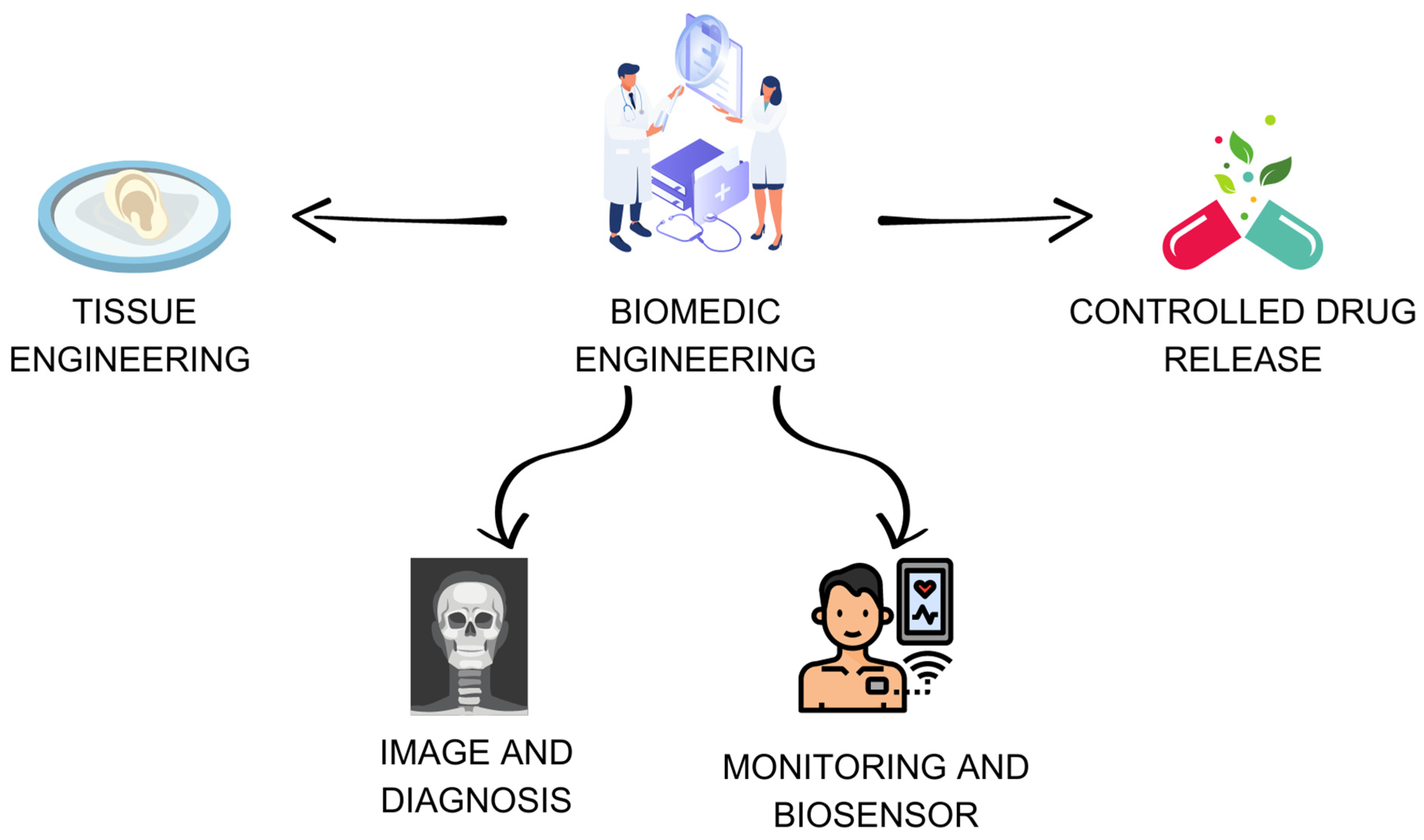
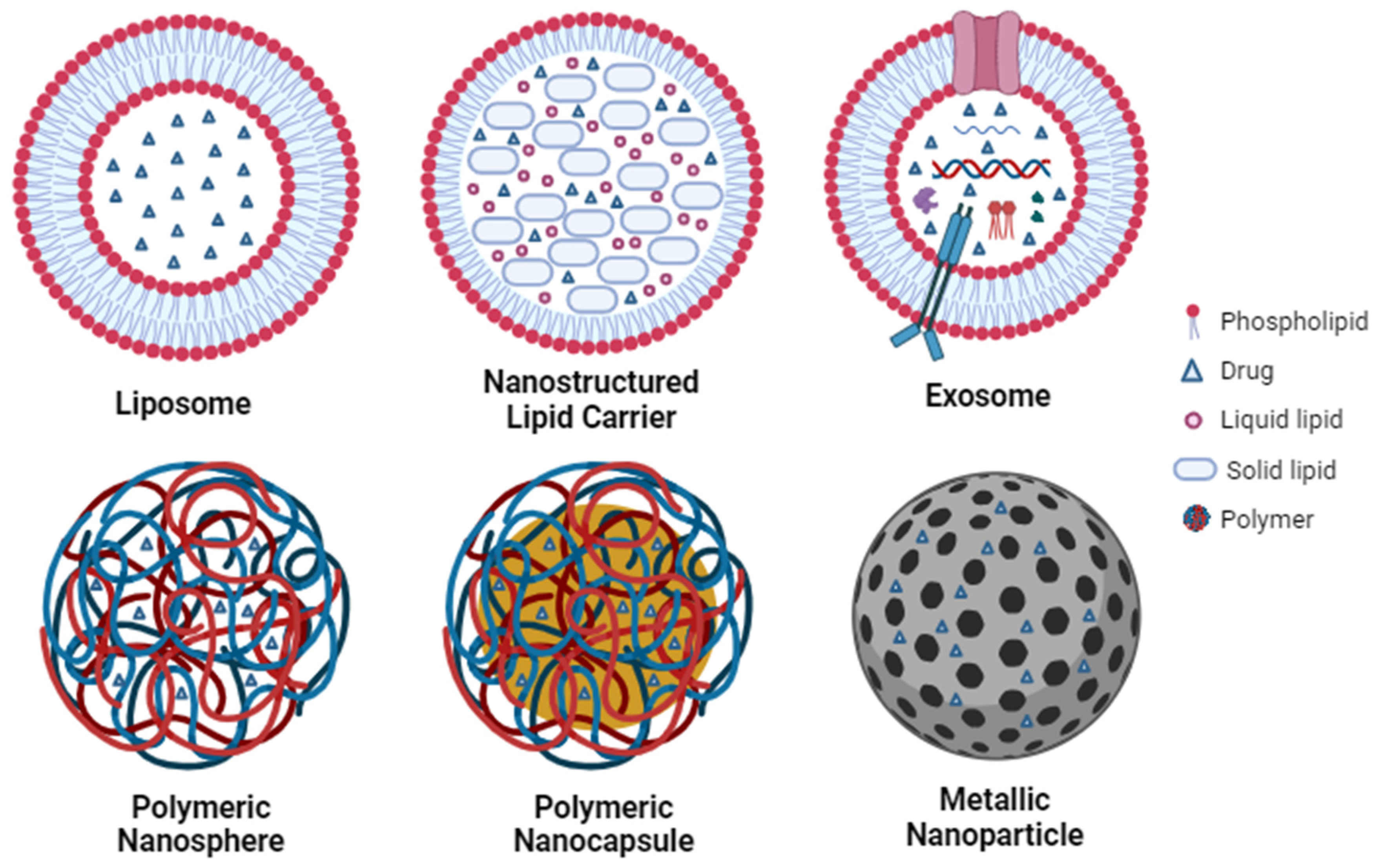
| Nanosystem | Composition | Therapeutic Application | Characterization | Reference |
|---|---|---|---|---|
| Liposome | PEG-coated liposomal doxorubicin | Treatment of breast cancer | — | [134] |
| Liposome system carrying prostaglandin E-1 (PGE-1) | Treatment of cardiovascular diseases | — | [146] | |
| Liposome modified with pyrrolidinium surfactants containing a hydroxyethyl fragment | Transdermal delivery of non-steroidal anti-inflammatory drugs | — | [149] | |
| Liposome coated with PEG | Ischemic myocardium | — | [147] | |
| Exosome | Exosomes by bovine milk | Treatment of lung tumor | Size < 80 nm; PDI of 0.22 ± 0.06 | [155] |
| Exosomal formulations | Treatment of cancer | — | [157] | |
| Exosomes derived from adipose tissue-derived mesenchymal stem cells | Treatment of type 1 diabetes mellitus. | Size ≤ 100; diameter of 41.1 nm | [159] | |
| Exosomes containing curcumin | Treatment of Alzheimer’s disease | — | [160] | |
| Nanostructured lipid carrier | Nanostructured lipid carriers produced for cotransport of lapachone and doxorubicin | Treatment of breast cancer | — | [169] |
| Nanostructured lipid carriers encapsulated with lipophilic low molecular weight EGFR inhibitor | Hepatocellular carcinomas | — | [171] | |
| Nanostructured lipid carriers with omega-3 fatty acids | Reduce oxidation in fortified foods | — | [176] | |
| Nanostructured lipid carriers encapsulating sucupira essential oil | Treatment for diabetes mellitus | Size 148.1 ± 0.9815 nm; PDI 0.274 ± 0.029; ZP from −0.00236 ± 0.147 mV | [179] | |
| Nanostructured lipid carriers encapsulating essential oil of Piper aduncum | Anti-inflammatory activity | — | [180] | |
| Nanostructured lipid carriers encapsulating essential oil of the leaf of Pinus densiflora | Treatment against oral squamous cell carcinoma | 72 ± 1.5 and 120 ± 5.1 nm; PDI 0.1 to 0.29; stability index of 89% | [181] | |
| Polymeric nanoparticle | Polylactic-co-glycolic acid (PLGA) nanoparticles | Treatment of prostate cancer | — | [139] |
| Nanoparticles of hyaluronic acid and lipoic acid encapsulated with doxorubicin | Treatment of multiple myeloma | Size of 183 nm | [187] | |
| PLGA-based polymeric nanoparticles loaded with an immunogenic BCMA72-80 peptide | Treatment of cancer | Size 172 ± 0.73 nm; PDI of 0.20 ± 0.01; Zeta potential of −1.16 ± 0.18 mV | [188] | |
| PLGA nanoparticles containing the programmed cell death ligand (PD-L1) | Activation of dendritic cells and T cells | — | [60] | |
| Iron oxide nanoparticle | Magnetic iron oxide nanoparticles with modified dimercaptosuccinic acid | Inhibition of cell proliferation and greater induction of cell apoptosis | Diameter of 18.7 ± 5.61 nm | [220] |
| Fe3O4 nanoparticles loaded with paclitaxel | Treatment of tumor | — | [221] | |
| Gold nanoparticle | Gold nanoparticles loaded with oligonucleotides | Regulation of genes or immunomodulatory processes | — | [229] |
| Gold nanoparticles | Treatment of cancer | Size 4 nm | [224] | |
| Silver nanoparticle | Gemcitabine encapsulated in silver nanoparticles | Ovarian cancer cells | Size 20 nm; PDI 0.123; Zeta potential −33 mV; | [237] |
| Silver nanoparticles | Prevention of H3N2 influenza virus | Diameter of around 9.5 ± 0.8 nm; stable over 6 months at room temperature | [240] |
| Nanosystem | Characteristics | Application | Main Results | Reference |
|---|---|---|---|---|
| Bismuth oxide nanoparticles (HA-Bi2O3) | - | Bismuth oxide nanoparticles | Exhibit great promise for CT imaging-guided radiotherapy in the diagnosis and treatment of tumors | [291] |
| Perfluorocarbon liposomes with gold nanospheres | Size: 200 nm; light-to-heat conversion effect under 808 nm by NIR laser irradiation | Ultrasound imaging-guided photothermal chemotherapy | A remarkably enhanced ultrasound signal was detected. Exhibited a prominent photothermally reinforced chemotherapeutic effect | [292] |
| Nanobubble-paclitaxel liposome (NB-PTXLp) complexes | Entrapment efficiency of 85.4 ± 4.39% Conjugation efficiency ~98.7 ± 0.14%) Size: 200 nm | Ultrasound imaging and ultrasound-responsive drug delivery in cancer cells | The developed nanobubbles were found to exhibit more than 1-week echogenic stability as opposed to 6 h stability of the commercially available ultrasound contrast agent SonoVue | [293] |
| Magnetic-carbon-quantum-dots-probe-labeled apoferritin nanocages | Average diameters of ~5 nm Polydispersity index of Gd-CDs was 0.201 | Bioimaging and targeted therapy | Unique green photoluminescence and almost no toxicity Could serve as an excellent T1 contrast agent for MRI | [41] |
| Deoxyglucose-conjugated persistent luminescent nanoparticles | Hydrodynamic diameter: 500 nm. Zeta potential was about +1 mV | Diagnostic application in fibrosarcoma tumor model | Significant accumulation of nanoparticles to the tumor site. They showed a higher killing ability for cancer cells compared to normal cells | [294] |
| Functionalized upconverting nanoparticles (UCNPs) | Particle size of 29.0 ± 0.4 nm. Ligands added to the surface: 32 nm for ligand-free UCNPs, to 50 nm for EY-UCNPs, and 100 nm for the EY-PEG-UCNP, with polydispersity indexes of 0.6, 1.2, and 0.4, respectively | Nanophotosensitizers and deep tissue bioimaging agents for simultaneous therapeutic and diagnostic applications | The functionalized UCNPs present deep tissue NIR-II fluorescence under 808 nm excitation, thus demonstrating their potential as bioimaging agents in the NIR-II biological window | [295] |
| Multicore magnetic iron oxide nanoparticles | Low hydrodynamic size of 35 nm, with good monodispersity, PDI < 0.13. Stability over at least 6 months | Mediators for AC-magnetic field hyperthermia and as contrast agents for MRI | Potential application in MRI and magnetic hyperthermia. PEG can stabilize the nanoparticles, which can improve deposition. As well, there is potential for suspensions as T2-contrast agents with good bio-stability and persistent magnetic responses following uptake, which can mark liposome deposition and may provide local hyperthermic hot spots | [296] |
| Gold nanoparticles radiolabeled with 99mTc. | Au-citrate NPs with 13.8 ± 1.2 nm Au-MUAM-PADA ** NPs with 14.0 ± 0.9 nm, Au-MUAM-PADA-99mTc(CO)3 NPs with 13.9 ± 1.2 nm | Theragnostic applications | The multifunctional Au NPs could be used for targeted drug delivery and imaging of cancer cells as well as for monitoring the effectiveness of treatment | [297] |
| Gold nanorods | Particle sizes of 177.9 ± 19.3 nm for eAuNR (PDI 0.46 ± 0.14) and 149.5 ± 9 nm for mAuNR (PDI 0.32 ±0.07) *** | Nanomaterials for target delivery | Important delivery differences exhibited by extracellular vesicles (EVs) or cell membranes- coated nanorods, an understanding of which may be important to the design and development of nanomaterials that use these coatings for target and delivery | [298] |
| rGO-AuNPs- PEG * | Size: 160 nm | Photothermal agent | Very strong SERS signal, NIR-II PA signal and high photothermal efficiency against tumor upon 1061 nm laser irradiation | [299] |
| Lanthanide-activated nanoparticles | - | Biological application: bioimaging, X-ray imaging, MRI, oncotherapy, photodynamic therapy, etc. | Nanosystems with multiple modalities of bioimaging, oncotherapy, and neuromodulation | [300,301] |
| Core/shell lead sulfide/cadmium sulfide (CdS) quantum dots (CSQDs) | Core size to ~5.4 nm for ~1600 nm emission, forming an ~1.5 nm thick CdS shell | Bright fluorescent probe emitting at ~1600 nm in the NIR | Exhibited long blood circulation time, real-time imaging, CSQDs are excreted through the biliary pathway without toxicity effects | [302] |
| SPIONs (iron-oxide NPs) | Surface modification 1,5-dihydroxy-1,5,5-tris-phosphonopentyl-phosphonic acid (di-HMBPs) | Osteoporosis | High affinity for calcium ions/hydroxyapatite for MRI use | [303] |
| Carbon nanotubes | 99mTc-labeled carbon nanotubes | Active bone metabolism | High affinity for hydroxyapatite for photoacoustic imaging use | [304] |
| Quantum dots | Surface modification with various antibodies | Binding to unique cell populations in bone marrow | Targeted cell imaging | [305] |
Disclaimer/Publisher’s Note: The statements, opinions and data contained in all publications are solely those of the individual author(s) and contributor(s) and not of MDPI and/or the editor(s). MDPI and/or the editor(s) disclaim responsibility for any injury to people or property resulting from any ideas, methods, instructions or products referred to in the content. |
© 2023 by the authors. Licensee MDPI, Basel, Switzerland. This article is an open access article distributed under the terms and conditions of the Creative Commons Attribution (CC BY) license (https://creativecommons.org/licenses/by/4.0/).
Share and Cite
Silva, D.F.; Melo, A.L.P.; Uchôa, A.F.C.; Pereira, G.M.A.; Alves, A.E.F.; Vasconcellos, M.C.; Xavier-Júnior, F.H.; Passos, M.F. Biomedical Approach of Nanotechnology and Biological Risks: A Mini-Review. Int. J. Mol. Sci. 2023, 24, 16719. https://doi.org/10.3390/ijms242316719
Silva DF, Melo ALP, Uchôa AFC, Pereira GMA, Alves AEF, Vasconcellos MC, Xavier-Júnior FH, Passos MF. Biomedical Approach of Nanotechnology and Biological Risks: A Mini-Review. International Journal of Molecular Sciences. 2023; 24(23):16719. https://doi.org/10.3390/ijms242316719
Chicago/Turabian StyleSilva, Debora F., Ailime L. P. Melo, Ana F. C. Uchôa, Graziela M. A. Pereira, Alisson E. F. Alves, Maria C. Vasconcellos, Francisco H. Xavier-Júnior, and Marcele F. Passos. 2023. "Biomedical Approach of Nanotechnology and Biological Risks: A Mini-Review" International Journal of Molecular Sciences 24, no. 23: 16719. https://doi.org/10.3390/ijms242316719