Article

Characterization and Seasonal Modulation of Adenosine A1 Receptors in the Arctic Ground Squirrel Brain

1
Institute of Arctic Biology, University of Alaska, Fairbanks, AK 99775, USA
2
Center for Transformative Research in Metabolism, University of Alaska, Fairbanks, AK 99775, USA
*
Author to whom correspondence should be addressed.
Current address: Department of Life Sciences, University of Nevada, Las Vegas, NV 89119, USA.
Int. J. Mol. Sci. 2023, 24(2), 1598; https://doi.org/10.3390/ijms24021598
Submission received: 24 November 2022 / Revised: 7 January 2023 / Accepted: 8 January 2023 / Published: 13 January 2023

Abstract

:
Hibernation is an adaptation that allows animals such as the Arctic ground squirrel (AGS) to survive the absence of food or water during the winter season. Understanding mechanisms of metabolic suppression during hibernation torpor promises new therapies for critical care. The activation of the Adenosine A1 receptor (A1AR) has been shown to be necessary and sufficient for entrance into hibernation with a winter season sensitization to the agonist, but the role of the A1AR in seasonal sensitization is unknown. In the current study, we characterize the A1AR in the forebrain, hippocampus and hypothalamus of summer and torpid AGS. For the first time, we define the pharmacological characteristics of the A1AR agonist, N6-cyclohexyladenosine and the A1AR antagonist dipropylcyclopentylxanthine (DPCPX) in the AGS brain. In addition, we test the hypothesis that increased A1AR agonist efficacy is responsible for sensitization of the A1AR during the torpor season. The resulting 35S-GTPγS binding data indicate an increase in agonist potency during torpor in two out of three brain regions. In addition to 35S-GTPγS binding, [3H]DPCPX saturation and competition assays establish for the first-time pharmacological characteristics for the A1AR agonist, N6-cyclohexyladenosine and the A1AR antagonist dipropylcyclopentylxanthine (DPCPX) in AGS brain.

1. Introduction

Torpor is a fundamental metabolic state of energy conservation. Hibernation, defined by prolonged torpor lasting days to two or more weeks, interrupted by brief 12–24 h episodes of euthermic metabolism, represents some of the most extreme examples of mammalian energy conservation. Evidence suggests that hibernation is an extension of sleep that involves the modulation of thermogenesis [1]. In ground squirrels, suppression of thermogenesis is sufficient to account for the initial fall in metabolic rate, after which a decline in core body temperature suppresses metabolic rate further through thermodynamic influence on metabolic processes [2]. Adenosine, a ubiquitous neuromodulator present in most tissues, modulates thermogenesis in the CNS [3,4]. The purine binds to four subtypes of G-protein coupled receptors, designated the adenosine A1, A2A, A2B and A3 receptors (A1AR, A2aAR, A2bAR, and A3AR, respectively). In the brain, A1AR and A2AAR are widely expressed with only a small amount of A3AR, whereas A2BAR is only present in peripheral tissues. A1AR and A3AR receptors propagate their signal through Gi/o G-proteins and are neuroinhibitory. In contrast, the A2AAR and A2BAR interact with Gs proteins and are neuroexcitatory.
Stimulation of A1AR within the CNS is necessary for the onset of torpor in hibernating Arctic Ground Squirrels (AGS; Urocitellus parryii); however, the response is regulated by a process entrained to a circannual rhythm that governs seasonal sensitivity to the A1AR agonist N6-cyclohexyladenosine (CHA). Evidence from our laboratory suggests that the winter season enhances the influence of CHA on thermogenesis within the median preoptic nucleus and the rostral raphe pallidus to inhibit the premotor sympathetic neurons within the rPA that drive thermogenesis [5]. The mechanism underlying seasonal sensitivity to CHA is unknown but may involve changes in adenosinergic tone within thermoregulatory nuclei in the hypothalamus or changes at the level of the A1AR.
Sleep and thermogenesis are modulated, in part, by A1AR [6] and A2AR [7]. Hibernation, sleep and thermogenesis show seasonal rhythms in ground squirrels [1,8] and humans [9], but it is unknown if the properties of A1AR agonists or the expression of A1AR vary with season. In the current study, we establish for the first-time pharmacological characteristics of the adenosine agonist CHA and antagonist DPCPX in AGS brain tissue. In addition, we test the hypothesis that a seasonal shift in the potency of CHA, the A1AR agonist most studied regarding A1AR agonist-induced hibernation and torpor, is associated with the seasonal expression of hibernation in AGS.

2. Results

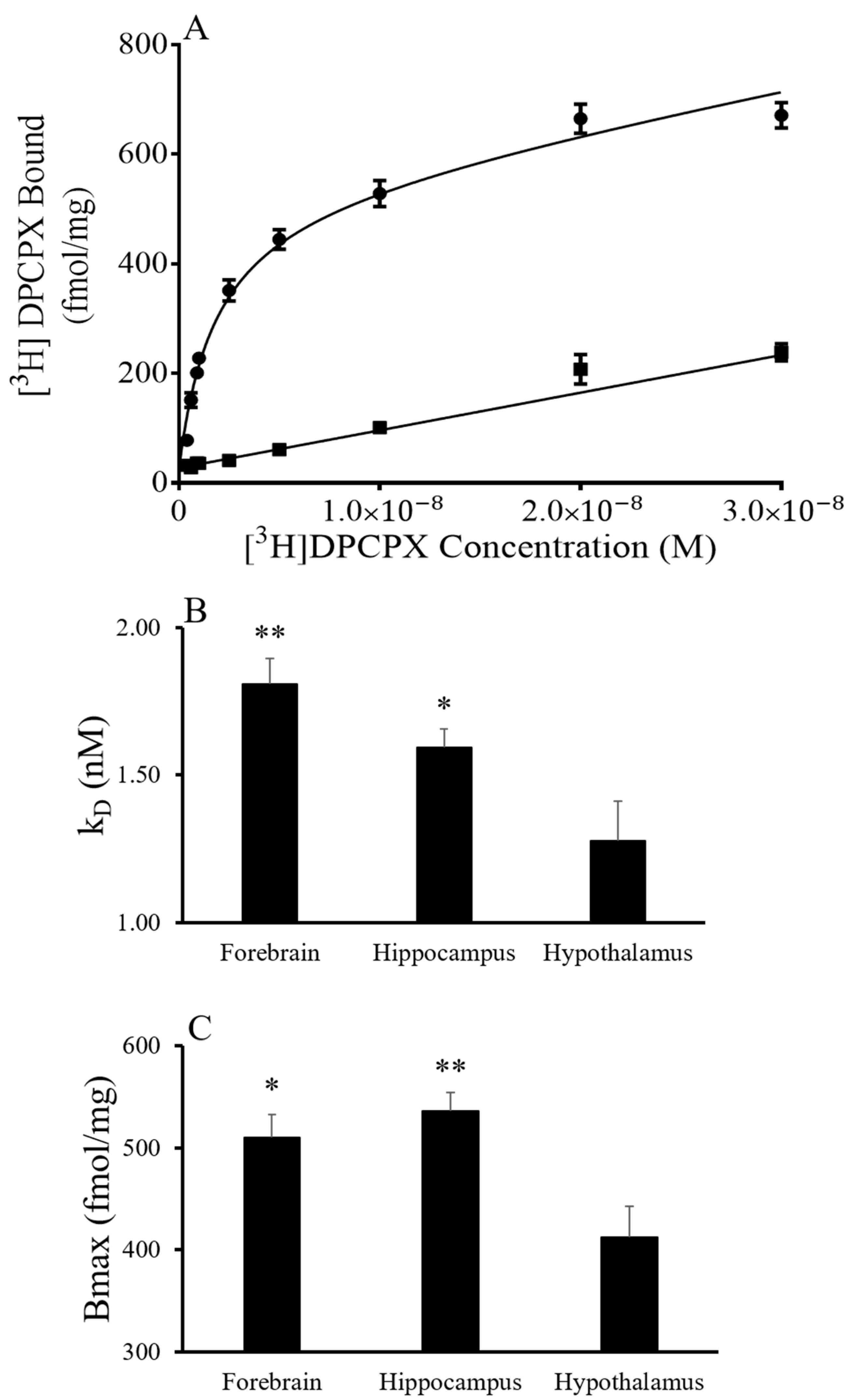
To characterize the A1AR, saturation and displacement assays were conducted in the forebrain, hippocampus and hypothalamus of AGS euthanized during the summer and torpid state in the winter season. Saturation experiments using [3H] DPCPX indicated a single site model and yielded KD and Bmax values that showed an effect of brain region but not of the season (Figure 1). The saturation curves for [3H] DPCPX binding were similar in the forebrain, hypothalamus and hippocampus of tissue collected in summer and from torpid AGS in winter (Figure 1). The results indicate that the hypothalamus has a lower KD and Bmax than the forebrain and hippocampus independent of the season (p = 0.0017, two-way ANOVA, the main effect of the region for KD; p = 0.0022, the main effect of the region for Bmax).
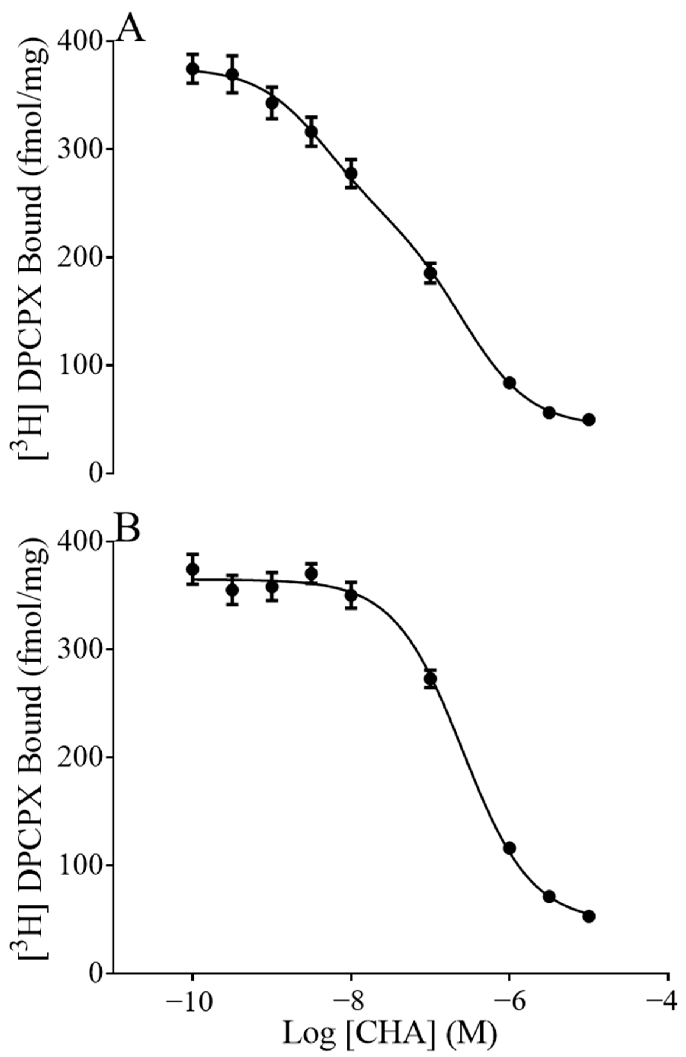
To ask if the fraction of receptors in the high or low affinity states was associated with seasonal sensitivity to CHA, we displaced [3H]DPCPX with CHA. As expected, displacement assays fit a two-site model indicating the presence of a low and high affinity site (pkiHi,PkiLo). Furthermore, in the presence of GTP, only the pkiLo site was detected, as high concentrations of GTP promote the disassociation of G-proteins from GPCRs (Figure 2). As expected, the pkiLo established by the two-site model did not differ from the GTP shift pkiLo, arguing that the GTP shift did indeed isolate the low affinity site. Here we found that the fraction of receptors in the high affinity state (Fraction High) was greater during torpor than during summer (p = 0.0071, two-way ANOVA, main effect of season, Table 1). In addition, there was an effect of region on the affinity of CHA at the low affinity site where the pkiLo in the hippocampus was significantly less than the pkiLo in the forebrain and hypothalamus, regardless of season (p < 0.0001, two-way ANOVA, the main effect of region, with post-hoc Tukey p < 0.0001 hippocampus vs. forebrain; p < 0.0001 hippocampus vs. hypothalamus). The ratio of dissociation constants at the high and low affinity sites (pkiHi/pkiLo) was also greater in hippocamps than in other regions (p < 0.001 two-way ANOVA, main effect of region). Post-hoc analysis showed that the hippocampus differed from the hypothalamus (p < 0.001, Tukey) and trended towards being different from the forebrain (p = 0.094).
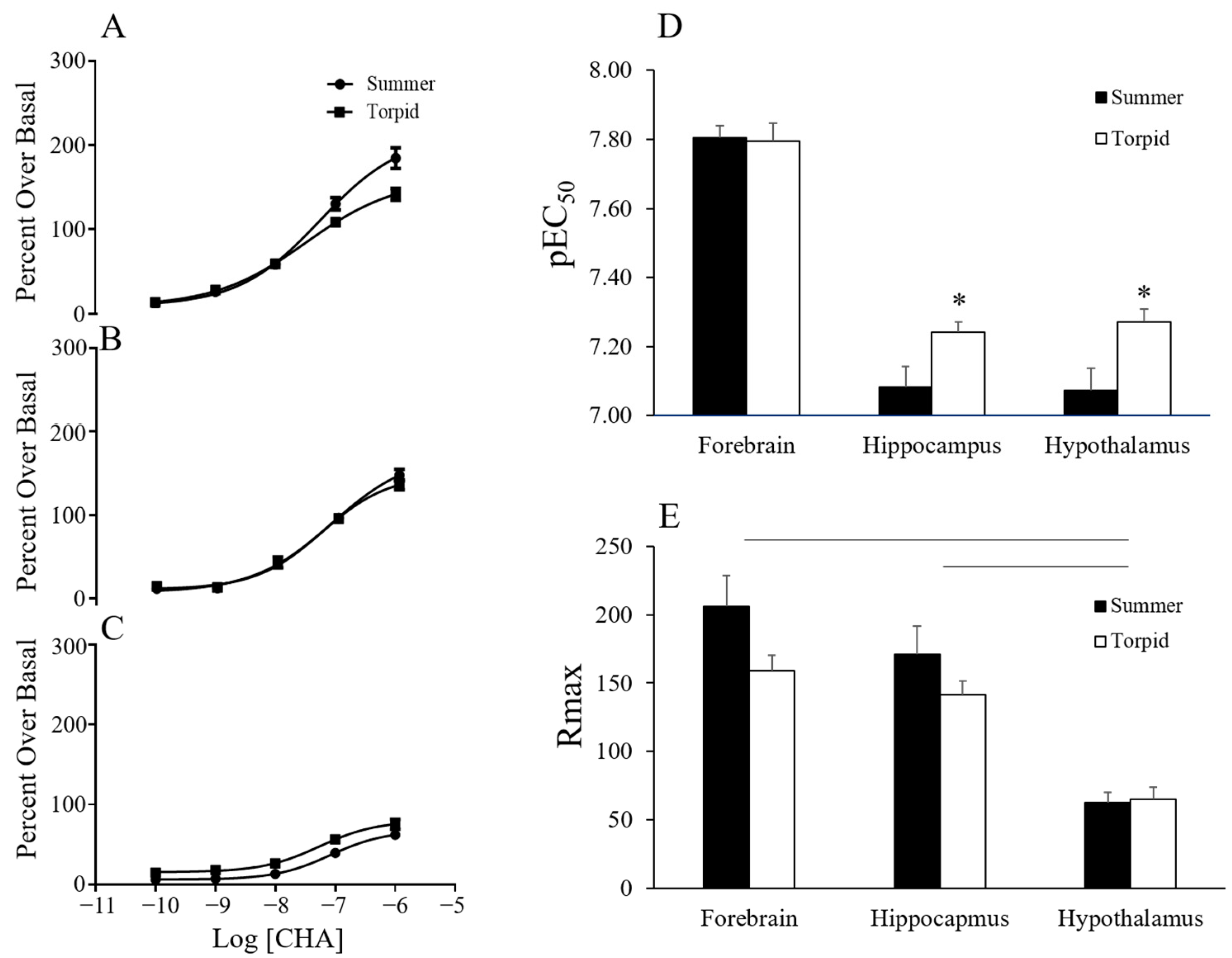
The functional response of CHA was investigated by agonist-induced 35S-GTPγS binding. Concentrations of CHA up to 1 µM stimulated 35S-GTPγS binding in a single-site manner. The resulting data revealed an effect of season and brain region on the pEC50 as well as an effect of region on the maximum signal (Rmax) (Figure 3). CHA was more potent in tissue collected during torpor than during summer (p < 0.005, two-way ANOVA, the main effect of the season). A trend towards an interaction between region and season (p < 0.091) led us to perform further one-way analyses over the season that showed increased potency of CHA in torpor in the hypothalamus (p < 0.05, t-test) and in the hippocampus (p < 0.05, t-test) but not in the forebrain (Figure 3). The Rmax was greatest in the forebrain and hippocampus and lowest in the hypothalamus (p < 0.0001 two-way ANOVA, the main effect of region, with post-hoc Tukey p < 0.0001 hippocampus vs. hypothalamus; p < 0.0001 hypothalamus vs. forebrain). During the characterization of the functional response of CHA, a low hillslope was observed at concentrations greater than 1 µM. A low hillslope is indicative of negative cooperativity or a second lower affinity binding site. CHA has been shown to have a low affinity for the A3AR in the rat brain [10], and therefore A3AR could be stimulated by high concentrations of CHA. We hypothesized that CHA at concentrations above 1 µM would stimulate the A3AR resulting in a low hillslope. To eliminate A3AR binding, we repeated the experiment in the presence of an A3AR antagonist (MRS 1334; [11]. In AGS, summer and torpid forebrain tissue, pre-blocked with 500 nM of MRS 1334, was stimulated with up to 100 µM CHA and 35S-GTPγS binding was measured (Figure 4). The inclusion of MRS1334 did not have an effect on the hillslope of CHA-induced 35S-GTPγS binding in either season, providing evidence that the A3AR stimulation was not contributing to the low hillslope. The potency of CHA, as indicated by the pEC50, decreased in the presence of MRS1334, and the decrease was greater in tissue from torpid ASG than in tissue from summer AGS (p < 0.0001, two-way ANOVA, the main effect of treatment; p < 0.05 treatment x season). Interestingly, the Rmax only increased in the torpid tissue (p < 0.05, t-test torpid MRS1334 vs. vehicle).
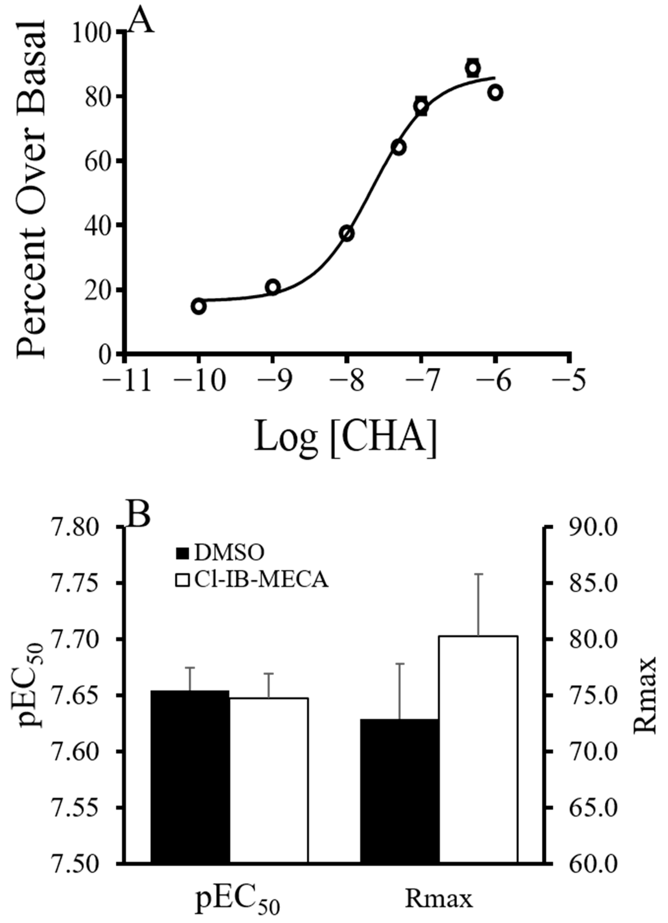
It has been well established that GPCRs form dimers and higher order oligomers with GPCRs which can affect agonist signaling. If the effect of blocking A3AR on Rmax or pEC50 was due to A1AR and A3AR cross-talk, we asked if stimulating A3AR would cause a change in Rmax or pEC50. The A3AR was stimulated with 132 nM 2-Chloro-N6-(3-iodobenzyl)-adenosine-5′-N-methyluronamide (Cl-IB–MECA). CHA-stimulated GDP/GTP exchange in the forebrain of summer AGS was then measured at concentrations up to 1.0 µM CHA using 35S-GTPγS binding assay (Figure 5). There was no effect of Cl-IB-MECA on Rmax or pEC50.

3. Discussion

This study is the first to characterize A1AR in the brain of summer and torpid AGS and to identify changes at the receptor level that may contribute to the seasonal shift in sensitivity to torpor-inducing effects of CHA. Results indicate that the potency of A1AR agonists, reflected by the EC50 in the GTP binding assay, increases in the hippocampus and hypothalamus during torpor when compared to the summer season. However, the shift in potency could not be explained by an increase in affinity or efficacy with the assays used and sample size available from the tissue bank. These findings have implications for the seasonality of sleep and thermoregulation in humans. as well as the seasonal efficacy of therapeutics.

3.1. Characterization of A1AR Ligand Binding and Agonist-Induced GDP/GTP Exchange

Direct ligand binding and competition assays have, for the first time, characterized the receptor pharmacology of CHA and DPCPX in AGS brain tissue. As expected, [3H] DPCPX saturation experiments produced a one-site binding curve at the A1AR [12]. KD for DPCPX was similar to KD’s reported in rat brain and smooth muscle preparations as well as sheep pineal membranes [13,14,15]), although the affinity of ligands for adenosine receptors are species dependent [16]. Bmax for DPCPX binding was consistent between brain regions, as would be expected given the ubiquitous distribution of A1AR in the brain.
CHA was found to induce GDP/GTP exchange as expected for an A1AR agonist, as shown in rat brain tissue. GTP produced a characteristic shift to a single low affinity binding site (Kilo); the magnitude of this shift represented by the ratio of the pKiHi/pKiLo, a measure of agonist efficacy, was similar between seasons in the hypothalamus and hippocampus where potency was found to be greater in torpid/compared to summer. In the forebrain, where potency did not change with the season, we found evidence of an increased abundance of receptors in the high affinity state. A higher fraction of receptors in the high affinity state could indicate a greater proportion of functional receptors coupled to G-protein. Although an increase in efficacy could not explain seasonal differences in potency in the hippocampus or hypothalamus, we saw a clear difference in efficacy between brain regions. The maximal cellular response to agonist-stimulated GDP/GTP exchange, another measure of agonist efficacy, varied between brain regions with more than a 5-fold difference between the highest response in the forebrain and the lowest response in the hypothalamus.

3.2. Potential Interaction between GPCR

A hillslope of less than one was noted at higher concentrations of CHA which was not due to binding at the A3AR site, although other binding sites could not be ruled out. Given that the A2BAR is not normally expressed in the brain and CHA has a very low affinity for the A2AAR, the second site is most likely not another adenosine receptor. The low Hill slope at higher concentrations of CHA could also be due to negative cooperativity that occurs when the A1AR forms homomers [17].
Interestingly, inhibition of the A3AR decreased the potency of CHA independent of season, suggesting positive cross-talk between the A1AR and the A3AR. However, results argued against cross-talk because stimulating the A3AR did not increase the potency of CHA. Alternatively, 500 nM of MRS 1334 may have inhibited the A1 receptor.
In summary, evidence supports a role for increased efficacy of CHA in the hypothalamus and hippocampus during the winter season as a mechanism that may contribute to seasonal sensitivity to CHA. Direct ligand binding and measurements of GDP/GTP exchange in AGS brain tissue yield results consistent with the behavior of A1AR ligands in other species. Evidence for a seasonal change in the receptor pharmacology of A1AR agonists demonstrates that endogenous rhythms may influence drug-receptor interactions. Similar seasonal influences in humans could have clinical implications for the vast pharmacopeia of GPCR ligands. Although the results reported here cannot explain the mechanism for altered efficacy, further study of the mechanism would have translational significance for developing A1AR agonists as therapeutics. The means to increase CNS efficacy would minimize individual differences in response to CHA reported previously [18,19] and potentially decrease peripheral side-effects such as hypotension and bradycardia by decreasing the therapeutic dose of agonist. Although A1AR agonists have limited translational potential due to peripheral side-effects, combining a CNS active agonist with an antagonist that does not cross the BBB shows promise as a means to target CNS sites of action [20]. A limitation of the study is that we did not include a winter group that was not torpid. There is a possibility that characteristics of the A1AR or CHA activation of the A1AR change between interbout arousal and torpor. Although we cannot rule out this possibility, we expect that by using the same assay temperature in summer and winter/torpid tissues, we avoided many of the influences of torpor that could have confounded the interpretation of a seasonal effect. Seasonal alteration of CHA pharmacokinetics or endogenous levels of adenosine may also play a role but were beyond the scope of this study. Translating hibernation for human medicine and defining mechanisms that underly seasonal sleep drive and sensitivity to therapeutics have broad implications for the future of therapeutics.

4. Materials and Methods

4.1. Arctic Ground Squirrels

AGS tissue was obtained from a tissue bank (courtesy of B. Barnes, Fairbanks, AK, USA). All animal procedures were approved by the UAF Institutional Animal Care and Use Committee (protocol #06-44). AGS were captured near 66°38′ N, 149°38′ W under permit from the Alaska Department of Fish & Game. Animals were housed at 22 °C on an 18:6 day: night cycle (5/2011–8/2011) and at 2 °C on a 4:20 day: night cycle (8/2011-time of tissue collection; 1/2011). Hibernation was monitored using the “shavings added” method, where hibernation (torpor) is indicated when shavings placed on the back of the AGS remain undisturbed 24 h later [21,22]. All tissue was harvested from adult male AGS during the summer season or while torpid during the winter season. The summer season was defined as AGS that were captured after the previous hibernation season and kept in captivity for two months before tissue collection. Tissue from torpid AGS were collected during the winter season, after at least six to eleven torpor bouts and at least ten but not more than thirteen days in the current torpor bout.
Summer AGS were euthanized by decapitation under a surgical plane of anesthesia (isoflurane, 4% mixed with 100% medical grade oxygen, delivered at 1.5 L/min until unresponsive to a toe pinch). Torpid AGS were euthanized without being aroused from torpor and did not require anesthesia nor breathe at a rate sufficient to absorb the gas anesthesia. Immediately following euthanasia, the brain was removed, and the hippocampus, hypothalamus and remaining forebrain were isolated and frozen in liquid nitrogen. All tissue was stored at −80 °C until use.

4.2. Isolation of Plasma Membrane for 35S GTPγS and [3H]DPCPX Binding Experiments

AGS membranes from the forebrain, hippocampus, hypothalamus and brainstem were isolated as described previously with modifications [23]. Briefly, tissue was homogenized on ice using an all glass Dounce homogenizer (10–15 strokes) in 20× volume homogenization buffer containing 10 mM HEPES, 2 IU/mL ADA, 640 mM sucrose and protease inhibitor tablets (Roche, Indianapolis, IN, USA) and then further homogenized by polytron for 10–15 s. The suspension was centrifuged at 1000× g for 10 min at 4 °C. Resultant supernatant was centrifuged at 48,000× g for 15 min at 4 °C. Pellets were resuspended in Resuspension buffer containing 10 mM HEPES, 2 IU/mL ADA and protease inhibitor tablets. The suspension was centrifuged at 48,000× g for 15 min at 4 °C. Pellets of AGS hippocampus and hypothalamus were suspended in a solution containing 6 mM HEPES, 122 µM GDP and 2.4 IU/mL ADA, the forebrain was suspended in 6 mM HEPES, 77 µM GDP, and 0.5 IU/mL ADA and both were incubated at room temperature under gentle rocking for 60 min and then centrifuged at 48,000× g for 30 min. Subsequent pellets were resuspended in a Resuspension buffer and stored at −80 °C until use.

4.3. [3H] DPCPX Binding

To ask if membrane expression of the A1AR increased in the winter season, we performed saturation experiments to determine the KD and Bmax of [3H] DPCPX binding to A1AR were conducted on the membrane of the forebrain, hippocampus and hypothalamus of winter and summer AGS following the guidelines of (Hulme 2010) with modification [22]. On the day of the experiment, aliquots of summer and torpid AGS were thawed on ice. The protein content of each animal was determined by protein analysis (Bio-Rad, Hercules, CA, USA) followed by centrifugation at 48,000× g for 30 min at 4 °C. The pellet was then resuspended in a solution containing 50 mM HEPES and 2 IU/mL ADA. Saturation experiments were performed by incubating 100 µg/mL protein with nine concentrations of [3H] DPCPX ranging between 0.4 and 30 nM in the presence of 50 mM HEPES and 2 IU/mL ADA. Non-specific binding was defined in the presence of 7 µM cyclopentyltheophylline (CPT). The solution was allowed to incubate for 90 min at room temperature, and the membrane bound ligand was isolated as described below.

4.4. 35S-GTPγS Binding

To investigate the functional response of CHA activation of the A1AR in summer and torpid animals, 35S-GTPγS binding experiments were performed as described previously with modifications [23]. On the day of the experiment, aliquots of summer and torpid AGS were thawed on ice. The protein content was then determined by protein analysis (Bio-Rad, Hercules, CA, USA) followed by centrifugation at 48,000× g for 30 min at 4 °C. The pellet was then resuspended in Assay buffer containing 50 mM HEPES, 200 mM NaCl, 10 mM MgCl2, 40 µM GDP, 100 µM Saponin, 1 IU/mL ADA and 1 mM DTT at pH 7.4. 100 µg per ml protein was incubated with 400 pM of 35S-GTPγS in a total volume of 100 µL for 90 min under gentle rocking at 37 °C. The non-specific activity was determined in the presence of 5 µM GTPγS. The constitutive activity was defined as binding in the absence of CHA. The reaction was terminated by rapid vacuum filtration, and then each well was washed three times with 200 µL of ice cold 50 mM HEPES. The plate was allowed to dry overnight. 40 µL of scintillation cocktail (PerkinElmer, Waltham, MA, USA) was added to each well, and 35S activity was determined in a 1450 Microbeta plus microplate scintillation counter (PerkinElmer, Waltham, MA, USA) utilizing a one-minute counting time. The effect of the A3AR was determined by preincubating the membrane on ice with an A3AR antagonist (MRS 1334, 500 nM) or agonist (Cl-IB-MECA, 132 nM) for at least one hour before conducting the 35S-GTPγS binding experiment.
To ask if the efficacy of CHA or the percentage of A1AR receptors in the high or low affinity state could explain the seasonal difference in response, KiLo was determined by displacing 1 nM [3H] DPCPX with nine concentrations of CHA ranging between 100 pM and 10 µM in the presence of 100 µg/mL protein, 50 mM HEPES, 2 IU/mL ADA and 1 mM GTP. KiHi was defined by displacing 1 nM [3H] DPCPX (PerkinElmer, Waltham, MA, USA) with CHA in the same manner as the KiLo experiments but without GTP. The solution was allowed to equilibrate for 90 min at room temperature, as indicated by kinetic experiments. Free and bound [3H] DPCPX was separated through an Inotech glass fiber filter pad (0.35 mM thickness/0.75 µM retention) (Inotech Bio. Sys., Derwood, MD, USA) by rapid filtration (0.5 mL per sec per well) with a cell harvester (Tomtec, Hamden, CT, USA). The filter was then allowed to dry overnight. The next morning each well was isolated and placed in a scintillation vial. Scintillation cocktail (PerkinElmer, Waltham, MA, USA) was added, and radioactivity was determined (1450 Microbeta Plus, PerkinElmer, Waltham, MA, USA) with a five-minute count per well. Unlabeled agonists and antagonists were obtained from Sigma (St. Louis, MO, USA).

4.5. Data Analysis

35S-GTPγS specific binding was determined by subtracting non-specific binding from overall binding. Specific binding was converted to percent over constitutive receptor activity. pEC50, Hill slope and span were determined using the function Log(agonist) vs. response—variable slope (four parameters) in Graphpad Prism 5 (v 5.04) (Graphpad Software, La Jolla, CA, USA).
[3H] DPCPX bound was converted from cpm to fmol per mg protein, and the specific binding was calculated. A sum of squares F-test was used to determine if a one-site or two-site model was appropriate, and then the KD and Bmax were calculated using the appropriate model using Graphpad Prism. KiLo for the displacement of [3H] DPCPX in the presence of GTP was calculated using the average KD (one or two site—Fit Ki). KiHi was calculated for the displacement of [3H] DPCPX without GTP (one or two site—Fit Ki) using the average KD and KiLo. Fraction Hi is the fraction of all the sites that have a high affinity for the competitor. It is calculated by Graphpad prism via the below equations.
Part1 = FractionHi * Span/(1 + 10^(X − LogEC50Hi))
Part2 = (1 − FractionHi) * Span/(1 + 10^(X − LogEC50Lo))
Data were analyzed by two-way ANOVA across brain region and season, followed by Tukey post-hoc comparisons or t-tests where indicated (R Studio). The significance threshold was defined as p < 0.05. Data are shown as mean ± SEM.

Author Contributions

Conceptualization, Z.C. and K.D.; methodology and formal analysis, Z.C.; writing—original draft preparation, Z.C.; writing—review and editing, Z.C. and K.D.; supervision, project administration, funding acquisition, K.D. All authors have read and agreed to the published version of the manuscript.

Funding

The research reported in this publication was supported by an Institutional Development Award (IDeA) from the National Institute of General Medical Sciences of the National Institutes of Health under grant numbers P20GM130443 and 2P20GM103395. The content is solely the responsibility of the authors and does not necessarily reflect the official views of the NIH. The APC was funded by P20GM130443.

Institutional Review Board Statement

All animal procedures were approved by the UAF Institutional Animal Care and Use Committee (protocol #06-44).

Informed Consent Statement

Not applicable.

Data Availability Statement

The data presented in this study are available on request from the corresponding author.

Acknowledgments

We thank Brian Barnes for the AGS tissue used in this study, Jeanette Moore for helpful discussions and Sarah Zieschang for manuscript formatting and submission.

Conflicts of Interest

Kelly Drew has a financial interest in Be Cool Pharmaceutics.

References

  1. Walker, J.M.H.E.; Berber, R.J.; Heller, H.C. Hibernation and circannual rhythms of sleep. Physiol. Zool. 1980, 53, 8–11. [Google Scholar] [CrossRef]
  2. Geiser, F. Ecological Physiology of Daily Torpor and Hibernation; Springer: Berlin/Heidelberg, Germany, 2021; pp. 133–136. [Google Scholar]
  3. Tupone, D.; Madden, C.J.; Morrison, S.F. Central Activation of the A1 Adenosine Receptor (A1AR) Induces a Hypothermic, Torpor-Like State in the Rat. J. Neurosci. 2013, 33, 14512–14525. [Google Scholar] [CrossRef] [PubMed] [Green Version]
  4. Ticho, S.R.; Radulovacki, M. Role of adenosine in sleep and temperature regulation in the preoptic area of rats. Pharmacol. Biochem. Behav. 1991, 40, 33–40. [Google Scholar] [CrossRef]
  5. Frare, C.; Jenkins, M.E.; McClure, K.M.; Drew, K.L. Seasonal decrease in thermogenesis and increase in vasoconstriction explain seasonal response to N6-cyclohexyladenosine-induced hibernation in the Arctic ground squirrel (Urocitellus parryii ). J. Neurochem. 2019, 151, 316–335. [Google Scholar] [CrossRef] [PubMed]
  6. Porkka-Heiskanen, T.; Kalinchuk, A.V. Adenosine, energy metabolism and sleep homeostasis. Sleep Med. Rev. 2011, 15, 123–135. [Google Scholar] [CrossRef]
  7. Lazarus, M.; Oishi, Y.; Bjorness, T.E.; Greene, R.W. Gating and the Need for Sleep: Dissociable Effects of Adenosine A1 and A2A Receptors. Front. Neurosci. 2019, 13, 740. [Google Scholar] [CrossRef] [PubMed]
  8. Olson, J.M.; Jinka, T.R.; Larson, L.K.; Danielson, J.J.; Moore, J.T.; Carpluck, J.; Drew, K.L. Circannual rhythm in body temperature, torpor, and sensitivity to A(1) adenosine receptor agonist in arctic ground squirrels. J. Biol. Rhythms. 2013, 28, 201–207. [Google Scholar] [CrossRef] [Green Version]
  9. Yetish, G.; Kaplan, H.; Gurven, M.; Wood, B.; Pontzer, H.; Manger, P.R.; Siegel, J.M. Natural sleep and its seasonal variations in three pre-industrial societies. Curr. Biol. 2015, 25, 2862–2868. [Google Scholar] [CrossRef] [Green Version]
  10. Mazzoni, M.R.; Buffoni, R.S.; Giusti, L.; Lucacchini, A. Characterization of a low affinity binding site for N6-substituted adenosine derivatives in rat testis membranes. J. Recept. Signal Transduct. 1995, 15, 905–929. [Google Scholar] [CrossRef]
  11. Hill, S.J.; May, L.T.; Kellam, B.; Woolard, J. Allosteric interactions at adenosine A1and A3receptors: New insights into the role of small molecules and receptor dimerization. Br. J. Pharmacol. 2014, 171, 1102–1113. [Google Scholar] [CrossRef]
  12. Lohse, M.; Klotz, K.-N.; Lindenborn-Fotinos, J.; Reddington, M.; Schwabe, U.; Olsson, R.A. 8-Cyclopentyl-1,3-dipropylxanthine (DPCPX) ? a selective high affinity antagonist radioligand for A1 adenosine receptors. Naunyn-Schmiedeberg’s Arch. Pharmacol. 1987, 336, 204–210. [Google Scholar] [CrossRef]
  13. León, D.; Albasanz, J.L.; Fernández, M.; Ruíz, M.A.; Martín, M. Down-regulation of rat brain adenosine A1 receptors at the end of pregnancy. J. Neurochem. 2004, 88, 993–1002. [Google Scholar] [CrossRef] [PubMed]
  14. Peachey, J.; Hourani, S.; Kitchen, I. The binding of 1,3-[3H]-dipropyl-8-cyclopentylxanthine to adenosine Ai receptors in rat smooth muscle preparations. Br. J. Pharmacol. 1994, 113, 1249–1256. [Google Scholar] [CrossRef] [PubMed]
  15. Falcón, J.; Privat, K.; Ravault, J.-P. Binding of an adenosine A1 receptor agonist and adenosine A1 receptor antagonist to sheep pineal membranes. Eur. J. Pharmacol. 1997, 337, 325–331. [Google Scholar] [CrossRef] [PubMed]
  16. Klotz, K.-N.; Vogt, H.; Tawfik-Schlieper, H. Comparison of A1 adenosine receptors in brain from different species by radioligand binding and photoaffinity labelling. Naunyn-Schmiedeberg’s Arch. Pharmacol. 1991, 343, 196–201. [Google Scholar] [CrossRef] [PubMed]
  17. Gracia, E.; Moreno, E.; Cortés, A.; Lluís, C.; Mallol, J.; McCormick, P.J.; Canela, E.I.; Casadó, V. Homodimerization of adenosine A1 receptors in brain cortex explains the biphasic effects of caffeine. Neuropharmacology 2013, 71, 56–69. [Google Scholar] [CrossRef]
  18. Jinka, T.R.; Carlson, Z.A.; Moore, J.T.; Drew, K.L. Altered thermoregulation via sensitization of A1 adenosine receptors in dietary-restricted rats. Psychopharmacology 2010, 209, 217–224. [Google Scholar] [CrossRef] [Green Version]
  19. Bailey, I.R.; Laughlin, B.; Moore, L.A.; Bogren, L.K.; Barati, Z.; Drew, K.L. Optimization of Thermolytic Response to A1 Adenosine Receptor Agonists in Rats. J. Pharmacol. Exp. Ther. 2017, 362, 424–430. [Google Scholar] [CrossRef] [Green Version]
  20. Jinka, T.R.; Combs, V.M.; Drew, K.L. Translating Drug-Induced Hibernation to Therapeutic Hypothermia. ACS Chem. Neurosci. 2015, 6, 899–904. [Google Scholar] [CrossRef] [Green Version]
  21. Lyman, C.P. The oxygen consumption and temperature regulation of hibernating hamsters. J. Exp. Zool. 1948, 109, 55–78. [Google Scholar] [CrossRef]
  22. Pengelley, E.T.; Fisher, K.C. Rhythmical arousal from hibernation in the golden-mantled ground squirrel, citellus lateralis tescorum. Can. J. Zool. 1961, 39, 15. [Google Scholar] [CrossRef]
  23. Giuntini, J.; Giusti, L.; Lucacchini, A.; Mazzoni, M.R. Modulation of A1 adenosine receptor signaling by peroxynitrite. Biochem. Pharmacol. 2003, 67, 375–383. [Google Scholar] [CrossRef] [PubMed]
Figure 1. Kinetics of [3H] DPCPX binding in AGS with results from summer and torpid groups combined. Binding of [3H] DPCPX in the forebrain was similar in appearance to that seen in the hippocampus and hypothalamus (A). [3H] DPCPX demonstrated nanomolar affinity for the A1AR in the three tissues tested. The affinity of [3H] DPCPX binding was lower in the hypothalamus than in the other brain regions (B). Also, the number of A1AR, indicated by Bmax, in the hypothalamus during the torpid season was less than in the forebrain and hippocampus (C) ** p < 0.01, * p < 0.05 vs. hypothalamus, Tukey, n = 4–8.
Ijms 24 01598 g001
Figure 2. [3H] DPCPX displaced with CHA in the presence or absence of GTP. Summer forebrain binding curves were best fit by a two-site model in the absence of 1 µM GTP (A) and one-site in the presence of 1 µM GTP (B). The general characteristics of these graphs were conserved in the winter season of the forebrain as well as the hippocampus and hypothalamus (not shown).
Ijms 24 01598 g002
Figure 3. CHA-induced 35S-GTPγS binding in summer and torpid AGS brain tissue. Three brain tissues were analyzed: forebrain (n = 6, 5; summer, torpid) (A), hippocampus (n = 7, 7; summer, torpid) (B) and hypothalamus (n = 7, 6; summer, torpid) (C). (D) An increase in pEC50 indicates an increase in the potency of CHA in the hippocampus and hypothalamus in torpid vs. summer AGS; * p < 0.05, t-test. Horizontal lines in (E) indicate differences between groups, p < 0.05.
Ijms 24 01598 g003
Figure 4. CHA (100 pM to 100 µM)-induced GDP/GTP exchange measured by 35S-GTPγS binding in the presence of A3AR antagonist, MRS 1334 dissolved in DMSO. Inhibiting A3AR had no effect on the Hill slope of CHA-induced GDP/GTP exchange in the forebrain of summer (hillslope = 0.55 ± 0.042, 0.66 ± 0.024, n = 3, 3; DMSO, MRS 1334) (A) or torpid (hillslope = 0.61 ± 0.56, 0.56 ± 0.042, n = 3, 3; DMSO, MRS 1334) AGS (B), showing that the low Hill slope was not due to CHA binding to the A3AR at higher concentrations. MRS 1334 reduced the potency of CHA in summer (C) and torpid (D) tissues. In torpid tissue, preincubation with MRS 1334 increased Rmax when compared with DMSO (D). * p < 0.01, † p < 0.05 vs. DMSO, t-test.
Ijms 24 01598 g004
Figure 5. CHA-induced GDP/GTP exchange measure with 35S-GTPγS binding in the presence of A3AR agonist. Preincubation with Cl-IB-MECA (n = 4, 4; DMSO, Cl-IB-MECA) (A) did not decrease the hill slope of the 100 pM–1 µM dose range as would be expected if the A3AR stimulation was inducing the low hill slope at higher concentrations of CHA (DMSO hillslope = 1.06 ± 0.0864; Cl-IB-MECA hillslope = 1.08 ± 0.0407). The presence or absence of Cl-IB-MECA had no effect on any pharmacological properties of CHA-induced 35S-GTPγS binding (B).
Ijms 24 01598 g005
Table 1. Characterization of CHA binding in summer and torpid AGS.
Region and Parameter StudiedSummerTorpid
Forebrain
[3H]DPCPX Displacement with CHA + GTP
pKiLo6.77 ± 0.0266.76 ± 0.020
[3H]DPCPX Displacement with CHA
pKiHi8.56 ± 0.0548.66 ± 0.024
Fraction High0.41 ± 0.0330.55 ± 0.024
pKiHi/pKiLo1.261.28
Hippocampus
[3H]DPCPX Displacement with CHA + GTP
pKiLo6.42 ± 0.0416.49 ± 0.074
[3H]DPCPX Displacement with CHA
pKiHi8.82 ± 0.0258.78 ± 0.021
Fraction High0.51 ± 0.0160.53 ± 0.020
pKiHi/pKiLo1.371.35
Hypothalamus
[3H]DPCPX Displacement with CHA + GTP
pKiLo6.77 ± 0.0306.84 ± 0.021
[3H]DPCPX Displacement with CHA
pKiHi8.39 ± 0.0138.39 ± 0.017
Fraction High0.42 ± 0.0660.55 ± 0.066
pKiHi/pKiLo1.241.23
Disclaimer/Publisher’s Note: The statements, opinions and data contained in all publications are solely those of the individual author(s) and contributor(s) and not of MDPI and/or the editor(s). MDPI and/or the editor(s) disclaim responsibility for any injury to people or property resulting from any ideas, methods, instructions or products referred to in the content.

