Article

Transcriptomic Profiling of Embryogenic and Non-Embryogenic Callus Provides New Insight into the Nature of Recalcitrance in Cannabis

by
Mohsen Hesami
1,
Marco Pepe
1,
Maxime de Ronne
2,3,4,
Mohsen Yoosefzadeh-Najafabadi
1,
Kristian Adamek
1,
Davoud Torkamaneh
2,3,4,5 and
Andrew Maxwell Phineas Jones
1,*
1
Department of Plant Agriculture, University of Guelph, Guelph, ON N1G 2W1, Canada
2
Département de Phytologie, Université Laval, Quebec, QC G1V 0A6, Canada
3
Institut de Biologie Intégrative et des Systèmes (IBIS), Université Laval, Quebec, QC G1V 0A6, Canada
4
Centre de Recherche et d’innovation sur les Végétaux (CRIV), Université Laval, Quebec, QC G1V 0A6, Canada
5
Institut Intelligence et Données (IID), Université Laval, Quebec, QC G1V 0A6, Canada
*
Author to whom correspondence should be addressed.
Int. J. Mol. Sci. 2023, 24(19), 14625; https://doi.org/10.3390/ijms241914625
Submission received: 31 July 2023 / Revised: 14 September 2023 / Accepted: 22 September 2023 / Published: 27 September 2023

Abstract

:
Differential gene expression profiles of various cannabis calli including non-embryogenic and embryogenic (i.e., rooty and embryonic callus) were examined in this study to enhance our understanding of callus development in cannabis and facilitate the development of improved strategies for plant regeneration and biotechnological applications in this economically valuable crop. A total of 6118 genes displayed significant differential expression, with 1850 genes downregulated and 1873 genes upregulated in embryogenic callus compared to non-embryogenic callus. Notably, 196 phytohormone-related genes exhibited distinctly different expression patterns in the calli types, highlighting the crucial role of plant growth regulator (PGRs) signaling in callus development. Furthermore, 42 classes of transcription factors demonstrated differential expressions among the callus types, suggesting their involvement in the regulation of callus development. The evaluation of epigenetic-related genes revealed the differential expression of 247 genes in all callus types. Notably, histone deacetylases, chromatin remodeling factors, and EMBRYONIC FLOWER 2 emerged as key epigenetic-related genes, displaying upregulation in embryogenic calli compared to non-embryogenic calli. Their upregulation correlated with the repression of embryogenesis-related genes, including LEC2, AGL15, and BBM, presumably inhibiting the transition from embryogenic callus to somatic embryogenesis. These findings underscore the significance of epigenetic regulation in determining the developmental fate of cannabis callus. Generally, our results provide comprehensive insights into gene expression dynamics and molecular mechanisms underlying the development of diverse cannabis calli. The observed repression of auxin-dependent pathway-related genes may contribute to the recalcitrant nature of cannabis, shedding light on the challenges associated with efficient cannabis tissue culture and regeneration protocols.

1. Introduction

Plant cells exhibit high plasticity, lending itself to cell differentiation [1]. In response to stresses such as pathogen infection and/or wounding, plants generate tumor-like tissue referred to as calli, which are unorganized cell masses [2]. It has been known for centuries that plants produce callus, the formation of which is a critical part of the grafting process. Callus formation in response to debarking was first documented more than two centuries ago [3]. The etymology of the term “callus” can be traced back to the Latin word “callum”, meaning hardness [4]. In the early periods of plant biology, the term “callus” was used to describe the extensive proliferation of cells and the accumulation of “callose” that formed from tissue injury [4]. In 1902, the German botanist Gottlieb Haberlandt observed the formation of undifferentiated masses of cells in the wounds of plant tissue and hypothesized that these callus tissues could be used to regenerate entire plants. In the years that followed, many researchers attempted to induce callus formation in plant tissues using various methods, such as wounding, hormone treatments, and culturing plants on nutrient media [2]. In 1934, the American plant physiologist Frederick White was the first to successfully induce callus formation using aseptic techniques [5], representing a tremendous breakthrough for plant tissue culture. He demonstrated that callus could be produced from tobacco explants cultured on a nutrient agar medium supplemented with coconut milk [5].
The discovery of callus formation through plant tissue culture is a significant milestone in plant biology research [6], sparking the development of many tissue culture techniques and revolutionizing the way that plants are propagated, studied, and manipulated [7]. Among the many applications of plant tissue culture, the production of secondary metabolites with important pharmaceutical, agricultural, and industrial values (e.g., alkaloids, flavonoids, and terpenoids) is of principal interest [8]. It is possible to control the large-scale production of secondary metabolites using in vitro callus cultures. Callus culture has also been used to study plant development and differentiation [2]. By manipulating culture conditions, researchers can induce calli to differentiate into specific cell types, such as roots, shoots, somatic embryos, or even tracheary elements [9]. This has enhanced the study of molecular mechanisms regulating plant development, leading to the discovery of key genes and pathways involved in plant differentiation, and has ultimately been regarded as the foundational approach for modern plant biotechnology [1].
Plant growth regulators (PGRs) play critical roles in regulating callus formation [4,10]. Auxins and cytokinins are the most commonly used hormones in plant tissue culture due to their roles in cell division and differentiation [6]. The balance between these hormones is critical for the induction and maintenance of callus [11]. In general, intermediate ratios of cytokinin and auxin lead to callus formation, while high cytokinin-to-auxin and auxin-to-cytokinin ratios, respectively, result in shoot and root generation [7,12]. The process of callus formation involves a series of molecular events that are still poorly understood. However, researchers have made significant strides in unraveling the underlying mechanisms by generating gain- and loss-of-function callus mutants in model plants [2,9,13]. Despite these advancements, recalcitrant plants of economic importance, such as cannabis, remain understudied and represent considerable research opportunities.
Callogenesis represents a significant biotechnological approach in cannabis for various purposes, including plant propagation via indirect organogenesis and somatic embryogenesis [14], genetic transformation and genome editing [15], secondary metabolite production [8], and bioenergy generation [16]. Despite recent studies that focus on optimizing in vitro cannabis propagation through callus, regeneration rates have been insufficient, posing significant obstacles for stable gene transformation and genome editing [14,17]. Consequently, a deeper understanding of cannabis callogenesis is necessary and can be attained by examining the transcriptomic profiles of various callus types.
Callus cells, derived from differentiated somatic cells, exhibit a striking characteristic known as pluripotency, which enables them to generate a diverse range of cell types and organs [1]. However, while callus cells are theoretically capable of differentiating into various cell types, in practice, not all calli are competent to do so. The common notion that callus is a mass of undifferentiated cells is not completely accurate, since there are different types of callus, suggesting that they are not necessarily fully de-differentiated, nor homogenous [6]. It has been shown that the calli of cannabis, like those of other plants, can be categorized into two main groups (i.e., embryogenic and non-embryogenic callus) based on their morphological characteristics (Figure 1). The non-embryogenic callus can be friable (Figure 1A) or compact callus (Figure 1B) with no sign of organ regeneration. While this type of callus is not suitable for regeneration, it can be useful for secondary metabolite production [7]. Embryogenic callus shows apparent organ regeneration such as shoot, root, and somatic embryos, which are respectively known as shooty (Figure 1C), rooty (Figure 1D), and embryonic (Figure 1E) calli [4]. Pluripotency in plant callus cells is characterized by their ability to self-renew, divide, and develop into differentiated cells [9]. Upon callus induction, somatic cells undergo various levels of dedifferentiation, reverting to a more primitive state resembling embryonic cells [6]. This dedifferentiation process is accompanied by significant changes in gene expression profiles, including the downregulation of cell type-specific markers and the upregulation of pluripotency-associated genes [1]. It has also been documented that distinct gene expression profiles can be observed in different types of calli [13].
To better understand gene expression patterns associated with embryogenic and non-embryogenic calli in plants, transcriptomic profiling through RNA sequencing (RNA-Seq) has been performed in different plants such as Cucumis melo [18], Picea balfouriana [19], Hordeum vulgare [20], Coffea arabica [21], Kalopanax septemlobus [22], and Gossypium hirsutum [23]. The mechanisms of gene regulation in the formation of various calli types have been studied, and a plethora of genes with differential expression, including those encoding transcription factors, cell cycle regulators, and secondary metabolism enzymes, have been determined in different plants [2]. For instance, genes encoding somatic embryogenesis receptor-like kinase (SERK), leafy cotyledon1 (LEC1), and auxin response factor (ARF) play pivotal roles in embryonic callus formation [24]. However, until now, there existed no study on global transcriptional changes and their regulation in relation to cannabis callogenesis. As an important industrial and medicinal plant that displays recalcitrance to regeneration, cannabis is a relevant specimen for conducting such a study.
To further analyze the mechanism of formation of different types of calli in cannabis and better guide in vitro culture in the future, this study was performed to compare the transcriptional profiles of various types of calli (i.e., non-embryogenic, rooty, and embryonic callus). Different classes of genes that are upregulated, downregulated, and/or activated in the aforementioned types of cannabis calli are presented and discussed to provide insight into the nature of cannabis recalcitrance.

2. Results

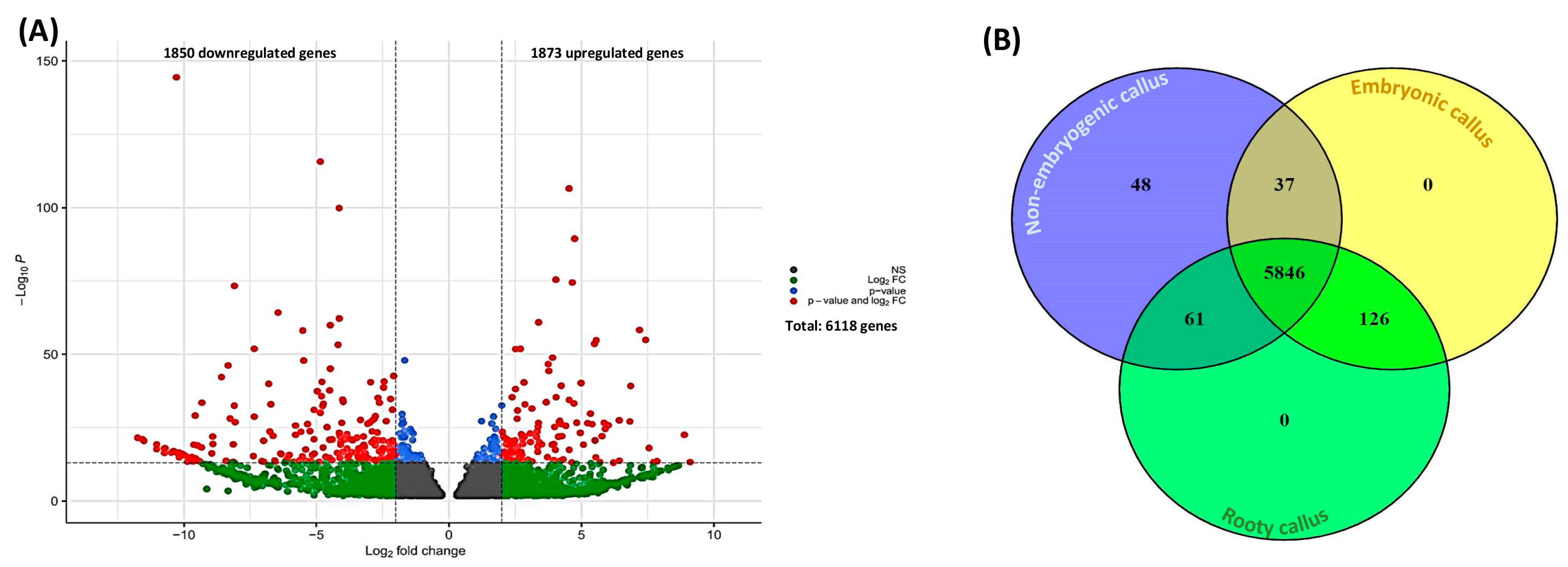
2.1. Differential Gene Expression and GO Enrichment of Different Types of Calli

A high-depth RNA sequencing was achieved with an average of 17.3, 26.3, and 23.6 million 150 bp paired-end reads for non-embryogenic calli, rooty calli, and embryonic calli, respectively. On average, 88.28% of the reads were correctly mapped to the cannabis reference genome. Of 16,417 expressed genes (Table S1), we found that 6118 genes were differentially expressed (1850 downregulated (logFC ≤ −1) and 1873 were upregulated (logFC ≥ 1) genes) within different callus types (Figure 2A and Table S2). Among these genes, 1265 genes were uncharacterized genes (unknown function).
The GO enrichment analysis revealed that 52.4% of upregulated genes were categorized in the molecular function, followed by the biological process (35.2%) and cellular component (12.4%), with 90, 91, and 90 subcategories, respectively. We then performed hierarchal clustering to classify subcategories based on their frequencies. This resulted in three clusters, where the first cluster covered 45.71% of gene product properties, followed by the second cluster (34.73%) and the third cluster (19.56%). The most pronounced enriched categories were the molecular function (10.11%), biological process (8.36%), binding (7.40%), cellular process (7.15%), metabolic process (6.61%), and catalytic activity (6.08%), respectively (Figure 3).
The top 10 upregulated genes in embryogenic calli (i.e., embryonic callus and rooty callus) were beta-galactosidase (LOC115706125), receptor-like protein 35 (LOC115725246), programmed cell death protein 4-like (LOC115707713), 1-aminocyclopropane-1-carboxylate oxidase homolog 1 (LOC115723447), guanine nucleotide-binding protein subunit beta-like protein (LOC115709330), 60S ribosomal protein L31 (LOC115705178), F-box/LRR protein (LOC115705507), acid phosphatase 1 (LOC115705442), protein DETOXIFICATION 29 (LOC115714995), and 60S acidic ribosomal protein P1 (LOC115705054) (Table 1).
On the other hand, GO analysis revealed that 54.66% of downregulated genes were categorized in the molecular function, followed by the biological process (33.52%) and cellular component (11.82%), with 94, 94, and 91 subcategories, respectively. The subcategories were arranged into three clusters using hierarchal clustering. The first cluster covered 47.24% of gene product properties, followed by the second cluster (34.66%) and the third cluster (18.10%). The most pronounced enriched categories were the molecular function (10.35%), biological process (8.61%), binding (7.93%), cellular process (7.36%), metabolic process (6.54%), and catalytic activity (6.45%), respectively (Figure 3).
The top 10 downregulated genes in embryogenic calli were beta-galactosidase-like (LOC115705878), LRR receptor-like serine/threonine-protein kinase IOS1 (LOC115704132), metal-nicotianamine transporter YSL3 (LOC115716639), guanine nucleotide-binding protein subunit beta-like protein (LOC115709323), protein TIFY 10A-like (LOC115706817), SNF1-related protein kinase regulatory subunit gamma-1 (LOC115711284), peroxidase 24 (LOC115721742), probable 2-oxoglutarate-dependent dioxygenase (LOC115702721), ferredoxin-nitrite reductase (LOC115702931), and neutral ceramidase 2 (LOC115697518) (Table 1).
Among DEGs, 5846 genes were differentially expressed in all types of calli (Figure 2B). In addition, 48 genes were unique to non-embryogenic callus, whereas 126 genes were uniquely expressed in embryogenic calli (i.e., rooty callus and embryonic callus) (Figure 2B and Table S3). The shared gene expression profiles between non-embryogenic callus and embryonic callus were found to consist of 37 genes (Figure 2B and Table S3). In contrast, a larger set of 61 genes was found to be shared between non-embryogenic callus and rooty callus (Figure 2B and Table S3).
Among the unique genes in non-embryogenic callus, 28 genes were uncharacterized genes (unknown function). The expression magnitudes of unique non-embryogenic callus genes ranged from −11.02 (logFC) to −7.22 (logFC) (Table S3). Cytochrome P450 71D9 (LOC115695663), cytidine deaminase 1 (LOC115705713), CASP-like protein 4D1 (LOC115707613), pentatricopeptide repeat-containing protein (LOC115710618), F-box/LRR-repeat protein (LOC115707558), mitogen-activated protein kinase 6 (LOC115704245), and probable S-adenosylmethionine-dependent methyltransferase (LOC115710298) are some examples of unique genes in non-embryogenic callus (Table S3). Based on the results of the GO, 65.05% of product properties of unique genes in non-embryogenic callus were categorized in the molecular function, followed by the biological process (32.14%) and cellular component (2.81%), respectively. In addition, the molecular function contained 28 subcategories, the biological process contained 27 subcategories, and the cellular component contained 11 subcategories. The most pronounced enriched categories were the molecular function (12.25%), binding (10.2%), biological process (8.67%), cellular process (8.67%), metabolic process (8.16%), and catalytic activity (7.14%), respectively (Figure 4). The lowest pronounced enriched category was the generation of precursor metabolites and energy (0.26%).
Among the unique genes in embryogenic calli (i.e., rooty callus and embryonic callus), 58 genes were uncharacterized genes (unknown function). The expression magnitudes of unique non-embryogenic callus genes ranged from 3.97 (logFC) to 9.1 (logFC) (Table S3). The mediator of RNA polymerase II transcription subunit 15a (LOC115708122), B3 domain-containing transcription factor VRN1 (LOC115697055), transcription factor MYB60 (LOC115721852), ATP-dependent DNA helicase pfh1-like (LOC115700347), ethylene-responsive transcription factor ERN1-like (LOC115723957), NAC domain-containing protein 43-like (LOC115708111), and G2/mitotic-specific cyclin S13-7 (LOC115718744) are some examples of unique genes in embryogenic callus (Table S3). Based on the results of the GO, 57.69% of the product properties of unique genes in embryogenic callus were categorized in the molecular function, followed by the biological process (31.80%) and cellular component (10.51%), respectively. In addition, the molecular function contained 37 subcategories, the biological process contained 37 subcategories, and the cellular component contained 28 subcategories. The most pronounced enriched categories were the molecular function (10%), biological process (9%), cellular process (8.11%), catalytic activity (8.05%), metabolic process (7.58%), and binding (7.43%), respectively (Figure 4). The lowest pronounced enriched category was anatomical structure development (0.11%). It is notable that some genes unique to embryogenic callus were categorized in categories not found in non-embryogenic callus (i.e., response to stress, cellular component organization, carbohydrate binding, carbohydrate metabolic process, cell wall, cytoplasm, DNA-binding transcription factor activity, external encapsulating structure, extracellular region, membrane, nuclease activity, nucleus, transcription regulator activity, and transporter activity).

2.2. Differentially Expressed Phytohormones-Related Genes

Genes related to the biosynthesis and signaling of phytohormones play pivotal roles in the formation of different types of calli. In total, 196 phytohormone-related genes were differentially expressed in different types of calli (Table S4). Of these, 65 phytohormone-related genes were downregulated (logFC ≤ −1) in embryogenic calli (i.e., rooty callus and embryonic callus), while 64 were upregulated (logFC ≥ 1) (Table S4). Ethylene-related genes were the phytohormone class with the most abundant DEGs (44 genes, Figure 5A), followed by auxin (43 genes, Figure 5B), cytokinin (42 genes, Figure 5C), abscisic acid (26 genes, Figure 5D), gibberellic acid (18 genes, Figure 6A), salicylic acid (10 genes, Figure 6B), brassinosteroid (8 genes, Figure 6C), and jasmonic acid (5 genes, Figure 6D), respectively.

2.3. Transcription Factor-Related Genes

Transcription factors play crucial roles in regulating the complex processes of callus development. Our results showed that 42 different transcription factor classes were differentially expressed in different types of calli (Table S5). MYB was the first class of transcription factors with the most abundant genes (45 genes), followed by C2H2 (40 genes), ERF (26 genes), AP2 (24 genes), bHLH (21 genes), bZIP (21 genes), B3 (20 genes), WRKY (19 genes), GRAS (16 genes), NAC (16 genes), ARF (15 genes), G2-like (12 genes), ARR-B (11 genes), C3H (11 genes), GATA (10 genes), FAR1 (9 genes), HD-ZIP (9 genes), MADS (9 genes), HB-other (8 genes), LBD (8 genes), Trihelix (8 genes), CO-like (6 genes), Dof (6 genes), TALE (6 genes), TCP (6 genes), BES1 (5 genes), CAMTA (5 genes), HSF (5 genes), NF-YB (5 genes), SBP (5 genes), NF-YA (4 genes), DBB (3 genes), RAV (3 genes), GRF (2 genes), NF-YC (2 genes), YABBY (2 genes), ZF-HD (2 genes), WOX (1 gene), EIL (1 gene), SRS (1 gene), and Whirly (1 gene), respectively (Table S5).
Specific genes with known functions in the formation of different types of calli were individually searched in the DEGs. For instance, ARF 19 (LOC115709608), AINTEGUMENTA-like 5 (ALI5, LOC115695698), APETALA2/ethylene response factor (AP2/ERF, LOC115725443), and cyclin-D3-3 (CYCD3-3, LOC115702731), which are involved in callus formation, were differentially expressed in all types of calli. On the other hand, many genes with crucial roles in somatic embryogenesis (e.g., LEAFY COTYLEDON 2 (LEC2), BABY BOOM (BBM, LOC115718863), and AGAMOUS-LIKE15 (AGL15, LOC115710309)) were not differentially expressed, which might be due to the epigenetic regulations.

2.4. Epigenetic Machinery-Related Genes

The reason for the lack of expression of embryogenesis-related genes can open a new window in our understanding of the recalcitrant nature of cannabis to in vitro culture. Hence, DEGs were searched to detect epigenetic machinery-related genes. In general, 247 DEGs related to different epigenetic machineries (e.g., chromatin remodeling, DNA methylation, and histone modification) were detected (Table S6). Among these genes, 61 genes were downregulated (logFC ≤ −1) in embryogenic calli, while 68 genes were upregulated (logFC ≥ 1) in embryogenic calli (Table S6). For instance, histone deacetylase 6 (HDAC6, LOC115703044) and CHROMATIN REMODELING 35 (LOC115724492), which target embryogenesis-related transcription factors (e.g., LEC1 and LEC2), were upregulated in embryogenic callus (Table S6), showing the pivotal roles played by epigenetics in cannabis somatic embryogenesis.

3. Discussion

The study of different types of calli is of utmost importance in the field of plant biotechnology and to a deeper understanding of the fundamental factors leading to plant regeneration and/or recalcitrance [25,26,27]. Callus is a mass of undifferentiated cells that can theoretically be induced to differentiate into different plant organs, such as roots, shoots, and embryos [4,7]. The ability to produce callus is essential for various plant tissue culture applications, including genetic transformation [15], micropropagation [28], secondary metabolite production [8], and bioenergy generation [16]. While cannabis readily forms callus, most researchers have been unable to efficiently regenerate plants from it [14,15,29,30,31]. New information related to the genes involved in callus formation and subsequent development will aid in better understanding the cause of recalcitrance in cannabis and help to develop strategies for overcoming this recalcitrant nature.
In the current study, different types of cannabis calli (i.e., non-embryogenic, rooty, and embryonic callus) were produced in a medium containing auxin (i.e., 2,4-D) and cytokinin (i.e., kinetin), which has been shown to produce a variety of callus types. These callus types include both non-embryogenic and embryogenic calli. Embryogenic calli can result in the formation of different organs, including shoots, roots, or somatic embryos. These callus types were used to study differential gene expression profiles to gain insight into the nature of recalcitrance and regeneration. It is well-documented that auxin and cytokinin play fundamental roles in callogenesis [32,33]. Previous studies in model plants have also revealed that callogenesis using auxin and cytokinin is controlled through complex gene regulatory networks [2,4,6].
Recent genetic data from Arabidopsis have greatly enhanced our comprehension of the molecular mechanisms that control embryogenic callus formation [6]. Specifically, studies related to embryonic callus formation have elucidated the delicate equilibrium between cell proliferation in the apical domain, the processes of programmed cell death (PCD), and terminal differentiation in the basal domain [24,34,35]. In line with genetic data from model species, our results showed that the programmed cell death protein 4-like gene (LOC115707713) was upregulated in embryogenic calli.
Embryogenic callus forms in response to exogenous PGRs in tissue culture media and is initiated from a group of cells (or even a single cell) with similar genetic backgrounds and morphologies [36]. These stimuli act through receptor-like proteins (RLPs) to regulate several cellular mechanisms and ultimately alter gene transcription patterns [37]. Indeed, RLPs regulate several cellular mechanisms to support cell differentiation and/or redifferentiation as well as adaptation under in vitro culture conditions [38]. Our results also showed that RLP35 (LOC115725246) was upregulated in embryogenic callus (i.e., rooty callus and embryonic callus), showing the critical roles of RLPs in embryogenic callus formation.
Embryogenic callus formation is accompanied by changes in the cellular components and structure of the cell wall [24,39]. The GO analysis of our study indicated that 12.4% of upregulated gene product properties were categorized in the cellular components category. Modifications of the cell wall are crucial for maintaining the balance of forces required for the cellular architecture, the cell shape, and the determination of the division plane [40]. In addition, cell walls play pivotal roles in connecting cells with their neighboring cells. A wide range of signaling factors transported to cell walls from the cytoplasm can be exported through the apoplast into neighboring cells, which promotes embryogenic callus formation [41,42,43]. In line with previous studies in other plant species, our result also showed that the most pronounced enriched GO categories for upregulated genes were the molecular function, biological process, binding, cellular process, metabolic process, and catalytic activity, respectively. The high representation of molecular function among the enriched categories suggests the importance of altered molecular functions in the regulation of embryogenic potential. Changes in molecular functions can directly influence cellular processes and signaling events to ultimately impact developmental processes associated with embryogenesis [39]. The specific molecular functions enriched in the upregulated genes must still be elucidated, but they likely play essential roles in modulating cellular processes required for embryogenic potential [2,24,40,44].
Several cell wall-modifying enzymes (e.g., β-galactosidase and 1,4-alpha-glucan-branching enzyme) are typically differentially expressed in embryogenic calli [6,24]. Based on our results, embryogenic callus formation is accompanied by the up-regulation of β-galactosidase (LOC115706125), which removes terminal galactosyl residues from galactolipids, glycoproteins, and carbohydrates and can thus modify the architecture and structure of the cell wall, as well as other properties, such as intercellular attachments [45]. Similar results were previously reported in plants such as Pinus radiata [46], cassava [47], and cotton [23]. Our results also showed the upregulation of 1,4-alpha-glucan-branching enzyme 2-2, chloroplastic/amyloplastic-like gene (LOC115725315) in embryogenic callus. This gene plays a key role in the starch synthesis pathway through the removal of 1, 4-alpha-linked oligosaccharides and, subsequently, the formation of the alpha-1, 6-glucosidic linkages in glycogen [48]. The 1, 4-alpha-glucan branching enzyme can ultimately alter the mechanical properties of the cell wall [48]. Geraniol 8-hydroxylase is another gene involved in cellular components by producing indole-3-acetaldoxime from tryptophan, resulting in the promotion of embryogenic callus formation [49]. Our results showed that this gene (LOC115725467) was upregulated in the embryogenic callus. The 60S ribosomal proteins (e.g., 60S ribosomal protein L31, 60S acidic ribosomal protein P1) play important roles related to embryogenic callus formation due to their functions in protein production [37,40,50]. Our results showed that these proteins (LOC115705178 and LOC115705054) were upregulated in embryogenic calli.
Importantly, the upregulation of cell wall-related signaling transduction genes such as those for guanine nucleotide-binding proteins is necessary for embryogenic callus formation [51,52]. Our results also showed that guanine nucleotide-binding protein subunit beta-like protein (LOC115723447) was upregulated in embryogenic callus. Similar to our results, Polesi et al. [51] reported that guanine nucleotide-binding protein subunit beta-like protein is a unique protein in the embryogenic callus of Guadua chacoensis. The serine carboxypeptidase gene plays an important role in secondary metabolite biosynthesis [53]. The expression of this gene as the signal transduction gene for embryogenic callus in citrus [53] and Douglas fir [54] indicates its crucial role in embryogenic callus formation. In line with these studies, our results revealed that serine carboxypeptidase (LOC115700502) was upregulated in embryogenic calli. The SNF1-related protein kinase regulatory subunit gamma-1-like is connected to another signal transduction cascade that regulates the expression of carbohydrate metabolism-related genes and thereby plays a crucial role in embryogenic callus formation [55]. Our results showed the upregulation of this gene (LOC115711285), demonstrating its role in cell wall-related signaling transduction in embryogenic calli.
Our results also demonstrated that protein DETOXIFICATION 29 (LOC115714995) was upregulated in the embryogenic calli. Since high levels of unstable and damaged proteins are usual in callogenesis due to stressful in vitro conditions, detoxification proteins are crucial for embryogenic callus formation [50].
In addition to top up- and top down-regulated genes, we performed a specific search to find phytohormone-related genes. Genes related to the biosynthesis and signaling of phytohormones such as auxin [56], cytokinin [57], gibberellic acid [58], abscisic acid [59], ethylene [60], salicylic acid [11], jasmonic acid [61], and brassinosteroid [62] play pivotal roles in the formation of different types of calli. Our results showed that 196 phytohormone-related genes were differentially expressed in different types of calli, demonstrating the importance of PGRs in callogenesis.
In addition to PGRs, transcription factors play crucial roles in regulating the complex process of indirect somatic embryogenesis [13]. Through their ability to bind to specific DNA sequences, transcription factors control the expression of genes involved in embryogenic cell formation, proliferation, and differentiation [63]. They act as key molecular switches, orchestrating the activation or repression of target genes, thereby modulating the intricate signaling networks and molecular events required for somatic embryogenesis [13]. By fine-tuning the balance of gene expression, these transcription factors govern the transition of somatic cells into embryogenic cells. Subsequent embryo development can then proceed, making such transcription factors pivotal players in the regulation of this highly efficient and valuable plant regeneration pathway [2]. We observed 42 classes of transcription factors that were differentially expressed in different types of calli. Besides transcription factors, epigenetic modifications significantly impact the formation of different types of calli [9]. Epigenome reprogramming, facilitated by chromatin-modifying factors, is responsible for altering the chromatin states of genes [64]. These factors play critical roles in orchestrating comprehensive changes to the global transcriptome during callogenesis [2,9]. Three distinct mechanisms (i.e., DNA methylation, histone modification, and nucleosome remodeling) are involved in modifying the chromatin structure [65,66]. As a result, the compaction of chromatin is either decreased or increased, leading to alterations in the accessibility of chromatin for the transcription machinery [67]. Our results showed that 247 epigenetic-related genes were differentially expressed in all types of calli. To elucidate the gene regulatory networks of cannabis callogenesis, specific genes with known functions in Arabidopsis related to the formation of different types of calli were individually analyzed for expression profiles in non-embryogenic callus and embryogenic callus.
Previous studies using Arabidopsis have demonstrated that various members of the ASYMMETRIC LEAVES2-LIKE family, also referred to as LATERAL ORGAN BOUNDARIES DOMAIN (LBD) transcription factors, including LBD16 and LBD29, mediate auxin signaling downstream of AUXIN RESPONSE FACTOR 7 (ARF7) and ARF19 [68,69]. LBD16 activates E2 PROMOTER BINDING FACTOR a (E2Fa), a core cell cycle regulator, leading to enhanced cell proliferation [70]. Additionally, LBD29 plays a role in cell wall modification processes [71]. PECTIN METHYLESTERASE (PME), a cell wall modifier, is one of the targets of LBD29, ultimately promoting callogenesis [72]. Our results revealed that ARF19 (LOC115709608), several LBDs (e.g., LBD4 (LOC115708902), LBD12 (LOC115720935), LBD19 (LOC115706345), LBD30 (LOC115705047), LBD39 (LOC115713617), and LBD40 (LOC115699573)), and PME (LOC115704128) were differentially expressed among certain types of cannabis calli. However, ARF 7, LBD16, LBD 29, and E2Fa were not differentially expressed in our study, suggesting the importance of other pathways in cannabis callogenesis.
The role of the cytokinin-dependent pathway in callogenesis is less clear than that of the auxin-dependent pathway. However, type-B RESPONSE REGULATORs (RRs) can be considered a critical component in the cytokinin-dependent pathway. Previous studies showed that RR 2 overexpression promotes Arabidopsis callogenesis in cytokinin-containing media. A potential target of RR2 in promoting the reentry to the cell cycle is CYCD3;1. In addition, previous studies showed that the expression of CYCD3;1 is upregulated in cytokinin-containing media. In line with these results in Arabidopsis, our results showed that RR (LOC115711591) and CYCD3;1 (LOC115718739) were expressed in all types of cannabis calli.
Another candidate, APETALA2/Ethylene-responsive factor (AP2/ERF), plays a pivotal role in cytokinin-mediated callogenesis through linking cytokinin signaling to cell cycle regulation. The direct activation of OBF BINDING PROTEIN1 (OBP1) and CYCD3;1 is mediated by AP2/ERF. The OBP1 gene shortens the G1 phase duration, which facilitates reentry to the cell cycle. In addition, OBP1 can directly activate the CYCD3;3 gene, which ultimately results in callogenesis. Consistent with findings in model plants, our results revealed that AP2/ERF (LOC115725443), OBP1 (LOC115719739), CYCD3;1, and CYCD3;3 (LOC115702731) were differentially expressed in all types of cannabis calli, emphasizing the critical role of the cytokinin-dependent pathway in cannabis callogenesis.
Overall, our findings indicated that all the genes associated with the cytokinin-dependent pathway were differentially expressed. These results are key to unlocking mysteries related to the recalcitrant nature of in vitro cannabis and serve as important information for overcoming such obstacles. It is well-documented that the expression of auxin-related genes in non-recalcitrant plants is significantly higher than that of cytokinin genes. Previous studies in recalcitrant plants showed that if auxin-related genes are upregulated to a higher degree than cytokinin genes, the non-embryogenic callus can be converted to embryogenic callus [1,13,18,19,22,23,73]. Hence, the limited expression levels of auxin-dependent pathway-related genes may account for the recalcitrant nature of cannabis.
Callus formation appears to exhibit significant heterogeneity, as observed in the formation of calli derived from lateral root primordia, where the expression of root meristem markers is only partially regulated [74]. Notably, callus cells demonstrate the ability to differentiate into rooty calli and embryonic calli, suggesting the existence of shared pathways between these callus types [6]. The establishment and maintenance of pluripotency in plant callus cells are orchestrated by an intricate network of molecular regulators [2]. Transcription factors, such as members of the LEC1, LEC2, and WUSCHEL-related homeobox (WOX) families, play crucial roles in promoting pluripotency by activating key regulatory pathways [13,24]. These transcription factors interact with chromatin modifiers to regulate the epigenetic landscape of callus cells [9] (Figure 7).
The acetylation of histone 3 (H3) and H4, which are tightly regulated by histone deacetylases (HDACs) and histone acetyltransferases (HATs), plays a pivotal positive role in expressing embryogenesis-related genes [9]. Tanaka et al. [75] showed that the upregulation of HDACs repressed embryogenic markers such as ABSCISIC ACID INSENSITIVE 3 (ABI3), FUSCA3 (FUS3), the LEAFY COTYLEDON 1 (LEC1), and LEC2, suggesting that embryogenic calli failed to make the transition necessary to allow for somatic embryogenesis. Our results showed that HDAC (LOC115703044) was upregulated in embryogenic calli. In addition, LEC2 (LOC115712445) was not expressed in all types of calli, suggesting that HDAC upregulation had resulted in the repression of LEC2 (Figure 7).
The chromatin remodeling factors (CRFs) mediate crosstalk between histone acetylation and histone methylation. Previous studies revealed that the upregulation of CRFs leads to the repression of LEC1 and LEC2, resulting in the production of the changes necessary for embryogenic calli to enable somatic embryogenesis [44,76]. Indeed, CRFs may function by guiding both H3 lysine 27 trimethylation (H3K27me3) and HDACs [9]. Our results showed that CRFs such as CHROMATIN REMODELING 35 (LOC115724492) were upregulated in embryogenic calli. Thus, it seems that the combination of HDAC and CRF ensures the silencing of LEC2 in cannabis calli and likely inhibits somatic embryogenesis from occurring in embryogenic calli (Figure 7).
Critical roles related to histone modification are played by JUMONJI 30 (JMJ30) [65], the upregulation of which can repress WUS, LEC1, and LEC2 [77]. Our results showed that JMJ30 (LOC115721095) was downregulated in embryogenic calli. However, a previous study demonstrated that either Polycomb repressive complex (PRCs) or JMJ30 activity can be sufficient to suppress somatic embryogenesis [77,78].
It has been shown that members of the PRCs such as EMBRYONIC FLOWER 2 (EMF2), SWINGER (SWN), and CURLY LEAF (CLF) play crucial roles related to indirect somatic embryogenesis [13]. Chanvivattana et al. [79] revealed that double loss-of-function mutants in PRCs such as EMF2 in A. thaliana led to ectopic root formation and indirect somatic embryogenesis. Bouyer et al. [80] showed that PRCs repress embryogenesis-related genes such as LEC1, LEC2, WOUND INDUCED DEDIFFERENTIATION 3 (WIND3), WUSCHEL (WUS), BABY BOOM (BBM), and AGAMOUS-LIKE15 (AGL15). Ikeuchi et al. [81] also showed that PRC mutants overexpressing WIND3 and LEC2 promoted indirect somatic embryogenesis. In contrast to many embryogenesis-related genes, the expression of LEC1 depends on the type of explant and the presence of PGRs such as 2,4-D in the medium [82]. Our results also showed that LEC1 (LOC115724811) was upregulated in embryogenic calli, which might be due to the presence of 2,4-D in the medium. Mozgová et al. [82] showed that PRCs suppress somatic embryogenesis through the repression of their targets such as LEC2, AGL15, and BBM. However, PRCs had no effect on LEC1 expression in the presence of 2,4-D [82]. In line with these studies, our results showed that EMF2 (LOC115706711) was differentially expressed in all types of calli, which resulted in the repression of WIND3 (LOC115722430), BBM (LOC115718863), LEC2, and AGL15 (LOC115710309) (Figure 7). One of the members of the AINTEGUMENTA-LIKE (AIL) group of APETALA2/ETHYLENE RESPONSE FACTOR (AP2/ERF), BBM, plays an important role in somatic embryo induction [1,2]. The expression of BBM and AIL/PLETHORA (PLT) genes (e.g., PLT1, PLT2, PLT3, PLT5, or PLT7) induces somatic embryogenesis, resulting in the formation of somatic embryos [13]. It has also been shown that AIL5 overexpression induces somatic embryogenesis [83]. Generally, the overexpression of all members of AIL proteins, except AINTEGUMENTA (ANT) and AIL1, leads to the induction of somatic embryogenesis [13]. However, in a study, Zhang et al. [14] explored the utility of various morphogenic genes, such as SHOOT MERISTEMLESS (STM), ISOPENTENYL TRANSFERASE (IPT), GROWTH-REGULATING FACTOR (GRF), GRF-INTERACTING FACTOR (GIF), BBM, and WUS, in enhancing the process of indirect shoot regeneration in hemp cultivars. However, the introduction of these morphogenic genes only resulted in a marginal increase of less than 6% regeneration frequency. It can be concluded that the epigenetic state of callus cells might suppress the activity of these transcription factors, thus contributing to the recalcitrance of cannabis towards indirect regeneration.
Another transcription factor that plays an important role in the induction of somatic embryogenesis is RWP-RK DOMAIN-CONTAINING 4 (RKD4)/GROUNDED (GRD) [1]. While RKD4 is the only member of the RKD family that induces somatic embryogenesis, other members of RKD influence the development of the embryo sac [13]. However, no expression of RKD (LOC115717465) was detected in our study.
Previous studies showed that LEC1, LEC2, FUS3, and AGL15 regulate auxin production and signaling for the induction of somatic embryogenesis, leading to the production of somatic embryos [2]. The YUCCA10 (YUC10) gene encoding an auxin synthesis enzyme is induced by LEC1 [84]. In line with previous studies in model plants, our results also showed that YUC10 (LOC115719231) was differentially expressed in all types of calli. GYUC2 and YUC4 genes are activated by LEC2 [85]. To modulate the auxin-mediated signaling, the expression of INDOLE ACETIC ACID INDUCIBLE 30 (IAA30) as a negative auxin signaling regulator is induced by LEC2 and AGL15 [84]. Our results also showed that YUC2 (LOC115704335), YUC4 (LOC115702268), and IAA30 (LOC115714171) were not expressed due to the absence expression of LEC2 and AGL15.
Previous studies showed that a low ratio of gibberellic acid (GA) to abscisic acid (ABA) can promote somatic embryogenesis [13]. It has also been shown that AGL15 can be a positive regulator for the GIBBERELLIN 2-OXIDASE6 (GA2ox6) gene encoding a GA-degrading enzyme and a negative regulator for the GA3ox2 gene which generally causes a reduction in the endogenous GA concentration [86]. Furthermore, GA biosynthesis is downregulated by FUS3 through activating ABA biosynthesis and repressing GA3ox1 and GA3ox2 [87]. In contrast, our results showed that gibberellin dioxygenase genes (e.g., LOC115704748, LOC115705300, and LOC115710658) were upregulated in embryogenic calli, which is considered to be an additional sign of a failure to induce somatic embryogenesis in embryogenic callus.
Another transcription factor, WUSCHEL (WUS), plays an important role in embryogenic callus formation [4]. The role of WUS in somatic embryogenesis has been previously shown in Arabidopsis [88]. Type A-RR is the target of WUS. In fact, WUS represses Type A-RR and promotes embryogenic callus formation [89]. Our results also showed that WUS (LOC115702606) was upregulated in embryogenic calli which resulted in the repression of Type A-RR (LOC115721904), demonstrating the role of this gene in embryogenic callus formation. In addition, the expression of WIND2 by regulating type B-RR as its target promotes embryogenic callus formation in the cytokinin-dependent pathway [90]. Our results also showed that WIND2 (LOC115713946) was differentially expressed, which resulted in the expression of Type B-RR (LOC115711466), and finally promoted embryogenic callus formation. Overall, our findings indicated that the expression levels of genes associated with the cytokinin-dependent pathway resulted in embryogenic callus formation. However, the repression of auxin-dependent pathway genes led to the suppression of somatic embryogenesis. As discussed, for the callogenesis, it seems that the repression of auxin-dependent pathway-related genes may account for the recalcitrant nature of cannabis (Figure 7).

4. Materials and Methods

4.1. Plant Material Source, Callus Formation, and Sampling

The current study was carried out on drug-type cannabis (UP305; Up Cannabis, ON). The callogenesis was performed based on our previously developed protocol [17]. Different types of calli including non-embryogenic callus (Figure 8A) and embryogenic callus (i.e., rooty callus (Figure 8B) and embryonic callus (Figure 8C)) were all obtained based on visual cues, including lack of organization and signs of de novo roots and polarization. Leaf explants from in vitro plantlets were cultured on a medium composed of MS [91] basal salts (Phytotechnology Laboratories, Overland Park, KA, USA), B5 [92] vitamins (Phytotechnology Laboratories, Overland Park, KA, USA), 0.7% agar (Fisher Scientific, Waltham, MA, USA), 3% sucrose, 0.38 mg/L kinetin (Thermo Fisher Scientific, Waltham, MA, USA), and 0.46 mg/L 2,4-dichlorophenoxyacetic acid (2,4-D, Thermo Fisher Scientific, Waltham, MA, USA). The cultures were kept in a growth chamber at 25 ±  2 °C under a 16 h photoperiod with 40  ± 5 μmol m−2 s−1 light intensity. In order to minimize the background signals of gene expression that may arise from different culture boxes, non-embryogenic calli, rooty calli, and embryonic calli were selected from 10 individual culture boxes after 90 days. The calli were then combined into a single tube, flash frozen, and stored at −80 °C. This procedure was repeated three times to obtain three biological replicates of each.

4.2. RNA Isolation and RNA Sequencing

To isolate RNA samples, pooled calli were placed in 1 mL of TRIzol™ (Thermos Fisher Scientific, Waltham, MA, USA) reagent before being crushed within an RETSCH MM 400 mixer mill (Fisher Scientific, Waltham, MA, USA). Total RNAs were isolated using the TRIzol™ protocol according to the manufacturer’s instructions in conjunction with DNase. The integrity of the total purified RNA was determined using a 2100 Bioanalyzer™ (Agilent Technologies Inc., Santa Clara, CA, USA), and only the three best samples per condition were used for library preparation.
To prepare RNA libraries for sequencing, the quantity of starting material was adjusted to 1 µg per sample. The preparation of mRNA-seq was facilitated with the use of the NEBNext® Ultra™ II directional RNA library Prep kit for Illumina, according to the manufacturer’s instructions, at the Genomic Analysis Platform of the Institut de Biologie Intégrative et des Systèmes (Université Laval, Québec, QC, Canada). The quality of the cDNA library was evaluated using an Agilent 2100 Bioanalyzer™. The RNA libraries were then sequenced on an Illumina NovaSeq 6000 platform (2 × 150 bp read) at the Centre d’expertise et de services Génome Québec (Montreal, QC, Canada).

4.3. Transcriptomic Analyses

Raw reads underwent a preprocessing and filtering procedure to ensure good-quality data were obtained. Specifically, Trimmomatic [93] was used to remove Illumina adapters from the reads, followed by the use of the FASTX Toolkit (http://hannonlab.cshl.edu/fastx_toolkit/index.html, accessed on 4 April 2023 version 0.0.13.2) to trim nucleotides with quality scores below 25 and remove reads containing less than 70% base pairs. Clean reads were subsequently aligned to the Cannabis sativa reference genome (cs10; https://www.ncbi.nlm.nih.gov/assembly/GCA_900626175.2, accessed on 4 April 2023) using the STAR version 2.7.0a [94]. Gene abundance was inferred by utilizing the cs10 reference genome annotation obtained from NCBI (https://ftp.ncbi.nlm.nih.gov/genomes/all/GCF/900/626/175/GCF_900626175.2_cs10/GCF_900626175.2_cs10_genomic.gff.gz, accessed on 4 April 2023) to generate fragment per kilobase of transcript per million (FPKM) mapped reads values. Afterward, FeatureCounts v. 1.5.0 [95] was used to quantify the number of reads mapped to each detected gene, and differential expression analysis was conducted using the DESeq2 R package [96]. Genes with fewer than 50 reads were removed from the analyses, and genes with an adjusted p-value of no more than 0.05 were considered differentially expressed. The fold change for each condition was compared based on the average of different condition sets. The expression values of both the rooty callus and the embryonic callus, referred to as embryogenic callus, were averaged, and these averages were then compared to the average of the non-embryogenic type.
The gene sequences were retrieved using the biomartr R package version 1.0.2 [97] to call the correct gene-associated names, and the data were used to help elucidate the gene ontology (GO) enrichment of differentially expressed genes (DEGs) using rentrez version 1.2.3 and biomaRt version 2.50.3 [98].
The Ortho DB V11 [99] database provided by the National Center for Biotechnology Information (NCBI) was used to identify homologous genes of Arabidopsis thaliana in Cannabis sativa. We selected a set of Arabidopsis thaliana genes of interest for which we wanted to identify homologs in Cannabis sativa. These genes were chosen based on their known functions in callogenesis in Arabidopsis and their potential relevance to the formation of different types of calli in Cannabis sativa.

5. Conclusions

This study offers comprehensive insight into the dynamics of gene expression and molecular mechanisms that direct the development of various types of cannabis calli. These findings represent critical factors that will form the foundation of future research endeavors aimed at enhancing plant regeneration strategies, thereby advancing various cultivation and biotechnological applications related to this economically significant crop. The absence of a robust embryogenic system capable of yielding vigorous plants directly from cotyledon embryos represents a substantial challenge in the pursuit of understanding callus development on a deeper level. In our study, the term “embryogenic” refers to the observed developmental trajectory and molecular signatures of calli that exhibit specific characteristics suggestive of embryogenic potential. However, without the ability to produce fully developed, viable plants directly from these calli, we must exercise caution when labeling them as traditional embryogenic calli, that is, callus capable of producing somatic embryos. This work marks an essential initial step in unraveling the intricacies of cannabis callus development. Although we have identified notable distinctions in gene expression profiles characteristic of specific calli types and have highlighted key regulatory factors, it remains crucial to be cognizant of the possibility that these so-called “embryogenic” tissues may follow an unfamiliar developmental path. Such a path could be distinct from the conventional embryogenic pathway that leads to plant regeneration. To address this limitation and ensure the validity of our findings, future research endeavors should aim to establish a more robust and reproducible embryogenic system in cannabis. This would enable a direct comparison between calli capable of developing into healthy, vigorous plants and those that are not. Ultimately, future research in this area will allow us to better understand the molecular mechanisms underpinning successful plant regeneration. Conducting additional experiments to validate the functional roles of the identified genes is essential and may be achieved by incorporating CRISPR-based functional validation methods to more comprehensively determine the molecular mechanisms driving cannabis callus development.

Supplementary Materials

The following supporting information can be downloaded at: https://www.mdpi.com/article/10.3390/ijms241914625/s1.

Author Contributions

Conceptualization, M.H. and A.M.P.J.; project administration, A.M.P.J.; funding acquisition, D.T. and A.M.P.J.; supervision, A.M.P.J.; resources, A.M.P.J.; investigation, M.H., M.P. and M.d.R.; methodology, M.H. and A.M.P.J.; software, M.Y.-N.; data curation, M.H., M.Y.-N. and D.T.; formal analysis, M.H. and M.Y.-N.; visualization, M.H. and M.Y.-N.; validation, D.T. and A.M.P.J.; writing—original draft preparation, M.H.; writing—review and editing, M.H., M.P., M.d.R., M.Y.-N., K.A., D.T. and A.M.P.J. All authors have read and agreed to the published version of the manuscript.

Funding

This work was conducted as part of a collaborative research project funded by Brantmed Inc. and NSERC Alliance (#ALLRP555969-20 to A.M.P.J and D.T.).

Institutional Review Board Statement

Not applicable.

Informed Consent Statement

Not applicable.

Data Availability Statement

All data generated or analyzed during this study are included in this published article as supplementary information files.

Conflicts of Interest

The authors declare no conflict of interest. The funders had no role in the design of the study; in the collection, analyses, or interpretation of the data; in the writing of the manuscript; or in the decision to publish the results.

References

  1. Lardon, R.; Geelen, D. Natural Variation in Plant Pluripotency and Regeneration. Plants 2020, 9, 1261. [Google Scholar] [CrossRef] [PubMed]
  2. Ikeuchi, M.; Favero, D.S.; Sakamoto, Y.; Iwase, A.; Coleman, D.; Rymen, B.; Sugimoto, K. Molecular mechanisms of plant regeneration. Annu. Rev. Plant Biol. 2019, 70, 377–406. [Google Scholar] [CrossRef] [PubMed]
  3. Neely, D. Tree wounds and wound closure. J. Arboric. 1979, 5, 135–140. [Google Scholar] [CrossRef]
  4. Ikeuchi, M.; Sugimoto, K.; Iwase, A. Plant callus: Mechanisms of induction and repression. Plant Cell 2013, 25, 3159–3173. [Google Scholar] [CrossRef] [PubMed]
  5. White, P.R. Potentially unlimited growth of excised plant callus in an artificial nutrient. Am. J. Bot. 1939, 26, 59–64. [Google Scholar] [CrossRef]
  6. Fehér, A. Callus, dedifferentiation, totipotency, somatic embryogenesis: What these terms mean in the era of molecular plant biology. Front. Plant Sci. 2019, 10, 536. [Google Scholar] [CrossRef]
  7. Efferth, T. Biotechnology applications of plant callus cultures. Engineering 2019, 5, 50–59. [Google Scholar] [CrossRef]
  8. Hesami, M.; Pepe, M.; Baiton, A.; Jones, A.M.P. Current status and future prospects in cannabinoid production through in vitro culture and synthetic biology. Biotechnol. Adv. 2023, 62, 108074. [Google Scholar] [CrossRef]
  9. Lee, K.; Seo, P.J. Dynamic epigenetic changes during plant regeneration. Trends Plant Sci. 2018, 23, 235–247. [Google Scholar] [CrossRef]
  10. Hesami, M.; Daneshvar, M.H.; Yoosefzadeh-Najafabadi, M. Establishment of a Protocol for in vitro Seed Germination and Callus Formation of Ficus religiosa L., an Important Medicinal Plant. Jundishapur J. Nat. Pharm. Prod. 2018, 13, e62682. [Google Scholar] [CrossRef]
  11. Grzyb, M.; Kalandyk, A.; Mikuła, A. Effect of TIBA, fluridone and salicylic acid on somatic embryogenesis and endogenous hormone and sugar contents in the tree fern Cyathea delgadii Sternb. Acta Physiol. Plant 2017, 40, 1. [Google Scholar] [CrossRef]
  12. Jafari, M.; Daneshvar, M.H. Prediction and optimization of indirect shoot regeneration of Passiflora caerulea using machine learning and optimization algorithms. BMC Biotechnol. 2023, 23, 27. [Google Scholar] [CrossRef] [PubMed]
  13. Horstman, A.; Bemer, M.; Boutilier, K. A transcriptional view on somatic embryogenesis. Regeneration 2017, 4, 201–216. [Google Scholar] [CrossRef] [PubMed]
  14. Zhang, X.; Xu, G.; Cheng, C.; Lei, L.; Sun, J.; Xu, Y.; Deng, C.; Dai, Z.; Yang, Z.; Chen, X.; et al. Establishment of an Agrobacterium-mediated genetic transformation and CRISPR/Cas9-mediated targeted mutagenesis in Hemp (Cannabis sativa L.). Plant Biotechnol. J. 2021, 19, 1979–1987. [Google Scholar] [CrossRef]
  15. Hesami, M.; Baiton, A.; Alizadeh, M.; Pepe, M.; Torkamaneh, D.; Jones, A.M. Advances and perspectives in tissue culture and genetic engineering of cannabis. Int. J. Mol. Sci. 2021, 22, 5671. [Google Scholar] [CrossRef]
  16. Norouzi, O.; Hesami, M.; Pepe, M.; Dutta, A.; Jones, A.M.P. In vitro plant tissue culture as the fifth generation of bioenergy. Sci. Rep. 2022, 12, 5038. [Google Scholar] [CrossRef]
  17. Hesami, M.; Jones, A.M.P. Modeling and optimizing callus growth and development in Cannabis sativa using random forest and support vector machine in combination with a genetic algorithm. Appl. Microbiol. Biotechnol. 2021, 105, 5201–5212. [Google Scholar] [CrossRef]
  18. Zhang, H.; Chen, J.; Zhang, F.; Song, Y. Transcriptome analysis of callus from melon. Gene 2019, 684, 131–138. [Google Scholar] [CrossRef]
  19. Li, Q.; Zhang, S.; Wang, J. Transcriptome analysis of callus from Picea balfouriana. BMC Genom. 2014, 15, 553. [Google Scholar] [CrossRef]
  20. Xu, Z.; Wang, F.; Tu, Y.; Xu, Y.; Shen, Q.; Zhang, G. Transcriptome analysis reveals genetic factors related to callus induction in barley. Agronomy 2022, 12, 749. [Google Scholar] [CrossRef]
  21. Awada, R.; Lepelley, M.; Breton, D.; Charpagne, A.; Campa, C.; Berry, V.; Georget, F.; Breitler, J.-C.; Léran, S.; Djerrab, D.; et al. Global transcriptome profiling reveals differential regulatory, metabolic and hormonal networks during somatic embryogenesis in Coffea arabica. BMC Genom. 2023, 24, 41. [Google Scholar] [CrossRef] [PubMed]
  22. Lee, N.N.; Lee, S.-A.; Kang, M.-J.; Joo, H.-J.; Kim, J.-A.; Park, E.-J. Comparative transcriptome analysis between embryogenic and nonembryogenic callus of Kalopanax septemlobus. Forest Sci. Technol. 2020, 16, 145–153. [Google Scholar] [CrossRef]
  23. Wen, L.; Li, W.; Parris, S.; West, M.; Lawson, J.; Smathers, M.; Li, Z.; Jones, D.; Jin, S.; Saski, C.A. Transcriptomic profiles of non-embryogenic and embryogenic callus cells in a highly regenerative upland cotton line (Gossypium hirsutum L.). BMC Dev. Biol. 2020, 20, 25. [Google Scholar] [CrossRef] [PubMed]
  24. Smertenko, A.; Bozhkov, P.V. Somatic embryogenesis: Life and death processes during apical–basal patterning. J. Exp. Bot. 2014, 65, 1343–1360. [Google Scholar] [CrossRef] [PubMed]
  25. Hillebrands, L.; Lamshoeft, M.; Lagojda, A.; Stork, A.; Kayser, O. In vitro metabolism of tebuconazole, flurtamone, fenhexamid, metalaxyl-M and spirodiclofen in Cannabis sativa L. (hemp) callus cultures. Pest Manag. Sci. 2021, 77, 5356–5366. [Google Scholar] [CrossRef]
  26. Feeney, M.; Punja, Z.K. Tissue culture and Agrobacterium-mediated transformation of hemp (Cannabis sativa L.). Vitr. Cell. Dev. Biol. Plant 2003, 39, 578–585. [Google Scholar] [CrossRef]
  27. Thacker, X.; Thomas, K.; Fuller, M.; Smith, S.; DuBois, J. Determination of optimal hormone and mineral salts levels in tissue culture media for callus induction and growth of industrial hemp (Cannabis sativa L.). Agri. Sci. 2018, 9, 1250–1268. [Google Scholar] [CrossRef]
  28. Monthony, A.S.; Page, S.R.; Hesami, M.; Jones, A.M.P. The past, present and future of Cannabis sativa tissue culture. Plants 2021, 10, 185. [Google Scholar] [CrossRef]
  29. Monthony, A.S.; Kyne, S.T.; Grainger, C.M.; Jones, A.M.P. Recalcitrance of Cannabis sativa to de novo regeneration; a multi-genotype replication study. PLoS ONE 2021, 16, e0235525. [Google Scholar] [CrossRef]
  30. Adhikary, D.; Kulkarni, M.; El-Mezawy, A.; Mobini, S.; Elhiti, M.; Gjuric, R.; Ray, A.; Polowick, P.; Slaski, J.J.; Jones, M.P.; et al. Medical cannabis and industrial hemp tissue culture: Present status and future potential. Front. Plant Sci. 2021, 12, 627240. [Google Scholar] [CrossRef]
  31. Ingvardsen, C.R.; Brinch-Pedersen, H. Challenges and potentials of new breeding techniques in Cannabis sativa. Front. Plant Sci. 2023, 14, 1154332. [Google Scholar] [CrossRef] [PubMed]
  32. Asghar, S.; Ghori, N.; Hyat, F.; Li, Y.; Chen, C. Use of auxin and cytokinin for somatic embryogenesis in plant: A story from competence towards completion. Plant Growth Regul. 2023, 99, 413–428. [Google Scholar] [CrossRef]
  33. Raspor, M.; Motyka, V.; Kaleri, A.R.; Ninković, S.; Tubić, L.; Cingel, A.; Ćosić, T. Integrating the roles for cytokinin and auxin in de novo shoot organogenesis: From hormone uptake to signaling outputs. Int. J. Mol. Sci. 2021, 22, 8554. [Google Scholar] [CrossRef] [PubMed]
  34. Shi, C.; Luo, P.; Du, Y.-T.; Chen, H.; Huang, X.; Cheng, T.-H.; Luo, A.; Li, H.-J.; Yang, W.-C.; Zhao, P.; et al. Maternal control of suspensor programmed cell death via gibberellin signaling. Nat. Commun. 2019, 10, 3484. [Google Scholar] [CrossRef] [PubMed]
  35. Daneva, A.; Gao, Z.; Van Durme, M.; Nowack, M.K. Functions and regulation of programmed cell death in plant development. Annu. Rev. Cell Dev. Biol. 2016, 32, 441–468. [Google Scholar] [CrossRef]
  36. Sivanesan, I.; Nayeem, S.; Venkidasamy, B.; Kuppuraj, S.P.; Rn, C.; Samynathan, R. Genetic and epigenetic modes of the regulation of somatic embryogenesis: A review. Biol. Futur. 2022, 73, 259–277. [Google Scholar] [CrossRef]
  37. Gulzar, B.; Mujib, A.; Malik, M.Q.; Sayeed, R.; Mamgain, J.; Ejaz, B. Genes, proteins and other networks regulating somatic embryogenesis in plants. J. Genet. Eng. Biotechnol. 2020, 18, 31. [Google Scholar] [CrossRef]
  38. Hussain, A.; Asif, N.; Pirzada, A.R.; Noureen, A.; Shaukat, J.; Burhan, A.; Zaynab, M.; Ali, E.; Imran, K.; Ameen, A.; et al. Genome wide study of cysteine rich receptor like proteins in Gossypium sp. Sci. Rep. 2022, 12, 4885. [Google Scholar] [CrossRef]
  39. Elhiti, M.; Stasolla, C. Transduction of signals during somatic embryogenesis. Plants 2022, 11, 178. [Google Scholar] [CrossRef]
  40. Karami, O.; Aghavaisi, B.; Mahmoudi Pour, A. Molecular aspects of somatic-to-embryogenic transition in plants. J. Chem. Biol. 2009, 2, 177–190. [Google Scholar] [CrossRef]
  41. Pennell, R.I.; Janniche, L.; Scofield, G.N.; Booij, H.; de Vries, S.C.; Roberts, K. Identification of a transitional cell state in the developmental pathway to carrot somatic embryogenesis. J. Cell Biol. 1992, 119, 1371–1380. [Google Scholar] [CrossRef] [PubMed]
  42. Pérez-Pérez, Y.; Carneros, E.; Berenguer, E.; Solís, M.-T.; Bárány, I.; Pintos, B.; Gómez-Garay, A.; Risueño, M.C.; Testillano, P.S. Pectin de-methylesterification and AGP increase promote cell wall remodeling and are required during somatic embryogenesis of Quercus suber. Front. Plant Sci. 2019, 9, 1915. [Google Scholar] [CrossRef] [PubMed]
  43. Olivares-García, C.A.; Mata-Rosas, M.; Peña-Montes, C.; Quiroz-Figueroa, F.; Segura-Cabrera, A.; Shannon, L.M.; Loyola-Vargas, V.M.; Monribot-Villanueva, J.L.; Elizalde-Contreras, J.M.; Ibarra-Laclette, E.; et al. Phenylpropanoids are connected to cell wall fortification and stress tolerance in avocado somatic embryogenesis. Int. J. Mol. Sci. 2020, 21, 5679. [Google Scholar] [CrossRef] [PubMed]
  44. Ikeuchi, M.; Ogawa, Y.; Iwase, A.; Sugimoto, K. Plant regeneration: Cellular origins and molecular mechanisms. Development 2016, 143, 1442–1451. [Google Scholar] [CrossRef]
  45. Hou, F.; Du, T.; Qin, Z.; Xu, T.; Li, A.; Dong, S.; Ma, D.; Li, Z.; Wang, Q.; Zhang, L. Genome-wide in silico identification and expression analysis of beta-galactosidase family members in sweetpotato [Ipomoea batatas (L.) Lam]. BMC Genom. 2021, 22, 140. [Google Scholar] [CrossRef]
  46. Aquea, F.; Arce-Johnson, P. Identification of genes expressed during early somatic embryogenesis in Pinus radiata. Plant Physiol. Biochem. 2008, 46, 559–568. [Google Scholar] [CrossRef]
  47. Ma, Q.; Zhou, W.; Zhang, P. Transition from somatic embryo to friable embryogenic callus in cassava: Dynamic changes in cellular structure, physiological status, and gene expression profiles. Front. Plant Sci. 2015, 6, 824. [Google Scholar] [CrossRef]
  48. Liu, J.; Luo, Q.; Zhang, X.; Zhang, Q.; Cheng, Y. Identification of vital candidate microRNA/mRNA pairs regulating ovule development using high-throughput sequencing in hazel. BMC Dev. Biol. 2020, 20, 13. [Google Scholar] [CrossRef]
  49. Zhang, C.; Xu, X.; Xu, X.; Li, Y.; Zhao, P.; Chen, X.; Shen, X.; Zhang, Z.; Chen, Y.; Liu, S.; et al. Genome-wide identification, evolution analysis of cytochrome P450 monooxygenase multigene family and their expression patterns during the early somatic embryogenesis in Dimocarpus longan Lour. Gene 2022, 826, 146453. [Google Scholar] [CrossRef]
  50. Heringer, A.S.; Santa-Catarina, C.; Silveira, V. Insights from proteomic studies into plant somatic embryogenesis. Proteomics 2018, 18, 1700265. [Google Scholar] [CrossRef]
  51. Polesi, L.G.; Fraga, H.P.d.F.; Almeida, F.A.; Silveira, V.; Guerra, M.P. Comparative proteomic analysis and antioxidant enzyme activity provide new insights into the embryogenic competence of Guadua chacoensis (Bambusoideae, Poaceae). J. Proteom. 2023, 273, 104790. [Google Scholar] [CrossRef] [PubMed]
  52. Méndez-Hernández, H.A.; Ledezma-Rodríguez, M.; Avilez-Montalvo, R.N.; Juárez-Gómez, Y.L.; Skeete, A.; Avilez-Montalvo, J.; De-la-Peña, C.; Loyola-Vargas, V.M. Signaling overview of plant somatic embryogenesis. Front. Plant Sci. 2019, 10, 77. [Google Scholar] [CrossRef] [PubMed]
  53. Nakano, M.; Kigoshi, K.; Shimizu, T.; Endo, T.; Shimada, T.; Fujii, H.; Omura, M. Characterization of genes associated with polyembryony and in vitro somatic embryogenesis in Citrus. Tree Genet. Genomes 2013, 9, 795–803. [Google Scholar] [CrossRef]
  54. Gautier, F.; Eliášová, K.; Leplé, J.-C.; Vondráková, Z.; Lomenech, A.-M.; Le Metté, C.; Label, P.; Costa, G.; Trontin, J.-F.; Teyssier, C.; et al. Repetitive somatic embryogenesis induced cytological and proteomic changes in embryogenic lines of Pseudotsuga menziesii [Mirb.]. BMC Plant Biol. 2018, 18, 164. [Google Scholar] [CrossRef] [PubMed]
  55. Lippert, D.; Zhuang, J.; Ralph, S.; Ellis, D.E.; Gilbert, M.; Olafson, R.; Ritland, K.; Ellis, B.; Douglas, C.J.; Bohlmann, J. Proteome analysis of early somatic embryogenesis in Picea glauca. Proteomics 2005, 5, 461–473. [Google Scholar] [CrossRef]
  56. Tang, L.P.; Zhang, X.S.; Su, Y.H. Regulation of cell reprogramming by auxin during somatic embryogenesis. Abiotech 2020, 1, 185–193. [Google Scholar] [CrossRef]
  57. Wybouw, B.; De Rybel, B. Cytokinin—A developing story. Trends Plant Sci. 2019, 24, 177–185. [Google Scholar] [CrossRef]
  58. Igielski, R.; Kępczyńska, E. Gene expression and metabolite profiling of gibberellin biosynthesis during induction of somatic embryogenesis in Medicago truncatula Gaertn. PLoS ONE 2017, 12, e0182055. [Google Scholar] [CrossRef]
  59. Seldimirova, O.A.; Kudoyarova, G.R.; Kruglova, N.N.; Galin, I.R.; Veselov, D.S. Somatic embryogenesis in wheat and barley calli in vitro is determined by the level of indoleacetic and abscisic acids. Rus. J. Dev. Biol. 2019, 50, 124–135. [Google Scholar] [CrossRef]
  60. Neves, M.; Correia, S.; Cavaleiro, C.; Canhoto, J. Modulation of organogenesis and somatic embryogenesis by ethylene: An overview. Plants 2021, 10, 1208. [Google Scholar] [CrossRef]
  61. Zhang, G.; Liu, W.; Gu, Z.; Wu, S.; E, Y.; Zhou, W.; Lin, J.; Xu, L. Roles of the wound hormone jasmonate in plant regeneration. J. Exp. Bot. 2023, 74, 1198–1206. [Google Scholar] [CrossRef] [PubMed]
  62. Zhao, R.; Qi, S.; Cui, Y.; Gao, Y.; Jiang, S.; Zhao, J.; Zhang, J.; Kong, L. Transcriptomic and physiological analysis identifies a gene network module highly associated with brassinosteroid regulation in hybrid sweetgum tissues differing in the capability of somatic embryogenesis. Hortic. Res. 2022, 9, uhab047. [Google Scholar] [CrossRef]
  63. Alizadeh, M.; Hoy, R.; Lu, B.; Song, L. Team effort: Combinatorial control of seed maturation by transcription factors. Curr. Opin. Plant Biol. 2021, 63, 102091. [Google Scholar] [CrossRef] [PubMed]
  64. Us-Camas, R.; Rivera-Solís, G.; Duarte-Aké, F.; De-la-Peña, C. In vitro culture: An epigenetic challenge for plants. Plant Cell Tissue Organ Cult. 2014, 118, 187–201. [Google Scholar] [CrossRef]
  65. Lloyd, J.P.B.; Lister, R. Epigenome plasticity in plants. Nat. Rev. Genet. 2022, 23, 55–68. [Google Scholar] [CrossRef]
  66. Hesami, M.; Jones, A.M.P. Potential roles of epigenetic memory on the quality of clonal cannabis plants: Content and profile of secondary metabolites. In Medicinal Usage of Cannabis and Cannabinoids; Preedy, V.R., Patel, V.B., Martin, C.R., Eds.; Academic Press: Cambridge, MA, USA, 2023; Volume 1, pp. 91–104. [Google Scholar]
  67. Wójcikowska, B.; Wójcik, A.M.; Gaj, M.D. Epigenetic regulation of auxin-induced somatic embryogenesis in plants. Int. J. Mol. Sci. 2020, 21, 2307. [Google Scholar] [CrossRef]
  68. Okushima, Y.; Fukaki, H.; Onoda, M.; Theologis, A.; Tasaka, M. ARF7 and ARF19 regulate lateral root formation via direct activation of LBD/ASL genes in Arabidopsis. Plant Cell 2007, 19, 118–130. [Google Scholar] [CrossRef]
  69. Pandey, S.K.; Lee, H.W.; Kim, M.-J.; Cho, C.; Oh, E.; Kim, J. LBD18 uses a dual mode of a positive feedback loop to regulate ARF expression and transcriptional activity in Arabidopsis. Plant J. 2018, 95, 233–251. [Google Scholar] [CrossRef]
  70. Berckmans, B.; Vassileva, V.; Schmid, S.P.C.; Maes, S.; Parizot, B.; Naramoto, S.; Magyar, Z.; Kamei, C.L.A.; Koncz, C.; Bögre, L.; et al. Auxin-dependent cell cycle reactivation through transcriptional regulation of Arabidopsis E2Fa by lateral organ boundary proteins. Plant Cell 2011, 23, 3671–3683. [Google Scholar] [CrossRef]
  71. Fan, M.; Xu, C.; Xu, K.; Hu, Y. LATERAL ORGAN BOUNDARIES DOMAIN transcription factors direct callus formation in Arabidopsis regeneration. Cell Res. 2012, 22, 1169–1180. [Google Scholar] [CrossRef]
  72. Xu, C.; Cao, H.; Xu, E.; Zhang, S.; Hu, Y. Genome-Wide Identification of Arabidopsis LBD29 Target Genes Reveals the Molecular Events behind Auxin-Induced Cell Reprogramming during Callus Formation. Plant Cell Physiol. 2018, 59, 749–760. [Google Scholar] [CrossRef] [PubMed]
  73. Lu, H.; Xu, P.; Hu, K.; Xiao, Q.; Wen, J.; Yi, B.; Ma, C.; Tu, J.; Fu, T.; Shen, J. Transcriptome profiling reveals cytokinin promoted callus regeneration in Brassica juncea. Plant Cell Tissue Organ Cult. 2020, 141, 191–206. [Google Scholar] [CrossRef]
  74. Sugiyama, M. Historical review of research on plant cell dedifferentiation. J. Plant Res. 2015, 128, 349–359. [Google Scholar] [CrossRef] [PubMed]
  75. Tanaka, M.; Kikuchi, A.; Kamada, H. The Arabidopsis histone deacetylases HDA6 and HDA19 contribute to the repression of embryonic properties after germination. Plant Physiol. 2008, 146, 149–161. [Google Scholar] [CrossRef] [PubMed]
  76. Henderson, J.T.; Li, H.-C.; Rider, S.D.; Mordhorst, A.P.; Romero-Severson, J.; Cheng, J.-C.; Robey, J.; Sung, Z.R.; de Vries, S.C.; Ogas, J. PICKLE acts throughout the plant to repress expression of embryonic traits and may play a role in gibberellin-dependent responses. Plant Physiol. 2004, 134, 995–1005. [Google Scholar] [CrossRef] [PubMed]
  77. Lee, K.; Park, O.-S.; Seo, P.J. JMJ30-mediated demethylation of H3K9me3 drives tissue identity changes to promote callus formation in Arabidopsis. Plant J. 2018, 95, 961–975. [Google Scholar] [CrossRef]
  78. Furuta, K.; Kubo, M.; Sano, K.; Demura, T.; Fukuda, H.; Liu, Y.-G.; Shibata, D.; Kakimoto, T. The CKH2/PKL chromatin remodeling factor negatively regulates cytokinin responses in Arabidopsis calli. Plant Cell Physiol. 2011, 52, 618–628. [Google Scholar] [CrossRef]
  79. Chanvivattana, Y.; Bishopp, A.; Schubert, D.; Stock, C.; Moon, Y.-H.; Sung, Z.R.; Goodrich, J. Interaction of Polycomb-group proteins controlling flowering in Arabidopsis. Development 2004, 131, 5263–5276. [Google Scholar] [CrossRef]
  80. Bouyer, D.; Roudier, F.; Heese, M.; Andersen, E.D.; Gey, D.; Nowack, M.K.; Goodrich, J.; Renou, J.-P.; Grini, P.E.; Colot, V.; et al. Polycomb repressive complex 2 controls the embryo-to-seedling phase transition. PLOS Genet. 2011, 7, e1002014. [Google Scholar] [CrossRef]
  81. Ikeuchi, M.; Iwase, A.; Rymen, B.; Harashima, H.; Shibata, M.; Ohnuma, M.; Breuer, C.; Morao, A.K.; de Lucas, M.; De Veylder, L.; et al. PRC2 represses dedifferentiation of mature somatic cells in Arabidopsis. Nat. Plants 2015, 1, 15089. [Google Scholar] [CrossRef]
  82. Mozgová, I.; Muñoz-Viana, R.; Hennig, L. PRC2 represses hormone-induced somatic embryogenesis in vegetative tissue of Arabidopsis thaliana. PLOS Genet. 2017, 13, e1006562. [Google Scholar] [CrossRef]
  83. Tsuwamoto, R.; Yokoi, S.; Takahata, Y. Arabidopsis EMBRYOMAKER encoding an AP2 domain transcription factor plays a key role in developmental change from vegetative to embryonic phase. Plant Mol. Biol. 2010, 73, 481–492. [Google Scholar] [CrossRef]
  84. Braybrook, S.A.; Stone, S.L.; Park, S.; Bui, A.Q.; Le, B.H.; Fischer, R.L.; Goldberg, R.B.; Harada, J.J. Genes directly regulated by LEAFY COTYLEDON2 provide insight into the control of embryo maturation and somatic embryogenesis. Proc. Natl. Acad. Sci. USA 2006, 103, 3468–3473. [Google Scholar] [CrossRef]
  85. Junker, A.; Mönke, G.; Rutten, T.; Keilwagen, J.; Seifert, M.; Thi, T.M.N.; Renou, J.-P.; Balzergue, S.; Viehöver, P.; Hähnel, U.; et al. Elongation-related functions of LEAFY COTYLEDON1 during the development of Arabidopsis thaliana. Plant J. 2012, 71, 427–442. [Google Scholar] [CrossRef]
  86. Zheng, Y.; Ren, N.; Wang, H.; Stromberg, A.J.; Perry, S.E. Global identification of targets of the Arabidopsis MADS domain protein AGAMOUS-Like15. Plant Cell 2009, 21, 2563–2577. [Google Scholar] [CrossRef] [PubMed]
  87. Gazzarrini, S.; Tsuchiya, Y.; Lumba, S.; Okamoto, M.; McCourt, P. The transcription factor FUSCA3 controls developmental timing in arabidopsis through the hormones gibberellin and abscisic acid. Dev. Cell 2004, 7, 373–385. [Google Scholar] [CrossRef] [PubMed]
  88. Lu, L.; Holt, A.; Chen, X.; Liu, Y.; Knauer, S.; Tucker, E.J.; Sarkar, A.K.; Hao, Z.; Roodbarkelari, F.; Shi, J.; et al. miR394 enhances WUSCHEL-induced somatic embryogenesis in Arabidopsis thaliana. New Phytol. 2023, 238, 1059–1072. [Google Scholar] [CrossRef]
  89. Yang, C.; Bratzel, F.; Hohmann, N.; Koch, M.; Turck, F.; Calonje, M. VAL- and AtBMI1-mediated H2Aub initiate the switch from embryonic to postgerminative growth in arabidopsis. Curr. Biol. 2013, 23, 1324–1329. [Google Scholar] [CrossRef] [PubMed]
  90. Iwase, A.; Mitsuda, N.; Koyama, T.; Hiratsu, K.; Kojima, M.; Arai, T.; Inoue, Y.; Seki, M.; Sakakibara, H.; Sugimoto, K.; et al. The AP2/ERF transcription factor WIND1 controls cell dedifferentiation in arabidopsis. Curr. Biol. 2011, 21, 508–514. [Google Scholar] [CrossRef]
  91. Murashige, T.; Skoog, F. A revised medium for rapid growth and bio assays with tobacco tissue cultures. Physiol. Plant 1962, 15, 473–497. [Google Scholar] [CrossRef]
  92. Gamborg, O.L.; Miller, R.A.; Ojima, K. Nutrient requirements of suspension cultures of soybean root cells. Exp. Cell Res. 1968, 50, 151–158. [Google Scholar] [CrossRef] [PubMed]
  93. Bolger, A.M.; Lohse, M.; Usadel, B. Trimmomatic: A flexible trimmer for Illumina sequence data. Bioinformatics 2014, 30, 2114–2120. [Google Scholar] [CrossRef]
  94. Dobin, A.; Gingeras, T.R. Mapping RNA-seq reads with STAR. Curr. Protoc. Bioinform. 2015, 51, 11.14.11–11.14.19. [Google Scholar] [CrossRef] [PubMed]
  95. Liao, Y.; Smyth, G.K.; Shi, W. featureCounts: An efficient general purpose program for assigning sequence reads to genomic features. Bioinformatics 2014, 30, 923–930. [Google Scholar] [CrossRef] [PubMed]
  96. Love, M.I.; Huber, W.; Anders, S. Moderated estimation of fold change and dispersion for RNA-seq data with DESeq2. Genome Biol. 2014, 15, 550. [Google Scholar] [CrossRef] [PubMed]
  97. Drost, H.-G.; Paszkowski, J. Biomartr: Genomic data retrieval with R. Bioinformatics 2017, 33, 1216–1217. [Google Scholar] [CrossRef] [PubMed]
  98. Durinck, S.; Spellman, P.T.; Birney, E.; Huber, W. Mapping identifiers for the integration of genomic datasets with the R/Bioconductor package biomaRt. Nat. Protoc. 2009, 4, 1184–1191. [Google Scholar] [CrossRef]
  99. Kuznetsov, D.; Tegenfeldt, F.; Manni, M.; Seppey, M.; Berkeley, M.; Kriventseva, E.V.; Zdobnov, E.M. OrthoDB v11: Annotation of orthologs in the widest sampling of organismal diversity. Nucleic Acids Res. 2023, 51, D445–D451. [Google Scholar] [CrossRef]
Figure 1. Different types of calli in cannabis including non-embryogenic calli such as (A) friable callus and (B) compact callus and embryogenic calli such as (C) shooty callus, (D) rooty callus, and (E) embryonic callus.
Ijms 24 14625 g001
Figure 2. (A) Volcano plot and (B) Venn diagram of differentially expressed genes.
Ijms 24 14625 g002
Figure 3. GO enrichment of down- and up-regulated differentially expressed genes.
Ijms 24 14625 g003
Figure 4. GO enrichment of differentially expressed genes between embryogenic and non-embryogenic calli.
Ijms 24 14625 g004
Figure 5. Differentially expressed phytohormones-related genes including (A) ethylene, (B) auxin, (C) cytokinin, and (D) abscisic acid. (See Table S4 for gene descriptions and more details). EC: embryonic callus, RC: rooty callus, NEC: non-embryogenic callus.
Ijms 24 14625 g005
Figure 6. Differentially expressed phytohormones-related genes including (A) gibberellic acid, (B) salicylic acid, (C) brassinosteroid, and (D) jasmonic acid. (See Table S4 for gene descriptions and more details). EC: embryonic callus, RC: rooty callus, NEC: non-embryogenic callus.
Ijms 24 14625 g006
Figure 7. Schematic representation of the molecular regulation of Cannabis sativa somatic embryogenesis.
Ijms 24 14625 g007
Figure 8. Different types of cannabis calli from this study. (A) Non-embryogenic callus, (B) rooty callus, and (C) embryonic callus are shown.
Ijms 24 14625 g008
Table 1. A list of the top 10 upregulated and downregulated genes in embryogenic calli.
Gene IDGene DescriptionBase Meanlog2FCStatp-ValuePadjRCNECEC
Upregulated genes
LOC115706125beta-galactosidase1935.1924.53247121.999062.94E-1071.61E-1032271.333123.33334521.667
LOC115725246receptor-like protein 35544.57634.74184820.131783.89E-901.28E-861027.33321917.3333
LOC115707713programmed cell death protein 4-like1672.3964.03530818.471843.48E-769.52E-732525127.66673351.667
LOC1157234471-aminocyclopropane-1-carboxylate oxidase homolog 11043.5493.38350116.566111.23E-611.83E-581507.333124.66672067.667
LOC115709330guanine nucleotide-binding protein subunit beta-like protein1077.1332.70010415.261221.39E-521.14E-491922.333165.66671726.667
LOC11570517860S ribosomal protein L31666.70657.19192416.197055.29E-596.68E-561300.6674.6666671128
LOC115705507F-box/LRR protein 394.51152.51605215.253941.55E-521.21E-4962774686
LOC115705442acid phosphatase 1462.39993.91165814.808611.29E-499.61E-47721.666737.66667896.3333
LOC115714995protein DETOXIFICATION 29391.09125.55922315.692951.69E-551.73E-52671.66679.666667724.6667
LOC11570505460S acidic ribosomal protein P1561.46937.42532815.712471.24E-551.36E-521021.6673.6666671013.333
Downregulated genes
LOC115705878beta-galactosidase-like2081.346−10.283−25.65553.7E-1456E-1417.5732076231.4465.020056
LOC115704132LRR receptor-like serine/threonine-protein kinase IOS1578.948−4.85181−22.93632E-1161.7E-11273.009821608.24855.58613
LOC115716639metal-nicotianamine transporter YSL3412.6694−4.14058−21.29081.4E-1005.67E-97130.23991048.47159.29757
LOC115706817protein TIFY 10A-like273.1394−6.44788−17.01666.18E-651.13E-6112.01685798.27739.124129
LOC115711284SNF1-related protein kinase regulatory subunit gamma-1358.2644−4.13545−16.74416.25E-631.03E-5951.28097968.566754.94563
LOC115721742peroxidase 24166.997−4.47462−16.42571.25E-601.71E-5710.53092469.353421.10658
LOC115702721probable 2-oxoglutarate-dependent dioxygenase At3g50210446.0996−5.51294−16.17158.01E-599.4E-5615.631271294.35428.31303
LOC115702931ferredoxin--nitrite reductase, chloroplastic-like425.0991−4.18426−15.46855.66E-545.16E-5198.756761115.15861.38266
LOC115697518neutral ceramidase 2119.8976−5.47992−14.6481.39E-489.48E-4618.49675333.82557.37046
LOC115709323guanine nucleotide-binding protein subunit beta-like protein451.4577−2.67595−12.50526.99E-362.5E-33160.6091032.158161.6061
RC: rooty callus; NEC: non-embryogenic callus; EC: embryonic callus.
Disclaimer/Publisher’s Note: The statements, opinions and data contained in all publications are solely those of the individual author(s) and contributor(s) and not of MDPI and/or the editor(s). MDPI and/or the editor(s) disclaim responsibility for any injury to people or property resulting from any ideas, methods, instructions or products referred to in the content.

