Article

Isoalantolactone Suppresses Glycolysis and Resensitizes Cisplatin-Based Chemotherapy in Cisplatin-Resistant Ovarian Cancer Cells

KM Convergence Research Division, Korea Institute of Oriental Medicine, Daejeon 34054, Republic of Korea
Int. J. Mol. Sci. 2023, 24(15), 12397; https://doi.org/10.3390/ijms241512397
Submission received: 7 July 2023 / Revised: 28 July 2023 / Accepted: 2 August 2023 / Published: 3 August 2023
(This article belongs to the Special Issue Natural Compounds in Cancer Therapy and Prevention)

Abstract

:
Cisplatin is a potent chemotherapeutic drug for ovarian cancer (OC) treatment. However, its efficacy is significantly limited due to the development of cisplatin resistance. Although the acquisition of cisplatin resistance is a complex process involving various molecular alterations within cancer cells, the increased reliance of cisplatin-resistant cells on glycolysis has gained increasing attention. Isoalantolactone, a sesquiterpene lactone isolated from Inula helenium L., possesses various pharmacological properties, including anticancer activity. In this study, isoalantolactone was investigated as a potential glycolysis inhibitor to overcome cisplatin resistance in OC. Isoalantolactone effectively targeted key glycolytic enzymes (e.g., lactate dehydrogenase A, phosphofructokinase liver type, and hexokinase 2), reducing glucose consumption and lactate production in cisplatin-resistant OC cells (specifically A2780 and SNU-8). Importantly, it also sensitized these cells to cisplatin-induced apoptosis. Isoalantolactone–cisplatin treatment regulated mitogen-activated protein kinase and AKT pathways more effectively in cisplatin-resistant cells than individual treatments. In vivo studies using cisplatin-sensitive and resistant OC xenograft models revealed that isoalantolactone, either alone or in combination with cisplatin, significantly suppressed tumor growth in cisplatin-resistant tumors. These findings highlight the potential of isoalantolactone as a novel glycolysis inhibitor for treating cisplatin-resistant OC. By targeting the dysregulated glycolytic pathway, isoalantolactone offers a promising approach to overcoming drug resistance and enhancing the efficacy of cisplatin-based therapies.

1. Introduction

Ovarian cancer (OC) constitutes a significant global health concern, as it represents one of the leading causes of cancer-related deaths among women [1]. The standard treatment procedure for OC includes surgery followed by chemotherapy [2]. Cisplatin, along with other drugs, is considered an effective first-line chemotherapy option [3]. As a platinum-based chemotherapeutic agent, cisplatin has demonstrated remarkable efficacy in cancer management [4]. However, despite the favorable initial response, most patients develop resistance to cisplatin after repeated exposure [5]. Overcoming drug resistance is crucial for improving patient outcomes and increasing the effectiveness of existing therapies.
The acquisition of cisplatin resistance is a complex process involving various molecular alterations within cancer cells [6]. With an increasing understanding of the mechanisms by which cancer cells develop chemoresistance, various mechanisms have been implicated in this process, including variations in drug metabolism, mutations in drug targets, changes in DNA synthesis and repair, the formation of cancer stem cells, immunosuppression, the inactivation of apoptotic genes, and the activation of anti-apoptotic genes [7,8]. Among these mechanisms, alterations in cellular metabolism have gained increasing attention [9]. One of the prominent metabolic adaptations observed in cancer cells is the upregulation of glycolysis, a metabolic pathway that generates energy and biosynthetic precursors for rapid cell proliferation [10]. This metabolic shift, known as the Warburg effect, allows cancer cells to thrive in the hypoxic and nutrient-limited tumor microenvironment [11]. Concerning cisplatin resistance, the dysregulated glycolytic pathway provides a survival advantage to resistant cancer cells. Consequently, targeting the dysregulated glycolytic pathway presents a promising strategy for overcoming cisplatin resistance in cancer [12,13]. Given that inhibiting glycolysis may disrupt the metabolic adaptations that enable cancer cells to survive and proliferate in the presence of cisplatin, identifying effective glycolysis inhibitors capable of sensitizing cisplatin-resistant OC cells to therapy holds significant therapeutic potential.
Natural products have been incorporated with therapy for treating various diseases, including cancer, for thousands of years in Asian countries, especially Korea, China, and Japan [14,15]. Increasing studies have suggested that synergizing natural products with conventional chemotherapies can effectively improve chemotherapy responses via multiple molecular mechanisms [16]. Based on the close relationship between cancer metabolic reprogramming and chemoresistance mentioned above, many compounds derived from natural products could reverse chemotherapeutic drug resistance through metabolic regulation. In addition, natural products are considered neoadjuvants for resensitizing chemotherapy from the metabolic perspective, opening up new avenues for overcoming chemotherapy resistance [17]. Isoalantolactone, a sesquiterpene lactone derived from Inula helenium L., is known for its diverse biological activities, including anti-inflammatory [18,19] and anticancer properties [20,21]. However, its potential as a glycolysis inhibitor in cisplatin-resistant OC remains largely unexplored. In this study, comprehensive in vitro and in vivo analyses of the effect of isoalantolactone on glycolysis inhibition, sensitization of cisplatin-resistant OC cells to apoptosis, and suppression of tumor growth in cisplatin-resistant xenograft models were performed. These findings highlight the potential of isoalantolactone as a novel therapeutic agent for overcoming cisplatin resistance in OC and provide a basis for further exploration of combination therapies targeting cancer metabolism.

2. Results

2.1. Isoalantolactone Markedly Inhibits the Growth of A2780cisR and SNU-8cisR OC Cells

Initially, the significant differences between cisplatin-sensitive and resistant OC cells by using three human OC cell lines (A2780, PEO1, and SNU-8) were evaluated by confirming cell viability. Cisplatin was more cytotoxic to cisplatin-sensitive A2780 cells (A2780sen), PEO1 cells (PEO1sen), and SNU-8 cells (SNU-8sen) than cisplatin-resistant A2780 cells (A2780cisR), PEO1 cells (PEO1cisR), and SNU-8 cells (SNU-8cisR), with a substantial difference between their half-maximal inhibitory concentration (IC50) values (Figure 1A), indicating that cisplatin-resistant cells were well established. To determine the effect of isoalantolactone on the proliferation of cisplatin-sensitive and resistant OC cells, OC cells were exposed to different concentrations of isoalantolactone for 24 h. Isoalantolactone was more cytotoxic against A2780cisR and SNU-8cisR than A2780sen and SNU-8sen cells, respectively (Figure 1B). Further evaluation of the effect of isoalantolactone on apoptosis using Annexin V staining indicated that the numbers of apoptotic cells were significantly increased in both A2780cisR and SNU-8cisR compared with A2780sen and SNU-8sen cells, respectively (Figure 1C and Figure S1). However, PEO1sen and PEO1cisR showed no significant differences in cell proliferation and apoptosis. Collectively, these results indicate that isoalantolactone effectively regulates cisplatin-resistant cells, particularly A2780 and SNU-8 OC cells.

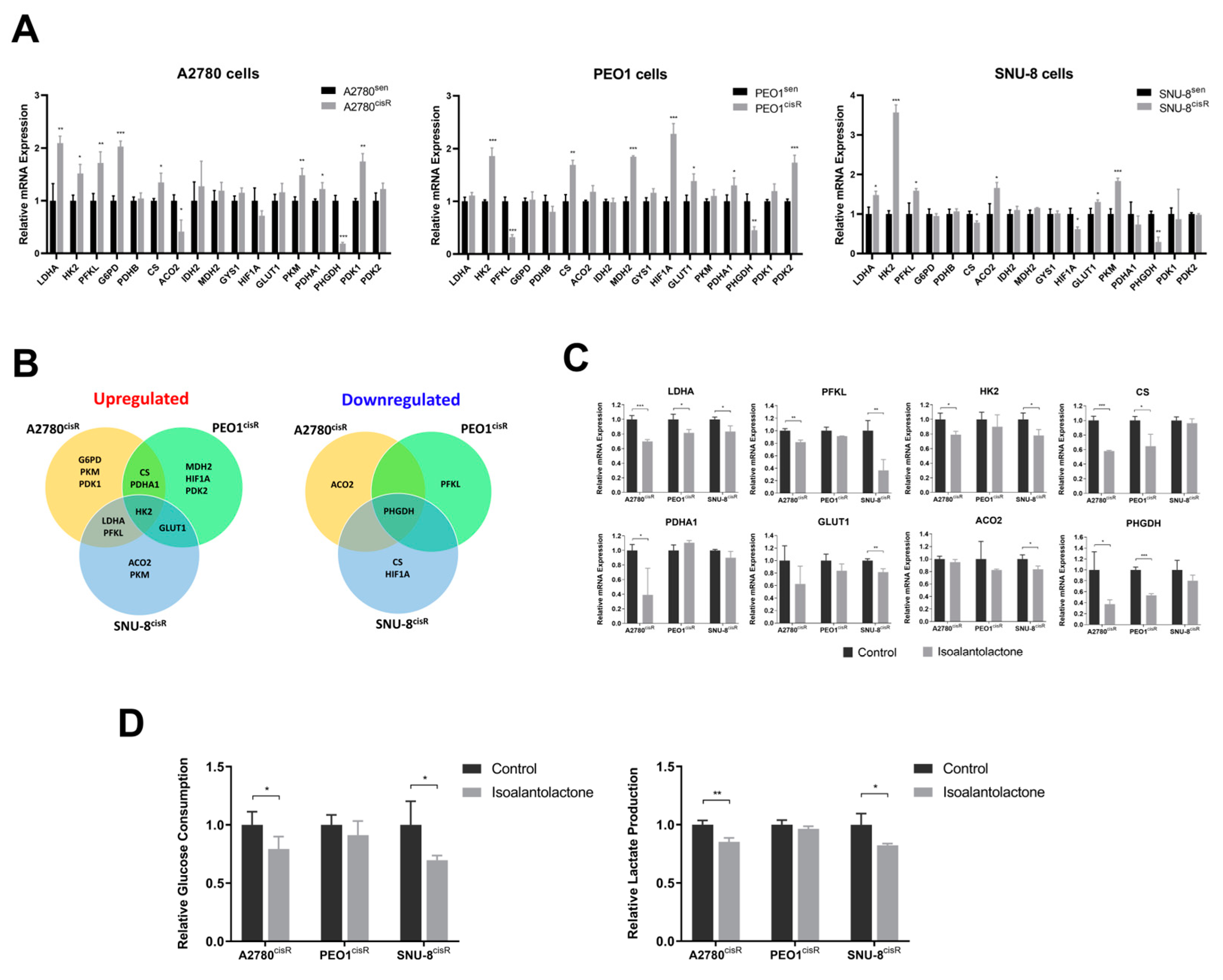
2.2. Isoalantolactone Inhibits Elevated Glycolysis in A2780cisR and SNU-8cisR OC Cells

The alteration in glycolytic metabolic genes in cisplatin-sensitive and resistant OC cells was investigated. A comparative mRNA expression analysis of the two cell types revealed increased glycolysis-related mRNA expression in cisplatin-resistant OC cells (Figure 2A). Specifically, A2780cisR cells exhibited greater mRNA expression levels of lactate dehydrogenase A (LDHA), hexokinase 2 (HK2), phosphofructokinase liver type (PFKL), glucose 6-phosphate dehydrogenase (G6PD), citrate synthase (CS), pyruvate kinase M1/2 (PKM), pyruvate dehydrogenase E1α (PDHA1), and pyruvate dehydrogenase kinase 1 (PDK1) than A2780sen cells. While PEO1cisR cells demonstrated higher mRNA expression levels of HK2, CS, malate dehydrogenase 2 (MDH2), hypoxia-inducible factor 1α (HIF1A), glucose transporter protein type 1 (GLUT1), PDHA1, and pyruvate dehydrogenase kinase 2 (PDK2) than PEO1sen cells, SNU-8cisR cells exhibited greater mRNA expression levels of LDHA, HK2, PFKL, aconitase 2 (ACO2), GLUT1, and PKM than SNU-8sen cells. Given the upregulation of the overlapping candidates LDHA, PFKL, CS, PDHA1, HK2, and GLUT1 and the downregulation of phosphoglycerate dehydrogenase (PHGDH) in cisplatin-resistant OC cells (Figure 2B), the inhibitory effect of isoalantolactone on the glycolysis of cisplatin-resistant OCs was mainly focused. As shown in Figure 2C, isoalantolactone effectively inhibited glycolysis by targeting key enzymes, including LDHA (in A2780cisR, PEO1cisR, SNU-8cisR), PFKL and HK2 (in A2780cisR, SNU-8cisR), and CS and PHGDH (in A2780cisR, PEO1cisR). Furthermore, the evaluation for the determination of whether isoalantolactone induces a metabolic shift in glycolysis in cisplatin-resistant OC cells revealed that isoalantolactone treatment significantly decreased glucose consumption and lactate production in A2780cisR and SNU-8cisR cells (Figure 2D) but failed to do so in PEO1cisR cells. Collectively, these findings suggest that isoalantolactone suppresses glycolysis in cisplatin-resistant OC cells by reducing glycolytic enzymes, leading to decreased glucose consumption and lactate production in cisplatin-resistant OC A2780cisR and SNU-8cisR cells.

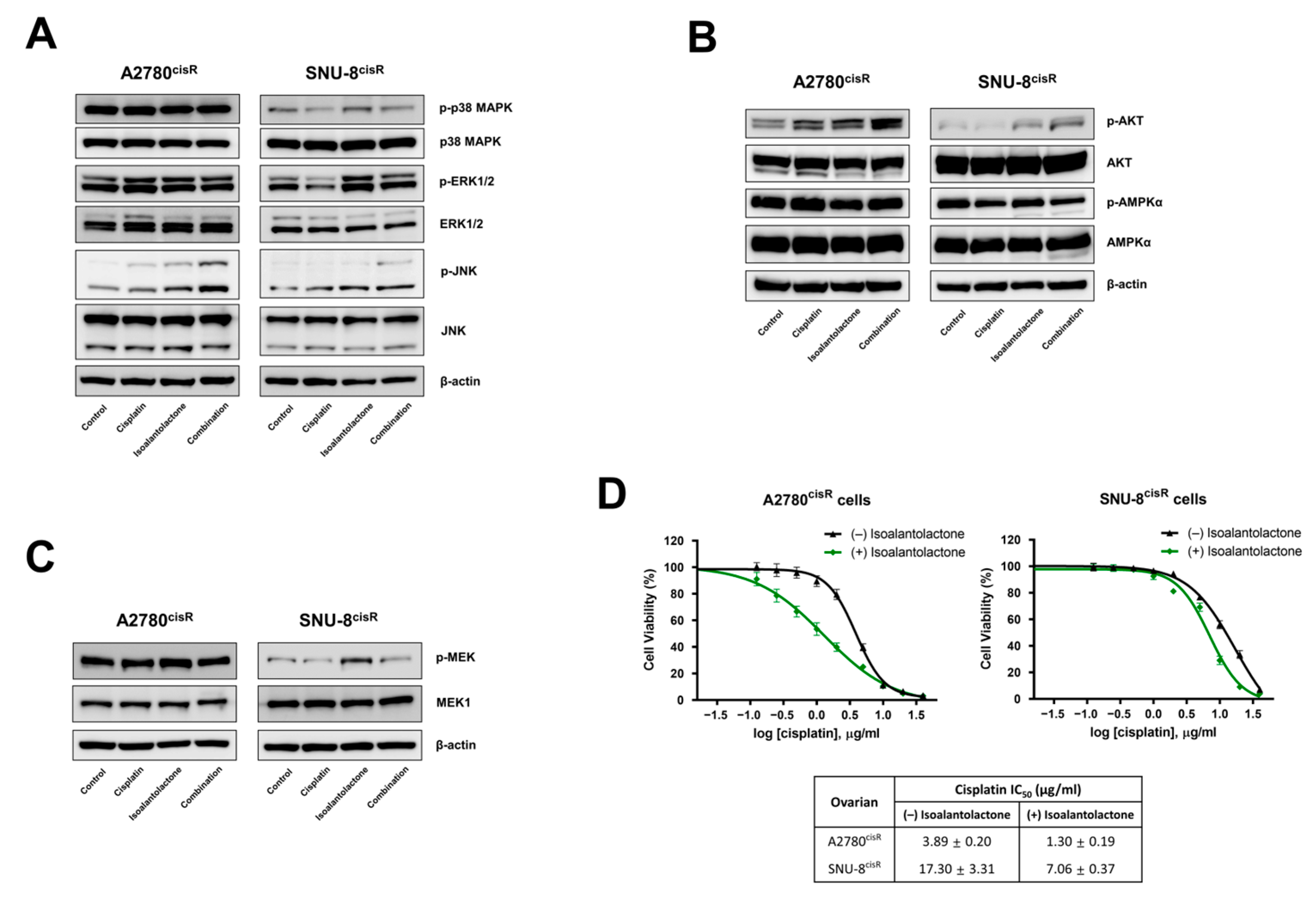
2.3. Isoalantolactone Increases the Sensitivity of A2780cisR and SNU-8cisR OC Cells to Cisplatin via the Apoptotic Pathway

Next, we investigated whether the decreased glycolysis caused by isoalantolactone could affect the sensitivity of cisplatin-resistant OC cells to cisplatin. The results showed that isoalantolactone combined with cisplatin increased apoptosis (Figure 3A and Figure S2), indicating that isoalantolactone treatment sensitized A2780cisR and SNU-8cisR OC cells to cisplatin. The evaluation of the effect of isoalantolactone on apoptosis-related proteins revealed that isoalantolactone increased the expression of cleaved caspase-3, caspase-8, and PARP1 and decreased the expression of anti-apoptotic genes such as cyclin D1 and mcl-1 (Figure 3B). Notably, the Caspase-Glo 3/7 Assay Kit was used to confirm that combination treatment induces apoptosis in OC cells, revealing that isoalantolactone–cisplatin treatment significantly increased caspase-3/7 activities, which agreed with the apoptosis results (Figure 3C). Overall, these results suggest that isoalantolactone suppresses glycolysis and sensitizes cisplatin-resistant cells to cisplatin by inducing mitochondria-mediated apoptosis and caspase activation in A2780cisR and SNU-8cisR cells.

2.4. Isoalantolactone Potentiates Cisplatin-Induced Apoptosis by Regulating the Survival Signaling Pathway

AKT, mitogen-activated protein kinase (MAPK), and AMP-activated protein kinase (AMPK) pathways are important for glucose homeostasis in cancer cell survival [22]. To explore the mechanism underlying the effect of isoalantolactone on cisplatin resistance-associated signaling pathways, the protein expression of MAPK signaling pathways was examined. Isoalantolactone–cisplatin treatment resulted in increased phosphorylation of JNK, whereas the total form of JNK remained unchanged in both A2780cisR and SNU-8cisR cells (Figure 4A). Isoalantolactone treatment induced increased phosphorylation of p38 MAPK and ERK1/2, whereas cisplatin treatment decreased levels of these proteins in SNU-8cisR cells. However, they had no effect in A2780cisR cells. In addition, the combination treatment increased AKT phosphorylation in both A2780cisR and SNU-8cisR cells but did not affect AMPKα phosphorylation (Figure 4B). Furthermore, the evaluation of the effect of isoalantolactone treatment on the MEK1 pathway, which is associated with ERK signaling and cisplatin resistance in cancer cells [23], revealed that isoalantolactone increased MEK1 phosphorylation (Figure 4C). The decreased IC50 values of cisplatin with isoalantolactone treatment in A2780cisR and SNU-8cisR cells were also confirmed (Figure 4D). Overall, these data suggest that isoalantolactone enhances the cisplatin sensitivity of cisplatin-resistant OC cells by regulating the MAPK and AKT survival signaling pathways.

2.5. Isoalantolactone–Cisplatin Treatment Inhibits A2780cisR Xenograft Tumor Growth

To investigate the antitumor effects of isoalantolactone in vivo, A2780sen and A2780cisR cells were used to construct a cisplatin-sensitive and resistant OC model (Figure 5A). In the A2780sen xenograft model, cisplatin treatment dramatically reduced tumor volume (Figure 5B) and tumor weight (Figure 5C) compared with the control group. However, isoalantolactone slightly decreased the tumor volume and tumor weight, although it was not significant. In the A2780cisR xenograft model, isoalantolactone treatment suppressed tumor volume (Figure 5D) and tumor weight (Figure 5E), while cisplatin treatment failed to do so. However, the combination treatment further decreased tumor volume and tumor weight. Isoalantolactone and/or cisplatin treatment did not affect the body weight of the mice compared with the control group, indicating that mouse health remained intact (Figure S3). Overall, these in vivo data suggest that isoalantolactone–cisplatin treatment can effectively overcome cisplatin resistance in OC.

2.6. Network Pharmacology Analysis Predicts the Target Pathways of Isoalantolactone-Regulated Glycolysis for OC Treatment

Network pharmacology provides a systemic approach to unraveling the potential multiple actions of natural product-derived compounds [24]. Information regarding the 105 potential protein targets for isoalantolactone was obtained using Pharmmapper and SwissTarget. Information regarding the 535 target genes of the disease was obtained using GeneCards. The 25 common gene targets are shown in Figure 6A. Potential targets were submitted to the STRING database, and the protein–protein interaction (PPI) network was constructed. The predicted PPI network showed that a functional link may exist between those target genes of isoalantolactone (Figure 6B). Furthermore, Gene Ontology (GO) and the Kyoto Encyclopedia of Genes and Genomes (KEGG) pathway enrichment analyses were performed. As shown in Figure 6C, the biological process terms for GO analysis included signal transduction, MAPK activity, and phosphorylation. The cellular component terms for GO analysis included macromolecular complex, cytoplasm, and mitochondrion. The molecular function terms for GO analysis included protein kinase activity, protein phosphatase binding, and MAPK activity. In addition, the KEGG pathway enrichment analysis of potential core targets indicated that genes might influence the VEGF, MAPK, and AKT pathways (Figure 6D). These results indicate that isoalantolactone may act on OC-related genes and that the MAPK pathway may be a potential treatment pathway.

3. Discussion

The mechanism of cisplatin resistance is extremely complex and multifactorial. One of the key mechanisms underlying cisplatin resistance in cancer is dysregulated cellular metabolism, particularly upregulated glycolysis [25]. Glucose is mainly used in OC cells to produce ATP and to maintain redox homeostasis and energy balance. Previous studies demonstrated that cisplatin-resistant OC cells require a higher demand for glucose and are more sensitive to glucose deprivation [26,27]. Similarly, in this study, cisplatin-resistant OC cells exhibited an enhanced glycolytic phenotype, proven by the upregulation of glycolytic enzymes such as LDHA, PFKL, and HK2. Among these enzymes, HK2-encoding gene is commonly upregulated in cisplatin-resistant cell lines used in experiments. HK2 phosphorylates glucose to generate glucose-6-phosphate, the first step in glycolysis [28]. A high expression of HK2 in tumors promotes tumor growth by maintaining higher glycolysis rates in cancer cells. In addition, HK2 overexpression has been correlated to chemoresistance in tumors [29]. Interestingly, cisplatin treatment reduced HK2 expression in cisplatin-sensitive OC cells but not in cisplatin-resistant OC cells [30]. HK2-targeting inhibitors combined with cisplatin could represent a novel approach to overcome cisplatin resistance. LDHA controls the conversion of pyruvate to lactate. Inhibiting glycolysis by targeting LDHA could restore cisplatin sensitivity in cisplatin-resistant cells [31]. Targeting PFKL, a rate-limiting glycolysis enzyme, could reduce glucose consumption and lactate production [32]. Therefore, small molecules targeting glycolytic enzymes have recently gained more attention in chemoresistance.
Natural products are a potential source of drug discovery. Small molecules derived from natural products can exhibit various pharmacological properties, including anticancer activities [33]. Although isoalantolactone exhibits anticancer activities in several types of cancer cells [34,35,36,37], including imatinib-resistant chronic myeloid leukemia [38], its anticancer effect in the cisplatin-resistant model and whether it can regulate cancer metabolism remain largely unknown. In particular, cisplatin resistance remains a significant challenge in OC treatment, necessitating the exploration of novel therapeutic approaches [39,40]. In this study, the potential of isoalantolactone as a glycolysis inhibitor was investigated to overcome cisplatin resistance in OC. The results demonstrated that isoalantolactone effectively inhibited the growth of cisplatin-resistant OC cells, particularly A2780cisR and SNU-8cisR cells. These findings suggest that isoalantolactone exhibits stronger cytotoxicity toward cisplatin-resistant OC cells than cisplatin-sensitive cells, highlighting its potential as a promising therapeutic agent. In addition, isoalantolactone effectively targeted these key enzymes, resulting in significantly inhibited glycolysis in cisplatin-resistant OC A2780cisR and SNU-8cisR cells. It reduced glucose consumption and lactate production, suggesting the disruption of the glycolytic pathway. These findings highlight the potential of isoalantolactone as a glycolysis inhibitor in cisplatin-resistant OC cells.
While the upregulation of glycolysis plays a crucial role in enabling cancer cells to evade cisplatin-induced apoptosis [41,42], its inhibition can enhance the chemotherapeutic sensitivity of OC to cisplatin [43]. Importantly, isoalantolactone sensitized cisplatin-resistant OC cells to cisplatin-induced apoptosis, and isoalantolactone–cisplatin treatment resulted in increased apoptosis compared with the individual treatments. This suggests that isoalantolactone inhibits glycolysis and enhances the efficacy of cisplatin by promoting apoptotic cell death in cisplatin-resistant OC cells. Many signaling pathways are associated with cisplatin resistance, as they promote cell survival and proliferation [44]. The activation of MAPK and AKT signaling pathways in cisplatin resistance has been demonstrated [45,46]. Isoalantolactone–cisplatin treatment was associated with the inhibition of survival signaling pathways, as indicated by the increased phosphorylation of JNK and AKT. These findings suggest that isoalantolactone overcomes cisplatin resistance by modulating survival signaling pathways and inducing apoptosis. Although isoalantolactone has a therapeutic effect on the recovery of chemoresistance through alteration in glucose metabolism and survival signaling pathways, further studies are required to clarify their relationship and the exact mechanism of isoalantolactone.
In this study, in vivo analyses using cisplatin-sensitive and resistant OC xenograft models were conducted to clarify the potential of isoalantolactone in overcoming cisplatin resistance. While cisplatin treatment alone showed limited efficacy in reducing tumor growth in the cisplatin-resistant model, isoalantolactone treatment significantly suppressed tumor growth. Surprisingly, isoalantolactone–cisplatin treatment further enhanced the antitumor effect, indicating a synergistic interaction between these treatments. These results suggest that isoalantolactone, either alone or in combination with cisplatin, can effectively overcome cisplatin resistance in OC. In addition, a network pharmacology analysis revealed the potential targets and pathways associated with isoalantolactone-regulated glycolysis for OC treatment. The analysis identified several key pathways, including the VEGF, MAPK, and AKT pathways, which have been implicated in cancer progression and drug resistance [47,48]. These findings provide a theoretical basis for the therapeutic potential of isoalantolactone in OC and suggest the involvement of specific pathways that may be targeted by isoalantolactone to overcome cisplatin resistance.

4. Materials and Methods

4.1. Chemicals and Reagents

Gibco (Grand Island, NY, USA) supplied RPMI-1640 medium, fetal bovine serum (FBS), and Dulbecco’s phosphate-buffered saline (DPBS). ChemFaces (Wuhan, China) supplied isoalantolactone. Antibodies against cleaved PARP1, cleaved caspase-3, cleaved caspase-8, cyclin D1, mcl-1, p-p38 MAPK, p38 MAPK, p-ERK1/2, ERK1/2, p-AKT, AKT, p-AMPKα, AMPKα, p-MEK, MEK, and β-actin were purchased from Cell Signaling Technology (Beverly, MA, USA). Santa Cruz Biotechnology (Santa Cruz, CA, USA) supplied antibodies against p-JNK and JNK. Cisplatin, dimethyl sulfoxide (DMSO), and Tween 80 were purchased from Sigma-Aldrich (St. Louis, MO, USA).

4.2. Cell Culture

The European Collection of Authenticated Cell Cultures supplied A2780sen, PEO1sen, A2780cisR, and PEO1cisR OC cells. SNU-8sen OC cells were purchased from the Korean Cell Line Bank. SNU-8cisR OC cells were established via continuous exposure to increasing doses of cisplatin over 1 year. The cells were maintained in an RPMI-1640 medium containing 10% FBS, penicillin 100 U/mL, and streptomycin 100 μg/mL at 37 °C in a 5% CO2 humidified atmosphere.

4.3. Cell Viability Assay

Cell proliferation was determined using a cell counting kit-8 assay (CCK-8, Dojindo, Kumamoto, Japan). Briefly, cells at the density of 1 × 10⁴ cells/well (A2780 and PEO1) and 5 × 10³ cells/well (SNU-8) were seeded into 96-well plates and incubated overnight. Then, different concentrations of cisplatin and/or isoalantolactone were added to each well. After 24 h or 48 h, CCK-8 was added. The absorbance of the CCK-8 reagent was detected at 450 nm using a Spectramax i3 microplate reader (Molecular Devices, San Jose, CA, USA). IC50 of cisplatin was calculated using GraphPad Prism 9 (San Diego, CA, USA).

4.4. Apoptosis Analysis

A flow cytometric analysis of apoptosis was performed to evaluate the apoptosis-inducing effects of isoalantolactone. Here, 5 × 10⁵ of A2780cisR cells and 2.5 × 10⁵ of SNU-8cisR were seeded into 6-well plates and maintained in a 37 °C incubator overnight. Afterward, the cells were exposed to a medium containing isoalantolactone (10 μM), followed by cisplatin (2 μg/mL for A2780cisR cells and 5 μg/mL for SNU-8cisR cells) for 48 h. The cells were detached, washed in DBPS, and resuspended in Annexin V binding buffer, followed by the addition of Annexin V/FITC and propidium iodide and 15 min incubation in the dark. The cells were resuspended in 400 μL of Annexin V binding buffer. The stained cells were acquired and analyzed using a BD LSRFortessa™ X-20 flow cytometer (BD Biosciences, San Jose, CA, USA) and FlowJo 10.8 (BD Biosciences).

4.5. Quantitative RT-PCR

Total RNA from A2780, PEO1, and SNU-8 cells was isolated using an RNeasy Plus Mini Kit (Qiagen, Valencia, CA, USA). The purity and concentration of RNA were measured using NanoDrop (Thermo Scientific, Waltham, MA, USA). Total RNA (1 μg) was converted to cDNA using iScript™ Advanced cDNA Synthesis Kit (Bio-Rad, Hercules, CA, USA). Table 1 provides the primer sequences. Gene expression analysis was performed using the SsoAdvanced SYBR Green SuperMix Kit (Bio-Rad) on the Bio-Rad CFX Connect Real-Time System (Bio-Rad). The relative gene level was calculated using comparative delta Ct and normalized using the ACTB gene.

4.6. Metabolic Assays

Alteration in cellular metabolism regarding glycolysis was measured using the Glucose-Glo ™ Assay and Lactate-Glo ™ Assay (Promega, Madison, WI, USA). The cells at densities of 1 × 10⁴/well (A2780cisR cells) and 5 × 10³/well (SNU-8cisR cells) were seeded in 96-well plates and incubated overnight. The cells were treated with isoalantolactone (10 μM), followed by cisplatin (2 μg/mL for A2780cisR cells and 5 μg/mL for SNU-8cisR cells) for 24 h. The culture and control media were collected and incubated with glucose detection reagent and lactate detection reagent for 1 h, respectively. The luminescence intensity was measured using a microplate luminometer (Molecular Devices). Glucose consumption and lactate production were calculated by subtracting from the concentrations of glucose and lactate in the control medium.

4.7. Western Blotting

Cell lysates were prepared using NP40 Cell Lysis Buffer (Invitrogen, Carlsbad, CA, USA), supplemented with Halt™ Protease and Phosphatase Inhibitor Cocktail (Thermo Fisher Scientific, Waltham, MA, USA). Equal amounts of protein, determined with a Qubit™ Protein Assay Kit (Invitrogen), were prepared in 4× LDS sample buffer (Invitrogen) and 10× sample reducing agent (Invitrogen) and heated at 70 °C for 10 min. The samples were separated on Bolt 4 to 12% Bis-Tris gels in Bolt MOPS SDS running buffer (Thermo Fisher Scientific), and the proteins were transferred onto a nitrocellulose membrane (Invitrogen) using the SureLock Tandem Transfer System (Thermo Fisher Scientific). The membrane was blocked with EveryBlot Blocking Buffer (Bio-Rad), incubated with primary antibodies overnight, and incubated with secondary antibodies conjugated with horseradish peroxidase. The bands were developed using Clarity ECL Western Blotting Substrates (Bio-Rad) and visualized using an ImageQuant LAS 4000 mini (GE Healthcare, Chicago, IL, USA).

4.8. Measurement of Caspase-3/7 Activity

The caspase-3/7 activities were evaluated using a Caspase-Glo 3/7 Assay Kit (Promega) following the manufacturer’s instructions. Briefly, cells at densities of 5 × 10³ cells/well (A2780) and 2.5 × 10³ cells/well (SNU-8) were cultured in 96-well plates and incubated overnight, followed by exposure to a medium containing isoalantolactone (10 μM) and subsequently cisplatin (2 μg/mL for A2780cisR cells and 5 μg/mL for SNU-8cisR cells) for 48 h. Caspase-3/7 reagent was added to each well and incubated for 1 h at room temperature. The luminescence intensity was measured using a microplate luminometer (Molecular Devices).

4.9. Tumor-Bearing Mice and Treatment

Specific pathogen-free five-week-old female Balb/c-nude mice were purchased from Saeron Bio (Uiwang, Republic of Korea) and acclimated for one week prior to experimental use. The mice were housed under the following environmental conditions: temperature 23 °C; humidity 50%; 12 h light–dark cycle. The animals were fed a standard chow diet (Purina Co., Seoul, Republic of Korea) and given free access to drinking water. A2780sen and A2780cisR tumor-bearing mice were prepared by being injected subcutaneously with 2 × 10⁶ cells into the right flank. After the tumor challenge, Balb/c-nude mice were intraperitoneally injected with 20 mg/kg isoalantolactone every day and 2.5 mg/kg cisplatin every three days for 17 days. Cisplatin was dissolved in PBS. Isoalantolactone was dissolved in PBS containing 2% DMSO and 2% Tween 80. All experimental procedures were approved by the Animal Care and Use Committee of the Korea Institute of Oriental Medicine (Approval Number: 21-032). Tumor volume was measured via caliper measurement and calculated using the following formula: length × width2 × 1/2. Body weight and tumor size were recorded.

4.10. Systematic Pharmacological Analysis of Isoalantolactone

Using GeneCards (https://genecards.weizmann.ac.il/v3/, accessed on 25 May 2023), the target genes of the disease were acquired as the keyword of “ovarian cancer” and “glycolysis”. The PubChem (https://pubchem.ncbi.nlm.nih.gov/, accessed on 17 October 2022) database was used to obtain the structure of isoalantolactone. Pharmmapper (https://www.lilab-ecust.cn/pharmmapper/index.html, accessed on 17 October 2022) and SwissTarget (https://www.swisstargetprediction.ch/, accessed on 17 October 2022) databases with the “Homo sapiens” species setting were utilized to predict the protein targets of isoalantolactone. The predicted protein targets were converted into the predicted gene targets using the UniProt database (https://www.uniport.org/, accessed on 25 May 2023). The disease–gene targets and drug-prediction gene targets were combined through Venny 2.1 software (https://bioinfogp.cnb.csic.es/tools/venny/index.html, accessed on 25 May 2023) to obtain common gene targets. The STRING database (https://cn.string-db.org/, accessed on 1 June 2023) was used to construct a protein–protein interaction network. GO and KEGG pathway enrichment analyses were performed on the David online analysis platform (https://david.ncifcrf.gov/, accessed on 2 June 2023) to screen the GO processes and signaling pathways involved in the potential common targets of isoalantolactone and diseases. The bubble chart and bar graph were plotted using an online platform (https://www.bioinformatics.com.cn/, accessed on 2 June 2023) for visualization.

4.11. Statistical Analysis

Statistical analysis was performed using GraphPad Prism 9.0. Error bars represent mean ± standard deviation (SD), except for tumor volume curves, which represent mean ± standard error of the mean. Statistical analysis of significance was performed using a two-tailed Student’s t-test. A value of p < 0.05 was considered statistically significant.

5. Conclusions

In conclusion, this study demonstrates the efficacy of isoalantolactone as a glycolysis inhibitor in overcoming cisplatin resistance in OC. Isoalantolactone effectively targets glycolytic enzymes and suppresses glycolysis. In addition, isoalantolactone supports the action of anticancer drugs by increasing the sensitivity of OC cells to cisplatin and helping to overcome cisplatin resistance. These findings highlight the potential of isoalantolactone as a novel therapeutic strategy for overcoming cisplatin resistance in OC. Further studies are warranted to elucidate the precise molecular mechanisms underlying its action and to evaluate its clinical potential.

Supplementary Materials

The following supporting information can be downloaded at: https://www.mdpi.com/article/10.3390/ijms241512397/s1.

Funding

This work was supported by the National Research Foundation (NRF) of Korea (NRF-2020R1C1C1004573).

Institutional Review Board Statement

Not applicable.

Informed Consent Statement

Not applicable.

Data Availability Statement

The data presented in this study are available on request from the corresponding author.

Conflicts of Interest

The author declares no conflict of interest.

References

  1. Torre, L.A.; Islami, F.; Siegel, R.L.; Ward, E.M.; Jemal, A. Global cancer in women: Burden and trends. Cancer Epidemiol. Biomark. Prev. 2017, 26, 444–457. [Google Scholar] [CrossRef] [Green Version]
  2. Chandra, A.; Pius, C.; Nabeel, M.; Nair, M.; Vishwanatha, J.K.; Ahmad, S.; Basha, R. Ovarian cancer: Current status and strategies for improving therapeutic outcomes. Cancer Med. 2019, 8, 7018–7031. [Google Scholar] [CrossRef] [PubMed] [Green Version]
  3. Guarneri, V.; Piacentini, F.; Barbieri, E.; Conte, P.F. Achievements and unmet needs in the management of advanced ovarian cancer. Gynecol. Oncol. 2010, 117, 152–158. [Google Scholar] [CrossRef]
  4. Brown, A.; Kumar, S.; Tchounwou, P.B. Cisplatin-based chemotherapy of human cancers. J. Cancer Sci. Ther. 2019, 11, 97. [Google Scholar] [PubMed]
  5. Song, M.; Cui, M.; Liu, K. Therapeutic strategies to overcome cisplatin resistance in ovarian cancer. Eur. J. Med. Chem. 2022, 232, 114205. [Google Scholar] [CrossRef]
  6. Koberle, B.; Tomicic, M.T.; Usanova, S.; Kaina, B. Cisplatin resistance: Preclinical findings and clinical implications. Biochim. Biophys. Acta 2010, 1806, 172–182. [Google Scholar] [CrossRef] [PubMed]
  7. Dasari, S.; Tchounwou, P.B. Cisplatin in cancer therapy: Molecular mechanisms of action. Eur. J. Pharmacol. 2014, 740, 364–378. [Google Scholar] [CrossRef] [Green Version]
  8. Rebucci, M.; Michiels, C. Molecular aspects of cancer cell resistance to chemotherapy. Biochem. Pharmacol. 2013, 85, 1219–1226. [Google Scholar] [CrossRef] [PubMed]
  9. Wang, L.; Zhao, X.; Fu, J.; Xu, W.; Yuan, J. The role of tumour metabolism in cisplatin resistance. Front. Mol. Biosci. 2021, 8, 691795. [Google Scholar] [CrossRef]
  10. Hay, N. Reprogramming glucose metabolism in cancer: Can it be exploited for cancer therapy? Nat. Rev. Cancer 2016, 16, 635–649. [Google Scholar] [CrossRef] [Green Version]
  11. Sun, L.; Suo, C.; Li, S.T.; Zhang, H.; Gao, P. Metabolic reprogramming for cancer cells and their microenvironment: Beyond the Warburg effect. Biochim. Biophys. Acta Rev. Cancer 2018, 1870, 51–66. [Google Scholar] [CrossRef] [PubMed]
  12. Xu, Y.; Gao, W.; Zhang, Y.; Wu, S.; Liu, Y.; Deng, X.; Xie, L.; Yang, J.; Yu, H.; Su, J.; et al. ABT737 reverses cisplatin resistance by targeting glucose metabolism of human ovarian cancer cells. Int. J. Oncol. 2018, 53, 1055–1068. [Google Scholar] [CrossRef] [PubMed] [Green Version]
  13. Varghese, E.; Samuel, S.M.; Liskova, A.; Samec, M.; Kubatka, P.; Busselberg, D. Targeting glucose metabolism to overcome resistance to anticancer chemotherapy in breast cancer. Cancers 2020, 12, 2252. [Google Scholar] [CrossRef] [PubMed]
  14. Dutta, S.; Mahalanobish, S.; Saha, S.; Ghosh, S.; Sil, P.C. Natural products: An upcoming therapeutic approach to cancer. Food Chem. Toxicol. 2019, 128, 240–255. [Google Scholar] [CrossRef]
  15. Zhang, X.; Qiu, H.; Li, C.; Cai, P.; Qi, F. The positive role of traditional Chinese medicine as an adjunctive therapy for cancer. Bio Sci. Trends 2021, 15, 283–298. [Google Scholar] [CrossRef] [PubMed]
  16. Lin, S.R.; Chang, C.H.; Hsu, C.F.; Tsai, M.J.; Cheng, H.; Leong, M.K.; Sung, P.J.; Chen, J.C.; Weng, C.F. Natural compounds as potential adjuvants to cancer therapy: Preclinical evidence. Br. J. Pharmacol. 2020, 177, 1409–1423. [Google Scholar] [CrossRef] [PubMed] [Green Version]
  17. Guo, W.; Tan, H.Y.; Chen, F.; Wang, N.; Feng, Y. Targeting cancer metabolism to resensitize chemotherapy: Potential development of cancer chemosensitizers from traditional Chinese medicines. Cancers 2020, 12, 404. [Google Scholar] [CrossRef] [Green Version]
  18. Ding, Y.H.; Song, Y.D.; Wu, Y.X.; He, H.Q.; Yu, T.H.; Hu, Y.D.; Zhang, D.P.; Jiang, H.C.; Yu, K.K.; Li, X.Z.; et al. Isoalantolactone suppresses LPS-induced inflammation by inhibiting TRAF6 ubiquitination and alleviates acute lung injury. Acta Pharmacol. Sin. 2019, 40, 64–74. [Google Scholar] [CrossRef] [Green Version]
  19. He, G.; Zhang, X.; Chen, Y.; Chen, J.; Li, L.; Xie, Y. Isoalantolactone inhibits LPS-induced inflammation via NF-κB inactivation in peritoneal macrophages and improves survival in sepsis. Biomed. Pharmacother. 2017, 90, 598–607. [Google Scholar] [CrossRef]
  20. Wu, M.; Zhang, H.; Hu, J.; Weng, Z.; Li, C.; Li, H.; Zhao, Y.; Mei, X.; Ren, F.; Li, L. Isoalantolactone inhibits UM-SCC-10A cell growth via cell cycle arrest and apoptosis induction. PLoS ONE 2013, 8, e76000. [Google Scholar] [CrossRef] [PubMed]
  21. Jin, C.; Zhang, G.; Zhang, Y.; Hua, P.; Song, G.; Sun, M.; Li, X.; Tong, T.; Li, B.; Zhang, X. Isoalantolactone induces intrinsic apoptosis through p53 signaling pathway in human lung squamous carcinoma cells. PLoS ONE 2017, 12, e0181731. [Google Scholar] [CrossRef] [Green Version]
  22. Schultze, S.M.; Hemmings, B.A.; Niessen, M.; Tschopp, O. PI3K/AKT, MAPK and AMPK signalling: Protein kinases in glucose homeostasis. Expert Rev. Mol. Med. 2012, 14, e1. [Google Scholar] [CrossRef] [Green Version]
  23. Kong, L.R.; Chua, K.N.; Sim, W.J.; Ng, H.C.; Bi, C.; Ho, J.; Nga, M.E.; Pang, Y.H.; Ong, W.R.; Soo, R.A.; et al. MEK inhibition overcomes cisplatin resistance conferred by SOS/MAPK pathway activation in squamous cell carcinoma. Mol. Cancer Ther. 2015, 14, 1750–1760. [Google Scholar] [CrossRef] [PubMed] [Green Version]
  24. Hao da, C.; Xiao, P.G. Network pharmacology: A Rosetta Stone for traditional Chinese medicine. Drug Dev. Res. 2014, 75, 299–312. [Google Scholar] [CrossRef]
  25. Cocetta, V.; Ragazzi, E.; Montopoli, M. Links between cancer metabolism and cisplatin resistance. Int. Rev. Cell Mol. Biol. 2020, 354, 107–164. [Google Scholar] [PubMed]
  26. Catanzaro, D.; Gaude, E.; Orso, G.; Giordano, C.; Guzzo, G.; Rasola, A.; Ragazzi, E.; Caparrotta, L.; Frezza, C.; Montopoli, M. Inhibition of glucose-6-phosphate dehydrogenase sensitizes cisplatin-resistant cells to death. Oncotarget 2015, 6, 30102–30114. [Google Scholar] [CrossRef] [PubMed] [Green Version]
  27. Tian, M.; Chen, X.S.; Li, L.Y.; Wu, H.Z.; Zeng, D.; Wang, X.L.; Zhang, Y.; Xiao, S.S.; Cheng, Y. Inhibition of AXL enhances chemosensitivity of human ovarian cancer cells to cisplatin via decreasing glycolysis. Acta Pharmacol. Sin. 2021, 42, 1180–1189. [Google Scholar] [CrossRef]
  28. Tan, V.P.; Miyamoto, S. HK2/hexokinase-II integrates glycolysis and autophagy to confer cellular protection. Autophagy 2015, 11, 963–964. [Google Scholar] [CrossRef] [Green Version]
  29. Suh, D.H.; Kim, M.A.; Kim, H.; Kim, M.K.; Kim, H.S.; Chung, H.H.; Kim, Y.B.; Song, Y.S. Association of overexpression of hexokinase II with chemoresistance in epithelial ovarian cancer. Clin. Exp. Med. 2014, 14, 345–353. [Google Scholar] [CrossRef]
  30. Zhang, X.Y.; Zhang, M.; Cong, Q.; Zhang, M.X.; Zhang, M.Y.; Lu, Y.Y.; Xu, C.J. Hexokinase 2 confers resistance to cisplatin in ovarian cancer cells by enhancing cisplatin-induced autophagy. Int. J. Biochem. Cell Biol. 2018, 95, 9–16. [Google Scholar] [CrossRef]
  31. Li, G.; Li, Y.; Wang, D.Y. Overexpression of miR-329-3p sensitizes osteosarcoma cells to cisplatin through suppression of glucose metabolism by targeting LDHA. Cell Biol. Int. 2021, 45, 766–774. [Google Scholar] [CrossRef]
  32. Zheng, C.; Yu, X.; Liang, Y.; Zhu, Y.; He, Y.; Liao, L.; Wang, D.; Yang, Y.; Yin, X.; Li, A.; et al. Targeting PFKL with penfluridol inhibits glycolysis and suppresses esophageal cancer tumorigenesis in an AMPK/FOXO3a/BIM-dependent manner. Acta Pharm. Sin. B 2022, 12, 1271–1287. [Google Scholar] [CrossRef]
  33. Newman, D.J.; Cragg, G.M. Natural products as sources of new drugs over the nearly four decades from 01/1981 to 09/2019. J. Nat. Prod. 2020, 83, 770–803. [Google Scholar] [CrossRef] [Green Version]
  34. Yan, Y.Y.; Zhang, Q.; Zhang, B.; Yang, B.; Lin, N.M. Active ingredients of Inula helenium L. exhibits similar anti-cancer effects as isoalantolactone in pancreatic cancer cells. Nat. Prod. Res. 2020, 34, 2539–2544. [Google Scholar] [CrossRef] [PubMed]
  35. Hu, F.; Yang, P. Isoalantolactone exerts anticancer effects on human HEC-1-B endometrial cancer cells via induction of ROS mediated apoptosis and inhibition of MEK/ERK signalling pathway. Acta Biochim. Pol. 2022, 69, 453–458. [Google Scholar] [CrossRef] [PubMed]
  36. Wu, F.; Shao, R.; Zheng, P.; Zhang, T.; Qiu, C.; Sui, H.; Li, S.; Jin, L.; Pan, H.; Jin, X.; et al. Isoalantolactone enhances the antitumor activity of doxorubicin by inducing reactive oxygen species and DNA damage. Front. Oncol. 2022, 12, 813854. [Google Scholar] [CrossRef]
  37. Kim, M.Y.; Lee, H.; Ji, S.Y.; Kim, S.Y.; Hwangbo, H.; Park, S.H.; Kim, G.Y.; Park, C.; Leem, S.H.; Hong, S.H.; et al. Induction of apoptosis by isoalantolactone in human hepatocellular carcinoma Hep3B cells through activation of the ROS-dependent JNK signaling pathway. Pharmaceutics 2021, 13, 1627. [Google Scholar] [CrossRef] [PubMed]
  38. Yin, S.S.; Chen, C.; Liu, Z.; Liu, S.L.; Guo, J.H.; Zhang, C.; Zhang, Q.W.; Gao, F.H. Isoalantolactone mediates the degradation of BCR-ABL protein in imatinib-resistant CML cells by down-regulating survivin. Cell Cycle 2023, 22, 1407–1420. [Google Scholar] [CrossRef]
  39. Parker, R.J.; Eastman, A.; Bostick-Bruton, F.; Reed, E. Acquired cisplatin resistance in human ovarian cancer cells is associated with enhanced repair of cisplatin-DNA lesions and reduced drug accumulation. J. Clin. Investig. 1991, 87, 772–777. [Google Scholar] [CrossRef] [Green Version]
  40. Li, J.; Feng, Q.; Kim, J.M.; Schneiderman, D.; Liston, P.; Li, M.; Vanderhyden, B.; Faught, W.; Fung, M.F.; Senterman, M.; et al. Human ovarian cancer and cisplatin resistance: Possible role of inhibitor of apoptosis proteins. Endocrinology 2001, 142, 370–380. [Google Scholar] [CrossRef]
  41. Li, F.L.; Liu, J.P.; Bao, R.X.; Yan, G.; Feng, X.; Xu, Y.P.; Sun, Y.P.; Yan, W.; Ling, Z.Q.; Xiong, Y.; et al. Acetylation accumulates PFKFB3 in cytoplasm to promote glycolysis and protects cells from cisplatin-induced apoptosis. Nat. Commun. 2018, 9, 508. [Google Scholar] [CrossRef] [Green Version]
  42. Guo, W.; Zhang, Y.; Chen, T.; Wang, Y.; Xue, J.; Zhang, Y.; Xiao, W.; Mo, X.; Lu, Y. Efficacy of RNAi targeting of pyruvate kinase M2 combined with cisplatin in a lung cancer model. J. Cancer Res. Clin. Oncol. 2011, 137, 65–72. [Google Scholar] [CrossRef] [PubMed]
  43. Loar, P.; Wahl, H.; Kshirsagar, M.; Gossner, G.; Griffith, K.; Liu, J.R. Inhibition of glycolysis enhances cisplatin-induced apoptosis in ovarian cancer cells. Am. J. Obstet. Gynecol. 2010, 202, 371.e1–371.e8. [Google Scholar] [CrossRef] [PubMed]
  44. Achkar, I.W.; Abdulrahman, N.; Al-Sulaiti, H.; Joseph, J.M.; Uddin, S.; Mraiche, F. Cisplatin based therapy: The role of the mitogen activated protein kinase signaling pathway. J. Transl. Med. 2018, 16, 96. [Google Scholar] [CrossRef] [PubMed] [Green Version]
  45. Peng, D.J.; Wang, J.; Zhou, J.Y.; Wu, G.S. Role of the Akt/mTOR survival pathway in cisplatin resistance in ovarian cancer cells. Biochem. Biophys. Res. Commun. 2010, 394, 600–605. [Google Scholar] [CrossRef] [PubMed] [Green Version]
  46. Brozovic, A.; Osmak, M. Activation of mitogen-activated protein kinases by cisplatin and their role in cisplatin-resistance. Cancer Lett. 2007, 251, 1–16. [Google Scholar] [CrossRef]
  47. Banerjee, S.; Kaye, S.B. New strategies in the treatment of ovarian cancer: Current clinical perspectives and future potential. Clin. Cancer Res. 2013, 19, 961–968. [Google Scholar] [CrossRef] [Green Version]
  48. Moserle, L.; Jimenez-Valerio, G.; Casanovas, O. Antiangiogenic therapies: Going beyond their limits. Cancer Discov. 2014, 4, 31–41. [Google Scholar] [CrossRef] [Green Version]
Figure 1. Isoalantolactone potently inhibits the growth of cisplatin-resistant A2780 (A2780cisR) and SNU-8 (SNU-8cisR) ovarian cancer (OC) cells: (A) The resistance phenotype in cisplatin-sensitive and resistant cells of OC A2780, PEO1, and SNU-8 cells. Cells were treated with cisplatin (0–40 µg/mL) and incubated for 48 h. Cell viability was assessed using a cell counting kit-8 assay (CCK-8). The half-maximal inhibitory concentration (IC50) values of cisplatin are shown. (B) Effect of isoalantolactone on the proliferation of cisplatin-sensitive and resistant OC cells. Cells were treated with isoalantolactone (0–20 µM) for 24 h. CCK-8 assay was performed. IC50 values of isoalantolactone are shown. (C) Flow cytometric analysis of apoptosis in cisplatin-sensitive and resistant OC cells. Cells were treated with isoalantolactone (0–15 µM) for 48 h. Annexin V staining was performed. Data are representative of three independent experiments as mean ± standard deviation (SD). ** p < 0.01; *** p < 0.001.
Ijms 24 12397 g001
Figure 2. Isoalantolactone alters the elevated glucose metabolism in A2780cisR and SNU-8cisR OC cells: (A) Alteration in glycolytic metabolic genes of cisplatin-sensitive and resistant A2780, PEO1, and SNU-8 OC cells. The mRNA level was analyzed via quantitative RT-PCR analysis. (B) The upregulated (left) and downregulated (right) genes in A2780cisR, PEO-1cisR, and SNU-8cisR OC cells. The overlapping genes between OC cells are indicated in the Venn diagram. (C) The effect of isoalantolactone on the mRNA expression of the overlapping genes, LDHA, PFKL, HK2, CS, PDHA1, GLUT1, ACO2, and PHGDH. (D) The effect of isoalantolactone on glucose consumption and lactate production. A2780cisR, PEO1cisR, and SNU-8cisR OC cells were treated with 10 µM isoalantolactone for 24 h. Glucose consumption and lactate production were measured in the culture media from the cells. The results are presented as mean ± SD (n = 4). * p < 0.05; ** p < 0.01; *** p < 0.001. LDHA, lactate dehydrogenase A; HK2, hexokinase 2; PFKL, phosphofructokinase liver type; G6PD, glucose 6-phosphate dehydrogenase; PDHB, pyruvate dehydrogenase β; CS, citrate synthase; ACO2, aconitase 2; IDH2, isocitrate dehydrogenase 2; MDH2, malate dehydrogenase 2; GYS1, glycogen synthase 1; HIF1A, hypoxia-inducible factor 1α; GLUT1, glucose transporter protein type 1; PKM, pyruvate kinase M1/2; PDHA1, pyruvate dehydrogenase E1α; PHGDH, phosphoglycerate dehydrogenase; PDK1, pyruvate dehydrogenase kinase 1; PDK2, pyruvate dehydrogenase kinase 2.
Ijms 24 12397 g002
Figure 3. Isoalantolactone sensitizes A2780cisR and SNU-8cisR OC cells to cisplatin via an apoptotic pathway. Cells were treated with 10 µM isoalantolactone combined with cisplatin 2 µg/mL (A2780cisR) and 5 µg/mL (SNU-8cisR) for 48 h: (A) Flow cytometric analysis of apoptosis in A2780cisR and SNU-8cisR OC cells. (B) The combined effect of isoalantolactone–cisplatin on the expression of apoptotic proteins. Cell lysates from A2780cisR and SNU-8cisR OC cells were confirmed using Western blotting. (C) The combined effect of isoalantolactone–cisplatin on caspase-3/7 activities. The caspase 3/7 activity was evaluated using the Caspase-Glo 3/7 Assay Kit. The results are presented as mean ± SD (n = 3). *** p < 0.001.
Ijms 24 12397 g003
Figure 4. Isoalantolactone combined with cisplatin regulates the survival signaling pathways in A2780cisR and SNU-8cisR OC cells. Cells were treated with cisplatin 2 µg/mL (A2780cisR) and 5 µg/mL (SNU-8cisR) or in combination with 10 µM isoalantolactone for 24 h: (A) The combined effect of isoalantolactone–cisplatin on the MAPK signaling pathway. The phosphorylation of p38 MAPK, ERK1/2, and JNK was confirmed using Western blotting. (B) The combined effect of isoalantolactone–cisplatin on AKT and AMPK signaling. The phosphorylation of AKT and AMPK was confirmed using Western blotting. (C) The combined effect of isoalantolactone–cisplatin on MEK signaling. Cell lysates from A2780cisR and SNU-8cisR OC cells were confirmed using Western blotting. (D) The attenuation of cisplatin resistance by isoalantolactone in A2780cisR and SNU-8cisR OC cells. Cells were treated with 10 μM isoalantolactone, followed by cisplatin (0–40 µg/mL) for 48 h. Cell viability was assessed using CCK-8 assay. IC50 values are shown.
Ijms 24 12397 g004
Figure 5. Isoalantolactone combined with cisplatin inhibits A2780cisR xenograft tumor growth: (A) Timeline of A2780sen and A2780cisR cell inoculation and treatment with cisplatin and isoalantolactone. A2780sen and A2780cisR cells (2 × 10⁶ cells) were inoculated subcutaneously into Balb/c-nude mice. One week after cell injection, the mice were divided into four groups (n = 6, each group) as follows: control, cisplatin, isoalantolactone, and combination (cisplatin–isoalantolactone) groups. Mice were intraperitoneally administered isoalantolactone (20 mg/kg) every day and/or cisplatin (2.5 mg/kg) every three days. Tumor volumes were measured three times a week. (B) Time courses of A2780sen cell xenograft tumor growth and visual comparison of dissected A2780sen tumor tissues. (C) A2780sen tumor weight. (D) Time courses of A2780cisR cell xenograft tumor growth and visual comparison of dissected A2780cisR tumor tissues. (E) A2780cisR tumor weight. The results are presented as mean ± standard error of the mean for tumor volume and SD for tumor weight (n = 6); ns, not significant; ** p < 0.01; *** p < 0.001.
Ijms 24 12397 g005
Figure 6. Network pharmacology analysis predicts the target pathways of isoalantolactone-regulated glycolysis for OC treatment: (A) Venn diagram of targets between diseases and isoalantolactone. The 535 target genes of the disease were obtained from GeneCards. The 105 potential protein targets for isoalantolactone were obtained from Pharmmapper and SwissTarget. The 25 overlapping gene targets are shown. (B) The protein–protein interaction (PPI) network diagram. The common targets of diseases and isoalantolactone were uploaded to the STRING database. Homo Sapiens was selected to construct the PPI network. (C) Gene Ontology (GO) functional enrichment analyses. Green represents biological process (BP), orange represents cellular component (CC), and purple represents molecular function (MF) terms for GO analysis. (D) The Kyoto Encyclopedia of Genes and Genomes pathway enrichment analysis of potential core targets.
Ijms 24 12397 g006
Table 1. List of primer sequences for quantitative RT-PCR.
Target GenesForward SequencesReverse Sequences
LDHAGGA TCT CCA ACA TGG CAG CCT TAGA CGG CTT TCT CCC TCT TGC T
HK2GAG TTT GAC CTG GAT GTG GTT GCCCT CCA TGT AGC AGG CAT TGC T
PFKLAAG AAG TAG GCT GGC ACG ACG TGCG GAT GTT CTC CAC AAT GGA C
G6PDCTG TTC CGT GAG GAC CAG ATC TTGA AGG TGA GGA TAA CGC AGG C
PDHBTGT AAC TGT GGA AGG AGG CTG GCAT CAG CAC CAG TGA CAC GAA C
CSCAC AGG GTA TCA GCC GAA CCA ACCA ATA CCG CTG CCT TCT CTG T
ACO2CAA TCG TCA CCT CCT ACA ACA GGGTC TCT GGG TTG AAC TTG AGG G
IDH2AGA TGG CAG TGG TGT CAA GGA GCTG GAT GGC ATA CTG GAA GCA G
MDH2CTG GAC ATC GTC AGA GCC AAC AGGA TGA TGG TCT TCC CAG CAT G
GYS1CCG CTA TGA GTT CTC CAA CAA GGAGA AGG CAA CCA CTG TCT GCT C
HIF1ATAT GAG CCA GAA GAA CTT TTA GGCCAC CTC TTT TGG CAA GCA TCC TG
GLUT1TTG CAG GCT TCT CCA ACT GGA CCAG AAC CAG GAG CAC AGT GAA G
PKMATG GCT GAC ACA TTC CTG GAG CCCT TCA ACG TCT CCA CTG ATC G
PDHA1GGA TGG TGA ACA GCA ATC TTG CCTCG CTG GAG TAG ATG TGG TAG C
PHGDHCTT ACC AGT GCC TTC TCT CCA CGCT TAG GCA GTT CCC AGC ATT C
PDK1CAT GTC ACG CTG GGT AAT GAG GCTC AAC ACG AGG TCT TGG TGC A
PDK2TGC CTA CGA CAT GGC TAA GCT CGAC GTA GAC CAT GTG AAT CGG C
ACTBCAC CAT TGG CAA TGA GCG GTT CAGG TCT TTG CGG ATG TCC ACG T
LDHA, lactate dehydrogenase A; HK2, hexokinase 2; PFKL, phosphofructokinase liver type; G6PD, glucose 6-phosphate dehydrogenase; PDHB, pyruvate dehydrogenase β; CS, citrate synthase; ACO2, aconitase 2; IDH2, isocitrate dehydrogenase 2; MDH2, malate dehydrogenase 2; GYS1, glycogen synthase 1; HIF1A, hypoxia-inducible factor 1α; GLUT1, glucose transporter protein type 1; PKM, pyruvate kinase M1/2; PDHA1, pyruvate dehydrogenase E1α; PHGDH, phosphoglycerate dehydrogenase; PDK1, pyruvate dehydrogenase kinase 1; PDK2, pyruvate dehydrogenase kinase 2; ACTB, β-actin.
Disclaimer/Publisher’s Note: The statements, opinions and data contained in all publications are solely those of the individual author(s) and contributor(s) and not of MDPI and/or the editor(s). MDPI and/or the editor(s) disclaim responsibility for any injury to people or property resulting from any ideas, methods, instructions or products referred to in the content.

