Article

DNA-Binding One Finger Transcription Factor PhDof28 Regulates Petal Size in Petunia

1
College of Landscape Architecture, Nanjing Forestry University, Nanjing 210037, China
2
Co-Innovation Center for Sustainable Forestry in Southern China, Nanjing Forestry University, Nanjing 210037, China
3
Shenzhen Institute of Advanced Technology, Chinese Academy of Sciences, Shenzhen 518000, China
4
Key Laboratory of Horticultural Plant Biology, Ministry of Education, College of Horticulture and Forestry Sciences, Huazhong Agricultural University, Wuhan 430070, China
*
Authors to whom correspondence should be addressed.
Int. J. Mol. Sci. 2023, 24(15), 11999; https://doi.org/10.3390/ijms241511999
Submission received: 21 June 2023 / Revised: 10 July 2023 / Accepted: 17 July 2023 / Published: 26 July 2023
(This article belongs to the Special Issue Advances in Research for Ornamental Plants Breeding)

Abstract

:
Petal size is a key indicator of the ornamental value of plants, such as Petunia hybrida L., which is a popular ornamental species worldwide. Our previous study identified a flower-specific expression pattern of a DNA-binding one finger (Dof)-type transcription factor (TF) PhDof28, in the semi-flowering and full-flowering stages of petunia. In this study, subcellular localization and activation assays showed that PhDof28 was localized in the cell nucleus and could undergo in vitro self-activation. The expression levels of PhDof28 tended to be significantly up-regulated at the top parts of petals during petunia flower opening. Transgenic petunia ‘W115’ and tobacco plants overexpressing PhDof28 showed similar larger petal phenotypes. The cell sizes at the middle and top parts of transgenic petunia petals were significantly increased, along with higher levels of endogenous indole-3-acetic acid (IAA) hormone. Interestingly, the expression levels of two TFs, PhNAC100 and PhBPEp, which were reported as negative regulators for flower development, were dramatically increased, while the accumulation of jasmonic acid (JA), which induces PhBPEp expression, was also significantly enhanced in the transgenic petals. These results indicated that PhDof28 overexpression could increase petal size by enhancing the synthesis of endogenous IAA in petunias. Moreover, a JA-related feedback regulation mechanism was potentially activated to prevent overgrowth of petals in transgenic plants. This study will not only enhance our knowledge of the Dof TF family, but also provide crucial genetic resources for future improvements of plant ornamental traits.

1. Introduction

Petunia hybrida L. is an herbaceous flower in the family Solanaceae, with wide distribution in gardens due to its high ornamental and economic values. Petals are important flower organs that not only send strong and recognizable visual signals to attract pollinators [1], but are also vital ornamental organs in petunias whose sizes significantly influence the plant’s ornamental value. Thus, exploring the molecular regulatory mechanism of petunia petal size is crucial for the improvement of its ornamental and economic values.
After the emergence of petal primordium, subsequent petal development can generally be divided into petal cell division and cell expansion stages, which both involve numerous complex regulatory networks [2,3]. It has been demonstrated that endogenous plant hormones, such as gibberellin acid (GA), ethylene, indole-3-acetic acid (IAA), and jasmonic acid (JA), play critical roles in the determination of petal size by activating related signal transduction pathways, and the antagonistic effects between ethylene and GA has been well illustrated in the rose petal expansion process [4,5,6]. However, the molecular mechanism of other participating hormones in the petal expansion process is not well understood.
Transcription factors (TFs) are extensively involved in the regulation of plant growth and development. Recently, a number of TFs associated with petal cell division have been identified, and their underlying molecular regulation of petal proliferation have been verified [5,7]. However, while some petal expansion regulatory genes have been characterized, their roles in petal expansion remain to be elucidated. In Arabidopsis thaliana L., the BIGPETALp basic helix-loop-helix (AtBPEp) and auxin response factor 8 (AtARF8) TFs—which are both preferentially expressed in the petals—have been shown to interact and influence the petal size by inhibiting petal cell expansion, while the expression of AtBPEp was shown to be positively correlated with endogenous JA levels [7,8]. The nucleus-localized NAC TF RhNAC100 and GRAS subfamily TF RhGAI1 in Rosa hybrida L. was shown to repress the expansion of petal cells by directly inhibiting the expression of the cell expansion-related cellulose synthase RhCesA2 gene [3,9]. A member of the teosinte proliferating cell factor (TCP) TF family, CmTCP20, exhibiting nuclear localization but with no transcriptional activation, was shown to promote Chrysanthemum morifolium Ramat. petal elongation/expansion by interacting with a key JA signaling pathway regulator CmJAZ1-like gene [10].
Members of the Dof TF family are plant-specific regulators, which contain a conserved Dof domain with C2-C2 single zinc finger structures at their N terminal. The conserved domain can both interact with proteins or bind to the cis-motif sequences on the promoters of target genes. Dof TFs have been reported to widely participate in the regulation of plant organ development [11]. In apples (Malus pumila Mill.), the expression level of MdDof24 was shown to significantly increase during flower development [12]. In Moso Bamboo [Phyllostachys edulis (Carrière) J. Houz.], the expression profiles of PheDof12, PheDof14, and PheDof16 were highly correlated with floral organ development, and the genes were predicted to play critical roles in the development of flowers [13]. Notably, the nucleus-localized AtDof5.4 gene in Arabidopsis exhibited a higher expression level in the petals, and its overexpression could inhibit the proliferation and expansion of flower organ cells; similar functions were also detected for AtDof4.1 [14,15]. However, the regulatory functions of Dof TF family members in petunia organ development have been rarely reported.
Our previous study on the petunia Dof TF family identified PhDof28, which showed specific expression in flower organs and association with petunia petal development [16]. The purpose of this study was to further functionally characterize PhDof28 roles in petal cell expansion and supply gene resource for the petal size improvement breeding of ornamental plants. These results will potentially deepen the understanding of the Dof TF family’s functions in the regulation of petal expansion in plants.

2. Results

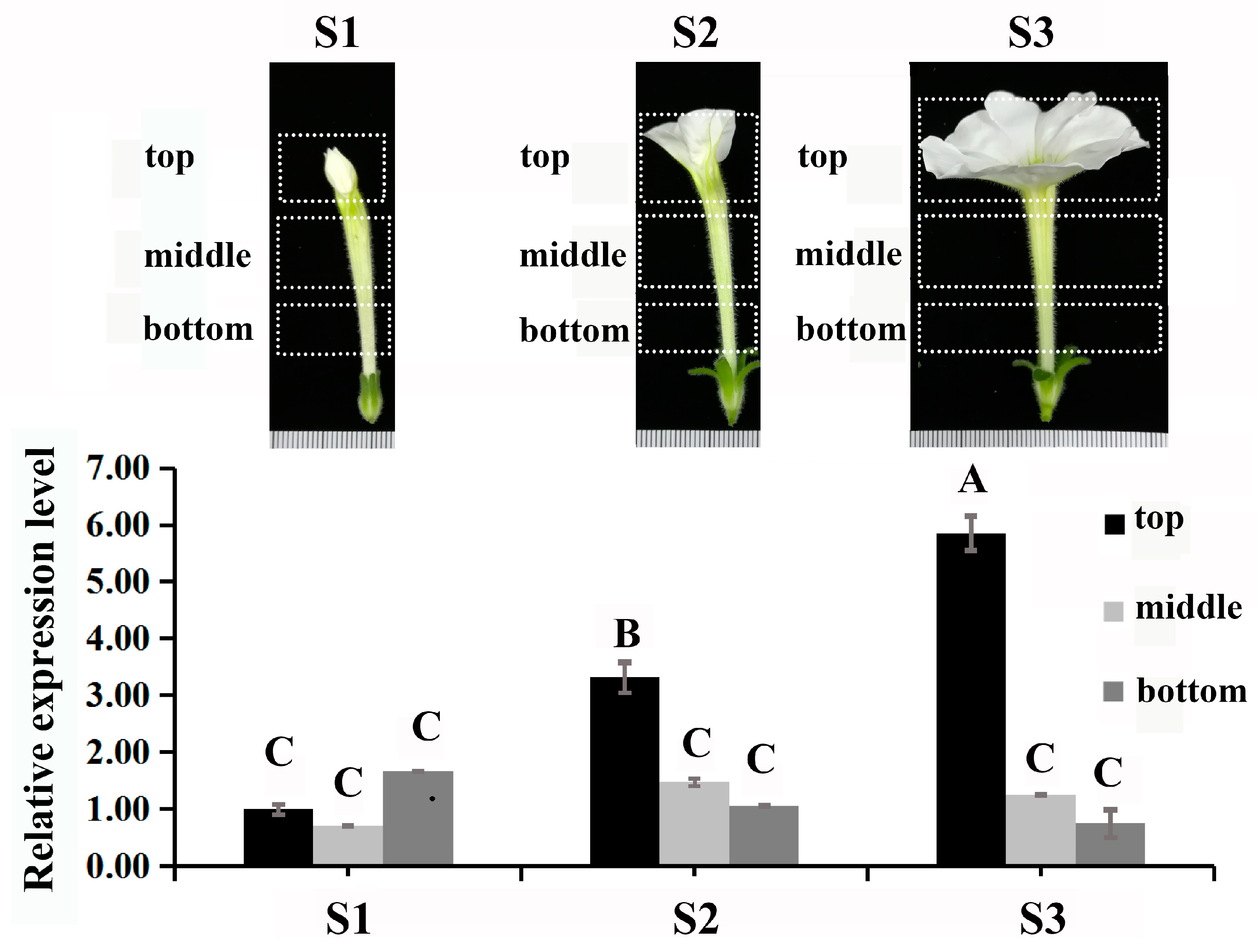
2.1. Sequence Features, Subcellular Localization, Self-Activation, and Expression of PhDof28

The PhDof28 sequence protein constituted 301 amino acids with a highly conserved C2-C2 domain (Figure 1A). A phylogenetic tree revealed that PhDof28 could cluster well with other Dof family members from Solanaceae (Figure 1B). Subcellular localization analysis indicates that PhDof28 was localized in the nucleus, while a transcriptional self-activation assay exhibited that pGBKT7::PhDof28 could grow well on sd/-trp and sd/-trp-ade plates and turn the sd/-trp-ade culture medium containing x-alpha-gal blue, which indicated self-activation (Figure 1D). The results of qRT-PCR indicated that PhDof28 was preferentially expressed in the top region of petals, with an up-regulated trend from the pre- to full-flowering stages (Figure 2).

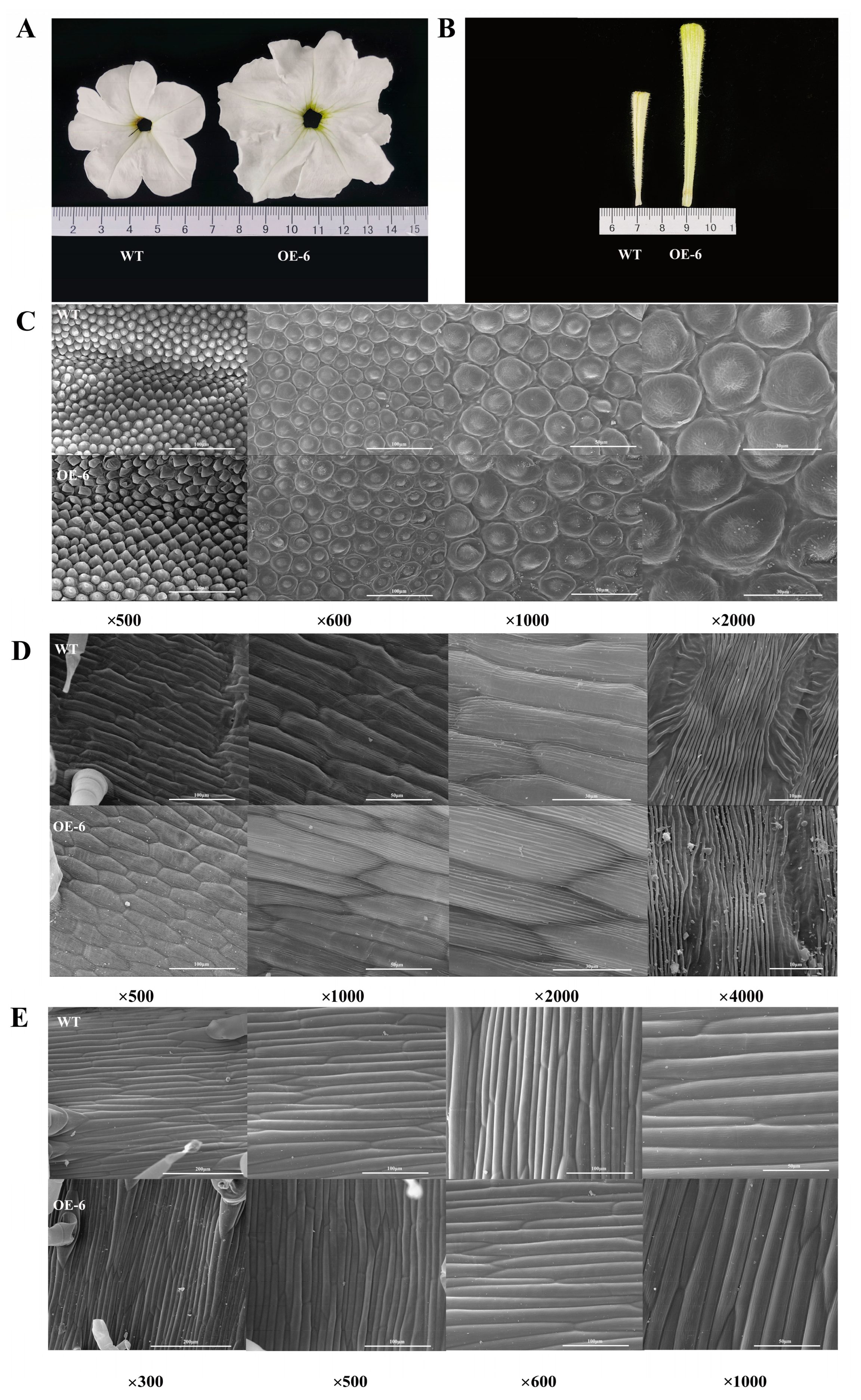
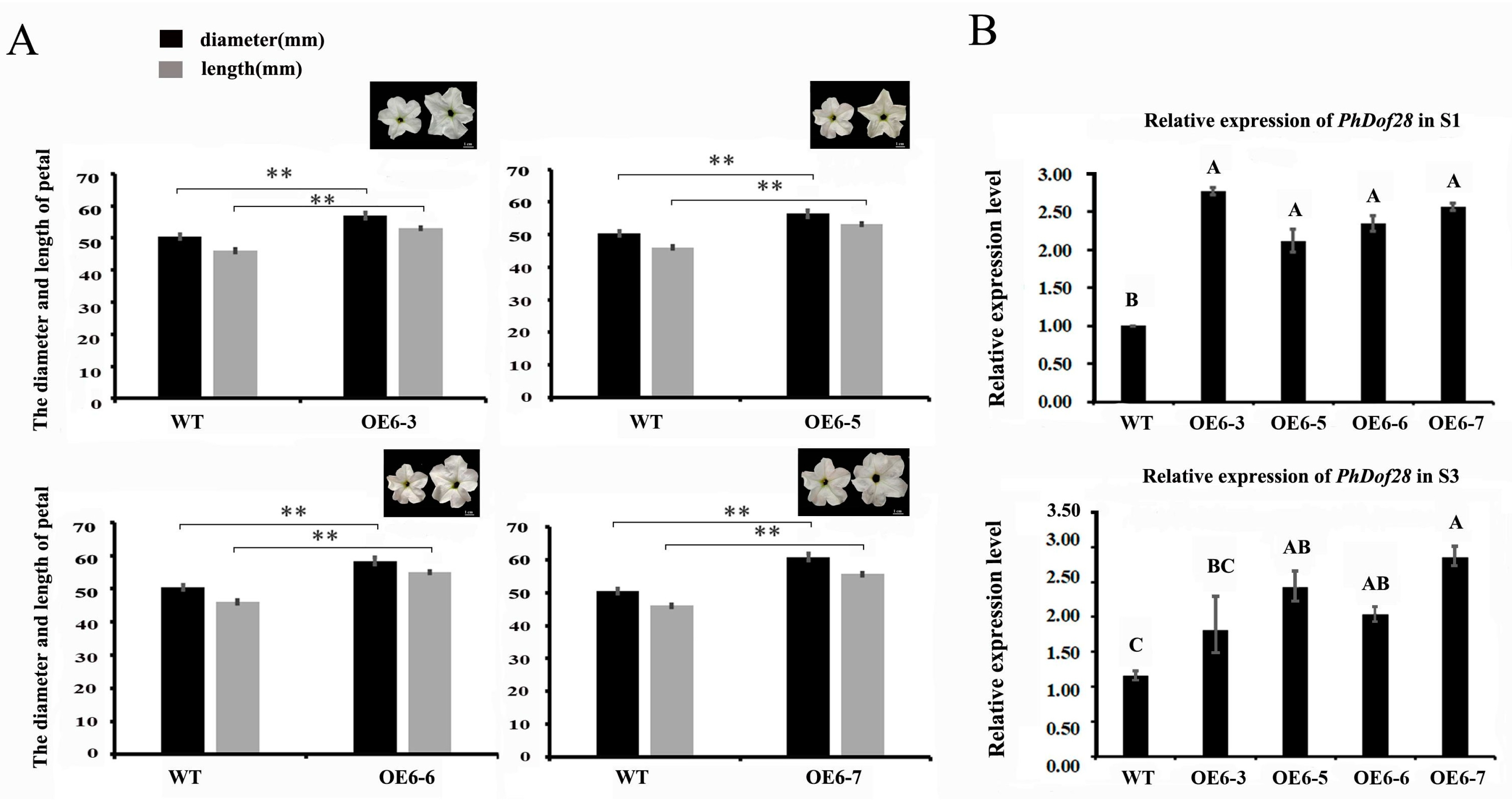
2.2. Phenotypic Characteristics of Transgenic Petunia Plants

PCR detection was used to obtain seven transgenic petunia plants (Figure S1), of which three plants had bigger petals, and OE-6 line was selected for systematic phenotypic observation. Compared to the wild-type plant, transgenic petunia overexpressing PhDof28 exhibited much larger flower diameter and a longer corolla tube (Figure 3A,B). The phenotype analysis of OE-6 petal cells at the top, middle, and bottom sections at the full-flowering stage using SEM (Scanning Electron Microcopy) showed that the top part of transgenic petunia petals had larger cells than that of wild type petals, and the cellular morphology of transgenic petals was relatively irregular (Figure 3C,D). However, no morphological differences were observed in the cells of the bottom part between wild-type and OE-6 petals (Figure 3E).
To test whether the enlarged petal phenotype could be stably inherited in the offspring, the F1 plants of OE-6 line, including OE6-3, OE6-5, OE6-6, and OE6-7 were selected for further phenotypic observations. The results showed that the phenotypes of F1 and F0 plants were similar, especially at the top part of the petals (Figure 4A), while the expression levels of PhDof28 in the F1 petals at the pre- and full-flowering stages were significantly up-regulated compared to those of the wild-type petunia (Figure 4B), which indicated stable inheritance of the large petal phenotype in the transgenic petunia offspring.

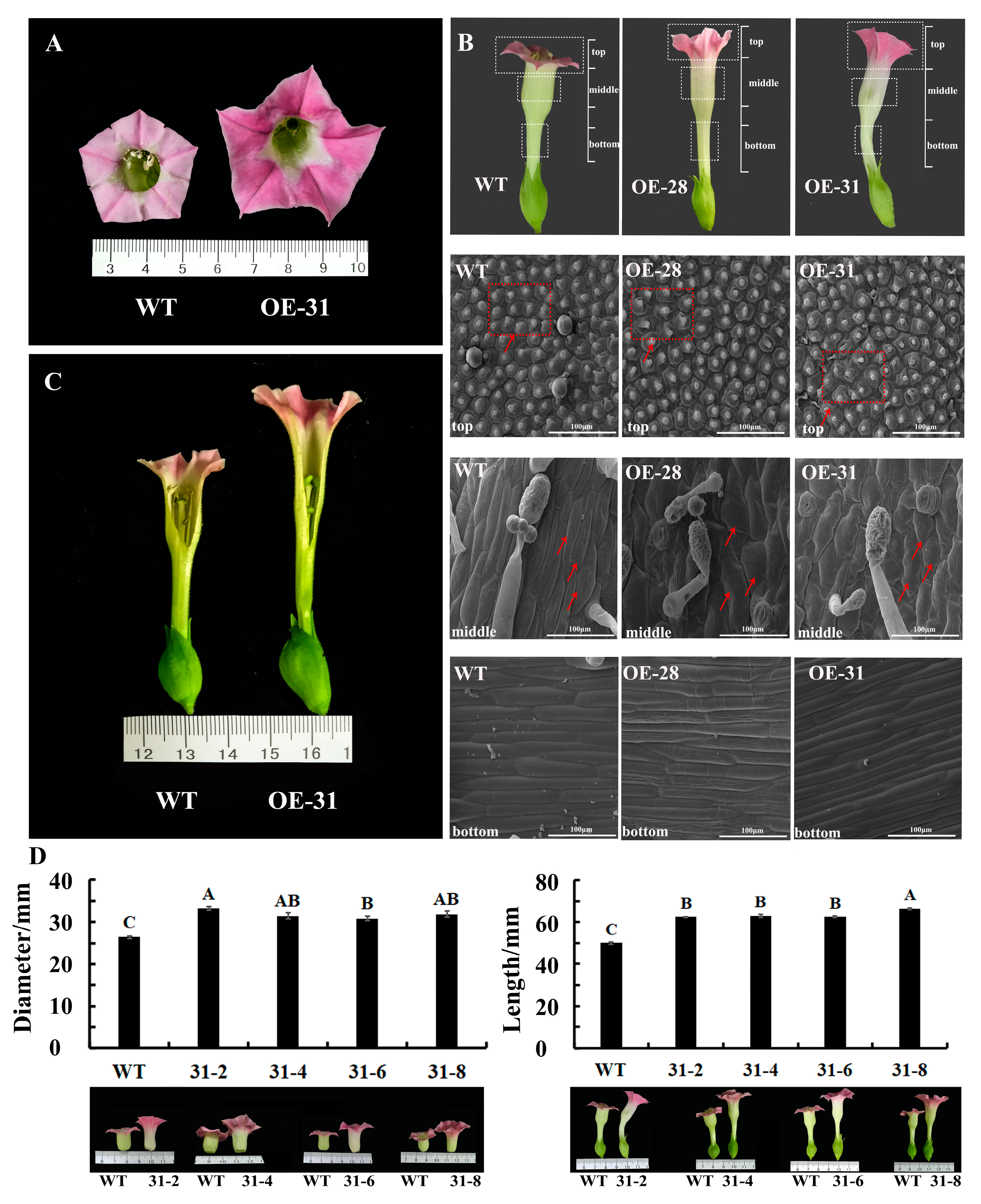
2.3. Phenotypic Characteristics of Transgenic Tobacco Plants

A total of six positive transgenic tobacco plants were isolated (Figure S2), and OE-31 and OE-28 with bigger petals were selected for further phenotypic observation. Results showed that tobacco plants overexpressing PhDof28 shared similar phenotypes with those of transgenic petunia (Figure 4 and Figure 5A,C), which confirmed that PhDof28 overexpression could promote petal elongation and expansion. The SEM observation revealed that transgenic OE-28 and OE-31 tobacco plants had larger cells, which were more loosely arranged as compared to the wild-type at the top part of the petal (Figure 5B). Meanwhile, cells in the middle petal parts of the transgenic tobacco were wider and larger than those of wild-type petunia (Figure 5B), while cells at the bottom part of the petal of transgenic tobacco plants were much longer than those of the wild-type petunia, which was consistent with the elongated flower phenotype (Figure 5B).
Subsequently, the F1 plants of the tobacco OE-31 line, including OE31-2, OE31-4, OE31-6, and OE31-8, were selected to further test whether the elongated petal phenotype could be inherited in a stable manner. As a result, a semi-quantitative PCR showed that PhDof28 was overexpressed in all the F1 plants (Figure S3). Phenotypic observations indicated that the diameter and length of F1 transgenic tobacco flowers were significantly larger than those of the wild-type (Figure 5D), which demonstrated stable phenotype inheritance.

2.4. The Endogenous Hormone Contents in Transgenic Petunia

The contents of six endogenous hormones, including IAA, JA, GA, abscisic acid (ABA), zeatin (ZT), and brassinosteroid (BR) were determined in the petals of transgenic petunia plants overexpressing PhDof28. Results indicated that the contents of IAA in transgenic petals were increased compared to those of wild-type plants. JA and GA contents were significantly higher in transgenic petunia only at the full-flowering stage and at the semi-flowering stage separately. The transgenic petunia maintained the same ABA levels as the wild-type plants. The contents of ZT and BR decreased significantly in the transgenic plants, while both maintained a consistent level at semi- and full-flowering stages (Figure 6).

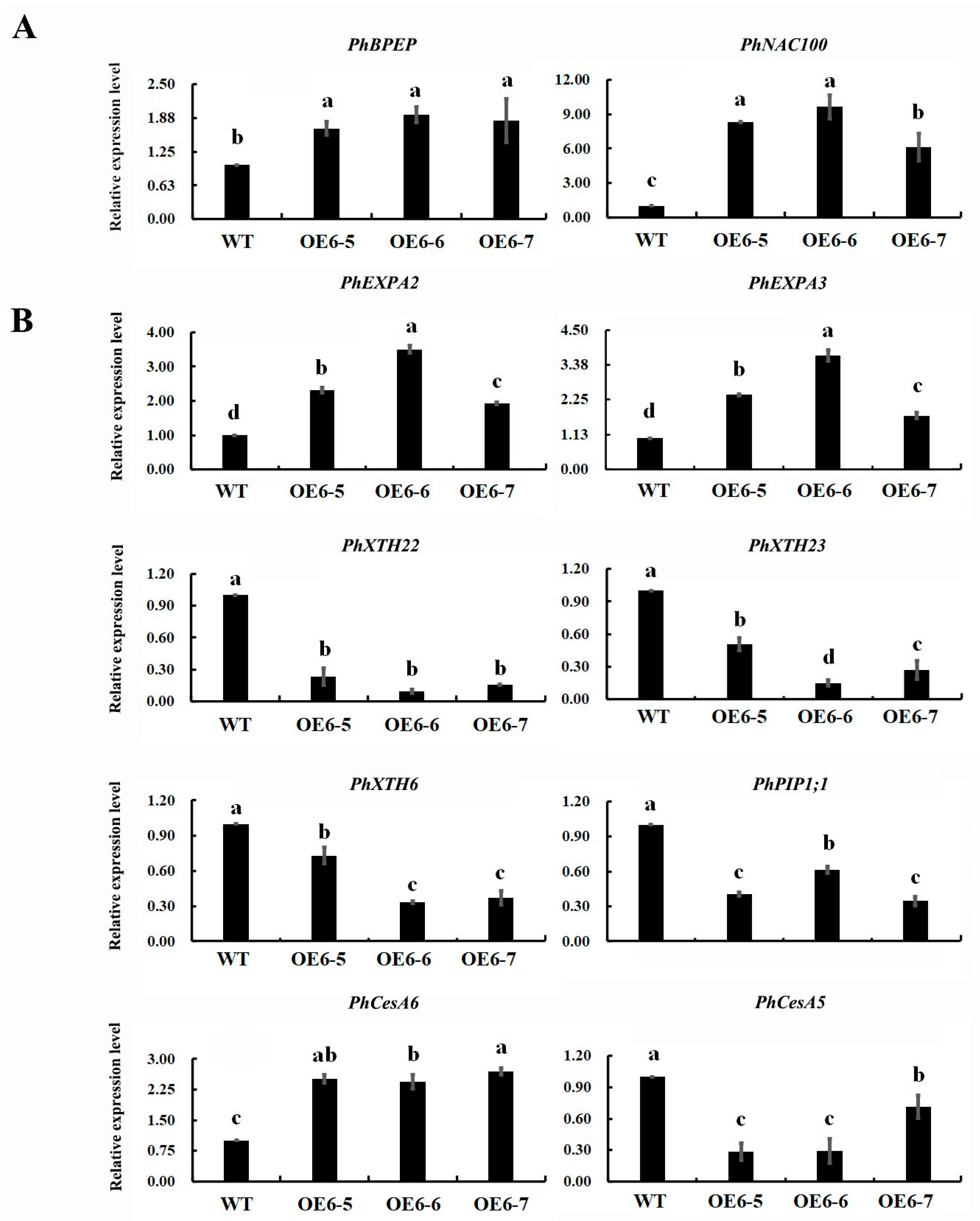
2.5. Expression Profile of Cell Expansion Associated Genes in Transgenic Petunia Petals

Two petal size TF inhibitors, including a bHLH TF gene, BIGPETAL (PhBPEp), and a NAC domain-containing protein 100 (PhNAC100), as well as eight cell expansion associated structural genes, including aquaporin (PhPIP1;1), cellulose synthase (PhCesA5/6), xyloglucan endotransglucosylase (PhXTH6/22/23), and expansion (PhEXPA2/3), were selected, and their expression profiles explored in transgenic petunia petals overexpressing PhDof28 during the pre-flowering stage. As a result, the levels of gene expression of PhBPEp and PhNAC100 were significantly up-regulated compared to the wild-type, while the levels of PhEXPA2, PhEXPA3, and PhCesA6 were increased in transgenic petunia. In contrast, the levels of gene expression of PhCesA5, PhXTH6, PhXTH22, PhXTH23, and PhPIP1;1 in the transgenic petunia were significantly down-regulated (Figure 7).

3. Discussion

3.1. Overexpression of PhDof28 Causes Petal Expansion in Petunia and Tobacco

Flower size is mainly influenced by petal cell division and expansion, and it represents one of the most vital determinants of the economic value of ornamental plants [17]. The Dof proteins are plant-specific TFs containing a C2-C2 single zinc finger structural domain that is highly conserved, and are widely participated in plant development [11]. Our recently reported Dof-type PhDof28 TF gene [16], with preferential expression in petunia plant petals, was selected for further functional analysis to determine its roles in the petunia petal expansion process. Generally, TFs have nucleus localization and transcriptional self-activation properties [18], and those specifically expressed in plant petals are usually predicted to be associated with distinct biological processes [15,19]. Consistently, this study determined that PhDof28 had a nuclear localization and transcriptional self-activation activity (Figure 1). Moreover, its expression was significantly up-regulated at the top part of petunia petals during flower opening (Figure 2), which confirmed its typical TF properties and potential petal expansion regulatory activity in petunias.
Stable transformation of PhDof28 in the petunia ‘W115’ and tobacco plants produced a larger transgenic petal phenotype than in wild-type plants (Figure 3 and Figure 5), which could also be stably inherited in the F1 plants (Figure 4). These results demonstrated that PhDof28 could significantly increase the cell sizes in the top parts of flower petals. Interestingly, the Arabidopsis AtDof5.4 gene, which is an ortholog of PhDof28 with preferential expression in the petals, inhibited petal cell proliferation and expansion, and its overexpression could significantly suppress petal expansion in transgenic Arabidopsis [15]. Despite having highly conserved functions during the evolutionary process, functional differentiation has been reported in some orthologous genes originating from a common ancestor [20]. For example, overexpression of AtMYB010 could induce a variety of growth and developmental abnormalities, such as stunted growth, multi-branching, early flowering, and thin stems in Arabidopsis. In contrast, overexpression of its orthologous GmMYB010 gene could not produce similar phenotypes in soybean [Glycine max (L.) Merr.]. In addition, overexpression of the maize (Zea mays L.) MYB59 TF significantly reduced the seeds’ germination rates in transgenic tobacco plants [21]; however, overexpression of its orthologous OsMYBAS1 gene promoted germination of transgenic rice seeds [22]. Given these reports, it is reasonable to speculate that PhDof28 and its orthologous AtDof5.4 gene might have undergone functional differentiation during plant evolution.

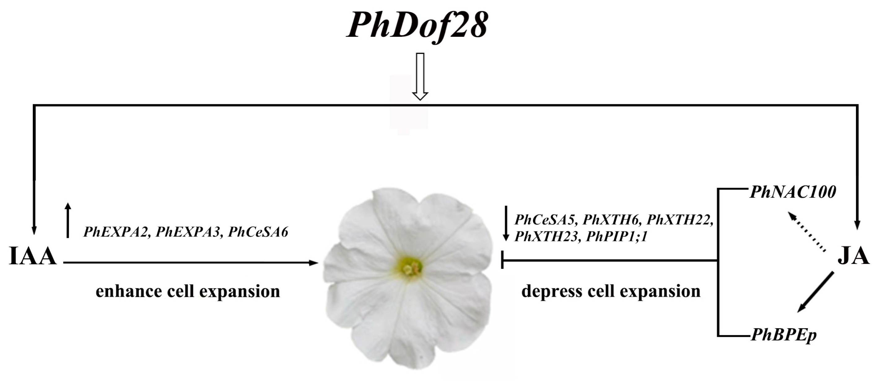
3.2. PhDof28 Regulates Petal Size by Triggering IAA and JA-Related Pathways

The expansion of petal cells has been reported to mainly rely on the regulation of plant hormone signaling, and the synergistic effects of endogenous plant hormones are critical determinants of plant petal size [4,5]. IAA is most extensively involved in the regulation of the development of plants by activating cell-wall-expansion-related genes, which in turn loosen and synthesize the cell wall [23,24,25]. In this study, IAA content in floral expansion stages of transgenic petunia petals overexpressing PhDof28 were remarkably higher than that of wild-type plants (Figure 6). Moreover, transgenic petunia plants showed highly increased expression levels of two previously reported cell-wall-loosening PhEXPA2 and PhEXPA3 genes [26], as well as a cell wall synthesis PhCesA6 gene [27]. Together, these results suggested that PhDof28 overexpression could enhance the accumulation of IAA in petunia petals and promote the expression of cell-expansion-associated genes.
The mutant Arabidopsis, opr3 with a defect in JA synthesis, showed pronounced petal cell expansion, which was significantly reduced after JA treatment, indicating that JA could inhibit expansion of petal cells [28]. Furthermore, the AtBPEp gene, which is induced by JA, could inhibit petal cell size by disrupting the petal cell expansion in A. thaliana [5,29]. Interestingly, JA content along with the expression of petal growth inhibitor PhBPEp and PhNAC100 genes were both increased in the petals of transgenic petunias overexpressing PhDof28 (Figure 6 and Figure 7A). On the contrary, the expression levels of cell-expansion-associated genes, which involved PhCesA5, PhXTH6, PhXTH22, PhXTH23, and PhPIP1;1, were all decreased (Figure 7B). These above results suggested that overexpression of PhDof28 can accelerate petal expansion by promoting IAA-related pathways in petunias. Moreover, to prevent excessive petal growth, transgenic plants could also trigger their regulatory feedback mechanism by inducing JA accumulation, leading to increased expression of petal size inhibitor TFs, such as PhBPEp (Figure 8).

4. Materials and Methods

4.1. Plant Materials

The P. hybrida L. ‘W115’ and Nicotiana tabacum L. were grown in the greenhouse at the Nanjing Forestry University, Nanjing, China, at 24 °C under 15 h light/9 h dark conditions. The different parts of petunia petals (top, middle, and bottom) were collected at pre-, semi-, and full-flowering stages, then frozen and stored in liquid nitrogen at −80 °C for use in RNA extraction. Leaves of Nicotiana benthamiana Domin grown at 16/8 h light/dark (144 µmol m−2 s−1) and 25 ± 2 °C for 30 days were selected for subcellular localization analysis.

4.2. qRT-PCR Analysis

Total RNA was isolated from different parts of petal samples using the RNA Prep Pure Plant Total RNA Kit (Aidlab, Beijing, China). The Reverse Transcription Kit (TransGen, Beijing, China) was used for reverse transcription of RNA samples. The qRT-PCR was performed using TB Green™ Premix Ex Taq™ (TaKaRa, Kusatsu, Japan). Specific qRT-PCR primers were designed using Primer Premier 5.0 [30]. qRT-PCR was carried out following the method previously described [31] with three biological and technical replicates per sample. Calculation of relative gene expression levels was carried out using the 2−ΔΔCt method [32].

4.3. Subcellular Localization and Transcriptional Self-Activation Assay

The GFP::pCAMBIA1300-PhDof28 fusion vector was constructed for subcellular localization and transferred into strain GV3101 of Agrobacterium tumefaciens Smith and Townsend, and then infiltrated into tobacco (N. benthamiana Domin) leaves for instant expression to observe the transient subcellular localization of PhDof28. The infiltrated plants were grown for 48 h, then the injected leaves were cut into small pieces of about 0.5 × 0.5 cm, placed on glass slides, and stained with fluorescent DAPI (4′,6-diamidino-2-phenylindole) dye. The fluorescence signal was examined with an LSM710 microscope (Zeiss, Jena, Germany).
The coding sequence of the PhDof28 gene was converted into the pGBKT7 vector for self-activation assay, and then transferred to Saccharomyces cerevisiae strain AH109 (WeidiBio, Shanghai, China) to acquire a self-activating yeast vector. The transformed yeast cells were cultivated in the dark for three days on selective media of sd/-trp, sd/-trp-ade, and sd/-trp-de + x-alpha-gal in a constant incubator at 30 °C to observe self-activation capacity of the target gene.

4.4. Stable Transformation of Petunia and Tobacco Plants

A pCAMBIA2300::PhDof28 overexpression vector was constructed and transformed into the A. tumefaciens EHA105 strain. The vector was subsequently transferred into the 20-day-old tobacco and petunia seedlings using the leaf disc method as mentioned previously [31,33], and PCR validation was conducted, with 35S-F and PhDof28-R as primers (Table S1). After developing more than three flowers, transgenic plants with a good phenotype were selected to measure flower diameter and length. The F1 plants were obtained through selfing and harvesting to continue observing their phenotype.

4.5. Scanning Electron Microscope (SEM) Analysis of Petal Cells

The F0 transgenic flowers of tobacco, petunia, and wild type plants from the top, middle, and bottom parts were selected, then cut into 0.5 × 0.5 cm2 sections (Figure 2). This process was completely carried out in the formaldehyde-acetic-acid-ethanol fixative to avoid oxidation of petal tissues. The cellular morphologies of petals were then observed under 300×, 500×, 600×, 1000×, and 2000× magnifications using SEM (JEOL, Tokyo, Japan).

4.6. Measurement of Hormone Content

To quantify hormone content, wild-type and F1 transgenic petunia petals at the semi- and full-flowering stages were separately gathered in four biological replicates. Samples were ground in liquid nitrogen, then extracted with an extraction buffer prior to measurement of hormone content in petals with the ELISA method [34,35].

4.7. Screening for Petal Candidate Genes Associated with Cell Expansion

To explore the function of PhDof28 in regulating the petal size, the cDNA sequences of previously reported structural genes and TFs potentially regulating petal size were retrieved from the TAIR (https://www.arabidopsis.org/, accessed on 10 July 2023) and the National Center for Biotechnology Information (NCBI) database (https://www.ncbi.nlm.nih.gov/, accessed on 10 July 2023). Then, the full-length amino acid sequences were used to probe their corresponding orthologous members in the petunia genome database (https://solgenomics.net/organism/Petunia_axillaris/genome, accessed on 10 July 2023). In addition, qRT-PCR primers were designed to explore the expression levels of retrieved candidate genes in the F1 transgenic petunia petal at the pre-flowering stage (Table S1).

4.8. Data Analysis

The mean ± standard error (SE) of at least three biological replicates were calculated for all data results. The data in this study were statistically analyzed using independent samples t-test or one-way ANOVA in SPSS, and the variability of the data was inferred by Duncan’s multiple range test at the p < 0.05 * or p < 0.01 ** level.

5. Conclusions

This study identified the nucleus-localized transcriptional activator PhDof28 gene, which showed preferential expression at the top parts of petunia flower petals. Transgenic tobacco and petunia ‘W115’ plants overexpressing PhDof28 could enhance their petal sizes by promoting the expression levels of cell-expansion-related genes, such as PhEXPA2, PhEXPA3, and PhCesA6. Interestingly, the expression levels of some cell-expansion-related genes, including PhCesA5, PhXTH6, PhXTH22, PhPIP1;1, and PhXTH23, were inhibited, while the petal expansion inhibitor TFs, PhNAC100 and PhBPEp, were significantly up-regulated. In addition, increased levels of plant endogenous IAA and JA hormones, which induce and inhibit petal elongation, respectively, were observed in transgenic plant petals. These results indicated that PhDof28 overexpression could increase the rate of petal elongation by promoting the accumulation of IAA. In contrast, to avoid excessive petal elongation, transgenic plants triggered a JA-related feedback regulation mechanism by enhancing the expression of petal inhibitor genes. This study will not only help deepen the comprehension of the functions of Dof TF family members in the regulation of plant ornamental traits, but also provide new genetic resources for future molecular breeding and improvement of petunias.

Supplementary Materials

The following supporting information can be downloaded at: https://www.mdpi.com/article/10.3390/ijms241511999/s1.

Author Contributions

Y.Y.: Conceptualization, Methodology, Supervision, Writing—review and editing. W.Z.: Formal analysis, Data curation, Writing—review and editing. H.S.: Visualization, Data curation, Writing—original draft. H.W.: Formal analysis. J.D.: Investigation, Data curation, Methodology. H.H. and L.W.: Conceptualization, Project administration. All authors have read and agreed to the published version of the manuscript.

Funding

This research was funded by the National Natural Science Foundation of China (Grant Nos. 31601785 and 32071828), the Natural Science Fund for Colleges and Universities in Jiangsu Province (Grant No. 20KJB220005), and the Priority Academic Program Development of Jiangsu Higher Education Institutions (PAPD).

Institutional Review Board Statement

Not applicable.

Informed Consent Statement

Not applicable.

Data Availability Statement

All data in this study can be found in the manuscript or supplemental materials.

Conflicts of Interest

The authors declare no conflict of interest.

References

  1. Baumann, K.; Perez-Rodriguez, M.; Bradley, D.; Venail, C.; Bailey, P.; Jin, H.; Koes, R.; Roberts, K.; Martin, C. Control of Cell and Petal Morphogenesis by R2R3 MYB Transcription Factors. Development 2007, 134, 1691–1701. [Google Scholar] [CrossRef] [PubMed] [Green Version]
  2. Meagher, T.R.; Gillies, A.C.M.; Costich, D.E. Genome Size, Quantitative Genetics and The Genomic Basis for Flower Size Evolution in Silene latifolia. Ann. Bot. 2005, 95, 247–254. [Google Scholar] [CrossRef] [Green Version]
  3. Pei, H.X.; Ma, N.; Tian, J.; Luo, J.; Chen, J.W.; Li, J.; Zheng, Y.; Chen, X.; Fei, Z.; Gao, J.P. An NAC Transcription Factor Controls Ethylene-regulated Cell Expansion in Flower Petals. Plant Physiol. 2013, 163, 775–791. [Google Scholar] [CrossRef] [PubMed] [Green Version]
  4. Hepworth, J.; Lenhard, M. Regulation of Plant Lateral-organ Growth by Modulating Cell Number and Size. Curr. Opin. Plant Biol. 2014, 17, 36–42. [Google Scholar] [CrossRef]
  5. Huang, T.B.; Irish, V.F. Gene Networks Controlling Petal Organogenesis. J. Exp. Bot. 2016, 67, 61–68. [Google Scholar] [CrossRef] [Green Version]
  6. Chen, C.X.; Hussain, N.; Wang, Y.R.; Li, M.T.; Liu, L.; Qin, M.Z.; Ma, N.; Gao, J.P.; Sun, X.M. An Ethylene-inhibited NF-YC Transcription Factor RhNF-YC9 Regulates Petal Expansion in Rose. Hortic. Plant J. 2020, 6, 419–427. [Google Scholar] [CrossRef]
  7. Varaud, E.; Brioudes, F.; Szécsi, J.; Leroux, J.; Brown, S.; Rechenmann, C.P.; Bendahmane, M. AUXIN RESPONSE FACTOR8 Regulates Arabidopsis Petal Growth by Interacting with the bHLH Transcription Factor BIGPETALp. Plant Cell 2011, 23, 973–983. [Google Scholar] [CrossRef] [Green Version]
  8. Brioudes, F.; Joly, C.; Szécsi, J.; Varaud, E.; Leroux, J.; Bellvert, F.; Bertrand, C.; Bendahmane, M. Jasmonate Controls Late Development Stages of Petal Growth in Arabidopsis thaliana. Plant J. 2009, 60, 1070–1080. [Google Scholar] [CrossRef]
  9. Luo, J.; Ma, N.; Pei, H.X.; Chen, J.W.; Li, J.; Gao, J.P. A DELLA Gene, RhGAI1, is a Direct Target of EIN3 and Mediates Ethylene-regulated Rose Petal Cell Expansion via Repressing the Expression of RhCesA2. J. Exp. Bot. 2013, 64, 5075–5084. [Google Scholar] [CrossRef] [Green Version]
  10. Wang, J.J.; Guan, Y.X.; Ding, L.; Li, P.R.; Zhao, W.Q.; Jiang, J.F.; Chen, S.M.; Chen, F.D. The CmTCP20 Gene Regulates Petal Elongation Growth in Chrysanthemum morifolium. Plant Sci. 2019, 280, 248–257. [Google Scholar] [CrossRef]
  11. Liu, Y.; Liu, N.N.; Deng, X.; Liu, D.M.; Li, M.F.; Cui, D.D.; Hu, Y.K.; Yan, Y.M. Genome-wide Analysis of Wheat DNA-binding with One Finger (Dof) Transcription Factor Genes: Evolutionary Characteristics and Diverse Abiotic Stress Responses. BMC Genom. 2020, 21, 276–294. [Google Scholar] [CrossRef] [Green Version]
  12. Yang, Q.; Chen, Q.J.; Zhu, Y.D.; Li, T.Z. Identification of MdDof Genes in Apple and Analysis of Their Response to Biotic or Abiotic Stress. Funct. Plant Biol. 2018, 45, 528–541. [Google Scholar] [CrossRef] [PubMed]
  13. Cheng, Z.C.; Hou, D.; Liu, J.; Li, X.Y.; Xie, L.H.; Ma, Y.J.; Gao, J. Characterization of Moso Bamboo (Phyllostachys edulis) Dof Transcription Factors in Floral Development and Abiotic Stress Responses. Genome 2018, 61, 151–156. [Google Scholar] [CrossRef]
  14. Ahmad, M.; Rim, Y.; Chen, H.; Kim, J.Y. Functional Characterization of Arabidopsis Dof Transcription Factor AtDof4.1. Russ. J. Plant Physiol. 2013, 60, 116–123. [Google Scholar] [CrossRef]
  15. Xu, P.P.; Chen, H.Y.; Ying, L.; Cai, W.M. AtDOF5.4/OBP4, a DOF Transcription Factor Gene that Negatively Regulates Cell Cycle Progression and Cell Expansion in Arabidopsis thaliana. Sci. Rep. 2016, 6, 27705–27718. [Google Scholar] [CrossRef] [PubMed] [Green Version]
  16. Yue, Y.Z.; Du, J.H.; Li, Y.; Thomas, H.R.; Frank, M.H.; Wang, L.G.; Hu, H.R. Insight into The Petunia Dof Transcription Factor Family Reveals a New Regulator of Male-sterility. Ind. Crops Prod. 2021, 161, 113196. [Google Scholar] [CrossRef]
  17. Norikoshi, R.; Shibata, T.; Ichimura, K. Cell Division and Expansion in Petals During Flower Development and Opening in Eustoma grandiflorum. Hortic. J. 2016, 85, 154–160. [Google Scholar] [CrossRef] [Green Version]
  18. Xie, F.F.; Chen, C.B.; Chen, J.Y.; Chen, J.X.; Hua, Q.Z.; Shah, K.; Zhang, Z.K.; Zhao, J.T.; Hu, G.B.; Chen, J.Y.; et al. Betalain Biosynthesis in Red Pulp Pitaya is Regulated via HuMYB132: A R-R Type MYB Transcription Factor. BMC Plant Biol. 2023, 23, 28–42. [Google Scholar] [CrossRef]
  19. Sagawa, J.M.; Stanley, L.E.; Lafountain, A.M.; Frank, H.A.; Liu, C.; Yuan, Y.W. An R2R3-MYB Transcription Factor Regulates Carotenoid Pigmentation in Mimulus lewisii Flowers. New Phytol. 2016, 209, 1049–1057. [Google Scholar] [CrossRef]
  20. Zhang, H.Y.; Shi, Y.Q.; Sun, M.D.; Hu, X.Z.; Hao, M.Y.; Shu, Y.; Zhou, X.R.; Hu, Q.; Li, C.; Mei, D.S. Functional Differentiation of BnVTE4 Gene Homologous Copies in α-tocopherol Biosynthesis Revealed by CRISPR/Cas9 Editing. Front. Plant Sci. 2022, 13, 850914–850923. [Google Scholar] [CrossRef]
  21. Zhai, K.H.; Zhao, G.W.; Jiang, H.Y.; Sun, C.X.; Ren, J.Y. Overexpression of Maize ZmMYB59 Gene Plays a Negative Regulatory Role in Seed Germination in Nicotiana tabacum and Oryza sativa. Front. Plant Sci. 2020, 11, 564665–564674. [Google Scholar] [CrossRef]
  22. Wu, R.; Ding, Y.Q.; Li, C.Y.; Wu, B.K.; Huang, Z.J.; Li, Z.N.; Wang, X.M.; Zhao, G.W. An R2R3-type Transcription Factor OsMYBAS1 Regulates Seed Germination Under Artificial Accelerated Aging in Transgenic Rice (Oryza sativa L.). Agronomy 2022, 12, 1955. [Google Scholar] [CrossRef]
  23. Su, Y.H.; Liu, Y.B.; Bai, B.; Zhang, X.S. Establishment of Embryonic Shoot-root Axis is Involved in Auxin and Cytokinin Response during Arabidopsis Somatic Embryogenesis. Front. Plant Sci. 2015, 5, 781–792. [Google Scholar] [CrossRef] [PubMed] [Green Version]
  24. Majda, M.; Robert, S. The Role of Auxin in Cell Wall Expansion. Int. J. Mol. Sci. 2018, 19, 951–972. [Google Scholar] [CrossRef] [Green Version]
  25. Du, M.M.; Edgar, P.S.; William, M.G. Rapid Auxin-mediated Cell Expansion. Annu Rev. Plant Biol. 2020, 71, 379–402. [Google Scholar] [CrossRef]
  26. Rose, J.K.C.; Braam, J.; Fry, S.C.; Nishitani, K. The XTH Family of Enzymes Involved in Xyloglucan Endotransglucosylation and Endohydrolysis: Current Perspectives and A New Unifying Nomenclature. Plant Cell Physiol. 2002, 43, 1421–1435. [Google Scholar] [CrossRef] [PubMed] [Green Version]
  27. Mutwil, M.; Debolt, S.; Persson, S. Cellulose Synthesis: A Complex Complex. Curr. Opin. Plant Biol. 2008, 11, 252–257. [Google Scholar] [CrossRef]
  28. Stintzi, A.; Browse, J. The Arabidopsis Male-sterile Mutant, opr3, Lacks the 12-oxophytodienoic Acid Reductase Required for Jasmonate Synthesis. Proc. Natl. Acad. Sci. USA 2000, 97, 10625–10630. [Google Scholar] [CrossRef]
  29. Szécsi, J.; Joly, C.; Bordji, K.; Varaud, E.; Cock, J.M.; Dumas, C.; Bendahmane, M. BIGPETALp, A bHLH Transcription Factor is Involved in The control of Arabidopsis Petal Size. EMBO J. 2006, 25, 3912–3920. [Google Scholar] [CrossRef] [Green Version]
  30. Singh, V.K.; Mangalam, A.K.; Dwivedi, S.; Naik, S. Primer Premier: Program for Design of Degenerate Primers from A Protein Sequence. Biotechniques 1998, 24, 318–319. [Google Scholar] [CrossRef] [Green Version]
  31. Yue, Y.Z.; Yang, D.; Sun, J.; Peng, H.; Yin, C.Q.; Guo, R.; Ning, G.G.; Hu, H.R. A Novel PhLRR Gene Promoter is Sufficient for Engineering Male Sterility in Petunia. Plant Mol. Biol. Rep. 2016, 34, 940–977. [Google Scholar] [CrossRef]
  32. Livak, K.J.; Schmittgen, T.D. Analysis of Relative Gene Expression Data Using Real-Time Quantitative PCR and the 2−ΔΔCT Method. Methods 2020, 172, 103990. [Google Scholar] [CrossRef]
  33. Huang, X.; Yue, Y.Z.; Sun, J.; Peng, H.; Yang, Z.N.; Bao, M.Z.; Hu, H.R. Characterization of A Fertility-related SANT/MYB gene (PhRL) from Petunia. Sci. Hortic. 2015, 183, 152–159. [Google Scholar] [CrossRef]
  34. Gu, H.; Ding, W.J.; Shi, T.T.; Ouyang, Q.X.; Yang, X.L.; Yue, Y.Z.; Wang, L.G. Integrated Transcriptome and Endogenous Hormone Analysis Provides New Insights into Callus Proliferation in Osmanthus fragrans. Sci. Rep. 2022, 12, 7609–7622. [Google Scholar] [CrossRef] [PubMed]
  35. Yang, X.L.; Ding, W.J.; Yue, Y.Z.; Xu, C.; Wang, X.; Wang, L.G. Cloning and Expression Analysis of Three Critical Triterpenoid Pathway Genes in Osmanthus fragrans. Electron. J. Biotechnol. 2018, 36, 1–8. [Google Scholar]
Figure 1. Sequence pcharacteristics of PhDof28. (A) The PhDof28 cDNA and its encoded amino acid sequences showing the conserved Dof domain; (B) The phylogenetic tree of PhDof28; (C) GFP fluorescent signals showing the nucleus localization of PhDof28; (D) The transcriptional self-activation analysis of PhDof28. * Means the termination codon.
Ijms 24 11999 g001
Figure 2. Expression levels of PhDof28 in different parts of petunia ‘W115’ at three stages of petal development. S1: Pre-flowering stage; S2: Semi-flowering stage; S3: Full-flowering stage. The data are mean values of three biological and three technical replicates. The error bar represents SE; alphabetical letters above the bars denote significant differences between groups at p < 0.01 (Duncan’s test).
Ijms 24 11999 g002
Figure 3. Phenotypic observation of petunia petals overexpressing PhDof28. (A) The top part of the petal; (B) Middle and bottom parts of the petal; (C) SEM (Scanning Electron Microcopy) observations of the top part of the petal; (D) SEM observations of middle part of the petal; (E) SEM observations of the bottom part of the petal.
Ijms 24 11999 g003
Figure 4. Petal phenotypes and expression levels of PhDof28 in F1 transgenic petunia. (A) Petal diameter and flower length of the F1 transgenic petunia; (B) Expression levels of PhDof28 in transgenic petunia petals at S1 and S3 stages. S1: pre-flowering stage, S3: semi-flowering stage. The data are mean values of three biological and three technical replicates. The error bar represents the SE. The asterisks and the alphabetical letters denote the significant difference between values at p < 0.01 (Duncan’s test).
Ijms 24 11999 g004
Figure 5. Phenotypic analysis of petals and SEM observation of petal cells from transgenic tobacco. (A) The front diameter of wild-type and OE-31 flowers; (B) SEM images of petal cells at the top, middle, and bottom parts of transgenic OE-31 and OE-28 tobacco plants. The red boxes and arrows indicate different petal cell morphology; (C) Sections of wild type and OE-31 flowers; (D) Statistical summary of F1 transgenic tobacco phenotypes. The alphabetical letters above the bars denote significant differences between groups at p < 0.01 (Duncan’s test).
Ijms 24 11999 g005
Figure 6. The contents of six endogenous hormones in the transgenic and wild-type petunia petals at S2 and S3 stages. S2: full-flowering stage, S3: semi-flowering stage. The data are mean values of four biological and three technical replicates. The error bar represents SE. Alphabetical letters above the bars denote the significant difference between groups at p < 0.05 (Duncan’s test).
Ijms 24 11999 g006
Figure 7. Expression levels of cell expansion related genes in the transgenic petunia petals. (A) The expression levels of PhBPEp and PhNAC100 TFs; (B) the expression levels of eight cell expansion-related structural genes. The data are mean value of three biological and technical replicates. The error bar represents SE. Alphabetical letters above the bars denote significant difference between groups at p < 0.05 (Duncan’s test).
Ijms 24 11999 g007
Figure 8. Hypothetical model illustrating the role of PhDof28 in the regulation of petal size in petunias. The overexpression of PhDof28 accelerated the content of IAA in petunia petals, and promotes the expression of cell-expansion-related genes, PhEXPA2, PhEXPA3, and PhCeSA6. Moreover, to prevent excessive petal growth, transgenic plants could also trigger their regulatory feedback mechanism by increasing JA accumulation, leading to increased expression of PhBPEp; at the same time, PhNAC100, PhXTH6, PhXTH22, PhCeSA5, PhXTH23 and PhPIP1;1 were down-regulated to depress cell expansion.
Ijms 24 11999 g008
Disclaimer/Publisher’s Note: The statements, opinions and data contained in all publications are solely those of the individual author(s) and contributor(s) and not of MDPI and/or the editor(s). MDPI and/or the editor(s) disclaim responsibility for any injury to people or property resulting from any ideas, methods, instructions or products referred to in the content.

