Article

Serpin Family A Member 1 Is Prognostic and Involved in Immunological Regulation in Human Cancers

1
Department of Pathology, Department of Clinical Biobank, Medical School of Nantong University, Nantong 226001, China
2
Department of Clinical Biobank, Affiliated Hospital of Nantong University, Nantong 226001, China
*
Author to whom correspondence should be addressed.
These authors contributed equally to this work.
Int. J. Mol. Sci. 2023, 24(14), 11566; https://doi.org/10.3390/ijms241411566
Submission received: 16 May 2023 / Revised: 5 July 2023 / Accepted: 12 July 2023 / Published: 17 July 2023
(This article belongs to the Special Issue Cancer Biomarkers and Bioinformatics)

Abstract

:
Serpin family A member 1 (SERPINA1) encodes a protease inhibitor participating in many human diseases, but its value in immunoregulation and prognosis of human cancers remains unclear. In this study, through comprehensive analysis of data from The Cancer Genome Atlas (TCGA) datasets, we found that SERPINA1 was dysregulated in many cancers compared with normal tissues. SERPINA1 expression was significantly associated with prognosis, immune subtype, molecular subtype, immune checkpoint (ICP) genes, tumor mutational burden (TMB), microsatellite instability (MSI), and the estimation of stromal and immune cells in malignant tumor tissues using expression data (ESTIMATE) score. There was a strong connection between SERPINA1 expression and tumor-infiltrating lymphocytes, and SERPINA1 showed significant relation to gene markers of immune cells in digestive tumors. Fluorescence-based multiplex immunohistochemistry confirmed that SERPINA1 protein expression was related to clinicopathologic features and immune infiltrates in hepatic cancer. This study suggests that SERPINA can potentially serve as a novel biomarker for cancer prognosis and immunotherapy.

1. Introduction

Immune-based therapies have revolutionized the treatment of advanced cancer. The application of immune checkpoint inhibitor therapy targeting the PD-1/PD-L1 or CTLA-4 pathway is a breakthrough for many types of cancer [1]. The tumor microenvironment (TME) comprises tumor cells, immune cells, stromal cells, intricate cytokines, and chemokines, providing a favorable environment for tumor growth [2]. Tumor cells secrete growth factors and cytokine and regulate interstitial and immune cells’ behaviors. The interaction between tumor cells and the TME influences the response to immunotherapy [3]. Therefore, it is necessary to identify novel prognostic biomarkers of immunotherapy.
Serpin family A member 1 (SERPINA1) is located on chromosome 14 (14q32.1), encoding the protein Alpha1-antitrypsin (A1AT) that is a highly conserved protein and the dominant protease inhibitor, the inhibition capacity of which accounts for more than 90% of all plasma proteases [4]. SERPINA1 is expressed mainly in hepatocytes but is also synthesized in mononuclear phagocytes, neutrophils, and intestinal epithelial cells [5]. SERPINA1 plays a crucial role in the maintenance of cell homeostasis through irreversibly inhibiting a variety of serine endopeptidases. Previous literature has paid much attention to the pathological process and treatment of SERPINA1 deficiency in the liver and lung. Recently, a growing body of research has confirmed that SERPINA1 shows a tremendous influence on multiple tumors, such as lung cancer [6], gastric cancer [7], and breast cancer [8]. However, the systematical analysis of SERPINA1 in prognosis and TME regulation in cancers is rare.
In this study, the role of SERPINA1 in prognosis and immunological regulation in tumors was analyzed through multiple bioinformatics approaches. The differential expressions and prognostic values of SERPINA1 in different cancers were explored comprehensively. We also analyzed the potential connection between SERPINA1 level and immune and molecular subtypes, biomarkers of therapeutic efficacy, and tumor-infiltrating lymphocytes. Furthermore, the role of SERPINA1A in liver hepatocellular carcinoma (LIHC) was detected using fluorescence-based multiplex immunohistochemistry (mIHC) to confirm the results we acquired. The study was designed to elucidate the potential of SERPINA1 in tumor prognosis and immunotherapy, thus providing novel insight into the antitumor strategy.

2. Results

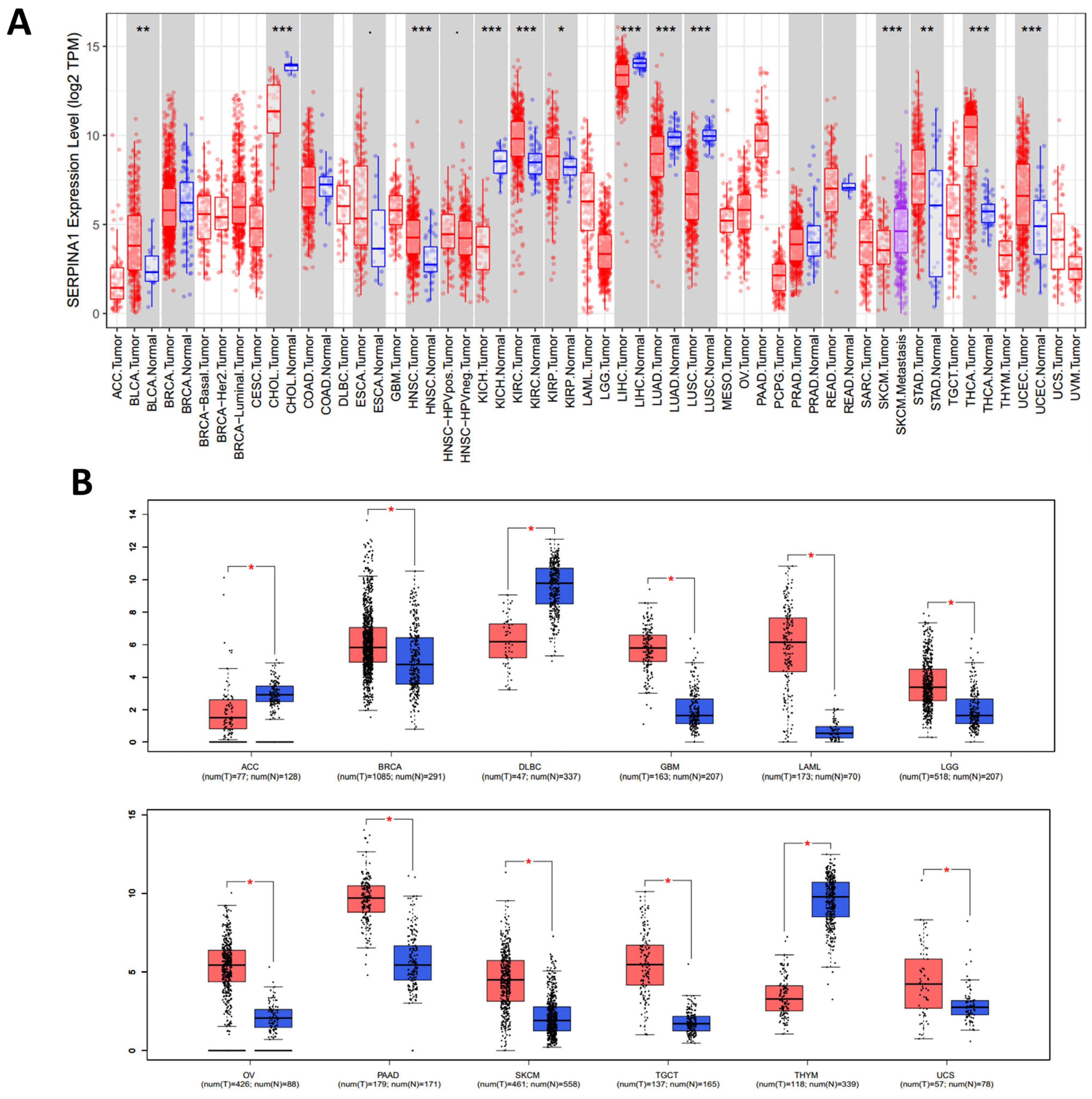
2.1. Pan-Cancer Differential Expressions of SERPINA1 between Tumor and Normal Tissues

The TIMER database confirmed that SERPINA1 mRNA expression was significantly higher in BLCA (bladder urothelial carcinoma), HNSC (head and neck squamous cell carcinoma), KIRC (kidney renal clear cell carcinoma), KIRP (kidney renal papillary cell carcinoma), STAD (stomach adenocarcinoma), THCA (thyroid carcinoma), and UCEC (uterine corpus endometrial carcinoma) than in paired normal tissues. Meanwhile, SERPINA1 expression was significantly lower in CHOL (cholangiocarcinoma), LIHC, LUAD (lung adenocarcinoma), and LUSC (lung squamous cell carcinoma) (Figure 1A). The GEPIA database was utilized to replenish the analysis of cancers without corresponding normal tissues, and the results showed that SERPINA1 expression was overexpressed in most cancers except ACC (adrenocortical carcinoma), DLBC (lymphoid neoplasm diffuse large B-cell lymphoma), and THYM (thymoma) (Figure 1B).
In addition, we detected the diagnostic value of SERPINA1 to differentiate tumors from normal tissue via the ROC curve (Figure 2). SERPINA1 had the potential to work as a diagnostic marker in BLCA (AUC = 0.711), CHOL (AUC = 0.952), ESAD (esophagus adenocarcinoma, AUC = 0.737), GBM (glioblastoma multiforme, AUC = 0.966), HNSC (AUC = 0.757), KICH (kidney chromophobe, AUC = 0.997), KIRC (AUC = 0.773), LIHC (AUC = 0.723), LUAD (AUC = 0.737), LUADLUSC (lung adenocarcinoma and lung squamous cell carcinoma, AUC = 0.854), LUSC (AUC = 0.984), OSCC (oral squamous cell carcinoma, AUC = 0.701), STAD (AUC = 0.703), THCA (AUC = 0.873), and UCEC (AUC = 0.715).

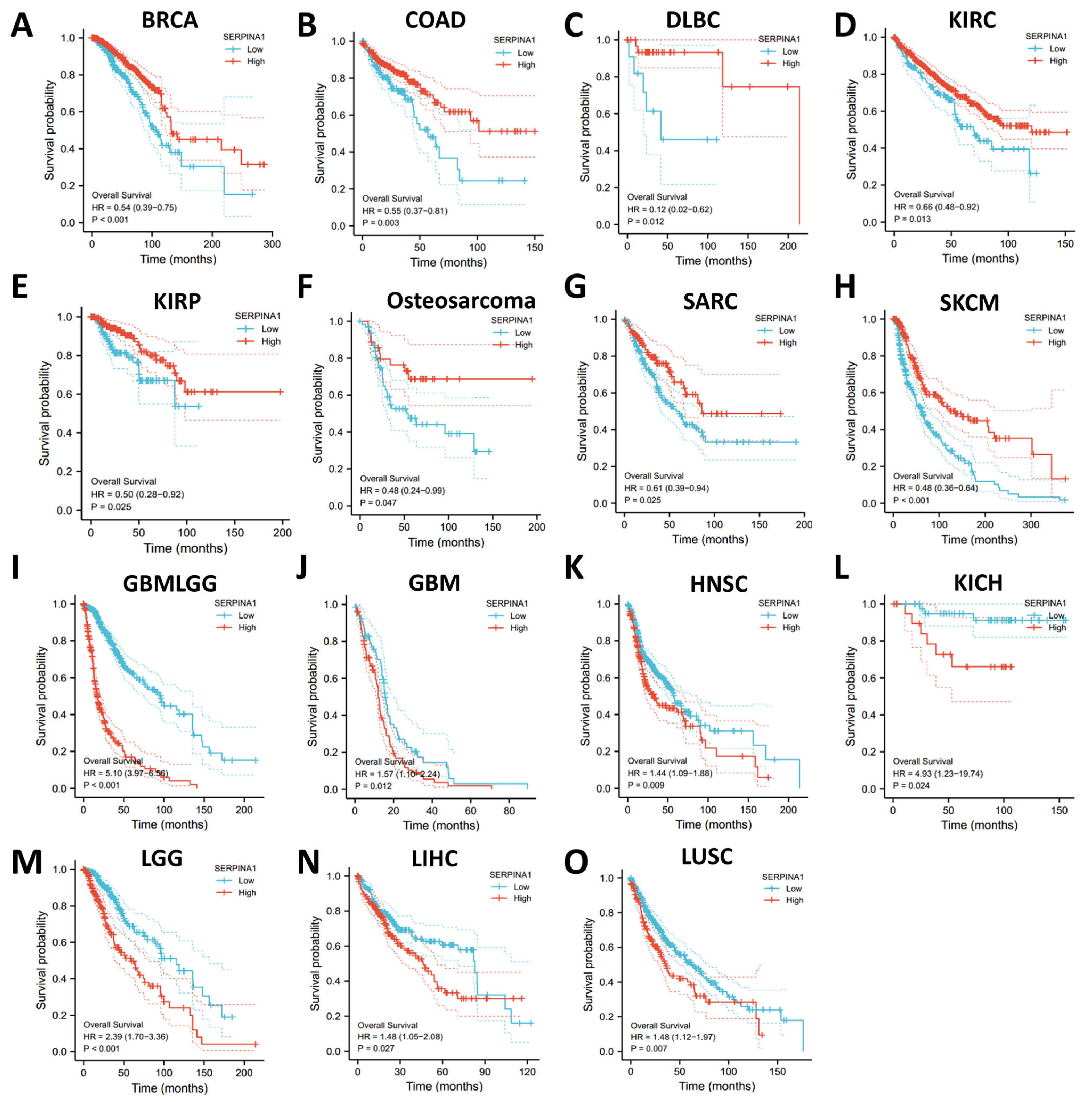
2.2. Dual Prognostic Role of SERPINA1 in Human Cancers

The prognostic role of SERPINA1 was identified using different databases. In the Xiantao database, higher SERPINA1 expression was connected to better overall survival (OS) in BRCA (breast invasive carcinoma), COAD (colon adenocarcinoma), DLBC, KIRC, KIRP, osteosarcoma, SARC (sarcoma), and SKCM (skin cutaneous melanoma) (Figure 3A–H). However, higher SERPINA1 expression was associated with poorer OS in GBMLGG (glioma), GBM, HNSC, KICH, LGG (brain lower-grade glioma), LIHC, and LUSC (Figure 3I–O). Furthermore, SERPINA1 expression in most of these cancers was also related to disease-specific survival (DSS) (Figure S1). In the Kaplan–Meier plotter database, higher SERPINA1 expression was connected to better OS in CESC (cervical squamous cell carcinoma and endocervical adenocarcinoma), THCA, and UCEC. In comparison, higher SERPINA1 expression was associated with poorer OS in ESCA (esophageal carcinoma), TGCT (testicular germ cell tumors), and THYM (Figure S2). These results suggested that SERPINA1 expression had the potential to be a prognostic biomarker in various cancers.

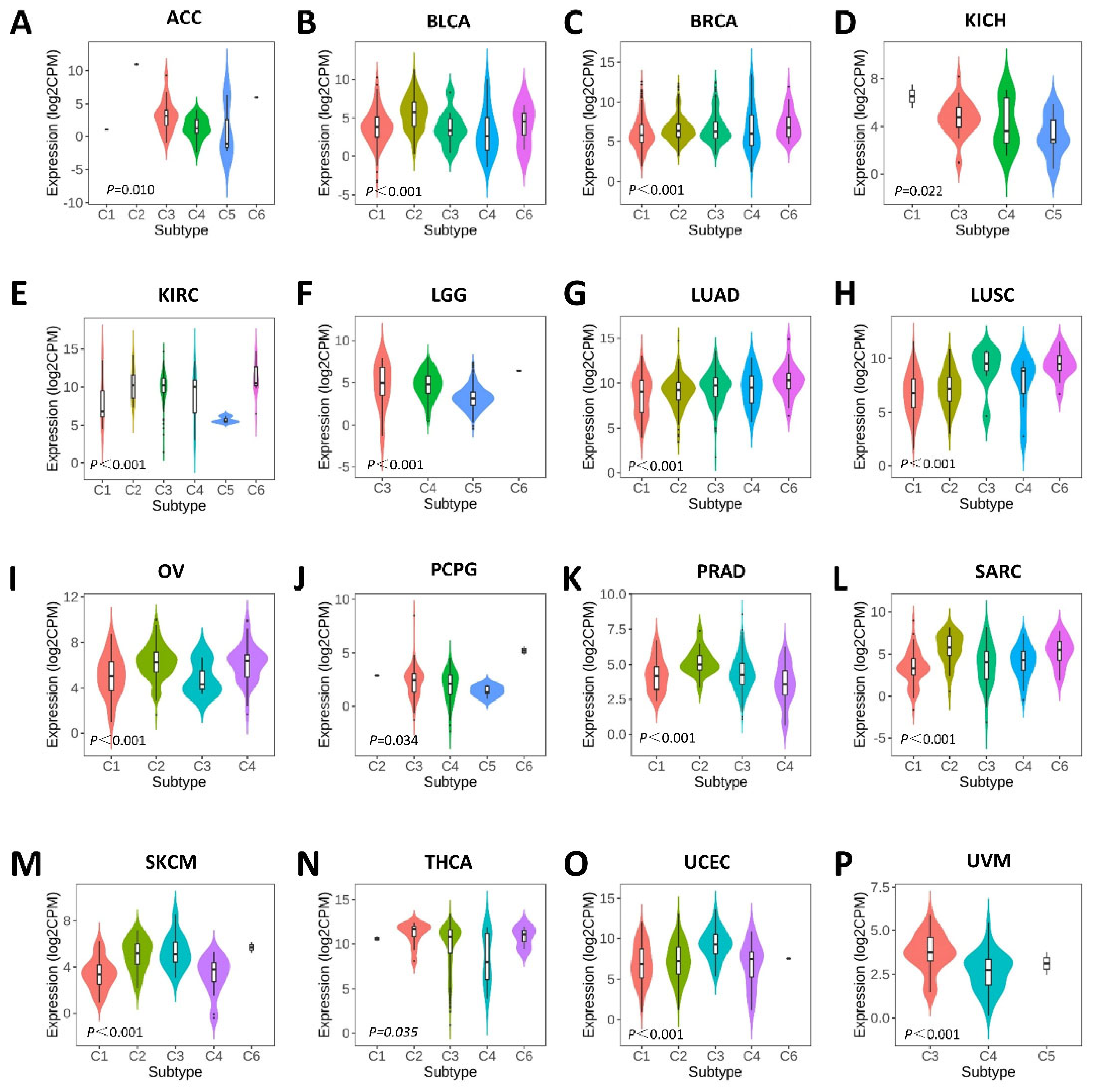
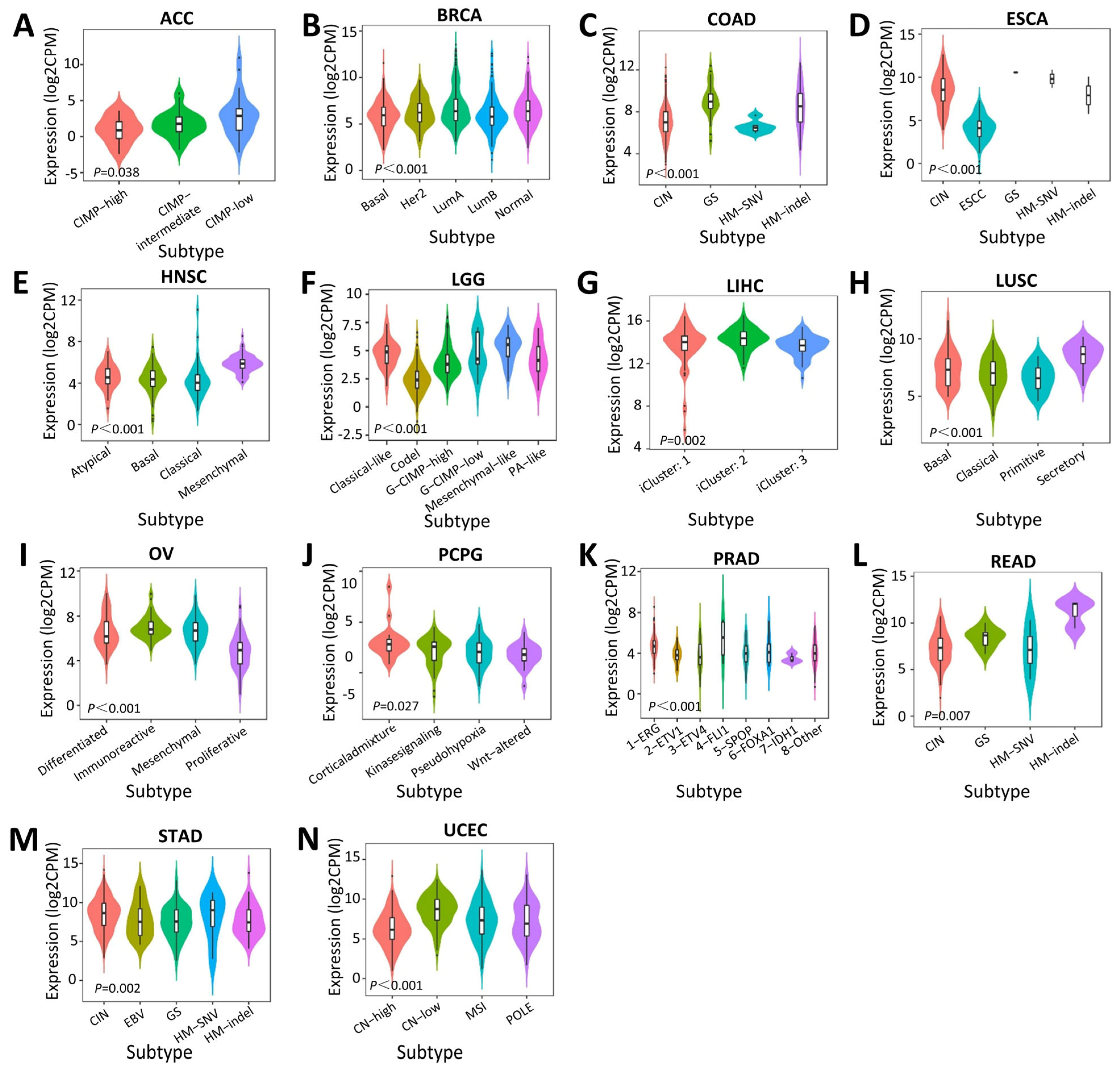
2.3. Associations between SERPINA1 and Immune and Molecular Subtypes

Then, we explored the influence of SERPINA1 on immune and molecular subtypes in human tumors through the TISIDB web portal. SERPINA1 expression was confirmed to be associated with immune subtypes in ACC, BLCA, BRCA, KICH, KIRC, LGG, LUAD, LUSC, OV (ovarian serous cystadenocarcinoma), PCPG (pheochromocytoma and paraganglioma), PRAD (prostate adenocarcinoma), SARC, SKCM, THCA, UCEC, and UVM (uveal melanoma) (Figure 4). Different molecular subtypes showed different SERPINA1 expressions in ACC, BRCA, COAD, ESCA, HNSC, LGG, LIHC, LUSC, OV, PCPG, PRAD, READ, STAD, and UCEC (Figure 5). The results above demonstrated that SERPINA1 expression is closely related to the immune and molecular subtypes of various cancers.

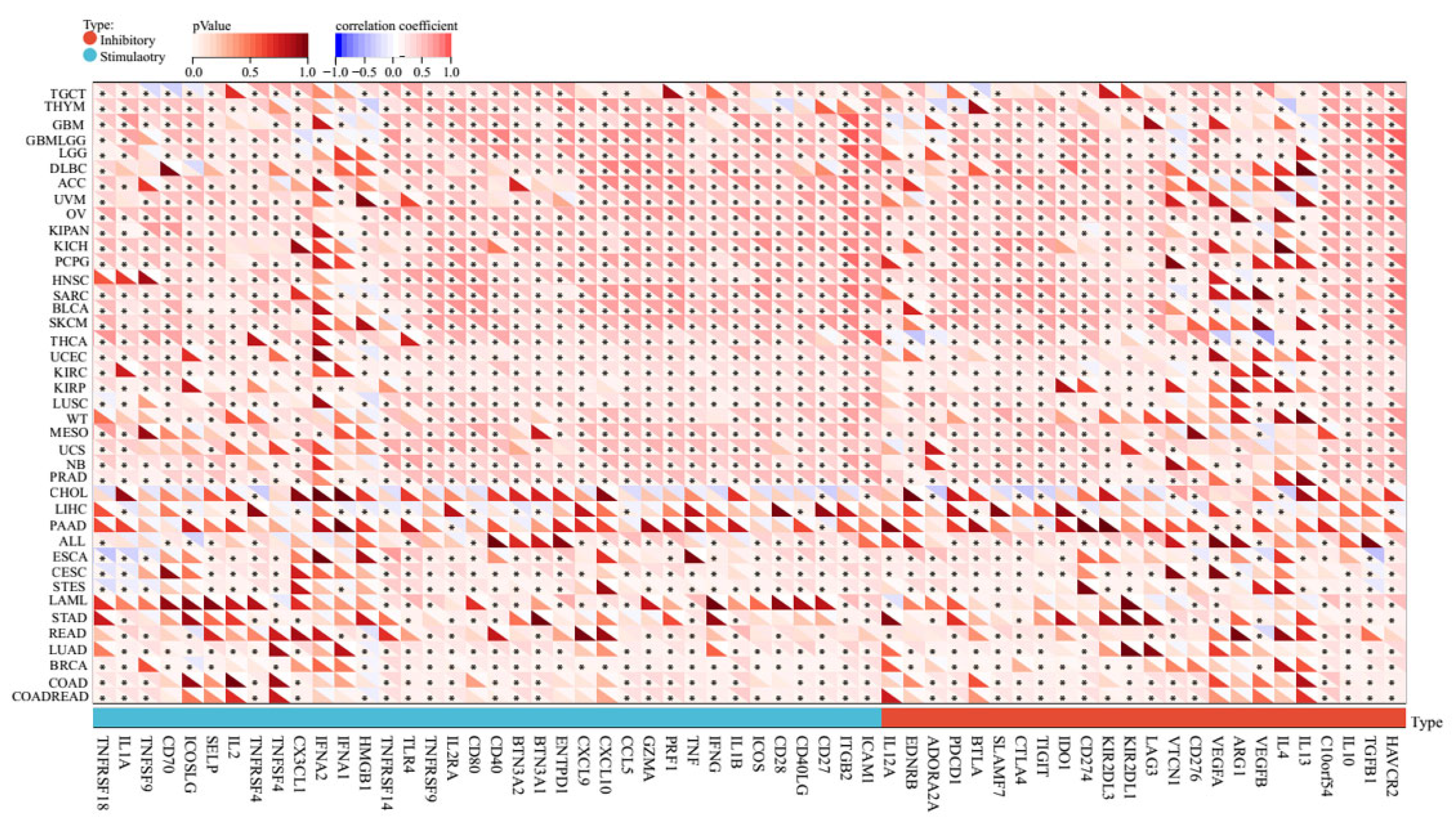
2.4. Pan-Cancer Relationships between AERPINA1 and Immune Checkpoint (ICP) Genes

ICP blockades have shown unprecedented advances in tumor immunotherapy [9]. To explore the potential role of SERPINA1 in immunotherapy, we analyzed the relationship between SERPINA1 expression and ICP genes, which significantly affect immune cell infiltration, through the SangerBox database [10]. SERPINA1 expression is positively related to ICP genes in most cancer types, especially GBMLGG, OV, KIPAN (Pan-kidney cohort), and PRAD, in which more than 90% of 60 ICP genes were connected to SERPINA1 expression (Figure 6). These results indicated that high SERPINA1 expression might forecast the satisfactory outcome of immunotherapy targeting ICP genes, and SERPINA1 might be a novel immunotherapy target for its influence on ICP genes. Furthermore, SERPINA1 related to a few ICP genes only in CHOL and PAAD, which suggested that corresponding patients with high SERPINA1 expression might respond poorly to immunotherapy referring to ICP genes. Based on the findings above, we considered SERPINA1 a potential prognostic biomarker or a novel therapeutic target for immunotherapy in human cancers.

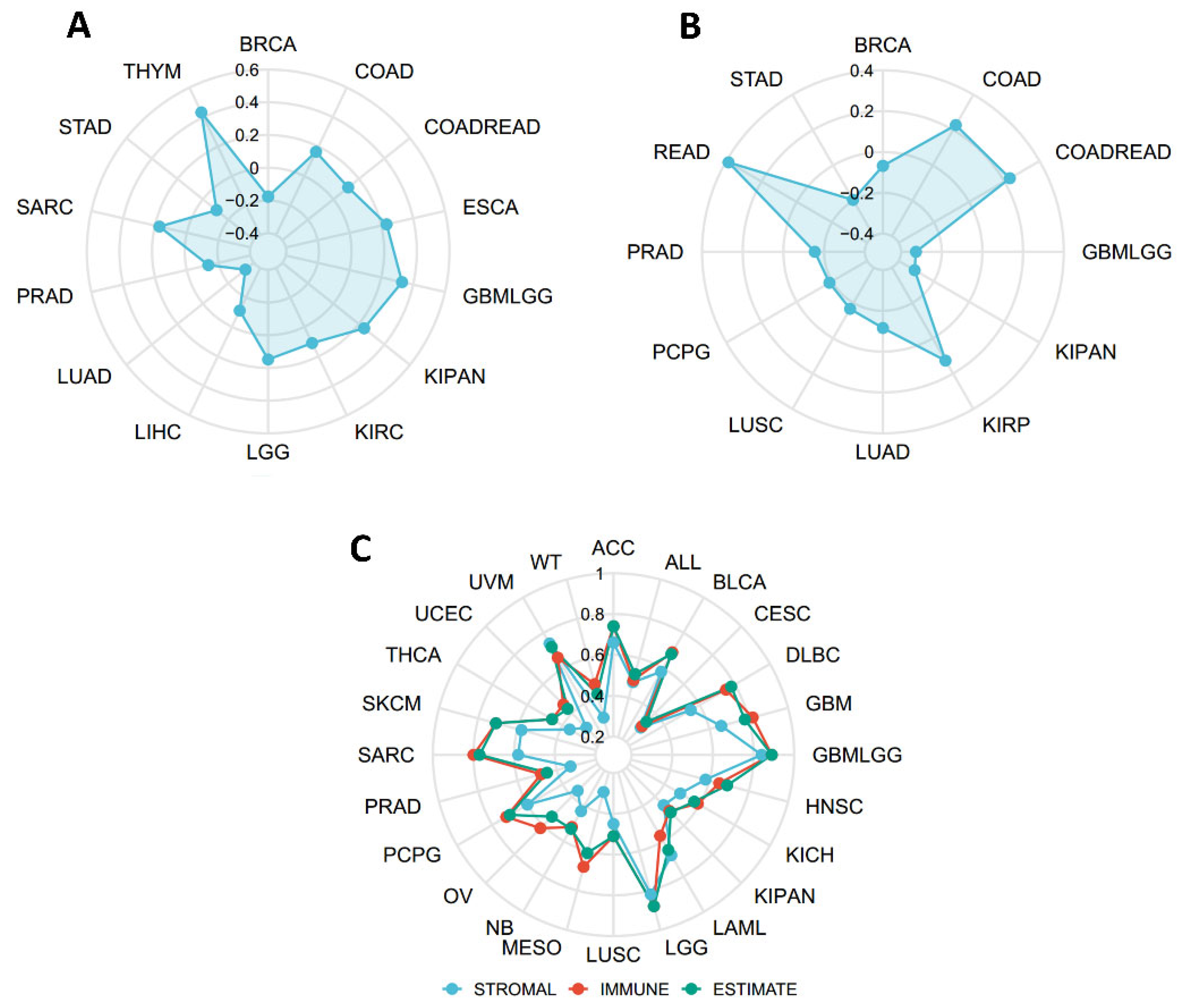
2.5. Connections between SERPINA1 and Tumor Mutational Burden (TMB), Microsatellite Instability (MSI), and Estimation of Stromal and Immune Cells in Malignant Tumor Tissues Using Expression Data (ESTIMATE)

To determine the immune value of SERPINA1 in the tumor microenvironment (TME), we explored the connections between SERPINA1 expression and TMB and MSI, which have been confirmed to influence immunotherapy efficacy significantly [11,12]. Results analyzed using the SangerBox database demonstrated that SERPINA1 expression was positively associated with TMB in COAD, COADREAD, ESCA, GBMLGG, KIPAN, KIRC, LGG, SARC, and THYM and negatively connected to TMB in BRCA, LIHC, LUAD, PRAD, and STAD (Figure 7A). As for MSI and SERPINA1 expression, there were positive associations in COAD, COADREAD, KIRP, and READ, and negative correlations in BRCA, GBMLGG, KIRPAN, LUAD, LUSC, PCPG, PRAD, and STAD (Figure 7B). Subsequently, we detected a relationship between SERPINA1 and three scores of ESTIMATE. The result confirmed significant positive associations between SERPINA1 expression and all three scores in ACC, ALL, BLCA, CESC, DLBC, GBM, GBMLGG, HNSC, KICH, KIPAN, LAML, LGG, LUSC, MESO (mesothelioma), NB (neuroblastoma), OV, PCPG, PRAD, SARC, SKMC, THCA, UCEC, UVM, and WT (high-risk Wilms tumor) (Figure 7C). These results suggested that SERPINA1 may play a vital role in antitumor immunity via affecting the composition of the TME.

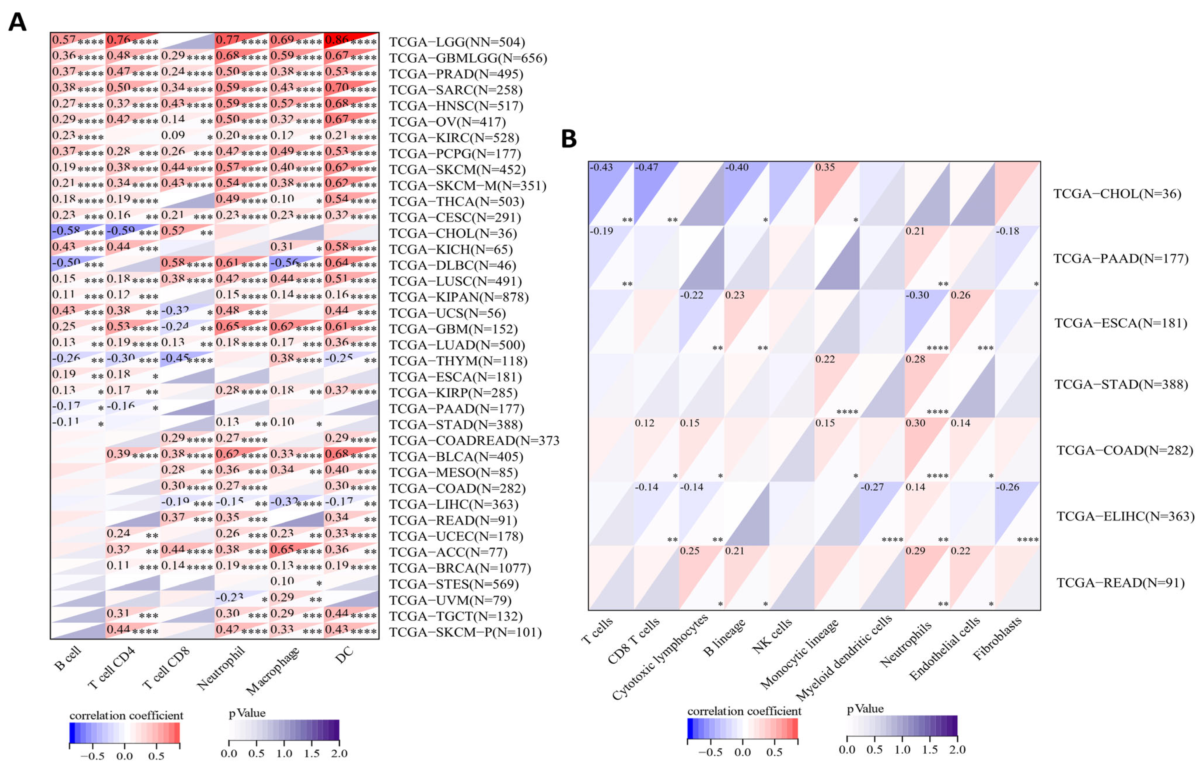
2.6. Correlations between SERPINA1 and Immune Cell Infiltration in the TME

As mentioned above, different immune subtypes of various cancers showed different SERPINA1 expressions. We further explored the correlations between SERPINA1 expression and immune cells in the TME through the Timer 2.0 website. The result demonstrated that SERPINA1 expression presented strong connections to B cells in 25 cancer types, CD4+ T cells in 28 cancer types, CD8+ T cells in 25 cancer types, neutrophils in 32 cancer types, macrophages in 31 cancer types, and dendritic cells (DCs) in 32 cancer types (Figure 8A).
Then, we focused on the relationships between SERPINA1 expression and immune cells in seven digestive system tumors using the R package MCPcounter. The results confirmed that SERPINA1 expression was associated with T cells, CD8+ T cells, the B lineage, and the monocytic lineage in CHOL; T cells, neutrophils, and fibroblasts in PAAD; cytotoxic lymphocytes, the B lineage, neutrophils, and endothelial cells in ESCA; the monocytic lineage and neutrophils in STAD; CD8+ T cells, cytotoxic lymphocytes, the monocytic lineage, neutrophils, and endothelial cells in COAD; CD8+ T cells, cytotoxic lymphocytes, myeloid dendritic cells, neutrophils, and fibroblasts in LIHC; cytotoxic lymphocytes, the B lineage, neutrophils, and endothelial cells in READ (Figure 8B). These findings strongly indicated the influential role of SERPINA1 expression in digestive cancers.
Finally, we analyzed the relationship between SERPINA1 expression and different gene markers of immune cells via the TIMER database. SERPINA1 expression was found to relate to CD8+ T cells, macrophages, DCs, and Th17 cells in digestive cancers (Table S1). For example, SERPINA1 showed close connections to CD8A in CD8+ T cells, CD68 in tumor-associated macrophages, IRF6 in M1 macrophages, NRP1 in DCs, STAT3 and IL17A in Th17 in most digestive cancers. ESCA (n = 184) and LIHC (n = 371) were examples to illustrate the potential immune value of SERPINA1. As shown in Table 1, SERPINA1 had a solid connection to all enrolled markers of CD8+ T cells, general T Cells, B cells, M2 macrophages, neutrophils, and Treg cells in ESCA. SERPINA1 in LIHC also showed a good relationship with most gene markers in neutrophils and natural killer (NK) cells. These results further suggested that SERPINA1 might be vital in regulating immune cell infiltration.

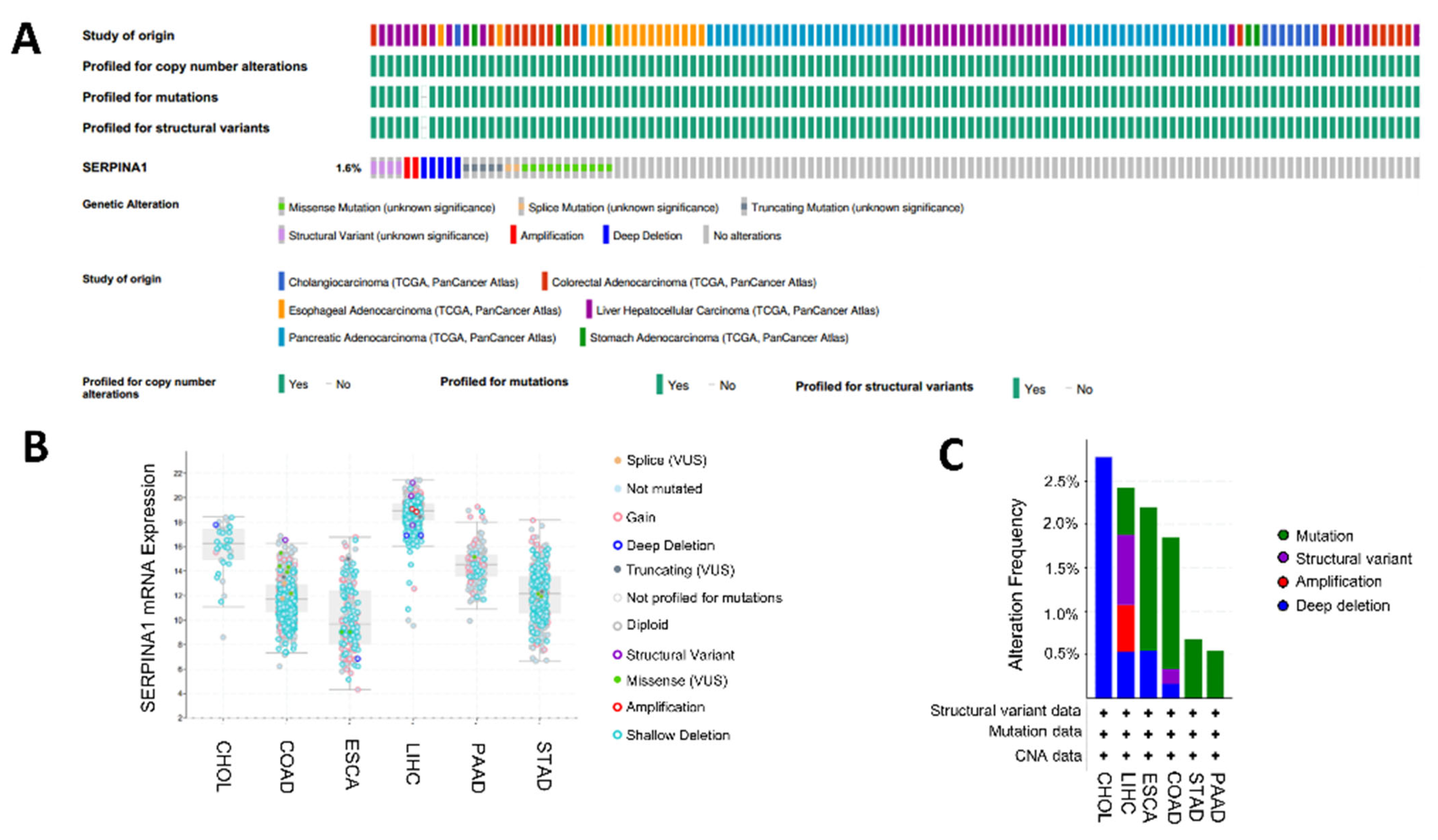
2.7. Alteration of SERPINA1 Gene in Different Subgroups of Digestive Carcinoma

Genomic alteration of SERPINA1 was explored in digestive cancers except for READ, which was absent on the cBioPortal website. The result proved that the incidence of SERPINA1 gene alteration was 1.6% (Figure 9A). Various types of SERPINA1 gene alterations indeed led to variations in SERPINA1 expression (Figure 9B). Furthermore, copy number variation (CNV) showed a relatively lower frequency in STAD and PAAD (Figure 9C). Then, we analyzed the associations between SERPINA and clinicopathologic features in seven digestive cancers via the clinical data from the TCGA database. For example, SERPINA1 expression was significantly connected to adjacent inflammation, tumor size, pathologic stage, and AFP level in LIHC (Table S2). The relationship between SERPINA1 and clinical features in the other six cancers is presented in Tables S3–S8. There was a weak correlation between SERPINA1 expression and characteristics in STAD and PAAD, which were accompanied by fewer CNVs. The results above suggested that genomic alteration of SERPINA1 occurs in cancers, and differential SERPINA1 expression might regulate cancer progression.

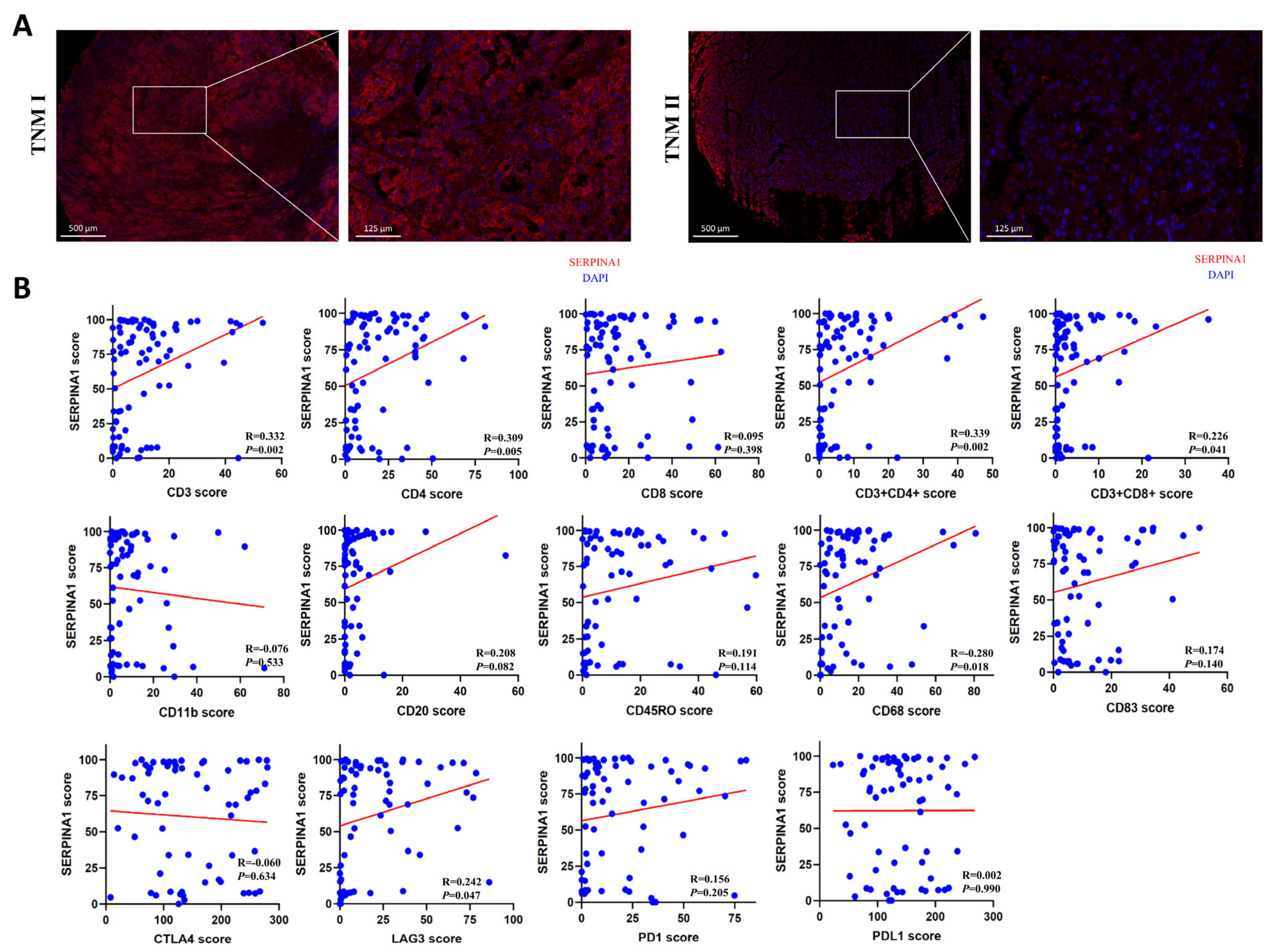
2.8. The Influence of SERPINA1 Protein on Clinicopathologic Features and Immune Infiltrates in LIHC

We detected SERPINA1 protein expression in 86 LIHC tissues through fluorescence-based mIHC to confirm the results above. SERPINA1 protein was detected in the cytoplasm and mesenchyme (Figure 10A). SERPINA protein expression was significantly related to vascular invasion, tumor size, and TNM stage (Table 2). Then, we analyzed the association between the protein level of SERPINA1 and biomarkers of some immune cells in these LIHC tissues. The results revealed that SERPINA1 was significantly connected to CD3 (p = 0.002), CD4 (p = 0.005), CD3 + CD4+ (p = 0.002), CD3 + CD8+ (p = 0.041), CD68 (p = 0.018), and LAG3 (p = 0.047) (Figure 10B), which meant SERPINA1 protein expression was positively associated with T helper (Th) cells, cytotoxic lymphocytes (CTLs), and macrophages.

3. Discussion

An inflammatory microenvironment is a prerequisite and promoter for virtually all cancers. A growing body of research shows that inflammation greatly influences the TME composition, especially on the plasticity of cancer cells and stromal cells [13]. Tumorigenic inflammation blocks anti-tumor immunity and exerts direct pro-tumor signaling on cancer cells [14]. As a serine protease inhibitor, SERPINA1 protein (A1AT) is well known for its anti-inflammatory effect, probably attributed to the inhibition on protease which is an inflammatory vital driver [15]. Furthermore, SERPINA1 protein (A1AT) can inhibit pro-inflammatory cytokine release in monocytes via the NFκB pathway [16]. However, SERPINA1 protein (A1AT) was also confirmed to activate leukocytes and play an inflammatory role [17]. In addition, SERPINA1 protein (A1AT) is inclined to promote the production of Tregs, decrease lymphocyte infiltration, and inhibit the differentiation and maturation of DCs [18,19,20]. Cancer cells can destroy surrounding tissues through releasing elastase, plasmin, and cathepsin, then spread locally, while SERPINA1 protein (A1AT) can inactivate these enzymes. These studies suggested that SERPINA1 has the potential to influence the TME and might be a promising biomarker for immunotherapy, but the role of SERPINA1 in TME regulation and cancer prognosis is unclear.
In this study, we first explored the differential expressions of SERPINA1 in tumor and normal tissues through multiple databases. Previous studies have demonstrated that SERPINA1 was overexpressed in pancreatic and breast cancer tissues but downregulated in lung cancer [21,22,23]. Our findings showed that SERPINA1 was upregulated in most cancer tissues except CHOL, LIHC, LUAD, LUSC, ACC, DLBC, and THYM. These results suggested that SERPINA1 may play a different role in different cancers.
Previous research confirmed serum SERPINA1 as a potential biomarker for cancer diagnosis and prognosis [22,24,25]. However, serum SERPINA1 may show little association with SERPINA1 expression in tissues [23]. Then, the association between SERPINA1 expression in cancer tissues and the prognosis was analyzed. Our results, obtained from the Xiantao web tool and Kaplan–Meier plotter database, demonstrated that SERPINA1 expression in tissues had a significant prognostic value for many cancers. Next, we detected the relationship between SERPINA1 expression and immune subtypes and molecular subtypes of human cancers via the TIMER database and the R package. The results confirmed that SERPINA1 expressions in different immune subtypes and molecular subtypes of most cancers were significantly different, indicating that SERPINA1 may affect the immune microenvironment and cancer prognosis. Moreover, we confirmed that SERPINA1 expression was significantly associated with clinical features in seven digestive cancers, similar to a paper proving that different SERPINA1 levels exist in patients with varying characteristics in colorectal cancer [26].
ICPs are inhibitory receptors expressed on T cells or other immune cells, and tumors can upregulate ICPs to achieve immune escape [27]. Therefore, the expression of ICPs in the TME can influence the clinical efficacy of immune checkpoint blockade (ICB) treatment. Our results based on the SangerBox database demonstrated that SERPINA1 expression was significantly connected to ICP genes in most cancer types, especially GBMLGG, OV, KIPAN, and PRAD, which suggested that patients with these cancers may respond well to ICB treatment. Patients with higher TMB can generate a more significant neoantigen load and induce a more robust neoantigen-specific T cell response, suggesting that patients benefit more from ICB treatment [28]. MSI is caused by functional defects in DNA mismatch repair, and patients with high MSI scores have better immunotherapy outcomes [29]. Our results found that SERPINA1 expression was associated with TMB and MSI in some cancers, positively or negatively, indicating that SERPINA1 plays a different role in the prediction of immunotherapy effect in various human cancers. Above results suggested that SERPINA1 might be important in cancer immunotherapy.
The ESTIMATE score is utilized to speculate the proportion of stromal and immune cells in the TME [30]. Through the SangerBox database, we demonstrated that SERPINA1 was positively connected to immune scores, stromal scores, and ESTIMATE scores in many cancers. Tumor-infiltrating lymphocytes (TILs) have an outsized influence on cancer prognosis and immune therapy [31,32]. This study confirmed that SERPINA1 strongly connects to TILs, which means a high probability of regulating the TME. For example, SERPINA1 expression was closely related to CD4+ T cells, CD8+ T cells, neutrophils, and macrophages in most cancer types. Particularly in ESCA, SERPINA1 was connected to PDCD1 and CTLA4, which are classical targets of immunotherapy [33,34]. Furthermore, SERPINA1 demonstrated significant association with CD8+ T cells, general T cells, B cells, M2 macrophages, neutrophils, and Treg cells in ESCA. These results suggested that SERPINA1 might affect immunotherapy efficiency through regulating TIL compositions in the TME.
Finally, we explored the SERPINA1 protein (A1AT) in LIHC tissues via fluorescence-based mIHC to preliminarily prove the results acquired. Similar to the findings regarding the SERPINA1 gene, SERPINA1 protein (A1AT) in LIHC tissues was associated with clinical features. In addition, the positive relationship between SERPINA1 level and some biomarkers of immune cells suggested the SERPINA1 protein (A1AT) might affect the infiltrations of Th cells, CTLs, and macrophages, as well as the efficacy of immunotherapy targeting LAG3.
Although we performed a systematic analysis on the role of SERPINA1 in pan-cancer, and cross-certified the result through different databases and R package, the study has some limitations. First, systematic bias may be caused by the difference in RNA-seq data from different databases. Second, the function of the SERPINA1 protein should be verified in vitro and in vivo. Third, we preliminarily deduced that SERPINA1 had a close connection to the immune microenvironment, but there was no direct evidence to prove the influence of SERPINA1 on immunotherapy efficiency and prognosis. In the future, more investigations are needed to certify the prognostic and immunological value of SERPINA1 and explore the underlying mechanism.

4. Materials and Methods

4.1. Differential Expression Analysis

The TIMER 2.0 tool (http://timer.cistrome.org/ (accessed on 15 March 2022)) and the GEPIA database (http://gepia.cancer-pku.cn/ (accessed on15 March 2022)) were utilized to compare the differential expressions of SERPINA1 in various cancer tissues and normal tissues [35,36].

4.2. Survival Analysis

The relationships between SERPINA1 expression and OS or DSS were analyzed using the Xiantao web tool (https://www.xiantao.love/ (accessed on 15 March 2022)) and Kaplan–Meier plotter database (https://kmplot.com/ (accessed on 15 March 2022)) [37,38].

4.3. Analysis of SERPINA1 Expression and Immune and Molecular Subtypes

The TISIDB web portal (http://cis.hku.hk/TISIDB/ (accessed on 16 March 2022)) was used to detect the associations between SERPINA1 expression and immune or molecular subtypes of different cancers [39].

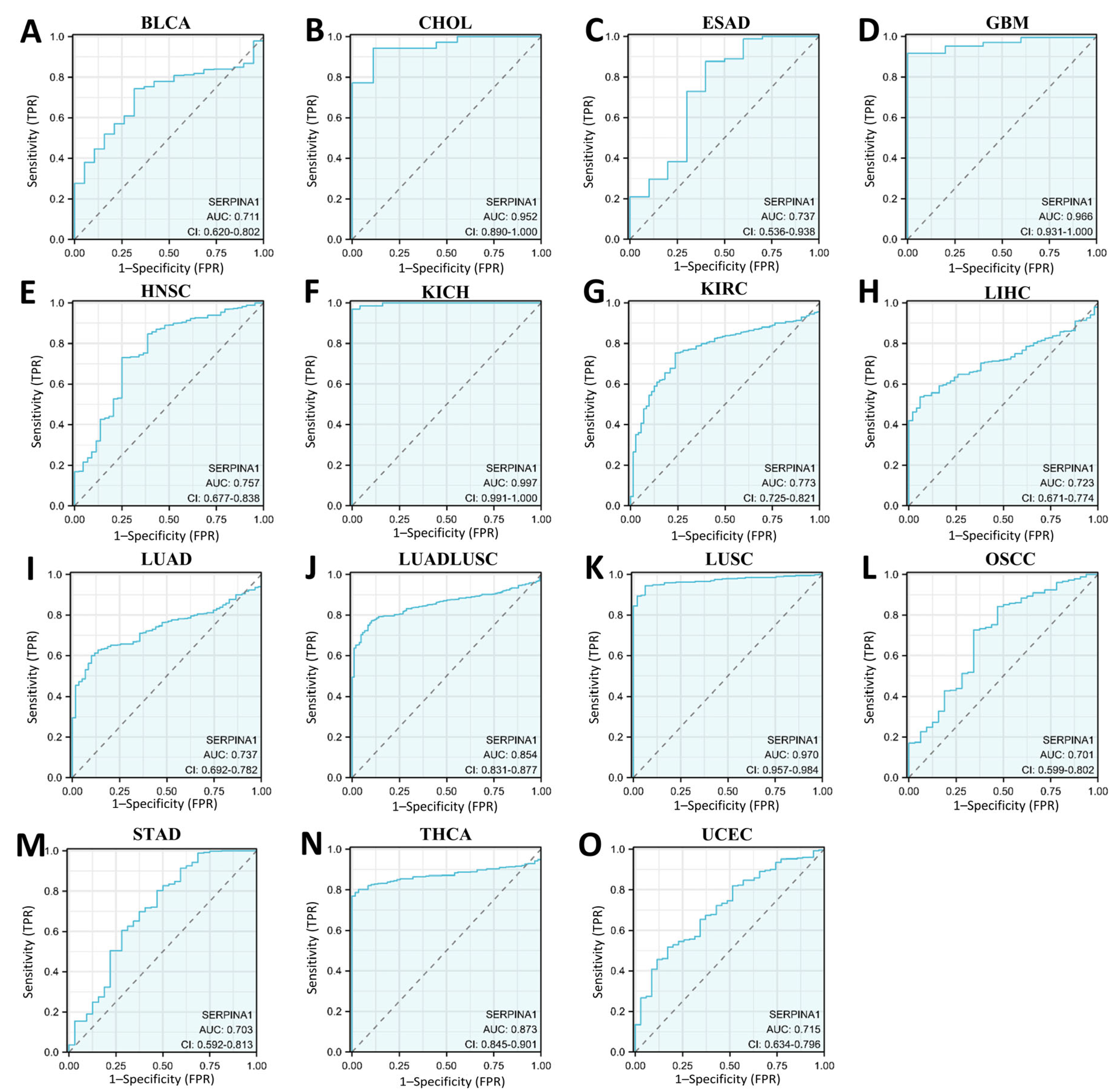
4.4. Analysis of SERPINA1 Expression and Biomarkers of Therapeutic Efficacy

To explore the relationships between SERPINA1 expression and ICP genes, TMB, MSI, and ESTIMATE, the SangerBox database (http://sangerbox.com/ (accessed on 20 March 2022)) was utilized comprehensively [10].

4.5. Analysis of SERPINA1 Expression and Immune Cell Infiltration

The associations between SERPINA1 level and six immune cells were explored in pan-cancers through the Timer 2.0 website. Then, we analyzed the connections between SERPINA1 expression and the absolute abundances of 8 immune cells and two stromal cells in seven digestive system tumors using the R package MCPcounter. The relationships between SERPINA1 expression and marker genes of immune cells were explored via the TIMER database.

4.6. Analysis of SERPINA1 Genomic Alterations

The cBioPortal for Cancer Genomics (https://docs.cbioportal.org/ (accessed on 10 April 2022)) was used to confirm the genomic alterations of SERPINA1 in digestive system tumors.

4.7. Analysis of SERPINA1 Expression and Clinicopathologic Features

The RNAseq data of seven digestive cancers were downloaded from the TCGA database (https://portal.gdc.cancer.gov (accessed on 10 April 2022)). Data without clinical information were discarded, and the R package STATS was utilized to analyze the associations between SERPINA1 expression and clinicopathologic features in each digestive cancer.

4.8. Tissue Samples and Patient Data

All hepatic tissue samples were collected from tumor immunotherapy LIHC patients admitted to the First Affiliated Hospital of Nantong University. A total of 86 LIHC tissue samples were detected in this study. None of the patients received any chemotherapy or radiation before surgery or biopsy. The study was approved by the Ethics Committee of Nantong University, and informed consent was provided by patients or their guardians.

4.9. Fluorescence-Based mIHC

H&E sections were observed for each tissue sample to determine the correct site. An array (12 × 10) was designed on the blank wax block, and the Quick Ray Master UATM-272A (UNITMA, Seoul, Republic of Korea) was used to drill holes into the receptor module. Tissue columns with a diameter of 1.5 mm were taken out from donor tissue blocks and then inserted into corresponding holes in receptor blocks. The receptor blocks were cut into slices with a thickness of 3 μm and placed on polylysine-coated glass slides. The slices were dewaxed and hydrated, boiled in 0.01 mM citrate buffer (pH 6.0) to retrieve antigens, and incubated with 5% goat serum to block the nonspecific site. The nuclei were stained with DAPI (#C1005, Beyotime, Shanghai, China). Then, the sections were incubated with anti-alpha 1 Antitrypsin antibody (1:200, #ab207303, Abcam, Cambridge, MA, USA) at 4 °C overnight. Opal polymer HRP Ms + Rb (#ARH1001EA, Perkin Elmer, Waltham, MA, USA) was added to the slices and incubated at dark for 3 h. Fluoroshield with DAPI (#F6057, Sigma, St. Louis, MO, USA) was used to stain nuclei and seal the sections.

4.10. Statistical Analysis

The experimental data of mIHC were statistically analyzed using IBM SPSS Statistics v26 (Endicott, New York, NY, USA). The correlation between SERPINA1 protein expression and clinicopathologic features was investigated using the Pearson Chi-square test. p < 0.05 was considered statistically significant.

5. Conclusions

Above all, SERPINA1 shows potential value for the diagnosis and prognosis of many human cancers and may be involved in the immune regulation of the TME. Further study about SERPINA1 is necessary for cancer immunotherapy and prognosis.

Supplementary Materials

The following supporting information can be downloaded at: https://www.mdpi.com/article/10.3390/ijms241411566/s1. Figure S1. The disease specific survival curves of human cancers with different SERPINA1 expressions. Figure S2. The overall survival curves of human cancers with different SERPINA1 expressions in the Kaplan–Meier plotter database. Table S1. The relationship between SERPINA1 expression and different gene markers of immune cells. Table S2. The relationships between SERPINA1 expression and clinical features in LIHC. Table S3. The relationships between SERPINA1 expression and clinical features in STAD. Table S4. The relationships between SERPINA1 expression and clinical features in CHOL. Table S5. The relationships between SERPINA1 expression and clinical features in COAD. Table S6. The relationships between SERPINA1 expression and clinical features in READ. Table S7. The relationships between SERPINA1 expression and clinical features in PAAD. Table S8. The relationships between SERPINA1 expression and clinical features in ESCA.

Author Contributions

Conceptualization, X.K. and J.J.; bioinformatic analysis, X.K. and J.L.; mIHC, J.L. and M.X.; data curation, X.K., J.L. and J.Z.; writing—review and editing, X.K. and J.L.; supervision, J.J.; project administration, J.J.; funding acquisition, J.J. All authors have read and agreed to the published version of the manuscript.

Funding

This research was funded by the National Natural Science Foundation of China (81761128018 and 81572871) and the Natural Science Foundation of Jiangsu Province (BK20151277).

Institutional Review Board Statement

The study was conducted according to the guidelines of the Declaration of Helsinki and approved by the Ethics Committee of Nantong University (2018-K020).

Informed Consent Statement

Informed consent was obtained from all subjects involved in the study. Written informed consent has been obtained from the patients to publish this paper.

Data Availability Statement

All data generated or analyzed are included in the current manuscript and they are available from the corresponding author upon reasonable request.

Conflicts of Interest

The authors declare no conflict of interest.

Abbreviations

ACCAdrenocortical carcinoma
BLCA Bladder urothelial carcinoma
BRCABreast invasive carcinoma
CESCCervical squamous cell carcinoma and endocervical adenocarcinoma
CHOLCholangiocarcinoma
CNVCopy number variation
COAD Colon adenocarcinoma
CTLCytotoxic lymphocyte
DCDendritic cell
DLBC Lymphoid neoplasm diffuse large B-cell lymphoma
ESAD Esophagus adenocarcinoma
ESCA Esophageal carcinoma
ESTIMATEEstimation of Stromal and Immune cells in Malignant Tumor tissues using Expression data
GBM Glioblastoma multiforme
GBMLGGGlioma
HNSC Head and neck squamous cell carcinoma
ICPImmune checkpoint
KICH Kidney chromophobe
KIPAN Pan-kidney cohort
KIRC Kidney renal clear cell carcinoma
KIRPKidney renal papillary cell carcinoma
LGG Brain lower grade glioma
LIHCLiver hepatocellular carcinoma
LUADLung adenocarcinoma
LUADLUSCLung adenocarcinoma and lung squamous cell carcinoma
LUSC Lung squamous cell carcinoma
MESO Mesothelioma
MSIMicrosatellite instability
NBNeuroblastoma
NKNatural killer cell
OSOverall survival
OSCC Oral squamous cell carcinoma
OV Ovarian serous cystadenocarcinoma
PCPGPheochromocytoma and paraganglioma
PRADProstate adenocarcinoma
SARCSarcoma
SERPINA1Serpin family A member 1
SKCM Skin cutaneous melanoma
STAD Stomach adenocarcinoma
TCGAThe Cancer Genome Atlas
TGCT Testicular germ cell tumors
THCA Thyroid carcinoma
THYM Thymoma
TMBTumor mutational burden
TMETumor microenvironment
UCECUterine corpus endometrial carcinoma
UVMUveal melanoma
WT High-risk Wilms tumor

References

  1. Ruf, B.; Heinrich, B.; Greten, T.F. Immunobiology and immunotherapy of HCC:spotlight on innate and innate-like immune cells. Cell. Mol. Immunol. 2021, 18, 112–127. [Google Scholar] [CrossRef] [PubMed]
  2. Xiang, S.; Li, J.; Shen, J.; Zhao, Y.; Xiao, Z. Identification of Prognostic Genes in the Tumor Microenvironment of Hepatocellular Carcinoma. Front. Immunol. 2021, 12, 653836. [Google Scholar] [CrossRef] [PubMed]
  3. Shen, H.; Yu, X.; Yang, F.; Zhang, Z.; Shen, J.; Sun, J.; Choksi, S.; Jitkaew, S.; Shu, Y. Reprogramming of Normal Fibroblasts into Cancer-Associated Fibroblasts by miRNAs-Mediated CCL2/VEGFA Signaling. PLoS Genet. 2016, 12, e1006244. [Google Scholar] [CrossRef] [PubMed] [Green Version]
  4. Kloth, J.N.; Gorter, A.; Fleuren, G.J.; Oosting, J.; Jordanova, E.S. Elevated expression of SerpinA1 and SerpinA3 in HLA-positive cervical carcinoma. J. Pathol. 2008, 215, 222–230. [Google Scholar] [CrossRef] [PubMed]
  5. Seynes, C.D.; Ged, C.; Verneuil, H.D.; Chollet, N.; Balduyck, M.; Raherison, C. Identification of a novel alpha1-antitrypsin variant. Respir. Med. Case Rep. 2016, 20, 64–67. [Google Scholar] [CrossRef]
  6. Enewold, L.; Mechanic, L.E.; Bowman, E.D.; Platz, E.A.; Alberg, A.J. SERPINA1 and ELA2 polymorphisms are not associated with COPD or lung cancer. Anticancer. Res. 2012, 32, 3923–3928. [Google Scholar]
  7. Yang, J.; Xiong, X.; Wang, X.; Guo, B.; He, K.; Huang, C. Identification of peptide regions of SERPINA1 and ENOSF1 and their protein expression as potential serum biomarkers for gastric cancer. Tumour Biol. 2015, 36, 5109–5118. [Google Scholar] [CrossRef]
  8. Chan, H.J.; Li, H.; Zheng, L.; Yuan, Y.C.; Chen, S. SERPINA1 is a direct estrogen receptor target gene and a predictor of survival in breast cancer patients. Oncotarget 2015, 6, 25815–25827. [Google Scholar] [CrossRef] [Green Version]
  9. Morad, G.; Helmink, B.A.; Sharma, P.; Wargo, J.A.J.C. Hallmarks of response, resistance, and toxicity to immune checkpoint blockade. Cell 2021, 184, 5309–5337. [Google Scholar] [CrossRef]
  10. Hu, J.; Qiu, D.; Yu, A.; Hu, J.; Deng, H.; Li, H.; Yi, Z.; Chen, J.; Zu, X. YTHDF1 Is a Potential Pan-Cancer Biomarker for Prognosis and Immunotherapy. Front. Oncol. 2021, 11, 607224. [Google Scholar] [CrossRef]
  11. Liu, L.; Bai, X.; Wang, J.; Tang, X.R.; Dong, Z.Y. Combination of TMB and CNA Stratifies Prognostic and Predictive Responses to Immunotherapy Across Metastatic Cancer. Clin. Cancer Res. 2019, 25, 7413–7423. [Google Scholar] [CrossRef] [Green Version]
  12. Luchini, C.; Bibeau, F.; Ligtenberg, M.J.; Singh, N.; Nottegar, A.; Bosse, T.; Miller, R.; Riaz, N.; Douillard, J.Y.; Andre, F.; et al. ESMO recommendations on microsatellite instability testing for immunotherapy in cancer, and its relationship with PD-1/PD-L1 expression and tumour mutational burden: A systematic review-based approach. Ann. Oncol. 2019, 30, 1232–1243. [Google Scholar] [CrossRef] [Green Version]
  13. Greten, F.R.; Grivennikov, S.I. Inflammation and Cancer: Triggers, Mechanisms, and Consequences. Immunity 2019, 51, 27–41. [Google Scholar] [CrossRef]
  14. Denk, D.; Greten, F.R. Inflammation: The incubator of the tumor microenvironment. Trends Cancer 2022, 8, 901–914. [Google Scholar] [CrossRef]
  15. Kaneva, M.K.; Muley, M.M.; Krustev, E.; Reid, A.R.; Souza, P.R.; Dell’Accio, F.; McDougall, J.J.; Perretti, M. Alphantitrypsin reduces inflammation and exerts chondroprotection in arthritis. FASEB J. 2021, 35, e21472. [Google Scholar] [CrossRef]
  16. Bai, X.; Bai, A.; Tomasicchio, M.; Hagman, J.R.; Buckle, A.M.; Gupta, A.; Kadiyala, V.; Bevers, S.; Serban, K.A.; Kim, K.; et al. α1-Antitrypsin Binds to the Glucocorticoid Receptor with Anti-Inflammatory and Antimycobacterial Significance in Macrophages. J. Immunol. 2022, 209, 1746–1759. [Google Scholar] [CrossRef]
  17. Ehlers, M.R. Immune-modulating effects of alpha-1 antitrypsin. Biol. Chem. 2014, 395, 1187–1193. [Google Scholar] [CrossRef]
  18. Marcondes, A.M.; Karoopongse, E.; Lesnikova, M.; Margineantu, D.; Welte, T.; Dinarello, C.A.; Hockenbery, D.; Janciauskiene, S.; Deeg, H.J. α-1-Antitrypsin (AAT)-modified donor cells suppress GVHD but enhance the GVL effect: A role for mitochondrial bioenergetics. Blood 2014, 124, 2881–2891. [Google Scholar] [CrossRef] [Green Version]
  19. Tawara, I.; Sun, Y.; Lewis, E.C.; Toubai, T.; Evers, R.; Nieves, E.; Azam, T.; Dinarello, C.A.; Reddy, P. Alpha-1-antitrypsin monotherapy reduces graft-versus-host disease after experimental allogeneic bone marrow transplantation. Proc. Natl. Acad. Sci. USA 2012, 109, 564–569. [Google Scholar] [CrossRef]
  20. Elshikha, A.S.; Lu, Y.; Chen, M.-J.; Akbar, M.; Zeumer, L.; Ritter, A.; Elghamry, H.; Mahdi, M.A.; Morel, L.; Song, S. Alpha 1 Antitrypsin Inhibits Dendritic Cell Activation and Attenuates Nephritis in a Mouse Model of Lupus. PLoS ONE 2016, 11, e0156583. [Google Scholar] [CrossRef] [Green Version]
  21. Trachte, A.L.; Suthers, S.E.; Lerner, M.R.; Hanas, J.S.; Jupe, E.R.; Sienko, A.E.; Adesina, A.M.; Lightfoot, S.A.; Brackett, D.J.; Postier, R.G. Increased expression of alpha-1-antitrypsin, glutathione S-transferase p and vascular endothelial growth factor in human pancreatic adenocarcinoma. Am. J. Surg. 2002, 184, 642–647. [Google Scholar] [CrossRef] [PubMed]
  22. López-Árias, E.; Aguilar-Lemarroy, A.; Jave-Suárez, L.F.; Morgan-Villela, G.; Mariscal-Ramírez, I.; Martínez-Velázquez, M.; Álvarez, Á.H.; Gutiérrez-Ortega, A.; Gutiérrez, R.H. Alpha 1-antitrypsin: A novel tumor-associated antigen identified in patients with early-stage breast cancer. Electrophoresis 2012, 33, 2130–2137. [Google Scholar] [CrossRef] [PubMed]
  23. Ercetin, E.; Richtmann, S.; Delgado, B.M.; Gomez-Mariano, G.; Schneider, M.A. Clinical Significance of SERPINA1 Gene and Its Encoded Alpha1-antitrypsin Protein in NSCLC. Cancers 2019, 11, 1306. [Google Scholar] [CrossRef] [PubMed] [Green Version]
  24. Pirisi, M.; Fabris, C.; Soardo, G.; Toniutto, P.; Bartoli, E. Prognostic value of serum alpha-1-antitrypsin in hepatocellular carcinoma. Eur. J. Cancer 1996, 32A, 221–225. [Google Scholar] [CrossRef]
  25. Wu, C.-C.; Lu, Y.-T.; Yeh, T.-S.; Chan, Y.-H.; Dash, S.; Yu, J.-S. Identification of Fucosylated SERPINA1 as a Novel Plasma Marker for Pancreatic Cancer Using Lectin Affinity Capture Coupled with iTRAQ-Based Quantitative Glycoproteomics. Int. J. Mol. Sci. 2021, 22, 6079. [Google Scholar] [CrossRef]
  26. Kwon, C.H.; Park, H.J.; Choi, J.H.; Lee, J.R.; Kim, H.K.; Jo, H.-J.; Kim, H.S.; Oh, N.; Song, G.A.; Park, D.Y. Snail and serpinA1 promote tumor progression and predict prognosis in colorectal cancer. Oncotarget 2015, 6, 20312–20326. [Google Scholar] [CrossRef] [Green Version]
  27. Topalian, S.L.; Taube, J.M.; Anders, R.A.; Pardoll, D.M. Mechanism-driven biomarkers to guide immune checkpoint blockade in cancer therapy. Nat. Rev. Cancer 2016, 16, 275–287. [Google Scholar] [CrossRef]
  28. McGrail, D.J.; Pilié, P.G.; Rashid, N.U.; Voorwerk, L.; Slagter, M.; Kok, M.; Jonasch, E.; Khasraw, M.; Heimberger, A.B.; Lim, B.; et al. High tumor mutation burden fails to predict immune checkpoint blockade response across all cancer types. Ann. Oncol. 2021, 32, 661–672. [Google Scholar] [CrossRef]
  29. Zhao, P.; Li, L.; Jiang, X.; Li, Q. Mismatch repair deficiency/microsatellite instability-high as a predictor for anti-PD-1/PD-L1 immunotherapy efficacy. J. Hematol. Oncol. 2019, 12, 54. [Google Scholar] [CrossRef]
  30. Yoshihara, K.; Shahmoradgoli, M.; Martínez, E.; Vegesna, R.; Kim, H.; Torres-Garcia, W.; Treviño, V.; Shen, H.; Laird, P.W.; Levine, D.A.; et al. Inferring tumour purity and stromal and immune cell admixture from expression data. Nat. Commun. 2013, 4, 2612. [Google Scholar] [CrossRef] [Green Version]
  31. Okadome, K.; Baba, Y.; Yagi, T.; Kiyozumi, Y.; Ishimoto, T.; Iwatsuki, M.; Miyamoto, Y.; Yoshida, N.; Watanabe, M.; Baba, H. Prognostic Nutritional Index, Tumor-infiltrating Lymphocytes, and Prognosis in Patients with Esophageal Cancer. Ann. Surg. 2020, 271, 693–700. [Google Scholar] [CrossRef]
  32. Zhang, Y.; Zhang, Z. The history and advances in cancer immunotherapy:understanding the characteristics of tumor-infiltrating immune cells and their therapeutic implications. Cell. Mol. Immunol. 2020, 17, 807–821. [Google Scholar] [CrossRef]
  33. Ribas, A.; Dummer, R.; Puzanov, I.; VanderWalde, A.; Andtbacka, R.H.I.; Michielin, O.; Olszanski, A.J.; Malvehy, J.; Cebon, J.; Fernandez, E. Oncolytic Virotherapy Promotes Intratumoral T Cell Infiltration and Improves Anti-PD-1 Immunotherapy. Cell 2017, 170, 1109–1119.e10. [Google Scholar] [CrossRef] [Green Version]
  34. Chen, D.; Menon, H.; Verma, V.; Guo, C.; Ramapriyan, R.; Barsoumian, H.; Younes, A.; Hu, Y.; Wasley, M.; Cortez, M.A.; et al. Response and outcomes after anti-CTLA4 versus anti-PD1 combined with stereotactic body radiation therapy for metastatic non-small cell lung cancer: Retrospective analysis of two single-institution prospective trials. J. Immunother. Cancer 2020, 8, e000492. [Google Scholar] [CrossRef] [Green Version]
  35. Li, T.; Fu, J.; Zeng, Z.; David, C.; Li, J.; Chen, Q.; Li, B.; Liu, X.S. TIMER2.0 for analysis of tumor-infiltrating immune cells. Nucleic Acids Res. 2020, 48, W509–W514. [Google Scholar] [CrossRef]
  36. Tang, Z.; Li, C.; Kang, B.; Gao, G.; Li, C.; Zhang, Z. GEPIA: A web server for cancer and normal gene expression profiling and interactive analyses. Nucleic Acids Res. 2017, 45, W98–W102. [Google Scholar] [CrossRef] [Green Version]
  37. Lánczky, A.; Győrffy, B. Web-Based Survival Analysis Tool Tailored for Medical Research (KMplot): Development and Implementation. J. Med. Internet Res. 2021, 23, e27633. [Google Scholar] [CrossRef]
  38. Chu, L.; Yi, Q.; Yan, Y.; Peng, J.; Li, Z.; Jiang, F.; He, Q.; Ouyang, L.; Wu, S.; Fu, C.; et al. A Prognostic Signature Consisting of Pyroptosis-Related Genes and SCAF11 for Predicting Immune Response in Breast Cancer. Front. Med. 2022, 9, 882763. [Google Scholar] [CrossRef]
  39. Ru, B.; Ngar, W.C.; Tong, Y.; Zhong, J.Y.; Zhong, S.; Wu, W.C.; Chu, K.C.; Yiu, W.C.; Ying, L.C.; Chen, I. TISIDB: An integrated repository portal for tumor–immune system interactions. Bioinformatics 2019, 35, 4200–4202. [Google Scholar] [CrossRef]
Figure 1. SERPINA1 expression in human cancers. (A) SERPINA1 expression in different human cancers and normal tissues according to the TIMER 2.0 database. (B) SERPINA1 expression in some human cancers and normal tissues according to the GEPIA database. Red, cancer tissues. Blue, normal tissues. * p < 0.05, ** p < 0.01, *** p < 0.001.
Ijms 24 11566 g001
Figure 2. The ROC curves of SERPINA1: (A) in BLCA; (B) in CHOL; (C) in ESAD; (D) in GBM; (E) in HNSC; (F) in KICH; (G) in KIRC; (H) in LIHC; (I) in LUAD; (J) in LUADLUSC; (K) in LUSC; (L) in OSCC; (M) in STAD; (N) in THCA; (O) in UCEC.
Ijms 24 11566 g002
Figure 3. The overall survival curves of human cancers with different SERPINA1 expressions: (A) in BRCA; (B) in COAD; (C) in DLBC; (D) in KIRC; (E) in KIRP; (F) in Osteosarcoma; (G) in SARC; (H) in SKCM; (I) in GBMLGG; (J) in GBM; (K) in HNSC; (L) in KICH; (M) in LGG; (N) in LIHC; (O) in LUSC.
Ijms 24 11566 g003
Figure 4. The SERPINA1 expressions in different immune subtypes: (A) in ACC; (B) in BLCA; (C) in BRCA; (D) in KICH; (E) in KIRC; (F) in LGG; (G) in LUAD; (H) in LUSC; (I) in OV; (J) in PCPG; (K) in PRAD; (L) in SARC; (M) in SKMC; (N) in THCA; (O) in UCEC; (P) in UVM. C1, wound healing. C2, IFN-gamma dominant. C3, inflammatory. C4, lymphocyte depleted. C5, immunologically quiet. C6, TGF-β dominant.
Ijms 24 11566 g004
Figure 5. The SERPINA1 expressions in different molecular subtypes: (A) in ACC; (B) in BRCA; (C) in COAD; (D) in ESCA; (E) in HNSC; (F) in LGG; (G) in LIHC; (H) in LUSC; (I) in OV; (J) in PCPG; (K) in PRAD; (L) in READ; (M) in STAD; (N) in UCEC.
Ijms 24 11566 g005
Figure 6. Pan-cancer association between SERPINA1 expression and ICP genes.
Ijms 24 11566 g006
Figure 7. The connection between SERPINA1 expression and biomarkers of therapeutic efficacy in human cancers. (A) The connection between SERPINA1 and TMB. (B) The connection between SERPINA1 and MSI. (C) The connection between SERPINA1 and ESTIMATE score.
Ijms 24 11566 g007
Figure 8. The relationship between SERPINA1 expression and tumor-infiltrating immune cells: (A) in pan-cancer; (B) in seven digestive cancers. * p < 0.05, ** p < 0.01, *** p < 0.001, **** p < 0.0001.
Ijms 24 11566 g008
Figure 9. Genomic alterations of SERPINA1 in different subgroups of digestive carcinoma. (A) OncoPrint of SERPINA2 gene alterations in cancer cohort. (B) The main types of SERPINA1 alterations in digestive cancers. (C) Details of SERPINA1 alterations in digestive cancers.
Ijms 24 11566 g009
Figure 10. The relationships between SERPINA1 protein, clinicopathologic features, and immune infiltrates in LIHC. (A) The protein expression of SERPINA1 in LIHC tissues with different TNM stages. (B) The connection between SERPINA1 and biomarkers of immune cells. Blue dot, LIHC tissue sample. Red line, a line with linear regression.
Ijms 24 11566 g010
Table 1. Relationship between SERPINA1 and related markers of immune cells in ESCA and LIHC.
DescriptionGene MarkersESCALIHC
NonePurityNonePurity
RpRpRpRp
CD8+ T CellCD8A0.2290.002 *0.1940.0090.1060.041 *0.0110.834
CD8B0.321<0.001 *0.293<0.001 *0.0690.184−0.0200.718
T Cell (general)CD3D0.358<0.001 *0.328<0.001 *0.1440.006 *0.0530.328
CD3E0.321<0.001 *0.282<0.001 *0.228<0.001 *0.1330.014 *
CD20.384<0.001 *0.354<0.001 *0.191<0.001 *0.0950.078
B CellCD190.362<0.001 *0.325<0.001 *0.0820.1140.0060.905
CD79A0.277<0.001 *0.238<0.001 *0.218<0.001 *0.1160.031 *
MonocyteCD860.0660.3750.0190.8010.1140.029 *−0.0030.961
CSF1R0.282<0.001 *0.257<0.001 *0.0980.059−0.0020.680
TAMCCL20.1320.07440.0880.2420.0830.112−0.0450.407
CD680.258<0.001 *0.258<0.001 *0.1610.002 *0.0690.199
IL100.1250.0900.0870.2470.1190.022 *0.0160.760
M1NOS2 0.451<0.001 *0.47<0.001 *0.0350.4990.0220.680
IRF5−0.286<0.001 *−0.317<0.001 *−0.1090.036 *−0.1330.013 *
PTGS20.060.4170.0430.5710.195<0.001 *0.0870.106
M2CD1630.339<0.001 *0.318<0.001 *0.1610.002 *0.0700.197
VSIG40.2380.001 *0.2060.005 *0.1010.052−0.0030.949
MS4A4A0.287<0.001 *0.253<0.001 *0.172<0.001 *0.0650.229
NeutrophilsCEACAM80.293<0.001 *0.274<0.001 *0.0740.1550.0580.284
ITGAM0.251<0.001 *0.2340.001 *−0.0630.226−0.1620.003 *
CCR70.428<0.001 *0.401<0.001 *0.278<0.001 *0.1869<0.001 *
NK CellKIR2DL10.1230.0940.0940.210−0.0460.367−0.0620.250
KIR2DL30.0350.6360.0330.665−0.0610.240−0.1180.028 *
KIR2DL40.0780.290.050.501−0.1180.023 *−0.1630.002 *
KIR3DL10.1710.020 *0.1530.040 *−0.0630.023 *−0.1090.042 *
KIR3DL2−0.0490.508−0.0920.221−0.0290.573−0.0840.121
KIR2DL300.99600.999−0.0610.240−0.1180.028 *
KIR2DS40.0420.5710.0410.583−0.0780.135−0.0850.117
Dendritic CellHLA-DPB10.4<0.001 *0.38<0.001 *0.150.004 *0.0550.307
HLA-DQB10.362<0.001 *0.329<0.001 *0.110.034 *0.0180.743
HLA-DRA0.456<0.001 *0.441<0.001 *0.1670.001 *0.0780.151
HLA-DPA10.431<0.001 *0.414<0.001 *0.178<0.001 *0.0890.094
CD1C0.0630.3930.0020.9770.217<0.001 *0.1310.015 *
NRP10.1780.016 *0.1420.058 *0.0600.2500.0210.697
ITGAX0.355<0.001 *0.24<0.001 *0.1260.015 *0.0270.624
Th1TBX210.308<0.001 *0.272<0.001 *0.1310.012 *0.0340.526
STAT40.275<0.001 *0.2370.001 *0.120.021 *0.0430.422
STAT10.1110.1340.0850.2500.175<0.001 *0.1360.011 *
IFNG0.2050.005 *0.1720.021 *−0.0220.669−0.0830.122
TNF0.0660.3730.0460.5370.1550.003 *0.0750.165
IL12A0.2010.006 *0.190.011 *0.0080.884−0.0400.454
IL12B0.262<0.001 *0.2270.002 *0.1310.012 *0.0490.368
Th2GATA30.070.3400.0340.6490.1070.040 *−0.0020.965
STAT60.278<0.001 *0.302<0.001 *−0.0400.444−0.0500.352
STAT5A0.444<0.001 *0.432<0.001 *−0.1160.026 *−0.189<0.001 *
IL130.1730.019 *0.1460.050−0.0990.057−0.120.026 *
TfhBCL6−0.504<0.001 *−0.512<0.001 *0.0190.7110.0110.833
IL210.090.2250.0590.4320.0450.3920.0120.821
Th17STAT30.0590.4270.0470.5310.1610.002 *0.1290.016 *
IL17A0.378<0.001 *0.379<0.001 *0.0790.1310.0670.218
TregFOXP30.255<0.001 *0.2220.003 *0.0250.633−0.0350.518
CCR80.314<0.001 *0.282<0.001 *0.179<0.001 *0.1070.048 *
STAT5B0.1890.010 *0.1930.009 *−0.0220.6770.0130.814
TGFB1−0.508<0.001 *−0.574<0.001 *0.205<0.001 *0.1130.036 *
T cell exhaustionPDCD10.301<0.001 *0.272<0.001 *0.1190.021 *0.0280.608
CTLA40.292<0.001 *0.261<0.001 *0.1140.028 *0.0290.589
LAG30.1430.0520.1060.158−0.0320.537−0.0910.091
HAVCR20.256<0.001 *0.2270.002 *0.0800.126−0.0470.380
GZMB0.1150.1200.0730.333−0.0130.801−0.0930.085
* p < 0.05.
Table 2. SERPINA1 protein expression level and LIHC patient clinicopathological characteristics.
CharacteristicsnSERPINA1 Expression (%)χ2p
Low or NoHigh
Total8637 (43.02)49 (56.98)
Gender 0.0540.816
Male6428 (43.75)36 (56.25)
Female229 (40.91)13 (59.09)
Age 0.0420.837
<607633 (43.42)43 (56.58)
≥60104 (40.00)6 (60.00)
Hepatitis B virus infection 2.4470.118
0102 (20.00)8 (80.00)
17635 (46.05)41 (53.95)
Differentiation 3.0460.218
Well83 (37.50)5 (62.50)
Moderate6224 (38.71)38 (61.29)
Poor1610 (62.50)6 (37.50)
Vascular invasion 6.3940.011 *
04614 (30.43)32 (69.57)
14023 (57.50)17 (42.50)
T 4.8790.027 *
T14213 (30.95)29 (69.05)
T2 + T34424 (54.55)20 (45.45)
N 2.7120.100
N08435 (41.67)49 (58.33)
N122 (100.00)0 (0.00)
TNM stage 4.8790.027 *
4213 (30.95)29 (69.05)
Ⅱ + Ⅲ4424 (54.55)20 (45.45)
* p < 0.05.
Disclaimer/Publisher’s Note: The statements, opinions and data contained in all publications are solely those of the individual author(s) and contributor(s) and not of MDPI and/or the editor(s). MDPI and/or the editor(s) disclaim responsibility for any injury to people or property resulting from any ideas, methods, instructions or products referred to in the content.

