Article

Whole Exome Sequencing of 20 Spanish Families: Candidate Genes for Non-Syndromic Pediatric Cataracts

by
Patricia Rodríguez-Solana
1,†,
Natalia Arruti
2,3,†,
María Nieves-Moreno
2,3,
Rocío Mena
1,4,
Carmen Rodríguez-Jiménez
1,
Marta Guerrero-Carretero
2,
Juan Carlos Acal
2,
Joana Blasco
2,
Jesús M. Peralta
2,
Ángela Del Pozo
4,5,
Victoria E. F. Montaño
1,4,
Lucía De Dios-Blázquez
5,
Celia Fernández-Alcalde
2,
Carmen González-Atienza
1,
Eloísa Sánchez-Cazorla
1,
María de Los Ángeles Gómez-Cano
4,6,
Luna Delgado-Mora
4,6,
Susana Noval
2,3 and
Elena Vallespín
1,3,4,*
1
Molecular Ophthalmology Section, Institute of Medical and Molecular Genetics (INGEMM), IdiPaz, La Paz University Hospital, 28046 Madrid, Spain
2
Department of Pediatric Ophthalmology, IdiPaz, La Paz University Hospital, 28046 Madrid, Spain
3
European Reference Network on Eye Diseases (ERN-EYE), La Paz University Hospital, 28046 Madrid, Spain
4
Biomedical Research Center in the Rare Diseases Network (CIBERER), Carlos II Health Institute (ISCIII), 28029 Madrid, Spain
5
Clinical Bioinformatics Section, Institute of Medical and Molecular Genetics (INGEMM), IdiPaz, CIBERER, La Paz University Hospital, 28046 Madrid, Spain
6
Clinical Genetics Section, Institute of Medical and Molecular Genetics (INGEMM), IdiPaz, CIBERER, La Paz University Hospital, 28046 Madrid, Spain
*
Author to whom correspondence should be addressed.
These authors contributed equally to this work.
Int. J. Mol. Sci. 2023, 24(14), 11429; https://doi.org/10.3390/ijms241411429
Submission received: 25 May 2023 / Revised: 23 June 2023 / Accepted: 11 July 2023 / Published: 13 July 2023

Abstract

:
Non-syndromic pediatric cataracts are defined as opacification of the crystalline lens that occurs during the first years of life without affecting other organs. Given that this disease is one of the most frequent causes of reversible blindness in childhood, the main objective of this study was to propose new responsible gene candidates that would allow a more targeted genetic approach and expand our genetic knowledge about the disease. We present a whole exome sequencing (WES) study of 20 Spanish families with non-syndromic pediatric cataracts and a previous negative result on an ophthalmology next-generation sequencing panel. After ophthalmological evaluation and collection of peripheral blood samples from these families, WES was performed. We were able to reach a genetic diagnosis in 10% of the families analyzed and found genes that could cause pediatric cataracts in 35% of the cohort. Of the variants found, 18.2% were classified as pathogenic, 9% as likely pathogenic, and 72.8% as variants of uncertain significance. However, we did not find conclusive results in 55% of the families studied, which suggests further studies are needed. The results of this WES study allow us to propose LONP1, ACACA, TRPM1, CLIC5, HSPE1, ODF1, PIKFYVE, and CHMP4A as potential candidates to further investigate for their role in pediatric cataracts, and AQP5 and locus 2q37 as causal genes.

1. Introduction

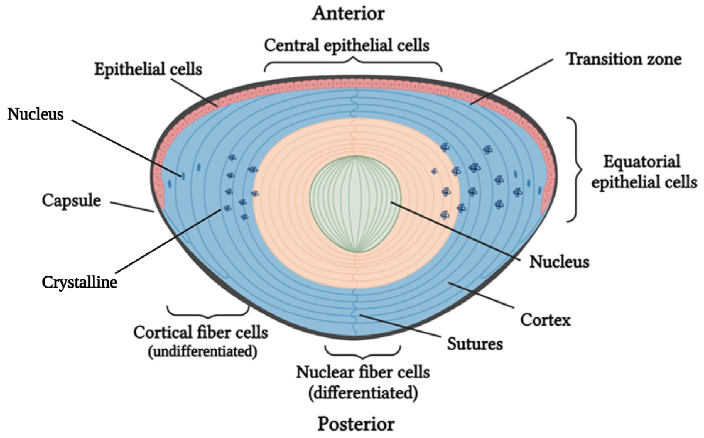
1.1. Cataract Formation: Lens Structure and Function

Cataracts are defined as opacification of the crystalline lens and are differentiated according to whether they appear during the first years of life (congenital and infantile) or progressively with age (“age-related”) [1].
The lens is a transparent structure that, through a process of accommodation, focuses and refracts light onto the retina. There, photoreceptors detect it and convert it into visual signals that are transmitted by the optic nerve to various parts of the brain. The lens is derived from the superficial ectoderm and underlying mesenchyme and belongs to the so-called anterior tissues of the eye. Together with the cornea, it forms part of the refractive system [2].
Structurally, the lens consists of the capsule, the epithelium, the fibrous cells, the transition zone at the lens equator, and the cortex (Figure 1) [2].
The capsule has a protective function against external pathogens and facilitates the differentiation of lens cells and their adhesion to the epithelium [2]. The epithelium consists of progenitor cells that divide continuously throughout life. The transition zone is composed of epithelial progenitor cells, which, after mitosis, begin their differentiation into fibrous cells. As they differentiate, these fibers produce a considerable amount of lens protein, called crystallin, and degrade their nuclei and organelles by ubiquitination or autophagy [3]. During the process of differentiation, the fibers migrate toward the center of the lens, overlapping; thus, the nucleus of the lens is made up of old differentiated fibers, whereas the more undifferentiated fibers are located in the periphery, leading to the cortex [4].
Figure 1. Diagram of the structural formation of the lens, which consists of the capsule, epithelial cells, and undifferentiated fibrous cells at the periphery, forming the cortex, and mature fibrous cells toward the interior, forming the nucleus [4]. Created by BioRender [5].
Ijms 24 11429 g001
The correct functioning of the lens depends on its transparency. This transparency is achieved by the precise organization of its cells, the compact packing of proteins, and the correct degradation of nuclei and organelles. Alterations in the stability or structure of the lens can lead to light scattering, causing decreased vision [6].

1.2. Classification of Pediatric Cataracts

Pediatric cataracts refer to opacification of the lens that occurs during the first years of life. Several classification systems have been developed based on onset, morphology, and laterality.
Based on the onset, pediatric cataracts can be differentiated into congenital cataracts (defined as any lens opacity present at birth or within the first year of life) and infantile cataracts (diagnosed in older babies or children). The diagnosis of lens opacity at a later age does not exclude a congenital onset, unless the red reflex was properly checked at birth [7].
Regarding morphology, the most widely used classification system was first proposed by Merin et al., in 1971 [8]. In addition to morphology, knowing the location of the opacity is essential for its characterization and classification [9].
According to laterality, cataracts are called bilateral if both eyes are affected or unilateral if only one eye is involved.
Bilateral congenital cataracts are generally more common than unilateral congenital cataracts, which are mostly caused by local abnormalities, such as persistent foveal vasculature [7,10].

1.3. Pediatric Cataracts: Prevalence and Etiology

Pediatric cataracts are among the most common causes of preventable severe childhood visual impairment or blindness, with an incidence of 1.8–3.6/10,000 per year. The prevalence ranges from 0.32 to 22.9/10,000 children [7]. Underdeveloped and low-income areas have a higher prevalence, probably due to socioeconomic conditions and a higher risk of infectious diseases, such as rubella or herpes simplex virus, which are strongly associated with cataracts.
Furthermore, pediatric cataracts have a varying etiology, with the majority being idiopathic (62%); the remaining 38% being hereditary (25%) or non-hereditary (13%), associated with metabolic pathologies, trauma, or secondary cataracts associated with syndromic diseases [10].
Hereditary cataracts can be divided into syndromic (if they are part of a systemic disease involving other organs) and non-syndromic (in which the eye is the only organ affected).

1.4. Mechanism of Pediatric Cataract Development

It has been shown that α-crystallin interacts only with partially denatured proteins that tend to aggregate [11]; thus, most denatured or partially denatured crystallins are expected to be bound to α-crystallin. However, if the mutation produced is severe, some crystallins might not be bound and solubilized by α-crystallin [12]. Furthermore, if the amount of denatured crystallins or the speed of denaturation exceeds the capacity of the α-crystallin to act, this could lead to the presence of heavy aggregates that scatter light and damage the systems responsible for the homeostasis of the lens cells, compromising their survival.
Regarding the structural organization of the lens, the destruction of its microarchitecture observed in cataracts is the result of cell damage and degeneration caused by mutant crystallins [13]. Evidence from several studies [12,13] suggests that the damage observed is the result of a direct toxic effect of the mutant protein on lens cells, either by escaping the action of the chaperone (α-crystallin) or by saturating it.
One way in which crystallins can directly damage lens cells is through the induction of an unfolded protein response (UPR), followed by apoptosis. The UPR is a set of adaptive intracellular signaling pathways that reduces the stress produced in the endoplasmic reticulum as a consequence of misfolded protein accumulation. Under conditions of homeostasis, heat shock protein 5 (HSPA5) binds to at least three endoplasmic reticulum sensors (IRE1, ATF6, and EIF2AK3), keeping them in an inactive state. In the presence of large amounts of unfolded protein, HSPA5 dissociates from these three sensors, activating them and initiating the UPR. When activated, this response results in increased chaperone synthesis, reduced transcription, and increased protein degradation through upregulation of endoplasmic reticulum-associated degradation protein levels. If these measures do not restore cellular integrity, the apoptotic pathway is initiated [12]. The lens histology observed in many types of hereditary cataracts could be due to the toxicity generated from mediators released by the UPR and apoptosis. Indeed, several studies in the literature support this theory [14,15,16].

1.5. Genetics of Pediatric Cataracts

Congenital cataracts were the first disease with autosomal dominant inheritance mapped in humans [17]. Although this pattern of inheritance predominates in pediatric cataracts of hereditary origin, autosomal recessive [18] and X-linked [19] patterns of inheritance have also been reported.
Non-syndromic congenital cataracts typically have complete penetrance, with autosomal dominant cataracts more common than recessive cataracts. Their expressivity is highly variable; thus, the same mutation within an affected family can give rise to various phenotypes. Furthermore, it is possible that the same type of cataract can be caused by mutations at different loci, making it a polygenic disease [6]. This complicated genotype–phenotype correlation complicates the genetic diagnosis of patients.
Currently, some 71 loci mapped to 56 cataract-causing genes have been identified [20]. This information is recorded in Cat-Map, a database that presents a chromosomal map together with the spectrum of mutations associated with hereditary cataracts, updated in real time [21]. The genetic causes of Mendelian cataracts can be grouped according to the cellular processes affected, suggesting that these pathways are critical for the correct development or homeostasis of the lens. Thus far, the most frequently implicated genes are those for crystallins (33%), developmental or transcription factors (26%), connexins (18%), membrane proteins and transporters (11%), genes involved in lipid metabolism (8%), and intermediate filament and chaperone proteins (4%) [20].

1.5.1. Crystallin Protein Genes

Crystallins comprise over 90% of the lens’ proteins and have the most fundamental place in the lens’ structure [22]. They are responsible for maintaining the transparency and structural organization of the lens. There are three types of crystallins: α, β, and γ [23]. The α-crystallins are a multimeric protein complex composed of two oligomers, αA-crystallins and αB-crystallins, which are encoded by two genes located on different chromosomes, CRYAA (21q22.3) and CRYAB (11q22-q22.3) [24]. The α-crystallins belong to the family of small heat shock proteins (sHsps). They are molecular chaperones that associate with misfolded proteins by helping facilitate their rearrangement or promote their degradation by other chaperones and cofactors [25].
Genetic mutations that cause decreased expression of functional αA-crystallins and αB-crystallins typically result in recessively inherited cataracts, such that their residual activity is sufficient to maintain lens transparency during childhood, even though this can result in increased susceptibility to environmental damage or age-related cataracts. An example is the p.Arg54Cys mutation in CRYAA, which generates total congenital cataracts in homozygosis; whereas, when it is present in heterozygosis, it results in small point opacities [26]. However, missense mutations, which result in a deleterious mutant protein, often cause autosomal dominant cataracts as a consequence of blocking the activity of the normal crystallin by a dominant negative mechanism or by causing damage to the homeostasis of the lens.
On the other hand, β-crystallins can be made up of various numbers of monomers, resulting in dimers to octamers, whereas γ-crystallins are monomers [24]. In contrast to α-crystallins, most mutations that occur in these proteins produce an unstable protein that denatures immediately or under stress conditions, precipitating in the cytosol, scattering light, and leading to cataracts [27].

1.5.2. Membrane Protein Genes

The elongation undergone by epithelial cells in their transition to fibrous cells requires the synthesis of large amounts of membrane proteins and lipids. Thus, any alteration that interferes with the synthesis of these structural components can lead to cataracts.
In relation to lipid synthesis, there are several studies that associate inherited cataracts with mutations in enzymes involved in the synthesis of various types of lipids, such as acylglycerol kinase [28] or lanosterol synthase [29].
In reference to membrane proteins, ion or solute transporters and proteins that maintain gap junctions are directly related to the appearance of bilateral congenital cataracts. Mutations in the SLC16A12 gene, which affects an active transmembrane transporter of monocarboxylic acids, can cause juvenile cataracts with dominant inheritance [30]. Mutations in DNMBP, a guanine nucleotide exchange factor that regulates cell junction configurations, have also been described [31].
Connexins are a family of proteins consisting of hemichannels that mediate communication between the intra- and extracellular milieu and gap junctions that allow cell–cell communication between adjacent cells. The lack of lens vascularization means that cell nutrition and metabolite transport is performed through an extensive network of connexin channels. Mutations affecting some of these proteins can lead to cataractogenesis [32]. An estimated 22% of familial cataracts are due to mutations in the GJA3 and GJA8 genes coding for connexin 46 and connexin 50, respectively [33].

1.5.3. Cytoskeletal Protein Genes

Beaded fiber-specific proteins belong to the family of intermediate filament proteins and form specific structures of the lens. The BFSP1 gene and BFSP2 gene encode filaments whose functions are not yet clearly understood; however, they play an important role in maintaining lens transparency [34]. This role is illustrated by animal studies showing the presence of cataracts in BFSP1 and BFSP2 knock-out mice [35]. In addition, many cases of pediatric cataracts of dominant or recessive inheritance caused by mutations in BFSP1 have been reported in the scientific literature [36,37]. In fact, pediatric cataracts are the phenotype associated with BFSP1 and BFSP2 in the Online Mendelian Inheritance in Man (OMIM) database [38,39].

1.5.4. Transcription Factors and Lens Growth Factors

One of the transcription factors most recently associated with congenital cataracts is paired box gene 6 (PAX6). This transcription factor is involved in the development of lenses and central nervous system tissues. Mutations affecting the C-terminal domain have been associated with cataracts and corneal dystrophy [40].
EPHA2 also plays an essential role in the development of lens and central system tissues. It is part of the ephrin receptor tyrosine kinase family of receptors and its mutations can lead to dominant or recessive early onset cataracts or even increase the risk of age-related cataracts [6,41,42].

1.5.5. Degradation Proteins or Chaperones

As previously mentioned, lens formation requires extensive restructuring of its components and differentiation of epithelial cells into fibrous cells. Fundamental to this process is the removal of cellular organelles and high levels of protein degradation. Mutations that disrupt this degradation process can lead to cataracts. An example is mutations in FYCO1, a scaffolding protein active in the microtubule transport of autophagic vesicles, which cause autosomal recessive cataracts [43], suggesting that autophagic vesicles are important for organelle degradation in developing lens fiber cells, among other functions. Another example involves CHMP4B, which encodes a member of the charged multivesicular body/chromatin-modifying protein (CHMP) family of proteins. It is a component of the major subunits of the endosomal sorting complex required for transport III (ESCRT-III) machinery, which facilitates innumerable biological processes of membrane remodeling and scission [44]. One of these processes is autophagy, which is a mechanism of cellular self-degradation essential for the removal of aggregated proteins and damaged organelles. Thus, impaired autophagolysosomal degradation could explain cataract formation in the absence of proper CHMP4B function [45]. It has been reported that mutations in CHMP4B can cause polar and subcapsular cataracts [46].

1.5.6. Metabolism-Associated Genes

The robust metabolism of lens cells maintains cellular homeostasis, thus providing a reducing environment to cope with the oxidative damage to which lens cells are subjected, preventing osmotic stresses and inhibiting glycosylation products.
Several genes involved in metabolism have been reported to cause cataracts over time. GALK1 encodes galactokinase, which is a major enzyme for the metabolism of galactose. Its deficiency causes pediatric cataracts during infancy and presenile cataracts in the adult population [47]. ALDH1A1 encodes the next enzyme after alcohol dehydrogenase in the major pathway of alcohol metabolism. Studies on mice have shown that ALDH1A1 protects the eye against cataract formation via an enzymatic pathway by detoxifying particulate matter generated by oxidative stress [48].

1.6. Symptoms and Treatment of Pediatric Cataracts

As previously discussed, late diagnosis of congenital cataracts can result in their mislabeling as infantile cataracts. Early diagnosis of congenital cataracts and prompt surgical intervention are essential to preventing irreversible amblyopia. If retinal blurring occurs during the critical period of visual development, vision could be lost. Therefore, therapeutic measures during the period of sensory plasticity are of vital importance to prevent blindness.
The most common clinical sign of congenital cataract is leukocoria, which is the white coloration of the pupil when examined under a beam of light. Other common signs are nystagmus (defined as involuntary, rapid, and repetitive movement of the eyes) and strabismus (the loss of parallel alignment of the eyes) [49]. In addition to these manifestations, non-syndromic congenital cataracts can occur in association with other ocular defects that compromise eye function, such as aniridia (absence of iris), microcornea (horizontal diameter less than 11 mm), or microphthalmia (small eye size) [50,51].
Currently, the only treatment for congenital cataracts is surgery. It has been shown that increasing age at the time of surgery correlates with poorer visual outcomes. The optimal time period for cataract removal is 8–10 weeks. Lambert and colleagues showed in their study that 88% of children operated on before 10 weeks of age achieved a score of 20/80 or better, compared with those operated on after 10 weeks, with a score of 20/100 or poorer [52]. Depending on the age of the patients and the anatomical characteristics of the eye, an intraocular lens (IOL) may be implanted after lensectomy, a process called pseudophakia. If only a lensectomy is performed, the eye is left aphakic and refractive correction with contact lenses or thick glasses is necessary [53].
Some studies have shown that visual outcomes in patients with bilateral congenital cataracts appear to be better after primary IOL implantation compared with aphakia corrected with contact lenses [54], although the rate of re-operations in the IOL-implanted children was significantly higher than in the left aphakic group [55,56,57].
The identification of the precise genetic mutation facilitates accurate genetic counseling, providing families with information and ongoing support for their condition, which is crucial for these patients. It also provides a more accurate prognosis, which is particularly important in syndromic cases [58].
All this demonstrates the importance of early detection of pediatric cataracts; it is therefore essential to expand our genetic knowledge of the disease. Therefore, the aim of this study is to propose new candidate genes responsible for non-syndromic pediatric cataracts, which will serve as a reference for other researchers and will allow us to offer better health care to patients with cataracts at the National Health System’s La Paz University Hospital, given that it is a reference center for the diagnosis and treatment of this pathology (Centers, Services and Reference Units, CSUR).

2. Results

2.1. Ophthalmological Examination

Of the 23 patients included in our study, 45 eyes were analyzed (1 patient had a unilateral cataract; this patient was included in our study, as was the sibling of another patient with bilateral cataracts). The results are summarized in Table 1.
Lensectomy was performed in 28 eyes. Eighteen eyes remained unoperated by the end of the study period because the cataracts were visually insignificant or visual acuity remained above driving-standard levels. Surgery was performed before the age of 1 year in 16 (59%) eyes. Postoperative glaucoma was found in six eyes. Eight eyes had nystagmus at the end of the study period and ten eyes had microphthalmia (Figure 2).
The most common type of cataracts in our cohort were nuclear and lamellar, present in 12 eyes in both types (Figure 3).

2.2. Molecular Genetics

After performing a whole exome sequencing (WES) analysis, we were able to identify the causal gene for pediatric cataracts in 10% (2/20) of the cohort and thus concluded the genetic diagnosis. In 35% (7/20) of the cohort, genes were found which, due to their relationship with the crystalline lens or with the cataract formation process, we considered to possibly be candidate genes responsible for the phenotype, although it would be necessary to perform further studies to confirm this. As for the remaining 55% (11/20) of the families, the result of the WES was inconclusive (Figure 4). Of the 10% of families with a genetic diagnosis and 35% with candidate genes, we found 11 variants. Of these, 18.2% (2/11) were classified as pathogenic (P), 9% (1/11) as likely pathogenic (LP), and 72.8% (8/11) as a variant of uncertain significance (VUS) (Table 2, Figure 5). As for pathogenic variants, a variant in AQP5 (NM_001651.3: c.152T>C:p.(Leu51Pro)) and a deletion at the 2q37.3 locus have been previously described, demonstrating that this specific variant causes congenital cataracts.

3. Discussion

As previously mentioned, pediatric cataracts have a highly variable expression. Together with the polygenicity and heterogeneity described, this variability makes it difficult to describe new genes associated with this phenotype. In addition, given that the exome occupies less than 2% of the entire human genome, it is possible that the WES study would not be able to identify the cause of the pathology. All this could explain why 55% of the families analyzed by WES attained inconclusive results, necessitating further studies on the variants found. Moreover, at present, the cost-effectiveness ratio of WES versus whole genome sequencing shows that it would be interesting to implement the latter in all the patients who remain without a genetic diagnosis to date [64] and, whenever possible, to start using this technique as the first diagnostic method.
However, the study of the whole exome remains an alternative solution in some cases. In 10% of the families analyzed, a genetic diagnosis was achieved. A de novo variant was found in the OFT-00040 family proband in the AQP5 gene, NM_001651.3:c.152T>C:p.(Leu51Pro), which had been previously reported in a study from Qingdao of posterior subcapsular congenital cataracts [63], demonstrating the causality of the variant. In the other diagnosed family, OFT-00350, a de novo deletion was found in the 661.2 Kb proband in the region 2q37.3. This region had been associated with the occurrence of bilateral posterior polar cataracts in two Chinese families [60]. This phenotype also coincided with the phenotype of our patient, which showed that the deleted region was clearly associated with pediatric cataracts.
In 35% of the cohort, genes were found that could be candidates for causing pediatric cataracts due to their relationship with the pathologic processes of the disease. In some cases, the evidence was stronger than in others, as in the CHMP4A and PIKFYVE genes. However, we considered it important to report the findings to serve as a precedent for other researchers.
This result highlights the current need to search for new candidate genes that will help us delimit the genetic etiology of pediatric cataracts, thus contributing to improving patient care. The results obtained are specifically discussed below.
The proband in the family OFT-00247 was heterozygous for the mutation in the LONP1 gene, (OMIM 605490), NM_004793.3:c.1939G>A:p.(Glu647Lys), inherited from his father, who did not have lens opacity.
LONP1 encodes a mitochondrial matrix protein that belongs to the Lon family of ATP-dependent proteases. This protein mediates the selective degradation of misfolded, unassembled, or oxidatively damaged polypeptides in the mitochondrial matrix. It could also have a chaperone function [65]. Diseases associated with LONP1 include cerebral, ocular, dental, auricular, and skeletal anomalies syndrome, which has pediatric cataracts as an associated phenotype [66]. In Patel, Nisha et al.’s study [67], the LONP1 gene was the second most commonly mutated gene in the pediatric cataract cohort after the crystallin genes, affecting five families. Our variant had previously been reported in homozygosity in two siblings with congenital cataracts and mild dysmorphia [59]. Although our patient was heterozygous for the variant and did not present mild dysmorphia, mutations in LONP1 associated with non-syndromic pediatric cataracts present in heterozygosis have been described in the literature [68]. Moreover, variable expressivity of the phenotype in individuals from the same family described in the literature could explain the paternal inheritance of the variant without lens opacities in the father [68]. It was also possible that, taking into account what has been described about the NM_004793.3:c.1939G>A:p.(Glu647Lys) variant, there was a second event in the proband that we were not able to find through the whole exome study.
The missense variant found [59] causes the substitution of a negatively charged amino acid and glutamic acid (E) for a positively charged amino acid and a lysine (K), at position 647. Given that the positions that remain conserved in a protein throughout evolution are relevant to its function and that the Pdel value (probability of a deleterious effect of the variant as calculated by the Panther Classification System) is 0.85, the variant could have a possibly harmful effect on the protein function [69]. This hypothesis is reinforced by studying possible changes in protein structure by employing tools such as Polymorphism Phenotyping v2 (PolyPhen-2) [70].
The importance of protein degradation in the eye as explained above suggests the pathogenic nature of the variant; however, further functional studies would be necessary to conclusively determine its pathogenic nature. Therefore, this variant is classified as of uncertain significance by the ACMG criteria [71].
In the OFT-00289 family, both siblings were heterozygous for the variant in the ACACA gene (OMIM 200350), NM_198839.2:c.1126C>T:p.(Arg376Cys), inherited from their healthy mother through germline mosaicism. Acetyl-CoA carboxylase alpha (ACACA) encodes one form of the complex multifunctional enzyme system acetyl-CoA carboxylase (ACC). The isoform encoded by ACACA maintains the regulation of fatty acid synthesis, producing malonyl-CoA, which is the first intermediary in cytosolic fatty acid synthesis and is a key metabolite for long-chain saturated fatty acid production.
Although the ACACA gene has been ruled out as a causal gene for pediatric cataracts in a previous study [72], many studies have shown the metabolic relevance of this enzyme. Activity-impeded ACACA reduces cytoplasmic membrane fluidity and impairs mobilities of transmembrane receptors, ultimately impairing cell membrane-dependent biological processes [73]. Moreover, inhibition of ACC can increase the intracellular acetyl-CoA level and stimulate the influx of calcium into the cells [74], which in the lens have been demonstrated to possibly be responsible for carboxypeptidase E activation and formation of a truncated form of αB-crystallin in cataracts [75].
The missense variant found had extremely low frequency in gnomAD population databases, affecting a gene with a gnomAD Z-score greater than 3.09 (7.24) and for which the predictive tools suggested a deleterious effect of the gene. In addition, this variant was the only one that both affected siblings did not share with their parents. However, due to insufficient evidence to date, it was classified as a VUS.
In reference to family OFT-00334, the female proband presented bilateral cataracts and was heterozygous for the mutation in the TRPM1 (OMIM 603576) gene, NM_001252024.2:c.4720dup:p.(Ser1574LysfsTer7), inherited from her mother, who did not have cataracts. It was possible that there was a second mutation that we were not able to find through exome sequencing.
Although TRPM1 encodes a calcium-permeable cation channel, which has been associated with autosomal recessive congenital stationary night blindness (CSNB), a case has recently been described in a 4-week-old boy of non-consanguineous parents affected by unilateral nuclear cataracts [76]. Most cases of unilateral pediatric cataracts are presumed to be sporadic; however, some families with unilateral cataracts have been reported, suggesting in some cases a germline mutation as an underlying cause [77].
Saleh E. et al. reported two novel presumed pathogenic variants that were identified in blood and lens epithelial cells in TRPM1. Following the genetic diagnosis, full-field electroretinography showed ON-bipolar signaling defects in both rods and cones compatible with CSNB [76], a phenotype that was not initially suspected. Moreover, homozygous animal models for TRPM1 have also been described, showing incipient posterior cortical cataracts and immature cataracts [78]. In addition, an important paralog of TRPM1 is TRPM3, with approximately 57% amino acid sequence identity. Heterozygous mutations in this gene have been linked to early onset cataracts with or without other eye abnormalities [79].
Regarding the genetic study of our proband, the frameshift variant found generated a premature stop codon that produced a truncated protein in a gene where loss of function is a known mechanism of disease. This characteristic, coupled with the extremely low frequency in gnomAD population databases, classified the variant as likely pathogenic by the ACMG criteria. Although our patient had bilateral cataracts and not unilateral cataracts, we considered it of interest to report as a candidate gene for future functional studies related to pediatric cataracts.
The male proband of the OFT-00350 family, who presented bilateral posterior polar cataracts, was heterozygous for a de novo deletion of 661.2 Kb, located in locus 2q37.3, which included 17 genes classified as pathogenic by the ACMG guidelines for copy number variants (CNVs) [80]. The deletion was found by a CNV study obtained from exome sequencing, confirmed by a single nucleotide polymorphism (SNP) array in the proband and excluded in the parents.
Currently, 71 cataract-associated loci have been mapped [20], among which the 2q37-qter locus was described in 2012 [60]. This article describes two Chinese families in which all affected members had a clear diagnosis of non-syndromic bilateral posterior polar congenital cataracts that were present after birth and developed during childhood. Genetic scans based on dinucleotide repeat microsatellite markers allowed the authors to describe a chromosomal region near the terminal end of chromosome 2q implicated in cataract development. This region comprised more than 40 known genes, of which they classified CXCR7, AQP12B, and SEPT2 as candidate genes based on their position and functionality.
The deleted region in our patient shared only one of the genes proposed as a candidate by Ouyang, the gene for aquaporin 12B (AQP12B). However, it also included the AQP12A gene, which was not specified in Ouyang’s article. Both genes encode transmembrane transporters of water and small neutral solutes.
Given that the lens is an avascular non-innervated organ, a hydrostatic pressure gradient generated by the activity of pumps, channels, and other transport proteins directs the supply of nutrients and the removal of metabolic waste from the lens. This mechanism substitutes for blood flow and maintains the ocular properties of the lens involved in transparency and light refraction [81]. Various studies have shown the development of early onset bilateral cataracts in AQP0 and AQP1 knockout mice [82,83]. Because of its importance as described in the lens, it is possible that the AQP12B or AQP12A present in the deletion loci in our patient were playing an essential role in the phenotype.
Although its pathogenicity should be verified with additional studies, the 661.2 kb deletion has been classified as pathogenic by the ACMG guidelines [80], suggesting its involvement in the development of pediatric cataracts.
In family OFT-00338, both siblings were heterozygous for the variant NM_016929.4:c.514C>T:p.(Arg172Trp) in the CLIC5 gene (OMIM 607293), which was inherited from their affected mother, who also presented the variant in heterozygosity.
This gene encodes a member of the chloride intracellular channel (CLIC) family of chloride ion channels. Although mutations in the human CLIC5 gene have been linked with progressive autosomal recessive non-syndromic sensorineural hearing impairment with or without vestibular dysfunction, CLIC5 has recently been localized to cilia and/or centrosomes in the lens, describing an unknown essential role in normal fiber cell organization and formation of the lens suture in the eye [84]. Given that the sutures form along the optical axis of the lens, their precise organization is important for light transmission.
As for the missense variant found, it involves the replacement of arginine, which is basic and polar, by tryptophan, which is neutral and slightly polar, at codon 172 of the CLIC5 protein. Its population frequency in gnomAD is extremely low; however, algorithms developed to predict the effect of missense changes on protein structure and function are either unavailable or do not agree on the potential impact of this missense change (SIFT: “Deleterious”; PolyPhen-2: “Possibly Damaging”; Align-GVGD: “Class C0”) [70,85,86]. It has been classified as a VUS. Therefore, despite the insufficient and contradictory information available on this variant, recent evidence on the CLIC5 gene in the lens supports proposing it as a possible causal gene for the OFT-00338 family phenotype.
In reference to family OFT-00346, the female proband was heterozygous for the frameshift variant NM_002157.2:c.61_62insACCA:p.(Ser21AsnfsTer5) in the HSPE1 gene (OMIM 600141) and for the deletion NM_024410.4:c.678_686del:p.(Cys227_Pro229del) in ODF1 (OMIM 182878). Both undescribed variants appeared de novo in the patient, whose parents were healthy.
The HSPE1 gene encodes an important heat shock protein, which functions as a chaperonin, preventing protein misfolding and promoting the refolding and proper assembly of unfolded polypeptides generated under stress conditions in the mitochondrial matrix. Together with Hsp60, it facilitates the correct folding of imported proteins [87].
The ODF1 gene is considered by sequence conservation to be a member of the sHsps family of proteins, of which crystallins and the HSPE1 gene mentioned above also form a part [88]. For this reason, the ODF1 gene is alternatively called heat shock protein beta-10 (HSPB10).
The sHsps belong to molecular chaperones, which protect prokaryotic and eukaryotic cells against the deleterious effects of stress, preventing stress-induced irreversible aggregation of damaged proteins and facilitating renaturation of bound substrates cooperating with other molecular chaperones [89]. Several articles demonstrate a protective role of sHsps against eye diseases such as cataracts and glaucoma [90]. Therefore, the sHsps family is implicated in cataract formation, because aggregation of misfolded proteins is one of the most common pathogenic mechanisms in cataracts.
The frameshift variant found in the HSPE1 gene has not been previously described and its population frequency in gnomAD is extremely low (0.0%). Moreover, a premature stop codon is generated that can cause loss of normal protein function by truncation or nonsense-mediated mRNA decay, resulting in an absent or altered protein product. However, given that there is thus far insufficient information about the variant, it has been classified as a VUS.
The deletion found in the ODF1 gene has an extremely low frequency in the gnomAD population databases. Also, the resulting protein is shorter in length due to the in-frame deletion in a non-repeat region, which affects the functionality of the protein. Based on ACMG criteria, the variant has been classified as a VUS.
Lastly, although functional models are needed to prove the pathogenicity of the variants, the important role played by the HSPE1 and ODF1 genes in the lens and the de novo presence of the variants in the proband led us to propose both genes as causal for pediatric cataracts.
The OFT gene panel study previously performed by our group in family OFT-00215 [91] revealed that the male proband was heterozygous for the variant NM_014290.2:c.1085C>T:p.(Pro362Leu) in the TDRD7 gene. Given that it had autosomal-recessive inheritance and the second mutation was not found, the study was extended to WES, which showed that he was also heterozygous for NM_015040.3:c.5844+3A>G in the PIKFYVE gene (OMIM 604632), inherited from his unaffected mother.
This gene encodes a lipid kinase that phosphorylates phosphatidylinositol in phosphoinositide lipid formation, which is a signaling molecule that regulates critical cell processes. PIKFYVE is the only source of phosphatidylinositol 3,5-bisphosphate (PI(3,5)P2) whose levels dynamically and transiently change in response to specific stimuli. In mammalian cells, physiological signals such as insulin and growth factors cause an acute elevation of its levels, suggesting that this lipid plays key roles in cellular homeostasis and in adaptation [92]. In addition, many studies have indicated that PIKFYVE plays important roles in lysosomes, causing the appearance of enlarged vacuoles [93], as well as in ionic homeostasis and membrane dynamics [94] and in transcription regulation [92].
Although PIKFYVE is associated with corneal dystrophy, disruption of PIKFYVE has recently been reported to cause pediatric cataracts in humans and zebrafish [95]. In this article, a possible pathogenic variant, NM_015040.4:c.5828G>A:p.(Gly1943Glu), was found in PIKFYVE by WES in a Chinese family with congenital cataracts and without corneal defects. The mutation was located in the phosphatidylinositol phosphate kinase (PIPK) domain of the PIKFYVE protein. Functional studies of the variant in animal models have confirmed that the PIKFYVE gene might also be involved in the development of early onset cataracts in zebrafish [95].
Regarding the genetic study of our proband, the splicing variant found (NM_015040.3:c.5844+3A>G) was located in an intronic region of the PIPK domain, which could affect splicing by affecting the essential kinase activity of the enzyme, as shown in recent functional studies [95]. This variant has not been previously described and it has an extremely low frequency in the gnomAD population database. However, computational prediction tools unanimously support a benign effect on the gene for a splice region variant. Therefore, due to contradictory information regarding this variant, it has been classified as a VUS by the ACMG guidelines [71].
In family OFT-00235, the male proband had a biallelic mutation in CHMP4A (OMIM 610051). He presented the nonsense variant, NM_014169.5:c.406C>T:p.(Gln136Ter), inherited from his unaffected father, and the frameshift variant, NM_014169.5:c.65del:p.(Ala22GlufsTer5), inherited from his healthy mother.
CHMP4A belongs to the chromatin-modifying protein/charged multivesicular body protein (CHMP) family. This protein comprises part of ESCRT-III, a complex involved in the formation of multivesicular bodies, which are delivered to lysosomes, allowing the degradation of membrane proteins such as stimulated growth factor receptors, lysosomal enzymes, and lipids [96]. In addition, the GeneCards database [97] associates CHMP4A with cataract 32 [98], which includes early onset lamellar cataracts, the phenotype presented by our proband. An important paralog of this gene is CHMP4B, which has been described as necessary for lens growth and differentiation in animal models [99] and as responsible for autosomal dominant infantile cataracts [100].
The biallelic mutation has been classified as a VUS because current information on these variants is thus far contradictory. However, knowing the importance of protein and metabolic degradation in the lens, it would be expected that an alteration in the CHMP4A pathway would lead to lens opacities, thus classifying this gene as a possible causative gene for pediatric cataracts. Even so, functional studies of the variants would be necessary to determine their pathogenic effect.
For family OFT-00040, the female proband was heterozygous for a de novo missense variant in the aquaporin 5 (AQP5) (OMIM 600442) gene, NM_001651.3:c.152T>C:p.(Leu51Pro). AQP5 encodes for a small transmembrane water channel protein, which plays a redundant role in water transport in the eye, lung, and sweat glands [101].
As discussed for other members of the aquaporin family (AQP12B, AQP0, AQP1) in the OFT-00350 family, water flow is essential for the maintenance of transparency and light refraction in the lens. In addition to these aquaporins, AQP5 has been reported to be expressed in epithelial cells and in lens fiber cells [102,103] and its expression changes as a function of fiber cell differentiation from an intracellular localization in differentiating fiber cells to the plasma membrane of mature fiber cells due to the loss of fiber cell nuclei. This characterization would indicate that AQP5 also plays a key role in the maintenance of lens properties. Recently, mutations in AQP5 have been directly related to the development of autosomal dominant congenital cataracts [63].
The missense variant found in our proband has been previously described in a family from Qingdao with posterior subcapsular congenital cataracts [63]. Tang, Suzhen et al. observed that the lens of AQP5-KO mice showed mild opacity at approximately 6 months of age, thus demonstrating that the mutation NM_001651.3:c.152T>C:p.(Leu51Pro) in AQP5 was associated with autosomal dominant congenital cataracts. Therefore, the variant has been classified as pathogenic, taking into account the following ACMG criteria: its extremely low frequency in the gnomAD database; computational prediction tools unanimously supporting a deleterious effect for a missense variant; de novo appearance in a patient with a consistent phenotype; existence of well-established functional studies that show the damaging effect on the gene or gene product. For these reasons, we can conclude that the mutation NM_001651.3:c.152T>C:p.(Leu51Pro) in the AQP5 gene was the causal gene for pediatric cataracts in the proband.

4. Materials and Methods

A combined ophthalmological and genetic study was performed by the Multidisciplinary Unit of Ophthalmogenetics at La Paz University Hospital, in accordance with the principles of the Declaration of Helsinki, and it was approved by the hospital’s ethics committee.
The inclusion criteria were the following: (1) Spanish pediatric patients with cataracts; (2) absence of syndromic phenotype; (3) negative result in sequencing study implementing a panel (OFTv2.1) that included 39 known non-syndromic pediatric cataract disease genes. Whenever possible, the patients’ relatives (parents, aunts, uncles, or siblings) were included in the study. In this way, 23 affected children from 20 unrelated families were recruited. After signing the informed consent, WES was performed.

4.1. Ophthalmological Studies

The demographic data collected during this study were eye laterality, cataract type, microphthalmia, nystagmus, age at surgery, and development of postoperative glaucoma.

4.2. Genetic Studies

Second, a genetic analysis was performed using genomic DNA obtained from leukocytes isolated from a peripheral blood sample. This was performed in the pre-analytical area of our institute, employing the Chemagic Magnetic Separation Module I (Chemagen, PerkinElmer, Waltham, MA, USA). DNA concentrations were measured by quantification with a NanoDrop ND-1000 spectrophotometer (Thermo Fisher Scientific, Waltham, MA, USA). Library preparation was performed with the Nextera DNA Exome Illumina DNA Prep with Enrichment (Illumina, San Diego, CA, USA) and IDT for Illumina DNA/RNA UD Indexes Set, A, B, C, or D, Tagmentation. Sequencing was performed on the high-quality sequencers, HiSeq4000 and NovaSeq6000, capturing 19,433 genes with the xGen™ Exome Research Panel v2 IDT.
The first analysis was then performed by the Bioinformatics of Medical and Molecular Genetics (INGEMM) team, which developed an analytical algorithm that identified SNPs and CNVs (insertions and deletions of small DNA fragments (indels)) within the capture regions of the exome.
The system comprised a sample pre-processing step, alignment of reads to a reference genome, identification and functional annotation of variants, and variant filtering. All these steps employed open tools widely used in the scientific community, as well as proprietary tools. In addition, all steps were robustly designed and included parameters that allowed the process to be monitored and the appropriate quality controls to deliver a reliable report on the variants in question.
The bioinformatics analysis was performed by the Clinical Bioinformatics Unit of the INGEMM center, employing the following software tools: trimmomatic-0.36, bowtie2-align version 2.0.6, picard-tools 1.141, samtools version 1.3.1, bedtools v2.26, and GenomeAnalysisTK version 3.3-0. The databases used were dbNSFP version 3.5, dbSNP v151, ClinVar date 20180930, ExAC-1, SIFT ensembl 66, Polyphen-2 v2.2.2, MutationAssessor release 3, FATHMM v2.3, CADD v1.4, and dbscSNV1.1.
CNV analysis was conducted utilizing in-house software called LACONv, specifically designed to calculate the copy number dosage of individual exons in targeted gene panels with the appropriate adjustment for GC sequence content. The copy number dosage of each captured region in the test sample was compared against all other samples in the same panel run to control for the batch effect. The P-values were computed using Mann–Whitney U tests; CNVs were deemed statistically significant if the value exceeded 0.05.
The second data analysis consisted of assessing the clinical pathogenic significance of the variants found in the patients by accessing multiple databases. These included PubMed [104], GeneCards [97], Decipher [105], gnomAD [106], UCSC Genome Browser [107], and specific databases such as Cat-Map, which is an online reference chromosomal map for inherited and age-related cataracts in humans, mice, and other vertebrates [108]. A literature search was performed for the gene and the variant found. The variants were then classified according to American College of Medical Genetics and Genomics (ACMG) prioritization [71,80] criteria as pathogenic, probably pathogenic, a VUS, probably benign, or benign, using the Varsome and Franklin database [109,110].
In some cases, candidate variants were included in GeneMatcher [111], an open-access website that allows connections to be established between researchers around the world who share an interest in the same gene. Depending on the type of variant, validation was performed by Sanger sequencing or by other techniques, such as SNP arrays.

5. Conclusions

In conclusion, WES was performed on 20 families with pediatric cataracts, reaching a diagnosis rate of 10%, in which two known mutations were found in the AQP5 gene (OMIM 600442) and in the 2q37 locus.
Furthermore, WES allowed us to propose several candidate genes that could cause pediatric cataracts due to their relationship with the lens and its transparency in 35% of the families. These genes are LONP1 (OMIM 605490), ACACA (OMIM 200350), TRPM1 (OMIM 603576), CLIC5 (OMIM 607293), HSPE1 (OMIM 600141), ODF1 (OMIM 182878), PIKFYVE (OMIM 604632), and CHMP4A (OMIM 610051).
In these cases, it will be necessary to continue with studies at the genomic, functional, familiar, and population levels to determine whether these genes are causative of pediatric cataracts and whether the combination of mutations in several genes could be causing them, following an oligogenic inheritance.
It is important to share the data obtained in public databases as well as to establish collaborations (e.g., ERN-EYE) to add knowledge and to be able to make new hypotheses about the genetic causes of pediatric cataracts, especially in undiagnosed families. In our study, we were not able to find variants that we suspected might be causing the pediatric cataract phenotype in 55% of the families.
Furthermore, we would consider extending the study in undiagnosed families, using new techniques such as long-read sequencing, whole genome sequencing, transcriptome analysis, or DNA methylation, which would allow us to reach a genetic diagnosis that would facilitate accurate genetic counseling.

Author Contributions

Conceptualization, P.R.-S., N.A., S.N. and E.V.; Methodology, M.N.-M., R.M., C.R.-J., M.G.-C., J.C.A., J.B., J.M.P., V.E.F.M. and C.F.-A.; Formal analysis, P.R.-S., N.A., Á.D.P. and L.D.D.-B.; Investigation, P.R.-S., N.A., C.G.-A. and E.S.-C.; Resources, C.F.-A., M.d.L.Á.G.-C. and L.D.-M.; Writing—original draft, P.R.-S. and N.A.; Writing—review & editing, S.N. and E.V.; Visualization, S.N. and E.V.; Supervision, S.N. and E.V. All authors have read and agreed to the published version of the manuscript.

Funding

This study has been funded by Instituto de Salud Carlos III (ISCIII) through the project “18/1234” and co-funded by ERDF, “A way to make Europe” and by ONCE grant number 2020/0197782.

Institutional Review Board Statement

The study was conducted according to the guidelines of the Declaration of Helsinki and approved by the Ethics Committee of La Paz University Hospital of Madrid (protocol code PI-4016 approved on the 3 February 2020).

Informed Consent Statement

Informed consent was obtained from all participants involved in the study.

Data Availability Statement

Not applicable.

Acknowledgments

We are grateful to the patients and their families.

Conflicts of Interest

The authors declare no conflict of interest.

Appendix A

Table A1. American College of Medical Genetics and Genomics criteria.
CodeDescription
PVS1Null variant (nonsense, frameshift, canonical ± 1 or 2 splice sites, initiation codon, single or multiexon deletion) in a gene where loss of function is a known mechanism of disease.
PS1Same amino acid change as a previously established pathogenic variant regardless of nucleotide change.
PS2De novo (both maternity and paternity confirmed) in a patient with the disease and no family history.
PS3Well-established in vitro or in vivo functional studies supporting a damaging effect on the gene or gene product.
BS1Allele frequency is greater than expected for the disorder.
PM1Located in a mutational hotspot and/or critical and well-established functional domain (e.g., active site of an enzyme) with no benign variation.
PM2Absent from controls (or at extremely low frequency if recessive) in Exome Sequencing Project, 1000 Genomes Project, and Exome Aggregation Consortium.
PM3For recessive disorders, detected in trans with a pathogenic variant.
PM4Protein length changes as a result of in-frame deletions/insertions in a non-repeat region or stop-loss variants.
PM5Novel missense change at an amino acid residue where a different missense change determined to be pathogenic has previously been observed.
PP1Co-segregation with disease in multiple affected family members in a gene definitively known to cause the disease.
PP2Missense variant in a gene that has a low rate of benign missense variation and in which missense variants are a common mechanism of disease.
PP3Multiple lines of computational evidence support a deleterious effect on the gene or gene product (e.g., conservation, evolutionary, splicing impact).
PP5Reputable source recently reports variant as pathogenic, but the evidence is not available to the laboratory to perform an independent evaluation.
BP4Multiple lines of computational evidence suggest no impact on gene or gene product (conservation, evolutionary, splicing impact, etc.).
BP6Reputable source recently reports variant as benign, but the evidence is not available for the laboratory to perform an independent evaluation.
Table A2. American College of Medical Genetics and Genomics criteria for copy number variant (CNV).
Evidence TypeEvidence
Section 1: Initial Assessment of Genomic Content
Copy number loss content1A. Contains protein coding or other known functionally important elements
1B. Does NOT contain protein coding or any known functionally important elements
Section 2: Overlap with Established/Predicted Haploinsufficient (HI) or Established Benign Genes/Genomic Regions
(Skip to Section 3 if your copy number loss DOES NOT overlap these types of genes/regions)
Overlap with ESTABLISHED HI genes or genomic regions and consideration of reason for referral2A. Complete overlap of an established HI gene/genomic region
2B. Partial overlap of an established HI genomic region
-
The observed CNV does NOT contain the known causative gene or critical region for this established HI genomic region OR;
-
Unclear if known causative gene or critical region is affected OR;
-
No specific causative gene or critical region has been established for this HI genomic region.
2C. Partial overlap with the 5′ end of an established HI gene (3′ end of the gene not involved);
2C-1. And coding sequence is involved;
2C-2. And only the 5′ UTR is involved.
2D. Partial overlap with the 3′ end of an established HI gene (5′ end of the gene not involved);
2D-1 And only the 3′ untranslated region is involved;
2D-2. And only the last exon is involved. Other established pathogenic variants have been reported in this exon;
2D-3. And only the last exon is involved. No other established pathogenic variants have been reported in this exon;
2D-4. And it includes other exons in addition to the last exon. Nonsense-mediated decay is expected to occur.
2E. Both breakpoints are within the same gene (intragenic CNV; gene-level sequence variant).
Overlap with ESTABLISHED benign genes or genomic regions2F. Completely contained within an established benign CNV region.
2G. Overlaps an established benign CNV but includes additional genomic material.
Haploinsufficiency Predictors2H. Two or more HI predictors suggest that AT LEAST ONE gene in the interval is HI.
Section 3: Evaluation of Gene Number
Number of protein-coding RefSeq genes wholly or partially included in the copy number loss3A. 0–24 genes
3B. 25–34 genes
3C. 35+ genes
Section 4: Detailed Evaluation of Genomic Content Using Cases from the Published Literature, Public Databases, and/or Internal Lab Data
(Skip to Section 5 if either your CNV overlapped with an established HI gene/region in Section 2 OR there have been no reports associating either the CNV or any genes within the CNV with human phenotypes caused by loss of function (LOF) or copy number loss)
Individual case evidence, de novo occurrencesReported proband (from the literature, public databases, or internal lab data) has either:
-
A complete deletion of or a LOF variant within a gene encompassed by the observed copy number loss OR;
-
An overlapping copy number loss similar in genomic content to the observed copy number loss AND;
4A. The reported phenotype is highly specific and relatively unique to the gene or genomic region;
4B. The reported phenotype is consistent with the gene/genomic region, is highly specific, but not necessarily unique to the gene/genomic region;
4C. The reported phenotype is consistent with the gene/genomic region but is not highly specific and/or has high genetic heterogeneity.
Individual case evidence, inconsistent phenotype4D. The reported phenotype is NOT consistent with what is expected for the gene/genomic region or not consistent in general.
Individual case evidence, unknown inheritance4E. Reported proband has a highly specific phenotype consistent with the gene/genomic region, but the inheritance of the variant is unknown.
Individual case evidence, segregation among similarly affected family members4F. 3–4 observed segregations;
4G. 5–6 observed segregations;
4H. 7 or more observed segregations.
Individual case evidence, non-segregations4I. Variant is NOT found in another individual in the proband’s family
AFFECTED with a consistent specific well-defined phenotype (no known phenocopies).
4J. Variant IS found in another individual in the proband’s family
UNAFFECTED with the specific well-defined phenotype observed in the proband.
4K. Variant IS found in another individual in the proband’s family
UNAFFECTED with the non-specific phenotype observed in the proband.
Case-control and population evidence4L. Statistically significant increase amongst observations in cases (with a consistent specific well-defined phenotype) compared with controls.
4M. Statistically significant increase amongst observations in cases (without a consistent non-specific phenotype OR unknown phenotype) compared with controls.
4N. No statistically significant difference between observations in cases and controls.
4O. Overlap with common population variation.
Section 5: Evaluation of Inheritance Pattern/Family History for Patient Being Studied
Observed copy number loss is DE NOVO5A. Use appropriate category from de novo scoring section in Section 4.
Observed copy number loss is INHERITED5B. Patient with specific well-defined phenotype and no family history. CNV is inherited from an apparently unaffected parent.
5C. Patient with non-specific phenotype and no family history. CNV is inherited from an apparently unaffected parent.
5D. CNV segregates with a consistent phenotype observed in the patient’s family.
Observed copy number loss, NON-
SEGREGATIONS
5E. Use appropriate category from non-segregation section in Section 4.
Other5F. Inheritance information is unavailable or uninformative.
5G. Inheritance information is unavailable or uninformative. The patient phenotype is non-specific but is consistent with what has been described in similar cases.
5H. Inheritance information is unavailable or uninformative. The patient phenotype is highly specific and consistent with what has been described in similar cases.
Figure A1. Diagram of pedigree of the 20 probands studied.
Ijms 24 11429 g0a1aIjms 24 11429 g0a1bIjms 24 11429 g0a1c

References

  1. Liu, Y.-C.; Wilkins, M.; Kim, T.; Malyugin, B.; Mehta, J.S. Cataracts. Lancet 2017, 390, 600–612. [Google Scholar] [CrossRef]
  2. Huang, B.; He, W. Molecular characteristics of inherited congenital cataracts. Eur. J. Med. Genet. 2010, 53, 347–357. [Google Scholar] [CrossRef]
  3. Miesfeld, J.B.; Brown, N.L. Eye organogenesis: A hierarchical view of ocular development. Curr. Top. Dev. Biol. 2019, 132, 351–393. [Google Scholar] [CrossRef]
  4. Khan, L.; Shaheen, N.; Hanif, Q.; Fahad, S.; Usman, M. Genetics of congenital cataract, its diagnosis and therapeutics. Egypt. J. Basic Appl. Sci. 2018, 5, 252–257. [Google Scholar] [CrossRef]
  5. BioRender. Available online: https://biorender.com/ (accessed on 2 January 2023).
  6. Hejtmancik, J.F. Congenital cataracts and their molecular genetics. Semin. Cell Dev. Biol. 2008, 19, 134–149. [Google Scholar] [CrossRef] [PubMed] [Green Version]
  7. Khokhar, S.; Pillay, G.; Dhull, C.; Agarwal, E.; Mahabir, M.; Aggarwal, P. Paediatric cataract. Indian J. Ophthalmol. 2017, 65, 1340. [Google Scholar] [CrossRef] [PubMed]
  8. Merin, S.; Crawford, J.S. The etiology of congenital cataracts. A survey of 386 cases. Can. J. Ophthalmol. 1971, 6, 178–182. [Google Scholar] [PubMed]
  9. Lenhart, P.D.; Lambert, S.R. Current management of infantile cataracts. Surv. Ophthalmol. 2022, 67, 1476–1505. [Google Scholar] [CrossRef]
  10. Li, J.; Chen, X.; Yan, Y.; Yao, K. Molecular genetics of congenital cataracts. Exp. Eye Res. 2020, 191, 107872. [Google Scholar] [CrossRef]
  11. Sathish, H.A.; Koteiche, H.A.; Mchaourab, H.S. Binding of destabilized βB2-crystallin mutants to α-crystallin: The role of a folding intermediate. J. Biol. Chem. 2004, 279, 16425–16432. [Google Scholar] [CrossRef] [Green Version]
  12. Moreau, K.L.; King, J.A. Cataract-Causing Defect of a Mutant γ-Crystallin Proceeds through an Aggregation Pathway which Bypasses Recognition by the α-Crystallin Chaperone. PLoS ONE 2012, 7, e37256. [Google Scholar] [CrossRef] [Green Version]
  13. Ma, Z.; Yao, W.; Theendakara, V.; Chan, C.C.; Wawrousek, E.; Hejtmancik, J.F. Overexpression of human γC-crystallin 5 bp duplication disrupts lens morphology in transgenic mice. Investig. Ophthalmol. Vis. Sci. 2011, 52, 5369–5375. [Google Scholar] [CrossRef] [PubMed] [Green Version]
  14. Andley, U.P.; Goldman, J.W. Autophagy and UPR in alpha-crystallin mutant knock-in mouse models of hereditary cataracts. Biochim. Biophys. Acta 2016, 1860 Pt B, 234–239. [Google Scholar] [CrossRef] [Green Version]
  15. Alapure, B.V.; Stull, J.K.; Firtina, Z.; Duncan, M.K. The unfolded protein response is activated in connexin 50 mutant mouse lenses. Exp. Eye Res. 2012, 102, 28–37. [Google Scholar] [CrossRef] [Green Version]
  16. Li, L.; Fan, D.-B.; Zhao, Y.-T.; Li, Y.; Kong, D.-Q.; Cai, F.-F.; Zheng, G.-Y. Two novel mutations identified in ADCC families impair crystallin protein distribution and induce apoptosis in human lens epithelial cells. Sci. Rep. 2017, 7, 17848. [Google Scholar] [CrossRef] [Green Version]
  17. Renwick, J.H.; Lawler, S.D. Probable linkage between a congenital cataract locus and the Duffy blood group locus. Ann. Hum. Genet. 1963, 27, 67–76. [Google Scholar] [CrossRef]
  18. Kandaswamy, D.K.; Prakash, M.V.S.; Graw, J.; Koller, S.; Magyar, I.; Tiwari, A.; Berger, W.; Santhiya, S.T. Application of WES towards molecular investigation of congenital cataracts: Identification of novel alleles and genes in a hospital-based cohort of south India. Int. J. Mol. Sci. 2020, 21, 9569. [Google Scholar] [CrossRef]
  19. Sun, W.; Xiao, X.; Li, S.; Guo, X.; Zhang, Q. Exome Sequencing of 18 Chinese Families with Congenital Cataracts: A New Sight of the NHS Gene. PLoS ONE 2014, 9, e100455. [Google Scholar] [CrossRef] [PubMed]
  20. Shiels, A.; Hejtmancik, J.F. Inherited cataracts: Genetic mechanisms and pathways new and old. Exp. Eye Res. 2021, 209, 108662. [Google Scholar] [CrossRef] [PubMed]
  21. Shiels, A.; Bennett, T.M.; Hejtmancik, J.F. Cat-Map: Putting cataract on the map. Mol. Vis. 2010, 16, 2007–2015. [Google Scholar]
  22. Weisschuh, N.; Aisenbrey, S.; Wissinger, B.; Riess, A. Identification of a novel CRYBB2 missense mutation causing congenital autosomal dominant cataract. Mol. Vis. 2012, 18, 174–180. [Google Scholar] [PubMed]
  23. Graw, J. Genetics of crystallins: Cataract and beyond. Exp. Eye Res. 2009, 88, 173–189. [Google Scholar] [CrossRef]
  24. Wistow, G. The human crystallin gene families. Hum. Genom. 2012, 6, 26. [Google Scholar] [CrossRef] [Green Version]
  25. Clark, A.R.; Lubsen, N.H.; Slingsby, C. sHSP in the eye lens: Crystallin mutations, cataract and proteostasis. Am. J. Ophthalmol. 2012, 44, 1687–1697. [Google Scholar] [CrossRef]
  26. Khan, A.O.; Aldahmesh, M.A.; Meyer, B. Recessive Congenital Total Cataract with Microcornea and Heterozygote Carrier Signs Caused by a Novel Missense CRYAA Mutation (R54C). Am. J. Ophthalmol. 2007, 144, 949–952.e2. [Google Scholar] [CrossRef] [PubMed]
  27. Ma, Z.; Yao, W.; Chan, C.-C.; Kannabiran, C.; Wawrousek, E.; Hejtmancik, J.F. Human βA3/A1-crystallin splicing mutation causes cataracts by activating the unfolded protein response and inducing apoptosis in differentiating lens fiber cells. Biochim. Biophys. Acta 2016, 1862, 1214–1227. [Google Scholar] [CrossRef]
  28. Fnon, N.F.; Hassan, H.H.; Ali, H.M.; Sobh, Z.K. Sengers syndrome: A rare case of cardiomyopathy combined with congenital cataracts in an infant: Post-mortem case report. Cardiovasc. Pathol. 2021, 54, 107371. [Google Scholar] [CrossRef] [PubMed]
  29. Zhao, L.; Chen, X.-J.; Zhu, J.; Xi, Y.-B.; Yang, X.; Hu, L.-D.; Ouyang, H.; Patel, S.H.; Jin, X.; Lin, D.; et al. Lanosterol reverses protein aggregation in cataracts. Nature 2015, 523, 607–611. [Google Scholar] [CrossRef]
  30. Castorino, J.J.; Gallagher-Colombo, S.M.; Levin, A.V.; FitzGerald, P.G.; Polishook, J.; Kloeckener-Gruissem, B.; Ostertag, E.; Philp, N.J. Juvenile Cataract-Associated Mutation of Solute Carrier SLC16A12 Impairs Trafficking of the Protein to the Plasma Membrane. Investig. Opthalmol. Vis. Sci. 2011, 52, 6774–6784. [Google Scholar] [CrossRef] [PubMed] [Green Version]
  31. Ansar, M.; Chung, H.-L.; Taylor, R.L.; Nazir, A.; Imtiaz, S.; Sarwar, M.T.; Manousopoulou, A.; Makrythanasis, P.; Saeed, S.; Falconnet, E.; et al. Bi-allelic Loss-of-Function Variants in DNMBP Cause Infantile Cataracts. Am. J. Hum. Genet. 2018, 103, 568–578. [Google Scholar] [CrossRef] [Green Version]
  32. Quan, Y.; Du, Y.; Tong, Y.; Gu, S.; Jiang, J.X. Connexin Gap Junctions and Hemichannels in Modulating Lens Redox Homeostasis and Oxidative Stress in Cataractogenesis. Antioxidants 2021, 10, 1374. [Google Scholar] [CrossRef]
  33. Berthoud, V.M.; Gao, J.; Minogue, P.J.; Jara, O.; Mathias, R.T.; Beyer, E.C. Connexin Mutants Compromise the Lens Circulation and Cause Cataracts through Biomineralization. Int. J. Mol. Sci. 2020, 21, 5822. [Google Scholar] [CrossRef]
  34. Ramachandran, R.D.; Perumalsamy, V.; Hejtmancik, J.F. Autosomal recessive juvenile onset cataract associated with mutation in BFSP1. Hum. Genet. 2007, 121, 475–482. [Google Scholar] [CrossRef] [Green Version]
  35. Alizadeh, A.; Clark, J.; Seeberger, T.; Hess, J.; Blankenship, T.; FitzGerald, P.G. Targeted deletion of the lens fiber cell-specific intermediate filament protein filensin. Investig. Ophthalmol. Vis. Sci. 2003, 44, 5252–5258. [Google Scholar] [CrossRef] [Green Version]
  36. Li, S.; Zhang, J.; Cao, Y.; You, Y.; Zhao, X. Novel mutations identified in Chinese families with autosomal dominant congenital cataracts by targeted next-generation sequencing. BMC Med. Genet. 2019, 20, 196. [Google Scholar] [CrossRef] [PubMed]
  37. Jones, J.L.; McComish, B.J.; Staffieri, S.E.; Souzeau, E.; Kearns, L.S.; Elder, J.E.; Charlesworth, J.C.; Mackey, D.A.; Ruddle, J.B.; Taranath, D.; et al. Pathogenic genetic variants identified in Australian families with paediatric cataract. BMJ Open Ophthalmol. 2022, 7, e001064. [Google Scholar] [CrossRef]
  38. Entry—#611391—Cataract 33, Multiple Types; Ctrct33—Omim. Available online: https://www.omim.org/entry/611391 (accessed on 8 March 2023).
  39. Entry—#611597—Cataract 12, Multiple Types; Ctrct12—Omim. Available online: https://www.omim.org/entry/611597 (accessed on 8 March 2023).
  40. Nieves-Moreno, M.; Noval, S.; Peralta, J.; Palomares-Bralo, M.; del Pozo, A.; Garcia-Miñaur, S.; Santos-Simarro, F.; Vallespin, E. Expanding the Phenotypic Spectrum of PAX6 Mutations: From Congenital Cataracts to Nystagmus. Genes 2021, 12, 707. [Google Scholar] [CrossRef]
  41. Jarwar, P.; Sheikh, S.A.; Waryah, Y.M.; Ujjan, I.U.; Riazuddin, S.; Waryah, A.M.; Ahmed, Z.M. Biallelic Variants in EPHA2 Identified in Three Large Inbred Families with Early-Onset Cataract. Int. J. Mol. Sci. 2021, 22, 10655. [Google Scholar] [CrossRef] [PubMed]
  42. Tan, W.; Hou, S.; Jiang, Z.; Hu, Z.; Yang, P.; Ye, J. Association of EPHA2 polymorphisms and age-related cortical cataract in a Han Chinese population. Mol. Vis. 2011, 17, 1553–1558. [Google Scholar] [PubMed]
  43. Iqbal, H.; Khan, S.Y.; Zhou, L.; Irum, B.; Ali, M.; Ahmed, M.R.; Shahzad, M.; Ali, M.H.; Naeem, M.A.; Riazuddin, S.; et al. Mutations in FYCO1 identified in families with congenital cataracts. Mol. Vis. 2020, 26, 334–344. [Google Scholar]
  44. Zhou, Y.; Bennett, T.M.; White, T.W.; Shiels, A. Charged multivesicular body protein 4b forms complexes with gap junction proteins during lens fiber cell differentiation. FASEB J. 2023, 37, e22801. [Google Scholar] [CrossRef]
  45. Sagona, A.P.; Nezis, I.P.; Stenmark, H. Association of CHMP4B and Autophagy with Micronuclei: Implications for Cataract Formation. BioMed Res. Int. 2014, 2014, 974393. [Google Scholar] [CrossRef] [PubMed] [Green Version]
  46. Wang, X.; Wang, D.; Wang, Q.; Huang, W.; Dongye, M.; Zhang, X.; Lin, D.; Lin, Z.; Li, J.; Hu, W.; et al. Broadening the Mutation Spectrum in GJA8 and CHMP4B: Novel Missense Variants and the Associated Phenotypes in Six Chinese Han Congenital Cataracts Families. Front. Med. 2021, 8, 713284. [Google Scholar] [CrossRef] [PubMed]
  47. Janzen, N.; Illsinger, S.; Meyer, U.; Shin, Y.S.; Sander, J.; Lücke, T.; Das, A.M. Early Cataract Formation Due to Galactokinase Deficiency: Impact of Newborn Screening. Arch. Med. Res. 2011, 42, 608–612. [Google Scholar] [CrossRef]
  48. Lassen, N.; Bateman, J.B.; Estey, T.; Kuszak, J.R.; Nees, D.W.; Piatigorsky, J.; Duester, G.; Day, B.J.; Huang, J.; Hines, L.M.; et al. Multiple and Additive Functions of ALDH3A1 and ALDH1A1: Cataract phenotype and ocular oxidative damage in Aldh3a1(-/-)/Aldh1a1(-/-) knock-out mice. J. Biol. Chem. 2007, 282, 25668–25676. [Google Scholar] [CrossRef] [Green Version]
  49. Moreira, J.; Ribeiro, I.; Mota, Á.; Gonçalves, R.; Coelho, P.; Maio, T.; Tenedório, P. Paediatric Cataracts: A Retrospective Study of 12 Years (2004–2016). Acta Med. Port. 2017, 30, 169–174. [Google Scholar] [CrossRef] [PubMed]
  50. Makrygianni, E.A.; Papadimas, G.K.; Vartzelis, G.; Georgala, M.; Tzetis, M.; Poulou, M.; Kitsiou-Tzeli, S.; Pons, R. Congenital Cataracts, Facial Dysmorphism, and Neuropathy Syndrome: Additional Clinical Features. Pediatr. Neurol. 2017, 67, e5–e6. [Google Scholar] [CrossRef]
  51. Zhang, H.; Chen, Z.; He, K.; Chang, P.; Zhao, Y.; Huang, X.; Li, J.; Jin, Z.; Zhao, Y.-E. Unique presentation of congenital cataract concurrent with microcornea, microphthalmia plus posterior capsule defect in monozygotic twins caused by a novel GJA8 mutation. Eye 2019, 33, 686–689. [Google Scholar] [CrossRef] [Green Version]
  52. Lambert, S.R.; Lynn, M.J.; Reeves, R.; Plager, D.A.; Buckley, E.G.; Wilson, M.E. Is there a latent period for the treatment of children with dense bilateral congenital cataracts? J. AAPOS 2006, 10, 30–36. [Google Scholar] [CrossRef]
  53. Chan, W.H.; Biswas, S.; Ashworth, J.L.; Lloyd, I.C. Congenital and infantile cataract: Aetiology and management. Eur. J. Paediatr. 2012, 171, 625–630. [Google Scholar] [CrossRef]
  54. Birch, E.E.; Cheng, C.; Stager, D.R., Jr.; Felius, J. Visual acuity development after the implantation of unilateral in-traocular lenses in infants and young children. J. AAPOS 2005, 9, 527–532. [Google Scholar] [CrossRef] [PubMed]
  55. Autrata, R.; Řehuřek, J.; Vodičková, K. Visual Results after Primary Intraocular Lens Implantation or Contact Lens Correction for Aphakia in the First Year of Age. Int. J. Ophthalmol. 2005, 219, 72–79. [Google Scholar] [CrossRef]
  56. Solebo, A.L.; Russell-Eggitt, I.; Cumberland, P.M.; Rahi, J.S.; British Isles Congenital Cataract Interest Group. Risks and outcomes associated with primary intraocular lens implantation in children under 2 years of age: The IoLunder2 cohort study. Br. J. Ophthalmol. 2015, 99, 1471–1476. [Google Scholar] [CrossRef] [Green Version]
  57. Infant Aphakia Treatment Study Group; Lambert, S.R.; Buckley, E.G.; Drews-Botsch, C.; DuBois, L.; Hartmann, E.; Lynn, M.J.; Plager, D.A.; Wilson, M.E. The Infant Aphakia Treatment Study: Design and clinical measures at enrollment: Design and clinical measures at enrollment. Arch. Ophthalmol. 2010, 128, 21–27. [Google Scholar] [CrossRef] [Green Version]
  58. Gillespie, R.L.; Black, G.C.M. The Role of Molecular Genetics in the Assessment of Children with Congenital Cataract. In Congenital Cataract; Springer International Publishing: Cham, Switzerland, 2016; pp. 29–54. [Google Scholar]
  59. Ma, A.; Grigg, J.R.; Flaherty, M.; Smith, J.; Minoche, A.E.; Cowley, M.J.; Nash, B.M.; Ho, G.; Gayagay, T.; Lai, T.; et al. Genome sequencing in congenital cataracts improves diagnostic yield. Hum. Mutat. 2021, 42, 1173–1183. [Google Scholar] [CrossRef] [PubMed]
  60. Ouyang, S.; Gao, L.; Zhang, L.; Zheng, Y.; Cao, W.; Feng, G.; He, L.; Liu, P. A new locus in chromosome 2q37-qter is associated with posterior polar cataract. Graefe’s Arch. Clin. Exp. Ophthalmol. 2012, 250, 907–913. [Google Scholar] [CrossRef]
  61. VCV001174689.6—ClinVar—NCBI. Available online: https://www.ncbi.nlm.nih.gov/clinvar/variation/1174689/?new_evidence=true (accessed on 25 March 2023).
  62. gnomAD. SNV: 14-24679928-G-A(GRCh37). Available online: https://gnomad.broadinstitute.org/variant/14-24679928-G-A?dataset=gnomad_r2_1 (accessed on 25 March 2023).
  63. Tang, S.; Di, G.; Hu, S.; Liu, Y.; Dai, Y.; Chen, P. AQP5 regulates vimentin expression via miR-124–3p.1 to protect lens transparency. Exp. Eye Res. 2021, 205, 108485. [Google Scholar] [CrossRef]
  64. Lavelle, T.A.; Feng, X.; Keisler, M.; Cohen, J.T.; Neumann, P.J.; Prichard, D.; Schroeder, B.E.; Salyakina, D.; Espinal, P.S.; Weidner, S.B.; et al. Cost-effectiveness of exome and genome sequencing for children with rare and undiagnosed conditions. Genet. Med. 2022, 24, 1349–1361. [Google Scholar] [CrossRef] [PubMed]
  65. Bota, D.A.; Davies, K.J. Mitochondrial Lon protease in human disease and aging: Including an etiologic classification of Lon-related diseases and disorders. Free Radic. Biol. Med. 2016, 100, 188–198. [Google Scholar] [CrossRef] [Green Version]
  66. GeneCards Human Gene Database. LONP1 Gene—GeneCards. Available online: https://www.genecards.org/cgi-bin/carddisp.pl?gene=LONP1&keywords=LONP1 (accessed on 28 March 2023).
  67. Patel, N.; Anand, D.; Monies, D.; Maddirevula, S.; Khan, A.O.; Algoufi, T.; Alowain, M.; Faqeih, E.; Alshammari, M.; Qudair, A.; et al. Novel phenotypes and loci identified through clinical genomics approaches to pediatric cataract. Hum. Genet. 2017, 136, 205–225. [Google Scholar] [CrossRef] [Green Version]
  68. Khan, A.O.; AlBakri, A. Clinical features of LONP1 -related infantile cataract. J. AAPOS 2018, 22, 229–231. [Google Scholar] [CrossRef]
  69. Available online: http://www.pantherdb.org/tools/csnpScore.do (accessed on 28 March 2023).
  70. Grid Gateway Error. Available online: http://genetics.bwh.harvard.edu/ggi/cgi-bin/ggi2.cgi (accessed on 28 March 2023).
  71. Richards, S.; Aziz, N.; Bale, S.; Bick, D.; Das, S.; Gastier-Foster, J.; Grody, W.W.; Hegde, M.; Lyon, E.; Spector, E.; et al. Standards and guidelines for the interpretation of sequence variants: A joint consensus recommendation of the American College of Medical Genetics and Genomics and the Association for Molecular Pathology. Genet. Med. 2015, 17, 405–424. [Google Scholar] [CrossRef] [Green Version]
  72. Hansen, L.; Comyn, S.; Mang, Y.; Lind-Thomsen, A.; Myhre, L.; Jean, F.; Eiberg, H.; Tommerup, N.; Rosenberg, T.; Pilgrim, D. The myosin chaperone UNC45B is involved in lens development and autosomal dominant juvenile cataract. Eur. J. Hum. Genet. 2014, 22, 1290–1297. [Google Scholar] [CrossRef] [PubMed] [Green Version]
  73. Stoiber, K.; Nagło, O.; Pernpeintner, C.; Zhang, S.; Koeberle, A.; Ulrich, M.; Werz, O.; Müller, R.; Zahler, S.; Lohmüller, T.; et al. Targeting de novo lipogenesis as a novel approach in anti-cancer therapy. Br. J. Cancer 2018, 118, 43–51. [Google Scholar] [CrossRef] [PubMed] [Green Version]
  74. Wang, Y.; Yu, W.; Li, S.; Guo, D.; He, J.; Wang, Y. Acetyl-CoA Carboxylases and Diseases. Front. Oncol. 2022, 12, 836058. [Google Scholar] [CrossRef] [PubMed]
  75. Nakajima, E.; David, L.L.; Riviere, M.A.; Azuma, M.; Shearer, T.R. Human and Monkey Lenses Cultured with Calcium Ionophore Form αB-Crystallin Lacking the C-Terminal Lysine, a Prominent Feature of Some Human Cataracts. Investig. Opthalmol. Vis. Sci. 2009, 50, 5828–5836. [Google Scholar] [CrossRef]
  76. Saleh, E.; Pechhacker, M.G.; Vig, A.; Mehta, M.; Maynes, J.; Tumber, A.; Tavares, E.; Vincent, A.; Mireskandari, K.; Heon, E. Unilateral cataract and congenital stationary night blindness in a child with novel variants in TRPM1. J. AAPOS 2022, 26, 202–205. [Google Scholar] [CrossRef]
  77. Wirth, M.G.; Russell-Eggitt, I.M.; Craig, J.E.; Elder, J.E.; Mackey, D.A. Aetiology of congenital and paediatric cataract in an Australian population. Br. J. Ophthalmol. 2002, 86, 782–786. [Google Scholar] [CrossRef]
  78. Sandmeyer, L.S.; Bellone, R.R.; Archer, S.; Bauer, B.S.; Nelson, J.; Forsyth, G.; Grahn, B.H. Congenital stationary night blindness is associated with the leopard complex in the miniature horse: Miniature horse congenital stationary night blindness is associated with the leopard complex. Vet. Ophthalmol. 2012, 15, 18–22. [Google Scholar] [CrossRef]
  79. Zhou, Y.; Bennett, T.M.; Shiels, A. Mutation of the TRPM3 cation channel underlies progressive cataract development and lens calcification associated with pro-fibrotic and immune cell responses. FASEB J. 2021, 35, e21288. [Google Scholar] [CrossRef]
  80. Riggs, E.R.; Andersen, E.F.; Cherry, A.M.; Kantarci, S.; Kearney, H.; Patel, A.; Raca, G.; Ritter, D.I.; South, S.T.; Thorland, E.C.; et al. Technical standards for the interpretation and reporting of constitutional copy-number variants: A joint consensus recommendation of the American College of Medical Genetics and Genomics (ACMG) and the Clinical Genome Resource (ClinGen). Genet. Med. 2020, 22, 245–257. [Google Scholar] [CrossRef] [Green Version]
  81. Giannone, A.A.; Li, L.; Sellitto, C.; White, T.W. Physiological Mechanisms Regulating Lens Transport. Front. Physiol. 2021, 12, 818649. [Google Scholar] [CrossRef]
  82. Kumari, S.S.; Gupta, N.; Shiels, A.; FitzGerald, P.G.; Menon, A.G.; Mathias, R.T.; Varadaraj, K. Role of Aquaporin 0 in lens biomechanics. Biochem. Biophys. Res. Commun. 2015, 462, 339–345. [Google Scholar] [CrossRef] [Green Version]
  83. Ruiz-Ederra, J.; Verkman, A.S. Accelerated Cataract Formation and Reduced Lens Epithelial Water Permeability in Aquaporin-1-Deficient Mice. Investig. Opthalmol. Vis. Sci. 2006, 47, 3960–3967. [Google Scholar] [CrossRef] [Green Version]
  84. Fan, J.; Lerner, J.; Wyatt, M.K.; Cai, P.; Peterson, K.; Dong, L.; Wistow, G. The klotho-related protein KLPH (lctl) has preferred expression in lens and is essential for expression of clic5 and normal lens suture formation. Exp. Eye Res. 2018, 169, 111–121. [Google Scholar] [CrossRef]
  85. SIFT—Predict Effects of Nonsynonmous/Missense Variants. Available online: https://sift.bii.a-star.edu.sg/ (accessed on 9 April 2023).
  86. Use—Align GVGD. Available online: http://agvgd.hci.utah.edu/agvgd_input.php (accessed on 9 April 2023).
  87. GeneCards Human Gene Database. HSPE1 Gene—GeneCards. Available online: https://www.genecards.org/cgi-bin/carddisp.pl?gene=HSPE1&keywords=hspe1 (accessed on 9 April 2023).
  88. Kappé, G.; Franck, E.; Verschuure, P.; Boelens, W.C.; Leunissen, J.A.M.; de Jong, W.W. The human genome encodes 10 α-crystallin–related small heat shock proteins: HspB1–10. Cell Stress Chaperones 2003, 8, 53. [Google Scholar] [CrossRef]
  89. Laskowska, E. Small heat shock proteins—Role in apoptosis, cancerogenesis and diseases associated with protein aggregation. Postep. Biochem. 2007, 53, 19–26. [Google Scholar]
  90. Sooraj, K.; Shukla, S.; Kaur, R.; Titiyal, J.S.; Kaur, J. The protective role of HSP27 in ocular diseases. Mol. Biol. Rep. 2022, 49, 5107–5115. [Google Scholar] [CrossRef]
  91. Fernández-Alcalde, C.; Nieves-Moreno, M.; Noval, S.; Peralta, J.M.; Montaño, V.E.F.; del Pozo, Á.; Santos-Simarro, F.; Vallespín, E. Molecular and Genetic Mechanism of Non-Syndromic Congenital Cataracts. Mutation Screening in Spanish Families. Genes 2021, 12, 580. [Google Scholar] [CrossRef]
  92. Rivero-Ríos, P.; Weisman, L.S. Roles of PIKfyve in multiple cellular pathways. Curr. Opin. Cell Biol. 2022, 76, 102086. [Google Scholar] [CrossRef] [PubMed]
  93. Ikonomov, O.C.; Sbrissa, D.; Shisheva, A. Mammalian Cell Morphology and Endocytic Membrane Homeostasis Require Enzymatically Active Phosphoinositide 5-Kinase PIKfyve. J. Biol. Chem. 2001, 276, 26141–26147. [Google Scholar] [CrossRef] [PubMed] [Green Version]
  94. Freeman, S.A.; Uderhardt, S.; Saric, A.; Collins, R.F.; Buckley, C.M.; Mylvaganam, S.; Boroumand, P.; Plumb, J.; Germain, R.N.; Ren, D.; et al. Lipid-gated monovalent ion fluxes regulate endocytic traffic and support immune surveillance. Science 2020, 367, 301–305. [Google Scholar] [CrossRef] [PubMed]
  95. Mei, S.; Wu, Y.; Wang, Y.; Cui, Y.; Zhang, M.; Zhang, T.; Huang, X.; Yu, S.; Yu, T.; Zhao, J. Disruption of PIKFYVE causes congenital cataract in human and zebrafish. eLife 2022, 11, e71256. [Google Scholar] [CrossRef] [PubMed]
  96. GeneCards Human Gene Database. CHMP4A Gene—GeneCards. Available online: https://www.genecards.org/cgi-bin/carddisp.pl?gene=CHMP4A&keywords=chmp4a (accessed on 12 April 2023).
  97. GeneCards Human Gene Database. GeneCards—Human Genes. Available online: https://www.genecards.org/ (accessed on 2 February 2023).
  98. Cataract 32, Multiple Types Disease: Malacards—Research Articles, Drugs, Genes, Clinical Trials. Available online: https://www.malacards.org/card/cataract_32_multiple_types (accessed on 12 April 2023).
  99. Zhou, Y.; Bennett, T.M.; Shiels, A. A charged multivesicular body protein (CHMP4B) is required for lens growth and differentiation. Differentiation 2019, 109, 16–27. [Google Scholar] [CrossRef] [PubMed]
  100. Shiels, A.; Bennett, T.M.; Knopf, H.L.; Yamada, K.; Yoshiura, K.-I.; Niikawa, N.; Shim, S.; Hanson, P.I. CHMP4B, a Novel Gene for Autosomal Dominant Cataracts Linked to Chromosome 20q. Am. J. Hum. Genet. 2007, 81, 596–606. [Google Scholar] [CrossRef] [Green Version]
  101. Kemény, K.K.; Ducza, E. Physiological Cooperation between Aquaporin 5 and TRPV4. Int. J. Mol. Sci. 2022, 23, 11634. [Google Scholar] [CrossRef]
  102. Bassnett, S.; Wilmarth, P.A.; David, L.L. The membrane proteome of the mouse lens fiber cell. Mol. Vis. 2009, 15, 2448–2463. [Google Scholar]
  103. Grey, A.C.; Walker, K.L.; Petrova, R.S.; Han, J.; Wilmarth, P.A.; David, L.L.; Donaldson, P.J.; Schey, K.L. Verification and spatial localization of aquaporin-5 in the ocular lens. Exp. Eye Res. 2013, 108, 94–102. [Google Scholar] [CrossRef] [Green Version]
  104. NCBI. Available online: https://pubmed.ncbi.nlm.nih.gov/ (accessed on 24 May 2022).
  105. DECIPHER v11.12: Mapping the Clinical Genome. Available online: https://www.deciphergenomics.org/ (accessed on 2 February 2023).
  106. gnomAD. Available online: https://gnomad.broadinstitute.org/ (accessed on 3 February 2023).
  107. UCSC Genome Browser Home. Available online: https://genome.ucsc.edu/ (accessed on 3 February 2023).
  108. Cat-Map. Available online: https://cat-map.wustl.edu/ (accessed on 3 February 2023).
  109. VarSome the Human Genomics Community. Available online: https://varsome.com/ (accessed on 24 March 2023).
  110. Franklin. Available online: https://franklin.genoox.com/clinical-db/hom (accessed on 3 February 2023).
  111. GeneMatcher. Available online: https://genematcher.org/ (accessed on 24 March 2023).
Figure 2. Representation of lensectomies performed in our cohort, age at surgery, and postoperative glaucoma, nystagmus, and microphthalmia.
Ijms 24 11429 g002
Figure 3. Cataract types in our cohort.
Ijms 24 11429 g003
Figure 4. Representation of the resolution capacity of whole exome sequencing in this study. We were able to find the causal genes (AQP5 and deletion in locus 2q37) in 10% of the cohort; in 35% of the families, we found genes that could be responsible for the formation of pediatric cataracts; in 55% of the cohort we did not find any conclusive results.
Ijms 24 11429 g004
Figure 5. Representation of the possible candidate genes for causing pediatric cataracts and the pathogenicity of the variants found.
Ijms 24 11429 g005
Table 1. Ophthalmologic findings.
Family IDRelationshipEyeCataract TypeMicrophthalmiaNystagmusAge at SurgeryGlaucoma
OFT-00172ProbandRENuclearYesNo<1 yearYes
LENuclearYesNo<1 yearNo
OFT-00247ProbandREPosterior polarYesNoN/AN/A
LEPFVYesNo<1 yearYes
OFT-00289ProbandREPosterior subcapsularNoNo>1 yearNo
LEPosterior subcapsularNoNo>1 yearNo
SisterRELamellarNoNo>1 yearNo
LELamellarNoNo>1 yearNo
OFT-00222ProbandRENuclearNoYes<1 yearYes
LECuneiformNoYesN/AN/A
OFT-00334ProbandREPosterior subcapsularNoNoN/AN/A
LEPosterior polarNoNoN/AN/A
OFT-00350ProbandREPosterior polarNoNo>1 yearNo
LEPosterior polarNoNoN/AN/A
OFT-00214ProbandRENuclearYesYes<1 yearYes
LENuclearYesYes<1 yearNo
OFT-00322ProbandREPosterior polarNoNoN/AN/A
LEPosterior subcapsularNoNoN/AN/A
OFT-00338ProbandREPosterior subcapsularNoNoN/AN/A
LEPosterior subcapsularNoNoN/AN/A
SisterRETransparent lensNoNoN/AN/A
LEPosterior lenticonusNoNo>1 yearNo
OFT-00346ProbandRELamellarNoNo>1 yearNo
LELamellarNoNo>1 yearNo
OFT-00302ProbandRELamellarYesNo<1 yearNo
LELamellarYesNo<1 yearNo
OFT-00215ProbandRELamellarNoNo>1 yearNo
LELamellarNoNo>1 yearNo
OFT-00235ProbandRELamellarNoNoN/AN/A
LELamellarNoNoN/AN/A
OFT-00377ProbandREPulverulentNoNoN/AN/A
LEPulverulentNoNoN/AN/A
ProbandREPulverulentNoNoN/AN/A
LEPulverulentNoNoN/AN/A
OFT-00040ProbandRENuclearNoYes<1 yearNo
LENuclearNoYes<1 yearNo
OFT-00446ProbandRENuclearYesYes<1 yearYes
LENuclearYesYes<1 yearYes
OFT-00345ProbandRELamellarNoNo>1 yearNo
LELamellarNoNo>1 yearNo
OFT-00456ProbandRENuclearNoNo<1 yearNo
LENuclearNoNo<1 yearNo
OFT-00487ProbandREPosterior subcapsularNoNoN/AN/A
LEPosterior subcapsularNoNoN/AN/A
OFT-00522ProbandRENuclearNoNo<1 yearNo
LEPosterior polarNoNo<1 yearNo
Abbreviations: RE—right eye, LE—left eye, PFV—persistent foveal vasculature, N/A—not applicable because no surgery was performed.
Table 2. Genetic results of non-syndromic pediatric cataracts in a Spanish cohort.
Family IDSexRelationshipGeneMutationLocationACMG Criteria *ACMG ResultCoding ImpactZygosityInheritanceDe Novo/
Inherited
Reported By
OFT-00172MProbandNCNC--------
OFT-00247FProbandLONP1NM_004793.3:c.1939G>A
p.(Glu647Lys)
Exon 13PM2, PP3, BP6VUSMissenseHeteroAD/ARPaternal(Ma, A., 2021) [59]
OFT-00289MProbandACACANM_198839.2:c.1126C>T
p.(Arg376Cys)
Exon 15PM2, PP2, PP3VUSMissenseHeteroAD/ARMaternal Germline Mosaic Novel **
FSister
OFT-00222MProbandNCNC--------
OFT-00334FProbandTRPM1NM_001252024.2:c.4720dup
p.(Ser1574LysfsTer7)
Exon 27PVS1, PM2LPFrameshiftHeteroARMaternalNovel **
OFT-00350MProbandLocus 2q37.3Deletion of 661,2 Kb
(chr2:241526318–242187541)
Chr 2 1A, 2A-2E, 2H, 3A, 5A P-HeteroAD/ARDe novo(Ouyang, 2012) [60]
OFT-00214FProbandNCNC--------
OFT-00322FProbandNCNC--------
OFT-00338MProbandCLIC5NM_016929.4:c.514C>T
p.(Arg172Trp)
Exon 5PM2, BS2VUSMissenseHeteroARMaternalClinVar [61]
FSister
OFT-00346FProbandHSPE1NM_002157.2:c.61_62insACCA: p.(Ser21AsnfsTer5)Exon 2PM2VUSFrameshiftHeteroAD/ARDe NovoNovel **
ODF1NM_024410.4:c.678_686del
p.(Cys227_Pro229del)
Exon 2PM2, PM4VUSInframe DeletionHeteroAD/ARDe NovoNovel **
OFT-00302FProbandNCNC--------
OFT-00215MProbandPIKFYVENM_015040.3:c.5844+3A>GExon 39PM2, PS3, BS4VUSSplicingHeteroADMaternalNovel **
OFT-00235MProbandCHMP4ANM_014169.5:c.406C>T
p.(Gln136Ter)
Exon 4PM2, BS2VUSNonsenseHeteroAD/ARPaternalgnomAD [62]
NM_014169.5:c.65del
p.(Ala22GlufsTer5)
Exon 2PM2VUSFrameshiftHeteroMaternalNovel **
OFT-00377FProbandNCNC--------
FSisterNCNC--------
OFT-00040FProbandAQP5NM_001651.3:c.152T>C
p.(Leu51Pro)
Exon 1PM2, PP3, PS2, PS3PMissenseHeteroADDe Novo(Tang, 2021) [63]
OFT-00446MProbandNCNC--------
OFT-00345FProbandNCNC--------
OFT-00456MProbandNCNC--------
OFT-00487FProbandNCNC--------
OFT-00522MProbandNCNC--------
Abbreviations: F—female; M—male; NC—not conclusive; P—pathogenic; LP—likely pathogenic; VUS—variant of uncertain significance; Hetero—heterozygous; AD—autosomal dominant; AR—autosomal recessive; ACMG—American College of Medical Genetics and Genomics. * ACMG Criteria in Appendix A, Table A1 and Table A2; ** Not previously reported in the literature.
Disclaimer/Publisher’s Note: The statements, opinions and data contained in all publications are solely those of the individual author(s) and contributor(s) and not of MDPI and/or the editor(s). MDPI and/or the editor(s) disclaim responsibility for any injury to people or property resulting from any ideas, methods, instructions or products referred to in the content.

