Article

Interplay between the RNA Chaperone Hfq, Small RNAs and Transcriptional Regulator OmpR Modulates Iron Homeostasis in the Enteropathogen Yersinia enterocolitica

Department of Molecular Microbiology, Institute of Microbiology, Faculty of Biology, University of Warsaw, Miecznikowa 1, 02-096 Warsaw, Poland
*
Authors to whom correspondence should be addressed.
These authors contributed equally to this work.
Present address: The Laboratory for Molecular Infection Medicine Sweden (MIMS), Umeå Centre for Microbial Research (UCMR), Department of Molecular Biology, Umeå University, SE-90187 Umeå, Sweden.
Int. J. Mol. Sci. 2023, 24(13), 11157; https://doi.org/10.3390/ijms241311157
Submission received: 19 June 2023 / Revised: 30 June 2023 / Accepted: 3 July 2023 / Published: 6 July 2023
(This article belongs to the Collection State-of-the-Art Molecular Microbiology in Poland)

Abstract

:
Iron is both essential for and potentially toxic to bacteria, so the precise maintenance of iron homeostasis is necessary for their survival. Our previous study indicated that in the human enteropathogen Yersinia enterocolitica, the regulator OmpR directly controls the transcription of the fur, fecA and fepA genes, encoding the ferric uptake repressor and two transporters of ferric siderophores, respectively. This study was undertaken to determine the significance of the RNA chaperone Hfq and the small RNAs OmrA and RyhB1 in the post-transcriptional control of the expression of these OmpR targets. We show that Hfq silences fur, fecA and fepA expression post-transcriptionally and negatively affects the production of FLAG-tagged Fur, FecA and FepA proteins. In addition, we found that the fur gene is under the negative control of the sRNA RyhB1, while fecA and fepA are negatively regulated by the sRNA OmrA. Finally, our data revealed that the role of OmrA results from a complex interplay of transcriptional and post-transcriptional effects in the feedback circuit between the regulator OmpR and the sRNA OmrA. Thus, the expression of fur, fecA and fepA is subject to complex transcriptional and post-transcriptional regulation in order to maintain iron homeostasis in Y. enterocolitica.

1. Introduction

The concentration of iron in the environment is critical for the control of bacterial metabolism and physiology [1,2]. Iron limitation can abolish bacterial growth, whereas a high intracellular iron concentration may be deleterious due to the generation of harmful reactive oxygen species [3]. Thus, the ability to maintain iron homeostasis is an important element in the functioning of bacteria, and the expression of genes involved in the transport and storage of iron has to be tightly controlled [4,5].
Multiple iron acquisition systems operate in bacteria for the uptake of iron from the environment [6,7,8]. The best characterized of these are based on the use of siderophores: low-molecular-weight chelators with a high affinity for ferric iron (Fe3+) ions. The characteristic feature of siderophores is their ability to capture ferric iron from the environment and transport it within the resulting complex into the bacterial cytosol via specific outer membrane (OM) receptors/transporters in a TonB-dependent manner [9]. Essentially, all genes encoding siderophore-based iron transport systems in enterobacteria are controlled by the Fur protein (ferric uptake regulator), an Fe2+-responsive transcription factor, the major regulator of iron homeostasis in these bacteria [4]. The expression of fur in Escherichia coli is controlled by several transcriptional regulators [10]. Another important layer of fur regulation in E. coli is post-transcriptional downregulation involving the small non-coding RNA (sRNA) RyhB [11].
Yersinia enterocolitica is the causative agent of yersiniosis, an enteric infection with symptoms including gastroenteritis, watery diarrhea and mesenteric lymphadenitis [12,13]. Yersiniosis is the third most commonly reported gastrointestinal infection in the EU/EEA after campylobacteriosis and salmonellosis [14]. Y. enterocolitica uses a variety of ways to obtain iron from the surrounding environment, including siderophore-mediated uptake and systems for acquiring iron from abundant heme or hemoproteins [15,16,17]. Pathogenic Y. enterocolitica strains can be divided into highly pathogenic members of biotype 1B, producing the siderophore yersiniabactin encoded on the chromosomal pathogenicity island HPI [18], and less pathogenic strains of biotypes 2–5 that are unable to produce this siderophore but can import foreign ferric siderophores via TonB-dependent OM transporters/receptors such as FecA (ferric citrate), FepA (ferric enterobactin, also known as ferrienterochelin), FoxA (ferrioxamine) and FcuA (ferrichrome) [19,20]. Almost all of the iron-scavenging mechanisms in Yersiniae are controlled by Fur [19,20,21,22].
Bacterial sRNAs act as key regulators of many cellular functions by modulating gene expression in response to various environmental signals [23,24,25,26,27,28,29]. This regulation is thought to allow bacteria to rapidly adapt to changes in the environment. The majority of bacterial sRNAs are trans-encoded, 50–150 nt in length, and form short imperfect base-pairing interactions with complementary sequences in their mRNA targets. The interaction of an sRNA with the mRNA target can positively or negatively impact gene expression at the level of translation initiation, mRNA stability or transcription termination [23]. Studies on Yersiniae have identified numerous sRNAs, including homologs of those found in E. coli [30,31,32,33]. A fairly high resolution transcriptome analysis revealed repertoires of 204 and 119 sRNAs expressed from the intergenic regions of Y. enterocolitica strains 8081v (bioserotype 1B/O:8) and Y1 (bioserotype 4/O:3), respectively [34]. Although comprehensive expression profiles of the identified sRNAs have been presented, the interaction partners and biological function of many of these trans-encoded sRNAs have yet to be identified.
Since the sRNA-binding sites on their mRNA targets are typically short (6–8 contiguous base pairs) and imperfect, many sRNAs in enterobacteria require the RNA chaperone Hfq for their stability and function [35,36,37]. Hfq, first described as a host factor required for the replication of the Qβ phage in E. coli [38], is a widely conserved protein that stabilizes sRNAs, facilitates sRNA-mRNA base pairing and modulates the degradation of target RNAs [23,37,39]. Homologs of E. coli Hfq have been described in many bacteria, including pathogenic Yersiniae [40]. It appears that Hfq, probably in combination with sRNAs, can control the expression of many virulence- and stress-associated genes in pathogenic bacteria [41], including Y. pestis [42], Y. pseudotuberculosis [43] and Y. enterocolitica [44,45].
Interestingly, out of ~100 sRNAs identified in E. coli, around 30 have been shown to be Hfq-dependent [23,39,46]. These include RyhB [29], which, in addition to its important role in maintaining iron homeostasis [47,48,49], acts as a regulator of various processes, such as biofilm formation and chemotaxis [50,51], acid resistance [52] and the repression of virulence genes [53]. Moreover, the homologous sRNAs OmrA and OmrB were discovered during two genome-wide searches in E. coli [54,55]. Both of these sRNAs participate in controlling the expression of a number of target mRNAs, including those encoding several OM proteins: protease OmpT and receptors for the ferric siderophores FepA, FecA and CirA [24,56]. OmrA and OmrB also repress the synthesis of some transcriptional regulators, such as CsgD for curli formation [57] and FlhDC for flagella biogenesis [58,59]. The mode of action of E. coli OmrA/B sRNAs has been studied in detail. The 5′ ends of these sRNAs are involved in the regulation of mRNA targets by base pairing, with the participation of Hfq and RNase E [56]. Interestingly, these sRNAs autoregulate their transcription in E. coli by a feedback control mechanism involving the two-component system (TCS) EnvZ/OmpR [24,60]. This system, comprising the EnvZ transmembrane sensor kinase and the transcriptional response regulator OmpR, modulates the expression of numerous genes in response to environmental changes such as altered osmolarity, pH and nutrient availability [61,62,63]. A large body of research has demonstrated that OmpR influences a wide variety of cellular processes in E. coli [64,65,66], Salmonella and Shigella [67,68], as well as pathogenic Yersinia species [69,70,71,72,73,74].
We recently reported that the OmpR regulator in Y. enterocolitica strain Ye9N (bioserotype 2/O:9) modulates the expression of fur, as well as the genes fecA and fepA (members of the Fur regulon), at the transcriptional level by directly binding to their promoters [22]. The objective of the present study was to unravel an additional post-transcriptional layer of control in the expression of these OmpR targets. Our investigation focused on the significance of the RNA chaperone Hfq in the production of the Fur repressor and the ferric siderophore transporters FecA and FepA. It was also intriguing to check whether the sRNA RyhB1 homolog is involved in the post-transcriptional regulation of fur. As only one gene with homology to E. coli omrA and omrB has been identified in the genomes of Yersiniae [30,31], this raised the question of whether this sRNA participates in the post-transcriptional regulation of fur or fecA and fepA in Y. enterocolitica. It was also interesting to check whether the regulatory loop between the Omr sRNAs and OmpR observed in E. coli exists in Y. enterocolitica and, if so, whether it exerts any effect on fecA and fepA expression.

2. Results

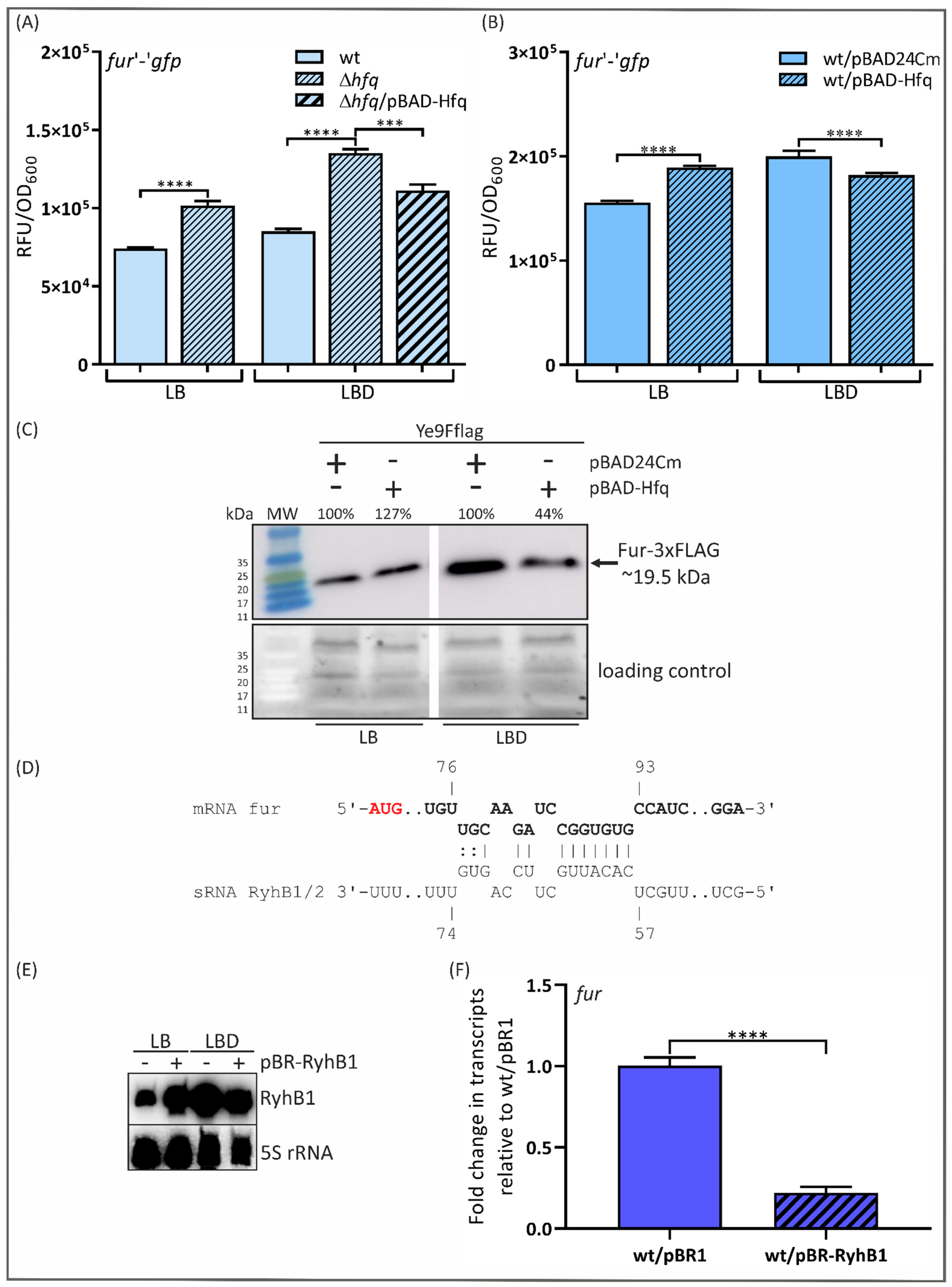
2.1. The RNA Chaperone Hfq and sRNA RyhB1 Impact Fur Expression in Y. enterocolitica 2/O:9, Strain Ye9N

To examine the role of Hfq in the expression of genes involved in iron uptake and regulation in Y. enterocolitica strain Ye9N (bioserotype 2/O:9), an isogenic Δhfq mutant lacking the Hfq-coding sequence was generated (strain Ye9hfq) (see Materials and Methods). Hfq typically acts with sRNAs to modulate mRNA stability and/or translation; thus, direct regulation by Hfq is usually dependent upon base pairing between the sRNA and its target mRNA near the ribosome-binding site. Bearing this in mind, the role of Hfq was tested using a plasmid carrying a translational fusion of fur with the gfp reporter gene. This plasmid, pFX-fur, was introduced into Ye9hfq and the wild-type strain Ye9N. In addition, the effect of the complementation of the Δhfq mutation or the overexpression of Hfq in the wild-type strain was tested by introducing a native copy of hfq under the control of the arabinose-inducible PBAD promoter in the plasmid pBAD-Hfq, with the empty vector pBAD24Cm as a control. These constructs are compatible with pFX plasmids, so they can be stably maintained together in the cell. To reveal the role of Hfq in iron-dependent fur regulation in Y. enterocolitica, the strains were grown in LB medium and under iron-starved conditions, i.e., LB supplemented with 150 µM dipyridyl, an iron chelator (LBD medium). It was previously suggested that iron may be involved in the regulation of fur transcription in E. coli [75] and Y. enterocolitica [22].
As shown in Figure 1A, the green fluorescence of Ye9hfq/pFX-fur increased by 27 and 37% relative to the parental strain Ye9N/pFX-fur in LB and LBD media, i.e., regardless of the iron concentration. The plasmid-borne copy of the hfq gene (pBAD-Hfq) introduced into Ye9hfq/pFX-fur grown in LBD only slightly decreased fur expression (~15%). Interesting data were obtained using the wild-type strain Ye9N/pFX-fur carrying the plasmid pBAD-Hfq. Hfq overexpression caused a slight increase in green fluorescence in the presence of abundant iron (LB medium) and decreased fluorescence under iron-starved conditions (LBD) (Figure 1B). These significant and opposite alterations in fur expression observed under iron-replete and iron-limiting conditions in the presence of increased Hfq are likely to reflect changes resulting from other putative regulatory factors/mechanisms whose activity is triggered by iron and depends on the level of Hfq. These results indicated that the link between Fur and Hfq might be more complex.
To further examine the role of Hfq in the iron-dependent regulation of fur, the abundance of the Fur protein was assessed using strain Ye9Fflag, which carries a chromosomal copy of a gene encoding an in-frame translational fusion of Fur with a 3×FLAG epitope peptide at the C-terminal end [22]. Plasmids pBAD24Cm and pBAD-Hfq were introduced into Ye9Fflag, and the obtained strains were grown in LB medium or under iron-limiting conditions (LBD). The Fur-3×FLAG protein was detected in whole-cell lysates by Western blotting using an anti-FLAG antibody. As shown in Figure 1C, the epitope-tagged Fur protein displayed strong and specific reactivity with this antibody. Interestingly, the amount of Fur-3×FLAG in the strain carrying pBAD-Hfq was increased (~27%) in LB medium and decreased (~56%) under iron-limiting conditions (LBD medium) compared with the strain carrying pBAD24Cm (Figure 1C). This opposite effect of Hfq on fur expression in iron-replete and iron-limiting media is consistent with the results obtained using the fur′-′gfp translational fusion under the same growth conditions. In addition, the amount of Fur-3×FLAG increased (~130%) in strain Ye9Fflag/pBAD24Cm in LBD compared with LB medium, confirming the importance of iron in regulating fur expression [22]. In summary, Fur abundance in Y. enterocolitica is modulated by the level of Hfq, and iron plays a role in this regulation.
Since the main function of Hfq is to act as a cofactor of sRNAs, Hfq/sRNA circuits might directly affect fur gene expression, and the interaction of Hfq/RyhB homologs may be responsible for the iron-dependent negative regulation of fur [11]. However, indirect regulation is also possible because Hfq could modulate the level of potential activator or repressor proteins involved in fur regulation. To study the effect of Hfq overexpression in a time-dependent manner, the Fur-3×FLAG level was monitored following hfq induction (30, 60, 90, 120 min after induction). However, there were no changes in the Fur protein level, which indicated both the direct and indirect Hfq-dependent regulation of fur.
It was previously determined that the sRNA RyhB, whose expression is negatively regulated by iron (Fe2+-Fur), may play a role in the control of fur expression in E. coli [11]. Interestingly, Yersinia species encode two RyhB homologs, named RyhB1 and RyhB2 [76,77]. The nucleotide sequences of the ryhB1 (108 nt) and ryhB2 (109 nt) genes in Y. enterocolitica are 64% and 63% identical to those of the E. coli homologs, respectively. By applying the IntaRNA program (version 5.0.7) to predict interactions between RNA molecules, we identified potential binding sites for both sRNAs within the fur mRNA (Figure 1D) and an additional target for base pairing with RyhB2 at the start of the coding sequence. To test whether either of these sRNAs is involved in the regulation of fur expression in Y. enterocolitica, the plasmid pBR-RyhB1 was created by cloning the Ye9N ryhB1 gene into the expression vector pBR-plac, named pBR1 (a kind gift from Prof. Susan Gottesman) [24]. Initially, we examined the abundance of ryhB1 in strains Ye9N and Ye9N/pBR-RyhB1 grown in LB or LBD by Northern blotting. The applied probe was designed to be specific for ryhB1 so will not bind to ryhB2. As shown in Figure 1E, the level of the ryhB1 transcript was increased in the wild-type strain grown in LBD compared to LB, suggesting the influence of iron on ryhB1 expression. Interestingly, the ryhB1 transcript was also present in strain Ye9N grown in LB, which may explain the regulatory effect of Hfq on fur expression under iron-replete conditions (Figure 1A). The ryhB1 mRNA was strikingly more abundant in Ye9N carrying the plasmid pBR-RyhB1. Moreover, the level of the ryhB1 transcript in this strain was slightly reduced under iron-limiting conditions. This decrease may result from specific degradation mechanisms that regulate the levels of sRNAs [78].
To determine the role of RyhB1 in regulating fur expression, RT-qPCR was performed (Figure 1F). The fur transcript level was significantly decreased by the presence of plasmid-borne RyhB1, suggesting a negative role for RyhB1 in the post-transcriptional regulation of fur in Y. enterocolitica. RyhB2 might play a similar role in fur regulation, but this has yet to be confirmed.

2.2. The Expression of fecA and fepA Is Silenced by Hfq

After exploring the role of Hfq and sRNA RyhB1 in the modulation of Fur expression, we turned our attention to FepA and FecA, the OM transporters/receptors for ferric citrate and ferric enterobactin, respectively. To test whether Hfq might control the expression of fecA and fepA, fecA’-’gfp and fepA’-’gfp translational fusions expressed from the plasmid pFX (pFX-fecA and pFX-fepA) were constructed and introduced into the wild-type strain Ye9N and Ye9hfq (∆hfq mutant). In addition, the effect of the complementation or overexpression of Hfq was studied by introducing pBAD-Hfq into Ye9hfq and the wild-type strain Ye9N carrying pFX-fecA or pFX-fepA. All tested strains were grown in LB and in LBD (iron-limiting conditions). As shown in Figure 2A, strain Ye9hfq exhibited increased fecA’-’gfp expression, irrespective of the iron concentration, compared to the wild-type strain (31% in LB and 36% in LBD). Increased expression of the fepA’-’gfp fusion was also observed in this ∆hfq mutant (52% in LB and LBD), independently of the iron concentration (Figure 2A). These results suggest that Hfq may participate in repressing fecA and fepA expression independently of iron. Interestingly, when the wild-type allele of hfq on the plasmid pBAD-Hfq was introduced into Ye9hfq and induced, the expression of fecA and fepA was slightly decreased, but not to wild-type levels.
The effects of hfq overexpression from the plasmid pBAD-Hfq in strains Ye9N/pFX-fecA and Ye9N/pFX-fepA were also analyzed (Figure 2B). Interestingly, an effect of increased Hfq on fecA’-’gfp was observed in cells grown under iron-limited conditions (LBD medium): fluorescence was slightly decreased (~10%) in comparison with the strain carrying the control vector pBAD24Cm, implying that Hfq inhibits fecA expression under these growth conditions. In comparison, the fepA’-’gfp strain carrying pBAD-Hfq displayed decreased fluorescence relative to the strain carrying pBAD24Cm in stationary-phase cultures grown in both LB and LBD medium (13% and 7%, respectively) (Figure 2B).
These results demonstrate that the lack of hfq leads to the upregulation of fecA and fepA expression irrespective of iron concentration. Therefore, Hfq participates in the repression of both genes. Hfq expressed from a multicopy plasmid had a negative effect on the expression of fecA in an iron-depleted culture or independently of iron in the case of fepA.
To check whether the Hfq-dependent regulation of fecA and fepA expression observed using translational fusions is reflected in the level of the encoded proteins, Western blotting analysis was performed (Figure 2C). To facilitate the detection of FecA and FepA, we prepared strains of Ye9N and Ye9hfq carrying a chromosomal copy of the desired gene encoding an in-frame translational fusion with a 3×FLAG epitope peptide at the C-terminal end (see Materials and Methods). The empty vector pBAD24Cm or plasmid pBAD-Hfq was then introduced into the FLAG-tagged strains. Western blot analysis with the anti-FLAG antibody was used to detect the FecA-3×FLAG and FepA-3×FLAG proteins in extracts prepared from cells grown to the stationary phase in LBD medium. As expected, significant increases in the levels of FecA-3×FLAG (~100%) and FepA-3×FLAG (~86%) were observed in the ∆hfq mutant background (strains Ye9hfqFecAflag and Ye9hfqFepAflag) in comparison with the parental strain Ye9N (strains Ye9FecAflag and Ye9FepAflag), all carrying the empty plasmid pBAD24Cm. The overexpression of hfq from pBAD-Hfq in Ye9hfq (complementation assay) decreased the abundance of FecA-3×FLAG (~41%) and FepA-3×FLAG (~35%) compared with the strains carrying pBAD24Cm, which supports the notion that Hfq negatively impacts FecA and FepA abundance under these conditions. It should be noted that we did not see significant changes in the amount of FecA-3×FLAG or FepA-3×FLAG when hfq was overexpressed in strains Ye9FecAflag and Ye9FepAflag grown to the stationary phase in LBD medium. Thus, in the wild-type strain Ye9N, additional Hfq provided by the plasmid pBAD-Hfq appeared to result in the decreased expression of the genes encoding the two Fe-siderophore transporters/receptors (fecA’-’gfp and fepA’-’gfp expression data), but the production of the proteins showed no significant changes (FecA-3×FLAG and FepA-3×FLAG protein abundance). This discrepancy between the reporter gene and Western blot data could be explained if the effect of Hfq on fecA and fepA expression detected using the gfp fusions is too modest to lead to a measurable decrease in FecA and FepA levels. However, the obtained results confirmed the participation of Hfq in repressing fecA and fepA expression in Y. enterocolitica.

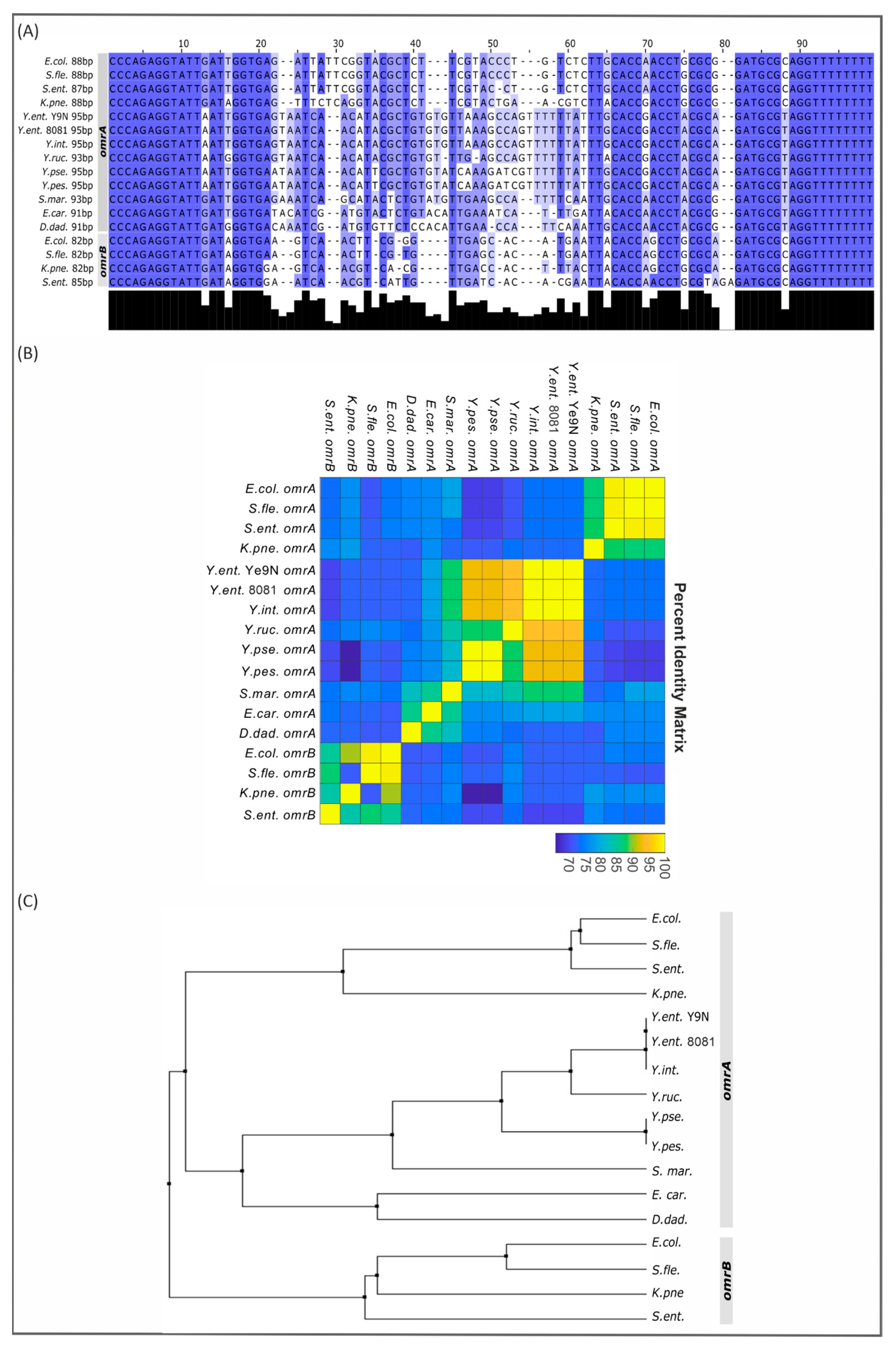
2.3. Partial Conservation of the omrA/B Locus in Gammaproteobacteria

Screens of the E. coli genome revealed the existence of two tandemly located sRNA genes, omrA and omrB, first named rygA/sraE and rygB [54,55]. The encoded sRNAs OmrA and OmrB are 88 nt and 82 nt in length, respectively, and share nearly identical sequences in their 5′ and 3′ regions [25,55]. Both sRNAs have been shown to downregulate the synthesis of several proteins, including outer membrane receptors for Fe-siderophores, i.e., CirA, FecA and FepA [25,56]. To examine the sequence conservation of OmrA and OmrB among Gammaproteobacteria, 13 genomes were searched for orthologs of these sRNAs: six Yersiniaceae species, namely, human pathogens Y. enterocolitica (2 strains of different bioserotypes—Ye9N (bioserotype 2/O:9) and 8081 (bioserotype 1B/O:8)), Y. pseudotuberculosis and Y. pestis, the fish pathogen Y. ruckeri, the environmental species Y. intermedia and Serratia marcescens (an opportunistic human pathogen), plus 4 Enterobacteriaceae species, E. coli, Shigella flexneri, Salmonella enterica subsp. Enterica and Klebsiella pneumoniae, and 2 members of the Pectobacteriaceae, the plant pathogens Erwinia carotovora and Dickeya dadantii. Multiple sequence alignment of the identified omrA and omrB genes was performed, the pairwise nucleotide identity was calculated, and an average distance tree was created (Figure 3). Interestingly, while both omrA and omrB genes could be identified in all studied members of the Enterobacteriaceae, only one Omr sRNA gene was recognized in Yersiniaceae and Pectobacteriaceae species. In addition, the Omr sRNAs in the Yersiniaceae are >90 nt in length, which is longer than both OmrA and OmrB in E. coli. A high level of sequence conservation was found within the first 21 and last 18 nucleotides of each sRNA, suggesting the importance of these regions for sRNA function. Analysis of the omr gene sequences from the selected bacteria provided phylogenetic information on this sRNA regulatory system. The highly conserved Omr sequences of Yersiniaceae are much closer to E. coli OmrA than OmrB. Omr in Y. enterocolitica strain Ye9N shares 93% sequence identity with those in Y. pseudotuberculosis and Y. pestis and 73% and 68% sequence identity with E. coli OmrA and OmrB, respectively. This analysis prompted us to identify the Omr sRNA in strain Ye9N as OmrA.
The genomic context of the omr genes in the analyzed Gammaproteobacteria is presented in Table S3. The omrA and omrB genes in the E. coli chromosome are tandemly located within the intergenic region between the genes aas, encoding 2-acylglycerophospho-ethanolamine acyl transferase/acyl-acyl carrier protein synthetase, and galR, encoding a transcriptional regulator. The same upstream/downstream genes flanking the omrA and omrB sRNA genes are present in the other Enterobacteriaceae species/strains examined (S. flexneri 301, S. enterica sv Typhimurium 14028S and K. pneumoniae subsp. pneumoniae HS11286). Analysis of the genome sequence of Y. enterocolitica strain Ye9N (bioserotype 2/O:9) localized the single omrA gene between the genes tnp, encoding IS110 family transposase, and aas. The aas and omrA genes are in the same transcriptional orientation, whereas tnp is oriented divergently. The same genomic organization is present in Y. enterocolitica strain 8081 of bioserotype 1B/O:8. Interestingly, the genomic context of the omrA gene varies in other Yersiniaceae species/strains. In Y. pseudotuberculosis IP 32953, Y. pestis A1122, Y. intermedia and S. marcescens KS10, omrA is located between the genes bisC, encoding the molybdopterin guanine dinucleotide-containing S/N-oxide reductase, and aas, while the genes flanking omrA in Y. ruckeri ATCC 29473 are aas and hp, encoding a hypothetical protein. In E. carotovora subsp. atroseptica SCRI1043, the omrA gene is located between aas and a gene encoding a methyl-accepting chemotaxis protein, while in D. dadantii 3937, the neighboring genes of omrA are quite different to those in all the other analyzed species: it is located between bglG, encoding the beta-glucoside bgl operon antiterminator, and lpxO, encoding Fe2+/alpha-ketoglutarate-dependent dioxygenase LpxO. In summary, this analysis revealed that the genomic context is highly conserved when both omrA and omrB are present but is only partially conserved when there is just one omr gene.

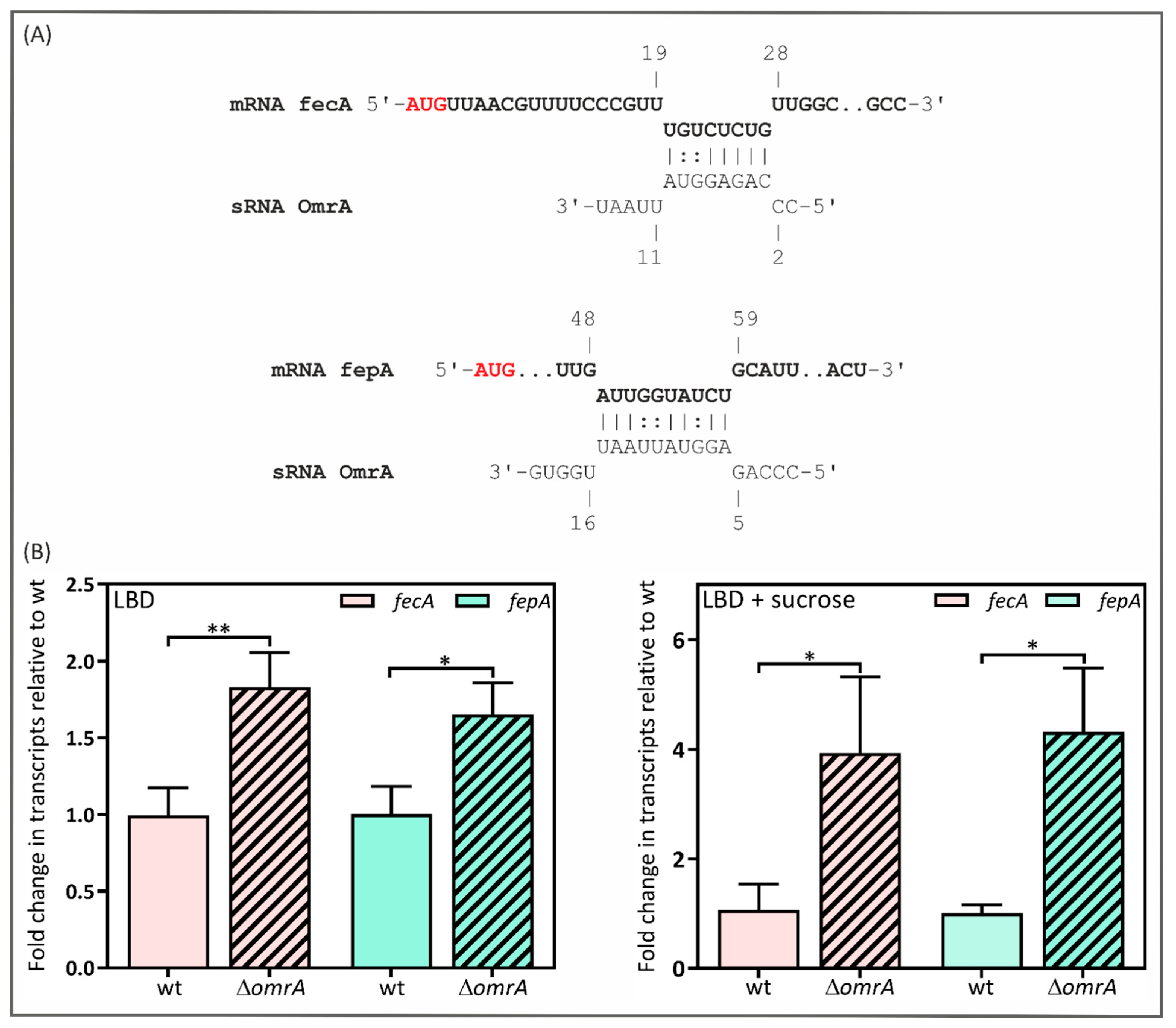
2.4. Y. enterocolitica’s OmrA sRNA Decreases fecA and fepA Levels

If OmrA from Y. enterocolitica directly regulates fecA or fepA transcript levels, we would expect to see base pairing between OmrA and the respective mRNAs. Using the software IntaRNA version 5.0.7, we were able to predict base pairing between OmrA and both analyzed mRNAs (Figure 4A). This analysis revealed complementarity between positions +20 and +27 nt of the fecA mRNA and between +49 and +58 nt of the fepA mRNA, downstream of the translation start site. The fecA mRNA region is complementary to nucleotides 3 to 10 of OmrA, while the fepA mRNA region is complementary to nucleotides 6 to 15 of OmrA. Interestingly, potential pairing with OmrA occurs within the early coding sequences of the fecA and fepA mRNAs instead of the 5′ UTR. To reveal whether this sRNA controls fecA and fepA expression, we measured the effect of omrA deletion on fecA and fepA transcript levels by RT-qPCR. An isogenic ∆omrA mutant was generated in Y. enterocolitica strain Ye9N (strain Ye9omrA) by marked allelic exchange using a construct based on the sacB-dependent suicide plasmid pDS132 (see Materials and Methods). This null mutation prevented the production of the OmrA mRNA (Figure S1). RT-qPCR analysis was performed using RNA isolated from the wild-type Y. enterocolitica strain Ye9N and the omrA null mutant grown in LBD and in LBD supplemented with 20% sucrose, known to induce omrA expression in E. coli [24]. As shown in Figure 4B, the deletion of omrA resulted in increased levels of the fecA and fepA transcripts (fecA displayed a 1.8-fold increase, and fepA increased 1.7-fold). More visible differences were observed when strains were grown in the iron-limited medium supplemented with sucrose (3.9-fold for fecA and 4.3-fold for fepA). The nature of the molecular interactions between OmrA and these mRNAs in Y. enterocolitica has yet to be determined and requires further study.

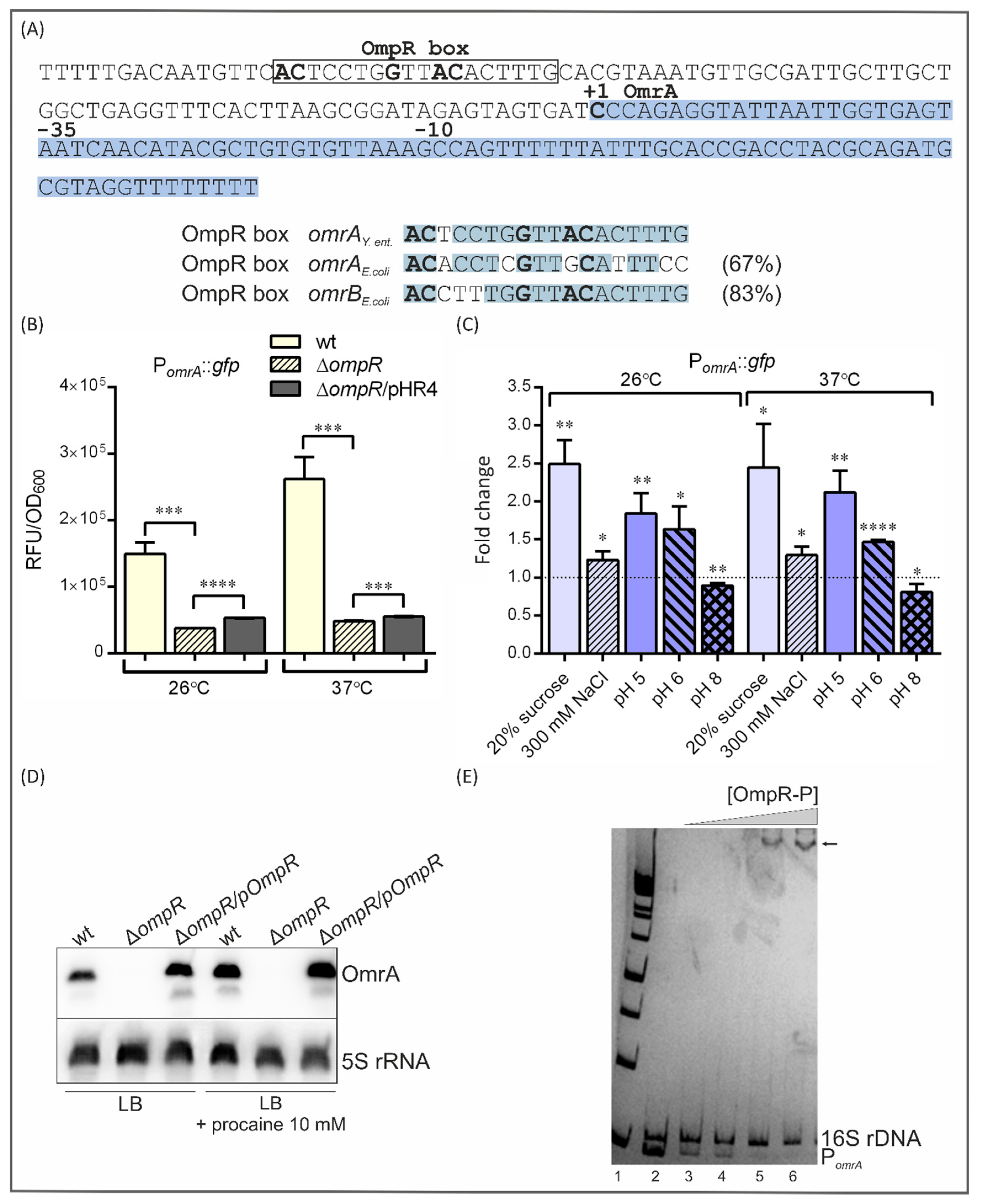
2.5. Influence of OmpR and Environmental Conditions on omrA Promoter Function

Previous studies in E. coli have identified the OmpR protein as a positive regulator of sRNA OmrA and OmrB transcription [24,60]. An examination of the Y. enterocolitica Ye9N omrA promoter sequence identified one putative OmpR-binding site (OmpR box) exhibiting 67% and 83% identity to the OmpR boxes found in the E. coli omrA and omrB promoter regions, respectively (Figure 5A). These OmpR boxes were recognized based on their similarity to OmpR-binding motifs found in the regulatory regions of the E. coli ompC and ompF porin genes [24,81]. The conserved motif GxxxC and AC base pairs are critical for OmpR binding [81,82,83]. The OmpR box in the Ye9N omrA gene promoter is located at −82 to −63 nt relative to the transcription initiation site (+1). The presence of a putative OmpR-binding motif in the Ye9N omrA regulatory sequence prompted us to examine the role of OmpR in the regulation of omrA transcription by creating a fusion of the omrA promoter with gfp in the plasmid vector pPROBE-TT’ (see Materials and Methods). This plasmid, pPOmrA, was introduced into the wild-type strain Ye9N and the ∆ompR mutant AR4 (constructed using a reverse genetic PCR-based strategy [84]). Complementation analysis was performed by transforming strain AR4/pPOmrA with the plasmid pHR4 expressing the active OmpR protein [84]. Cultures of strains carrying pPOmrA were grown to the stationary phase in LB medium at 26 °C and 37 °C, and green fluorescence was measured (Figure 5B).
The data presented in Figure 5B show that when grown at 26 °C or 37 °C, the ∆ompR mutant AR4 displayed a significant decrease (4-fold and 5.4-fold, respectively) in reporter gene expression compared to the parental strain Ye9N, suggesting that OmpR is involved in the positive regulation of the omrA gene. Complementation of the ompR mutation by OmpR expressed in trans (strain AR4/pPOmrA/pHR4) caused a slight increase in fluorescence compared to the ∆ompR mutant strain at both studied temperatures. The link between OmrA and OmpR might be complex, with the possible existence of a negative regulatory circuit whereby OmrA can limit ompR expression through feedback control (see 2.6). We cannot rule out the possibility that an optimal concentration of phosphorylated OmpR is required to control the amount of OmrA. Moreover, in the complementation assay, there will be an excess of OmpR relative to EnvZ, and this imbalance may influence the expression of omrA.
The activity of the omrA promoter in Ye9N was higher at 37 °C than at 26 °C (1.75-fold), indicating that the transcription of this gene is subject to temperature regulation. This thermoregulation was also observed in strain AR4, indicating that OmpR plays no part in the response of omrA to changes in temperature.
The EnvZ/OmpR TCS modulates the expression of several genes in E. coli and Salmonella in response to altered osmolarity and pH [62,63]. These environmental signals change the DNA-binding affinity of the transcriptional activator protein OmpR. Therefore, we next examined the influence of these environmental cues on the activity of the omrA promoter (Figure 5C). First, omrA promoter activity was assessed in strain Ye9N (Ye9N/pPOmrA) grown in LB medium to the exponential phase and then incubated for 1 h at 26 °C or 37 °C in LB supplemented with 20% sucrose or 0.3 M NaCl (Figure 5C). These high-osmolarity conditions caused increases in the activity of the omrA promoter of 2.5-fold in 20% sucrose and 1.2-fold in 0.3 M NaCl irrespective of the temperature. To determine whether the pH of the growth medium influences the expression of omrA, cultures were grown in LB medium to the exponential phase and then incubated for 1 h at 26 °C or 37 °C in LB medium at pH 5, 6 or 8. Acid pH significantly increased the activity of the PomrA::gfp transcriptional fusion, while this activity was significantly decreased at pH 8.0 (Figure 5C). No effect of temperature was observed. Procaine, a local anesthetic, is also known to activate the OmpR regulator [85,86]. Therefore, we examined the influence of procaine on the regulation of omrA expression by Northern blotting performed using RNA prepared from Ye9N and AR4 (∆ompR) cells grown to the exponential phase in the presence and absence of this compound. As shown in Figure 5D, the level of the omrA transcript was increased in strain Ye9N after the addition of 10 mM procaine. In strain AR4 (∆ompR), no omrA transcripts could be detected in cells grown in LB with or without procaine, confirming the importance of OmpR as an activator of omrA transcription in the exponential phase. Interestingly, as shown in Figure 5B, stationary-phase cells of the ∆ompR mutant displayed a significant decrease in reporter gene expression compared to the parental strain, although a residual level of omrA expression was still detected.
In summary, these results reveal the role of OmpR in the activation of omrA expression and imply that OmpR may directly influence omrA transcription, i.e., by binding to the promoter region of omrA. One potential OmpR-binding site was recognized in the omrA promoter region of strain Ye9N (Figure 5A). To determine whether this OmpR box functions as a binding site for OmpR in vitro, we performed an electrophoretic mobility shift assay (EMSA) using a 250 bp fragment encompassing the omrA regulatory region and purified OmpR-His6 protein [74], phosphorylated in vitro by acetyl-p. The DNA fragment was incubated with increasing amounts of the His-tagged OmpR-P protein. As a control, a 304 bp 16S rDNA fragment was included in the binding reactions. As shown in Figure 5E, slower-migrating OmpR-P/DNA complexes were observed in the presence of OmpR-P at concentrations of 37.3 µM and above. OmpR-P failed to interact with the 16S rDNA fragment used as a negative control. These results demonstrate that the regulation of omrA transcription by OmpR is mediated by the direct binding of this regulatory protein to the omrA promoter.

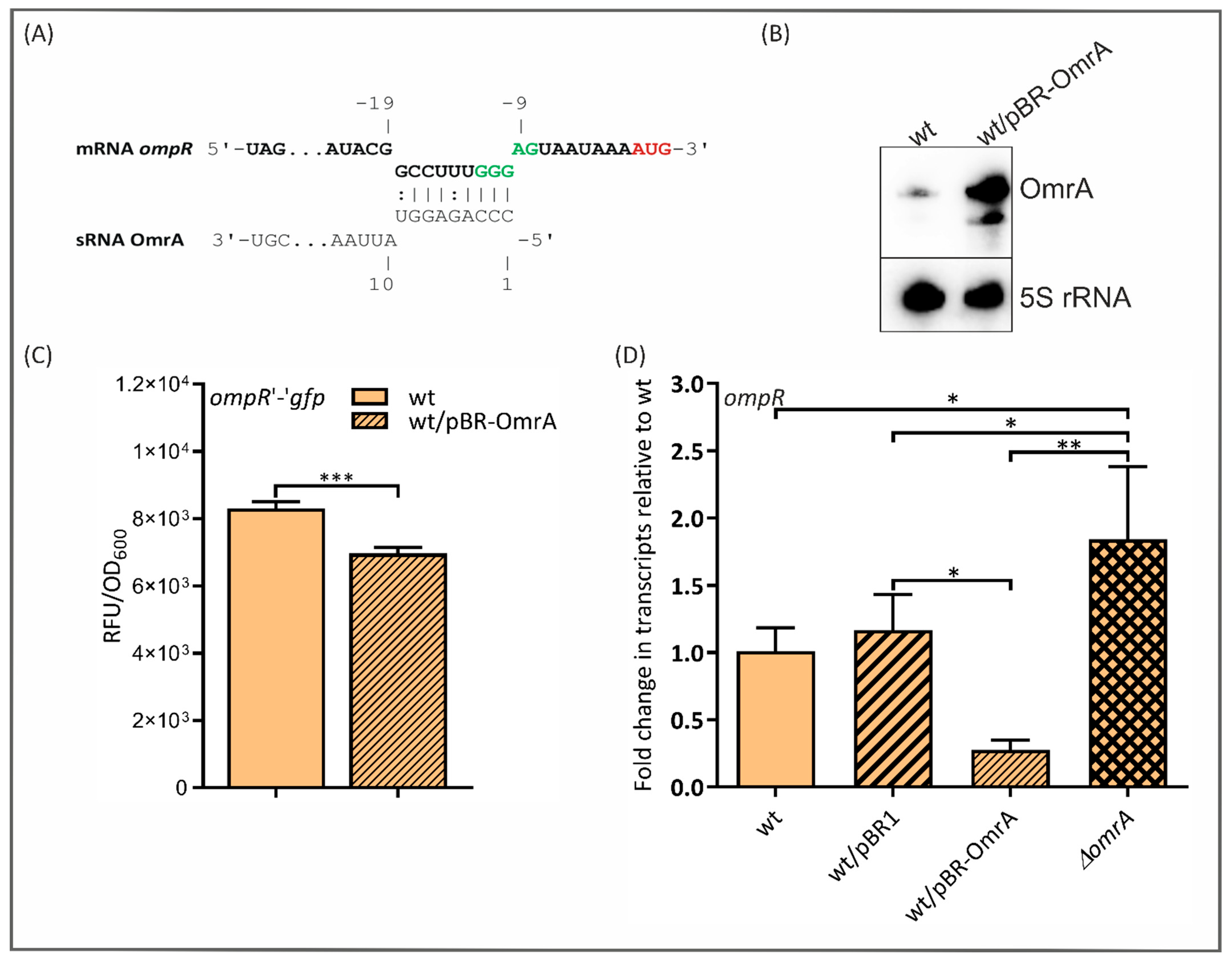
2.6. OmrA Regulates ompR Expression Post-Transcriptionally

Since two redundant E. coli sRNAs, OmrA and OmrB, downregulate the expression of the OmpR transcriptional regulator [24], we investigated the potential OmrA-mediated regulation of the ompR gene in Y. enterocolitica. The IntaRNA program yielded a short possible predicted pairing between OmrA and the Ye9N ompR mRNA near the RBS and translation initiation codon (Figure 6A). This involves the conserved 5′ end of OmrA and nts −10 to −18 of the ompR mRNA relative to AUG. The site of interaction of OmrA with ompR in Y. enterocolitica is extremely similar to the one reported in E. coli [56]. To test whether sRNA OmrA is involved in the regulation of ompR expression in Y. enterocolitica, the plasmid pFX-ompR (a kind gift from Prof. O. Rossier) [45] carrying an ompR’-’gfp translational fusion was applied. Moreover, the plasmid pBR-OmrA was created by cloning the Ye9N omrA gene into the expression vector pBR-plac [24], as in the case of sRNA ryhB1. The omrA transcript abundance was assessed in cells grown to the stationary phase in LB by Northern blotting. As shown in Figure 6B, the omrA mRNA (transcript of the chromosomal gene) was barely detectable in strain Ye9N but was strikingly more abundant in Ye9N carrying the plasmid pBR-OmrA. This demonstrated that ectopically expressed OmrA could be used to study the post-transcriptional regulation of ompR expression. As shown in Figure 6C, strain Ye9N carrying the ompR’-’gfp fusion with an enhanced level of OmrA, grown to the stationary phase, displayed a ~24% decrease in fluorescence intensity compared to the strain without the plasmid pBR-OmrA. Since the effect of OmrA on the ompR’- ’gfp fusion activity was quite modest, we used RT-qPCR to estimate the level of the ompR transcript in strains with different OmrA contents (Figure 6D). This analysis showed that the plasmid-borne OmrA significantly reduced ompR expression (3.6-fold). Moreover, the deletion of omrA resulted in an increased level of the ompR transcript. These data support the notion that OmrA represses the expression of ompR in Y. enterocolitica.

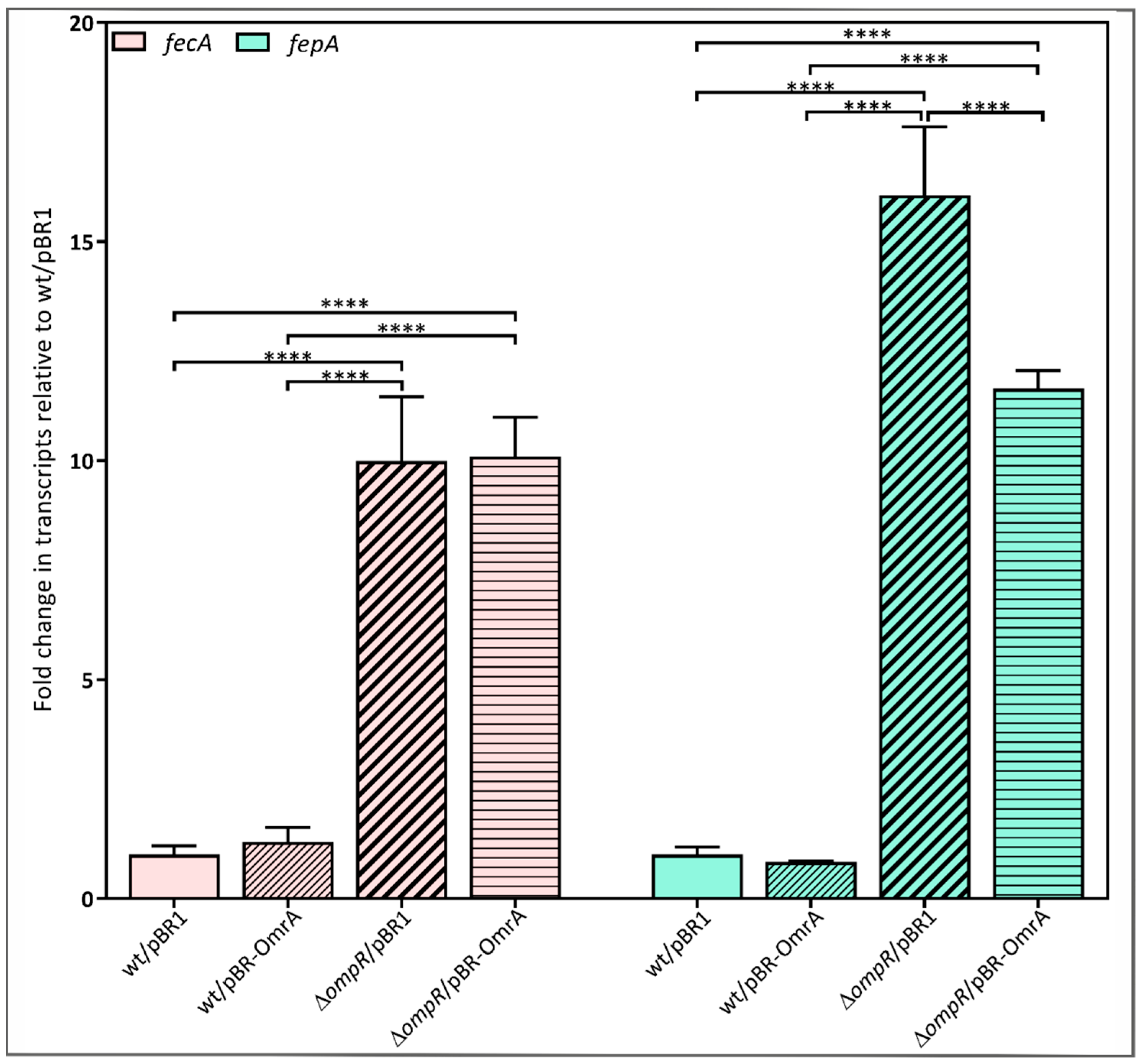
2.7. Requirement for OmpR in the OmrA-Dependent Regulation of fecA and fepA Expression
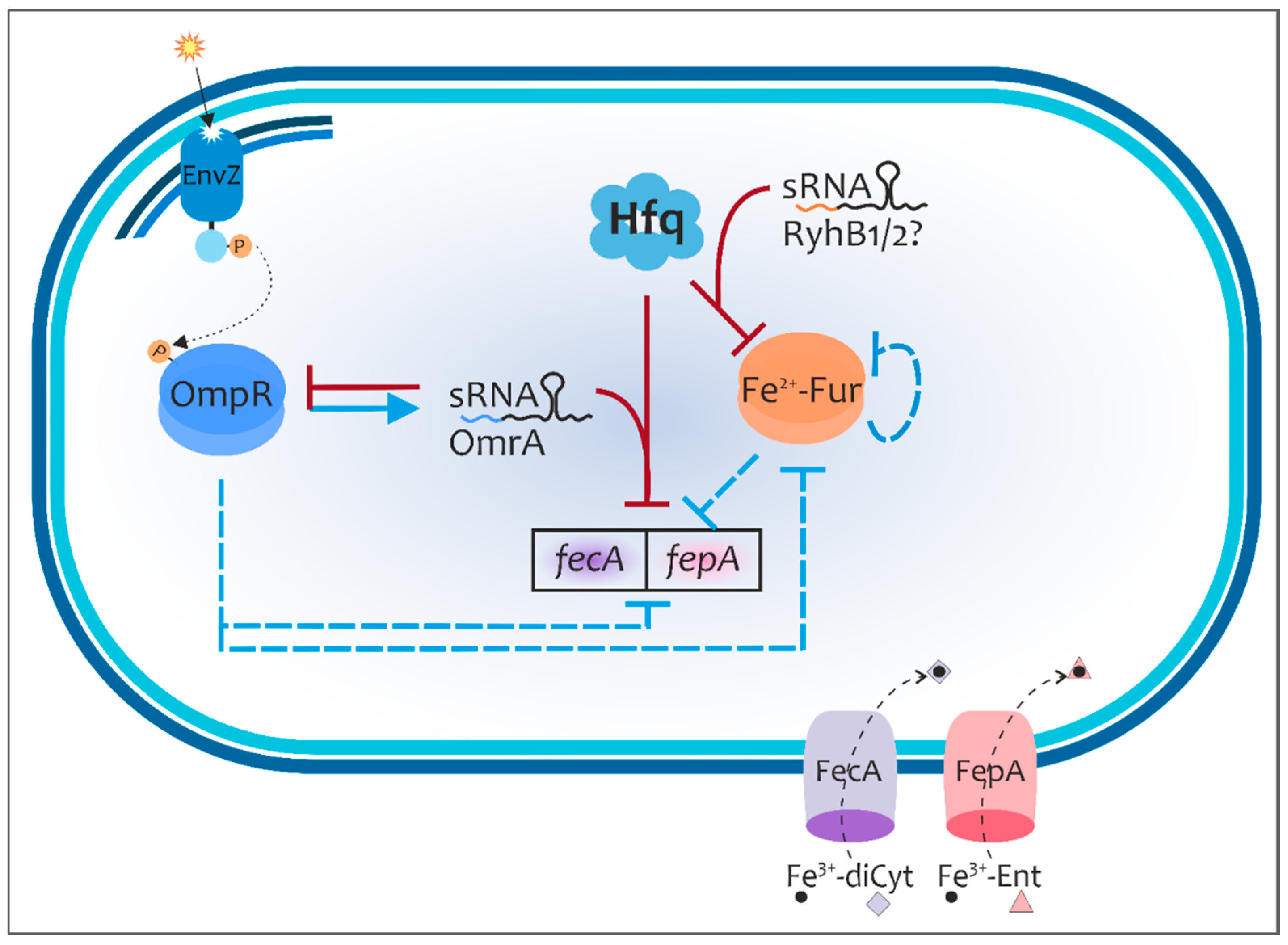
Following the demonstration that OmpR induces omrA transcription in Y. enterocolitica and there is negative feedback between sRNA OmrA and ompR expression, we decided to examine the role of OmrA in the post-transcriptional regulation of fecA and fepA in the ompR-null mutant background. The fecA and fepA transcripts were quantified in strain AR4 (∆ompR) and compared with the wild-type Ye9N, both carrying plasmid-borne omrA (pBR-OmrA) or the empty vector (pBR1). The results presented in Figure 7 reveal an increase in the levels of the fecA and fepA transcripts in the ΔompR strain AR4/pBR1 relative to the wild-type strain Ye9N/pBR1 (10-fold and 16-fold), confirming previous data showing that OmpR directly inhibits fecA and fepA transcription in Y. enterocolitica [22]. In light of our findings regarding OmrA, this effect may also result from the lack of this sRNA, since it is not expressed in the ΔompR mutant background. Interestingly, no effect of ectopically expressed OmrA on the fecA and fepA mRNA levels was observed in the wild-type strain, suggesting that a combination of transcriptional and post-transcriptional regulatory mechanisms involving OmrA and OmpR could inversely modulate the abundance of both transcripts. Surprisingly, only a moderate reduction in fepA mRNA levels (27%) was observed in the ∆ompR mutant background in the presence of plasmid-borne omrA, while the level of fecA transcripts remained unchanged. These results suggest that fepA expression, in contrast to fecA, may be silenced by OmrA independently of OmpR. Since the inhibitory effect on fepA mRNA levels in the presence of OmrA is modest, it is possible that the abundance/activity of other factors (Hfq, RNase E), known to be involved in OmrA-dependent regulation, may be decreased in the ∆ompR mutant background.

3. Discussion

In this study, we investigated whether the RNA chaperone Hfq influences the expression of the gene encoding Fur, the master regulator of iron homeostasis in Y. enterocolitica, and two genes belonging to the Fur regulon, fecA and fepA, encoding OM receptors/transporters for ferric citrate and ferric enterobactin, respectively. Two experimental approaches were employed: (i) measuring fluorescence produced by translational fusions of fur, fecA and fepA with gfp and (ii) monitoring the production of FLAG-tagged Fur, FepA and FecA proteins by immunoblotting.
The study of translational fusions of fur with gfp demonstrated that the absence of chromosomally encoded Hfq caused the upregulation of fur expression. Hfq-dependent silencing of fur expression was observed independently of the presence of iron ions. However, in wild-type cells, the overexpression of Hfq from a plasmid led to the downregulation of fur expression under iron-starved conditions, while upregulation was observed in the presence of iron. These iron-dependent alterations in fur expression seen in the presence of plasmid-borne Hfq suggest that the link between iron, Fur and Hfq might be complex. Western blot analysis confirmed the inhibitory effect of Hfq on the production of FLAG-tagged Fur in Y. enterocolitica. However, this polypeptide was clearly produced under iron-limited conditions, which suggests that Hfq-dependent production of Fur occurs in an iron-responsive manner. Hfq usually acts in conjunction with sRNAs to modulate mRNA stability and/or translation [39]. Therefore, we hypothesized the involvement of putative regulatory sRNAs, whose activity is triggered by iron starvation and which interact with Hfq to decrease Fur abundance.
Hfq-dependent sRNA RyhB, whose expression is negatively regulated by iron (Fe2+-Fur), may play a role in the post-transcriptional control of fur in E. coli [11,35]. The analysis of the Y. enterocolitica genome sequence revealed the presence of two RyhB homologs, named RyhB1 and RyhB2. The potential base pairing of RyhB1/RyhB2 with a single site in the coding sequence of the fur mRNA was recognized. The initial characterization of Y. enterocolitica RyhB1 revealed its role in the negative regulation of fur expression. The influence of RyhB2 on Y. enterocolitica fur has yet to be determined. Interestingly, reciprocal regulation between Fur and two RyhB homologs in Y. pestis has recently been demonstrated [87].
To clarify whether Hfq participates in the post-transcriptional regulation of the fecA and fepA genes, expression studies were performed under iron-replete and iron-limiting conditions (release from Fe+2-Fur repressor activity). A comparative analysis of strains with different Hfq contents, using plasmid-encoded translational fusions of fecA and fepA with a gfp reporter gene, indicated that Hfq represses the expression of these genes irrespective of iron. Interestingly, the silencing of fecA and fepA by overexpressed Hfq in iron-starved conditions was moderate, suggesting that an optimal concentration of Hfq is required to control the amounts of the transporters FecA and FepA. It was previously found that raising Hfq levels modestly increases the accumulation of sRNAs and hence the silencing of some target genes [88,89]. Finally, an analysis of the abundance of FLAG-tagged FecA and FepA expressed from their chromosomal loci under iron-depleted conditions confirmed that Hfq negatively affects the production of these proteins. The demonstration that Hfq regulates the expression of these ferric siderophore transporters is in line with the findings of a previous study showing that, in Y. enterocolitica bioserotype 1B/O:8 strains, Hfq inhibits the expression of the OM receptor FyuA for the uptake of Fe-yersiniabactin (Fe-Ybt), as well as the synthesis of Ybt [44]. The genes necessary for Ybt synthesis, uptake and regulation are localized within a High-Pathogenicity Island (HPI) that is not present in Y. enterocolitica strains of biotypes 2–5, which include strain Ye9N, the object of our research [18,90].
Our finding that Hfq affects the expression of proteins involved in iron acquisition and the master regulator Fur indicates that this RNA chaperone plays a key role in iron homeostasis in Y. enterocolitica. It is also worth adding that the expression of several OM proteins involved in the pathogenicity of Y. enterocolitica 1B/O:8 that are not regulated by iron is also regulated by Hfq [45]. It appears that Hfq is involved in both iron-dependent and -independent gene regulation in Y. enterocolitica, as is the case in E. coli [91] and Neisseria meningitidis [92]. Thus, we cannot exclude the possibility that Hfq may play both direct and indirect roles in the post-transcriptional regulation of the fecA and fepA genes in Y. enterocolitica.
Previously, Guillier and Gottesman [24,56] showed that the homologous sRNAs OmrA and OmrB in E. coli, bound to Hfq, exert post-transcriptional control over the expression of the genes cirA, fecA and fepA, encoding OM transporters for ferric siderophores, by pairing with and altering the stability of their mRNAs. Interestingly, while two redundant genes, omrA and omrB, are present in E. coli, only omrA was found in the genomes of the pathogenic Yersinia species Y. enterocolitica, Y. pestis and Y. pseudotuberculosis. Overall, the OmrA sequence is conserved in the genus Yersinia, but there are some differences in the core region compared with OmrA in E. coli. In light of the finding that Hfq can repress the expression of fecA and fepA in Y. enterocolitica, we sought to determine whether the sRNA OmrA participates in this regulation. It was also interesting because the homologous sRNAs might exhibit variations in their function that reflect the lifestyle of the bacteria concerned [93]. Notably, the sRNA OmrA in E. coli has been identified as a negative regulator of motility, which contrasts with the positive role it plays in Erwinia amylovora [94].
To confirm the participation of OmrA in the post-transcriptional inhibition of fecA and fepA expression, RT-qPCR analysis was performed to examine levels of the gene transcripts in the parental strain and a ∆omrA mutant. The results suggest that both analyzed genes are repressed by OmrA in Y. enterocolitica, especially in a medium supplemented with sucrose. These findings are in agreement with those of studies in E. coli, although fecA was shown to be a negative target for both OmrA and OmrB in this species, whereas only OmrA affected fepA expression [24].
Direct negative regulation by an Hfq-bound sRNA is usually dependent on base pairing between the sRNA and its target mRNA near the ribosome-binding site [37]. Potential base pairing between the conserved 5′ region of Y. enterocolitica OmrA and the fepA and fecA mRNAs was identified in their coding sequences. However, the importance of this predicted base pairing in silencing fecA and fepA requires experimental verification. This is particularly important in light of results concerning the pairing of the fepA transcript with sRNAs OmrA/B in E. coli. It was shown that OmrA/B represses fepA expression by targeting a stem-loop structure within the mRNA coding sequence at position +19, which seems to be necessary for the activation of translation initiation [95]. Our bioinformatic analysis showed that the fepA mRNA sequence in Y. enterocolitica differs considerably from that in E. coli (contains deletions and subtle single or multiple base variations), although a stem-loop-like structure is present at position +12 of the fepA coding sequence (Figure S2A). Moreover, OmrA may form a duplex with this predicted structure (Figure S2B). If, similarly to E. coli, this stem-loop region activates the translation of fepA, OmrA might repress fepA expression by targeting this structure. Notably, an analogous secondary structure within the fecA mRNA was not recognized using UNAFold software version 3.5. Further investigation of the Y. enterocolitica OmrA/fepA mRNA interaction is clearly required.
The present study revealed that omrA expression in Y. enterocolitica is induced in response to environmental signals such as high temperature and osmolarity or low pH, which suggests that the synthesis of OmrA is tightly regulated and restricted to highly specific conditions, like sRNAs in general [96]. While the mechanism of thermoregulation of omrA in Y. enterocolitica has yet to be characterized, our findings suggest that the EnvZ/OmpR TCS is responsible for regulating omrA expression in response to high osmolarity, low pH or the presence of procaine, all factors known to activate the EnvZ/OmpR TCS in E. coli [61,62,85,86,97]. By applying a PomrA::gfp transcriptional fusion and Northern blotting, we demonstrated that OmpR strongly and positively affects omrA expression. The in vitro binding of Y. enterocolitica OmpR to an omrA promoter region fragment, including a putative OmpR-binding site identified in silico, was confirmed. These findings demonstrate the direct involvement of OmpR in the positive regulation of sRNA OmrA in Y. enterocolitica. It should be noted that although the nucleotide sequence of Y. enterocolitica omrA exhibits higher similarity to E. coli omrA than to omrB (73% vs. 68%), the regulation of Y. enterocolitica omrA expression most closely resembles that observed for E. coli omrB [24,60].
We also found that the ompR mRNA may be a target for silencing by OmrA in Y. enterocolitica. Complementarity between the conserved 5′ end of OmrA and a region upstream of the start codon in the ompR mRNA, defined in silico, is very similar to that observed in E. coli and appears to be sufficient to downregulate ompR expression. Our results show that the transcription of omrA in Y. enterocolitica is activated by OmpR (probably phosphorylated OmpR), and in turn, OmrA represses the synthesis of OmpR. This is equivalent to the regulatory loop described for E. coli [24,56]. Given the existence of such a regulatory circuit, we performed RT-qPCR analysis to estimate the role of OmrA in the post-transcriptional regulation of fecA and fepA expression in the ompR mutant background. The absence of OmpR increased the abundance of both transcripts, confirming our previous finding that the expression of these ferric siderophore transporters is controlled at the level of transcription by the regulator OmpR [22]. The results of this study indicate that this effect may also be due to the lack of OmrA in the ompR mutant. Interestingly, we observed no significant effect of plasmid-borne OmrA on the levels of the fecA and fepA mRNAs in the wild-type strain, which might result from an uncharacterized regulatory mechanism operating in Y. enterocolitica. This unexpected finding may in part be due to the inhibitory effect of plasmid-borne OmrA on the level of OmpR, being the repressor of fecA and fepA transcription, resulting in the increased expression of both. On the other hand, OmrA may directly target both mRNAs, leading to their silencing. Interestingly, in the strain lacking OmpR, no effect of OmrA on fecA mRNA abundance was observed, and a moderate OmrA-dependent reduction in fepA transcript levels occurred. We assume that the absence of OmpR might decrease the level of other regulatory factors, resulting in the impairment of OmrA activity. It was previously shown that the activity of sRNAs and hence the stability of mRNAs may depend on two factors: RNase E and Hfq [35,56]. Nothing is currently known about the influence of OmpR on Hfq and RNase E expression in Y. enterocolitica. However, OmpR was found to induce Hfq expression in Salmonella enterica sv. Typhi [98].
Taken together, the results of this study indicate that the RNA chaperone Hfq and sRNA OmrA, and presumably RyhB homologs, are required in concert with the regulator OmpR for fine-tuning iron homeostasis in Y. enterocolitica by modulating the expression of the Fur regulator and iron uptake transporters for ferric citrate (FecA) and ferric enterobactin (FepA). Our previous studies on Y. enterocolitica have demonstrated that the expression of the regulator Fur and both transporters is controlled at the level of transcription by OmpR [22]. The present study has uncovered an additional layer of control for a number of these OmpR targets. We propose a model in which several regulatory factors influence the expression of fur, fecA and fepA at the transcriptional and post-transcriptional levels (Figure 8).

4. Materials and Methods

4.1. Strains, Plasmids, Media and Growth Conditions

The Y. enterocolitica and E. coli strains and plasmids used in this work are described in Table S1. Y. enterocolitica strain Ye9N of bioserotype 2/O:9 and its derivatives were used throughout. E. coli strains S17-1 λpir [99], CC118 λpir [100], TOP 10F′ (Thermo Fisher Scientific, Waltham, MA, USA), DH5α [101] and BL21(DE3) were used as hosts for recombinant plasmids. Unless otherwise indicated, Y. enterocolitica strains were routinely grown at 26 °C in LB broth (Lennox, BioShop, Burlington, ON, Canada) or on LB agar. When necessary, LB was supplemented with the appropriate antibiotics at the following concentrations: 25 μg/mL chloramphenicol (Cm, 20 µg/mL for E. coli), 40 µg/mL gentamicin (Gm; 10 µg/mL for E. coli), 50 µg/mL kanamycin (Km), 30 µg/mL nalidixic acid (Nal), 12.5 µg/mL tetracycline (Tet), 50 µg/mL trimethoprim (Tp) and 100 µg/mL spectinomycin (Sp). Iron depletion was achieved by growth in LB supplemented with an iron chelator (150 µM 2,2-dipyridyl; LBD). Hfq expression from pBAD-Hfq was induced by L-arabinose (2%) [101]. The effects of high osmolarity (20% sucrose or 0.3 M NaCl), different pH (pH 5.0, 6.0 and 8.0) and procaine (10 mM) were studied. Cells were grown overnight (to OD600 ~1.5), diluted to OD600 0.1 and grown for ~4 h until OD600 0.4 when the required compounds were added. To investigate the effect of acidic and alkaline conditions, the pH of LB medium was adjusted to 5.0 and 6.0 with MES buffer and 8.0 with MOPS buffer. Untreated cells were cultured in parallel as a control.

4.2. Molecular Biology Techniques

Standard DNA manipulation methods, i.e., polymerase chain reactions (PCRs), restriction digests, ligations and DNA electrophoresis, were performed as described previously [102]. Plasmid DNA was isolated using a GeneJET Plasmid Miniprep Kit (Thermo Fisher Scientific) or GeneMATRIX Plasmid Miniprep DNA Purification Kit (EURx, Gdańsk, Poland). Genomic DNA was isolated using a GeneMATRIX Bacterial & Yeast Genomic DNA Purification Kit (EURx). PCRs were performed with Phusion High-Fidelity DNA polymerase or DreamTaq DNA polymerase (Thermo Fisher Scientific). Oligonucleotide primers used for PCR were purchased from Genomed S.A. (Warsaw, Poland) and are listed in Table S2. For the purification of DNA fragments amplified by PCR or obtained by restriction digestion, a GeneMATRIX PCR/DNA Clean-Up Purification Kit (EURx) was used. DNA sequencing was performed by Eurofins Genomics (Ebersberg, Germany).

4.3. Generation of Null hfq and omrA Mutants in Y. enterocolitica Strains

Null hfq and omrA mutants of Y. enterocolitica Ye9N and AR4 (∆ompR) were produced by homologous recombination using constructs based on suicide plasmid pDS132 [103]. Appropriate overlap extension PCR products were generated to delete the hfq or omrA genes by allelic exchange at the native Y. enterocolitica loci, simultaneously introducing antibiotic resistance cassettes (Tp and Gm, respectively). Briefly, for each gene, three overlapping DNA fragments were PCR-amplified: fragment A (5′ fragment of hfq or omrA gene; Ye9 genomic DNA as the template), fragment B (3′ fragment of hfq or omrA gene; Ye9 genomic DNA as the template) and fragment C (Tp or Gm cassette; p34E-Tp or pBBR1MCS-5 as a template, lab collection). Mixtures of fragments A, B and C were then used as the template with pairs of flanking primers, dHfq(D)_F/dHfq(D)_R or dOmrA(D)_F/dOmrA(D)_R (Table S2), to generate the final PCR products. These DNA fragments were digested with XbaI (FastDigest, Thermo Fisher Scientific) and cloned into the corresponding site in pDS132. The obtained plasmid, pDShfq or pDSOmrA, was used to transform E. coli S17-1 λpir with selection on Tp or Gm. Finally, the plasmids were conjugally transferred from the donor E. coli strains into Y. enterocolitica strains, and transconjugants were selected with Cm. Integration of the plasmid into the chromosome after a single cross-over was verified by PCR. Selection for the second recombination event was performed by plating onto selective medium (Tp or Gm) containing 10% (w/v) sucrose. Colonies from sucrose selection medium were tested for Cm sensitivity. Deletion mutants were verified using colony PCR with primers flanking the expected site of mutagenesis, and the amplified fragments were sequenced.

4.4. Construction of Plasmid-Borne gfp Translational Fusions with fepA and fecA

Translational fusions of fepA and fecA with a gfp reporter gene were constructed using the Golden Gate technique [104] in plasmid pFX-P, Spr [105]. DNA fragments carrying the promoter, 5′ untranslated region (5′ UTR) and several codons of the gene of interest (18 codons of fecA and 13 of fepA) were amplified by PCR. The primers used for this purpose contained Eco31I sites and additional sequences designed to generate compatible ends with Eco31I-digested pFX-P (Table S2). In a 20 μL Golden Gate cloning reaction, 50 ng of vector was mixed with 100 ng of PCR product, 5 units of Eco31I (BsaI isoschizomer, Thermo Fisher Scientific) and 4.5 units of T4 DNA ligase (Thermo Fisher Scientific) in ligase buffer. Each reaction was incubated for 5 min at 37 °C, 10 min at 22 °C (for 50 cycles), followed by 5 min at 50 °C and 10 min at 65 °C, and then it was used to transform E. coli DH5α. Clones were selected on LB agar supplemented with Sp, and their identities were confirmed by PCR and DNA sequencing. Plasmids pFX-fecA or pFX-fepA carrying the fusions were then introduced by electroporation into the desired Y. enterocolitica strains. The green fluorescence of cultures was assessed using a TECAN Infinite® 200 PRO fluorometer (Tecan, Männedorf, Switzerland).

4.5. Construction of Strains Expressing FecA and FepA Carrying a 3×FLAG Epitope

The FLAG tag was fused to the C-terminus of recombinant FecA and FepA to facilitate protein detection. In-frame fusions of the fecA and fepA genes with the 3×FLAG epitope coding sequence were constructed by overlap extension PCR using primers listed in Table S2. Two overlapping DNA fragments (A and B) were PCR-amplified using Y. enterocolitica Ye9 genomic DNA as the template. Fragment A comprised 529 bp of the fecA gene without a STOP codon (or 605 bp of the fepA gene without a STOP codon) with an overhang of 20 bp representing the 1×FLAG coding sequence. Fragment B comprised the 3×FLAG coding sequence (69 bp) plus 175 bp downstream of the fecA STOP codon (or 582 bp downstream of the fepA STOP codon). Mixtures of amplicons A and B were then used as the template in PCR with pairs of flanking primers, 1FecAFLAGXba_F/4FLAGFecAXba_R or 1FepAFLAGXba_F/4FLAGFepAXba_R, to generate fragments of 761 bp and 1236 bp, respectively. These fragments were digested with XbaI and then cloned into the corresponding site of suicide vector pDS132 [103]. The resulting constructs pDSfecA-FLAG and pDSfepA-FLAG were verified by sequencing and then introduced into different Y. enterocolitica strains via conjugation from the donor E. coli S17-1 λpir. Transconjugants carrying pDSfecA-FLAG or pDSfepA-FLAG integrated into the chromosome were selected with Cm. To force the second cross-over recombination, strains were plated on medium supplemented with 10% (w/v) sucrose. Sucrose-resistant colonies that had lost Cm resistance were screened for the 3×FLAG coding sequence using colony PCR with primer pair FlagSpr1/FlagSpr2. The presence of intact fusions carrying a 3×FLAG epitope peptide coding sequence was confirmed by sequencing the PCR products using appropriate primers (Table S2).

4.6. Western Blotting

Y. enterocolitica strains were grown under the desired conditions until OD600 ~ 1. Volumes of 1 mL of each culture were then centrifuged, and the cell pellets were resuspended in OD600 ×100 μL of Laemmli buffer [102]. Samples were then heated at 95 °C for 10 min to solubilize cellular proteins, which were separated by SDS-PAGE. All gels were prepared using a TGX Stain-Free FastCast Acrylamide Kit (12%, BioRad, Hercules, CA, USA). The loading of equivalent amounts of protein was verified by UV-induced fluorescence with a GE Healthcare AI600 Imager (GE Healthcare, Chicago, IL, USA). Then, proteins were electroblotted onto a nitrocellulose membrane (Amersham Protran 0.2 µm Western Blotting Membrane; GE Healthcare) using a wet electroblotting system (100 V for 1 h; Bio-Rad). After blocking with 3.5% non-fat dried milk diluted in TBST, the membranes were incubated with mouse anti-FLAG monoclonal antibody (1:5000, Merck, Darmstadt, Germany). Then, the blot was washed with TBST and incubated with sheep anti-mouse IgG conjugated with HRP secondary antibody (1:8000, Merck). After washing the blot, positive immunoreactions were visualized using Clarity Western ECL Blotting Substrate (Bio-Rad) for HRP-based chemiluminescent detection with a GE Healthcare AI600 Imager.

4.7. Construction of a PomrA::gfp Transcriptional Fusion in Plasmid pPROBE-TT’

To obtain a transcriptional PomrA::gfp fusion, a 133 bp DNA fragment representing the omrA promoter region (−134/−1) upstream of the translational start site and containing a predicted OmpR-binding motif was amplified by PCR with the primer pair OmrA1E/OmrA133K (Table S2) using Y. enterocolitica Ye9N genomic DNA as the template. The obtained fragment was then purified, digested with restriction endonucleases EcoRI and KpnI (FastDigest, Thermo Fisher Scientific), whose unique sites were included in the primers, and ligated with the mobilizable vector pPROBE-TT’ cleaved with the same enzymes (Table S1). In this vector, the omrA promoter fragment was inserted upstream of the gfp gene to form plasmid pPomrA. The obtained PomrA::gfp fusion was verified by PCR and DNA sequencing with the primers used to amplify the promoter fragment (Table S2). The plasmid pPomrA and the empty parent vector pPROBE-TT’ were introduced into E. coli CC118 λpir by transformation and then transferred into Y. enterocolitica strains by triparental conjugation using E. coli DH5α containing the plasmid pRK2013 carrying tra and mob genes [106]. The presence of these constructs in the Y. enterocolitica transformants was confirmed by plasmid isolation.

4.8. Measurement of GFP Fluorescence

To study the effect of regulator OmpR and stress factors on omrA gene expression, early log-phase bacterial cultures (OD600 ~ 0.2) expressing GFP were incubated for ~2 h at 26 °C or 37 °C and then exposed to different stress conditions for 60 min or incubated until they reached early stationary phase (OD600 ~ 1). Next, 200 μL of the cultures were transferred to wells of Corning® 96 Well Black Polystyrene Microplates (black, clear bottom, Merck). Absorbance at 600 nm and GFP fluorescence (excitation 485 nm; emission 530 nm) were measured using a TECAN Infinite® 200 PRO microplate reader. GFP fluorescence was expressed as the relative fluorescence intensity (RFU) divided by the OD600 after subtracting the values of a blank sample. A culture of strain Ye9N carrying vector pPROBE-TT’ [107] was used to determine background fluorescence. Individual cultures were assayed in triplicate, and the reported values are the means from three independent cultures.

4.9. Generation of a Plasmid Overexpressing ryhB1 and omrA from a Foreign Promoter

Plasmid pBR-plac carrying a modified inducible PLlacO-1 promoter [108], originally used to clone omrA and omrB from E. coli, was received from Prof. Susan Gottesman [24]. The presence of an AatII restriction site in pBR-plac between positions −6 and −1 relative to the transcription start site and a unique EcoRI restriction site made it possible to clone ryhB1 and omrA of Y. enterocolitica Ye9N without including any possible regulatory sequences in the 5′ end. Oligonucleotides RyhB1-For and RyhB1-Rev or OmrA-For and OmrA-Rev were annealed, and the resulting duplexes were cloned into vector pBR-plac cleaved with AatII and EcoRI. E. coli DH5α was transformed with the ligation mixtures. The correctness of the insert in recombinant plasmid clones was confirmed by sequencing with two vector primers (pBR_F and pBR_R). The obtained constructs pBR-RyhB1 and pBR-OmrA were then introduced into Y. enterocolitica strains by electroporation. It should be noted that the expression of sRNAs from obtained plasmids in Y. enterocolitica did not require IPTG induction.

4.10. RNA Isolation and Northern Blot Analysis

The levels of transcripts were assessed by Northern blot analysis. Overnight cultures were diluted 100-fold in 10 mL of fresh medium and grown at 26 °C for about 6 h (until OD600 = 1). Total RNA was isolated using TRItidy™ (AppliChem, Darmstadt, Germany), as described previously [109]. The quality of the RNA was checked by gel electrophoresis and quantified using a Qubit 4 Fluorometer with a Qubit RNA IQ Assay Kit (Thermo Fisher Scientific). For each sample, 10 µg of total RNA was separated by denaturing polyacrylamide gel electrophoresis (6% acrylamide/bis 29:1, 7 M urea). The RNA was then electro-transferred to a Zeta-Probe® Blotting Membrane (Bio-Rad, Hercules, CA, USA). Crosslinking was performed by UV irradiation (1200 J, UVP HL-2000 HybriLinkerTM, Analytik Jena, Jena, Germany). The membrane was then incubated with DNA oligonucleotide probes labeled with 32P (Table S2). After washing to remove unbound probes, membranes were exposed to phosphor storage screens and analyzed using a Typhoon FLA 9000 biomolecular imager (GE Healthcare). Quantification was performed using ImageQuantTL (version 8.2, GE Healthcare). The presented blots are representative of at least two independent experiments.

4.11. RT-qPCR

Total RNA was isolated from cultures of Y. enterocolitica strains grown in LBD medium at 26 °C (each culture in triplicate). After the cultures had reached OD600 ~ 1, the cells were harvested by centrifugation, and RNA was isolated from the cell pellets using a Nucleo Spin RNA purification kit (Macherey-Nagel, Düren, Germany). RNA samples were treated with a TURBO DNA-freeTM kit (Invitrogen Waltham, MA, USA) to ensure the complete removal of contaminating DNA. The purity and quality of the RNA were assessed using a Qubit 4 fluorometer with a Qubit RNA IQ Assay Kit (Thermo Fisher Scientific). First-strand cDNA synthesis was performed using a Maxima H Minus First-Strand cDNA Synthesis Kit (Thermo Fisher Scientific). Real-time PCR was performed using 5x HOT FIREPol EvaGreen qPCR Mix Plus (Solis Biodyne, Tartu, Estonia) with a LightCycler 96 System (Roche, Basel, Switzerland). Oligonucleotide primers used for qPCR were purchased from Genomed S.A. (Warsaw, Poland) and are listed in Table S2. The levels of the amplified PCR products were normalized to that of a fragment amplified from the 16S rRNA reference gene. Fold changes were calculated using the Pfaffl method [110].

4.12. Electrophoretic Mobility Shift Assay (EMSA)

The binding of phosphorylated OmpR-His6 to the regulatory region of the omrA gene containing a putative OmpR-binding site was examined by the EMSA technique. Overproduction and purification of recombinant OmpR-His6 for in vitro DNA binding studies were performed as described previously [74]. Briefly, Y. enterocolitica OmpR protein with a His6 motif at its N-terminus was expressed from plasmid pETOmpR in E. coli BL21(DE3) and purified using Ni-NTA resin (Qiagen, Hilden, Germany). The concentration of the purified OmpR-His6 was determined using the RC DC protein assay (Bio-Rad). A 250 bp DNA fragment comprising the regulatory region upstream of omrA was amplified by PCR with primer pair EomrAYe_F/EomrAYe_R (Table S2), with Ye9N genomic DNA as the template. A fragment of 16S rDNA (304 bp, amplified using primers E16S304Ye_F/E16S304Ye_R, Table S2) was prepared for use as a non-specific binding control. The amplicons were purified using a GeneMatrix PCR/DNA Clean-Up Kit (EURx), and the DNA concentrations were determined with a NanoDrop 2000 spectrophotometer (Thermo Fisher Scientific). The process of in vitro OmpR-His6 phosphorylation by incubation with acetyl phosphate and binding of His-tagged OmpR-P was conducted as described previously [22,111]. The binding reactions contained the 250 bp omrA promoter fragment (0.3 pmol) and 304 bp 16S rDNA control fragment (0.3 pmol) mixed with increasing amounts of OmpR-P (12.4–74.6 μM) in OmpR-binding buffer (50 mM Tris-HCl pH 8.0, 100 mM KCl, 1 mM EDTA, 1 mM DTT, 20 mM MgCl2, 12% glycerol, 100 µg/mL BSA, 0.1% Triton X-100). OmpR-P binding to the DNA target was performed at 22 °C for 15 min. The OmpR-P/DNA complexes were separated by electrophoresis in 6% polyacrylamide gels under non-denaturing conditions (19:1 acrylamide/bisacrylamide, 0.2×TBE containing 2% glycerol) in 0.2×TBE running buffer for 3 h at 110 V. After staining the gels in 1×SYBRgreen solution (Invitrogen), DNA-containing bands were visualized using a GE Healthcare AI600 Imager.

4.13. Bioinformatic and Statistical Analyses

In silico analyses were performed on the Y. enterocolitica Ye9N (bioserotype 2/O:9) omrA locus obtained from a shotgun genome sequence (NCBI/GenBank: NZ_JAALCX010000053.1). Other bioinformatic analyses examined complete genome sequences available through the NCBI (https://www.ncbi.nlm.nih.gov/genbank/, accessed on 23 January 2023): Escherichia coli str. K-12 substr. MG1655 (NCBI tax. ID: 511145); Shigella flexneri serotype 2a str. 301 (NCBI tax. ID: 198214); Salmonella enterica subsp. enterica serovar Typhimurium str. 14028S (NCBI tax. ID: 588858); Klebsiella pneumoniae subsp. pneumoniae HS11286 (NCBI tax. ID: 1125630); Y. enterocolitica subsp. palearctica 8081 (NCBI tax. ID: 393305); Y. intermedia (NCBI tax. ID: 631); Y. ruckeri ATCC 29473 (NCBI tax. ID: 527005); Y. pseudotuberculosis IP 32953 (NCBI tax. ID: 273123); Y. pestis str. A1122 (NCBI tax. ID: 1035377); Serratia marcescens strain KS10 (NCBI tax. ID: 615); Erwinia carotovora subsp. atroseptica SCRI1043 (NCBI tax. ID: 218491); Dickeya dadantii 3937 (NCBI tax. ID: 198628). The program T-Coffee [79] was used for sequence alignments. For sequence alignment editing, visualization, analysis and phylogenic tree generation, the Jalview program was used [80]. A heatmap was created using MATLAB R2021b version 9.11 (https://se.mathworks.com/, accessed on 23 January 2023). Complementary nucleotide sequences in sRNAs RyhB or OmrA and mRNAs were identified using IntaRNA software version 5.0.7 (http://rna.informatik.uni-freiburg.de/IntaRNA, accessed on 23 January 2023). Secondary structures of mRNAs were predicted with UNAFold software version 3.5 (http://www.unafold.org/mfold/applications/rna-folding-form.php, accessed on 23 January 2023). Homology searches were performed with BLAST software version 2.9.0 (https://blast.ncbi.nlm.nih.gov/Blast.cgi, accessed on 23 January 2023) and Clustal Omega version 1.2.4 (https://www.ebi.ac.uk/Tools/msa/clustalo/, accessed on 23 January 2023). The diagram of the phylogenetic tree was created with Jalview version 2.8.2. Statistical analyses were performed using Prism 7 software (v.7.02, GraphPad, San Diego, CA, USA). One-way ANOVA or Student’s unpaired t-test was used to determine statistically significant differences.

5. Conclusions

Y. enterocolitica faces several different challenges during infection and colonization of the human body. Acquisition of iron is necessary for growth within the host, while on the other hand, its intracellular accumulation can be toxic for bacteria. Thus, tight regulation of iron acquisition is essential to fulfill the iron requirements of a pathogen while preventing harmful over-accumulation. The results of this study reveal that Hfq-mediated interaction of certain sRNAs with their target mRNAs (at least fur, fecA and fepA) might regulate diverse iron acquisition mechanisms, with the exact strategy tailored to conditions in specific ecological niches, including the availability of iron and siderophores in the host body. The tight regulation of FecA and FepA by a coherent feed-forward OmpR/sRNA OmrA regulatory loop may be a vital strategy for growth/survival in niches that vary in several physico-chemical parameters. High osmolarity and low pH, conditions characteristic of the gut environment and known to activate/phosphorylate OmpR, may induce the expression of OmrA, which acts to reduce the synthesis of the iron transporters FepA and FecA to prevent the acquisition of too much iron. The solubility of iron is known to increase in acidic conditions; therefore, the uptake of ferric siderophores by FepA and FecA receptors might be less necessary under these circumstances. It is noteworthy that post-transcriptional mechanisms that reduce the abundance of FecA and FepA may help Y. enterocolitica to evade the host’s immune surveillance by decreasing the levels of immunogenic epitopes in the outer membrane. Given the complexity of the OmpR and Fur regulatory circuits, post-transcriptional regulatory networks (Hfq/sRNAs) may be significant and contribute to the fitness of Y. enterocolitica.

Supplementary Materials

The following supporting information can be downloaded at: https://www.mdpi.com/article/10.3390/ijms241311157/s1.

Author Contributions

Conceptualization, A.R., K.J. and K.B.; investigation, K.J., J.K., P.G., P.R. and M.N.; data curation, A.R., K.B. and K.J.; writing—original draft preparation, A.R. and K.B.; writing—review and editing, A.R. and K.B.; visualization, A.R., K.J. and M.N.; supervision, A.R., K.B. and A.K.-B.; project administration, K.B. and A.R.; funding acquisition, K.B., A.R and K.J. All authors have read and agreed to the published version of the manuscript.

Funding

This research was funded by the National Science Center, Poland, OPUS grant no. UMO-2016/21/B/NZ6/01009. However, the construction of plasmid pBR-OmrA and the ompR expression analysis were performed and funded by the National Science Center, Poland (grant PRELUDIUM BIS no. UMO-2021/43/O/NZ6/00008). Moreover, the construction of plasmid pBR-RyhB1 and all related analyses were performed and funded by the National Science Center, Poland (grant PRELUDIUM no. UMO-2019/33/N/NZ1/00484). Publication was co-financed by resources from the University of Warsaw. For the purpose of Open Access, the author has applied a CC BY public copyright license to any Author Accepted Manuscript (AAM) version arising from this submission.

Data Availability Statement

The data that support the findings of this study are available from the corresponding author upon reasonable request.

Acknowledgments

We would like to express our gratitude to Ombeline Rossier for providing the plasmids pFX-fur, pFX-ompR and pBAD-Hfq and Susan Gottesman for the plasmid pBR-plac.

Conflicts of Interest

The authors declare no conflict of interest. The funders had no role in the design of the study; in the collection, analyses or interpretation of data; in the writing of the manuscript; or in the decision to publish the results.

References

  1. Andrews, S.; Norton, I.; Salunkhe, A.S.; Goodluck, H.; Aly, W.S.; Mourad-Agha, H.; Cornelis, P. Control of iron metabolism in bacteria. Metal Ions Life Sci. 2013, 12, 203–239. [Google Scholar] [CrossRef]
  2. Frawley, E.R.; Fang, F.C. The ins and outs of bacterial iron metabolism. Mol. Microbiol. 2014, 93, 609–616. [Google Scholar] [CrossRef] [Green Version]
  3. Winterbourn, C.C. Toxicity of iron and hydrogen peroxide: The Fenton reaction. Toxicol. Lett. 1995, 82–83, 969–974. [Google Scholar] [CrossRef]
  4. Hantke, K. Iron and metal regulation in bacteria. Curr. Opin. Microbiol. 2001, 4, 172–177. [Google Scholar] [CrossRef]
  5. Andrews, S.C.; Robinson, A.K.; Rodriguez-Quinones, F. Bacterial iron homeostasis. FEMS Microbiol. Rev. 2003, 27, 215–237. [Google Scholar] [CrossRef] [Green Version]
  6. Braun, V.; Hantke, K. Recent insights into iron import by bacteria. Curr. Opin. Chem. Biol. 2011, 15, 328–334. [Google Scholar] [CrossRef]
  7. Finkelstein, R.A.; Sciortino, C.V.; McIntosh, M.A. Role of iron in microbe-host interactions. Rev. Infect. Dis. 1983, 5 (Suppl. S4), S759–S777. [Google Scholar] [CrossRef]
  8. Sheldon, J.R.; Laakso, H.A.; Heinrichs, D.E. Iron Acquisition Strategies of Bacterial Pathogens. Microbiol. Spectr. 2016, 4. [Google Scholar] [CrossRef]
  9. Noinaj, N.; Guillier, M.; Barnard, T.J.; Buchanan, S.K. TonB-dependent transporters: Regulation, structure, and function. Annu. Rev. Microbiol. 2010, 64, 43–60. [Google Scholar] [CrossRef] [Green Version]
  10. Zheng, M.; Doan, B.; Schneider, T.D.; Storz, G. OxyR and SoxRS regulation of fur. J. Bacteriol. 1999, 181, 4639–4643. [Google Scholar] [CrossRef] [Green Version]
  11. Vecerek, B.; Moll, I.; Blasi, U. Control of Fur synthesis by the non-coding RNA RyhB and iron-responsive decoding. EMBO J. 2007, 26, 965–975. [Google Scholar] [CrossRef] [PubMed] [Green Version]
  12. Bottone, E.J. Yersinia enterocolitica: A panoramic view of a charismatic microorganism. CRC Crit. Rev. Microbiol. 1977, 5, 211–241. [Google Scholar] [CrossRef] [PubMed]
  13. Bottone, E.J. Yersinia enterocolitica: Revisitation of an Enduring Human Pathogen. Clin. Microbiol. Newsl. 2015, 37, 1–8. [Google Scholar] [CrossRef]
  14. ECDC. Annual Epidemiological Report on Communicable Diseases, European Centre for Disease Prevention and Control. 2020. Available online: http://www.ecdc.europa.eu (accessed on 9 September 2022).
  15. Pelludat, C.; Rakin, A.; Jacobi, C.A.; Schubert, S.; Heesemann, J. The yersiniabactin biosynthetic gene cluster of Yersinia enterocolitica: Organization and siderophore-dependent regulation. J. Bacteriol. 1998, 180, 538–546. [Google Scholar] [CrossRef] [PubMed] [Green Version]
  16. Caza, M.; Kronstad, J.W. Shared and distinct mechanisms of iron acquisition by bacterial and fungal pathogens of humans. Front. Cell. Infect. Microbiol. 2013, 19, 80. [Google Scholar] [CrossRef]
  17. Jaworska, K.; Nieckarz, M.; Ludwiczak, M.; Raczkowska, A.; Brzostek, K. OmpR-Mediated Transcriptional Regulation and Function of Two Heme Receptor Proteins of Yersinia enterocolitica Bio-Serotype 2/O:9. Front. Cell. Infect. Microbiol. 2018, 8, 333. [Google Scholar] [CrossRef]
  18. Carniel, E.; Guilvout, I.; Prentice, M. Characterization of a large chromosomal “high-pathogenicity island” in biotype 1B Yersinia enterocolitica. J. Bacteriol. 1996, 178, 6743–6751. [Google Scholar] [CrossRef] [Green Version]
  19. Bäumler, A.; Koebnik, R.; Stojiljkovic, I.; Heesemann, J.; Bottone, V.; Hantke, K. Survey on Newly Characterized Iron Uptake Systems of Yersinia enterocolitica. Zent. Bakteriol. 1993, 278, 416–424. [Google Scholar] [CrossRef]
  20. Straley, S.C.; Perry, R.D. Environmental modulation of gene expression and pathogenesis in Yersinia. Trends Microbiol. 1995, 3, 310–317. [Google Scholar] [CrossRef]
  21. Staggs, T.M.; Perry, R.D. Fur regulation in Yersinia species. Mol. Microbiol. 1992, 6, 2507–2516. [Google Scholar] [CrossRef]
  22. Jaworska, K.; Ludwiczak, M.; Murawska, E.; Raczkowska, A.; Brzostek, K. The regulator OmpR in Yersinia enterocolitica participates in iron homeostasis by modulating fur level and affecting the expression of genes involved in iron uptake. Int. J. Mol. Sci. 2021, 22, 1475. [Google Scholar] [CrossRef] [PubMed]
  23. Waters, L.S.; Storz, G. Regulatory RNAs in bacteria. Cell 2009, 136, 615–628. [Google Scholar] [CrossRef] [PubMed] [Green Version]
  24. Guillier, M.; Gottesman, S. Remodelling of the Escherichia coli outer membrane by two small regulatory RNAs. Mol. Microbiol. 2006, 59, 231–247. [Google Scholar] [CrossRef]
  25. Guillier, M.; Gottesman, S.; Storz, G. Modulating the outer membrane with small RNAs. Genes Dev. 2006, 20, 2338–2348. [Google Scholar] [CrossRef] [PubMed] [Green Version]
  26. Görke, B.; Vogel, J. Noncoding RNA control of the making and breaking of sugars. Genes Dev. 2008, 22, 2914–2925. [Google Scholar] [CrossRef] [PubMed] [Green Version]
  27. Romby, P.; Vandenesch, F.; Wagner, E.G. The role of RNAs in the regulation of virulence-gene expression. Curr. Opin. Microbiol. 2006, 9, 229–236. [Google Scholar] [CrossRef]
  28. Mika, F.; Hengge, R. Small regulatory RNAs in the control of motility and biofilm formation in E. coli and Salmonella. Int. J. Mol. Sci. 2013, 26, 4560–4579. [Google Scholar] [CrossRef] [Green Version]
  29. Masse, E.; Gottesman, S. A small RNA regulates the expression of genes involved in iron metabolism in Escherichia coli. Proc. Natl. Acad. Sci. USA 2002, 99, 4620–4625. [Google Scholar] [CrossRef]
  30. Koo, J.T.; Alleyne, T.M.; Schiano, C.A.; Jafari, N.; Lathem, W.W. Global discovery of small RNAs in Yersinia pseudotuberculosis identifies Yersinia-specific small, noncoding RNAs required for virulence. Proc. Natl. Acad. Sci. USA 2011, 108, E709–E717. [Google Scholar] [CrossRef]
  31. Schiano, C.A.; Lathem, W.W. Post-transcriptional regulation of gene expression in Yersinia species. Front. Cell. Infect. Microbiol. 2012, 9, 129. [Google Scholar] [CrossRef] [Green Version]
  32. Martínez-Chavarría, L.C.; Vadyvaloo, V. Yersinia pestis and Yersinia pseudotuberculosis infection: A regulatory RNA perspective. Front. Microbiol. 2015, 6, 956. [Google Scholar] [CrossRef] [PubMed]
  33. Yan, Y.; Su, S.; Meng, X.; Ji, X.; Qu, Y.; Liu, Z.; Wang, X.; Cui, Y.; Deng, Z.; Zhou, D.; et al. Determination of sRNA expressions by RNA-seq in Yersinia pestis grown in vitro and during infection. PLoS ONE 2013, 8, e74495. [Google Scholar] [CrossRef] [PubMed] [Green Version]
  34. Schmühl, C.; Beckstette, M.; Heroven, A.K.; Bunk, B.; Spröer, C.; McNally, A.; Overmann, J.; Dersch, P. Comparative transcriptomic profiling of Yersinia enterocolitica O:3 and O:8 reveals major expression differences of fitness- and virulence-relevant genes indicating ecological separation. Msystems 2019, 23, e00239-18. [Google Scholar] [CrossRef] [PubMed] [Green Version]
  35. Massé, E.; Escorcia, F.E.; Gottesman, S. Coupled degradation of a small regulatory RNA and its mRNA targets in Escherichia coli. Genes Dev. 2003, 17, 2374–2383. [Google Scholar] [CrossRef] [Green Version]
  36. Moll, I.; Leitsch, D.; Steinhauser, T.; Bläsi, U. RNA chaperone activity of the Sm-like Hfq protein. EMBO Rep. 2003, 4, 284–289. [Google Scholar] [CrossRef] [Green Version]
  37. Aiba, H. Mechanism of RNA silencing by Hfq-binding small RNAs. Curr. Opin. Microbiol. 2007, 10, 13413–13419. [Google Scholar] [CrossRef]
  38. Franze de Fernandez, M.T.; Eoyang, L.; August, J.T. Factor fraction required for the synthesis of bacteriophage Qbeta-RNA. Nature 1968, 219, 588–590. [Google Scholar] [CrossRef]
  39. Vogel, J.; Luisi, B.F. Hfq and its constellation of RNA. Nat. Rev. Microbiol. 2011, 9, 578–589. [Google Scholar] [CrossRef] [Green Version]
  40. Sun, X.; Zhulin, I.; Wartell, R.M. Predicted structure and phyletic distribution of the RNA-binding protein Hfq. Nucleic Acids Res. 2002, 30, 3662–3671. [Google Scholar] [CrossRef] [Green Version]
  41. Chao, Y.; Vogel, J. The role of Hfq in bacterial pathogens. Curr. Opin. Microbiol. 2010, 13, 24–33. [Google Scholar] [CrossRef]
  42. Geng, J.; Song, Y.; Yang, L.; Feng, Y.; Qiu, Y.; Li, G.; Guo, J.; Bi, Y.; Qu, Y.; Wang, W.; et al. Involvement of the post-transcriptional regulator Hfq in Yersinia pestis virulence. PLoS ONE 2009, 4, e6213. [Google Scholar] [CrossRef] [PubMed] [Green Version]
  43. Schiano, C.A.; Bellows, L.E.; Lathem, W.W. The small RNA chaperone Hfq is required for the virulence of Yersinia pseudotuberculosis. Infect. Immun. 2010, 78, 2034–2044. [Google Scholar] [CrossRef] [PubMed] [Green Version]
  44. Kakoschke, T.; Kakoschke, S.; Magistro, G.; Schubert, S.; Borath, M.; Heesemann, J.; Rossier, O. The RNA chaperone Hfq impacts growth, metabolism and production of virulence factors in Yersinia enterocolitica. PLoS ONE 2014, 9, e86113. [Google Scholar] [CrossRef] [Green Version]
  45. Kakoschke, T.K.; Kakoschke, S.C.; Zeuzem, C.; Bouabe, H.; Adler, K.; Heesemann, J.; Rossier, O. The RNA chaperone Hfq is essential for virulence and modulates the expression of four adhesins in Yersinia enterocolitica. Sci. Rep. 2016, 6, 29275. [Google Scholar] [CrossRef] [Green Version]
  46. Brantl, S. Bacterial chromosome-encoded small regulatory RNAs. Future Microbiol. 2009, 4, 85–103. [Google Scholar] [CrossRef]
  47. Wilderman, P.J.; Sowa, N.A.; FitzGerald, D.J.; FitzGerald, P.C.; Gottesman, S.; Ochsner, U.A.; Vasil, M.L. Identification of tandem duplicate regulatory small RNAs in Pseudomonas aeruginosa involved in iron homeostasis. Proc. Natl. Acad. Sci. USA 2004, 101, 9792–9797. [Google Scholar] [CrossRef] [PubMed]
  48. Mellin, J.R.; Goswami, S.; Grogan, S.; Tjaden, B.; Genco, C.A. A novel Fur- and iron-regulated small RNA, NrrF, is required for indirect Fur-mediated regulation of the sdhA and sdhC genes in Neisseria meningitides. J. Bacteriol. 2007, 189, 3686–3694. [Google Scholar] [CrossRef] [PubMed] [Green Version]
  49. Gaballa, A.; Antelmann, H.; Aguilar, C.; Khakh, S.K.; Song, K.B.; Smaldone, G.T.; Helmann, J.D. The Bacillus subtilis iron-sparing response is mediated by a Fur-regulated small RNA and three small, basic proteins. Proc. Natl. Acad. Sci. USA 2008, 105, 11927–11932. [Google Scholar] [CrossRef]
  50. Mey, A.R.; Craig, S.A.; Payne, S.M. Characterization of Vibrio cholerae RyhB: The RyhB regulon and role of RyhB in biofilm formation. Infect. Immun. 2005, 73, 5706–5719. [Google Scholar] [CrossRef] [Green Version]
  51. Davis, B.M.; Quinones, M.; Pratt, J.; Ding, Y.; Waldor, M.K. Characterization of the small untranslated RNA RyhB and its regulon in Vibrio cholera. J. Bacteriol. 2005, 187, 4005–4014. [Google Scholar] [CrossRef] [Green Version]
  52. Oglesby, A.G.; Murphy, E.R.; Iyer, V.R.; Payne, S.M. Fur regulates acid resistance in Shigella flexneri via RyhB and ydeP. Mol. Microbiol. 2005, 58, 1354–1367. [Google Scholar] [CrossRef] [PubMed]
  53. Murphy, E.R.; Payne, S.M. RyhB, an iron-responsive small RNA molecule, regulates Shigella dysenteriae virulence. Infect. Immun. 2007, 75, 470–3477. [Google Scholar] [CrossRef] [PubMed] [Green Version]
  54. Argaman, L.; Hershberg, R.; Vogel, J.; Bejerano, G.; Wagner, E.G.; Margalit, H.; Altuvia, S. Novel small RNA-encoding genes in the intergenic regions of Escherichia coli. Curr. Biol. 2001, 12, 941–950. [Google Scholar] [CrossRef] [PubMed] [Green Version]
  55. Wassarman, K.M.; Repoila, F.; Rosenow, C.; Storz, G.; Gottesman, S. Identification of novel small RNAs using comparative genomics and microarrays. Genes Dev. 2001, 15, 1637–1651. [Google Scholar] [CrossRef] [Green Version]
  56. Guillier, M.; Gottesman, S. The 5′ end of two redundant sRNAs is involved in the regulation of multiple targets, including their own regulator. Nucleic Acids Res. 2008, 36, 6781–6794. [Google Scholar] [CrossRef] [Green Version]
  57. Holmqvist, E.; Reimegård, J.; Sterk, M.; Grantcharova, N.; Römling, U.; Wagner, E.G. Two antisense RNAs target the transcriptional regulator CsgD to inhibit curli synthesis. EMBO J. 2010, 29, 1840–1850. [Google Scholar] [CrossRef] [Green Version]
  58. De Lay, N.; Gottesman, S. A complex network of small non-coding RNAs regulate motility in Escherichia coli. Mol. Microbiol. 2012, 86, 524–538. [Google Scholar] [CrossRef] [Green Version]
  59. Romilly, C.; Hoekzema, M.; Holmqvist, E.; Wagner, E.G.H. Small RNAs OmrA and OmrB promote class III flagellar gene expression by inhibiting the synthesis of anti-Sigma factor FlgM. RNA Biol. 2020, 17, 872–880. [Google Scholar] [CrossRef] [Green Version]
  60. Brosse, A.; Korobeinikova, A.; Gottesman, S.; Guillier, M. Unexpected properties of sRNA promoters allow feedback control via regulation of a two-component system. Nucleic Acids Res. 2016, 44, 9650–9666. [Google Scholar] [CrossRef] [Green Version]
  61. Cai, S.J.; Inouye, M. EnvZ-OmpR interaction and osmoregulation in Escherichia coli. J. Biol. Chem. 2002, 277, 24155–24161. [Google Scholar] [CrossRef] [Green Version]
  62. Stincone, A.; Daudi, N.; Rahman, A.S.; Antczak, P.; Henderson, I.; Cole, J.; Johnson, M.D.; Lund, P.; Falciani, F. A systems biology approach sheds new light on Escherichia coli acid resistance. Nucleic Acids Res. 2011, 39, 7512–7528. [Google Scholar] [CrossRef] [PubMed] [Green Version]
  63. Chakraborty, S.; Kenney, L.J. A New Role of OmpR in Acid and Osmotic Stress in Salmonella and E. coli. Front. Microbiol. 2018, 9, 2656. [Google Scholar] [CrossRef] [PubMed] [Green Version]
  64. Slauch, J.M.; Silhavy, T.J. Genetic analysis of the switch that controls porin gene expression in Escherichia coli K-12. J. Mol. Biol. 1989, 210, 281–292. [Google Scholar] [CrossRef] [PubMed]
  65. Shin, S.; Park, C. Modulation of flagellar expression in Escherichia coli by acetyl phosphate and the osmoregulator OmpR. J. Bacteriol. 1995, 177, 4696–4702. [Google Scholar] [CrossRef] [PubMed] [Green Version]
  66. Yamamoto, K.; Nagura, R.; Tanabe, H.; Fujita, N.; Ishihama, A.; Utsumi, R. Negative regulation of the bolA1p of Escherichia coli K-12 by the transcription factor OmpR for osmolarity response genes. FEMS Microbiol. Lett. 2000, 186, 257–262. [Google Scholar] [CrossRef]
  67. Bernardini, M.L.; Fontaine, A.; Sansonetti, P.J. The two-component regulatory system ompR-envZ controls the virulence of Shigella flexneri. J. Bacteriol. 1990, 172, 6274–6281. [Google Scholar] [CrossRef] [Green Version]
  68. Perkins, T.T.; Davies, M.R.; Klemm, E.J.; Rowley, G.; Wileman, T.; James, K.; Keane, T.; Maskell, D.; Hinton, J.C.D.; Dougan, G.; et al. ChIP-seq and transcriptome analysis of the OmpR regulon of Salmonella enterica serovars Typhi and Typhimurium reveals accessory genes implicated in host colonization. Mol. Microbiol. 2013, 87, 526–538. [Google Scholar] [CrossRef] [Green Version]
  69. Hu, Y.; Lu, P.; Wang, Y.; Ding, L.; Atkinson, S.; Chen, S. OmpR positively regulates urease expression to enhance acid survival of Yersinia pseudotuberculosis. Microbiology 2009, 155, 2522–2531. [Google Scholar] [CrossRef] [Green Version]
  70. Gao, H.; Zhang, Y.; Han, Y.; Yang, L.; Liu, X.; Guo, Z.; Tan, Y.; Huang, X.; Zhou, D.; Yang, R. Phenotypic and transcriptional analysis of the osmotic regulator OmpR in Yersinia pestis. BMC Microbiol. 2011, 11, 39. [Google Scholar] [CrossRef] [Green Version]
  71. Brzostek, K.; Skorek, K.; Raczkowska, A. OmpR, a central integrator of several cellular responses in Yersinia enterocolitica. Adv. Exp. Med. Biol. 2012, 954, 325–334. [Google Scholar] [CrossRef]
  72. Skorek, K.; Raczkowska, A.; Dudek, B.; Miętka, K.; Guz-Regner, K.; Pawlak, A.; Klausa, E.; Bugla-Płoskońska, G.; Brzostek, K. Regulatory protein OmpR influences the serum resistance of Yersinia enterocolitica O:9 by modifying the structure of the outer membrane. PLoS ONE 2013, 8, e79525. [Google Scholar] [CrossRef]
  73. Raczkowska, A.; Trzos, J.; Lewandowska, O.; Nieckarz, M.; Brzostek, K. Expression of the AcrAB components of the AcrAB-TolC multidrug efflux pump of Yersinia enterocolitica is subject to dual regulation by OmpR. PLoS ONE 2015, 10, e0124248. [Google Scholar] [CrossRef] [PubMed]
  74. Nieckarz, M.; Raczkowska, A.; Debski, J.; Kistowski, M.; Dadlez, M.; Heesemann, J.; Rossier, O.; Brzostek, K. Impact of OmpR on the membrane proteome of Yersinia enterocolitica in different environments: Repression of major adhesin YadA and heme receptor HemR. Environ. Microbiol. 2016, 18, 997–1021. [Google Scholar] [CrossRef] [PubMed]
  75. De Lorenzo, V.; Herrero, M.; Giovannini, F.; Neilands, J.B. Fur (ferric uptake regulation) protein and CAP (catabolite-activator protein) modulate transcription of fur gene in Escherichia coli. Eur. J. Biochem. 1988, 173, 537–546. [Google Scholar] [CrossRef] [PubMed]
  76. Deng, Z.; Meng, X.; Su, S.; Liu, Z.; Ji, X.; Zhang, Y.; Zhao, X.; Wang, X.; Yang, R.; Han, Y. Two sRNA RyhB homologs from Yersinia pestis biovar microtus expressed in vivo have differential Hfq-dependent stability. Res. Microbiol. 2012, 163, 413–418. [Google Scholar] [CrossRef]
  77. Acuña, L.G.; Barros, M.J.; Montt, F.; Peñaloza, D.; Núñez, P.; Valdés, I.; Gil, F.; Fuentes, J.A.; Calderón, I.L. Participation of two sRNA RyhB homologs from the fish pathogen Yersinia ruckeri in bacterial physiology. Microbiol. Res. 2021, 242, 126629. [Google Scholar] [CrossRef] [PubMed]
  78. Arraiano, C.M.; Andrade, J.M.; Domingues, S.; Guinote, I.B.; Malecki, M.; Matos, R.G.; Moreira, R.N.; Pobre, V.; Reis, F.P.; Saramago, M.; et al. The critical role of RNA processing and degradation in the control of gene expression. FEMS Microbiol. Rev. 2010, 34, 883–923. [Google Scholar] [CrossRef] [Green Version]
  79. Notredame, C.; Higgins, D.G.; Heringa, J. T-Coffee: A novel method for fast and accurate multiple sequence alignment. J. Mol. Biol. 2000, 302, 205–217. [Google Scholar] [CrossRef] [Green Version]
  80. Waterhouse, A.M.; Procter, J.B.; Martin, D.M.A.; Clamp, M.; Barton, G.J. Jalview version 2: A multiple sequence alignment and analysis workbench. Bioinformatics 2009, 25, 1189–1191. [Google Scholar] [CrossRef] [Green Version]
  81. Harlocker, S.L.; Bergstrom, L.; Inouye, M. Tandem binding of six OmpR proteins to the ompF upstream regulatory sequence of Escherichia coli. J. Biol. Chem. 1995, 270, 26849–26856. [Google Scholar] [CrossRef] [Green Version]
  82. Pratt, L.A.; Silhavy, T.J. Identification of base pairs important for OmpR-DNA interaction. Mol. Microbiol. 1995, 17, 565–573. [Google Scholar] [CrossRef]
  83. Kenney, L.J. Structure/function relationships in OmpR and other winged-helix transcription factors. Curr. Opin. Microbiol. 2002, 5, 135–141. [Google Scholar] [CrossRef]
  84. Brzostek, K.; Raczkowska, A.; Zasada, A. The osmotic regulator OmpR is involved in the response of Yersinia enterocolitica O:9 to environmental stresses and survival within macrophages. FEMS Microbiol. Lett. 2003, 228, 265–271. [Google Scholar] [CrossRef] [Green Version]
  85. Taylor, R.K.; Hall, M.N.; Silhavy, T.J. Isolation and characterization of mutations altering expression of the major outer membrane porin proteins using the local anaesthetic procaine. J. Mol. Biol. 1983, 166, 273–282. [Google Scholar] [CrossRef] [PubMed]
  86. Rampersaud, A.; Inouye, M. Procaine, a local anesthetic, signals through the EnvZ receptor to change the DNA binding affinity of the transcriptional activator protein OmpR. J. Bacteriol. 1991, 173, 6882–6888. [Google Scholar] [CrossRef] [PubMed] [Green Version]
  87. Ni, B.; Wu, H.S.; Xin, Y.Q.; Zhang, Q.W.; Zhang, Y.Q. Reciprocal regulation between Fur and two RyhB homologs in Yersinia pestis, and roles of RyhBs in biofilm formation. Biomed. Environ. Sci. 2021, 34, 299–308. [Google Scholar] [CrossRef] [PubMed]
  88. Hussein, R.; Lim, H.N. Disruption of small RNA signaling caused by competition for Hfq. Proc. Natl. Acad. Sci. USA 2011, 108, 1110–1115. [Google Scholar] [CrossRef] [PubMed]
  89. Moon, K.; Gottesman, S. Competition among Hfq-binding small RNAs in Escherichia coli. Mol. Microbiol. 2011, 82, 1545–1562. [Google Scholar] [CrossRef] [PubMed]
  90. Carniel, E. The Yersinia high-pathogenicity island: An iron-uptake island. Microbes Infect. 2001, 3, 561–569. [Google Scholar] [CrossRef]
  91. Vecerek, B.; Moll, I.; Afonyushkin, T.; Kaberdin, V.; Bläsi, U. Interaction of the RNA chaperone Hfq with mRNAs: Direct and indirect roles of Hfq in iron metabolism of Escherichia coli. Mol. Microbiol. 2003, 50, 897–909. [Google Scholar] [CrossRef]
  92. Mellin, J.R.; McClure, R.; Lopez, D.; Green, O.; Reinhard, B.; Genco, C. Role of Hfq in iron-dependent and -independent gene regulation in Neisseria meningitidis. Microbiology 2010, 156, 2316–2326. [Google Scholar] [CrossRef] [PubMed] [Green Version]
  93. Oglesby-Sherrouse, A.G.; Murphy, E.R. Iron-responsive bacterial small RNAs: Variations on a theme. Metallomics 2013, 4, 276–286. [Google Scholar] [CrossRef] [PubMed] [Green Version]
  94. Schachterle, J.K.; Zeng, Q.; Sundin, G.W. Three Hfq-dependent small RNAs regulate flagellar motility in the fire blight pathogen Erwinia amylovora. Mol. Microbiol. 2019, 111, 1476–1492. [Google Scholar] [CrossRef]
  95. Jagodnik, J.; Chiaruttini, C.; Guillier, M. Stem-loop structures within mRNA coding sequences activate translation initiation and mediate control by small regulatory RNAs. Mol. Cell. 2017, 5, 158–170.e3. [Google Scholar] [CrossRef] [PubMed] [Green Version]
  96. Gottesman, S.; Storz, G. Bacterial small RNA regulators: Versatile roles and rapidly evolving variations. Cold Spring Harb. Perspect. Biol. 2011, 3, a003798. [Google Scholar] [CrossRef] [Green Version]
  97. Russo, F.D.; Silhavy, T.J. EnvZ controls the concentration of phosphorylated OmpR to mediate osmoregulation of the porin genes. J. Mol. Biol. 1991, 222, 567–580. [Google Scholar] [CrossRef] [PubMed]
  98. Zhang, Y.; Xia, L.; Lin, L.; Tang, H.; Osei-Adjei, G.; Xu, S.; Zhang, Y.; Huang, X. Reciprocal regulation of OmpR and Hfq and their regulatory actions on the Vi polysaccharide capsular antigen in Salmonella enterica serovar Typhi. Curr. Microbiol. 2018, 6, 773–778. [Google Scholar] [CrossRef]
  99. Simon, R.; Priefer, U.; Pühler, A. A broad host range mobilization system for in vivo genetic engineering: Transposon mutagenesis in Gram-negative bacteria. Bio/Technology 1983, 1, 784–791. [Google Scholar] [CrossRef]
  100. Herrero, M.; de Lorenzo, V.; Timmis, K.N. Transposon vectors containing non-antibiotic resistance selection markers for cloning and stable chromosomal insertion of foreign genes in Gram-negative bacteria. J. Bacteriol. 1990, 172, 6557–6567. [Google Scholar] [CrossRef] [Green Version]
  101. Guzman, L.M.; Belin, D.; Carson, M.J.; Beckwith, J. Tight regulation, modulation, and high-level expression by vectors containing the arabinose PBAD promoter. J. Bacteriol. 1995, 177, 4121–4130. [Google Scholar] [CrossRef] [Green Version]
  102. Sambrook, J.; Russel, D. Molecular Cloning: A Laboratory Manual, 3rd ed.; Cold Spring Harbor Laboratory Press: Cold Spring Harbor, NY, USA, 2001. [Google Scholar]
  103. Philippe, N.; Alcaraz, J.-P.; Coursange, E.; Geiselmann, J.; Schneider, D. Improvement of pCVD442, a suicide plasmid for gene allele exchange in bacteria. Plasmid 2004, 51, 246–255. [Google Scholar] [CrossRef]
  104. Engler, C.; Kandzia, R.; Marillonnet, S. A one pot, one step, precision cloning method with high throughput capability. PLoS ONE 2008, 3, e3647. [Google Scholar] [CrossRef] [Green Version]
  105. Schmidtke, C.; Abendroth, U.; Brock, J.; Serrania, J.; Becker, A.; Bonas, U. Small RNA sX13: A multifaceted regulator of virulence in the plant pathogen Xanthomonas. PLoS Pathog. 2013, 9, e1003626. [Google Scholar] [CrossRef] [PubMed] [Green Version]
  106. Figurski, D.H.; Helinski, D.R. Replication of an origin-containing derivative of plasmid RK2 dependent on a plasmid function provided in trans. Proc. Natl. Acad. Sci. USA 1979, 76, 1648–1652. [Google Scholar] [CrossRef] [PubMed]
  107. Miller, W.G.; Leveau, J.H.; Lindow, S.E. Improved gfp and inaZ broad-host-range promoter-probe vectors. Mol. Plant-Microbe Interact. 2000, 11, 1243–1250. [Google Scholar] [CrossRef] [PubMed] [Green Version]
  108. Lutz, R.; Bujard, H. Independent and tight regulation of transcriptional units in Escherichia coli via the LacR/O, the TetR/O and AraC/I1-I2 regulatory elements. Nucleic Acids Res. 1997, 25, 1203–1210. [Google Scholar] [CrossRef]
  109. Chomczynski, P. A reagent for the single-step simultaneous isolation of RNA, DNA and proteins from cell and tissue samples. Biotechniques 1993, 15, 532–537. [Google Scholar]
  110. Pfaffl, M.W. A new mathematical model for relative quantification in real-time RT-PCR. Nucleic Acids Res. 2001, 29, e45. [Google Scholar] [CrossRef]
  111. Brzostek, K.; Brzostkowska, M.; Bukowska, I.; Karwicka, E.; Raczkowska, A. OmpR negatively regulates expression of invasin in Yersinia enterocolitica. Microbiology 2007, 153, 2416–2425. [Google Scholar] [CrossRef] [Green Version]
Figure 1. Post-transcriptional regulation of fur expression. (A,B) Influence of Hfq on fur expression. Green fluorescence of strain Ye9N and its derivatives carrying a fur′-′gfp translational fusion (pFX-fur) was measured in cultures grown to the stationary phase in LB or LBD. The presented data are the mean relative fluorescence values normalized to the OD600 of the culture (±standard deviation) from at least three independent experiments, each performed with three separate cultures per strain. Significance was calculated by one-way ANOVA (**** p < 0.0001, *** p < 0.001). (C) Influence of Hfq on the abundance of Fur-3×FLAG protein. Levels of Fur-3×FLAG were analyzed in Ye9Fflag cells carrying either the empty vector pBAD24Cm or the plasmid pBAD-Hfq, grown to the stationary phase in LB or LBD. Western blotting of cell lysates was performed with a monoclonal mouse anti-FLAG epitope antibody. The upper panel shows the immunoblot, and the lower panel shows part of the Bio-Rad Stain-Free gel as a loading control. The percentage values indicate the immunostained protein band intensities relative to Ye9Fflag/pBAD24Cm grown in LB or LBD. MW—color prestained protein marker. (D) Predicted base pairing between sRNAs RyhB1/RyhB2 and the fur transcript. The mRNA sequences are in bold, with the translation start codons marked in red. Nucleotides are numbered from the first base of the start codon. (E) Northern blot showing the level of ryhB1 mRNA in strains Ye9N (wt) and Ye9N/pBR-RyhB1 grown in LB or LBD. As a loading control, the level of 5S rRNA was examined. (F) The level of fur transcripts assessed by RT-qPCR in Ye9N (wt) and Ye9N/pBR-RyhB1 grown to early stationary phase in LBD. Relative fur transcript levels, normalized to the amount of 16S rRNA, are shown, taking the mRNA level in Ye9N/pBR1 as 1. The presented data are the mean values (±standard deviation) obtained from at least three independent experiments. Significance was calculated using Student’s unpaired t-test (**** p < 0.0001).
Ijms 24 11157 g001
Figure 2. Influence of Hfq on fecA and fepA expression. (A,B) Green fluorescence of strain Ye9N and its derivatives carrying a fecA’-’gfp translational fusion (pFX-fecA) or fepA’-’gfp translational fusion (pFX-fepA) measured in cultures grown to stationary phase in LB or LBD medium. The presented data are the mean relative fluorescence values normalized to the OD600 of the culture (±standard deviation) from at least three independent experiments, each performed with three separate cultures per strain. Significance was calculated by one-way ANOVA (**** p < 0.0001, ** p < 0.01). (C) Influence of Hfq on FecA and FepA abundance in Y. enterocolitica strains. Levels of FecA-3×FLAG (left panel) and FepA-3×FLAG (right panel) in strains Ye9N and Ye9hfq carrying plasmid pBAD-Hfq or empty vector pBAD24Cm grown to stationary phase in LBD. The percentage values indicate the immunostained protein band intensities relative to the Ye9hfq/pBAD24Cm. Western blotting of cell lysates was performed with a monoclonal mouse anti-FLAG epitope antibody. Part of the Bio-Rad Stain-Free gel is shown as a loading control. These results are representative of two independent experiments. MW—3-color prestained protein marker.
Ijms 24 11157 g002
Figure 3. Nucleotide sequence conservation of OmrA and OmrB sRNAs from Gammaproteobacteria. (A) Multiple sequence alignment of omrA and omrB genes from 12 species (13 strains) of Gammaproteobacteria (Escherichia coli str. K-12 substr. MG1655 (E.col.; NCBI taxid: 511145); Shigella flexneri serotype 2a str. 301 (S.fle.; NCBI taxid: 198214); Salmonella enterica subsp. enterica serovar Typhimurium str. 14028S (S.ent.; NCBI taxid: 588858); Klebsiella pneumoniae subsp. pneumoniae HS11286 (K.pne.; NCBI taxid: 1125630); Yersinia enterocolitica subsp. palearctica Ye9N bioserotype 2/O:9 (Y.ent. Ye9N; a shotgun genome sequence: Accession number NZ_JAALCX010000053.1); Yersinia enterocolitica subsp. enterocolitica 8081 (Y.ent. 8081; NCBI taxid: 393305); Yersinia intermedia (Y.int.; NCBI taxid: 631); Yersinia ruckeri ATCC 29473 (Y.ruc.; NCBI taxid: 527005); Yersinia pseudotuberculosis IP 32953 (Y.pse.; NCBI taxid: 273123); Yersinia pestis str. A1122 (Y.pes.; NCBI taxid: 1035377); Serratia marcescens strain KS10 (S.mar.; NCBI taxid: 615); Erwinia carotovora subsp. atroseptica SCRI1043 (E.car.; NCBI taxid: 218491); and Dickeya dadantii 3937 (D.dad.; NCBI taxid: 198628)). The alignment was performed using the T-Coffee tool and visualized in Jalview version 2.8.2 [79,80]. The nucleotides are colored by percentage identity; the most highly conserved nucleotides have the darkest color (dark blue signifies more than 80% nucleotide identity; medium blue represents more than 60% identity; light blue corresponds to more than 40% identity, and no color indicates identity below 40%). The level of identity is shown by a histogram at the bottom. The coding region lengths are given. (B) Heatmap indicating the percentage identity of omrA/omrB genes shared by two species. The identity matrix was calculated based on comparisons using the T-Coffee tool. The yellow–blue gradient bar represents the identity percentage scale. (C) Phylogenetic tree of omrA and omrB sequences in Gammaproteobacteria. The diagram was created with Jalview version 2.8.2 [80] based on matrices of average distances for the selected species.
Ijms 24 11157 g003
Figure 4. The Y. enterocolitica fecA and fepA genes are negative regulatory targets of sRNA OmrA. (A) Predicted base pairing between sRNA OmrA and the fecA and fepA transcripts. The mRNA sequences are in bold, with the translation start codons marked in red. Nucleotides are numbered from the first base of the start codon. (B) The levels of fecA and fepA transcripts assessed by RT-qPCR in Ye9N (wt) and Ye9omrA (∆omrA). The analysis was performed using RNA prepared from cells grown to the early stationary phase in LBD (left panel) and LBD supplemented with 20% sucrose (right panel). Relative fecA and fepA transcript levels, normalized to the amount of 16S rRNA, are shown, taking the mRNA level in Ye9N as 1. The presented data are the mean values (±standard deviation) obtained from at least three independent experiments. Significance was calculated using Student’s unpaired t-test (** p < 0.01, * p < 0.05).
Ijms 24 11157 g004
Figure 5. Influence of OmpR on omrA promoter function. (A) Putative OmpR-binding site (OmpR box) identified in the omrA regulatory region of Y. enterocolitica strain Ye9N (boxed). The 5′ end of the omrA transcript is shaded blue. Below, the OmpR boxes identified in the promoters of omrA and omrB in E. coli [24] are aligned with the OmpR box recognized in the Ye9N omrA promoter (% identity is shown). Nucleotides crucial for OmpR interaction are shown in bold. Identical nucleotides are shaded gray. (B) Analysis of omrA expression in the wild type (Ye9N), ∆ompR mutant (AR4) and strain AR4 complemented with pHR4, using a PomrA::gfp transcriptional fusion. Strains were cultivated in LB at 26 °C or 37 °C to stationary phase. The data represent mean fluorescence activity values normalized to the OD600 of the culture (±standard deviation) from a representative experiment performed using at least triplicate cultures of each strain. Significance was calculated using Student’s unpaired t-test (**** p < 0.0001, *** p < 0.001). (C) Fluorescence intensity of the wild-type strain Ye9N harboring the PomrA::gfp fusion plasmid grown in LB to exponential phase then exposed to high osmolarity or different pH conditions for 1 h at 26 °C or 37 °C. Fold change relative to strain Ye9N grown in LB is shown. Significance was calculated using Student’s unpaired t-test (**** p < 0.0001, ** p < 0.01, * p < 0.05). (D) Northern blot showing the levels of omrA mRNA in the wild-type strain Ye9N, ∆ompR mutant (strain AR4) and complemented strain AR4/pOmpR grown to exponential phase in LB with or without the addition of procaine (10 mM). As a loading control, the level of 5S rRNA was examined. (E) EMSA analysis to study the ability of His-tagged OmpR-P to bind to the promoter region of omrA in vitro. The 250 bp omrA promoter fragment was incubated without protein (lane 2) or with 12.4 μM (lane 3), 24.9 μM (lane 4), 37.3 μM (lane 5) or 49.7 μM (lane 6) His-tagged OmpR-P. Lane 1, MassRuler DNA Ladder Low Range. A 304 bp 16S rDNA fragment was included in the reaction mixtures as a competitor and negative control. Unbound DNA and protein/DNA complexes are indicated.
Ijms 24 11157 g005
Figure 6. OmrA inhibits ompR expression at the post-transcriptional level. (A) Predicted base pairing between sRNA OmrA and the ompR mRNA of Y. enterocolitica Ye9N. mRNA sequences are shown in bold. The AUG translation start codon and putative Shine–Dalgarno sequences are in bold and marked in red and green, respectively. (B) Northern blot showing the level of omrA mRNA in the wild-type strain Ye9N and Ye9N carrying pBR-OmrA. Strains were grown in LB to stationary phase. As a loading control, the level of 5S rRNA was examined. (C) Analysis of ompR expression in strain Ye9N carrying the ompR’-’gfp translational fusion (pFX-ompR) after growth in LB to stationary phase in the presence or absence of plasmid pBR-OmrA. The data represent mean fluorescence activity values normalized to the OD600 of the culture (±standard deviation) from at least three independent experiments, each with three separate cultures per strain. Significance was calculated using Student’s unpaired t-test (*** p < 0.001). (D) The level of ompR transcripts assessed by RT-qPCR in Ye9N, Ye9N/pBR1, Ye9N/pBR-OmrA and the ΔomrA mutant. The analysis was performed using RNA prepared from cells grown to early stationary phase in LB + 20% sucrose. Relative ompR transcript levels, normalized to the amount of 16S rRNA, are shown, taking the mRNA level in Ye9N as 1. The presented data are the mean values (±standard deviation) obtained from at least three independent experiments. Significance was calculated using one-way ANOVA (** p < 0.01, * p < 0.05).
Ijms 24 11157 g006
Figure 7. Levels of fecA and fepA transcripts in the wild-type (Ye9N) and ∆ompR strains (AR4) transformed with pBR-OmrA or the empty vector pBR1. The abundance of fecA and fepA mRNAs was assessed by RT-qPCR after the growth of these strains in LBD. Relative fecA and fepA transcript levels, normalized to the amount of 16S rRNA, are shown, taking the mRNA level in Ye9N/pBR1 as 1. The presented data are the mean values (±standard deviation) from at least two independent experiments. Significance was calculated using one-way ANOVA (**** p < 0.0001).
Ijms 24 11157 g007
Figure 8. Model for the interplay between regulatory proteins OmpR, Fur, Hfq and sRNAs RyhB1 and OmrA in the regulation of iron acquisition in Y. enterocolitica. The production of FecA and FepA, the OM transporters for ferric citrate and ferric enterobactin, is controlled by OmpR via direct regulation of the transcription of the fecA and fepA genes, plus fur, encoding the Fur protein, the major transcriptional repressor of fecA and fepA [22]. We hypothesize that the RNA chaperone Hfq acts in concert with different sRNAs to exert its post-transcriptional effects on the Fur repressor and transporters FecA and FepA. Hfq silences the expression of fur at the post-transcriptional level in stationary-phase cells grown in iron-depleted medium, with the participation of putative cofactors (sRNAs RyhB1/RyhB2?). Production of FecA and FepA is negatively controlled by Hfq at the post-transcriptional level. The silencing of fecA and fepA depends on the sRNA OmrA, and Hfq is important in this regulation. Finally, the production of the sRNA OmrA is controlled by OmpR through its direct positive effect on omrA transcription. In turn, OmrA post-transcriptionally controls the expression of OmpR; thus, a coherent feed-forward regulatory loop exists. Dashed blue lines—previously reported regulation of transcription; red and blue lines—regulatory pathways identified in this study (post-transcriptional and transcriptional regulation, respectively). Lines ending with a perpendicular bar indicate a negative interaction; lines ending with an arrowhead indicate a positive effect.
Ijms 24 11157 g008
Disclaimer/Publisher’s Note: The statements, opinions and data contained in all publications are solely those of the individual author(s) and contributor(s) and not of MDPI and/or the editor(s). MDPI and/or the editor(s) disclaim responsibility for any injury to people or property resulting from any ideas, methods, instructions or products referred to in the content.

