
Molecular Design of Magnetic Resonance Imaging Agents Binding to Amyloid Deposits


Abstract
:1. Introduction
2. Major Amyloid Visualization Techniques
2.1. Optical Imaging
2.2. Radio Imaging
2.3. MR Imaging
3. Molecular Design of MRI Fibril-Binding Probes
3.1. Fluorinated Probes Binding to Amyloid Plaques
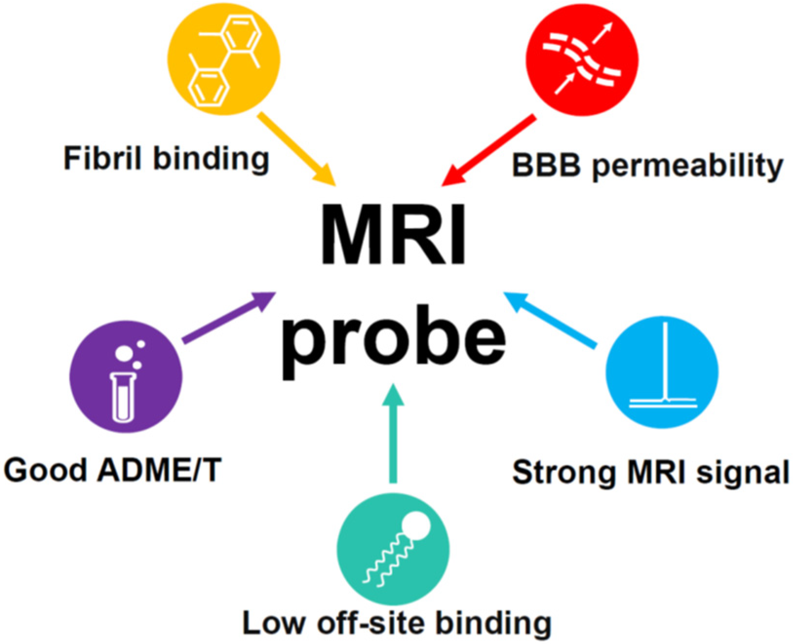
3.1.1. Requirements for an MRI Probe Candidate
- High affinity and specificity of binding to Aβ plaques: The affinity is usually determined in vitro using beta-amyloid binding assays based on radioligand binding, fluorescence titration, or other techniques. These experiments are not robust, and the value for the same compound may vary significantly depending on the exact conditions and methods used by different authors, which complicates our understanding of the structure–affinity relationships. The presence of certain molecular scaffolds discussed in the next section can guarantee a certain degree of affinity to beta-amyloid, which can be enhanced by various structural modifications. The approved PET probes have an affinity below 10 nM, and most of the compounds studied as potential probes for various imaging techniques have an affinity in the range of 1 to 1000 nM. The introduction of long, flexible chains carrying fluorinated groups into the molecule is likely to result in a decrease in the affinity [100]. Hence, the design of MRI probe candidates should be initiated from the precursors with the highest possible amyloid affinity.
- 2.
- Blood–brain barrier (BBB) permeability: The ability of amyloid probes to cross the BBB is indispensable for MRI imaging in vivo. Passive transmembrane diffusion of a compound through the BBB is possible for rather small lipophilic molecules. A molecular weight between 400 and 600 Da is usually considered as an upper border for BBB-permeable compounds. Under this threshold, the kinetic permeability logPS and the steady-state blood–brain partition coefficient logBB are, to some extent, correlated with 1-octanol-water partition coefficient logP (or logD for ionizable compounds) [101]. For larger molecules, the passive permeability rapidly decreases with the molecular size and is generally not correlated with logP. The cutoff value of logBB = 0 can be taken to classify compounds as permeable, which means an equal concentration of a compound on both sides of the barrier. The equivalent cutoff value for logPS is about –2 [101]. Despite the fact that the relationship of these quantities with logP is not strict, the value of logP (logD) > 1 is often recommended for the molecules that should pass the BBB. Additional commonly mentioned empirical rules for the BBB-permeable compounds are to form no more than eight hydrogen bonds with water; not to carry a negative charge, since the surface of the brain endothelial cells forming the BBB is itself negatively charged; and not to be a high-affinity serum protein binder [102,103]. For a more precise a priori logBB prediction, multiple QSAR models based on linear regressions or various machine learning approaches [104,105,106] have been developed.
- 3.
- Low binding to the membrane lipids in the brain: Binding to membrane lipids is thought to decrease the signal-to-noise ratio and can be reduced by lowering the probe’s lipophilicity. The upper border of the optimal logP (logD) value is about 3–4. The approved PET probes are moderately lipophilic, i.e., logD = 1.58 for florbetaben [110] and logP = 3.44 for flutemetamol [111], while for Shiga-X22, the predicted logP value equals 3.77 [67].
- 4.
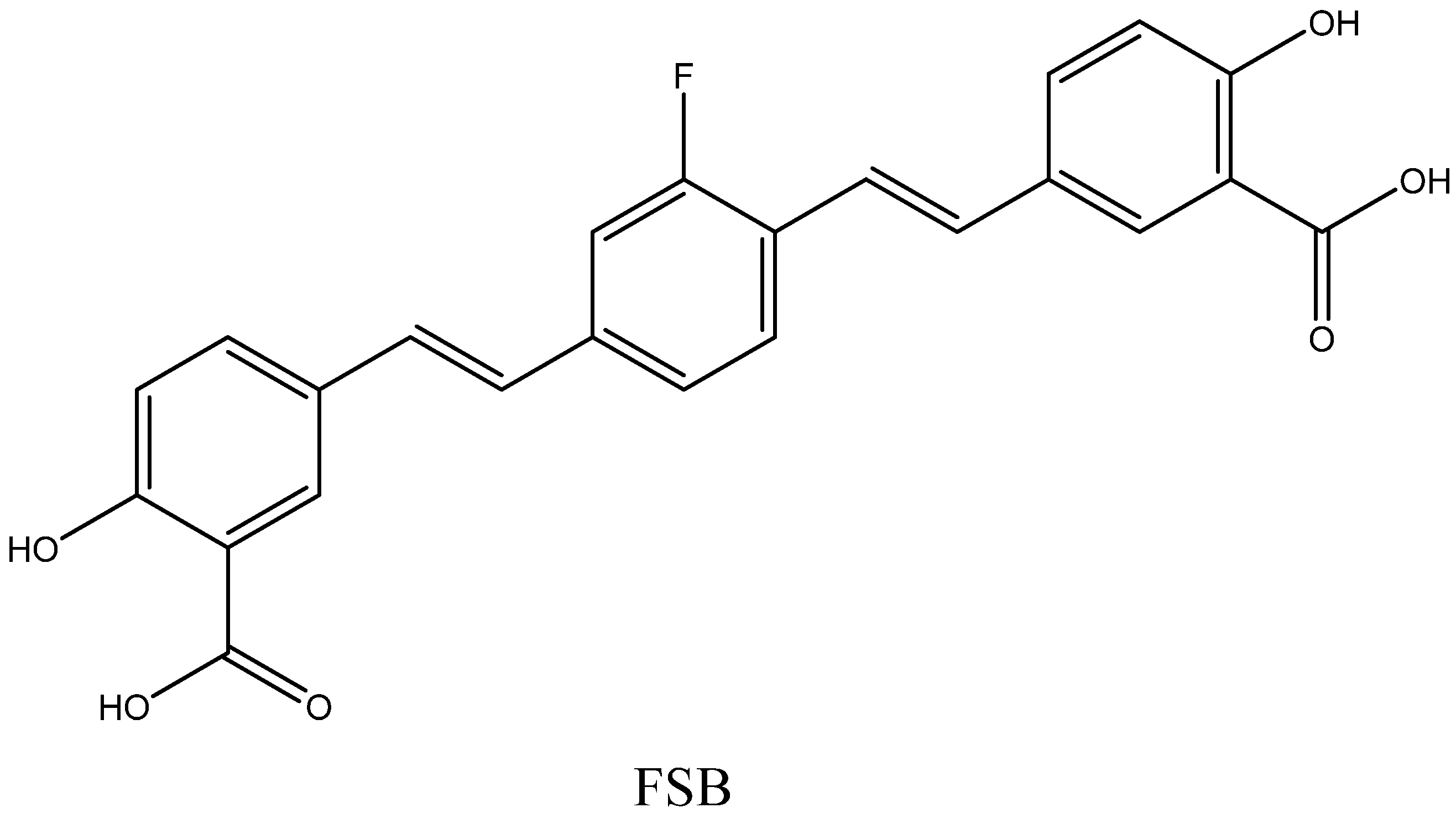
- Other ADMET properties: Sufficient in vivo stability, low toxicity, and an optimal clearance rate are necessary for the successful use of a compound as a diagnostic agent. Most of the known probe candidates have been studied in live mice and cell cultures and found to have low toxicity, with the exceptions of some compounds with long ethylene glycol chains [67] and possible nephrotoxicity of FSB at high doses [90].
- 5.
- Strong MRI signal. The NMR signal intensity of a compound is proportional to the number of fluorine atoms in its molecule [112,113]. The MRI probes that were studied in vivo contained one or two trifluoromethyl groups, and their signal-to-noise ratio was far from desirable [67,96]. However, as mentioned above, a larger number of fluorine atoms leads to higher hydrophobicity and increased off-target interactions with brain lipid components, as well as low solubility in water. An alternative strategy could be a simultaneous introduction of fluorines and polar groups, decreasing the hydrophobicity of a probe. The fluorine atoms should be arranged symmetrically in order to provide a single NMR peak. Moreover, fluorines should be separated from the amyloid-binding fragment by a flexible chain to avoid signal broadening and disappearance.
3.1.2. Molecular Fragments Providing Affinity to Aβ Fibrils
- 1.
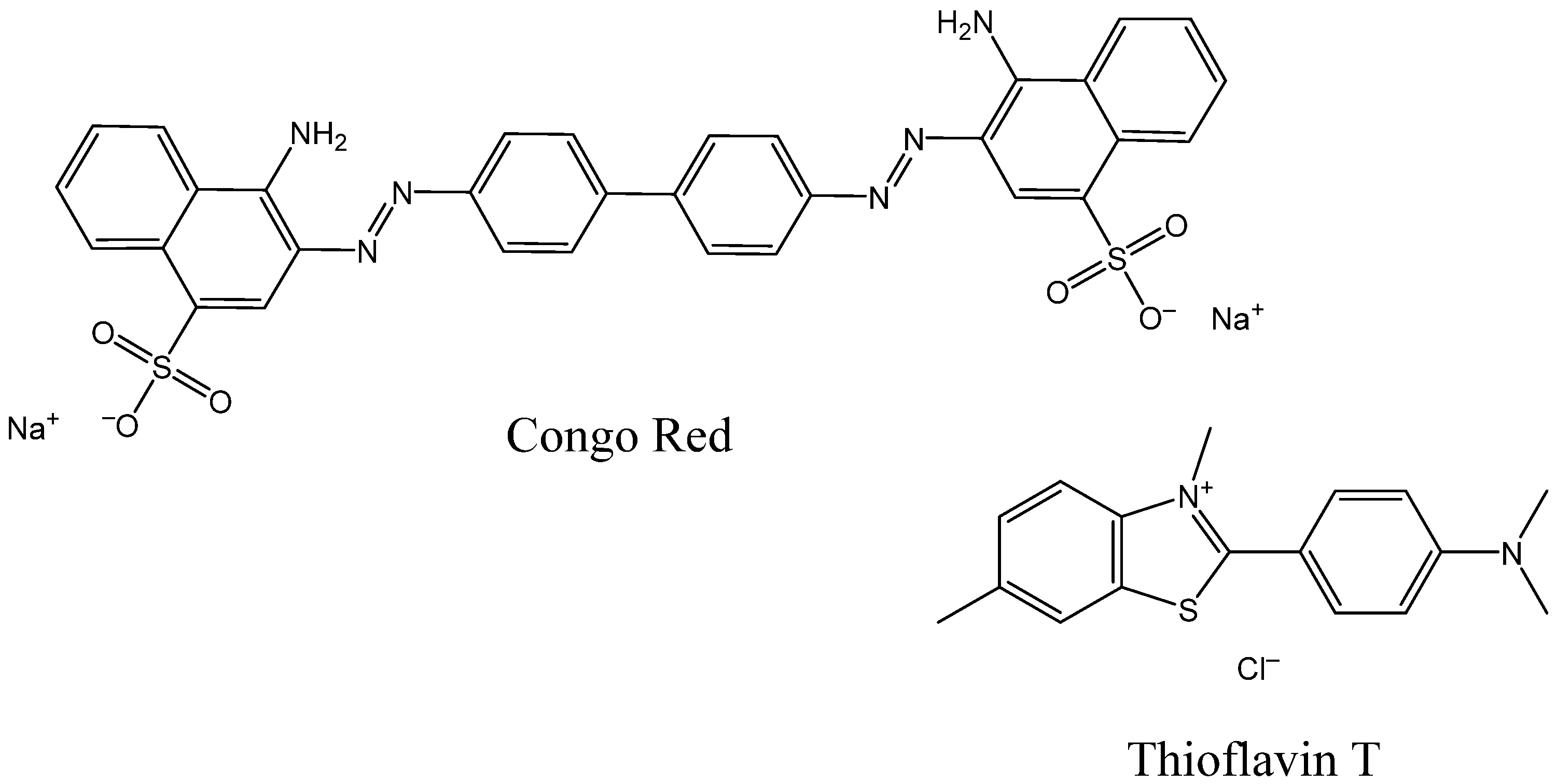
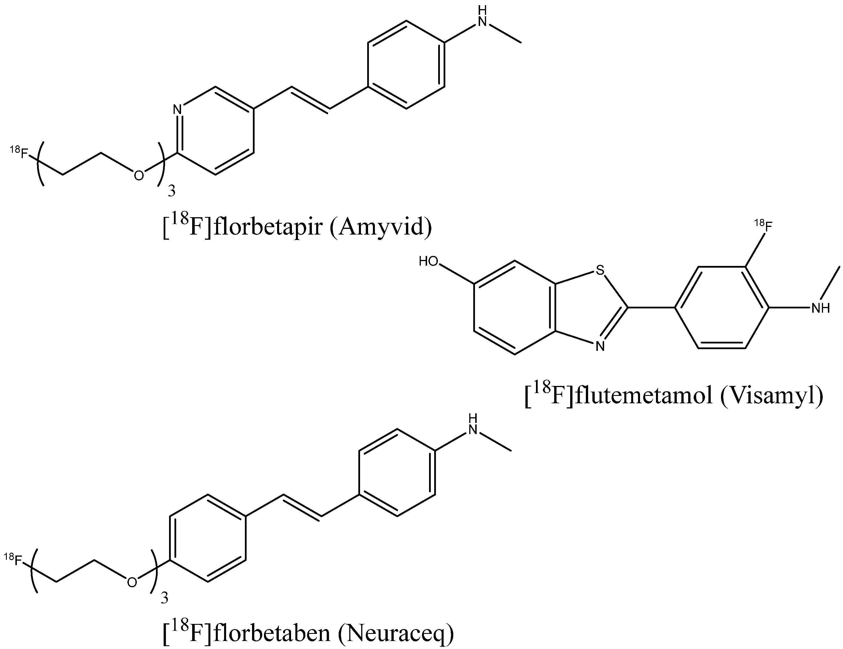
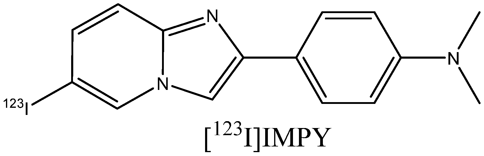
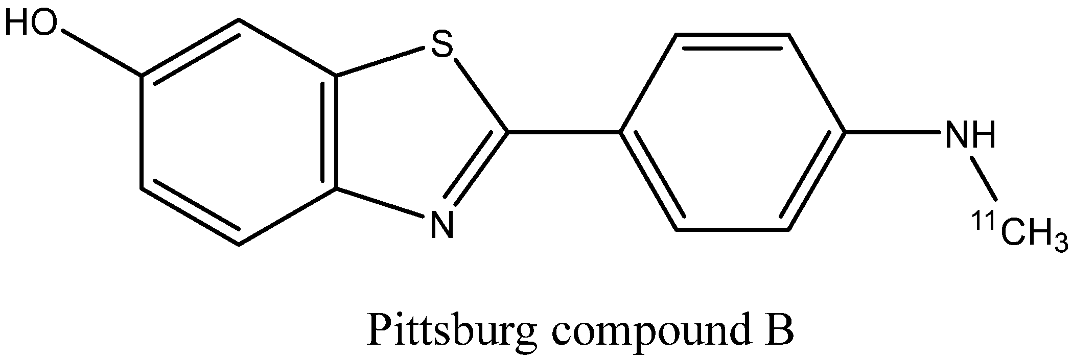
- Benzothiazole derivatives [30,126,127]: Prominent representatives are thioflavin T, flutemetamol, and Pittsburgh compound B (Figure 20) [128], a PET probe used in numerous early in vivo imaging experiments [129]. Most of the synthesized ligands contain an aromatic substituent or a conjugated double-bond system attached to the C2 atom. QSAR analysis shows that the monoalkyl amino group in the para position to benzothiazole increases the binding affinity [28]. Substituted benzoxazoles, benzothiophenes, benzofurans, imidazopyridines (e.g., IMPY), and other bioisosteric heterocycles [130,131] can also show strong fibril binding.

- 2.
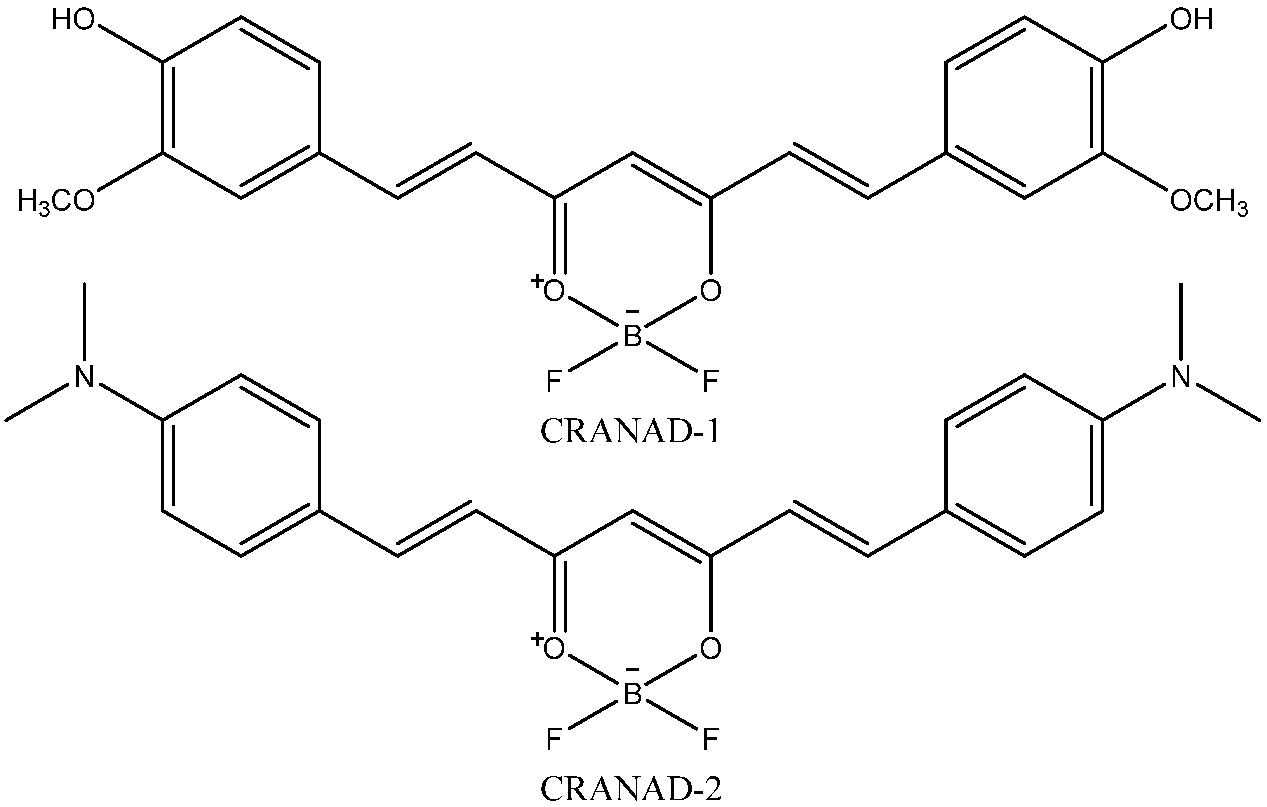
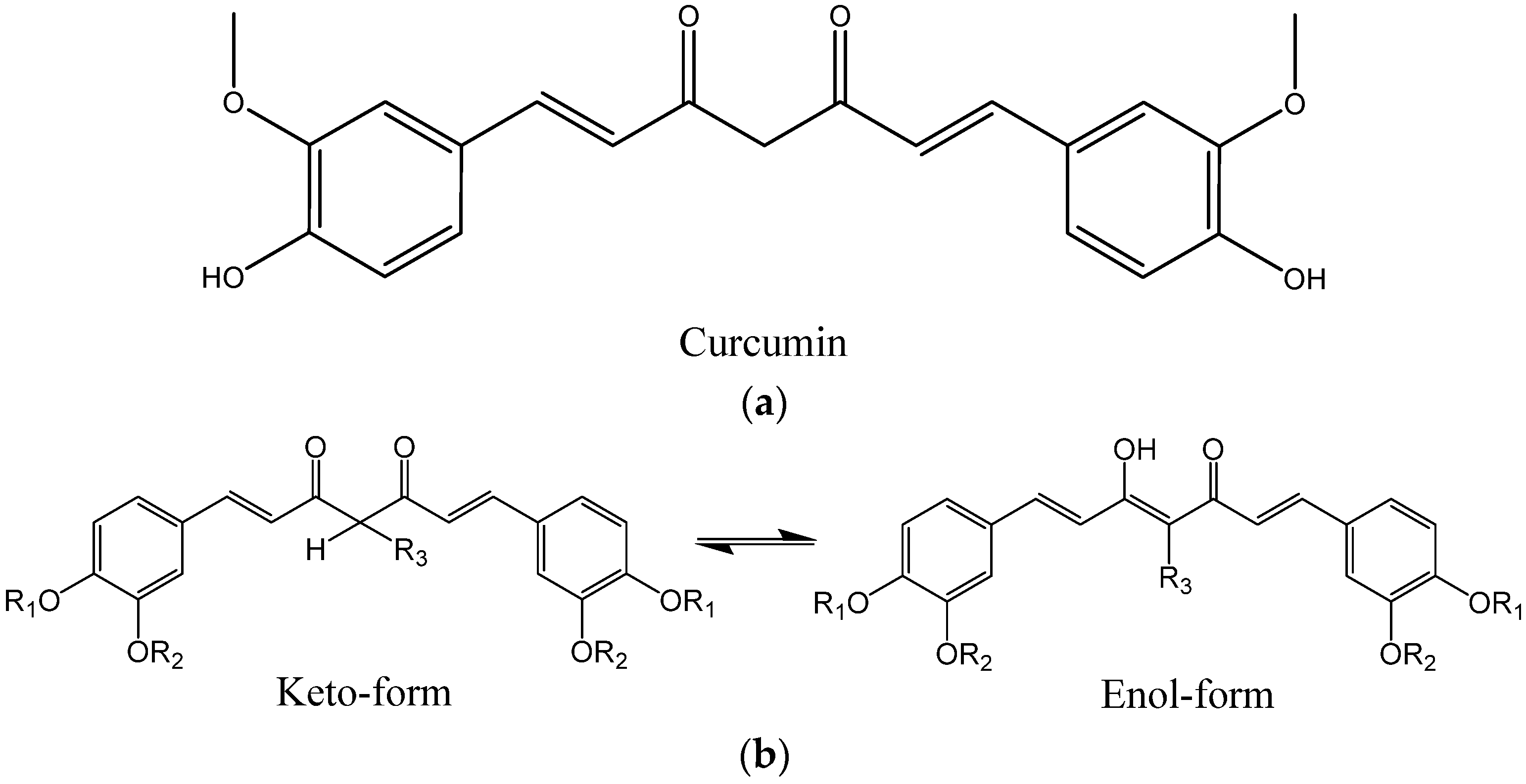
- Curcumin (Figure 21a) derivatives [45,132,133,134], which include CRANAD dyes and some of the Shiga-Y series compounds: It was reported that the enol form of curcumin derivatives (Figure 21b) exhibits a high affinity to Aβ fibrils, while the keto form shows weaker binding, leading to the bound state becoming predominantly enolic [94]. While curcumin itself is chemically unstable, weakly soluble in water, prone to non-specific binding with many different targets, and has a poor pharmacokinetic profile, some of its derivatives can be more suitable for probe development. The replacement of β-diketone with a difluoroboronate moiety in the CRANAD series or with a single carbonyl group [132] helps to overcome the pharmacokinetic limitations. Replacing the two phenyl rings of curcumin with pyridyls can strengthen the interaction with proton donor residues of Aβ [135]. The presence of a rather rigid linker between the aromatic rings, like in curcumin, seems to be a prerequisite for high Aβ affinity [136].

- 3.
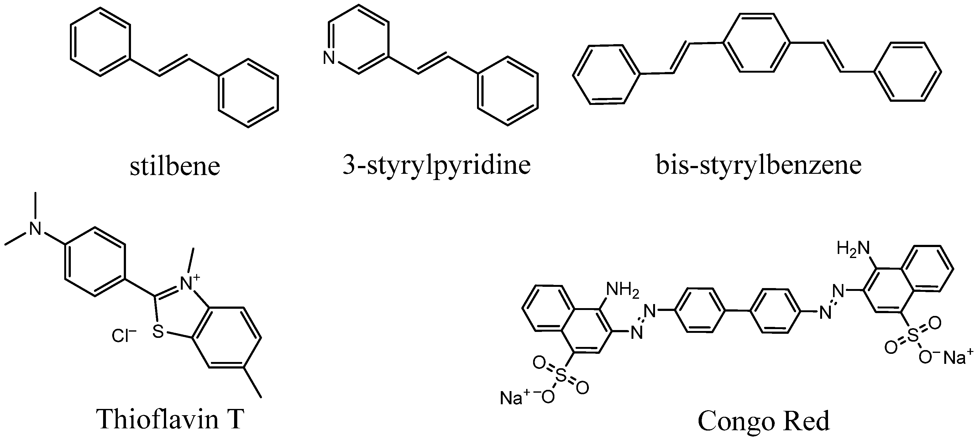
- Stilbenes, styrylpyridines, and bis-styrylbenzenes (Figure 22) [92,100,137,138]: These compounds also have aromatic rings separated by rigid conjugated double bonds. The aforementioned PET probes, florbetaben and florbetapir, as well as the FSB compound, belong to this group. The studies of their amyloid affinity were inspired by certain structural similarities of stilbenes with benzothiazoles (particularly thioflavin T) and bis-styrylbenzenes with Congo Red dye. Some non-stilbene (azo) fibril-binding Congo Red derivatives have also been obtained [38,139,140]. Bis-styrylbenzenes have also been modified by replacing the central benzene unit with naphthalene, thiophene, or other heterocyclic moieties [137].

- 4.
- Dicyanomethylene derivatives [30,141,142]. A well-known representative is the PET probe FDDNP (Figure 23a) [143], used for research purposes. These compounds are π-conjugated systems comprising double bonds and an aromatic ring, often with an electron donor dialkyl amino substituent in para-position (Figure 23b).

- 5.
3.2. Probes Binding to Non-Aβ Fibrils
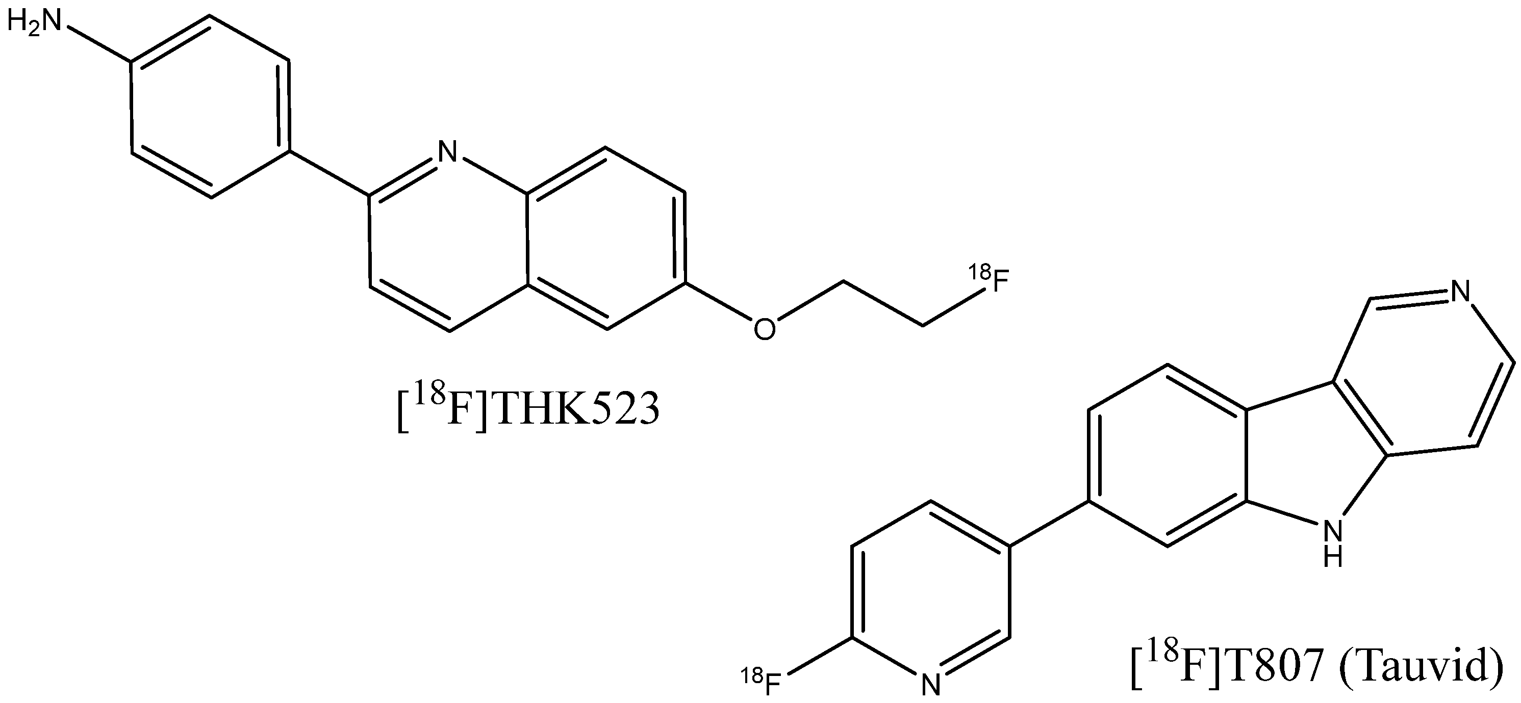
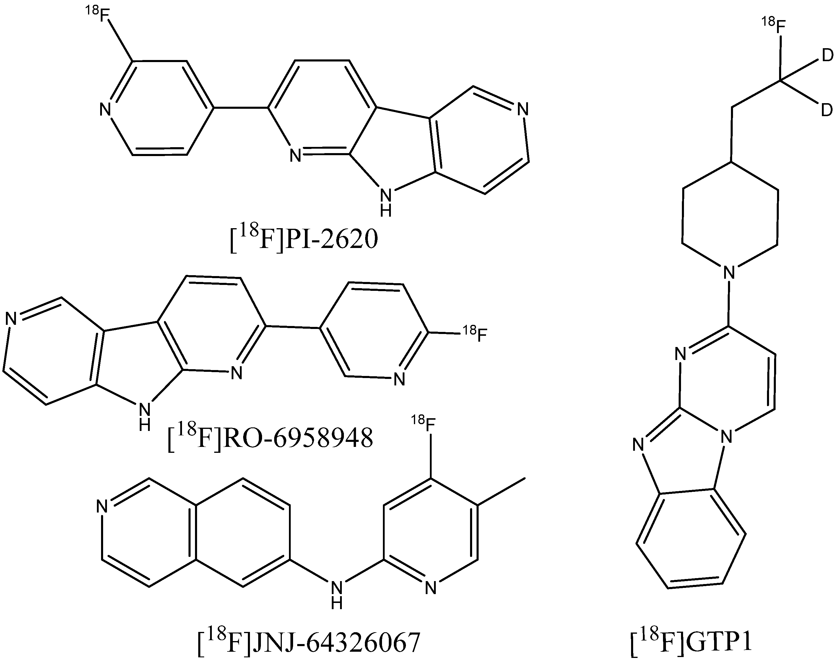
3.2.1. Imaging Tracers of Tau Pathology
3.2.2. Probes for Visualization of Non-AD-Related Amyloid Deposits
3.3. Metal Complex-Based Contrast Agents
4. Conclusions
Author Contributions
Funding
Conflicts of Interest
References
- 2020 Alzheimer’s Disease Facts and Figures. Alzheimer’s Dement. 2020, 16, 391–460. [CrossRef] [PubMed]
- Yeo, S.K.; Shepelytskyi, Y.; Grynko, V.; Albert, M.S. Molecular Imaging of Fluorinated Probes for Tau Protein and Amyloid-β Detection. Molecules 2020, 25, 3413. [Google Scholar] [CrossRef] [PubMed]
- Khan, S.; Barve, K.H.; Kumar, M.S. Recent Advancements in Pathogenesis, Diagnostics and Treatment of Alzheimer’s Disease. Curr. Neuropharmacol. 2020, 18, 1106–1125. [Google Scholar] [CrossRef] [PubMed]
- Jack, C.R.; Bennett, D.A.; Blennow, K.; Carrillo, M.C.; Dunn, B.; Haeberlein, S.B.; Holtzman, D.M.; Jagust, W.; Jessen, F.; Karlawish, J.; et al. NIA-AA Research Framework: Toward a Biological Definition of Alzheimer’s Disease. Alzheimer’s Dement. 2018, 14, 535–562. [Google Scholar] [CrossRef] [PubMed]
- McKhann, G.M.; Knopman, D.S.; Chertkow, H.; Hyman, B.T.; Jack, C.R.; Kawas, C.H.; Klunk, W.E.; Koroshetz, W.J.; Manly, J.J.; Mayeux, R.; et al. The Diagnosis of Dementia Due to Alzheimer’s Disease: Recommendations from the National Institute on Aging-Alzheimer’s Association Workgroups on Diagnostic Guidelines for Alzheimer’s Disease. Alzheimer’s Dement. 2011, 7, 263–269. [Google Scholar] [CrossRef] [PubMed] [Green Version]
- Weller, J.; Budson, A. Current Understanding of Alzheimer’s Disease Diagnosis and Treatment. F1000Research 2018, 7, 1161. [Google Scholar] [CrossRef] [PubMed] [Green Version]
- Hansson, O.; Seibyl, J.; Stomrud, E.; Zetterberg, H.; Trojanowski, J.Q.; Bittner, T.; Lifke, V.; Corradini, V.; Eichenlaub, U.; Batrla, R.; et al. CSF Biomarkers of Alzheimer’s Disease Concord with Amyloid-β PET and Predict Clinical Progression: A Study of Fully Automated Immunoassays in BioFINDER and ADNI Cohorts. Alzheimer’s Dement. 2018, 14, 1470–1481. [Google Scholar] [CrossRef]
- Budson, A.E.; Solomon, P.R. New Criteria for Alzheimer Disease and Mild Cognitive Impairment: Implications for the Practicing Clinician. Neurologist 2012, 18, 356–363. [Google Scholar] [CrossRef] [PubMed]
- Braak, H.; Braak, E. Morphological Criteria for the Recognition of Alzheimer’s Disease and the Distribution Pattern of Cortical Changes Related to This Disorder. Neurobiol. Aging 1994, 15, 355–356. [Google Scholar] [CrossRef]
- Hardy, J.A.; Higgins, G.A. Alzheimer’s Disease: The Amyloid Cascade Hypothesis. Science 1992, 256, 184–185. [Google Scholar] [CrossRef]
- Hardy, J.; Selkoe, D.J. The Amyloid Hypothesis of Alzheimer’s Disease: Progress and Problems on the Road to Therapeutics. Science 2002, 297, 353–356. [Google Scholar] [CrossRef] [PubMed] [Green Version]
- Selkoe, D.J.; Hardy, J. The Amyloid Hypothesis of Alzheimer’s Disease at 25 Years. EMBO Mol. Med. 2016, 8, 595–608. [Google Scholar] [CrossRef] [PubMed]
- Huang, L.-K.; Chao, S.-P.; Hu, C.-J. Clinical Trials of New Drugs for Alzheimer Disease. J. Biomed. Sci. 2020, 27, 18. [Google Scholar] [CrossRef] [PubMed] [Green Version]
- Bateman, R.J.; Xiong, C.; Benzinger, T.L.S.; Fagan, A.M.; Goate, A.; Fox, N.C.; Marcus, D.S.; Cairns, N.J.; Xie, X.; Blazey, T.M.; et al. Clinical and Biomarker Changes in Dominantly Inherited Alzheimer’s Disease. N. Engl. J. Med. 2012, 367, 795–804. [Google Scholar] [CrossRef] [PubMed] [Green Version]
- Janeiro, M.H.; Ardanaz, C.G.; Sola-Sevilla, N.; Dong, J.; Cortés-Erice, M.; Solas, M.; Puerta, E.; Ramírez, M.J. Biomarkers in Alzheimer’s Disease. Adv. Lab. Med. Adv. En Med. Lab. 2021, 2, 27–37. [Google Scholar] [CrossRef] [PubMed]
- Dudeffant, C.; Vandesquille, M.; Herbert, K.; Garin, C.M.; Alves, S.; Blanchard, V.; Comoy, E.E.; Petit, F.; Dhenain, M. Contrast-Enhanced MR Microscopy of Amyloid Plaques in Five Mouse Models of Amyloidosis and in Human Alzheimer’s Disease Brains. Sci. Rep. 2017, 7, 4955. [Google Scholar] [CrossRef] [PubMed] [Green Version]
- 2021 Alzheimer’s Disease Facts and Figures. Alzheimer’s Dement. 2021, 17, 327–406. [CrossRef]
- Rajan, K.B.; Weuve, J.; Barnes, L.L.; McAninch, E.A.; Wilson, R.S.; Evans, D.A. Population Estimate of People with Clinical Alzheimer’s Disease and Mild Cognitive Impairment in the United States (2020–2060). Alzheimer’s Dement. 2021, 17, 1966–1975. [Google Scholar] [CrossRef]
- Kaur, A.; New, E.J.; Sunde, M. Strategies for the Molecular Imaging of Amyloid and the Value of a Multimodal Approach. ACS Sens. 2020, 5, 2268–2282. [Google Scholar] [CrossRef]
- Arora, A.; Bhagat, N. Insight into the Molecular Imaging of Alzheimer’s Disease. Int. J. Biomed. Imaging 2016, 2016, 7462014. [Google Scholar] [CrossRef] [Green Version]
- Mori, T.; Maeda, J.; Shimada, H.; Higuchi, M.; Shinotoh, H.; Ueno, S.; Suhara, T. Molecular Imaging of Dementia: Molecular Imaging for Dementia. Psychogeriatrics 2012, 12, 106–114. [Google Scholar] [CrossRef] [Green Version]
- Furumoto, S.; Okamura, N.; Iwata, R.; Yanai, K.; Arai, H.; Kudo, Y. Recent Advances in the Development of Amyloid Imaging Agents. Curr. Top. Med. Chem. 2007, 7, 1773–1789. [Google Scholar] [CrossRef] [PubMed]
- Zhu, L.; Ploessl, K.; Kung, H.F. PET/SPECT Imaging Agents for Neurodegenerative Diseases. Chem. Soc. Rev. 2014, 43, 6683–6691. [Google Scholar] [CrossRef] [PubMed] [Green Version]
- Klunk, W.E.; Mathis, C.A. The Future of Amyloid-Beta Imaging: A Tale of Radionuclides and Tracer Proliferation. Curr. Opin. Neurol. 2008, 21, 683–687. [Google Scholar] [CrossRef] [PubMed] [Green Version]
- Uzuegbunam, B.C.; Librizzi, D.; Hooshyar Yousefi, B. PET Radiopharmaceuticals for Alzheimer’s Disease and Parkinson’s Disease Diagnosis, the Current and Future Landscape. Molecules 2020, 25, 977. [Google Scholar] [CrossRef] [PubMed] [Green Version]
- Hickey, J.L.; Donnelly, P.S. Diagnostic Imaging of Alzheimer’s Disease with Copper and Technetium Complexes. Coord. Chem. Rev. 2012, 256, 2367–2380. [Google Scholar] [CrossRef]
- Hayne, D.J.; Lim, S.; Donnelly, P.S. Metal Complexes Designed to Bind to Amyloid-β for the Diagnosis and Treatment of Alzheimer’s Disease. Chem. Soc. Rev. 2014, 43, 6701–6715. [Google Scholar] [CrossRef] [PubMed] [Green Version]
- Papagiannopoulou, D.; Hadjipavlou-Litina, D. Computational Modeling of Diagnostic Imaging Agents for Alzheimer’s Disease: Molecular Imaging Agents for the In Vivo Detection of Amyloid Plaques in Alzheimer’s Disease. In Computational Modeling of Drugs against Alzheimer’s Disease; Roy, K., Ed.; Neuromethods; Springer: New York, NY, USA, 2018; Volume 132, pp. 463–479. ISBN 978-1-4939-7403-0. [Google Scholar]
- Aliyan, A.; Cook, N.P.; Martí, A.A. Interrogating Amyloid Aggregates Using Fluorescent Probes. Chem. Rev. 2019, 119, 11819–11856. [Google Scholar] [CrossRef] [PubMed]
- Zhang, Y.; Ding, C.; Li, C.; Wang, X. Advances in Fluorescent Probes for Detection and Imaging of Amyloid-β Peptides in Alzheimer’s Disease. In Advances in Clinical Chemistry; Elsevier: Amsterdam, The Netherlands, 2021; Volume 103, pp. 135–190. ISBN 978-0-12-824616-0. [Google Scholar]
- Bertoncini, C.W.; Soledad Celej, M. Small Molecule Fluorescent Probes for the Detection of Amyloid Self-Assembly In Vitro and In Vivo. Curr. Protein Pept. Sci. 2011, 12, 206–220. [Google Scholar] [CrossRef]
- Yang, J.; Guo, Y.; Pistolozzi, M.; Yan, J. Research Progress of Multi-Functional Fluorescent Probes for Alzheimer’s Disease Monitoring. Dye. Pigment. 2021, 193, 109466. [Google Scholar] [CrossRef]
- Gyasi, Y.I.; Pang, Y.-P.; Li, X.-R.; Gu, J.-X.; Cheng, X.-J.; Liu, J.; Xu, T.; Liu, Y. Biological Applications of near Infrared Fluorescence Dye Probes in Monitoring Alzheimer’s Disease. Eur. J. Med. Chem. 2020, 187, 111982. [Google Scholar] [CrossRef] [PubMed]
- Teipel, S.J.; Grothe, M.; Lista, S.; Toschi, N.; Garaci, F.G.; Hampel, H. Relevance of Magnetic Resonance Imaging for Early Detection and Diagnosis of Alzheimer Disease. Med. Clin. N. Am. 2013, 97, 399–424. [Google Scholar] [CrossRef] [PubMed]
- Colliot, O.; Hamelin, L.; Sarazin, M. Magnetic Resonance Imaging for Diagnosis of Early Alzheimer’s Disease. Rev. Neurol. 2013, 169, 724–728. [Google Scholar] [CrossRef] [PubMed] [Green Version]
- Salerno, M.; Santo Domingo Porqueras, D. Alzheimer’s Disease: The Use of Contrast Agents for Magnetic Resonance Imaging to Detect Amyloid Beta Peptide inside the Brain. Coord. Chem. Rev. 2016, 327–328, 27–34. [Google Scholar] [CrossRef]
- Groenning, M. Binding Mode of Thioflavin T and Other Molecular Probes in the Context of Amyloid Fibrils—Current Status. J. Chem. Biol. 2010, 3, 1–18. [Google Scholar] [CrossRef] [PubMed] [Green Version]
- Yakupova, E.I.; Bobyleva, L.G.; Vikhlyantsev, I.M.; Bobylev, A.G. Congo Red and Amyloids: History and Relationship. Biosci. Rep. 2019, 39, BSR20181415. [Google Scholar] [CrossRef] [PubMed] [Green Version]
- Wu, C.; Scott, J.; Shea, J.-E. Binding of Congo Red to Amyloid Protofibrils of the Alzheimer Aβ9–40 Peptide Probed by Molecular Dynamics Simulations. Biophys. J. 2012, 103, 550–557. [Google Scholar] [CrossRef] [Green Version]
- Xue, C.; Lin, T.Y.; Chang, D.; Guo, Z. Thioflavin T as an Amyloid Dye: Fibril Quantification, Optimal Concentration and Effect on Aggregation. R. Soc. Open Sci. 2017, 4, 160696. [Google Scholar] [CrossRef] [Green Version]
- Noël, S.; Cadet, S.; Gras, E.; Hureau, C. The Benzazole Scaffold: A SWAT to Combat Alzheimer’s Disease. Chem. Soc. Rev. 2013, 42, 7747. [Google Scholar] [CrossRef]
- Kosaka, N.; Ogawa, M.; Choyke, P.L.; Kobayashi, H. Clinical Implications of Near-Infrared Fluorescence Imaging in Cancer. Future Oncol. 2009, 5, 1501–1511. [Google Scholar] [CrossRef] [Green Version]
- Cao, J.; Zhu, B.; Zheng, K.; He, S.; Meng, L.; Song, J.; Yang, H. Recent Progress in NIR-II Contrast Agent for Biological Imaging. Front. Bioeng. Biotechnol. 2020, 7, 487. [Google Scholar] [CrossRef] [PubMed] [Green Version]
- Teraphongphom, N.; Kong, C.S.; Warram, J.M.; Rosenthal, E.L. Specimen Mapping in Head and Neck Cancer Using Fluorescence Imaging: Specimen Mapping in HNC. Laryngoscope Investig. Otolaryngol. 2017, 2, 447–452. [Google Scholar] [CrossRef] [PubMed] [Green Version]
- Ran, C.; Xu, X.; Raymond, S.B.; Ferrara, B.J.; Neal, K.; Bacskai, B.J.; Medarova, Z.; Moore, A. Design, Synthesis, and Testing of Difluoroboron-Derivatized Curcumins as Near-Infrared Probes for in Vivo Detection of Amyloid-β Deposits. J. Am. Chem. Soc. 2009, 131, 15257–15261. [Google Scholar] [CrossRef] [PubMed] [Green Version]
- Ren, W.; Li, L.; Zhang, J.; Vaas, M.; Klohs, J.; Ripoll, J.; Wolf, M.; Ni, R.; Rudin, M. Non-Invasive Visualization of Amyloid-Beta Deposits in Alzheimer Amyloidosis Mice Using Magnetic Resonance Imaging and Fluorescence Molecular Tomography. Biomed. Opt. Express 2022, 13, 3809. [Google Scholar] [CrossRef] [PubMed]
- Li, H.; Wang, J.; Li, Y.; Chen, X.; Zhang, W.; Zhao, Y.; Liu, G.; Pan, J. Detection of Aβ Oligomers in Early Alzheimer’s Disease Diagnose by in Vivo NIR-II Fluorescence Imaging. Sens. Actuators B Chem. 2022, 358, 131481. [Google Scholar] [CrossRef]
- Miao, J.; Miao, M.; Jiang, Y.; Zhao, M.; Li, Q.; Zhang, Y.; An, Y.; Pu, K.; Miao, Q. An Activatable NIR-II Fluorescent Reporter for In Vivo Imaging of Amyloid-β Plaques. Angew. Chem. Int. Ed. 2023, 62, e202216351. [Google Scholar] [CrossRef] [PubMed]
- Yanagisawa, D.; Ibrahim, N.F.; Taguchi, H.; Morikawa, S.; Kato, T.; Hirao, K.; Shirai, N.; Sogabe, T.; Tooyama, I. Fluorine-19 Magnetic Resonance Imaging Probe for the Detection of Tau Pathology in Female RTg4510 Mice. J. Neuro Res. 2018, 96, 841–851. [Google Scholar] [CrossRef]
- Vaquero, J.J.; Kinahan, P. Positron Emission Tomography: Current Challenges and Opportunities for Technological Advances in Clinical and Preclinical Imaging Systems. Annu. Rev. Biomed. Eng. 2015, 17, 385–414. [Google Scholar] [CrossRef] [PubMed] [Green Version]
- Zanzonico, P. Positron Emission Tomography: A Review of Basic Principles, Scanner Design and Performance, and Current Systems. Semin. Nucl. Med. 2004, 34, 87–111. [Google Scholar] [CrossRef] [Green Version]
- Xu, C.; Mu, L.; Roes, I.; Miranda-Nieves, D.; Nahrendorf, M.; Ankrum, J.A.; Zhao, W.; Karp, J.M. Nanoparticle-Based Monitoring of Cell Therapy. Nanotechnology 2011, 22, 494001. [Google Scholar] [CrossRef] [Green Version]
- Accorsi, R. Brain Single-Photon Emission CT Physics Principles. AJNR Am. J. Neuroradiol. 2008, 29, 1247–1256. [Google Scholar] [CrossRef] [PubMed] [Green Version]
- Du, Y.; Zaidi, H. Single-Photon Emission Computed Tomography: Principles and Applications. In Encyclopedia of Biomedical Engineering; Elsevier: Amsterdam, The Netherlands, 2019; pp. 493–506. ISBN 978-0-12-805144-3. [Google Scholar]
- Wong, D.F.; Rosenberg, P.B.; Zhou, Y.; Kumar, A.; Raymont, V.; Ravert, H.T.; Dannals, R.F.; Nandi, A.; Brašić, J.R.; Ye, W.; et al. In Vivo Imaging of Amyloid Deposition in Alzheimer Disease Using the Radioligand 18F-AV-45 (Flobetapir F 18). J. Nucl. Med. 2010, 51, 913–920. [Google Scholar] [CrossRef] [PubMed] [Green Version]
- Curtis, C.; Gamez, J.E.; Singh, U.; Sadowsky, C.H.; Villena, T.; Sabbagh, M.N.; Beach, T.G.; Duara, R.; Fleisher, A.S.; Frey, K.A.; et al. Phase 3 Trial of Flutemetamol Labeled With Radioactive Fluorine 18 Imaging and Neuritic Plaque Density. JAMA Neurol. 2015, 72, 287. [Google Scholar] [CrossRef] [PubMed]
- Schipke, C.G.; Peters, O.; Heuser, I.; Grimmer, T.; Sabbagh, M.N.; Sabri, O.; Hock, C.; Kunz, M.; Kuhlmann, J.; Reininger, C.; et al. Impact of Beta-Amyloid-Specific Florbetaben PET Imaging on Confidence in Early Diagnosis of Alzheimer’s Disease. Dement. Geriatr. Cogn. Disord. 2012, 33, 416–422. [Google Scholar] [CrossRef] [PubMed] [Green Version]
- Clark, C.M.; Pontecorvo, M.J.; Beach, T.G.; Bedell, B.J.; Coleman, R.E.; Doraiswamy, P.M.; Fleisher, A.S.; Reiman, E.M.; Sabbagh, M.N.; Sadowsky, C.H.; et al. Cerebral PET with Florbetapir Compared with Neuropathology at Autopsy for Detection of Neuritic Amyloid-β Plaques: A Prospective Cohort Study. Lancet Neurol. 2012, 11, 669–678. [Google Scholar] [CrossRef] [PubMed]
- Sabri, O.; Seibyl, J.; Rowe, C.; Barthel, H. Beta-Amyloid Imaging with Florbetaben. Clin. Transl. Imaging 2015, 3, 13–26. [Google Scholar] [CrossRef] [PubMed] [Green Version]
- Salloway, S.; Gamez, J.E.; Singh, U.; Sadowsky, C.H.; Villena, T.; Sabbagh, M.N.; Beach, T.G.; Duara, R.; Fleisher, A.S.; Frey, K.A.; et al. Performance of [18F]Flutemetamol Amyloid Imaging against the Neuritic Plaque Component of CERAD and the Current (2012) NIA-AA Recommendations for the Neuropathologic Diagnosis of Alzheimer’s Disease. Alzheimer’s Dement. Diagn. Assess. Dis. Monit. 2017, 9, 25–34. [Google Scholar] [CrossRef]
- Yao, C.-H.; Lin, K.-J.; Weng, C.-C.; Hsiao, I.-T.; Ting, Y.-S.; Yen, T.-C.; Jan, T.-R.; Skovronsky, D.; Kung, M.-P.; Wey, S.-P. GMP-Compliant Automated Synthesis of [18F]AV-45 (Florbetapir F 18) for Imaging β-Amyloid Plaques in Human Brain. Appl. Radiat. Isot. 2010, 68, 2293–2297. [Google Scholar] [CrossRef]
- Lisova, K.; Wang, J.; Chao, P.H.; van Dam, R.M. A Simple and Efficient Automated Microvolume Radiosynthesis of [18F]Florbetaben. EJNMMI Radiopharm. Chem. 2020, 5, 30. [Google Scholar] [CrossRef]
- Wang, H.; Guo, X.; Jiang, S.; Tang, G. Automated Synthesis of [18F]Florbetaben as Alzheimer’s Disease Imaging Agent Based on a Synthesis Module System. Appl. Radiat. Isot. 2013, 71, 41–46. [Google Scholar] [CrossRef]
- Petrov, S.A.; Yusubov, M.S.; Beloglazkina, E.K.; Nenajdenko, V.G. Synthesis of Radioiodinated Compounds. Classical Approaches and Achievements of Recent Years. Int. J. Mol. Sci. 2022, 23, 13789. [Google Scholar] [CrossRef] [PubMed]
- Jacobson, O.; Kiesewetter, D.O.; Chen, X. Fluorine-18 Radiochemistry, Labeling Strategies and Synthetic Routes. Bioconjugate Chem. 2015, 26, 1–18. [Google Scholar] [CrossRef] [PubMed] [Green Version]
- Newberg, A.B.; Wintering, N.A.; Plössl, K.; Hochold, J.; Stabin, M.G.; Watson, M.; Skovronsky, D.; Clark, C.M.; Kung, M.-P.; Kung, H.F. Safety, Biodistribution, and Dosimetry of 123I-IMPY: A Novel Amyloid Plaque-Imaging Agent for the Diagnosis of Alzheimer’s Disease. J. Nucl. Med. 2006, 47, 748–754. [Google Scholar] [PubMed]
- Tooyama, I.; Yanagisawa, D.; Taguchi, H.; Kato, T.; Hirao, K.; Shirai, N.; Sogabe, T.; Ibrahim, N.F.; Inubushi, T.; Morikawa, S. Amyloid Imaging Using Fluorine-19 Magnetic Resonance Imaging (19F-MRI). Ageing Res. Rev. 2016, 30, 85–94. [Google Scholar] [CrossRef] [PubMed]
- Lu, F.-M.; Yuan, Z. PET/SPECT Molecular Imaging in Clinical Neuroscience: Recent Advances in the Investigation on CNS Diseases. Quant. Imaging Med. Surg. 2015, 5, 433–447. [Google Scholar] [CrossRef] [PubMed]
- Wahsner, J.; Gale, E.M.; Rodríguez-Rodríguez, A.; Caravan, P. Chemistry of MRI Contrast Agents: Current Challenges and New Frontiers. Chem. Rev. 2019, 119, 957–1057. [Google Scholar] [CrossRef] [PubMed]
- Yu, Z.; He, Q.; Yang, J.; Luo, M. A Supervised ML Applied Classification Model for Brain Tumors MRI. Front. Pharmacol. 2022, 13, 884495. [Google Scholar] [CrossRef] [PubMed]
- Petersen, S.E.; Aung, N.; Sanghvi, M.M.; Zemrak, F.; Fung, K.; Paiva, J.M.; Francis, J.M.; Khanji, M.Y.; Lukaschuk, E.; Lee, A.M.; et al. Reference Ranges for Cardiac Structure and Function Using Cardiovascular Magnetic Resonance (CMR) in Caucasians from the UK Biobank Population Cohort. J. Cardiovasc. Magn. Reson. 2017, 19, 18. [Google Scholar] [CrossRef] [Green Version]
- ACCF/ACR/SCCT/SCMR/ASNC/NASCI/SCAI/SIR 2006 Appropriateness Criteria for Cardiac Computed Tomography and Cardiac Magnetic Resonance Imaging. J. Am. Coll. Radiol. 2006, 3, 751–771. [CrossRef]
- Hughes, P.; Miranda, R.; Doyle, A.J. MRI Imaging of Soft Tissue Tumours of the Foot and Ankle. Insights Imaging 2019, 10, 60. [Google Scholar] [CrossRef]
- Aoki, T.; Fujisaki, A.; Terasawa, T.; Hayashida, Y.; Todoroki, Y.; Hirano, N.; Hisaoka, M.; Sakai, A.; Korogi, Y. Primary Site Identification of Soft-Tissue Mass: Things to Know in MRI Assessment. Magn. Reson. Imaging 2022, 55, 37–47. [Google Scholar] [CrossRef] [PubMed]
- Ng, S.N.; Axelsen, M.B.; Østergaard, M.; Pedersen, S.J.; Eshed, I.; Hetland, M.L.; Møller, J.M.; Terslev, L. Whole-Body Magnetic Resonance Imaging Assessment of Joint Inflammation in Rheumatoid Arthritis—Agreement with Ultrasonography and Clinical Evaluation. Front. Med. 2020, 7, 285. [Google Scholar] [CrossRef] [PubMed]
- Marzo-Ortega, H.; Tanner, S.F.; Rhodes, L.A.; Tan, A.L.; Conaghan, P.G.; Hensor, E.M.A.; Radjenovic, A.; O’Connor, P.; Emery, P.; McGonagle, D. Magnetic Resonance Imaging in the Assessment of Metacarpophalangeal Joint Disease in Early Psoriatic and Rheumatoid Arthritis. Scand. J. Rheumatol. 2009, 38, 79–83. [Google Scholar] [CrossRef] [PubMed]
- Hope, T.A.; Hope, M.D.; Purcell, D.D.; von Morze, C.; Vigneron, D.B.; Alley, M.T.; Dillon, W.P. Evaluation of Intracranial Stenoses and Aneurysms with Accelerated 4D Flow. Magn. Reson. Imaging 2010, 28, 41–46. [Google Scholar] [CrossRef] [PubMed]
- Coenegrachts, K. Magnetic Resonance Imaging of the Liver: New Imaging Strategies for Evaluating Focal Liver Lesions. World J. Radiol. 2009, 1, 72. [Google Scholar] [CrossRef]
- Halankar, J.; Jhaveri, K.; Metser, U. Cystic Lesions of the Pancreatico-Biliary Tree: A Schematic MRI Approach. Indian J. Radiol. Imaging 2017, 27, 167–176. [Google Scholar] [CrossRef] [PubMed]
- Miller, F.H.; Rini, N.J.; Keppke, A.L. MRI of Adenocarcinoma of the Pancreas. Am. J. Roentgenol. 2006, 187, W365–W374. [Google Scholar] [CrossRef] [PubMed]
- Huang, P.; Zhang, M. Magnetic Resonance Imaging Studies of Neurodegenerative Disease: From Methods to Translational Research. Neurosci. Bull. 2023, 39, 99–112. [Google Scholar] [CrossRef]
- Lee, S.-P.; Falangola, M.F.; Nixon, R.A.; Duff, K.; Helpern, J.A. Visualization of ?-Amyloid Plaques in a Transgenic Mouse Model of Alzheimer’s Disease Using MR Microscopy without Contrast Reagents. Magn. Reson. Med. 2004, 52, 538–544. [Google Scholar] [CrossRef] [Green Version]
- Bort, G.; Catoen, S.; Borderies, H.; Kebsi, A.; Ballet, S.; Louin, G.; Port, M.; Ferroud, C. Gadolinium-Based Contrast Agents Targeted to Amyloid Aggregates for the Early Diagnosis of Alzheimer’s Disease by MRI. Eur. J. Med. Chem. 2014, 87, 843–861. [Google Scholar] [CrossRef]
- Asher, K.A.; Bangerter, N.K.; Watkins, R.D.; Gold, G.E. Radiofrequency Coils for Musculoskeletal Magnetic Resonance Imaging. Top. Magn. Reson. Imaging 2010, 21, 315–323. [Google Scholar] [CrossRef] [PubMed] [Green Version]
- Yamaguchi, K.; Ueki, R.; Nonaka, H.; Sugihara, F.; Matsuda, T.; Sando, S. Design of Chemical Shift-Switching 19F Magnetic Resonance Imaging Probe for Specific Detection of Human Monoamine Oxidase A. J. Am. Chem. Soc. 2011, 133, 14208–14211. [Google Scholar] [CrossRef] [PubMed]
- Srinivas, M.; Heerschap, A.; Ahrens, E.T.; Figdor, C.G.; Vries, I.J.M. de 19F MRI for Quantitative in Vivo Cell Tracking. Trends Biotechnol. 2010, 28, 363–370. [Google Scholar] [CrossRef] [Green Version]
- Tirotta, I.; Dichiarante, V.; Pigliacelli, C.; Cavallo, G.; Terraneo, G.; Bombelli, F.B.; Metrangolo, P.; Resnati, G. 19F Magnetic Resonance Imaging (MRI): From Design of Materials to Clinical Applications. Chem. Rev. 2015, 115, 1106–1129. [Google Scholar] [CrossRef] [PubMed]
- Boehm-Sturm, P.; Mengler, L.; Wecker, S.; Hoehn, M.; Kallur, T. In Vivo Tracking of Human Neural Stem Cells with 19F Magnetic Resonance Imaging. PLoS ONE 2011, 6, e29040. [Google Scholar] [CrossRef] [PubMed]
- Tirotta, I.; Mastropietro, A.; Cordiglieri, C.; Gazzera, L.; Baggi, F.; Baselli, G.; Bruzzone, M.G.; Zucca, I.; Cavallo, G.; Terraneo, G.; et al. A Superfluorinated Molecular Probe for Highly Sensitive in Vivo 19F-MRI. J. Am. Chem. Soc. 2014, 136, 8524–8527. [Google Scholar] [CrossRef] [PubMed]
- Higuchi, M.; Iwata, N.; Matsuba, Y.; Sato, K.; Sasamoto, K.; Saido, T.C. 19F and 1H MRI Detection of Amyloid β Plaques in Vivo. Nat. Neurosci. 2005, 8, 527–533. [Google Scholar] [CrossRef] [PubMed]
- Sato, K.; Higuchi, M.; Iwata, N.; Saido, T.C.; Sasamoto, K. Fluoro-Substituted and 13C-Labeled Styrylbenzene Derivatives for Detecting Brain Amyloid Plaques. Eur. J. Med. Chem. 2004, 39, 573–578. [Google Scholar] [CrossRef] [PubMed]
- Flaherty, D.P.; Walsh, S.M.; Kiyota, T.; Dong, Y.; Ikezu, T.; Vennerstrom, J.L. Polyfluorinated Bis-Styrylbenzene β-Amyloid Plaque Binding Ligands. J. Med. Chem. 2007, 50, 4986–4992. [Google Scholar] [CrossRef] [PubMed]
- Amatsubo, T.; Morikawa, S.; Inubushi, T.; Urushitani, M.; Taguchi, H.; Shirai, N.; Hirao, K.; Kato, M.; Morino, K.; Kimura, H.; et al. Trifluoromethoxy-Benzylated Ligands Improve Amyloid Detection in the Brain Using 19F Magnetic Resonance Imaging. Neurosci. Res. 2009, 63, 76–81. [Google Scholar] [CrossRef]
- Yanagisawa, D.; Shirai, N.; Amatsubo, T.; Taguchi, H.; Hirao, K.; Urushitani, M.; Morikawa, S.; Inubushi, T.; Kato, M.; Kato, F.; et al. Relationship between the Tautomeric Structures of Curcumin Derivatives and Their Aβ-Binding Activities in the Context of Therapies for Alzheimer’s Disease. Biomaterials 2010, 31, 4179–4185. [Google Scholar] [CrossRef] [PubMed]
- Yanagisawa, D.; Amatsubo, T.; Morikawa, S.; Taguchi, H.; Urushitani, M.; Shirai, N.; Hirao, K.; Shiino, A.; Inubushi, T.; Tooyama, I. In Vivo Detection of Amyloid β Deposition Using 19F Magnetic Resonance Imaging with a 19F-Containing Curcumin Derivative in a Mouse Model of Alzheimer’s Disease. Neuroscience 2011, 184, 120–127. [Google Scholar] [CrossRef] [PubMed]
- Yanagisawa, D.; Taguchi, H.; Ibrahim, N.F.; Morikawa, S.; Shiino, A.; Inubushi, T.; Hirao, K.; Shirai, N.; Sogabe, T.; Tooyama, I. Preferred Features of a Fluorine-19 MRI Probe for Amyloid Detection in the Brain. J. Alzheimer’s Dis. 2014, 39, 617–631. [Google Scholar] [CrossRef] [PubMed]
- Yanagisawa, D.; Ibrahim, N.F.; Taguchi, H.; Morikawa, S.; Tomiyama, T.; Tooyama, I. Fluorine-19 Magnetic Resonance Imaging for Detection of Amyloid β Oligomers Using a Keto Form of Curcumin Derivative in a Mouse Model of Alzheimer’s Disease. Molecules 2021, 26, 1362. [Google Scholar] [CrossRef] [PubMed]
- Dai, Y.; Fang, T.; Xu, Y.; Jiang, T.; Qiao, J. Multi-fluorine Labeled Indanone Derivatives as Potential MRI Imaging Probes for β-Amyloid Plaques. Chem. Biol. Drug Des. 2023, 101, 650–661. [Google Scholar] [CrossRef] [PubMed]
- Yousaf, M.; Ahmad, M.; Bhatti, I.A.; Nasir, A.; Hasan, M.; Jian, X.; Kalantar-Zadeh, K.; Mahmood, N. In Vivo and In Vitro Monitoring of Amyloid Aggregation via BSA@FGQDs Multimodal Probe. ACS Sens. 2019, 4, 200–210. [Google Scholar] [CrossRef]
- Kung, H.F.; Choi, S.R.; Qu, W.; Zhang, W.; Skovronsky, D. 18F Stilbenes and Styrylpyridines for PET Imaging of Aβ Plaques in Alzheimer’s Disease: A Miniperspective. J. Med. Chem. 2010, 53, 933–941. [Google Scholar] [CrossRef] [Green Version]
- Carpenter, T.S.; Kirshner, D.A.; Lau, E.Y.; Wong, S.E.; Nilmeier, J.P.; Lightstone, F.C. A Method to Predict Blood-Brain Barrier Permeability of Drug-Like Compounds Using Molecular Dynamics Simulations. Biophys. J. 2014, 107, 630–641. [Google Scholar] [CrossRef] [Green Version]
- Kadry, H.; Noorani, B.; Cucullo, L. A Blood–Brain Barrier Overview on Structure, Function, Impairment, and Biomarkers of Integrity. Fluids Barriers CNS 2020, 17, 69. [Google Scholar] [CrossRef]
- Mikitsh, J.L.; Chacko, A.-M. Pathways for Small Molecule Delivery to the Central Nervous System across the Blood-Brain Barrier. Perspect. Med. Chem. 2014, 6, 11–24. [Google Scholar] [CrossRef] [PubMed] [Green Version]
- Abraham, M.H.; Ibrahim, A.; Zhao, Y.; Acree, W.E. A Data Base for Partition of Volatile Organic Compounds and Drugs from Blood/Plasma/Serum to Brain, and an LFER Analysis of the Data. J. Pharm. Sci. 2006, 95, 2091–2100. [Google Scholar] [CrossRef]
- Konovalov, D.A.; Coomans, D.; Deconinck, E.; Vander Heyden, Y. Benchmarking of QSAR Models for Blood-Brain Barrier Permeation. J. Chem. Inf. Model. 2007, 47, 1648–1656. [Google Scholar] [CrossRef] [PubMed]
- Faramarzi, S.; Kim, M.T.; Volpe, D.A.; Cross, K.P.; Chakravarti, S.; Stavitskaya, L. Development of QSAR Models to Predict Blood-Brain Barrier Permeability. Front. Pharmacol. 2022, 13, 1040838. [Google Scholar] [CrossRef]
- Pinheiro, R.G.R.; Coutinho, A.J.; Pinheiro, M.; Neves, A.R. Nanoparticles for Targeted Brain Drug Delivery: What Do We Know? Int. J. Mol. Sci. 2021, 22, 11654. [Google Scholar] [CrossRef] [PubMed]
- Dong, X. Current Strategies for Brain Drug Delivery. Theranostics 2018, 8, 1481–1493. [Google Scholar] [CrossRef] [PubMed]
- Bellettato, C.M.; Scarpa, M. Possible Strategies to Cross the Blood–Brain Barrier. Ital. J. Pediatr. 2018, 44, 131. [Google Scholar] [CrossRef] [PubMed] [Green Version]
- Yousefi, B.H.; von Reutern, B.; Scherübl, D.; Manook, A.; Schwaiger, M.; Grimmer, T.; Henriksen, G.; Förster, S.; Drzezga, A.; Wester, H.-J. FIBT versus Florbetaben and PiB: A Preclinical Comparison Study with Amyloid-PET in Transgenic Mice. EJNMMI Res. 2015, 5, 20. [Google Scholar] [CrossRef] [PubMed] [Green Version]
- Josephson, L.; Stratman, N.; Liu, Y.; Qian, F.; Liang, S.H.; Vasdev, N.; Patel, S. The Binding of BF-227-Like Benzoxazoles to Human α-Synuclein and Amyloid β Peptide Fibrils. Mol. Imaging 2018, 17, 153601211879629. [Google Scholar] [CrossRef] [PubMed]
- Jirak, D.; Galisova, A.; Kolouchova, K.; Babuka, D.; Hruby, M. Fluorine Polymer Probes for Magnetic Resonance Imaging: Quo Vadis? Magn. Reson. Mater. Phys. Biol. Med. 2019, 32, 173–185. [Google Scholar] [CrossRef] [Green Version]
- Chirizzi, C.; De Battista, D.; Tirotta, I.; Metrangolo, P.; Comi, G.; Bombelli, F.B.; Chaabane, L. Multispectral MRI with Dual Fluorinated Probes to Track Mononuclear Cell Activity in Mice. Radiology 2019, 291, 351–357. [Google Scholar] [CrossRef]
- Zhang, C.; Yan, K.; Fu, C.; Peng, H.; Hawker, C.J.; Whittaker, A.K. Biological Utility of Fluorinated Compounds: From Materials Design to Molecular Imaging, Therapeutics and Environmental Remediation. Chem. Rev. 2022, 122, 167–208. [Google Scholar] [CrossRef] [PubMed]
- Zhang, J.; Yuan, Y.; Li, Y.; Yang, H.; Zhang, H.; Chen, S.; Zhou, X.; Yang, Z.; Jiang, Z.-X. Synthesis of Branched Monodisperse Oligoethylene Glycols and 19F MRI-Traceable Biomaterials through Reductive Dimerization of Azides. J. Org. Chem. 2020, 85, 6778–6787. [Google Scholar] [CrossRef] [PubMed]
- Röder, C.; Vettore, N.; Mangels, L.N.; Gremer, L.; Ravelli, R.B.G.; Willbold, D.; Hoyer, W.; Buell, A.K.; Schröder, G.F. Atomic Structure of PI3-Kinase SH3 Amyloid Fibrils by Cryo-Electron Microscopy. Nat. Commun. 2019, 10, 3754. [Google Scholar] [CrossRef] [PubMed] [Green Version]
- van Gils, J.H.M.; van Dijk, E.; Peduzzo, A.; Hofmann, A.; Vettore, N.; Schützmann, M.P.; Groth, G.; Mouhib, H.; Otzen, D.E.; Buell, A.K.; et al. The Hydrophobic Effect Characterises the Thermodynamic Signature of Amyloid Fibril Growth. PLoS Comput. Biol. 2020, 16, e1007767. [Google Scholar] [CrossRef] [PubMed]
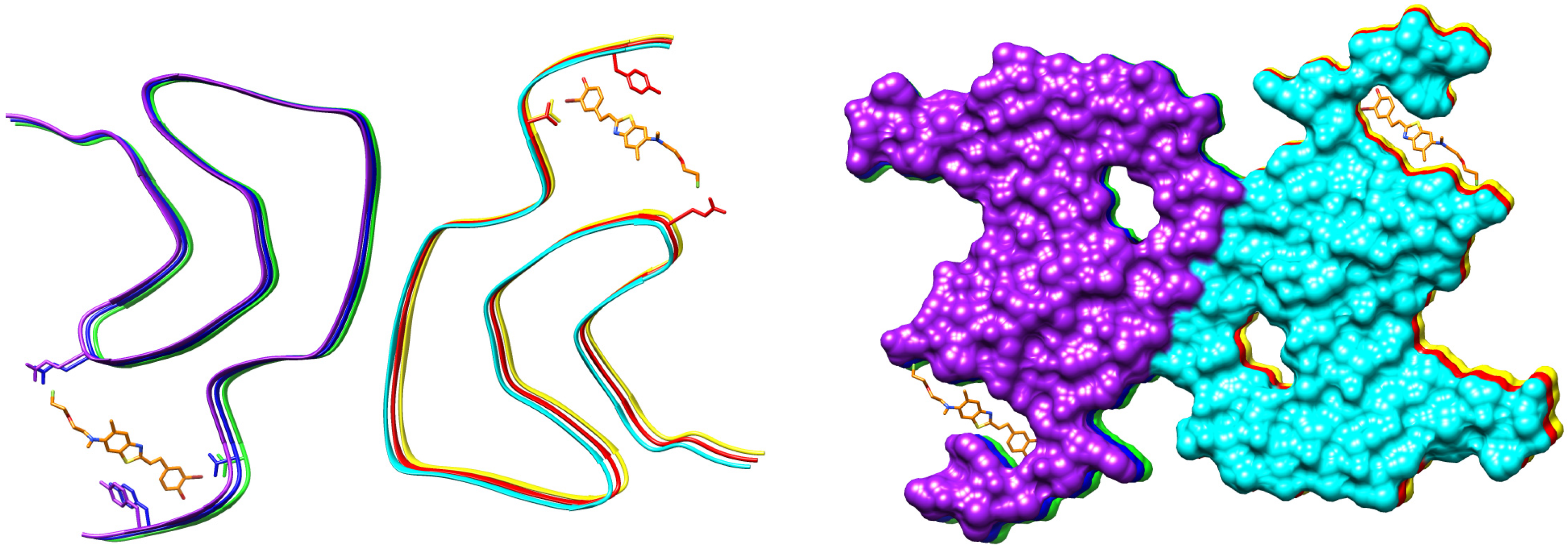
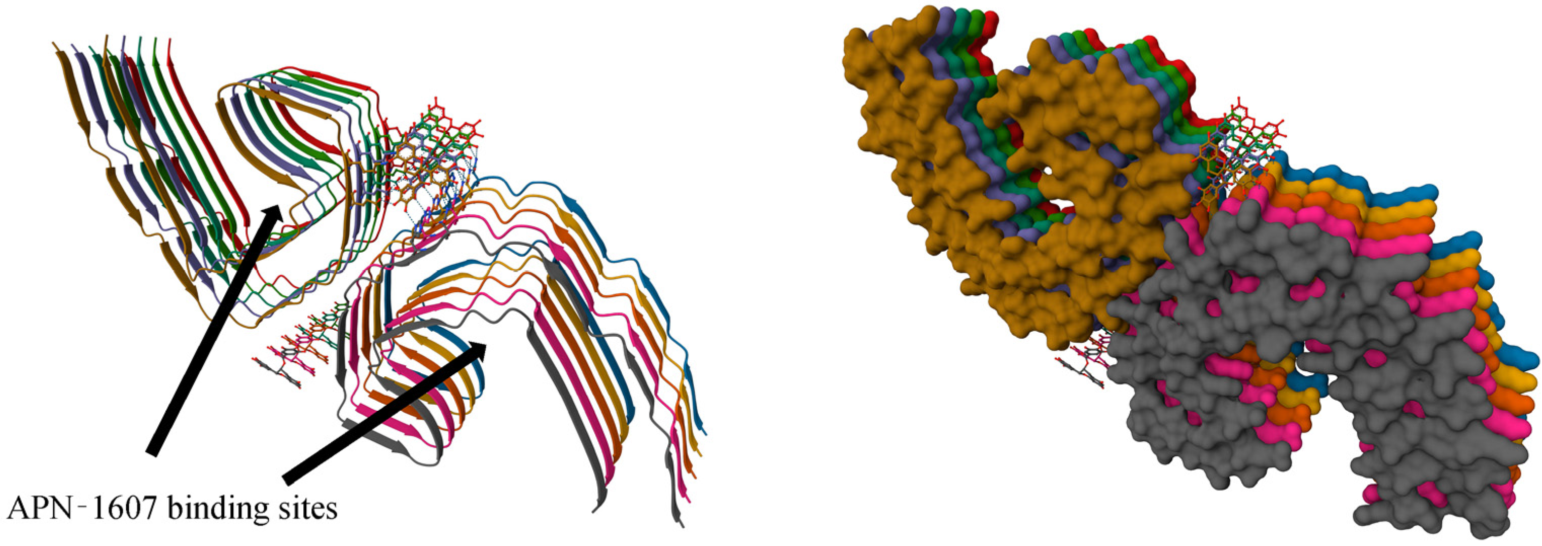
- Duan, P.; Chen, K.J.; Wijegunawardena, G.; Dregni, A.J.; Wang, H.K.; Wu, H.; Hong, M. Binding Sites of a Positron Emission Tomography Imaging Agent in Alzheimer’s β-Amyloid Fibrils Studied Using 19F Solid-State NMR. J. Am. Chem. Soc. 2022, 144, 1416–1430. [Google Scholar] [CrossRef] [PubMed]
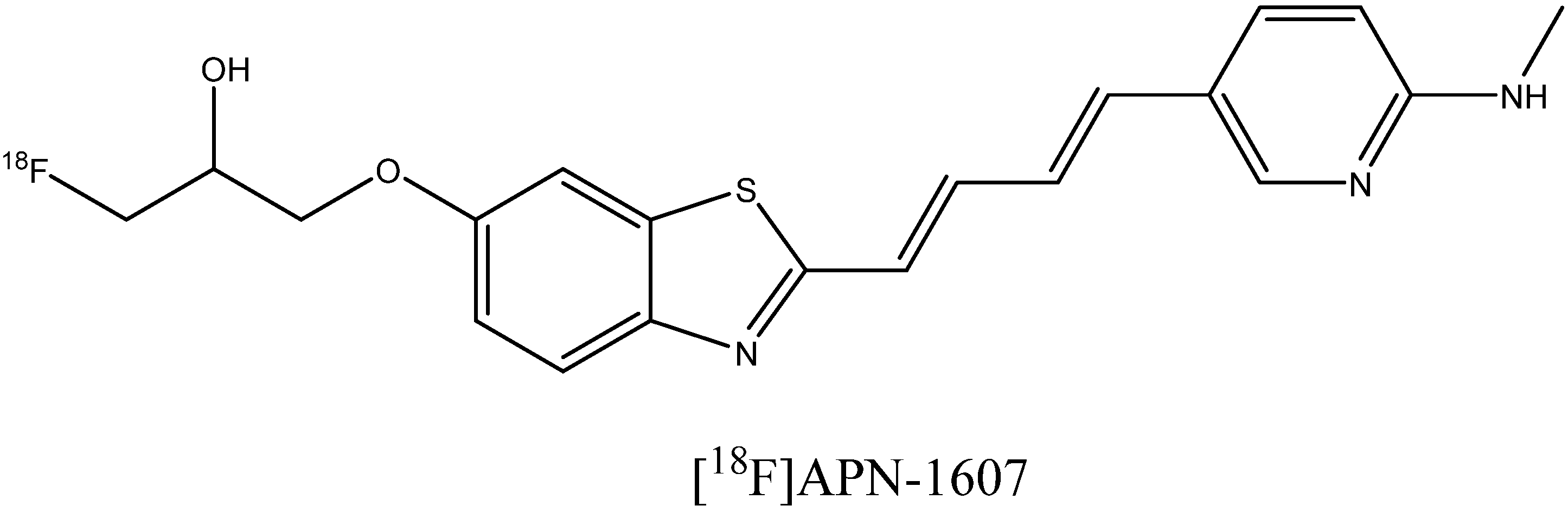
- Shi, Y.; Murzin, A.G.; Falcon, B.; Epstein, A.; Machin, J.; Tempest, P.; Newell, K.L.; Vidal, R.; Garringer, H.J.; Sahara, N.; et al. Cryo-EM Structures of Tau Filaments from Alzheimer’s Disease with PET Ligand APN-1607. Acta Neuropathol. 2021, 141, 697–708. [Google Scholar] [CrossRef] [PubMed]
- Frieg, B.; Gremer, L.; Heise, H.; Willbold, D.; Gohlke, H. Binding Modes of Thioflavin T and Congo Red to the Fibril Structure of Amyloid-β(1–42). Chem. Commun. 2020, 56, 7589–7592. [Google Scholar] [CrossRef] [PubMed]
- Schütz, A.K.; Soragni, A.; Hornemann, S.; Aguzzi, A.; Ernst, M.; Böckmann, A.; Meier, B.H. The Amyloid-Congo Red Interface at Atomic Resolution. Angew. Chem. Int. Ed. 2011, 50, 5956–5960. [Google Scholar] [CrossRef] [PubMed]
- Antonschmidt, L.; Matthes, D.; Dervişoğlu, R.; Frieg, B.; Dienemann, C.; Leonov, A.; Nimerovsky, E.; Sant, V.; Ryazanov, S.; Giese, A.; et al. The Clinical Drug Candidate Anle138b Binds in a Cavity of Lipidic α-Synuclein Fibrils. Nat. Commun. 2022, 13, 5385. [Google Scholar] [CrossRef] [PubMed]
- Seidler, P.M.; Murray, K.A.; Boyer, D.R.; Ge, P.; Sawaya, M.R.; Hu, C.J.; Cheng, X.; Abskharon, R.; Pan, H.; DeTure, M.A.; et al. Structure-Based Discovery of Small Molecules That Disaggregate Alzheimer’s Disease Tissue Derived Tau Fibrils in Vitro. Nat. Commun. 2022, 13, 5451. [Google Scholar] [CrossRef]
- Merz, G.E.; Chalkley, M.J.; Tan, S.; Tse, E.; Lee, J.; Prusiner, S.B.; Paras, N.A.; DeGrado, W.F.; Southworth, D.R. Stacked Binding of a Small Molecule PET Tracer to Alzheimer’s Tau Paired Helical Filaments. BioRxiv 2022. [Google Scholar] [CrossRef]
- Merz, G.E.; Chalkley, M.J.; Tan, S.K.; Tse, E.; Lee, J.; Prusiner, S.B.; Paras, N.A.; DeGrado, W.F.; Southworth, D.R. Stacked Binding of a PET Ligand to Alzheimer’s Tau Paired Helical Filaments. Nat. Commun. 2023, 14, 3048. [Google Scholar] [CrossRef] [PubMed]
- Watanabe, H.; Ono, M.; Ariyoshi, T.; Katayanagi, R.; Saji, H. Novel Benzothiazole Derivatives as Fluorescent Probes for Detection of β-Amyloid and α-Synuclein Aggregates. ACS Chem. Neurosci. 2017, 8, 1656–1662. [Google Scholar] [CrossRef] [PubMed]
- Dyrager, C.; Vieira, R.P.; Nyström, S.; Nilsson, K.P.R.; Storr, T. Synthesis and Evaluation of Benzothiazole-Triazole and Benzothiadiazole-Triazole Scaffolds as Potential Molecular Probes for Amyloid-β Aggregation. New J. Chem. 2017, 41, 1566–1573. [Google Scholar] [CrossRef] [Green Version]
- Mathis, C.A.; Wang, Y.; Holt, D.P.; Huang, G.-F.; Debnath, M.L.; Klunk, W.E. Synthesis and Evaluation of 11 C-Labeled 6-Substituted 2-Arylbenzothiazoles as Amyloid Imaging Agents. J. Med. Chem. 2003, 46, 2740–2754. [Google Scholar] [CrossRef] [PubMed]
- Herholz, K.; Ebmeier, K. Clinical Amyloid Imaging in Alzheimer’s Disease. Lancet Neurol. 2011, 10, 667–670. [Google Scholar] [CrossRef] [PubMed]
- Yang, Y.; Cui, M. Radiolabeled Bioactive Benzoheterocycles for Imaging β-Amyloid Plaques in Alzheimer’s Disease. Eur. J. Med. Chem. 2014, 87, 703–721. [Google Scholar] [CrossRef]
- Cui, M.; Ono, M.; Kimura, H.; Ueda, M.; Nakamoto, Y.; Togashi, K.; Okamoto, Y.; Ihara, M.; Takahashi, R.; Liu, B.; et al. Novel 18F-Labeled Benzoxazole Derivatives as Potential Positron Emission Tomography Probes for Imaging of Cerebral β-Amyloid Plaques in Alzheimer’s Disease. J. Med. Chem. 2012, 55, 9136–9145. [Google Scholar] [CrossRef]
- Gan, C.; Hu, J.; Nan, D.-D.; Wang, S.; Li, H. Synthesis and Biological Evaluation of Curcumin Analogs as β-Amyloid Imaging Agents. Future Med. Chem. 2017, 9, 1587–1596. [Google Scholar] [CrossRef]
- Si, G.; Zhou, S.; Xu, G.; Wang, J.; Wu, B.; Zhou, S. A Curcumin-Based NIR Fluorescence Probe for Detection of Amyloid-Beta (Aβ) Plaques in Alzheimer’s Disease. Dye. Pigment. 2019, 163, 509–515. [Google Scholar] [CrossRef]
- Zhang, X.; Tian, Y.; Li, Z.; Tian, X.; Sun, H.; Liu, H.; Moore, A.; Ran, C. Design and Synthesis of Curcumin Analogues for in Vivo Fluorescence Imaging and Inhibiting Copper-Induced Cross-Linking of Amyloid Beta Species in Alzheimer’s Disease. J. Am. Chem. Soc. 2013, 135, 16397–16409. [Google Scholar] [CrossRef] [PubMed] [Green Version]
- Zhang, X.; Tian, Y.; Zhang, C.; Tian, X.; Ross, A.W.; Moir, R.D.; Sun, H.; Tanzi, R.E.; Moore, A.; Ran, C. Near-Infrared Fluorescence Molecular Imaging of Amyloid Beta Species and Monitoring Therapy in Animal Models of Alzheimer’s Disease. Proc. Natl. Acad. Sci. USA 2015, 112, 9734–9739. [Google Scholar] [CrossRef] [PubMed]
- Reinke, A.A.; Gestwicki, J.E. Structure?Activity Relationships of Amyloid Beta-Aggregation Inhibitors Based on Curcumin: Influence of Linker Length and Flexibility. Chem. Biol. Drug Des. 2007, 70, 206–215. [Google Scholar] [CrossRef] [PubMed] [Green Version]
- Zhang, J.; Sandberg, A.; Konsmo, A.; Wu, X.; Nyström, S.; Nilsson, K.P.R.; Konradsson, P.; LeVine, H.; Lindgren, M.; Hammarström, P. Detection and Imaging of Aβ1-42 and Tau Fibrils by Redesigned Fluorescent X-34 Analogues. Chem. Eur. J. 2018, 24, 7210–7216. [Google Scholar] [CrossRef] [PubMed]
- Flaherty, D.P.; Kiyota, T.; Dong, Y.; Ikezu, T.; Vennerstrom, J.L. Phenolic Bis-Styrylbenzenes as β-Amyloid Binding Ligands and Free Radical Scavengers. J. Med. Chem. 2010, 53, 7992–7999. [Google Scholar] [CrossRef] [PubMed] [Green Version]
- Ishii, K.; Klunk, W.E.; Arawaka, S.; Debnath, M.L.; Furiya, Y.; Sahara, N.; Shoji, S.; Tamaoka, A.; Pettegrew, J.W.; Mori, H. Chrysamine G and Its Derivative Reduce Amyloid β-Induced Neurotoxicity in Mice. Neurosci. Lett. 2002, 333, 5–8. [Google Scholar] [CrossRef] [PubMed]
- Klunk, W.E.; Bacskai, B.J.; Mathis, C.A.; Kajdasz, S.T.; McLellan, M.E.; Frosch, M.P.; Debnath, M.L.; Holt, D.P.; Wang, Y.; Hyman, B.T. Imaging Aβ Plaques in Living Transgenic Mice with Multiphoton Microscopy and Methoxy-X04, a Systemically Administered Congo Red Derivative. J. Neuropathol. Exp. Neurol. 2002, 61, 797–805. [Google Scholar] [CrossRef] [Green Version]
- Zhang, M.; Fu, H.; Hu, W.; Leng, J.; Zhang, Y. Versatile Dicyanomethylene-Based Fluorescent Probes for the Detection of β-Amyloid in Alzheimer’s Disease: A Theoretical Perspective. Int. J. Mol. Sci. 2022, 23, 8619. [Google Scholar] [CrossRef]
- Cheng, Y.; Zhu, B.; Deng, Y.; Zhang, Z. In Vivo Detection of Cerebral Amyloid Fibrils with Smart Dicynomethylene-4H-Pyran-Based Fluorescence Probe. Anal. Chem. 2015, 87, 4781–4787. [Google Scholar] [CrossRef]
- Liu, J.; Kepe, V.; Žabjek, A.; Petrič, A.; Padgett, H.C.; Satyamurthy, N.; Barrio, J.R. High-Yield, Automated Radiosynthesis of 2-(1-{6-[(2-[18F]Fluoroethyl)(Methyl)Amino]-2-Naphthyl}ethylidene)Malononitrile ([18F]FDDNP) Ready for Animal or Human Administration. Mol. Imaging Biol. 2007, 9, 6–16. [Google Scholar] [CrossRef]
- Yang, W.; Wong, Y.; Ng, O.T.W.; Bai, L.-P.; Kwong, D.W.J.; Ke, Y.; Jiang, Z.-H.; Li, H.-W.; Yung, K.K.L.; Wong, M.S. Inhibition of Beta-Amyloid Peptide Aggregation by Multifunctional Carbazole-Based Fluorophores. Angew. Chem. Int. Ed. 2012, 51, 1804–1810. [Google Scholar] [CrossRef] [PubMed]
- Li, Y.; Chen, C.; Xu, D.; Poon, C.-Y.; Ho, S.-L.; Zheng, R.; Liu, Q.; Song, G.; Li, H.-W.; Wong, M.S. Effective Theranostic Cyanine for Imaging of Amyloid Species in Vivo and Cognitive Improvements in Mouse Model. ACS Omega 2018, 3, 6812–6819. [Google Scholar] [CrossRef]
- Dao, P.; Ye, F.; Liu, Y.; Du, Z.Y.; Zhang, K.; Dong, C.Z.; Meunier, B.; Chen, H. Development of Phenothiazine-Based Theranostic Compounds That Act Both as Inhibitors of β-Amyloid Aggregation and as Imaging Probes for Amyloid Plaques in Alzheimer’s Disease. ACS Chem. Neurosci. 2017, 8, 798–806. [Google Scholar] [CrossRef] [PubMed]
- Cao, Y.; Liu, X.; Zhang, J.; Liu, Z.; Fu, Y.; Zhang, D.; Zheng, M.; Zhang, H.; Xu, M.-H. Design of a Coumarin-Based Fluorescent Probe for Efficient In Vivo Imaging of Amyloid-β Plaques. ACS Chem. Neurosci. 2023, 14, 829–838. [Google Scholar] [CrossRef]
- Chen, X.; Li, Y.; Kang, J.; Ye, T.; Yang, Z.; Liu, Z.; Liu, Q.; Zhao, Y.; Liu, G.; Pan, J. Application of a Novel Coumarin-Derivative near-Infrared Fluorescence Probe to Amyloid-β Imaging and Inhibition in Alzheimer’s Disease. J. Lumin. 2023, 256, 119661. [Google Scholar] [CrossRef]
- Rajasekhar, K.; Narayanaswamy, N.; Murugan, N.A.; Viccaro, K.; Lee, H.-G.; Shah, K.; Govindaraju, T. Aβ Plaque-Selective NIR Fluorescence Probe to Differentiate Alzheimer’s Disease from Tauopathies. Biosens. Bioelectron. 2017, 98, 54–61. [Google Scholar] [CrossRef] [PubMed]
- Yan, J.; Zhu, J.; Zhou, K.; Wang, J.; Tan, H.; Xu, Z.; Chen, S.; Lu, Y.; Cui, M.; Zhang, L. Neutral Merocyanine Dyes: For in Vivo NIR Fluorescence Imaging of Amyloid-β Plaques. Chem. Commun. 2017, 53, 9910–9913. [Google Scholar] [CrossRef] [PubMed]
- Tan, H.; Zhou, K.; Yan, J.; Sun, H.; Pistolozzi, M.; Cui, M.; Zhang, L. Dual-Functional Red-Emitting Fluorescent Probes for Imaging Beta-Amyloid Plaques and Viscosity. Sens. Actuators B Chem. 2019, 298, 126903. [Google Scholar] [CrossRef]
- Yang, H.-L.; Fang, S.-Q.; Tang, Y.-W.; Wang, C.; Luo, H.; Qu, L.-L.; Zhao, J.-H.; Shi, C.-J.; Yin, F.-C.; Wang, X.-B.; et al. A Hemicyanine Derivative for Near-Infrared Imaging of β-Amyloid Plaques in Alzheimer’s Disease. Eur. J. Med. Chem. 2019, 179, 736–743. [Google Scholar] [CrossRef] [PubMed]
- Fitzpatrick, A.W.P.; Falcon, B.; He, S.; Murzin, A.G.; Murshudov, G.; Garringer, H.J.; Crowther, R.A.; Ghetti, B.; Goedert, M.; Scheres, S.H.W. Cryo-EM Structures of Tau Filaments from Alzheimer’s Disease. Nature 2017, 547, 185–190. [Google Scholar] [CrossRef] [PubMed] [Green Version]
- Chang, A.; Xiang, X.; Wang, J.; Lee, C.; Arakhamia, T.; Simjanoska, M.; Wang, C.; Carlomagno, Y.; Zhang, G.; Dhingra, S.; et al. Homotypic Fibrillization of TMEM106B across Diverse Neurodegenerative Diseases. Cell 2022, 185, 1346–1355. [Google Scholar] [CrossRef] [PubMed]
- Zhang, W.; Falcon, B.; Murzin, A.G.; Fan, J.; Crowther, R.A.; Goedert, M.; Scheres, S.H. Heparin-Induced Tau Filaments Are Polymorphic and Differ from Those in Alzheimer’s and Pick’s Diseases. eLife 2019, 8, e43584. [Google Scholar] [CrossRef] [PubMed]
- Shi, Y.; Zhang, W.; Yang, Y.; Murzin, A.G.; Falcon, B.; Kotecha, A.; van Beers, M.; Tarutani, A.; Kametani, F.; Garringer, H.J.; et al. Structure-Based Classification of Tauopathies. Nature 2021, 598, 359–363. [Google Scholar] [CrossRef] [PubMed]
- Falcon, B.; Zhang, W.; Murzin, A.G.; Murshudov, G.; Garringer, H.J.; Vidal, R.; Crowther, R.A.; Ghetti, B.; Scheres, S.H.W.; Goedert, M. Structures of Filaments from Pick’s Disease Reveal a Novel Tau Protein Fold. Nature 2018, 561, 137–140. [Google Scholar] [CrossRef] [PubMed]
- Falcon, B.; Zivanov, J.; Zhang, W.; Murzin, A.G.; Garringer, H.J.; Vidal, R.; Crowther, R.A.; Newell, K.L.; Ghetti, B.; Goedert, M.; et al. Novel Tau Filament Fold in Chronic Traumatic Encephalopathy Encloses Hydrophobic Molecules. Nature 2019, 568, 420–423. [Google Scholar] [CrossRef] [PubMed]
- Li, X.; Zhang, S.; Liu, Z.; Tao, Y.; Xia, W.; Sun, Y.; Liu, C.; Le, W.; Sun, B.; Li, D. Subtle Change of Fibrillation Condition Leads to Substantial Alteration of Recombinant Tau Fibril Structure. iScience 2022, 25, 105645. [Google Scholar] [CrossRef] [PubMed]
- Okamura, N.; Suemoto, T.; Furumoto, S.; Suzuki, M.; Shimadzu, H.; Akatsu, H.; Yamamoto, T.; Fujiwara, H.; Nemoto, M.; Maruyama, M.; et al. Quinoline and Benzimidazole Derivatives: Candidate Probes for In Vivo Imaging of Tau Pathology in Alzheimer’s Disease. J. Neurosci. 2005, 25, 10857–10862. [Google Scholar] [CrossRef] [PubMed] [Green Version]
- Fodero-Tavoletti, M.T.; Okamura, N.; Furumoto, S.; Mulligan, R.S.; Connor, A.R.; McLean, C.A.; Cao, D.; Rigopoulos, A.; Cartwright, G.A.; O’Keefe, G.; et al. 18F-THK523: A Novel in Vivo Tau Imaging Ligand for Alzheimer’s Disease. Brain 2011, 134, 1089–1100. [Google Scholar] [CrossRef] [Green Version]
- Okamura, N.; Furumoto, S.; Harada, R.; Tago, T.; Yoshikawa, T.; Fodero-Tavoletti, M.; Mulligan, R.S.; Villemagne, V.L.; Akatsu, H.; Yamamoto, T.; et al. Novel 18F-Labeled Arylquinoline Derivatives for Noninvasive Imaging of Tau Pathology in Alzheimer Disease. J. Nucl. Med. 2013, 54, 1420–1427. [Google Scholar] [CrossRef] [PubMed] [Green Version]
- Xia, C.; Arteaga, J.; Chen, G.; Gangadharmath, U.; Gomez, L.F.; Kasi, D.; Lam, C.; Liang, Q.; Liu, C.; Mocharla, V.P.; et al. [18F]T807, a Novel Tau Positron Emission Tomography Imaging Agent for Alzheimer’s Disease. Alzheimer’s Dement. 2013, 9, 666–676. [Google Scholar] [CrossRef] [PubMed]
- Lemoine, L.; Leuzy, A.; Chiotis, K.; Rodriguez-Vieitez, E.; Nordberg, A. Tau Positron Emission Tomography Imaging in Tauopathies: The Added Hurdle of Off-target Binding. Alzheimer’s Dement. Diagn. Assess. Dis. Monit. 2018, 10, 232–236. [Google Scholar] [CrossRef] [PubMed]
- Drake, L.R.; Pham, J.M.; Desmond, T.J.; Mossine, A.V.; Lee, S.J.; Kilbourn, M.R.; Koeppe, R.A.; Brooks, A.F.; Scott, P.J.H. Identification of AV-1451 as a Weak, Nonselective Inhibitor of Monoamine Oxidase. ACS Chem. Neurosci. 2019, 10, 3839–3846. [Google Scholar] [CrossRef] [PubMed]
- Kramer, V.; Brooks, A.F.; Haeger, A.; Kuljis, R.O.; Rafique, W.; Koeppe, R.A.; Raffel, D.M.; Frey, K.A.; Amaral, H.; Scott, P.J.H.; et al. Evaluation of [18F]- N -Methyl Lansoprazole as a Tau PET Imaging Agent in First-in-Human Studies. ACS Chem. Neurosci. 2020, 11, 427–435. [Google Scholar] [CrossRef] [PubMed]
- Pascoal, T.A.; Therriault, J.; Benedet, A.L.; Savard, M.; Lussier, F.Z.; Chamoun, M.; Tissot, C.; Qureshi, M.N.I.; Kang, M.S.; Mathotaarachchi, S.; et al. 18F-MK-6240 PET for Early and Late Detection of Neurofibrillary Tangles. Brain 2020, 143, 2818–2830. [Google Scholar] [CrossRef] [PubMed]
- Hostetler, E.D.; Walji, A.M.; Zeng, Z.; Miller, P.; Bennacef, I.; Salinas, C.; Connolly, B.; Gantert, L.; Haley, H.; Holahan, M.; et al. Preclinical Characterization of 18F-MK-6240, a Promising PET Tracer for In Vivo Quantification of Human Neurofibrillary Tangles. J. Nucl. Med. 2016, 57, 1599–1606. [Google Scholar] [CrossRef] [PubMed] [Green Version]
- Kroth, H.; Oden, F.; Molette, J.; Schieferstein, H.; Capotosti, F.; Mueller, A.; Berndt, M.; Schmitt-Willich, H.; Darmency, V.; Gabellieri, E.; et al. Discovery and Preclinical Characterization of [18F]PI-2620, a next-Generation Tau PET Tracer for the Assessment of Tau Pathology in Alzheimer’s Disease and Other Tauopathies. Eur. J. Nucl. Med. Mol. Imaging 2019, 46, 2178–2189. [Google Scholar] [CrossRef] [Green Version]
- Kuwabara, H.; Comley, R.A.; Borroni, E.; Honer, M.; Kitmiller, K.; Roberts, J.; Gapasin, L.; Mathur, A.; Klein, G.; Wong, D.F. Evaluation of 18F-RO-948 PET for Quantitative Assessment of Tau Accumulation in the Human Brain. J. Nucl. Med. 2018, 59, 1877–1884. [Google Scholar] [CrossRef] [Green Version]
- Honer, M.; Gobbi, L.; Knust, H.; Kuwabara, H.; Muri, D.; Koerner, M.; Valentine, H.; Dannals, R.F.; Wong, D.F.; Borroni, E. Preclinical Evaluation of 18F-RO6958948, 11C-RO6931643, and 11C-RO6924963 as Novel PET Radiotracers for Imaging Tau Aggregates in Alzheimer Disease. J. Nucl. Med. 2018, 59, 675–681. [Google Scholar] [CrossRef] [PubMed] [Green Version]
- Rombouts, F.J.R.; Declercq, L.; Andrés, J.-I.; Bottelbergs, A.; Chen, L.; Iturrino, L.; Leenaerts, J.E.; Mariën, J.; Song, F.; Wintmolders, C.; et al. Discovery of N-(4-[18F]Fluoro-5-Methylpyridin-2-Yl)Isoquinolin-6-Amine (JNJ-64326067), a New Promising Tau Positron Emission Tomography Imaging Tracer. J. Med. Chem. 2019, 62, 2974–2987. [Google Scholar] [CrossRef]
- Sanabria Bohórquez, S.; Marik, J.; Ogasawara, A.; Tinianow, J.N.; Gill, H.S.; Barret, O.; Tamagnan, G.; Alagille, D.; Ayalon, G.; Manser, P.; et al. [18F]GTP1 (Genentech Tau Probe 1), a Radioligand for Detecting Neurofibrillary Tangle Tau Pathology in Alzheimer’s Disease. Eur. J. Nucl. Med. Mol. Imaging 2019, 46, 2077–2089. [Google Scholar] [CrossRef]
- Brendel, M.; Barthel, H.; van Eimeren, T.; Marek, K.; Beyer, L.; Song, M.; Palleis, C.; Gehmeyr, M.; Fietzek, U.; Respondek, G.; et al. Assessment of 18F-PI-2620 as a Biomarker in Progressive Supranuclear Palsy. JAMA Neurol. 2020, 77, 1408. [Google Scholar] [CrossRef] [PubMed]
- Schonhaut, D.R.; McMillan, C.T.; Spina, S.; Dickerson, B.C.; Siderowf, A.; Devous, M.D.; Tsai, R.; Winer, J.; Russell, D.S.; Litvan, I.; et al. 18F-Flortaucipir Tau Positron Emission Tomography Distinguishes Established Progressive Supranuclear Palsy from Controls and Parkinson Disease: A Multicenter Study: Flortaucipir Tau PET in PSP. Ann. Neurol. 2017, 82, 622–634. [Google Scholar] [CrossRef] [PubMed] [Green Version]
- Su, Y.; Fu, J.; Yu, J.; Zhao, Q.; Guan, Y.; Zuo, C.; Li, M.; Tan, H.; Cheng, X. Tau PET Imaging with [18F]PM-PBB3 in Frontotemporal Dementia with MAPT Mutation. J. Alzheimer’s Dis. 2020, 76, 149–157. [Google Scholar] [CrossRef] [PubMed]
- Rojo, L.E.; Alzate-Morales, J.; Saavedra, I.N.; Davies, P.; Maccioni, R.B. Selective Interaction of Lansoprazole and Astemizole with Tau Polymers: Potential New Clinical Use in Diagnosis of Alzheimer’s Disease. J. Alzheimer’s Dis. 2010, 19, 573–589. [Google Scholar] [CrossRef] [PubMed]
- Sekijima, Y.; Yazaki, M.; Oguchi, K.; Ezawa, N.; Yoshinaga, T.; Yamada, M.; Yahikozawa, H.; Watanabe, M.; Kametani, F.; Ikeda, S. Cerebral Amyloid Angiopathy in Posttransplant Patients with Hereditary ATTR Amyloidosis. Neurology 2016, 87, 773–781. [Google Scholar] [CrossRef]
- Ezawa, N.; Katoh, N.; Oguchi, K.; Yoshinaga, T.; Yazaki, M.; Sekijima, Y. Visualization of Multiple Organ Amyloid Involvement in Systemic Amyloidosis Using 11C-PiB PET Imaging. Eur. J. Nucl. Med. Mol. Imaging 2018, 45, 452–461. [Google Scholar] [CrossRef] [PubMed]
- Antoni, G.; Lubberink, M.; Estrada, S.; Axelsson, J.; Carlson, K.; Lindsjö, L.; Kero, T.; Långström, B.; Granstam, S.-O.; Rosengren, S.; et al. In Vivo Visualization of Amyloid Deposits in the Heart with 11 C-PIB and PET. J. Nucl. Med. 2013, 54, 213–220. [Google Scholar] [CrossRef] [Green Version]
- Uneus, E.I.; Wilhelmsson, C.; Bäckström, D.; Anan, I.; Wixner, J.; Pilebro, B.; Riklund, K.; Ögren, M.; Ögreen, M.; Axelsson, J.; et al. Cerebellar and Cerebral Amyloid Visualized by [18F]Flutemetamol PET in Long-Term Hereditary V30M (p.V50M) Transthyretin Amyloidosis Survivors. Front. Neurol. 2022, 13, 816636. [Google Scholar] [CrossRef] [PubMed]
- Khor, Y.M.; Cuddy, S.; Harms, H.J.; Kijewski, M.F.; Park, M.-A.; Robertson, M.; Hyun, H.; Di Carli, M.F.; Bianchi, G.; Landau, H.; et al. Quantitative [18F]Florbetapir PET/CT May Identify Lung Involvement in Patients with Systemic AL Amyloidosis. Eur. J. Nucl. Med. Mol. Imaging 2020, 47, 1998–2009. [Google Scholar] [CrossRef] [PubMed]
- Ehman, E.C.; El-Sady, M.S.; Kijewski, M.F.; Khor, Y.M.; Jacob, S.; Ruberg, F.L.; Sanchorawala, V.; Landau, H.; Yee, A.J.; Bianchi, G.; et al. Early Detection of Multiorgan Light-Chain Amyloidosis by Whole-Body 18F-Florbetapir PET/CT. J. Nucl. Med. 2019, 60, 1234–1239. [Google Scholar] [CrossRef] [Green Version]
- Baratto, L.; Park, S.Y.; Hatami, N.; Gulaka, P.; Vasanawala, S.; Yohannan, T.K.; Herfkens, R.; Witteles, R.; Iagaru, A. 18F-Florbetaben Whole-Body PET/MRI for Evaluation of Systemic Amyloid Deposition. EJNMMI Res. 2018, 8, 66. [Google Scholar] [CrossRef] [PubMed]
- Liu, L.; Prime, M.E.; Lee, M.R.; Khetarpal, V.; Brown, C.J.; Johnson, P.D.; Miranda-Azpiazu, P.; Chen, X.; Clark-Frew, D.; Coe, S.; et al. Imaging Mutant Huntingtin Aggregates: Development of a Potential PET Ligand. J. Med. Chem. 2020, 63, 8608–8633. [Google Scholar] [CrossRef] [PubMed]
- Liu, L.; Johnson, P.D.; Prime, M.E.; Khetarpal, V.; Lee, M.R.; Brown, C.J.; Chen, X.; Clark-Frew, D.; Coe, S.; Conlon, M.; et al. [11C]CHDI-626, a PET Tracer Candidate for Imaging Mutant Huntingtin Aggregates with Reduced Binding to AD Pathological Proteins. J. Med. Chem. 2021, 64, 12003–12021. [Google Scholar] [CrossRef] [PubMed]
- Liu, L.; Johnson, P.D.; Prime, M.E.; Khetarpal, V.; Brown, C.J.; Anzillotti, L.; Bertoglio, D.; Chen, X.; Coe, S.; Davis, R.; et al. Design and Evaluation of [18F]CHDI-650 as a Positron Emission Tomography Ligand to Image Mutant Huntingtin Aggregates. J. Med. Chem. 2023, 66, 641–656. [Google Scholar] [CrossRef] [PubMed]
- Biancalana, M.; Koide, S. Molecular Mechanism of Thioflavin-T Binding to Amyloid Fibrils. Biochim. Biophys. Acta (BBA)—Proteins Proteom. 2010, 1804, 1405–1412. [Google Scholar] [CrossRef] [PubMed] [Green Version]
- Hawe, A.; Sutter, M.; Jiskoot, W. Extrinsic Fluorescent Dyes as Tools for Protein Characterization. Pharm. Res. 2008, 25, 1487–1499. [Google Scholar] [CrossRef] [Green Version]
- Sachchithanantham, S.; Wechalekar, A.D. Imaging in Systemic Amyloidosis. Br. Med. Bull. 2013, 107, 41–56. [Google Scholar] [CrossRef] [Green Version]
- Chen, W.; Dilsizian, V. Molecular Imaging of Amyloidosis: Will the Heart Be the Next Target After the Brain? Curr. Cardiol. Rep. 2012, 14, 226–233. [Google Scholar] [CrossRef]
- Cheng, K.T. 124I/125I-Fibril-Reactive Monoclonal Antibody. In Molecular Imaging and Contrast Agent Database (MICAD); National Center for Biotechnology Information (US): Bethesda, MD, USA, 2004. [Google Scholar]
- Kim, P.K.; Hong, Y.J.; Im, D.J.; Suh, Y.J.; Park, C.H.; Kim, J.Y.; Chang, S.; Lee, H.-J.; Hur, J.; Kim, Y.J.; et al. Myocardial T1 and T2 Mapping: Techniques and Clinical Applications. Korean J. Radiol. 2017, 18, 113. [Google Scholar] [CrossRef] [Green Version]
- Wang, T.K.M.; Hassan, O.K.A.; Jaber, W.; Xu, B. Multi-Modality Imaging of Cardiac Amyloidosis: Contemporary Update. World J. Radiol. 2020, 12, 87–100. [Google Scholar] [CrossRef]
- Baggiano, A.; Boldrini, M.; Martinez-Naharro, A.; Kotecha, T.; Petrie, A.; Rezk, T.; Gritti, M.; Quarta, C.; Knight, D.S.; Wechalekar, A.D.; et al. Noncontrast Magnetic Resonance for the Diagnosis of Cardiac Amyloidosis. JACC Cardiovasc. Imaging 2020, 13, 69–80. [Google Scholar] [CrossRef] [PubMed]
- Soundarya, S.; Sruthi, M.S.; Sathya Bama, S.; Kiruthika, S.; Dhiyaneswaran, J. Early Detection of Alzheimer Disease Using Gadolinium Material. Mater. Today Proc. 2021, 45, 1094–1101. [Google Scholar] [CrossRef]
- Kim, E.; Di Censo, D.; Baraldo, M.; Simmons, C.; Rosa, I.; Randall, K.; Ballard, C.; Dickie, B.R.; Williams, S.C.R.; Killick, R.; et al. In Vivo Multi-Parametric Manganese-Enhanced MRI for Detecting Amyloid Plaques in Rodent Models of Alzheimer’s Disease. Sci. Rep. 2021, 11, 12419. [Google Scholar] [CrossRef] [PubMed]
- Lacerda, S.; Morfin, J.-F.; Geraldes, C.F.G.C.; Tóth, É. Metal Complexes for Multimodal Imaging of Misfolded Protein-Related Diseases. Dalton Trans. 2017, 46, 14461–14474. [Google Scholar] [CrossRef] [PubMed]
- Poduslo, J.F.; Wengenack, T.M.; Curran, G.L.; Wisniewski, T.; Sigurdsson, E.M.; Macura, S.I.; Borowski, B.J.; Jack, C.R. Molecular Targeting of Alzheimer’s Amyloid Plaques for Contrast-Enhanced Magnetic Resonance Imaging. Neurobiol. Dis. 2002, 11, 315–329. [Google Scholar] [CrossRef] [PubMed] [Green Version]
- Poduslo, J.F.; Curran, G.L.; Peterson, J.A.; McCormick, D.J.; Fauq, A.H.; Khan, M.A.; Wengenack, T.M. Design and Chemical Synthesis of a Magnetic Resonance Contrast Agent with Enhanced in Vitro Binding, High Blood−Brain Barrier Permeability, and in Vivo Targeting to Alzheimer’s Disease Amyloid Plaques. Biochemistry 2004, 43, 6064–6075. [Google Scholar] [CrossRef] [PubMed] [Green Version]
- Matharu, B.; Spencer, N.; Howe, F.; Austen, B. Gadolinium-Complexed Aβ-Binding Contrast Agents for MRI Diagnosis of Alzheimer’s Disease. Neuropeptides 2015, 53, 63–70. [Google Scholar] [CrossRef]
- Plissonneau, M.; Pansieri, J.; Heinrich-Balard, L.; Morfin, J.-F.; Stransky-Heilkron, N.; Rivory, P.; Mowat, P.; Dumoulin, M.; Cohen, R.; Allémann, É.; et al. Gd-Nanoparticles Functionalization with Specific Peptides for ß-Amyloid Plaques Targeting. J. Nanobiotechnol. 2016, 14, 60. [Google Scholar] [CrossRef] [PubMed] [Green Version]
- Wadghiri, Y.Z.; Li, J.; Wang, J.; Hoang, D.M.; Sun, Y.; Xu, H.; Tsui, W.; Li, Y.; Boutajangout, A.; Wang, A.; et al. Detection of Amyloid Plaques Targeted by Bifunctional USPIO in Alzheimer’s Disease Transgenic Mice Using Magnetic Resonance Microimaging. PLoS ONE 2013, 8, e57097. [Google Scholar] [CrossRef] [Green Version]
- Xiong, Y.; Qu, Y.; Min, Z.; Wu, J.; Zhang, S.; Xue, Z. Amyloid Plaque Imaging with a Targeted MRI Contrast Agent in a Transgenic Mouse Model of Alzheimer’s Disease. Int. J. Nanomed. 2022, 17, 927–936. [Google Scholar] [CrossRef]
- Yang, C.-C.; Yang, S.-Y.; Chieh, J.-J.; Horng, H.-E.; Hong, C.-Y.; Yang, H.-C.; Chen, K.H.; Shih, B.Y.; Chen, T.-F.; Chiu, M.-J. Biofunctionalized Magnetic Nanoparticles for Specifically Detecting Biomarkers of Alzheimer’s Disease in Vitro. ACS Chem. Neurosci. 2011, 2, 500–505. [Google Scholar] [CrossRef] [Green Version]
- Poduslo, J.F.; Ramakrishnan, M.; Holasek, S.S.; Ramirez-Alvarado, M.; Kandimalla, K.K.; Gilles, E.J.; Curran, G.L.; Wengenack, T.M. In Vivo Targeting of Antibody Fragments to the Nervous System for Alzheimer’s Disease Immunotherapy and Molecular Imaging of Amyloid Plaques: Targeting of Antibody Fragments to AD Amyloid Plaques. J. Neurochem. 2007, 102, 420–433. [Google Scholar] [CrossRef] [PubMed]
- Vandesquille, M.; Li, T.; Po, C.; Ganneau, C.; Lenormand, P.; Dudeffant, C.; Czech, C.; Grueninger, F.; Duyckaerts, C.; Delatour, B.; et al. Chemically-Defined Camelid Antibody Bioconjugate for the Magnetic Resonance Imaging of Alzheimer’s Disease. mAbs 2017, 9, 1016–1027. [Google Scholar] [CrossRef] [PubMed] [Green Version]
- Sillerud, L.O.; Solberg, N.O.; Chamberlain, R.; Orlando, R.A.; Heidrich, J.E.; Brown, D.C.; Brady, C.I.; Vander Jagt, T.A.; Garwood, M.; Vander Jagt, D.L. SPION-Enhanced Magnetic Resonance Imaging of Alzheimer’s Disease Plaques in AβPP/PS-1 Transgenic Mouse Brain. J. Alzheimer’s Dis. 2013, 34, 349–365. [Google Scholar] [CrossRef] [PubMed]
- Margel, S.; Skaat, H.; Corem-Salkmon, E.; Grinberg, I.; Last, D.; Goez, D.; Mardor, Y. Antibody-Conjugated, Dual-Modal, near-Infrared Fluorescent Iron Oxide Nanoparticles for Antiamyloidgenic Activity and Specific Detection of Amyloid-β; Fibrils. Int. J. Nanomed. 2013, 8, 4063. [Google Scholar] [CrossRef] [PubMed] [Green Version]
- Choi, G.; Kim, H.-K.; Baek, A.R.; Kim, S.; Kim, M.J.; Kim, M.; Cho, A.E.; Lee, G.-H.; Jung, H.; Yang, J.; et al. Multifunctional Imaging of Amyloid-Beta Peptides with a New Gadolinium-Based Contrast Agent in Alzheimer’s Disease. J. Ind. Eng. Chem. 2020, 83, 214–223. [Google Scholar] [CrossRef]
- Wang, X.; Chan, H.N.; Desbois, N.; Gros, C.P.; Bolze, F.; Li, Y.; Li, H.W.; Wong, M.S. Multimodal Theranostic Cyanine-Conjugated Gadolinium(III) Complex for In Vivo Imaging of Amyloid-β in an Alzheimer’s Disease Mouse Model. ACS Appl. Mater. Interfaces 2021, 13, 18525–18532. [Google Scholar] [CrossRef]
- Utomo, R.Y.; Okada, S.; Sumiyoshi, A.; Aoki, I.; Nakamura, H. Development of an MRI Contrast Agent for Both Detection and Inhibition of the Amyloid-β Fibrillation Process. RSC Adv. 2022, 12, 5027–5030. [Google Scholar] [CrossRef]
- Martins, A.F.; Morfin, J.-F.; Kubíčková, A.; Kubíček, V.; Buron, F.; Suzenet, F.; Salerno, M.; Lazar, A.N.; Duyckaerts, C.; Arlicot, N.; et al. PiB-Conjugated, Metal-Based Imaging Probes: Multimodal Approaches for the Visualization of β-Amyloid Plaques. ACS Med. Chem. Lett. 2013, 4, 436–440. [Google Scholar] [CrossRef] [PubMed] [Green Version]
- Martins, A.F.; Morfin, J.-F.; Geraldes, C.F.G.C.; Tóth, É. Gd3+ Complexes Conjugated to Pittsburgh Compound B: Potential MRI Markers of β-Amyloid Plaques. J. Biol. Inorg. Chem. 2014, 19, 281–295. [Google Scholar] [CrossRef] [Green Version]
- Cheng, K.K.; Chan, P.S.; Fan, S.; Kwan, S.M.; Yeung, K.L.; Wáng, Y.-X.J.; Chow, A.H.L.; Wu, E.X.; Baum, L. Curcumin-Conjugated Magnetic Nanoparticles for Detecting Amyloid Plaques in Alzheimer’s Disease Mice Using Magnetic Resonance Imaging (MRI). Biomaterials 2015, 44, 155–172. [Google Scholar] [CrossRef] [PubMed]
- Zhou, J.; Fa, H.; Yin, W.; Zhang, J.; Hou, C.; Huo, D.; Zhang, D.; Zhang, H. Synthesis of Superparamagnetic Iron Oxide Nanoparticles Coated with a DDNP-Carboxyl Derivative for in Vitro Magnetic Resonance Imaging of Alzheimer’s Disease. Mater. Sci. Eng. C 2014, 37, 348–355. [Google Scholar] [CrossRef] [PubMed]
- Li, S.; He, H.; Cui, W.; Gu, B.; Li, J.; Qi, Z.; Zhou, G.; Liang, C.; Feng, X. Detection of Aβ Plaques by a Novel Specific MRI Probe Precursor CR-BSA-(Gd-DTPA)n in APP/PS1 Transgenic Mice. Anat. Rec. 2010, 293, 2136–2143. [Google Scholar] [CrossRef] [PubMed]
- Sedov, I.; Khaibrakhmanova, D. Molecular Mechanisms of Inhibition of Protein Amyloid Fibril Formation: Evidence and Perspectives Based on Kinetic Models. Int. J. Mol. Sci. 2022, 23, 13428. [Google Scholar] [CrossRef] [PubMed]
- Wang, C.; Wang, X.; Chan, H.; Liu, G.; Wang, Z.; Li, H.; Wong, M.S. Amyloid-β Oligomer-Targeted Gadolinium-Based NIR/MR Dual-Modal Theranostic Nanoprobe for Alzheimer’s Disease. Adv. Funct. Mater. 2020, 30, 1909529. [Google Scholar] [CrossRef]
- Badachhape, A.A.; Working, P.K.; Srivastava, M.; Bhandari, P.; Stupin, I.V.; Devkota, L.; Tanifum, E.A.; Annapragada, A.V.; Ghaghada, K.B. Pre-Clinical Dose-Ranging Efficacy, Pharmacokinetics, Tissue Biodistribution, and Toxicity of a Targeted Contrast Agent for MRI of Amyloid Deposition in Alzheimer’s Disease. Sci. Rep. 2020, 10, 16185. [Google Scholar] [CrossRef] [PubMed]
- Agyare, E.K.; Curran, G.L.; Ramakrishnan, M.; Yu, C.C.; Poduslo, J.F.; Kandimalla, K.K. Development of a Smart Nano-Vehicle to Target Cerebrovascular Amyloid Deposits and Brain Parenchymal Plaques Observed in Alzheimer’s Disease and Cerebral Amyloid Angiopathy. Pharm. Res. 2008, 25, 2674–2684. [Google Scholar] [CrossRef] [PubMed]
- Ansciaux, E.; Burtea, C.; Laurent, S.; Crombez, D.; Nonclercq, D.; Vander Elst, L.; Muller, R.N. In Vitro and in Vivo Characterization of Several Functionalized Ultrasmall Particles of Iron Oxide, Vectorized against Amyloid Plaques and Potentially Able to Cross the Blood-Brain Barrier: Toward Earlier Diagnosis of Alzheimer’s Disease by Molecular Imag: Molecular Contrast Agents for Alzheimer’s Disease. Contrast Media Mol. Imaging 2015, 10, 211–224. [Google Scholar] [CrossRef] [PubMed]
- Alzeca Biosciences, Inc. Proof-of-Concept Study of New Imaging Diagnostic in Patients With Suspected Alzheimer’s Disease; Alzeca Biosciences, Inc.: Houston, TX, USA, 2022. [Google Scholar]
- Tanifum, E.A.; Ghaghada, K.B.; Annapragada, A.V. Targeted Contrast Agents for MRI of Amyloid Deposition. U.S. Patent 11116854B2, 30 November 2021. [Google Scholar]


| Compound Names and Literature References | Target | Figure 1 |
|---|---|---|
| For 19F imaging | ||
| FSB [90] | Aβ | 8 |
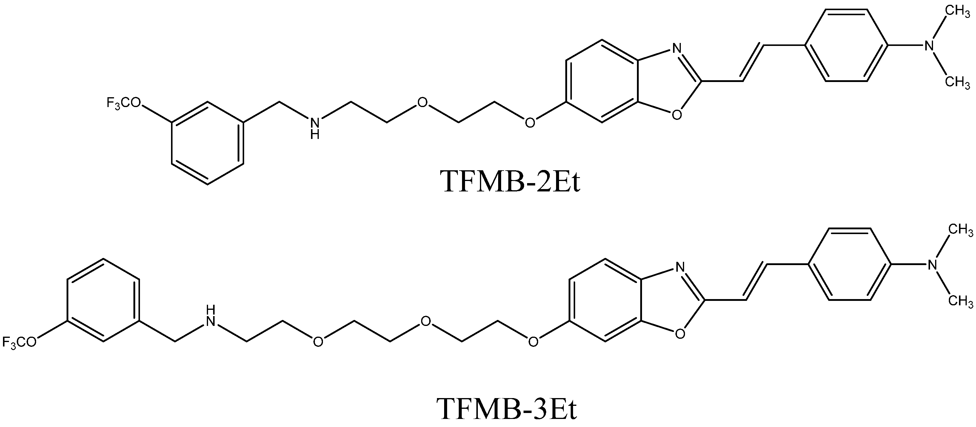
| TFMB-2Et, TFMB-3Et [93] | Aβ | 9 |
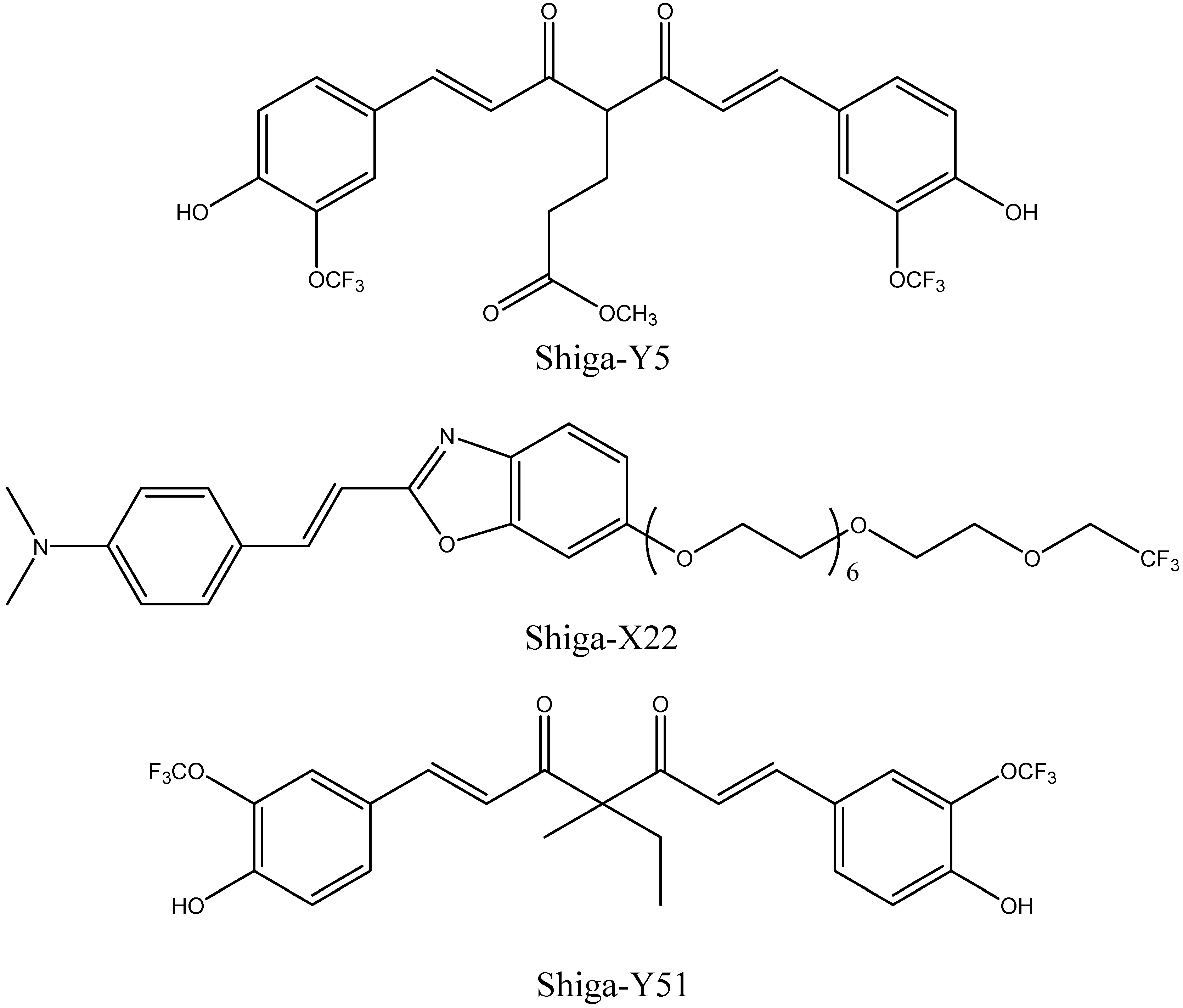
| Shiga-Y5 [94,95] | Aβ | 10 |
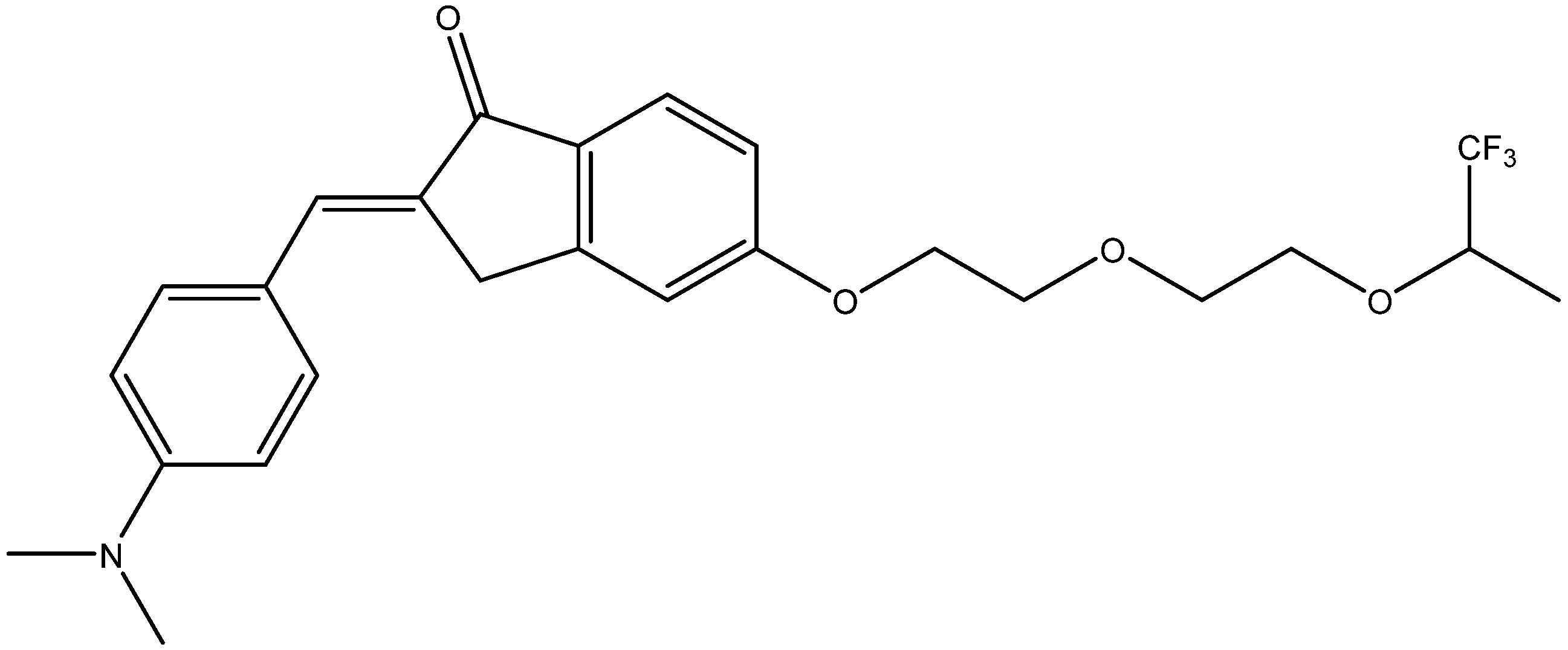
| Shiga-X22 [67,96] | Aβ | 10 |
| Shiga-Y51 [97] | Soluble Aβ aggregates | 10 |
| 7d [98] | Aβ | 13 |
| BSA-capped GQDs functionalized with hydrofluorinated glucose [99] | Aβ | – |
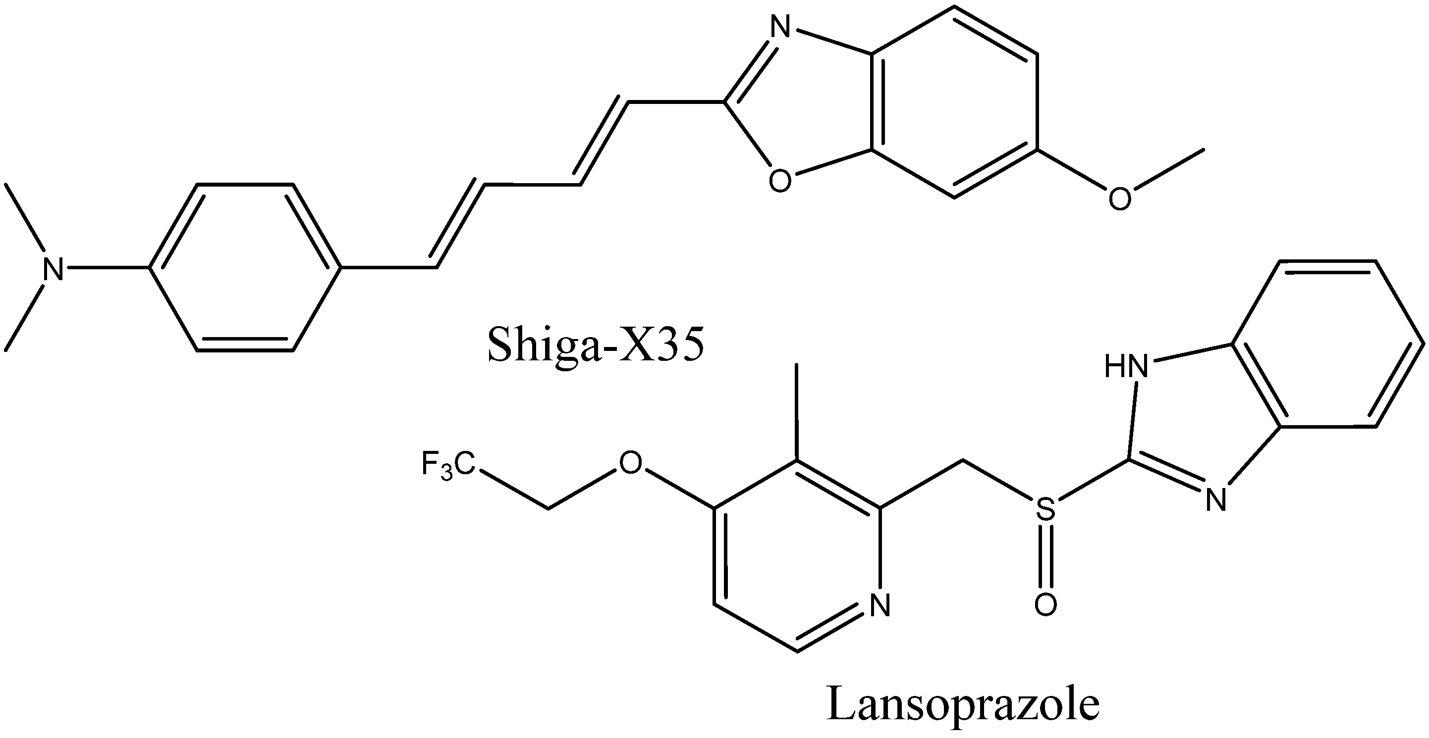
| Shiga-X35 [49] | Tau | 26 |
| Lansoprazole [177] | Tau | 26 |
| For 1H contrast imaging, metal-based | ||
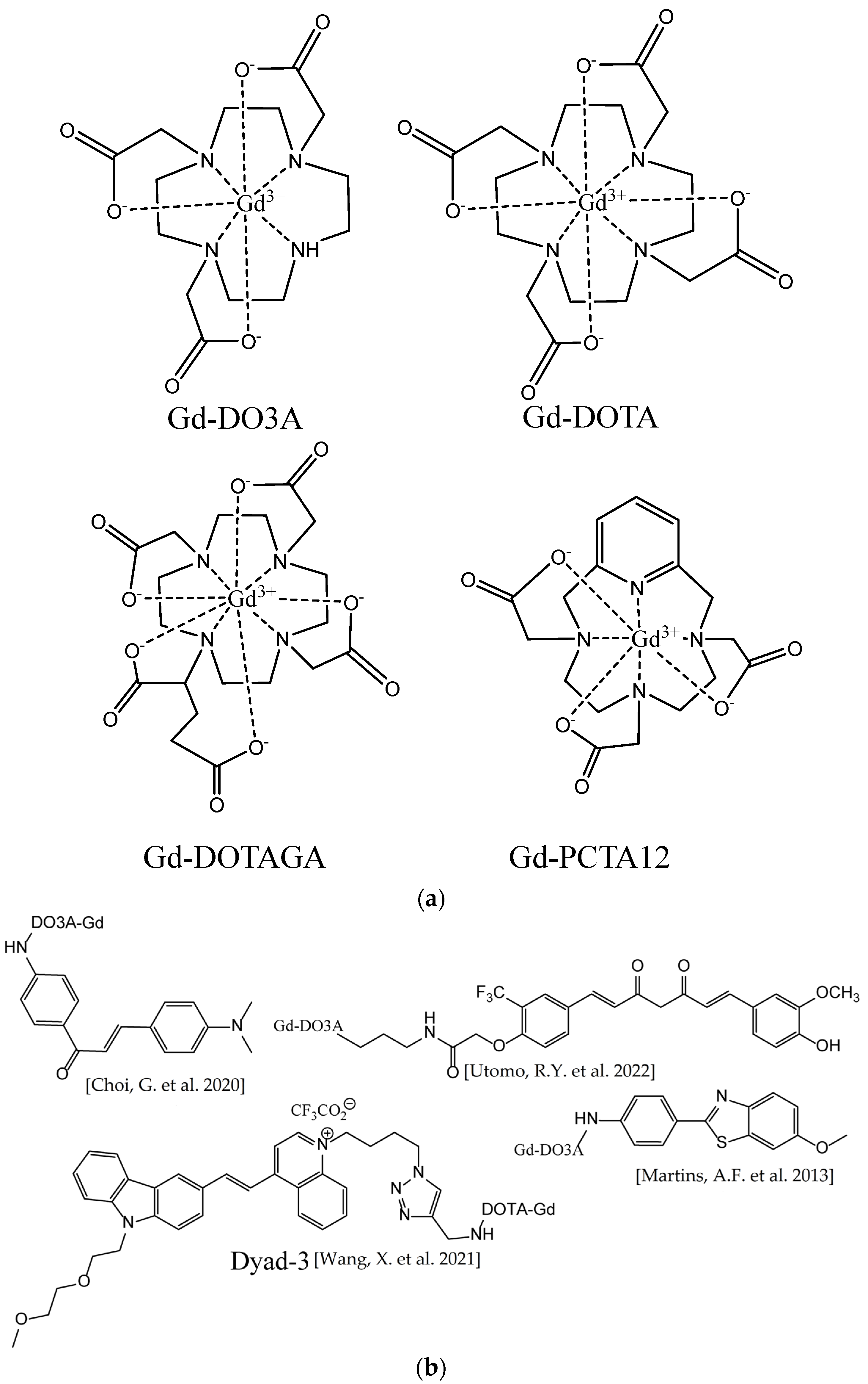
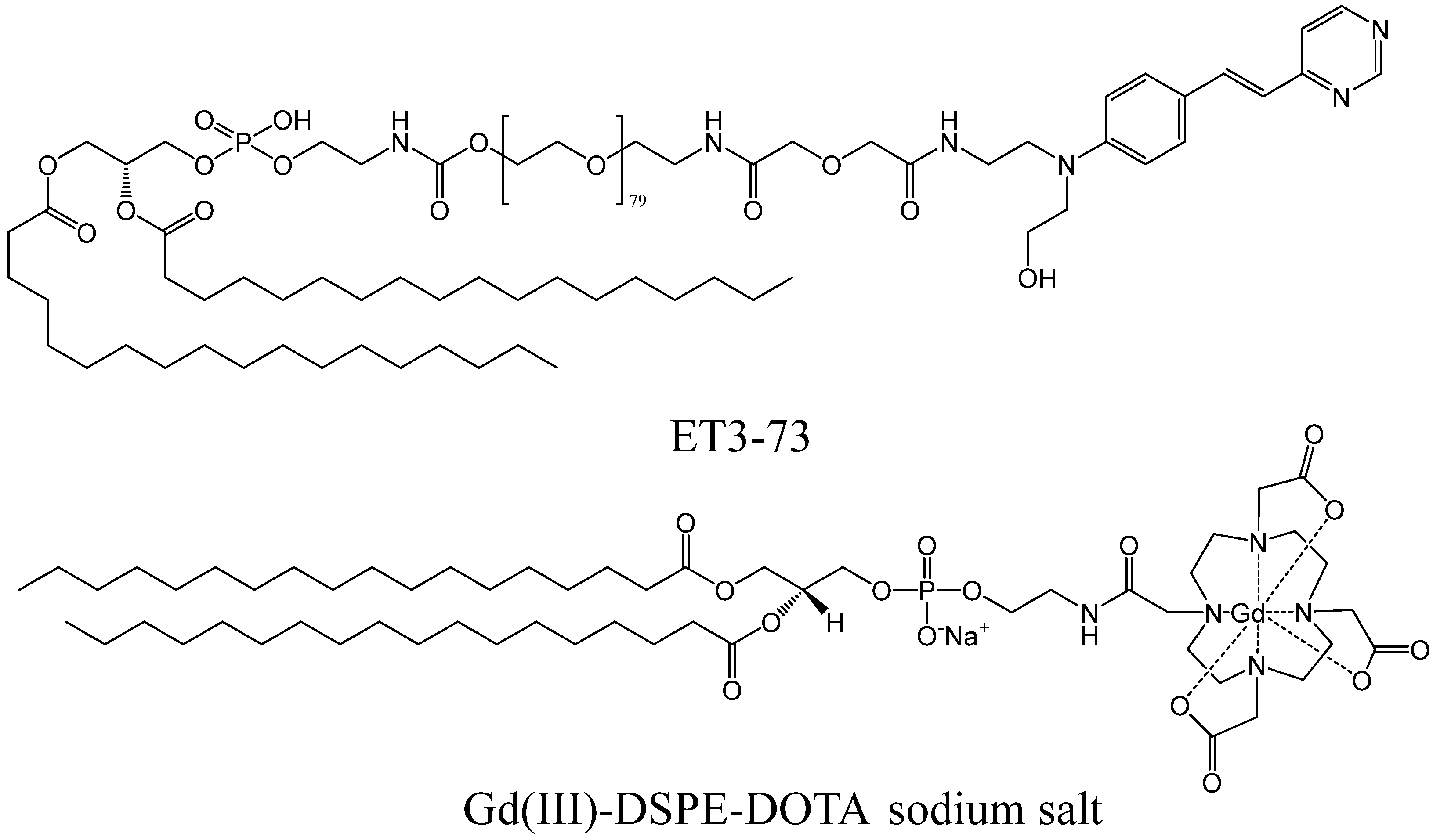
| Gd3+ chelates conjugated with Aβ binders [200,201,202,210,211,212,213,214] | Aβ | 29b |
| SPIONs conjugated with Aβ binders [203,204] | Aβ | – |
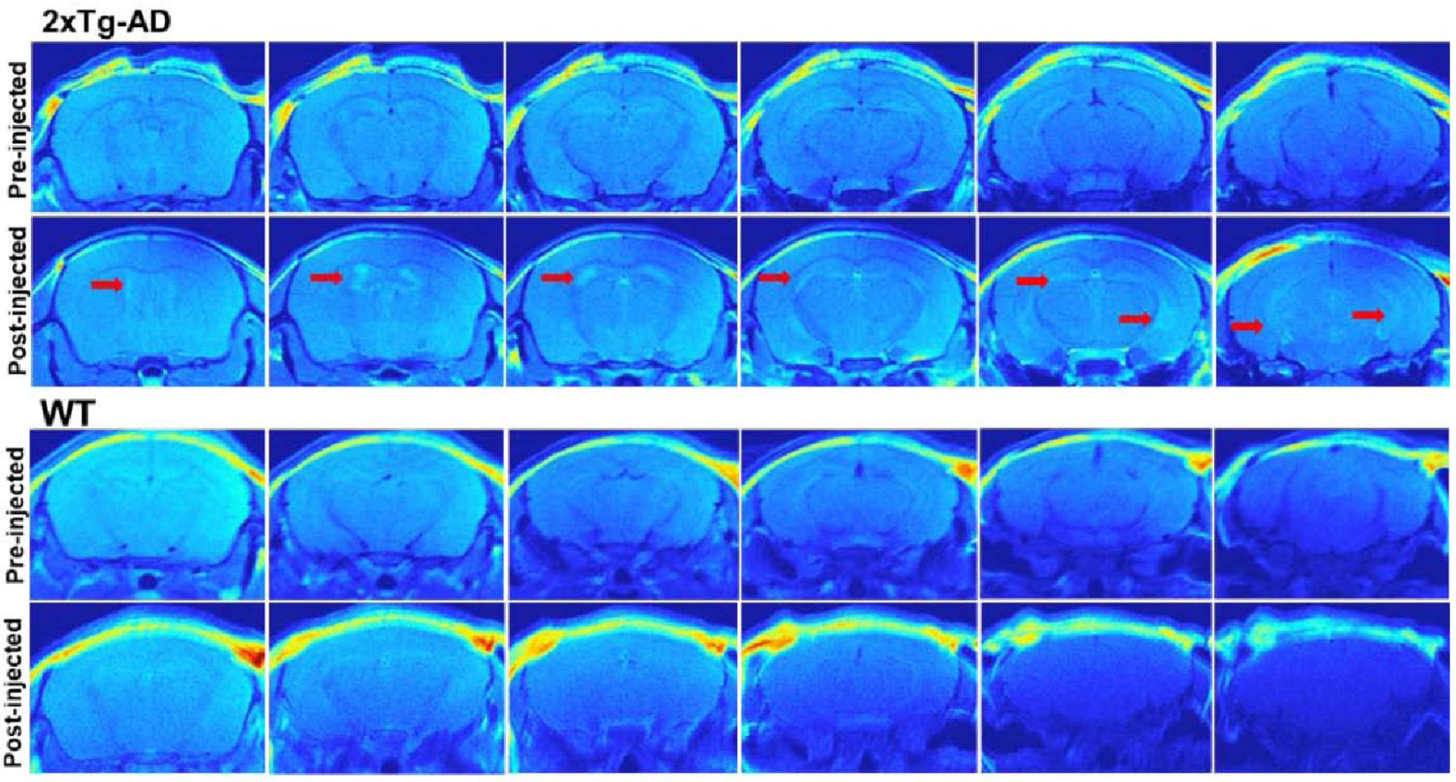
| ADx-001 [220,224] | Aβ | 31 |
Disclaimer/Publisher’s Note: The statements, opinions and data contained in all publications are solely those of the individual author(s) and contributor(s) and not of MDPI and/or the editor(s). MDPI and/or the editor(s) disclaim responsibility for any injury to people or property resulting from any ideas, methods, instructions or products referred to in the content. |
© 2023 by the authors. Licensee MDPI, Basel, Switzerland. This article is an open access article distributed under the terms and conditions of the Creative Commons Attribution (CC BY) license (https://creativecommons.org/licenses/by/4.0/).
Share and Cite
Nikiforova, A.; Sedov, I. Molecular Design of Magnetic Resonance Imaging Agents Binding to Amyloid Deposits. Int. J. Mol. Sci. 2023, 24, 11152. https://doi.org/10.3390/ijms241311152
Nikiforova A, Sedov I. Molecular Design of Magnetic Resonance Imaging Agents Binding to Amyloid Deposits. International Journal of Molecular Sciences. 2023; 24(13):11152. https://doi.org/10.3390/ijms241311152
Chicago/Turabian StyleNikiforova, Alena, and Igor Sedov. 2023. "Molecular Design of Magnetic Resonance Imaging Agents Binding to Amyloid Deposits" International Journal of Molecular Sciences 24, no. 13: 11152. https://doi.org/10.3390/ijms241311152