Article

Circular Intronic RNA circTTN Inhibits Host Gene Transcription and Myogenesis by Recruiting PURB Proteins to form Heterotypic Complexes

1
College of Animal Science and Technology, Hunan Agricultural University, Changsha 410128, China
2
Guangdong Laboratory for Lingnan Modern Agriculture, Guangzhou 510642, China
3
Institute of Subtropical Agriculture, Chinese Academy of Sciences, Changsha 410125, China
*
Authors to whom correspondence should be addressed.
Int. J. Mol. Sci. 2023, 24(12), 9859; https://doi.org/10.3390/ijms24129859
Submission received: 28 April 2023 / Revised: 24 May 2023 / Accepted: 5 June 2023 / Published: 7 June 2023

Abstract

:
Muscle cell growth plays an important role in skeletal muscle development. Circular RNAs (circRNAs) have been proven to be involved in the regulation of skeletal muscle growth and development. In this study, we explored the effect of circTTN on myoblast growth and its possible molecular mechanism. Using C2C12 cells as a functional model, the authenticity of circTTN was confirmed by RNase R digestion and Sanger sequencing. Previous functional studies have showed that the overexpression of circTTN inhibits myoblast proliferation and differentiation. Mechanistically, circTTN recruits the PURB protein on the Titin (TTN) promoter to inhibit the expression of the TTN gene. Moreover, PURB inhibits myoblast proliferation and differentiation, which is consistent with circTTN function. In summary, our results indicate that circTTN inhibits the transcription and myogenesis of the host gene TTN by recruiting PURB proteins to form heterotypic complexes. This work may act as a reference for further research on the role of circRNA in skeletal muscle growth and development.

1. Introduction

Skeletal muscle is an important component of the mammalian body, which plays an important role in energy transformation and posture maintenance [1]. The skeletal muscle development process includes three stages: (1) mesenchymal stem cells from mesoderm proliferate and differentiate into myoblasts, (2) myoblasts further differentiate to form primary myotubes and secondary myotubes, and (3) myotubes then fuse into muscle fibers [2]. This process is also known as myogenesis. Muscle fibers are the basic contractile units of skeletal muscle. They are surrounded by connective tissue layers and are formed into bundles to form skeletal muscle [3]. Skeletal muscle development is subject to myogenic regulatory factors (MRFs), paired box family (PAX), SIX family, and myocyte enhancer factor 2 (MEF2) [4,5]. In recent years, with the upsurge of research on non-coding RNAs (ncRNAs), the mechanism of circular RNAs (circRNAs) in skeletal muscle growth and development is also attracting researchers’ interest.
circRNAs are a class of non-coding RNA molecules that do not have a 5′ cap and 3′ poly(A) tail. Dicer [6] first discovered the potato spindle tuber “virus” in 1971 and called it viroid. In 1993, circular transcription of the Sry gene was found in mice, but the circRNAs discovered were thought to be low-abundance RNA molecules formed by the missplicing of exon transcripts [7]. With the rapid development of high-throughput sequencing, more circRNAs have been discovered by researchers [8]. Currently, circRNAs have been shown to be associated with myogenesis, the transformation of muscle fiber types, and muscle diseases [9,10,11]. For example, circZfp609 can act as a molecular sponge to adsorb miR-194-5p and isolate its inhibition of BCLAF1, thus inhibiting myogenic differentiation [12]. In addition, circRNAs can regulate the variable splicing of transcripts and parental gene transcription and translation by binding to RBPs [13,14,15]. However, the previous research on this mechanism has mainly been focused on the aspect of cancer treatment and less on the aspect of skeletal muscle development, which requires further exploration.
We previously found a circular intronic RNA circTTN produced by the TTN (Titin) gene through high-throughput sequencing [16]. circTTN was significantly differentially expressed in the skeletal muscle of Ningxiang pig at 30 d, 90 d, 150 d, and 210 d after birth, suggesting that circTTN may be involved in the regulation of skeletal muscle myogenesis. In vitro, functional validation models (C2C12 cells) showed that circTTN is a negative regulator of myogenesis and that circTTN recruits PURB protein to inhibit the transcription of the host gene TTN. Our study revealed the molecular regulatory mechanism of circTTN and provided a reference for further research on skeletal muscle growth and development.

2. Results

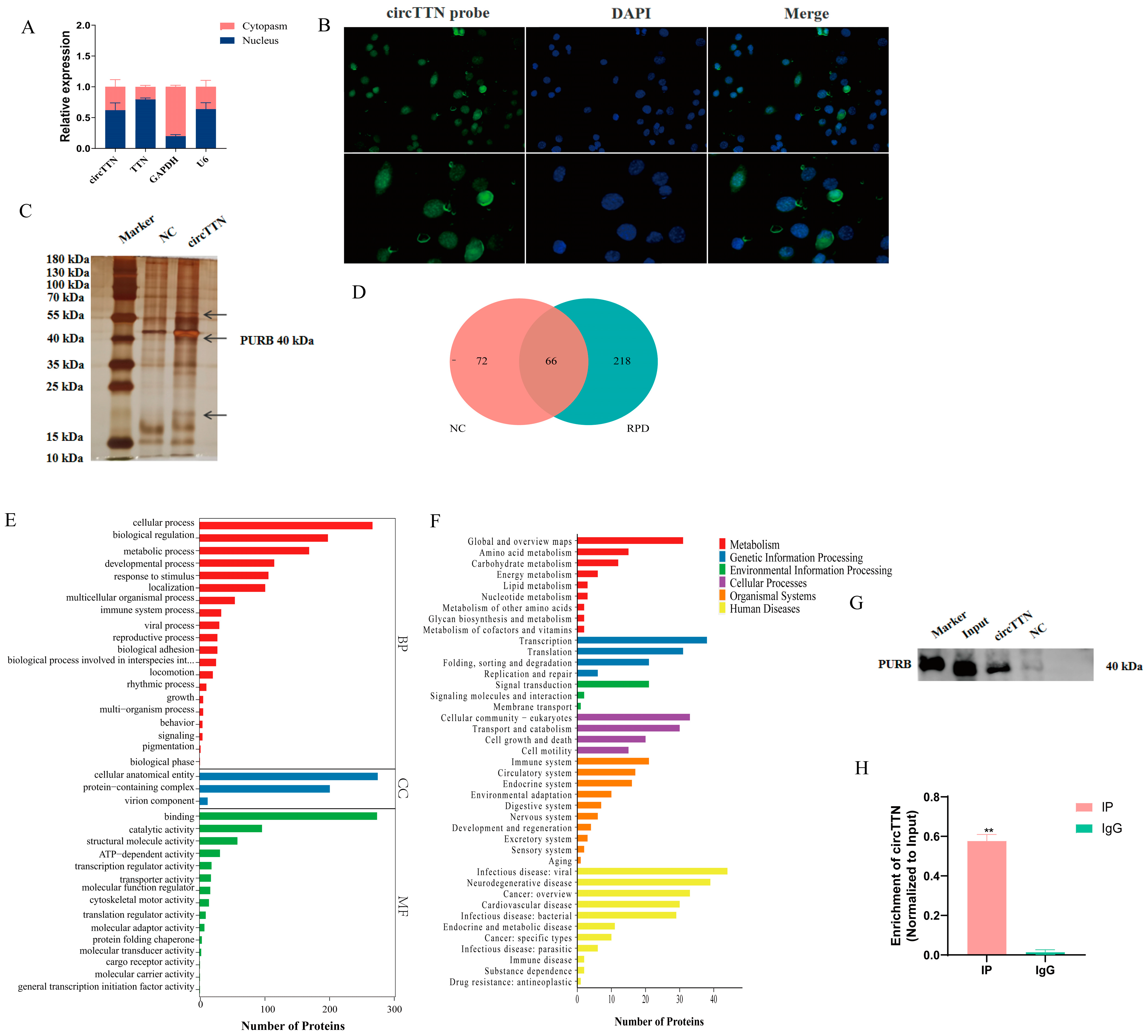
2.1. Characterization Analysis of circTTN

circTTN is derived from intron15 of the TTN gene; its length is 339 bases, and it is located in chromosome 15 (Chr15_85761324_85761662) (Figure 1A). First, the divergent primer and convergent primer were designed to perform qRT-PCR experiments using the cDNA and gDNA of muscle tissue as templates, and DNA gel electrophoresis was performed on the products. The convergent primer and divergent primer could amplify bands using cDNA as a template. Only the convergent primer could amplify a band when gDNA was used as the template (Figure 1B). The products of the divergent primer were analyzed using Sanger sequencing (Figure 1A). After treatment with RNase R, TTN and β-actin genes had no bands after electrophoresis, while circTTN had bands (Figure 1C). The above experimental results can confirm the authenticity and stability of circTTN.
Then, the expression of circTTN in different tissues of Ningxiang pig at four periods after birth were detected using qRT-PCR assay. The results showed that the expression of circTTN in the heart and muscle of Ningxiang pig at four stages was the highest (Figure 1D). In addition, circTTN was differentially expressed during the proliferation and differentiation of C2C12 cells (Figure 1E,F).

2.2. circTTN Inhibits Proliferation and Differentiation of C2C12 Cells and Host Gene TTN

To explore the function of circTTN, the circTTN overexpression vector was transfected into C2C12 cells, and the transfection efficiency of circTTN was more than 3000 times higher (p < 0.01) (Figure 2A), which can be used for subsequent experiments. First, qRT-PCR and Western blot were used to detect the expression of proliferation biomarker genes. The overexpression of circTTN significantly reduced the mRNA levels of CCND and CDK4 (p < 0.05); the mRNA expression level of PCNA was markedly decreased (p < 0.01) (Figure 2B). The protein expression levels of proliferating genes CCND and PCNA in the overexpression group were markedly lower than those in the control group (p < 0.01); the CDK4 protein expression level was significantly decreased (p < 0.05) (Figure 2C,D). The results of the CCK8 experiments displayed that there were no significant difference in the proliferative activity between the overexpressed group and the control group (p > 0.05) (Figure 2E); the EdU results showed that the number of positive proliferating cells in the overexpressed group was markedly decreased (p < 0.01) (Figure 2F,G). In addition, flow cytometry was used to detect circTTN expression during cell cycle process, and the number of cells in the overexpressed group was significantly reduced in the S phase (p < 0.01) (Figure 2H,I). In summary, circTTN inhibited the proliferation of C2C12 cells.
We further explored the effect of circTTN on the differentiation of C2C12 cells; the overexpressed circTTN vector was transfected into cells for induction differentiation culture, and then, qRT-PCR and a Western blot assay were used to detect the expression of differentiation biomarker genes. The overexpression of circTTN significantly inhibited the expression of differentiation biomarker genes (MyoD, MyHC, and MyoG) at the mRNA level (Figure 3A) (p < 0.01). At the protein level, the expression of MyoD and MyoG were markedly repressed (p < 0.01), significantly inhibiting the expression of MyHC (p < 0.05) (Figure 3B,C). In addition, an immunofluorescence assay was used to observe the changes in phenotypic myotube differentiation, and the number in differentiated myotubes in the circTTN overexpression group was significantly reduced compared with the control group (p < 0.05) (Figure 3D). These results indicate that circTTN inhibited the differentiation of C2C12 cells.
TTN is a coding gene that plays an important role in skeletal muscle. We intended to discover if circTTN acts on host gene TTN. The qRT-PCR results implied that circTTN overexpression significantly downregulated TTN gene expression during the proliferation period (Figure 4A) (p < 0.01). At the protein level, the overexpression of circTTN was markedly lower during proliferation compared with the control group (Figure 4B,C) (p < 0.01). Similarly, the effect of circTTN overexpression on the TTN gene during differentiation was consistent with that during proliferation, and the mRNA and protein levels of circTTN were significantly decreased compared with those of the control group (p < 0.01) (Figure 4D–F).

2.3. circTTN Interacts with PURB Protein

First, we determined the subcellular localization of circTTN by nucleoplasmic isolation and RNA fluorescence in situ hybridization experiments. GAPDH and U6 were used as reference genes in the cytoplasm and nucleus, respectively. The results showed that circTTN and TTN mRNA existed in the nucleus and cytoplasm, but most of them were located in the nucleus (Figure 5A). The results of the FISH experiment were consistent with the results of nucleoplasmic separation. (Figure 5B). These results suggest that circTTN may be regulated in the nucleus. circRNA can interact with RNA-binding proteins to influence the expression of parent genes. We then examined circTTN-bound proteins using an RNA pull-down assay. The circTTN group showed obvious bands of difference compared with the control NC group (Figure 5C). Subsequently, a total of 356 proteins were identified using mass spectrometry (MS) analysis, among which 218 different proteins were found in the circTTN group (RPD group) (Figure 5D, Table 1). Gene ontology (GO) functional enrichment analysis and Kyoto Encyclopedia of Genes and Genomes (KEGG) analysis were conducted, respectively. GO functional enrichment showed that these proteins were mainly enriched in cellular processes, protein complexes, regulatory translation activities, biological regulation, developmental processes, and binding. KEGG suggested that some proteins were enriched in transcription, translation, and metabolism (Figure 5F). These results implied that circTTN may bind to these proteins and play a certain function. Next, the expression of Pur-beta (PURB) protein in the RNA pull-down eluent was detected using a Western blot assay (Figure 5G), and it was found that the expression of PURB protein could be only detected in the circTTN group’s eluent. In addition, we reversely demonstrated an interaction between PURB protein and circTTN through an RIP experiment (Figure 5H). The results showed that the PURB antibody could be significantly enriched to circTTN (p < 0.01), further supporting the idea that circTTN interacts with PURB.

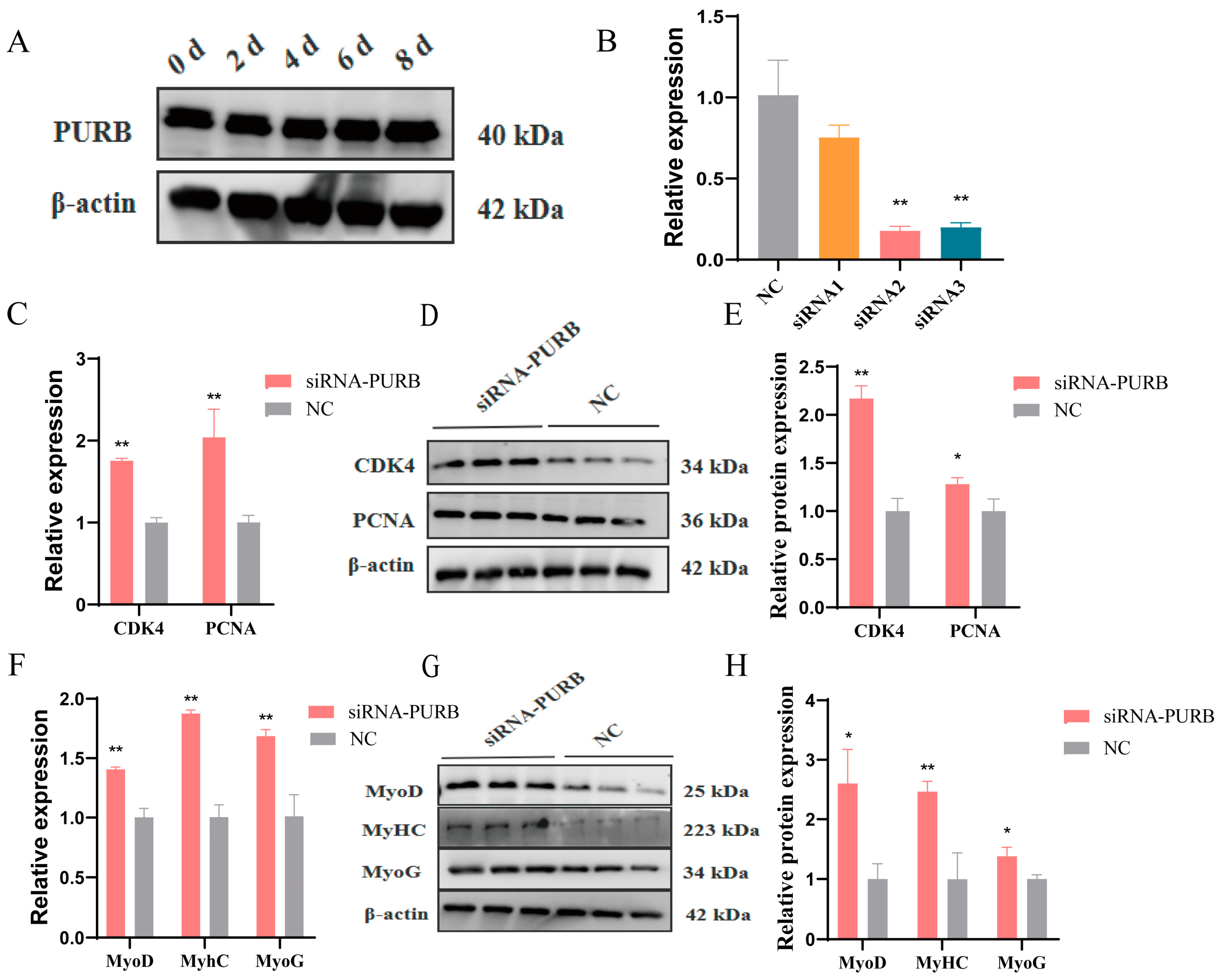
2.4. PURB Knockdown Promotes Proliferation and Differentiation of C2C12 Cells and Increase TTN Expression

Previously, we confirmed the interaction between PURB protein and circTTN. Next, we detected the expression of PURB in C2C12 cells using a Western blot assay (Figure 6A), and it was found that PURB was highly expressed in C2C12 cells. To further explore the effect of PURB on C2C12 cells, we designed 3 siRNA sequences, as shown in Figure 6B; qRT-PCR was used to select the one with the best knockdown effect, and siRNA2 had the best knockdown efficiency (p < 0.01) (5′-GUGGACUCCAAGCGUUUCUTT-3′), which can be used for subsequent experiments. siRNA-PURB was transfected into C2C12 cells, and siRNA-PURB significantly promoted the expression of CDK4 and PCNA genes at the mRNA level (Figure 6C) (p < 0.01). The protein expression level of CDK4 was markedly higher than that of the control group (p < 0.01), and the protein expression level of PCNA was significantly increased (Figure 6D,E) (p < 0.05). These results indicated that the knockdown PURB promoted the proliferation of C2C12. Similarly, siRNA-PURB was transfected into cells and induced differentiation was performed until myotubes were grown. As shown in Figure 6F, siRNA-PURB significantly upregulated the expression of differentiation biomarker genes (MyoD, MyHC, and MyoG) at the mRNA level (p < 0.01). siRNA-PURB markedly elevated the protein expression of MyHC (p < 0.01) and significantly promoted the protein expression of MyoD and MyoG (p < 0.05) (Figure 6G,H). In conclusion, PURB inhibited the proliferation and differentiation of C2C12 cells.
To explore the effect of PURB on the TTN gene, siRNA-PURB was transfected into C2C12 cells, and the effects of PURB on TTN during proliferation and differentiation were detected, respectively. The results show that the mRNA and protein levels of siRNA-PURB were significantly higher than those of the control group at the proliferation stage (p < 0.01) (Figure 7A–C). siRNA-PURB were markedly increased compared with the control group at the differentiation stage (p < 0.01) (Figure 7D), and the protein level was significantly elevated (p < 0.05) (Figure 7E,F). These results indicate that PURB inhibited TTN gene expression.

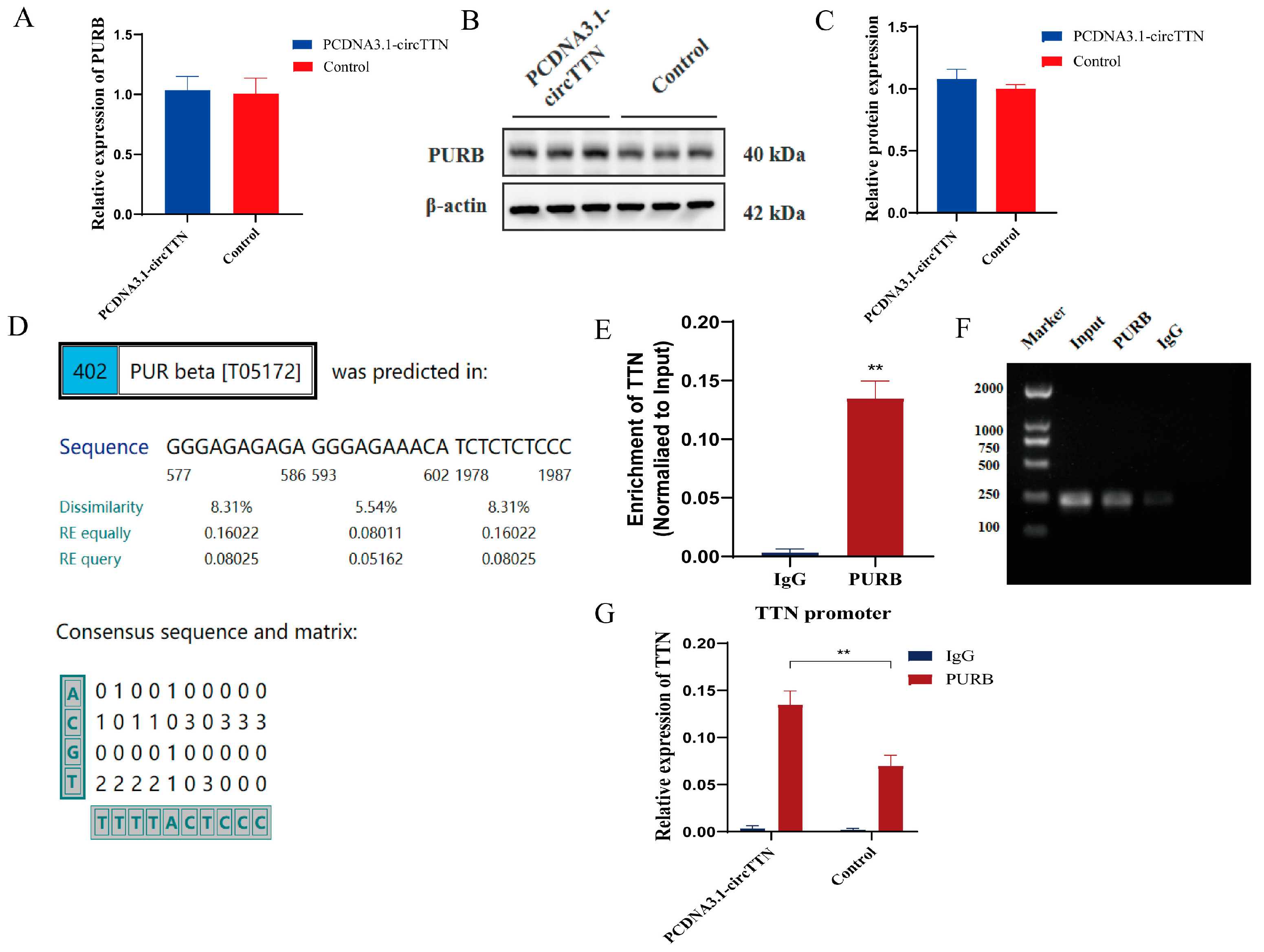
2.5. circTTN Inhibits TTN Transcription by Recruiting PURB

We intended to verify whether PURB are regulated by circTTN. After circTTN overexpression, PURB induced no significant changes in mRNA and protein levels (Figure 8A–C) (p < 0.05). This indicated that circTTN overexpression did not affect the PURB expression level. In a previous study, the effects of circTTN and PURB on the TTN gene were explored, respectively. Then, we utilized the UCSC Genome Browser (http://genome.ucsc.edu/ accessed on 25 August 2022) and PROMO database (https://alggen.lsi.upc.es/cgi-bin/promo_v3/promo/promoinit.cgi?dirDB=TF_8.3 accessed on 25 August 2022) for bioinformatics analysis. We found that the PURB binding motif was present at the TTN promoter (Figure 8D), and then, ChIP-qPCR was used to further verify the binding of PURB and the TTN promoter. PURB protein significantly bound to the TTN promoter (Figure 8E,F) (p < 0.01). After circTTN overexpression, we found that PURB protein bound to the TTN promoter was significantly higher than in the control group (p < 0.01) (Figure 8G). Our study suggested that circTTN inhibited the transcription of the host gene TTN by recruiting PURB protein.

3. Discussion

Skeletal muscle plays a role in coordinating movement and maintaining normal metabolism in organisms [17]. At present, circRNA is involved in skeletal muscle proliferation and differentiation as a positive or negative regulator of skeletal muscle myogenesis [18]. The C2C12 cell line is a valuable in vitro model, originally monocytes, which can rapidly integrate into multinucleated myotubes after culture with an induced differentiation solution [19]. Due to its unique biochemical characteristics, researchers can better understand the metabolism and differentiation of skeletal muscle at cellular and molecular levels [20]. Sun et al. [21] identified circCSDE with a stable and high expression in porcine skeletal muscle and studied the function of circCSDE on C2C12 cells, where they found that circCSDE1 and its target miR-21-3p jointly regulated the proliferation and differentiation of myoblasts. In their study, the mechanism of circCSDE regulating the growth and development of skeletal muscle in the porcine skeletal muscle injury model was verified. In our study, we first reported that circular intronic RNA circTTN acts as a negative regulator of myogenesis and inhibits the transcription of the host gene TTN by recruiting PURB protein in the nucleus (Figure 9).
circRNAs are usually expressed in a tissue-specific manner and perform different biological functions [22]. circRNAs regulate gene expression through the use of a variety of mechanisms. Some studies have shown that circRNA can act as a miRNA sponge or an effective competing endogenouse RNA (ceRNA) molecule [23,24], which is also one of the most classical mechanisms of circRNAs. circRNAs can also bind RBPs. Although the RBPs’ binding site of circRNA is less than that of the corresponding linear mRNA [25], there is still a lot of evidence that RBPs interacts with circRNAs; moreover, they are involved in almost every aspect of the circRNA life cycle, including generation, transcriptional regulation, post-transcriptional regulation, functional execution, specific modifications, and potential extracellular transport pathways [26,27,28]. In this study, we identified the authenticity of circTTN through the use of RNase R digestion and other methods, and then, detected the expression level of circTTN in Ningxiang pig tissues of different days of age and proliferative and differentiated C2C12 cells. It was found that circTTN was highly expressed in the heart and muscle tissues of Ningxiang pig at different periods. It was also differentially expressed in proliferating and differentiated C2C12 cells. This suggested that circTTN may be involved in the regulation of skeletal muscle development. Then, we demonstrated that circTTN inhibits the proliferation and differentiation of C2C12 cells. A previous study has shown that circTTN promotes the proliferation and differentiation of bovine myoblasts [29], suggesting that circTTN may have different effects on the proliferation and differentiation of myoblasts in different mammals. The reason may be that circRNA plays different functions due to different subcellular localization. Our study showed that circTTN is a negative regulator of muscle regeneration in skeletal muscle. Interestingly, we found that circTTN inhibited the expression of the host gene TTN during both proliferation and differentiation. In addition, we found that circTTN is mainly located in the nucleus and interacts with PURB protein. Our study showed that PURB appears to be involved in circTTN expression and function.
PURB is a highly conserved protein whose conserved region includes ssDNA and RNA domains. It is a DNA and RNA binding protein involved in the regulation of DNA replication and transcription [30] and can inhibit or promote gene transcription [31]. PURB has been reported to play different roles in many physiological and pathological processes [32]. It can interact with gonadotropin-releasing hormone1 (GnRH1) to control fish reproduction by stimulating the pituitary to release gonadotropin-releasing hormone1 [33]. Hariharan et al. [34] demonstrated that PURB protein negatively regulates the trans-activation of smooth muscle actin genes in myoblasts. Based on previous studies on PURB, in our study, we confirmed that PURB inhibits the proliferation and differentiation of C2C12 cells and the expression of the TTN gene, which was consistent with the function of circTTN.
Previous studies showed that PURB, as a transcription factor, can be recruited by linc-HOXA1 to mediate the transcriptional regulation of lincHOXA1 in embryonic stem cells [35]. A recent study indicated that LncCMRR functionally interacts with the transcriptional suppressor PURB to inhibit its binding potential in the Flk1 promoter region, thereby protecting Flk1 transcription during cardiac mesoderm formation [36]. By binding to PURB protein, Circ-Calm4 promoted the transcription of autophagy-related protein Beclin1, thereby regulating the hypoxia-induced autophagy of pulmonary artery smooth muscle [37]. circRNAs can not only act as miRNA molecular sponges or translation proteins to regulate muscle growth and development [38] but they also regulate muscle generation by interacting with RBPs. Pandey et al. [14] determined that PURB was distributed in the nucleus and cytoplasm of C2C12 cells, and it combined with circSamd4 located in the nucleus to participate in the transcription regulation of MHC, thus promoting myogenesis, which was also the first revelation that circRNA could interact with RBPs to regulate the growth and development of skeletal muscle. Chen et al. [39] pointed out that circMYBPC1 can directly bind MyHC protein, promote the differentiation of myoblasts, and possibly promote skeletal muscle regeneration. One study showed that LncMGPF promoted the expression of HuR in C2C12 cytoplasm, enhancing the stability of MyoD and MyoG mRNA, and thus promoting myogenesis [40,41]. In this study, we found that PURB and the TTN promoter have a binding motif through the use of the UCSC genome browser and PROMO database, and we found that circTTN overexpression promoted the binding of PURB and the TTN promoter and inhibited the generation of the TTN gene. The protein encoded by the TTN gene is the largest known protein and plays a key structural, developmental, and regulatory role in heart and skeletal muscle [42,43]. circTTN inhibits TTN transcription through PURB recruitment, which also indicates that circTTN affects skeletal muscle development through PURB recruitment. In addition, our study showed that circTTN or PURB negatively regulated the proliferation and differentiation of C2C12 cells; however, it was unclear whether circTTN inhibited the proliferation and differentiation of myoblasts by recruiting PURB. Furthermore, the role of circTTN in muscle regeneration was not explored in vivo, which is also a limitation of our study. These are something that needs to be addressed in future studies.

4. Materials and Methods

4.1. Experimental Animals and Samples

All the animals were obtained from Dalong Animal Husbandry Technology Co., LTD., Ningxiang City, Hunan Province. In total, 7 tissue samples (n = 3) were taken from Ningxiang pigs at 30 d, 90 d, 150 d, and 210 d after birth, including heart, liver, spleen, lung, kidney, longissimus dorsi muscle, and adipose. The sampling process strictly followed the asepsis rules. The samples were placed in liquid nitrogen where they were then brought back to the laboratory and finally stored in a freezer at −80 °C.

4.2. Culture and Differentiation of C2C12 Cells

C2C12 cells were purchased from Anweisci (Shanghai, China). The resuscitated cells were cultured with growth medium (CM) in 10 cm dishes. CM was composed of 89% DMEM (Gibco, Waltham, MA, USA), 10% fetal bovine serum (Gibco, Waltham, MA, USA), and 1% penicillin–streptomycin (Gibco, Waltham, MA, USA). When the cell confluence reached 80–90%, trypsin was used for digestion (Gibco, Waltham, MA, USA), and the medium was changed to differentiation medium (DM), which was composed of 2% horse serum and 98% DMEM. C2C12 cells were cultured in a 37 °C incubator containing 5% CO2.

4.3. RNA Extraction and cDNA Synthesis

An RNA Extraction kit (TIANGEN, Beijing, China) was used to extract total RNA. A nucleic acid concentration was used for the measurements using NanoDrop ND-2000 (Thermo Scientific, Waltham, MA, USA). The samples that needed to be processed with RNase R were digested using RNase R (JISAI, Guangzhou, China). Then, cDNA synthesis was performed using a RevertAid First Strand cDNA Synthesis Kit (Thermo Scientific, Waltham, MA, USA) according to the manufacturer’s instructions.

4.4. Quantitative Real-Time PCR

Quantitative real-time PCR (qRT-PCR) primer sequences were designed from the NCBI (https://www.ncbi.nlm.nih.gov/tools/primer-blast/ accessed on 10 November 2021) database, and primers were synthesized from Tsingke Biotechnology (Beijing, China). The primer sequences are shown in Table 2. qRT-PCR was performed using a CFX connect real-time system (Bio-Rad, Hercules, CA, USA), and qRT-PCR data were analyzed using the β-actin gene as the reference gene. The relative expression was calculated using the 2−ΔΔCt method.

4.5. Vector Construction and siRNA

The circTTN overexpression vector (PCDNA3.1-circTTN) and empty vector (control) were synthesized in JTS Scientific Biotechnology (Wuhan, China). The siRNA and NC of PURB were synthesized in GenePharma Biotechnology (Suzhou, China). Transfection was performed using a Lipofectamine 2000 reagent (Invitrogen, Waltham, MA, USA) according to the manufacturer’s instructions.

4.6. CCK8 and EdU Assays

Cell proliferation was detected through Cell Counting Kit-8 (CCK8). The cells were cultured after transfection and inoculated into 96-well plates when the confluence reached 90%. Then, they were cultured for 0 h, 24 h, 48 h, 72 h, and 96 h, respectively, and 10 μL of CCK8 reagent was added 4 h before each assay (Beyotime Biotechnology, Shanghai, China). The absorbance at 450 nm was measured using a microplate reader (Multiskan FC, Thermo Scientific, Waltham, MA, USA). Cell proliferation was detected using the EdU kit (Meilunbio Biotechnology, Shanghai, China). Similarly, the cells were inoculated into 96-well plates and cultured. The EdU solution was diluted with cell growth medium at a ratio of 500:1 to prepare 20 μM of EdU medium. Each well was added with 100 μL of EdU solution, incubated for 2 h, and then fixed with 4% paraformaldehyde, followed by 0.5% tritonX-100 osmotic solution, and finally, stained with EdU and Hoeches 33342.

4.7. Flow Cytometry

The cells were transfected when the cells grew to about 60%. After transfection, the cells were mixed with 70% ethanol and fixed at 4 °C for 2 h. Each tube of the sample was stained with 0.5 mL of staining solution (0.5 mL of staining buffer, 10 μL of propidium iodide storage, and 10 μL of RNaseA) and incubated at 37 °C for 30 min in the absence of light. Then, the treated cell suspension was measured using Cytek DxP Athena flow cytometry (Cytek, Fremont, CA, USA).

4.8. Immunofluorescence Staining

The cells were fixed with 4% paraformaldehyde for 30 min, incubated with 0.5% triton X-100 at room temperature for 20 min, and closed with 5% BSA (Bio Froxx, Frankfurt, Germany) for 2 h. Anti-MyHC monoclonal antibody was used for diluting 5% BAS (1:200, DSHB, Iowa, IA, USA) and was incubated overnight at 4 °C. Then, it was incubated with anti-mouse IgG (1:1000, Abbkine, Wuhan, China) for 2 h. Next, nuclei were stained using DAPI (Solarbio, Beijing, China) for 10 min without light. Finally, photographs were taken and analyzed under an inverted microscope (Axio Vert A1, ZEISS, Oberkochen, Germany).

4.9. Western Blot Analysis

The cell proteins were extracted with RIPA Lysis Buffer (Beyotime Biotechnology, Shanghai, China), and the protein concentration was quantified using a BCA protein quantitative/concentration assay kit (Meilunbio Biotechnology, Shanghai, China). The denatured protein samples were electrophoreted in Tris/Glycine/SDS Buffer (Epizyme, Biotechnology, Shanghai, China), and then, the proteins were transferred to the PDVF membrane for electrotransfer in the electrotransfer solution (Yamei, Biotechnology, Shanghai, China). The membranes were sealed with the sealing liquid for 2 h, and then β-actin (1:1000, TransGen, Beijing, China), CDK4 (1:1000, AiFang, Changsha, China), PCNA (1:2000, ZENGBIO, Chengdu, China), CCND (1:1000, CST, Boston, MA, USA), MyoD (1:1000, HUABIO, Hangzhou, China), MyoG (1:200, DSHB, Iowa, IA, USA), MyHC (1:1000, DSHB, Iowa, IA, USA), Titin (1:1000, Bioword, Minnesota, MN, USA), and PURB (1:1000, Proteintech, San Diego, CA, USA) antibodies were incubated at 4 °C overnight. Finally, the membranes were incubated with the secondary antibody (1:10,000, ZENBIO, Chengdu, China) for 2 h, and the protein bands were developed using an ECL chemiluminescent substrate kit (Biosharp, Guangzhou, China). All the protein levels were analyzed by β-actin protein reference normalization.

4.10. Nucleoplasmic Separation and RNA Fluorescence In Situ Hybridization

Nuclear and cytoplasmic RNA was extracted using a cytoplasmic and nuclearRNA purification kit (Norgen Biotek, Thorold, ON, Canada) and was quantified using qRT-PCR analysis. We made cell creep tablets, fixed them with 4% paraformaldehyde, and sent them to Pinuofei Biotechnology (Wuhan, China) for a RNA fluorescence in situ hybridization (FISH) experiment. The probe sequence of circTTN is 5′-UGAGGCUGCCAACCGGAACAAUGACG-3′.

4.11. RNA Pull-Down and Mass Spectrometry

Biotin-labeled circTTN probe and NC probe were provided by Ribo Biotechnology (Guangzhou, China). Pull-down protein experiments were performed using an RNA pulldown kit (BersinBio, Guangzhou, China). First, the circTTN probe and NC probe formed a secondary structure and were incubated with streptavidin magnetic beads, respectively. Then, 2 × 10⁷ cell samples were collected for protein extraction and the nucleic acid of protein samples were removed, and then, the probe–magnetic bead complex was rotatively incubated with cell extracts. Finally, the histone samples of the circTTN group and NC group were collected, and silver dyeing was performed with a silver dyeing kit (Beyotime, Shanghai, China). First, 30% ethanol was used for fixing, and then, silver sensitizing solution, silver solution, silver dyeing solution, and silver dyeing stop solution were added successively. The silver dyed adhesive strips were photographed and the results were analyzed. RNA pulldown protein samples were determined and analyzed using mass spectrometry in Genecreate Biotechnology (Wuhan, China).

4.12. RNA Immunoprecipitation (RIP) Analysis

An RNA Immunoprecipitation Kit (BersinBio, Guangzhou, China) was used for RIP analysis. First, 2 × 10⁷ cells were lysed to remove the DNA, and then the cell lysate samples were divided into 3 parts according to 0.8 mL (IP), 0.8 mL (IgG), and 0.1 mL (Input). IP and IgG samples were added with the corresponding experimental antibodies PURB (ProteintechSan Diego, CA, USA) and IgG (BersinBio, Guangzhou, China), respectively, and were incubated at 4 °C for 16 h. Then, they were rotatively incubated with balanced protein A/G beads for 1 h, followed by RNA eluting using an elution buffer. Finally, RNA was extracted with a mixture of phenol–chloroform–isoamyl alcohol, which was detected and analyzed using qRT-PCR according to the percentage input method.

4.13. ChIP-qPCR Analysis

A SimpleChIP® Plus Enzymatic Chromatin IP Kit (CST, Boston, MA, USA) was used for ChIP analysis. In brief, the cells were crosslinked with 1% formaldehyde for 10 min and incubated with 10× glycine for 5 min, followed by the addition of protease enzyme (PIC); then, chromatin fragment and diluted chromatin were added to the corresponding IP (PURB) and IgG immunoprecipitation antibody. They were then incubated overnight by rotation at 4 °C, followed by the addition of protein G magnetic beads. After incubation at 4 °C for 2 h, the DNA complex was cleaned with high-salt and low-salt washing solution, and chromatin was eluted with a ChIP Elution buffer and Proteinase K was added. Finally, DNA purification was performed. The combination of PURB and the TTN promoter were analyzed using qRT-PCR according to the percentage input method.

4.14. Statistical Analysis

We used Microsoft Excel to sort the data. We analyzed all the data using t-tests and a one-way ANOVA and calculated qRT-PCR data using the 2−ΔΔCt method. In addition, we analyzed RIP-qPCR and ChIP-qPCR using the percentage input method, counted the number of cells using Image J software (version 1.49), and mapped these results using GraphPad Prism 8.0 software. Data analysis was expressed as mean ± SEM, n = 3. * p < 0.05 and ** p < 0.01 denote a significant and extremely significant mean, respectively.

5. Conclusions

In conclusion, we verified a novel circular intronic RNA circTTN and validated its function in C2C12 cells. Our study shows that circTTN inhibits myoblast proliferation and differentiation and influences skeletal muscle development through the regulation of transcription using the circTTN/PURB/TTN axis. This study provides new insights and references for future comprehensive analyses of skeletal muscle growth and development.

Author Contributions

Data curation, H.M. and N.A.; formal analysis, K.W., B.C. and S.L.; methodology, X.L. (Xintong Li) and Z.Y.; supervision, Y.Y. and H.M.; validation, X.X. and X.L. (Xiaolin Liu); writing—original draft, N.A.; writing—review and editing, H.M., S.H. and Y.Y. All authors have read and agreed to the published version of the manuscript.

Funding

This research was funded by the Laboratory of Lingnan Modern Agriculture Project (NT2021005), Hunan Provincial Natural Science Joint Foundation (2022JJ50018), Changsha Municipal Natural Science Foundation (kq2202230), and Major science and technology projects in Yunnan Province (202202AE090032).

Institutional Review Board Statement

The Institutional Animal Care and Use Committee of Hunan Agricultural University evaluated and approved the experimental protocols under approval number 2013-06.

Informed Consent Statement

Not applicable.

Data Availability Statement

The data analyzed during the current study are available from the corresponding author on reasonable request.

Acknowledgments

We express thanks to all the members of the lab for their help.

Conflicts of Interest

The authors declare no conflict of interest.

References

  1. Wolfe, R.R. The underappreciated role of muscle in health and disease. Am. J. Clin. Nutr. 2006, 84, 475–482. [Google Scholar] [CrossRef] [PubMed] [Green Version]
  2. Bentzinger, C.F.; Wang, Y.X.; Rudnicki, M.A. Building muscle: Molecular regulation of myogenesis. Cold Spring Harb. Perspect. Biol. 2012, 4, a008342. [Google Scholar] [CrossRef] [PubMed] [Green Version]
  3. Charge, S.B.; Rudnicki, M.A. Cellular and molecular regulation of muscle regeneration. Physiol. Rev. 2004, 84, 209–238. [Google Scholar] [CrossRef] [PubMed]
  4. Hernandez-Hernandez, J.M.; Garcia-Gonzalez, E.G.; Brun, C.E.; Rudnicki, M.A. The myogenic regulatory factors, determinants of muscle development, cell identity and regeneration. Semin. Cell Dev. Biol. 2017, 72, 10–18. [Google Scholar] [CrossRef]
  5. Li, H.; Zhu, C.; Tao, Z.; Xu, W.; Song, W.; Hu, Y.; Zhu, W.; Song, C. MyoD and Myf6 gene expression patterns in skeletal muscle during embryonic and posthatch development in the domestic duck (Anas platyrhynchos domestica). J. Anim. Breed. Genet. 2014, 131, 194–201. [Google Scholar] [CrossRef]
  6. Diener, T.O. Potato spindle tuber “virus”. IV. A replicating, low molecular weight RNA. Virology 1971, 45, 411–428. [Google Scholar] [CrossRef]
  7. Capel, B.; Swain, A.; Nicolis, S.; Hacker, A.; Walter, M.; Koopman, P.; Goodfellow, P.; Lovell-Badge, R. Circular transcripts of the testis-determining gene Sry in adult mouse testis. Cell 1993, 73, 1019–1030. [Google Scholar] [CrossRef]
  8. Jeck, W.R.; Sorrentino, J.A.; Wang, K.; Slevin, M.K.; Burd, C.E.; Liu, J.; Marzluff, W.F.; Sharpless, N.E. Circular RNAs are abundant, conserved, and associated with ALU repeats. RNA 2013, 19, 141–157. [Google Scholar] [CrossRef] [Green Version]
  9. Li, B.; Yin, D.; Li, P.; Zhang, Z.; Zhang, X.; Li, H.; Li, R.; Hou, L.; Liu, H.; Wu, W. Profiling and Functional Analysis of Circular RNAs in Porcine Fast and Slow Muscles. Front. Cell Dev. Biol. 2020, 8, 322. [Google Scholar] [CrossRef]
  10. Wei, X.; Li, H.; Yang, J.; Hao, D.; Dong, D.; Huang, Y.; Lan, X.; Plath, M.; Lei, C.; Lin, F.; et al. Circular RNA profiling reveals an abundant circLMO7 that regulates myoblasts differentiation and survival by sponging miR-378a-3p. Cell Death Dis. 2017, 8, e3153. [Google Scholar] [CrossRef] [Green Version]
  11. Zheng, S.; Zhang, X.; Odame, E.; Xu, X.; Chen, Y.; Ye, J.; Zhou, H.; Dai, D.; Kyei, B.; Zhan, S.; et al. CircRNA-Protein Interactions in Muscle Development and Diseases. Int. J. Mol. Sci. 2021, 22, 3262. [Google Scholar] [CrossRef]
  12. Legnini, I.; Di Timoteo, G.; Rossi, F.; Morlando, M.; Briganti, F.; Sthandier, O.; Fatica, A.; Santini, T.; Andronache, A.; Wade, M.; et al. Circ-ZNF609 Is a Circular RNA that Can Be Translated and Functions in Myogenesis. Mol. Cell 2017, 66, 22–37. [Google Scholar] [CrossRef] [PubMed] [Green Version]
  13. Yu, Y.; Fang, L. CircRPAP2 regulates the alternative splicing of PTK2 by binding to SRSF1 in breast cancer. Cell Death Discov. 2022, 8, 152. [Google Scholar] [CrossRef] [PubMed]
  14. Pandey, P.R.; Yang, J.H.; Tsitsipatis, D.; Panda, A.C.; Noh, J.H.; Kim, K.M.; Munk, R.; Nicholson, T.; Hanniford, D.; Argibay, D.; et al. circSamd4 represses myogenic transcriptional activity of PUR proteins. Nucleic Acids Res. 2020, 48, 3789–3805. [Google Scholar] [CrossRef] [PubMed] [Green Version]
  15. Abdelmohsen, K.; Panda, A.C.; Munk, R.; Grammatikakis, I.; Dudekula, D.B.; De, S.; Kim, J.; Noh, J.H.; Kim, K.M.; Martindale, J.L.; et al. Identification of HuR target circular RNAs uncovers suppression of PABPN1 translation by CircPABPN1. RNA Biol. 2017, 14, 361–369. [Google Scholar] [CrossRef] [Green Version]
  16. Li, B.; Yang, J.; Yang, J.; He, J.; Gong, Y.; Xiao, Y.; Zeng, Q.; Xu, K.; Duan, Y.; He, J.; et al. Spatiotemporal Regulation and Functional Analysis of Circular RNAs in Skeletal Muscle and Subcutaneous Fat during Pig Growth. Biology 2021, 10, 841. [Google Scholar] [CrossRef]
  17. Yin, L.; Li, N.; Jia, W.; Wang, N.; Liang, M.; Yang, X.; Du, G. Skeletal muscle atrophy: From mechanisms to treatments. Pharmacol. Res. 2021, 172, 105807. [Google Scholar] [CrossRef]
  18. Chen, R.; Lei, S.; Jiang, T.; Zeng, J.; Zhou, S.; She, Y. Roles of lncRNAs and circRNAs in regulating skeletal muscle development. Acta Physiol. 2020, 228, e13356. [Google Scholar] [CrossRef]
  19. Ding, H.; Heng, B.; He, W.; Shi, L.; Lai, C.; Xiao, L.; Ren, H.; Mo, S.; Su, Z. Chronic reactive oxygen species exposure inhibits glucose uptake and causes insulin resistance in C2C12 myotubes. Biochem. Biophys. Res. Commun. 2016, 478, 798–803. [Google Scholar] [CrossRef]
  20. Wong, C.Y.; Al-Salami, H.; Dass, C.R. C2C12 cell model: Its role in understanding of insulin resistance at the molecular level and pharmaceutical development at the preclinical stage. J. Pharm. Pharmacol. 2020, 72, 1667–1693. [Google Scholar] [CrossRef]
  21. Sun, D.; An, J.; Cui, Z.; Li, J.; You, Z.; Lu, C.; Yang, Y.; Gao, P.; Guo, X.; Li, B.; et al. CircCSDE1 Regulates Proliferation and Differentiation of C2C12 Myoblasts by Sponging miR-21-3p. Int. J. Mol. Sci. 2022, 23, 12038. [Google Scholar] [CrossRef] [PubMed]
  22. Patop, I.L.; Wust, S.; Kadener, S. Past, present, and future of circRNAs. EMBO J. 2019, 38, e100836. [Google Scholar] [CrossRef]
  23. Dhawan, A.; Harris, A.L.; Buffa, F.M.; Scott, J.G. Endogenous miRNA sponges mediate the generation of oscillatory dynamics for a non-coding RNA network. J. Theor. Biol. 2019, 481, 54–60. [Google Scholar] [CrossRef] [PubMed]
  24. Panda, A.C. Circular RNAs Act as miRNA Sponges. Adv. Exp. Med. Biol. 2018, 1087, 67–79. [Google Scholar]
  25. You, X.; Vlatkovic, I.; Babic, A.; Will, T.; Epstein, I.; Tushev, G.; Akbalik, G.; Wang, M.; Glock, C.; Quedenau, C.; et al. Neural circular RNAs are derived from synaptic genes and regulated by development and plasticity. Nat. Neurosci. 2015, 18, 603–610. [Google Scholar] [CrossRef] [PubMed] [Green Version]
  26. Wang, Z.; Lei, X. A web server for identifying circRNA-RBP variable-length binding sites based on stacked generalization ensemble deep learning network. Methods 2022, 205, 179–190. [Google Scholar] [CrossRef]
  27. Yang, Y.; Hou, Z.; Ma, Z.; Li, X.; Wong, K.C. iCircRBP-DHN: Identification of circRNA-RBP interaction sites using deep hierarchical network. Brief. Bioinform. 2021, 22, bbaa27. [Google Scholar] [CrossRef]
  28. Wang, Z.; Lei, X. Matrix factorization with neural network for predicting circRNA-RBP interactions. BMC Bioinform. 2020, 21, 229. [Google Scholar] [CrossRef]
  29. Wang, X.; Cao, X.; Dong, D.; Shen, X.; Cheng, J.; Jiang, R.; Yang, Z.; Peng, S.; Huang, Y.; Lan, X. Circular RNA TTN Acts As a miR-432 Sponge to Facilitate Proliferation and Differentiation of Myoblasts via the IGF2/PI3K/AKT Signaling Pathway. Mol. Ther. Nucleic Acids 2019, 18, 966–980. [Google Scholar] [CrossRef] [Green Version]
  30. Kelm, R.J., Jr.; Wang, S.X.; Polikandriotis, J.A.; Strauch, A.R. Structure/function analysis of mouse Purbeta, a single-stranded DNA-binding repressor of vascular smooth muscle alpha-actin gene transcription. J. Biol. Chem. 2003, 278, 38749–38757. [Google Scholar] [CrossRef] [Green Version]
  31. Ji, J.; Tsika, G.L.; Rindt, H.; Schreiber, K.L.; McCarthy, J.J.; Kelm, R.J., Jr.; Tsika, R. Puralpha and Purbeta collaborate with Sp3 to negatively regulate beta-myosin heavy chain gene expression during skeletal muscle inactivity. Mol. Cell. Biol. 2007, 27, 1531–1543. [Google Scholar] [CrossRef] [PubMed] [Green Version]
  32. Johnson, E.M. The Pur protein family: Clues to function from recent studies on cancer and AIDS. Anticancer Res. 2003, 23, 2093–2100. [Google Scholar] [PubMed]
  33. Kelm, R.J., Jr.; Lamba, G.S.; Levis, J.E.; Holmes, C.E. Characterization of purine-rich element binding protein B as a novel biomarker in acute myelogenous leukemia prognostication. J. Cell. Biochem. 2018, 119, 2073–2083. [Google Scholar] [CrossRef] [PubMed]
  34. Hariharan, S.; Kelm, R.J., Jr.; Strauch, A.R. The Puralpha/Purbeta single-strand DNA-binding proteins attenuate smooth-muscle actin gene transactivation in myofibroblasts. J. Cell. Physiol. 2014, 229, 1256–1271. [Google Scholar] [CrossRef] [PubMed]
  35. Maamar, H.; Cabili, M.N.; Rinn, J.; Raj, A. linc-HOXA1 is a noncoding RNA that represses Hoxa1 transcription in cis. Genes Dev. 2013, 27, 1260–1271. [Google Scholar] [CrossRef] [PubMed] [Green Version]
  36. Yang, Y.; Wang, X.; Tan, Y.; Xu, Y.; Guo, X.; Wu, Y.; Wang, W.; Jing, R.; Zhu, F.; Ye, D.; et al. LncCMRR Plays an Important Role in Cardiac Differentiation by Regulating the Purb/Flk1 Axis. Stem Cells 2023, 41, 11–25. [Google Scholar] [CrossRef]
  37. Zhang, J.; Li, Y.; Chen, Y.; Yu, X.; Yu, X.; Wang, S.; Sun, H.; Zheng, X.; Zheng, X.; Zhang, L.; et al. Circ-calm4 regulates hypoxia-induced pulmonary artery smooth muscle autophagy by binding Purb. J. Mol. Cell. Cardiol. 2023, 176, 41–54. [Google Scholar] [CrossRef]
  38. Li, L.; Chen, Y.; Nie, L.; Ding, X.; Zhang, X.; Zhao, W.; Xu, X.; Kyei, B.; Dai, D.; Zhan, S.; et al. MyoD-induced circular RNA CDR1as promotes myogenic differentiation of skeletal muscle satellite cells. Biochim. Biophys. Acta Gene Regul. Mech. 2019, 1862, 807–821. [Google Scholar] [CrossRef]
  39. Chen, M.; Wei, X.; Song, M.; Jiang, R.; Huang, K.; Deng, Y.; Liu, Q.; Shi, D.; Li, H. Circular RNA circMYBPC1 promotes skeletal muscle differentiation by targeting MyHC. Mol. Ther. Nucleic Acids 2021, 24, 352–368. [Google Scholar] [CrossRef]
  40. Lv, W.; Zhao, S.; Hou, Y.; Tong, Q.; Peng, Y.; Li, J.; Xu, Z.; Zuo, B. Single Nucleotide Polymorphisms of Porcine lncMGPF Regulate Meat Production Traits by Affecting RNA Stability. Front. Cell Dev. Biol. 2021, 9, 731712. [Google Scholar] [CrossRef]
  41. Lv, W.; Jin, J.; Xu, Z.; Luo, H.; Guo, Y.; Wang, X.; Wang, S.; Zhang, J.; Zuo, H.; Bai, W.; et al. lncMGPF is a novel positive regulator of muscle growth and regeneration. J. Cachexia Sarcopenia Muscle 2020, 11, 1723–1746. [Google Scholar] [CrossRef] [PubMed]
  42. Chauveau, C.; Rowell, J.; Ferreiro, A. A rising titan: TTN review and mutation update. Hum. Mutat. 2014, 35, 1046–1059. [Google Scholar] [CrossRef] [PubMed]
  43. Gautel, M.; Djinovic-Carugo, K. The sarcomeric cytoskeleton: From molecules to motion. J. Exp. Biol. 2016, 219, 135–145. [Google Scholar] [CrossRef] [PubMed] [Green Version]
Figure 1. Characterization analysis of circTTN. (A) circTTN source diagram and the joint sequence by sanger sequencing. (B) The authenticity of circTTN was detected by divergent primer and convergent primer. (C) The stability of circTTN was determined by RNase R digestion. (D) The expression of circTTN in different tissues of Ningxiang pig at 30 d, 90 d, 150 d, and 210 d after birth was detected. (E) circTTN expression in C2C12 cells during proliferation period was detected. (F) circTTN expression during C2C12 cells differentiation was detected.
Ijms 24 09859 g001
Figure 2. Effect of overexpression of circTTN on proliferation of C2C12 cells. (A) Transfection efficiency was screened by qRT-PCR 24 h after transfection with circTTN overexpression (PCDNA3.1-circTTN) and empty vector (control). The transfection condition is 4.0 μg PCDNA3.1-circTTN/control and 10 μL Lipofectamine 2000 reagent. (B) The effect of circTTN overexpression was detected by qRT-PCR on proliferation biomarker genes at the mRNA level. (C) The effect of circTTN overexpression was detected by Western blot on proliferation biomarker genes at the protein level. (D) Gray scale analysis of protein band in B. (E) Detection of cell proliferative activity using CCK8 after transfection with PCDNA3.1-circTTN and control. (F) Cell proliferation was detected by EdU. Scale bars indicate 200 μm. (G) EdU-positive cells were counted. (H) Cell cycle changes were detected using flow cytometry. (I) Count the number of proliferating cells during the cell cycle. * p < 0.05 and ** p < 0.01. Results are presented as mean ± S.E.M. n = 3.
Ijms 24 09859 g002
Figure 3. Effect of overexpression of circTTN on differentiation of C2C12 cells. (A) At 72 h after transfection and differentiation, qRT-PCR was used to detect the effect of circTTN overexpression on the mRNA level of differentiation biomarker genes. (B) Effect of overexpression of circTTN on differentiation biomarker genes at the protein level. (C) Gray scale analysis of protein band in B. (D) The changes in circTTN overexpressed myotubes were detected by immunofluorescence at sixth day of differentiation. Scale bars indicate 200 μm. * p < 0.05 and ** p < 0.01. Results are presented as mean ± S.E.M. n = 3.
Ijms 24 09859 g003
Figure 4. Effect of overexpression of circTTN on TTN gene. (A) qRT-PCR assay was used to detect the effect of overexpressed circTTN on the proliferation stage of TTN. (B) Western blot assay was used to detect the effect of overexpressed circTTN on TTN. (C) Gray scale analysis of protein bands in figure B. (D) The effect of circTTN overexpression on TTN differentiation stage was detected by qRT-PCR. (E) Western blot assay was used to detect the effect of overexpressed circTTN on TTN at differentiation stage. (F) Protein band gray analysis statistics in figure (E). ** p < 0.01. Results are presented as mean ± S.E.M. n = 3.
Ijms 24 09859 g004
Figure 5. circTTN interacts with PURB protein. (A) The location of circTTN was determined by nucleoplasmic separation. (B) We determined the location of circTTN using FISH. 400× (upper images) and 1000× (bottom images) (C) In the RNA pull-down experimental silver glue diagram, arrows indicate differential proteins. (D) Protein statistics were identified by mass spectrometry. (E) Functional enrichment of protein GO was identified by mass spectrometry. (F) Protein KEGG analysis was identified by mass spectrometry. (G) The expression of Pur-beta (PURB) protein in RNA pull-down eluent was detected using WB. (H) RIP experiments reverse demonstrated that PURB protein interacts with circTTN. ** p < 0.01. Results are presented as mean ± S.E.M. n = 3.
Ijms 24 09859 g005
Figure 6. Knockdown effect of PURB on proliferation and differentiation of C2C12 cells. (A) PURB protein expression in naturally differentiated C2C12 cells. (B) Screening of siRNA sequences by qRT-PCR 24h after transfection with siRNA-PURB and NC. The transfection conditions are 100 pmol siRNA-PURB/NC and 5 μL Lipofectamine 2000 reagent. (C) The effect of siRNA-PURB proliferation biomarker genes at mRNA level. (D) The effect of siRNA-PURB on proliferation biomarker genes at the protein level. (E) Gray scale analysis of protein bands in (D). (F) At 72 h after transfection and differentiation, qRT-PCR was used to detect the effect of siRNA-PURB on the mRNA level of differentiation biomarker genes (G) The effect of siRNA-PURB on differentiation biomarker genes at the protein level. (H) Gray scale analysis of protein bands in Figure (G). * p < 0.05 and ** p < 0.01. Results are presented as mean ± S.E.M. n = 3.
Ijms 24 09859 g006
Figure 7. Effect of PURB knockdown on TTN gene. (A) qRT-PCR was used to detect the effect of PURB knockdown on TTN during proliferation stage. (B) Western blot assay was used to detect the effect of PURB knockdown on TTN during proliferation stage. (C) Gray scale analysis of protein bands in (B). (D) qRT-PCR assay was used to detect the effect of PURB knockdown on TTN during differentiation. (E) Western blot assay was used to detect the effect of PURB knockdown on TTN during differentiation. (F) Protein band gray analysis statistics (E). * p < 0.05 and ** p < 0.01. Results are presented as mean ± S.E.M. n = 3.
Ijms 24 09859 g007
Figure 8. circTTN inhibits TTN gene expression by recruiting PURB. (A) The effect of overexpression of circTTN on PURB mRNA level. (B) The effect of overexpression of circTTN on PURB at the protein level. (C) Gray scale analysis of protein bands in (B). (D) PROMO database predicted PURB binding to TTN promoter. (E) ChIP-qPCR showed that PURB antibody was enriched to TTN promoter. (F) Glue map of ChIP-qPCR demonstrated that PURB antibody was enriched to TTN promoter. (G) ChIP-qPCR showed that circTTN overexpression promoted PURB and TTN promoter aggregation. ** p < 0.01. Results are presented as mean ± S.E.M. n = 3.
Ijms 24 09859 g008
Figure 9. Molecular regulatory mechanism model of circTTN. Circular intronic RNA circTTN acts as a negative regulator of myogenesis and inhibits transcription of host gene TTN by recruiting PURB protein in the nucleus.
Ijms 24 09859 g009
Table 1. Possible circTTN interaction proteins identified by RNA pull-down RPD group (top 30).
AccessionsNamesUnusedDescription
Q6QAQ1ACTB191.97Actin, cytoplasmic 1
P02543VIM 149.62Vimentin
A0A287AU59ACACA133.63Acetyl-CoA carboxylase 1
A0A287BNK7PLEC133Uncharacterized protein
A0A287BHM1TPM191.59Tropomyosin alpha-1 chain
A0A5G2RI50MYO1C 83.87Unconventional myosin-Ic isoform b
Q2XVP4TUBA1B 61.55Tubulin alpha-1B chain
P02554Tubulin beta 60.54Tubulin beta chain
F6Q364ACTA2 59.23Uncharacterized protein
P02540DES 52.77Desmin
A0A287BQP1RBM25 36.79Uncharacterized protein
A0A288CG57EEF1A1 36.1Elongation factor 1-alpha
A0A286ZVS0SF3B335.56Splicing factor 3B subunit 3
F1SSA6MYH1033.34Myosin motor domain-containing protein
A0A287BSP1MYO5A 32.65Uncharacterized protein
A0A287A4H2PURB32.47Transcriptional activator protein Pur-beta
P80021ATPATP5F1A 31.56ATP synthase subunit alpha, mitochondrial
F1RUL1RBM1729.87Splicing factor 45
A0A287B1F6MPRIP29.21Uncharacterized protein
K9J4V0SNRNP200 29Protogenin isoform X1
I3L920CORO1C27.2Coronin
I3L650CALD126.54Uncharacterized protein
A0A287BIL8GN = HSPA5 26.378 kDa glucose-regulated protein
K7GLT8ATP5F1B 24.2ATP synthase subunit beta
A0A5G2R8T3MYO1E 23.9Uncharacterized protein
A0A286ZHW0SF3B1 21.89Splicing factor 3B subunit 1 isoform 1
F1S5A8DHX15 21.49RNA helicase
A0A286ZWK2HSPA820.98Uncharacterized protein
I3LLT2CAD20.86Aspartate carbamoyltransferase
A0A286ZXU2FLNA 19.38Uncharacterized protein
Table 2. Primer sequences information.
GenesPrimer (5′→3′)Length
circTTN (convergent primer)F: TGTCTCACTGCCTTGTCTGATG241 bp
R: TTAGCAGCTGGTTCAGTCACC
circTTN (divergent primer)F: AGCTGCTAAAGTGCCCATTCC139 bp
R: AAACAAAGAACATCAGACAAGGCA
β-actinF: GGACTGTTACTGAGCTGCGTT290 bp
R: CGCCTTCACCGTTCCAGTT
CDK4F: CGAGCGTAAGGCTGATGGAT177 bp
R: CCAGGCCGCTTAGAAACTGA
PCNAF: GCCGAGACCTTAGCCACATT229 bp
R: GTAGGAGACAGTGGAGTGGC
CCNDF: TCAAGTGTGACCCGGACTG235 bp
R: GCTCCTTCCTCTTTGCGGG
MyoDF: AGTGAATGAGGCCTTCGAGA169 bp
R: GCATCTGAGTCGCCACTGTA
MyoGF: CAATGCACTGGAGTTCGGT134 bp
R: CTGGGAAGGCAACAGACAT
MyHCF: CGGTCGAAGTTGCATCCCT141 bp
R: GAGCCTCGATTCGCTCCTTT
TTNF: GTCCTCCATCTCCTCCTGGT173 bp
R: GCGTTTTGTTGACCCTCACC
PURBF: GGAGCGACAGAGGGATAAGC181 bp
R: TAGCTCACTGGGGACGAGAA
TTN-promoterF: TGCCCACAAGTGTCTTCAACT179 bp
R: ACCTGTTCCTCTGATTGGCAG
Disclaimer/Publisher’s Note: The statements, opinions and data contained in all publications are solely those of the individual author(s) and contributor(s) and not of MDPI and/or the editor(s). MDPI and/or the editor(s) disclaim responsibility for any injury to people or property resulting from any ideas, methods, instructions or products referred to in the content.

