Article

Fatty Acid Composition of Dry and Germinating Pollen of Gymnosperm and Angiosperm Plants

by
Maria Breygina
1,*,†,
Alexander Voronkov
2,†,
Tatiana Ivanova
2 and
Ksenia Babushkina
1
1
Department of Plant Physiology, Biological Faculty, Lomonosov Moscow State University, Leninskiye gory 1-12, Moscow 119991, Russia
2
Timiryazev Institute of Plant Physiology, Russian Academy of Sciences, Botanicheskaya St. 35, Moscow 127276, Russia
*
Author to whom correspondence should be addressed.
These authors contributed equally to this work.
Int. J. Mol. Sci. 2023, 24(11), 9717; https://doi.org/10.3390/ijms24119717
Submission received: 11 May 2023 / Revised: 30 May 2023 / Accepted: 1 June 2023 / Published: 3 June 2023
(This article belongs to the Special Issue Function and Metabolism of Plant Lipids 2.0)

Abstract

:
A pollen grain is a unique haploid organism characterized by a special composition and structure. The pollen of angiosperms and gymnosperms germinate in fundamentally similar ways, but the latter also have important features, including slow growth rates and lower dependence on female tissues. These features are, to some extent, due to the properties of pollen lipids, which perform a number of functions during germination. Here, we compared the absolute content and the fatty acid (FA) composition of pollen lipids of two species of flowering plants and spruce using GC-MS. The FA composition of spruce pollen differed significantly, including the predominance of saturated and monoene FAs, and a high proportion of very-long-chain FAs (VLCFAs). Significant differences between FAs from integumentary lipids (pollen coat (PC)) and lipids of gametophyte cells were found for lily and tobacco, including a very low unsaturation index of the PC. The proportion of VLCFAs in the integument was several times higher than in gametophyte cells. We found that the absolute content of lipids in lily pollen is almost three times higher than in tobacco and spruce pollen. For the first time, changes in the FA composition were analyzed during pollen germination in gymnosperms and angiosperms. The stimulating effect of H2O2 on spruce germination also led to noticeable changes in the FA content and composition of growing pollen. For tobacco in control and test samples, the FA composition was stable.

1. Introduction

Pollen grains (PGs) are unique reproductive structures found in all seed plants, including both gymnosperms and angiosperms. They ensure the delivery of male gametes to the female gametophyte via a pollen tube (PT). The structure and main functions of PGs and PTs in the two plant groups are fundamentally similar, but there also are important differences, including, for example, growth rate, pollen wall construction, and the ability to interact with pollinators [1]. Undoubtedly, the systematic position of coniferous plants and the peculiarities of their reproduction are reflected in the biochemical composition of pollen grains. It should be noted that, in comparison with leaves, seeds, and other somatic tissues, the physiology of male gametophytes has been studied to a lesser extent: the majority of articles on conifers’ pollen germination appeared in the last two decades [1,2,3,4]. As for pollen biochemistry, there are several classic works on individual species, such as Pseudotsuga menziesii, Pinus ponderosa, Picea pungens, and Cycas revoluta [5,6,7,8].
PGs contain several lipidic structures, which play a key role in their development. The thick extracellular pollen wall, exine, is largely synthesized from acyl lipid and phenylpropanoid precursors, which together form the exceptionally stable biopolymer sporopollenin [9,10]. An additional extracellular lipidic matrix, pollen coat (PC), covers the interstices of the exine and performs functions in pollen dispersal and pollen–stigma recognition [11]. Two types of PC material exist in angiosperms: pollenkitt is the most common adhesive material present around PGs of almost all entomophilous angiosperms, whereas tryphine seems to be restricted to Brassicaceae [12]. In gymnosperms, no pollenkitt is produced (including Gnetum gnemon, which has a relatively advanced reproductive system with double fertilization [13,14]).
The coat lipids are not only important to protect pollen during the desiccation stage and transport, but are also involved in pollen attachment to stigma and the following interaction and signaling processes [12,15,16]. Another important function is less studied and includes attraction, feeding, and decontamination of pollinating insects. Fatty acids (FAs) are important in the reproduction, development, and nutrition of honeybees; the latter is provided mainly by palmitic (16:0) and oleic (9–18:1) acids [17]. Pollen with a high content of lauric (12:0), myristic (14:0), linoleic (9,12–18:2), and α-linolenic (9,12,15–18:3) acids, which are believed to inhibit the growth of spore-forming bacteria and other microbes that inhabit the brood combs of beehives. The bactericidal and antifungal properties of pollen are important for colony hygiene [17,18].
PC, as well as plant cuticle, has a high percentage of very-long-chain lipids (VLCL). In Arabidopsis, long chain lipids of tryphine were reported to be essential for proper pollen–stigma communication, resulting in pollen hydration on the dry stigma [19]. A substantial reduction in tryphine lipids in this species compromised pollen germination and, consequently, resulted in male sterility [20]. Jessen et al. investigated the role of two long-chain acyl-CoA synthetases (LACSs) in the production of tryphine lipids. Lacs1lacs4 knock-out mutants showed significant reductions in PC lipids and were conditionally sterile [20]. On the other hand, PC is not so important for pollen germination in Crocus, which also has a dry stigma: PGs with pollenkitt washed off after being deposited on the stigma emitted tubes that penetrated the papillae as effectively as that of untreated pollen [21].
In angiosperms, sporopollenin and PC precursors are both synthesized in tapetum—a specialized layer of nutritive cells within the anther—under the control of the sporophytic genome, but at different stages of development [10]. Sporopollenin precursors are exported from tapetum cells and deposited onto the surface of PGs [15]. Later during pollen development, the tapetal cells rupture, setting previously accumulated lipids and proteins free; these tapetum-derived substances overlay the exine and form the PC. During the last period of development, pollen also accumulates internal substances including polar lipids in the form of densely packed membranes, and neutral lipids such as triacylglycerols (TAGs) in lipid droplets (oil bodies) [15]. TAG synthesis is highly important for pollen development: thus, in Arabidopsis, a knockout mutant line, dgat1-1/dgat1-1PDAT1/pdat1-2, was almost unable to transmit mutant alleles for both genes through the male gametophyte. These plants produced close to 50% pollen that did not contain TAG, and its development was aborted after the microspore stage [22]. The synthesis of intracellular lipids in the PG is controlled by the gametophytic genome [11]. Therefore, the mature pollen contains two types of lipids: sporophyte-derived PC lipids and gametophyte-derived intracellular and membrane lipids [15]. During germination, the ratio of these groups changes: the proportion of membrane lipids, which are required in large quantities for PT growth, increases [15]. In angiosperms, sugars are taken up from the female apoplast and metabolized into lipids at a fast pace [23]. In tobacco pollen, the abundance of proteins involved in FA synthesis increases during PT growth until they make up ~1% of all proteins [24]. However, changes in the FA composition during germination are poorly studied: one recent study focused on multi–omics in pollen tubes (growing for more than 3 h) and described changes provoked by heat stress [25]; another study was dedicated to glycerolipid dynamics during pollen germination and pollen tube growth, and substantial changes in this group of lipids were found [26]. Both investigations were performed on the same object—tobacco. Important questions that we raised in this study were: (a) are dynamics of the FA composition similar in germinating pollen of coniferous and flowering plant; and (b) can factors that positively influence the efficiency of germination affect the FA composition of germinating pollen?

2. Results

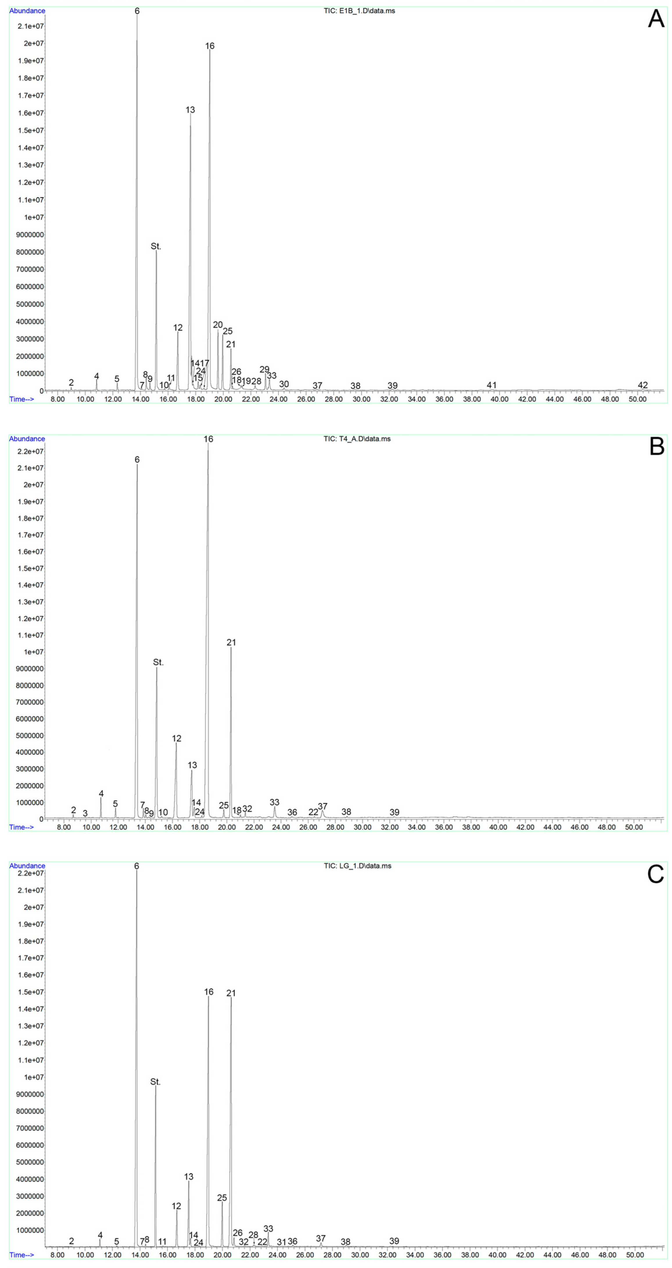
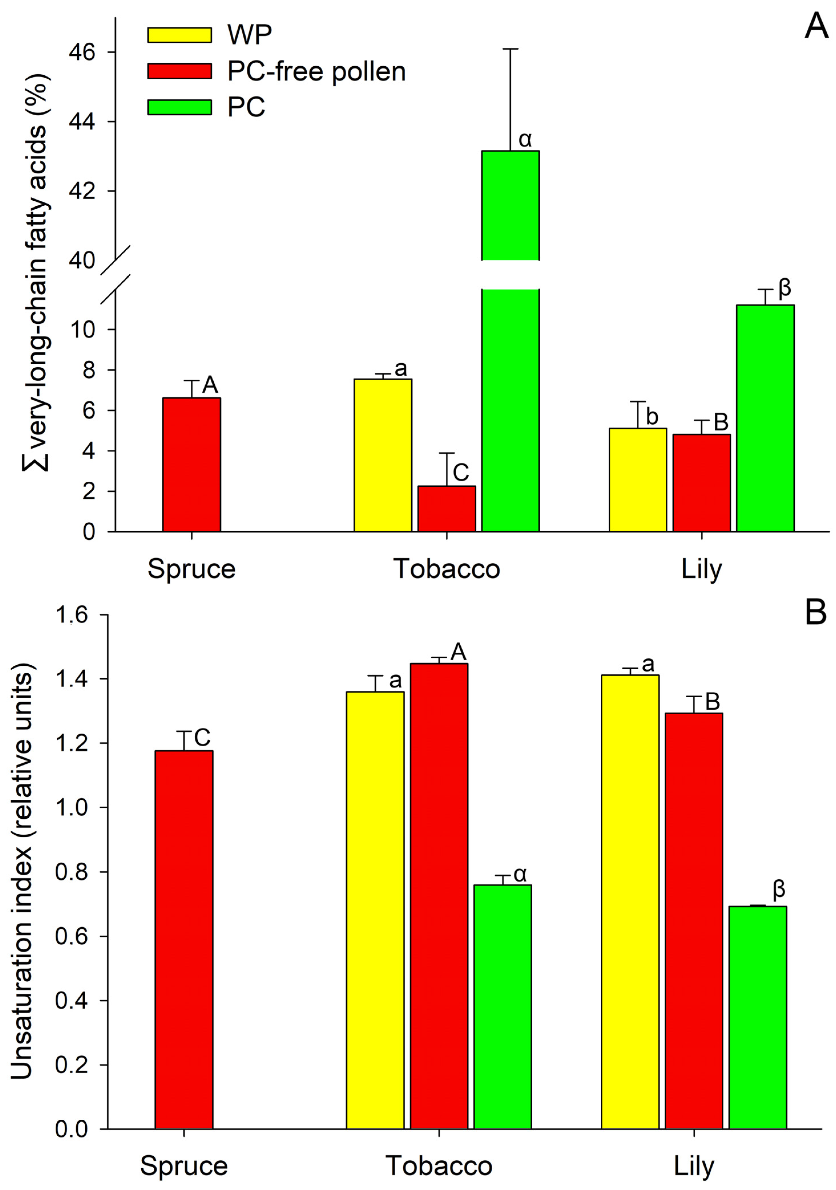
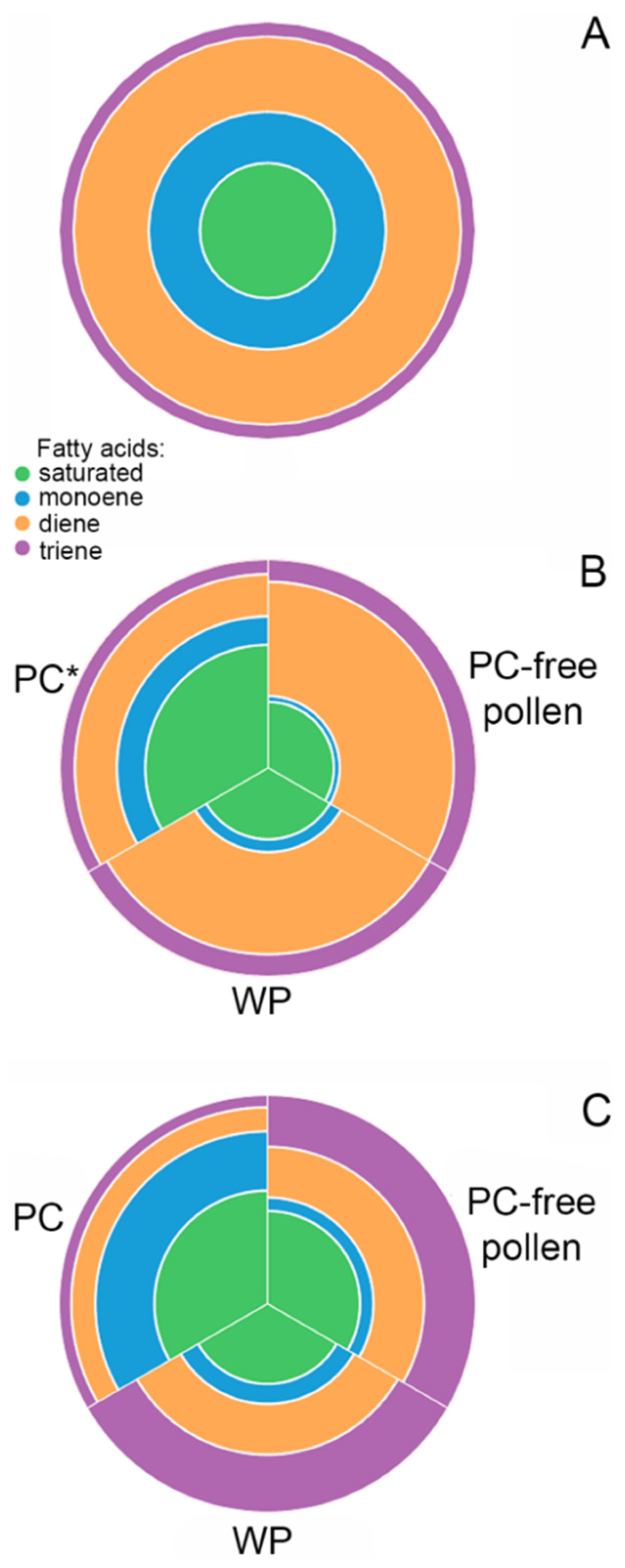
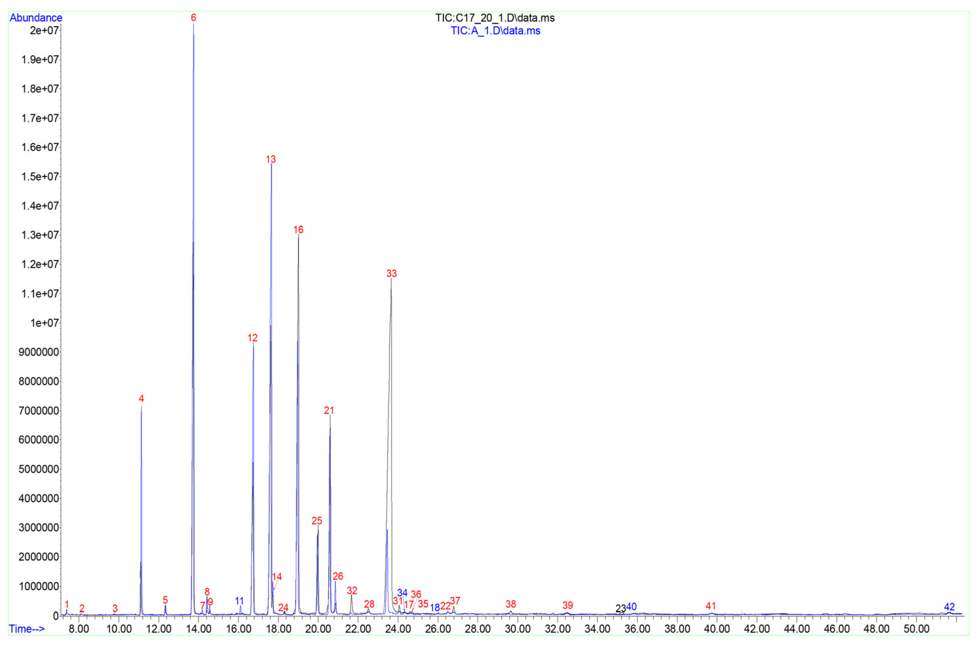
2.1. Fatty Acid Composition of Spruce Pollen

The total lipids of spruce pollen were represented by 31 types of individual C12–30 FAs. The major ones were 16:0, 9–18:1, and 9,12–18:2 acids (Table 1, Figure 1A).
Additionally of note is a significant proportion of stearic (18:0), pinolenic (5,9,12–18:3), and 9,12,15–18:3 FAs. The relative content of other FAs did not exceed 1% of the total amount. Very-long-chain FAs (VLCFAs—with more than 20 C atoms in the chain) of spruce pollen lipids made up 6.62% of the total FA content (Figure 2A), 2.71% of which accounted for arachidic FA (20:0) (Table 1).
The unsaturation index (UI) of spruce pollen lipids was 1.176 (Figure 2B); such a low value can be explained by a large proportion of saturated and monoene FAs and a low percentage of triene FAs (Figure 3A).

2.2. Fatty Acid Composition of Tobacco Pollen

Since tobacco and lily pollen, unlike spruce pollen, is covered with a PC whose composition is markedly different from the internal parts of the male gametophyte. We considered three samples which were analyzed separately: whole pollen (WP), PC, and PC-free pollen (with PC washed off with hexane).
The total lipids of tobacco WP were represented by 32 types of individual C10–28 FAs. The major ones were 16:0, 9,12–18:2, and 9,12,15–18:3 acids (Table 1, Figure 1B). There was also a significant proportion of 18:0, 9–18:1, behenic (22:0), and lignoceric (24:0) FAs. The relative content of the rest did not exceed 1% for each FA. VLCFAs accounted for ≈7.6% of the total FA and were represented by 12 individual FAs, with 3.77% belonging to 22:0 (Figure 2A).
In PC-free tobacco pollen, the number of individual C12–26 FAs decreased to 24 (Figure 4). The main FAs were the same as in WP: palmitic, linoleic, and α-linolenic (Table 1); these three acids accounted for ≈92% of the total amount. The relative amount of palmitoleic acid decreased by three times. The relative content of VLCFAs was ≈2.25% of all FAs, which was several times less than in WP (Figure 2A).
The total lipids of PC contained 30 individual C10–28 FAs. The main acids were 16:0 (relative content two times less than in WP), 9–18:1 (almost three times more than in WP), 9,12–18:2 (≈three times less than in WP) and 22:0 (this VLCFA dominated and accounted for almost a third of all PC FAs) (Table 1). It is important to note the differences in the lipid components of PC relative to WP: the content of minor FAs: 18:0, 20:0, eicosadienoic (11,14–20:2), heneicosanoic (21:0), tricosanoic (23:0), and 24:0 increased several times. At the same time, the proportion of 9,12,15–18:3 decreased (Table 1). Only in PC was the presence of a rare α-parinaric (9,11,13,15–18:4; PubChem CID 5460995) acid belonging to the group of conjugated FAs registered (Table 1; Figures S1 and S2) [27]. Due to the significant differences described above, there was more than a five fold increase in the proportion of VLCFAs compared to WP samples: up to 43.15% (Figure 2A).
Shifts of UI reflect the differences in the proportion of FAs with different numbers of double bonds in lipids of tobacco WP, PC-free pollen, and PC components (Figure 2B). The lowest lipid UI was observed in PC—0.759, which is a consequence of the high proportion of saturated and monoene FAs and the low proportion of dienes and trienes (Figure 3B). The UI of total lipids of WP and PC-free pollen did not differ significantly and were 1.359 and 1.447, respectively (Figure 2B).

2.3. Fatty Acid Composition of Lily Pollen

The total lipids of lily WP were represented by 34 types of individual C10–30 FAs. The major FAs were 16:0, 9,12–18:2 and 9,12,15–18:3; their relative content was more than 80% of the total (Table 1, Figure 1C). Note the minor content of 18:0, 9–18:1, and 20:0 acids; their impact did not exceed 1% (Table 1). VLCFAs accounted for 5.11% of total FAs and consisted of 15 individual species, with 2.4% belonging to 20:0 acid (Table 1; Figure 2A).
After PC components were washed off, the number of individual types of C12–26 FAs in lily pollen decreased to 24. The relative content of the major FAs did not change significantly, and they remained the same: palmitic, linoleic, and α-linolenic (Table 1). 18:0, 9–18:1 and 20:0 were present in minor amounts (Table 1). VLCFAs were represented by 10 individual species and accounted for 4.81% of the total identified FAs (Figure 2A).
The total lipids of lily PC contained 34 types of individual C10–30 FAs (Figure 4). The main FAs were 16:0, 18:0, 9–18:1, and 9,12–18:2 (Table 1). 12:0, 14:0, 20:0, gadoleic (11–20:1), 22:0, and 24:0 acids were registered in minor amounts in PC components (Table 1); the proportion of each remaining FA was less than 1% of the total. VLCFA accounted for 11.24% of the total PC FAs (Figure 2A), which is more than two times higher than in WP and PC-free pollen. Some minor FAs were registered only in PC components: capric (10:0), tridecanoic (13:0), 14-hexadecenoic (14–16:1), and others (Table 1).
UI of total lipids was calculated for WP, PC-free pollen, and PC components (Figure 2B). This indicator was close in WP (1.411) and after PC washout (1.293), which can be explained by a large proportion of polyene FAs. The UI of PC lipids was 0.692: saturated FAs dominated, and among the unsaturated FAs, monoene FAs accounted for the largest part (Figure 3C).

2.4. The Lipid Absolute Contents of Spruce, Tobacco and Lily Pollen

Absolute lipid content in spruce pollen and tobacco PC-free pollen did not differ significantly (Table 1). At the same time, in PC-free lily pollen, it was almost three times higher than in the two other species (Table 1). Lily PC also contained significantly more lipids than tobacco PC (Table 1).

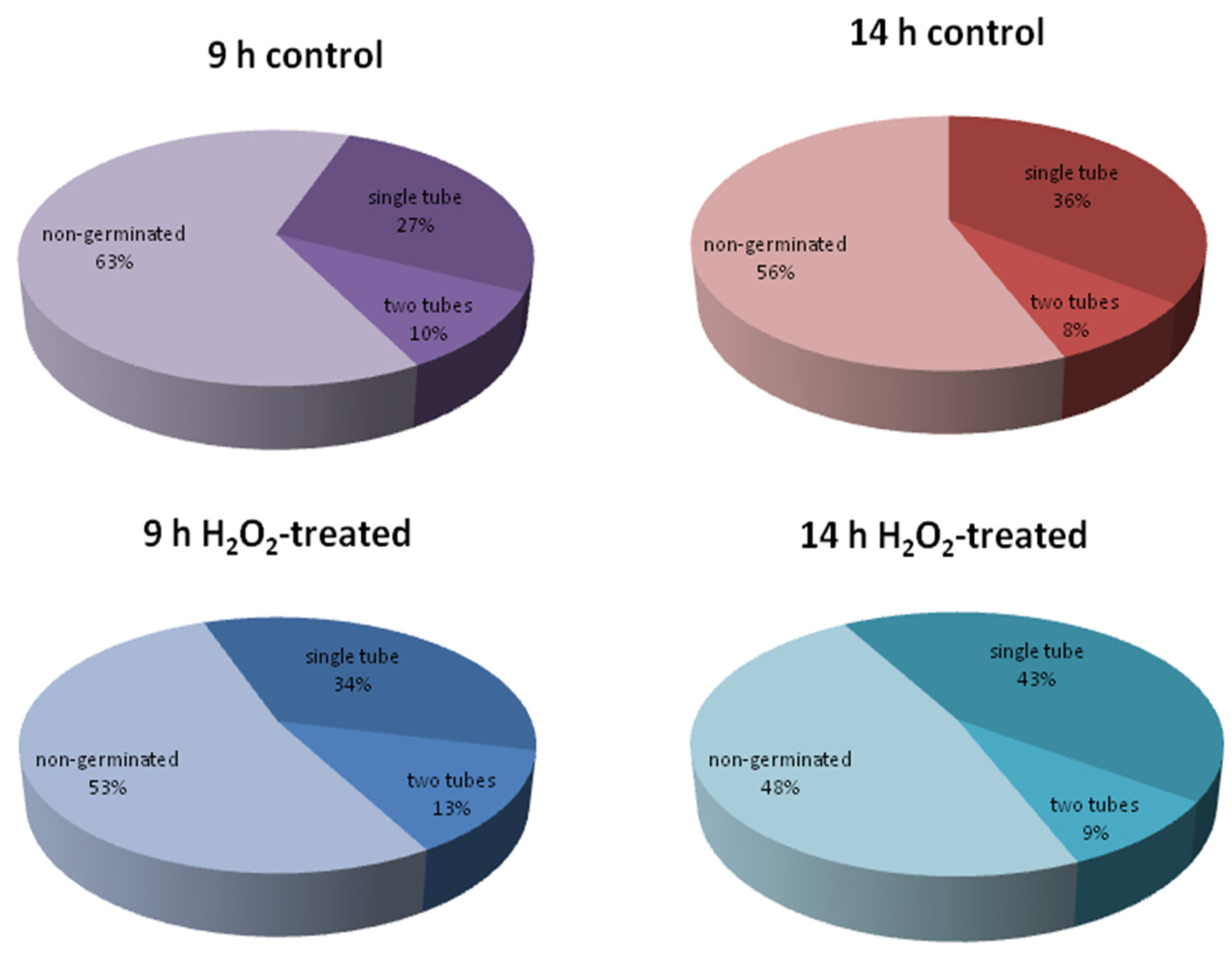
2.5. The Effect of Hydrogen Peroxide on Tobacco and Spruce Pollen Germination

To identify possible changes in the FA composition of pollen during germination, we decided to consider both germination on a standard incubation medium (control) and germination in the presence of a physiological regulator. The effects of toxic substances that reduce germination were outside the scope of this study. Therefore, we decided to test the effect of hydrogen peroxide, which was reported to affect pollen physiology positively [3,28,29].
We found that 100 µM H2O2 stimulated pollen germination in spruce, which was detected at both time points (Figure 5): at 14 h, the percent of germinated grains was 44 ± 3 and 52 ± 3 for control and H2O2, respectively; at 9 h, this difference was more pronounced as the total germination was lower (38 ± 2 and 48 ± 3, respectively). Next, we looked at whether the detected change in germination efficiency was reflected in the FA composition.
The same concentration of peroxide did not affect the efficiency of tobacco pollen germination: after one hour, it was 33.5 ± 2 and 36 ± 3 for control and H2O2, respectively (n = 4).

2.6. Dynamics of the Fatty Acid Composition and Lipid Absolute Content of Spruce Pollen during Germination with or without H2O2

We used two time points to monitor the germination process in spruce pollen. These points were chosen according to our previous studies [30,31], in which we found that after 9 h, exine forms one or two large gaps through which the pollen tube(s) comes out. At this time point, oxygen consumption and cytosolic pH reach their maximum. Nine hours is, therefore, considered the point of germination. At 14 h, the length of an average pollen tube reaches the diameter of pollen grain—this point is considered ongoing polar growth.
During germination and growth of spruce pollen in standard cultivation medium, no significant differences were recorded among the major FAs; only the content of 20:0 was significantly higher at the germination point (9 h). There were significant differences in the content of minor FAs: 12:0, palmitoleic (9–16:1), rumenic (9,11–18:2), 11–20:1, sciadonic (5,11,14–20:3), dihomo-γ-linolenic acid (8,11,14–20:3), pentacosylic (25:0) (Table S1). Some new FA or isomers appeared during pollen incubation. Thus, in growing spruce pollen tubes cis-6,11-eicosadienoic (6.11–20:2) was registered; during germination and growth, we did not detect melissic (30:0) FA, which was found in dry pollen.
When H2O2 was added to the cultivation medium during spruce pollen germination, the composition of lipid FAs changed. Among the major FAs, the relative amount of saturated 18:0 and 20:0 significantly decreased, but the content of unsaturated 9–18:1 and 9,12–18:2 increased. Peroxide addition significantly reduced the content of pentadecanoic (15:0), 7,10-hexadecadienoic (7,10–16:2), cis-10-heptadecenoic (10–17:1), 10,12-octadecadienoate (trans, cis-10,12–18:2), and 9,11–18:2, nonadecylic (19:0) FAs. Qualitative changes were also registered: 6,11–20:2 and 30:0 FAs were detected in peroxide-treated samples (Table S1). However, all H2O2-induced changes did not have a significant effect on UI, which was 1.123 and 1.214 for control and treated samples, respectively.
When H2O2 was added to the cultivation medium during PT growth, changes affected only minor FAs. The relative content of 10–17:1, 10,13-octadecadienoate (10,13–18:2), 11–20:1, and 8,11,14–20:3 FAs significantly decreased while 6,11–20:2 and 25:0 FAs significantly increased. Additionally, H2O2 initiated the appearance of 30:0, which was absent in control PT samples (Table S1). UI did not change significantly (1.188 and 1.236).
During spruce pollen germination and growth, the absolute content of lipids did not change. At the same time, the addition of H2O2 to the cultivation medium led to a significant increase in the content of lipids (Table S1).

2.7. Dynamics of the Fatty Acid Composition and Lipid Absolute Content of Tobacco Pollen during Germination with or without H2O2

Tobacco germinates in vitro much faster than spruce, so the corresponding points were shifted and were 1 h for germination and 3 h for tube growth. No significant differences were found in the qualitative or quantitative content of the major and even minor FA between all time points and control/treated pollen (Table S1). Only superminor FAs were affected: 10:0 was registered only at the germination stage, 13:0 was absent during germination in H2O2-treated suspensions and during PT growth, 10,13–18:2 was absent in dry pollen and appeared during germination and growth in all samples, 11,14–20:2 was recorded during pollen germination with H2O2 and during growth in control medium, 25:0 was absent during pollen germination and PT growth with H2O2, and cerotic (26:0) was present only in dry pollen and during control germination. In total, such changes affected no more than 1% of FAs of total lipids. The absolute content of lipids in tobacco pollen did not change during germination and the addition of growth both to the medium, with and without peroxide (Table S1).

3. Discussion

In this work, we considered several aspects of the lipid FA composition of pollen. On the one hand, we identified key differences between pollen FA compositions of coniferous and flowering plants. Since PC layer is absent in the pollen of gymnosperms, we compared it with PC-free pollen of flowering plants. Such a comparison includes two aspects: (1) gymnosperms have their own peculiarities of FA composition, characteristic of different tissues and organs [32,33]; (2) pollen has its own differences in FA composition from vegetative tissues of the same species [15].
For dry pollen, the set of major FAs was quite typical for pollen of Pinaceae: 16:0, 9–18:1, and 9,12–18:2 acids. For comparison, the major components in Pseudotsuga were 16:0, 9–18:1, and 9,12–18:2, whereas in Pinus they were 16:0, 18:0, 9–18:1, and 9,12–18:2 acids [34,35,36]. In a comparative study analyzing lipids and FAs of five species of pine, ginkgo, and alder, similar patterns were found: the proportion of saturated 16:0 and monoene 9–18:1 in all Pinus species was about two times higher than in angiosperm pollen (Alnus japonica). 9,12,15–18:3, on the contrary, was minor in pollen of all pine species (1–11%), while in alder it accounted for more than 26% [37]. The total contents of saturated fatty acids in three Pinus species was reported to vary from 51.27% (P. sylvestris) to 56.36% (P. sibirica) [35].
As for the less abundant FAs, in Picea pollen we found 5,9,12–18:3—one of Δ5 FAs—which has previously been found in the pollen of other gymnosperms, including Pinus yunnanensis [38], other Pinus species [35] and Cycas revoluta [6]; moreover, in the first case, its content decreased during storage and sterilization in parallel with the loss of pollen quality [38]. Gymnosperm seed lipids are also characterized by the presence of peculiar FAs containing Δ5-ethylenic bond, frequently referred to as Δ5-olefinic acids, with 18 or 20 carbon atoms, which are seldom encountered in angiosperms [39]. These acids are: taxoleic (5,9–18:2; PubChem CID 5312476) (Figure S3A), pinolenic (5,9,12–18:3; PubChem CID 5312495) (Figure S3B), coniferonic (5,9,12,15–18:4; PubChem CID 13751481), 5,11-eicosadienoic (5,11–20:2; PubChem CID 13751478), sciadonic (5,11,14–20:3; PubChem CID 445084) (Figure S3C), and juniperonic (5,11,14,17–20:4; PubChem CID 5312543) [40]. Half of these FAs were found in spruce pollen, which was a clear difference from pollen of tobacco and lily. Angiosperms, with a few exceptions such as Ranunculaceae, have lost the ability to synthesize such FAs. Hence, Δ5 FAs could be considered as an archaic FA marker. Among conifers, these FAs also occur with different frequencies: a few species that presented a low overall content of Δ5 FAs, grow in warm-temperate regions, compared to other pine species. It is hypothesized that Δ5-olefinic acids might be related to cold-acclimation [39]. Δ5 FAs are also present in lipids from algae and mosses, and little is known about their functions. In Chlamydomonas, two lines harboring independent mutant alleles of desaturase (CrDES) failed to synthesize 5,9,12–18:3 and were more sensitive to stress of endoplasmic reticulum than their parental lines [41]. However, there is no experimental confirmation of the protective function of 5,9,12,15–18:4 in higher plants.
VLCFAs made up 6.62% of the total FA content in Picea pollen, 2.71% of which accounted for 20:0. This is consistent with the data on FAs from leaves: relatively high amounts of VLCFAs were detected in different genera of Pinaceae (Pinus, Picea, Pseudotsuga, Abies, Cedrus, Keteleeria) and in Cupressaceae, in contrast with what was observed in angiosperms leaves [32]. VLCFAs possess distinctive biophysical properties in increasing lipid hydrophobicity, interdigitation in leaflets of lipid bilayer, and the transition from a fluid to a gel phase [42]. These unique properties allow VLCFAs to facilitate the generation and stabilization of the highly curved membrane domains required in the formation of highly specialized cells, such as the polarized ‘shmoo’ cell in yeast. Nothing is known about the participation of VLCFAs in pollen tube formation, since in flowering plants they are present in much greater quantities in the PC than in membranes. It can be assumed that they play an important role in polar growth in all seed plants, but in conifers their role is greater, since their content is higher. However, further research is needed to test this hypothesis.
Another notable difference between pollen lipids in spruce and the other two species studied is UI. For spruce pollen, it was significantly lower compared to tobacco and lily. One possible explanation is the growth rate, which is many times slower in the pollen of coniferous plants than the average of flowering plants [1]. Another possible explanation is the more severe germination conditions in the cone compared to a flower [43,44], and the respectively higher requirements for the resistance of spruce pollen membranes to unfavorable factors.
Considering the FA composition of tobacco and lily pollen, we confirmed and refined the patterns of lipid FA distribution in pollen of flowering plants reported for other angiosperm species [45,46]. For both species, PC was characterized by a radically different set of FAs. We found that in tobacco, the proportion of 9,12,15–18:3, which was dominant in PC-free pollen (54%), was significantly lower in PC (17%) while 22:0 acid grew from less than 1% to 30% (Table 1). The content of saturated FAs—18:0, 20:0, 21:0, 23:0, and 24:0—increased several times, which led to a more than five fold increase (Figure 3) in the proportion of VLCFAs (Figure 2A) and very low UI (Figure 2B). In lily, VLCFA in PC were more than two times higher than in PC-free pollen. Two FAs—saturated 16:0 and monoenoic 9–18:1—accounted for about 50% of the total PC FAs in this species. These results are in good agreement with data on other species, including those with a rather remote systematic position. Thus, in Brassica napus, PC is highly enriched medium- and long-chain saturated FAs while intracellular PG lipids are highly enriched in C18 polyunsaturates and, particularly, 9,12,15–18:3 [11]. However, the predominance of monoene and saturated FAs in PC is not characteristic of all angiosperm species: for example, in Crocus vernus pollenkitt, most FAs are unsaturated (9,12,15–18:3–53%) [21]. In Arabidopsis, PC has a high percent of VLCL which was shown to be essential for proper pollen–stigma communication, resulting in the hydration of the pollen on the dry stigma [19,20]. The differences in the FA composition between lipids of the PC and PG cells can be convincingly explained by the origin of these lipids: PC synthesis is controlled by the sporophytic genome, while intracellular lipids are synthesized by the gametophyte [15].
For PC of both species, we noted the presence of some rare minor FAs, which were absent in the washed pollen, including parinaric acid (9,11,13,15–18:4) in tobacco. In cell-free model systems, it was shown that the inclusion of polyunsaturated conjugated FAs, in particular 9,11,13,15–18:4, into lipid bilayers can lead to an increase in their resistance to flocculation caused by divalent cations [47,48] and drastically increase membrane dissipativity when exposed to UV irradiation [49,50]. Additionally, this FA has a strong fluorescence in UV region [51,52]. The significance of the accumulation of 9,11,13,15–18:4 in the PC requires further study. However, it can be assumed that this is an adaptive feature of pollen integuments to protect against ultraviolet exposure and other environmental factors. As a surface component with UV fluorescence, 9,11,13,15–18:4 can also attract pollinators. It should be noted that 9,11,13,15–18:4, as well as other superminor conjugated FAs (10,12–18:2, 9,11–18:2, and 9,11,13–18:3) identified in pollen, may be formed artificially from 9,12–18:2 and 9,12,15–18:3 FAs under alkaline conditions [53,54,55]. Thus, we analyze this result with caution; however, the standard procedure we used did not lead to the formation of conjugates in any studied samples (Table 1), so we can assume that 9,11,13,15–18:4 found in tobacco pollen is most likely of natural origin.
Analyzing the absolute content of lipids in pollen, we found that in lily it exceeds by several times that in tobacco and spruce; however, the proportion of surface lipids in the composition of whole pollen in tobacco is 2.5 times higher than in lily (≈12.5% and ≈5%, respectively). This indicates a high proportion of internal storage lipids in lily pollen. In classical works elucidating the ultrastructure of Lilium pollen tubes, lipid bodies have been described [56,57], which appear to contain the stored material necessary for the very rapid growth of the tube. At the same time, in tobacco, a significant proportion of lipids are located on the surface of the pollen grain and, presumably, are involved in the recognition and hydration of pollen on the stigma, which in this species is covered with a lipid-rich exudate [58].
In this study we did not limit ourselves to dry pollen but looked at the dynamics of the FA composition during germination and the effect of H2O2 on it, and eventually discovered the fundamental differences between spruce and tobacco. The main difference was that the FA composition and the absolute lipid content of tobacco pollen both in control and H2O2-treated samples were very stable. For tobacco, the lipidomes of pollen tubes have been studied under heat stress, and it was found that both glycerophospholipids and galactoglycerolipids increased in saturated acyl chains under heat stress, while triacylglycerols changed less in respect to desaturation but increased in abundance [25]. It is interesting to note that, in this study, the negative environmental factor changed the fatty acid composition of pollen tube lipids, and we obtained data on the same object on the complete stability of such composition under H2O2 treatment. This emphasizes that peroxide is a normal physiological factor and can be opposed to stress factors to which the pollen lipids are forced to adapt. At the same time, the profile of FAs in spruce pollen changed both in control (minor FAs) and H2O2-treated samples (both major and minor FAs). Among the major FAs, the relative amount of saturated 18:0 and 20:0 significantly decreased, but the content of unsaturated 9–18:1 and 9,12–18:2 increased, which affected the UI, although these changes were not statistically significant. An increase in the proportion of unsaturated FAs is in good agreement with an increase in the germination rate, which is more pronounced in 9 h samples. As for the stability of the FA composition of tobacco pollen, it can be speculatively considered an evolutionary acquisition, although this statement needs further research.

4. Materials and Methods

Plant material. Male cones from Picea pungens Engelm. were collected in Moscow State University Botanical Garden and kept at 25 °C until the scales opened to shed pollen, which was then stored at −20 °C. Defrosted pollen samples were used for cultivation.
Plants of Nicotiana tabacum L. var. Petit Havana SR1 were grown in a climatic chamber (25 °C, 16 h light) in vermiculite. The plants were watered with standard salt solution [59]. Anthers were removed just before flower opening and dried at 25 °C for three days. Mature pollen was preserved at −20 °C. Defrosted pollen was rehydrated in a humid chamber at 25 °C for 2 h before cultivation.
Lilium longiflorum Thunb. var. White Heaven anthers were collected from flowers bought in a local store and desiccated at 30 °C for 4 d; dry pollen was stored at −20 °C.
Pollen germination efficiency was assessed after 9 and 14 h of cultivation at 25 °C in standard medium containing, for Picea: 0.3 M sucrose, 1 mM CaCl2 and 1 mM H3BO3 in 15 mM MES-Tris buffer pH 5.5; for tobacco: 0.3 M sucrose, 1.6 mM H3BO3, 3 mM Ca(NO3)2, 0.8 mM MgSO4, and 1 mM KNO3 in 25 mM MES-Tris buffer, pH 5.8. Germinated PGs were fixed with 2% paraformaldehyde in 50 mM Na-phosphate buffer, pH 7.4 for minimum 30 min at 4 °C. Between 500 and 900 PGs were counted for each sample.
Lipid extraction. PC was washed off of lily and tobacco pollen with hexane (1:100 m/V, 2 × 1 min) and analyzed separately. About 500 mg pollen (with or without PC) were fixed separately in 50 mL boiling isopropyl alcohol for 1 h to preserve the native lipid composition. The samples were stored at +4 °C until extraction. To extract lipids, isopropanol was separated through a Schott glass filter into a 200 mL volumetric flask, and plant material was homogenized in a porcelain mortar and extracted as previously described with slight modifications [60]. Lipids were extracted three times sequentially in chloroform/methanol/H2O (30:20:1.7, by vol.) and chloroform/methanol/HNO3 (20:10:0.1, by vol.). All the extracts were transferred to the volumetric flask, neutral pH was adjusted with NH4OH, and the total volume was adjusted to 200 mL with isopropanol containing 0.001% ionol (butylated hydroxytoluene, Sigma-Aldrich, Saint Louis, MO, USA, 34750) as an antioxidant. The extracts were stored at +4 °C.
Preparation and analysis of fatty acid methyl esters. FA methyl esters (FAMEs) were prepared according to the previously described method with slight modifications [61]. A total of 50 μg of margaric acid (17:0) (Sigma Aldrich, Saint Louis, MO, USA, H3500) was added to 50 mL of extract as an internal standard. The aliquot (50 mL) of the extract (or PC in hexane) was evaporated to dryness using a rotary evaporator under standard conditions, and saponification was carried out in a boiling solution of 4% NaOH in methyl alcohol/water (1:1, by vol.). Then, the sample was evaporated to dryness using a rotary vacuum evaporator. H2O (1–2 mL) was added to the dried sample and unsaponifiable FAs were washed out several times with hexane until clearness. Then, a few drops of methyl orange were added to the remaining water-soluble fraction, and it was acidified with 20% H2SO4 to a pink color. Then, FAs were extracted six times with hexane. The collected hexane was evaporated, 3 mL of methanol and a few drops of acetyl chloride were added to the sample, and it was boiled for 1 h. Then, the sample was again evaporated, 1–2 mL of H2O and a few drops of methyl orange were added, and methyl esters of fatty acids were extracted six times with hexane. Following this, the hexane was evaporated and 500 μL of benzene was added. The extract in benzene was pipetted onto a chromatographic plate, and a mixture of hexane/diethyl ether/glacial acetic acid (8:2:0.1, by vol.) was used as the mobile phase. When the front passed to the top of the plate, the plate was removed and air dried for 1–2 min. Then, the plate was treated with a 0.001% solution of 2′,7′-dichlorofluorescein in ethanol and air-dried for 5–7 min. The FAME-containing zones were visualized in UV light (λ = 365 nm). Then, the sorbent from the FAME-containing zone of chromatographic plate was removed using a scalpel and transferred to a Schott glass filter, and the FAMEs were eluted from the sorbent by washing out with hexane six times [62].
The FAMEs were analyzed via gas chromatography-mass spectrometry (GC-MS) on an Agilent 7890A GC (Agilent, Santa Clara, CA, USA) with a quadrupole mass detector Agilent 5975C fitted with a 60-m capillary column DB-23 (inner diameter 0.25 mm, thickness of stationary phase-(50%-cyanopropyl)–methylpolysyloxane—250 µm). The prepared FAMEs were separated under the following conditions: carrier gas, helium at 1 mL/min; sample volume, 1 µL; split ratio, 4:1 (in numerous analyses, splitless injection was used); evaporator temperature, 260 °C. The oven temperature program was as follows: from 130 to 170 °C at 6.5 °C/min, to 215 °C at 2.75 °C/min (25 min hold at this temperature), to 240 °C at 40 °C/min (30 min hold at 240 °C). The operational temperature of the mass detector was set to 240 °C, and the ionization energy was set to 70 eV. To identify individual FAME species, NIST and Wiley search libraries and MSD ChemStation software, G1701EAE.0200.493 (Agilent, Santa Clara, CA, USA) were used, and the relative retention time and equal chain length (ECL) value were calculated for each peak [63].
UV/Vis-absorption curves of methyl esters of fatty acids.Two milligrams of FAMEs was dissolved in 1 mL of hexane, and the absorption spectra of the prepared solutions were measured at 200–500 nm using UV/Vis spectrophotometers (Shimadzu UV-2700, Kyoto, Japan or Hitachi 557, Tokyo, Japan).
Data and Statistical Analyses. To characterize the unsaturation level of lipid FAs, the UI [64] were calculated. All experiments were performed in triplicate with at least 3–5 independent executions. The data are presented in tables and a graph as the means ± SEM. Statistical analysis was performed using one-way ANOVA followed by post hoc analysis using Tukey’s honest significant difference (HSD) for unequal N tests. Different letters and symbols show significantly different values. Mean values were considered significantly different at p < 0.05. For pollen germination experiments, significant difference was evaluated by OriginLab 9.7 software (Northampton, MS, USA) according to Mann–Whitney test, p < 0.05.

5. Conclusions

In this work, dynamics and specific features of Picea pollen lipids were revealed, both in a state of physiological dormancy and at two time points during germination. In standard cultivation medium, no significant differences were recorded among the major FAs, though minor ones changed; under the effect of H2O2, which stimulated germination, we found significant changes in the FA composition and absolute lipid content. Picea pollen is characterized by a low degree of lipid unsaturation and a high proportion of VLCFAs. Some rare FAs were identified in pollen for the first time, and their possible functions are discussed. We analyzed the FA composition of pollen of representatives of monocots and dicots with wet stigmas in detail (surface lipids and cells separately), which made it possible to compare new data with the results previously reported for plants with dry stigmas—Crocus, Brassica, and Arabidopsis—and clarify general patterns and discuss the differences.

Supplementary Materials

The following supporting information can be downloaded at: https://www.mdpi.com/article/10.3390/ijms24119717/s1.

Author Contributions

Conceptualization, M.B.; fatty acid analysis, A.V. and T.I.; pollen germination analysis, K.B.; writing—original draft preparation, A.V. and T.I.; writing—review and editing, M.B. All authors have read and agreed to the published version of the manuscript.

Funding

This research was funded by the Russian Science Foundation (21-74-10054).

Institutional Review Board Statement

Not applicable.

Informed Consent Statement

Not applicable.

Data Availability Statement

The data presented in this study are available on request from the corresponding author. The data are not publicly available due to the University restrictions.

Acknowledgments

Spectroscopic analysis was partially supported by the Ministry of Science and Higher Education of the Russian Federation, state assignment No. 122042700043-9.

Conflicts of Interest

The authors declare no conflict of interest.

References

  1. Breygina, M.; Klimenko, E.; Schekaleva, O. Pollen germination and pollen tube growth in gymnosperms. Plants 2021, 10, 1301. [Google Scholar] [CrossRef] [PubMed]
  2. Chen, T.; Teng, N.; Wu, X.; Wang, Y.; Tang, W.; Šamaj, J.; Baluška, F.; Lin, J. Disruption of actin filaments by latrunculin B affects cell wall construction in Picea meyeri pollen tube by disturbing vesicle trafficking. Plant Cell Physiol. 2007, 48, 19–30. [Google Scholar] [CrossRef] [PubMed] [Green Version]
  3. Maksimov, N.; Evmenyeva, A.; Breygina, M.; Yermakov, I. The role of reactive oxygen species in pollen germination in Picea pungens (blue spruce). Plant Reprod. 2018, 18, 761–767. [Google Scholar] [CrossRef] [PubMed]
  4. Lazzaro, M.D.; Marom, E.Y.; Reddy, A.S.N. Polarized cell growth, organelle motility, and cytoskeletal organization in conifer pollen tube tips are regulated by KCBP, the calmodulin-binding kinesin. Planta 2013, 238, 587–597. [Google Scholar] [CrossRef]
  5. Sowa, S.; Connor, K.F. Biochemical changes during pollen germination measured in vivo by infrared spectroscopy. Plant Sci. 1995, 105, 23–30. [Google Scholar] [CrossRef]
  6. Sidorov, R.A.; Kuznetsova, E.I.; Pchelkin, V.P.; Zhukov, A.V.; Gorshkova, E.N.; Tsydendambaev, V.D. Fatty acid composition of the pollen lipids of Cycas revoluta Thunb. Grasas Y Aceites 2016, 67, 5. [Google Scholar] [CrossRef] [Green Version]
  7. Muren, R.C.; Ching, T.M.; Ching, K.I.M.K. Metabolic Study of Douglas-fir Pollen Germination in vitro. Physiol. Plant 1979, 46, 287–292. [Google Scholar] [CrossRef]
  8. Stanley, R.G. Pollen Chemistry and Tube Growth. In Pollen Development and Physiology; Elsevier: Amsterdam, The Netherlands, 1971; pp. 131–155. [Google Scholar]
  9. Shi, J.; Cui, M.; Yang, L.; Kim, Y.J.; Zhang, D. Genetic and Biochemical Mechanisms of Pollen Wall Development. Trends Plant Sci. 2015, 20, 741–753. [Google Scholar] [CrossRef] [PubMed]
  10. Li, N.; Xu, C.; Li-Beisson, Y.; Philippar, K. Fatty Acid and Lipid Transport in Plant Cells. Trends Plant Sci. 2016, 21, 145–158. [Google Scholar] [CrossRef] [Green Version]
  11. Piffanelli, P.; Ross, J.H.E.; Murphy, D.J. Biogenesis and function of the lipidic structures of pollen grains. Sex. Plant Reprod. 1998, 11, 65–80. [Google Scholar] [CrossRef]
  12. Pacini, E.; Hesse, M. Pollenkitt—Its composition, forms and functions. Flora Morphol. Distrib. Funct. Ecol. Plants 2005, 200, 399–415. [Google Scholar] [CrossRef]
  13. Carmichael, J.S.; Friedman, W.E. Double fertilization in Gnetum gnemon (Gnetaceae): Its bearing on the evolution of sexual reproduction within the gnetales and the anthophyte clade. Am. J. Bot. 1996, 83, 767–780. [Google Scholar] [CrossRef]
  14. Hesse, M. Pollenkitt is lacking in Gnetum gnemon (Gnetaceae). Plant Syst. Evol. 1980, 136, 41–46. [Google Scholar] [CrossRef]
  15. Ischebeck, T. Lipids in pollen—They are different. Biochim. Biophys. Acta—Mol. Cell Biol. Lipids 2016, 1861, 1315–1328. [Google Scholar] [CrossRef] [PubMed]
  16. Firon, N.; Nepi, M.; Pacini, E. Water status and associated processes mark critical stages in pollen development and functioning. Ann. Bot. 2012, 109, 1201–1213. [Google Scholar] [CrossRef] [Green Version]
  17. Manning, R. Fatty acids in pollen: A review of their importance for honey bees. Bee World 2001, 82, 60–75. [Google Scholar] [CrossRef]
  18. Manning, R.; Harvey, M. Fatty acids in honeybee-collected pollens from six endemic Western Australian eucalypts and the possible significance to the Western Australian beekeeping industry. Aust. J. Exp. Agric. 2002, 42, 217–223. [Google Scholar] [CrossRef]
  19. Preuss, D.; Lemieux, B.; Yen, G.; Davis, R.W. A conditional sterile mutation eliminates surface components from Arabidopsis pollen and disrupts cell signaling during fertilization. Genes Dev. 1993, 7, 974–985. [Google Scholar] [CrossRef] [Green Version]
  20. Jessen, D.; Olbrich, A.; Knüfer, J.; Krüger, A.; Hoppert, M.; Polle, A.; Fulda, M. Combined activity of LACS1 and LACS4 is required for proper pollen coat formation in Arabidopsis. Plant J. 2011, 68, 715–726. [Google Scholar] [CrossRef]
  21. Chichiriccò, G.; Pacini, E.; Lanza, B. Pollenkitt of some monocotyledons: Lipid composition and implications for pollen germination. Plant Biol. 2019, 21, 920–926. [Google Scholar] [CrossRef]
  22. Zhang, M.; Fan, J.; Taylor, D.C.; Ohlrogge, J.B. DGAT1 and PDAT1 acyltransferases have overlapping functions in Arabidopsis triacylglycerol biosynthesis and are essential for normal pollen and seed development. Plant Cell 2009, 21, 3885–3901. [Google Scholar] [CrossRef] [Green Version]
  23. Mellema, S.; Eichenberger, W.; Rawyler, A.; Suter, M.; Tadege, M.; Kuhlemeier, C. The ethanolic fermentation pathway supports respiration and lipid biosynthesis in tobacco pollen. Plant J. 2002, 30, 329–336. [Google Scholar] [CrossRef] [Green Version]
  24. Ischebeck, T.; Valledor, L.; Lyon, D.; Gingl, S.; Nagler, M.; Meijón, M.; Egelhofer, V.; Weckwerth, W. Comprehensive cell-specific protein analysis in early and late pollen development from diploid microsporocytes to pollen tube growth. Mol. Cell. Proteom. 2014, 13, 295–310. [Google Scholar] [CrossRef] [PubMed] [Green Version]
  25. Krawczyk, H.E.; Rotsch, A.H.; Herrfurth, C.; Scholz, P.; Shomroni, O.; Salinas-Riester, G.; Feussner, I.; Ischebeck, T. Heat stress leads to rapid lipid remodeling and transcriptional adaptations in Nicotiana tabacum pollen tubes. Plant Physiol. 2022, 189, 490–515. [Google Scholar] [CrossRef] [PubMed]
  26. Serrano, N.; Pejchar, P.; Soukupová, H.; Hubálek, M.; Potocký, M. Comprehensive analysis of glycerolipid dynamics during tobacco pollen germination and pollen tube growth. Front. Plant Sci. 2022, 13, 1028311. [Google Scholar] [CrossRef] [PubMed]
  27. Sklar, L.A.; Hudson, B.S.; Simoni, R.D. Conjugated polyene fatty acids as membrane probes: Preliminary characterization. Proc. Natl. Acad. Sci. USA 1975, 72, 1649–1653. [Google Scholar] [CrossRef] [Green Version]
  28. Hiscock, S.J.; Bright, J.; McInnis, S.M.; Desikan, R.; Hancock, J.T. Signaling on the stigma. Potential new roles for ROS and NO in plant cell signaling. Plant Signal. Behav. 2007, 2, 23–24. [Google Scholar] [CrossRef] [Green Version]
  29. Breygina, M.; Klimenko, E.; Shilov, E.; Podolyan, A.; Mamaeva, A.; Zgoda, V.; Fesenko, I. Hydrogen peroxide in tobacco stigma exudate affects pollen proteome and membrane potential in pollen tubes. Plant Biol. 2021, 23, 592–602. [Google Scholar] [CrossRef]
  30. Breygina, M.; Klimenko, E.; Podolyan, A.; Voronkov, A. Dynamics of pollen activation and the role of H+-ATPase in pollen germination in blue spruce (Picea pungens). Plants 2020, 9, 1760. [Google Scholar] [CrossRef]
  31. Breygina, M.; Maksimov, N.; Polevova, S.; Evmenyeva, A. Bipolar pollen germination in blue spruce (Picea pungens). Protoplasma 2019, 256, 941–949. [Google Scholar] [CrossRef]
  32. Mongrand, S.; Badoc, A.; Patouille, B.; Lacomblez, C.; Chavent, M.; Cassagne, C.; Bessoule, J.-J. Taxonomy of gymnospermae: Multivariate analyses of leaf fatty acid composition. Phytochemistry 2001, 58, 101–115. [Google Scholar] [CrossRef]
  33. Takagi, T.; Itabashi, Y. Cis-5-Olefinic Unusual Fatty Acids in Seed Lipids of Gymnospermae and Their Distribution in Triacylglycerols. Lipids 1982, 17, 716–723. [Google Scholar] [CrossRef]
  34. Ching, T.M.; Ching, K.K. Fatty acids in pollen of some coniferous species. Science 1962, 138, 890–891. [Google Scholar] [CrossRef]
  35. Erdyneeva, S.A.; Shiretorova, V.G.; Tykheev, Z.A.; Radnaeva, L.D. Fatty-Acid Composition of Pollen from Pinus sylvestris, P. sibirica, and P. pumila. Chem. Nat. Compd. 2021, 57, 741–742. [Google Scholar] [CrossRef]
  36. Andrikopoulos, N.K.; Siafaka-Kapadai, A.; Demopoulos, C.A.; Kapoulas, V.M. Lipids of Pinus halepensis pollen. Phytochemistry 1985, 24, 2953–2957. [Google Scholar] [CrossRef]
  37. Chung, H.K.; Lee, K.Y. A study of the chemical constituents of gymnosperm pollens. Seoul J. Med. 1977, 18, 125–136. [Google Scholar]
  38. Mei, S.; Yang, C.; Song, X.; Wang, T.; Wang, Y.; Geng, Y.; Wang, J.; Su, S. Analysis of the effect of sterilization and storage on the quality of Pinus yunnanensis pollen based on untargeted metabonomics. J. Food Process. Preserv. 2021, 45, e16033. [Google Scholar] [CrossRef]
  39. Wolff, R.L.; Comps, B.; Marpeau, A.M.; Deluc, L.G. Taxonomy of Pinus species based on the seed oil fatty acid compositions. Trees—Struct. Funct. 1997, 12, 113–118. [Google Scholar] [CrossRef]
  40. Song, L.; Wen, S.; Ye, Q.; Lou, H.; Gao, Y.; Bajpai, V.K.; Carpena, M.; Prieto, M.-A.; Simal-Gandara, J.; Xiao, J.; et al. Advances on delta 5-unsaturated-polymethylene-interrupted fatty acids: Resources, biosynthesis, and benefits. Crit. Rev. Food Sci. Nutr. 2023, 63, 767–789. [Google Scholar] [CrossRef]
  41. Yamaoka, Y.; Shin, S.; Choi, B.Y.; Kim, H.; Jang, S.; Kajikawa, M.; Yamano, T.; Kong, F.; Légeret, B.; Fukuzawa, H.; et al. The bZIP1 Transcription Factor Regulates Lipid Remodeling and Contributes to ER Stress Management in Chlamydomonas reinhardtii. Plant Cell 2019, 31, 1127–1140. [Google Scholar] [CrossRef] [Green Version]
  42. Paz Ramos, A.; Lague, P.; Lamoureux, G.; Lafleur, M. Effect of saturated very long-chain fatty acids on the organization of lipid membranes: A study combining 2H NMR spectroscopy and molecular dynamics simulations. J. Phys. Chem. B 2016, 120, 6951–6960. [Google Scholar] [CrossRef]
  43. Williams, C.G. Conifer Reproductive Biology; Springer: Dordrecht, The Netherlands, 2009; ISBN 978-1-4020-9601-3. [Google Scholar]
  44. Williams, J.H. Novelties of the flowering plant pollen tube underlie diversification of a key life history stage. Pnas 2008, 105, 11259–11263. [Google Scholar] [CrossRef] [Green Version]
  45. Rothnie, N.E.; Palmer, M.V.; Burke, D.G.; Sang, J.P.; Hilliard, E.P.; Salisbury, P.A.; Evans, D.E.; Knox, R.B.; Williams, E.G. Comparative analysis of fatty acids in pollen and seed of rapeseed. Phytochemistry 1987, 26, 1895–1897. [Google Scholar] [CrossRef]
  46. Piffanelli, P.; Ross, J.H.E.; Murphy, D.J. Intra-and extracellular lipid composition and associated gene expression patterns during pollen development in Brassica napus. Plant J. 1997, 11, 549–562. [Google Scholar] [CrossRef]
  47. Mansy, S.S.; Szostak, J.W. Thermostability of model protocell membranes. Proc. Natl. Acad. Sci. USA 2008, 105, 13351–13355. [Google Scholar] [CrossRef] [PubMed] [Green Version]
  48. Michaelian, K.; Rodríguez, O. Prebiotic fatty acid vesicles through photochemical dissipative structuring. Rev. Cuba. Química 2019, 31, 354–370. [Google Scholar]
  49. Fan, Y.; Fang, Y.; Ma, L. The self-crosslinked ufasome of conjugated linoleic acid: Investigation of morphology, bilayer membrane and stability. Colloids Surf. B Biointerfaces 2014, 123, 8–14. [Google Scholar] [CrossRef]
  50. Verma, P.; Kaur, K.; Wanchoo, R.K.; Toor, A.P. Esterification of acetic acid to methyl acetate using activated TiO2 under UV light irradiation at ambient temperature. J. Photochem. Photobiol. A Chem. 2017, 336, 170–175. [Google Scholar] [CrossRef]
  51. Barrantes, F.J. Fluorescence sensors for imaging membrane lipid domains and cholesterol. In Current Topics in Membranes; Elsevier: Amsterdam, The Netherlands, 2021; Volume 88, pp. 257–314. ISBN 9780323911146. [Google Scholar]
  52. Kuypers, F.A.; van den Berg, J.J.M.; Schalkwijk, C.; Roelofsen, B.; Kamp, J.A.F.O. den Parinaric acid as a sensitive fluorescent probe for the determination of lipid peroxidation. Biochim. Biophys. Acta (BBA)/Lipids Lipid Metab. 1987, 921, 266–274. [Google Scholar] [CrossRef] [Green Version]
  53. Lundberg, W. Alkali-isomerization reactions of unsaturated fatty acids. Am. J. Clin. Nutr. 1958, 6, 592–593. [Google Scholar] [CrossRef] [PubMed]
  54. Shayanmehr, M.-R.; Elhamirad, A.-H.; Armin, M. Different Conditions for Alkaline Isomerization and the Production of Conjugated Linoleic Acid (CLA) from Linoleic Acid. J. Biochem. Tech. 2018, 2, 1–8. [Google Scholar]
  55. Svetashev, V.I.; Imbs, A.B. Isomerization of octadecapentaenoic acid (18:5n-3) in algal lipid samples under derivatization for GC and GC-MS analysis. J. Phycol. 2014, 50, 322–327. [Google Scholar] [CrossRef]
  56. Pierson, E.S.; Lichtscheidl, I.K.; Derksen, J.A.N. Structure and Behaviour of Organelles in Living Pollen Tubes of Lilium longiflorum. J. Exp. Bot. 1990, 41, 1461–1468. [Google Scholar] [CrossRef]
  57. Rosen, W.G.; Gawlik, S.R.; Dashek, W.V.; Siegesmund, K.A. Fine structure and cytochemistry of lilium pollen tubes. Am. J. Bot. 1964, 51, 61–71. [Google Scholar] [CrossRef]
  58. Cresti, M.; Keijzer, C.J.; Tiezzi, A.; Ciampolini, F.; Focardi, S. Stigma of Nicotiana: Ultrastructural and biochemical studies. Am. J. Bot. 1986, 73, 1713–1722. [Google Scholar] [CrossRef]
  59. Nitsch, J.P. Deux espaces photoperiodiques de jours courts: Plumbago indica L. et P. zeyelanica. Bull. Bot. Fr. 1965, 112, 517–522. [Google Scholar] [CrossRef] [Green Version]
  60. Voronkov, A.S.; Ivanova, T.V. Fatty Acids Composition of the Epiphytic Ferns, Platycerium bifurcatum and Asplenium nidus, and the Terrestrial Fern, Asplenium trichomanes. Am. Fern J. 2021, 111, 117–128. [Google Scholar] [CrossRef]
  61. Voronkov, A.; Ivanova, T. Significance of Lipid Fatty Acid Composition for Resistance to Winter Conditions in Asplenium scolopendrium. Biology 2022, 11, 507. [Google Scholar] [CrossRef]
  62. Voronkov, A.S.; Ivanova, T.V.; Kuznetsova, E.I.; Kumachova, T.K. Adaptations of Malus domestica Borkh. (Rosaceae) Fruits Grown at Different Altitudes. Russ. J. Plant Physiol. 2019, 66, 922–931. [Google Scholar] [CrossRef]
  63. Ivanova, T.V.; Voronkov, A.S.; Kuznetsova, E.I.; Kumachova, T.K.; Zhirov, V.K.; Tsydendambaev, V.D. Lipid Fatty Acids from the Pericarp of Cydonia oblonga Mill. and Mespilus germanica L. are Involved in Plant Adaptation to Altitudinal Zonality. Dokl. Biochem. Biophys. 2019, 486, 229–233. [Google Scholar] [CrossRef]
  64. Lyons, J.M.; Wheaton, T.A.; Pratt, H.K. Relationship between the Physical Nature of Mitochondrial Membranes and Chilling Sensitivity in Plants. Plant Physiol. 1964, 39, 262–268. [Google Scholar] [CrossRef] [PubMed] [Green Version]
Figure 1. Chromatograms of FAMEs of spruce pollen (A) and PC-free pollen of tobacco (B) and lily (C). Numerals denote FAs according to their number in Table S1.
Ijms 24 09717 g001
Figure 2. Sum of very-long-chain fatty acids (A) and unsaturated index (B) in pollen of spruce, tobacco, and lily. The data are presented as the means ± SEM. Statistical analysis was performed using one-way ANOVA followed by post hoc analysis using Tukey’s honest significant difference for unequal N tests. Different letters show significantly different values for WP (a, b), PC-free pollen (A, B, C), PC (α, β). Mean values were considered significantly different at p < 0.05. Analysis was performed separately for WP (whole pollen), PC (pollen coat), and PC-free pollen (washed with hexane).
Ijms 24 09717 g002
Figure 3. The ratio of saturated and groups of unsaturated (mono-, di-, triene) fatty acids in pollen of spruce (A), tobacco (B) and lily (C) in % of the total FAMEs. *—tetraene also found (0.02%).
Ijms 24 09717 g003
Figure 4. Chromatograms of pollen coat (PC) FAMEs of tobacco (black) and lily (blue). Numbers indicate FAs according to their number in Table S1: red—FAs are identified in PC of both tobacco and lily pollen, black—FAs are identified only in tobacco PC, blue—FAs are identified only in lily PC.
Ijms 24 09717 g004
Figure 5. The effect of hydrogen peroxide on the germination of spruce pollen grains after 9 and 14 h of incubation. Means are presented. For each diagram, four suspensions were counted (n = 4), 400 to 900 cells in each. The level of germination in H2O2-treated samples significantly differs from the control at both times according to Mann–Whitney test, p < 0.05.
Ijms 24 09717 g005
Table 1. Fatty acid composition and absolute content of lipids (esterified FAs, μmol/g) of spruce, tobacco and lily pollen (mass % of the amount of FAMEs). Since spruce pollen does not have a pollen coat, we compared it with washed pollen of tobacco and lily (column “PC-free pollen”.
No.FASpruce
Pollen
TobaccoLily
WPPC-Free PollenPCWPPC-Free PollenPC
110:0-0.01 ± 0.01-0.08 ± 0.02βtrace #-0.19 ± 0.01α
212:00.10 ± 0.01A0.03 ± 0.01b0.06 ± 0.02B0.29 ± 0.04β0.22 ± 0.08a0.04 ± 0.01B2.97 ± 0.04α
313:0-0.01 ± 0.010.01 ± 0.010.02 ± 0.01αtrace-0.03 ± 0.01α
414:00.53 ± 0.06A0.49 ± 0.05b0.42 ± 0.07A1.22 ± 0.14β0.75 ± 0.08a0.44 ± 0.08A5.56 ± 0.27α
515:00.30 ± 0.03A0.36 ± 0.05a0.29 ± 0.06A0.26 ± 0.02α0.10 ± 0.01b0.08 ± 0.01B0.28 ± 0.01α
616:023.12 ± 1.57C24.91 ± 0.10b26.93 ± 0.76B11.65 ± 1.12β30.25 ± 1.09a37.35 ± 3.00A23.68 ± 1.01α
77–16:10.57 ± 0.05A0.53 ± 0.14a0.42 ± 0.06B0.50 ± 0.06α0.25 ± 0.01b0.17 ± 0.05C0.55 ± 0.01α
89–16:10.23 ± 0.01A0.14 ± 0.01b0.12 ± 0.01C0.13 ± 0.03β0.23 ± 0.01a0.19 ± 0.01B0.25 ± 0.02α
914–16:10.44 ± 0.04A0.07 ± 0.02a0.02 ± 0.01B0.12 ± 0.01α0.01 ± 0.01b-0.04 ± 0.01β
107,10–16:20.01 ± 0.01Atrace0.03 ± 0.01A----
1110–17:10.09 ± 0.02A---0.02 ± 0.010.01 ± 0.01B0.07 ± 0.03
1218:03.48 ± 0.26A1.75 ± 0.23b1.68 ± 0.17C4.82 ± 0.07β2.89 ± 0.37a2.61 ± 0.14B11.98 ± 0.90α
139–18:121.47 ± 0.94A4.32 ± 0.83b1.36 ± 0.09C11.82 ± 0.91β7.68 ± 0.53a4.75 ± 0.26B25.11 ± 1.18α
1411–18:11.21 ± 0.28A0.83 ± 0.03a0.86 ± 0.03B0.61 ± 0.09β0.68 ± 0.19a0.65 ± 0.05C0.91 ± 0.11α
155,9–18:20.33 ± 0.10------
169,12–18:233.79 ± 3.74B47.47 ± 1.80a54.10 ± 0.50A17.46 ± 1.54α23.97 ± 0.68b24.04 ± 1.44C10.99 ± 0.07β
1710,13–18:20.05 ± 0.010.07 ± 0.02a-0.79 ± 0.100.04 ± 0.04a-trace
1810,12–18:20.58 ± 0.09A0.60 ± 0.750.36 ± 0.06A-trace-0.13 ± 0.01
199,11–18:20.73 ± 0.09------
205,9,12–18:33.56 ± 0.49-----
219,12,15–18:32.53 ± 0.28C10.68 ± 1.64b10.97 ± 0.51B6.92 ± 0.39α27.69 ± 0.80a24.80 ± 0.84A5.79 ± 0.42α
229,11,13–18:3-trace0.02 ± 0.02A0.08 ± 0.01αtrace0.02 ± 0.01A0.13 ± 0.05α
239,11,13,15–18:4-trace-0.02 ± 0.01---
2419:00.26 ± 0.16A0.13 ± 0.01a0.10 ± 0.03A0.06 ± 0.01β0.07 ± 0.01b0.04 ± 0.02B0.13 ± 0.02α
2520:02.71 ± 0.15A0.92 ± 0.21b0.30 ± 0.05B2.70 ± 0.14α2.40 ± 0.41a2.57 ± 0.34A2.83 ± 0.17α
2611–20:10.48 ± 0.05B0.25 ± 0.15b-0.38 ± 0.01β0.71 ± 0.02a0.62 ± 0.01A1.19 ± 0.05α
2811,14–20:20.36 ± 0.06A0.37 ± 0.03a-1.11 ± 0.14α0.15 ± 0.02b0.15 ± 0.03B0.25 ± 0.08β
295,11,14–20:31.05 ± 0.14------
308,11,14–20:30.01 ± 0.01------
3111,14,17–20:3-0.12 ± 0.09a-0.35 ± 0.02α0.03 ± 0.01a0.02 ± 0.010.08 ± 0.02β
3221:0-trace0.05 ± 0.03A0.77 ± 0.09α0.10 ± 0.050.09 ± 0.03A0.18 ± 0.03β
3322:00.86 ± 0.14A3.77 ± 0.73a0.87 ± 0.09A29.77 ± 1.81α0.87 ± 0.31b0.89 ± 0.19A3.71 ± 0.16β
3413–22:1----trace-0.14 ± 0.01
3513,16–22:2-0.20 ± 0.05-0.75 ± 0.08αtrace-0.11 ± 0.01β
3623:0-0.16 ± 0.01a0.08 ± 0.03A1.40 ± 0.16α0.14 ± 0.01a0.06 ± 0.01A0.47 ± 0.08β
3724:00.76 ± 0.12A1.59 ± 0.40a0.93 ± 0.08A5.29 ± 0.55α0.42 ± 0.11b0.35 ± 0.07B1.34 ± 0.07β
3825:00.07 ± 0.03A0.09 ± 0.06a0.01 ± 0.01B0.13 ± 0.01α0.05 ± 0.05a0.02 ± 0.01B0.09 ± 0.01β
3926:00.16 ± 0.03A0.13 ± 0.07a0.01 ± 0.01C0.47 ± 0.08α0.08 ± 0.06a0.04 ± 0.01B0.54 ± 0.04α
4027:0----trace-0.01 ± 0.01
4128:00.15 ± 0.120.01 ± 0.01a-0.03 ± 0.01β0.14 ± 0.24a-0.23 ± 0.05α
4230:00.01 ± 0.01---0.02 ± 0.01-0.04 ± 0.01
FAs absolute content, µmol/g151.11 ± 5.52B160.85 ± 13.72b136.59 ± 11.60B19.88 ± 1.75β580.67 ± 79.11a430.68 ± 76.10A28.24 ± 3.65α
#—<0.01%. The data are presented in tables and a graph as the means ± SEM. Statistical analysis was performed using one-way ANOVA followed by post hoc analysis using Tukey’s honest significant difference (HSD) for unequal N tests. Different letters show significantly different values. Mean values were considered significantly different at p < 0.05. Analysis was performed separately for WP (a, b), PC-free pollen (A, B), PC (α, β).
Disclaimer/Publisher’s Note: The statements, opinions and data contained in all publications are solely those of the individual author(s) and contributor(s) and not of MDPI and/or the editor(s). MDPI and/or the editor(s) disclaim responsibility for any injury to people or property resulting from any ideas, methods, instructions or products referred to in the content.

