Review

Meta-Inflammation and New Anti-Diabetic Drugs: A New Chance to Knock Down Residual Cardiovascular Risk

1
Department of Cardiovascular Sciences, Fondazione Policlinico A. Gemelli, IRCCS, 00168 Rome, Italy
2
Department of Cardiovascular and Pneumological Sciences, Catholic University of Sacred Heart, 00168 Rome, Italy
*
Author to whom correspondence should be addressed.
Int. J. Mol. Sci. 2023, 24(10), 8643; https://doi.org/10.3390/ijms24108643
Submission received: 3 April 2023 / Revised: 28 April 2023 / Accepted: 8 May 2023 / Published: 12 May 2023

Abstract

:
Type 2 diabetes mellitus (DM) represents, with its macro and microvascular complications, one of the most critical healthcare issues for the next decades. Remarkably, in the context of regulatory approval trials, sodium-glucose cotransporter 2 inhibitors (SGLT2i) and glucagon-like peptide 1 receptor agonists (GLP-1 RAs) proved a reduced incidence of major adverse cardiovascular events (MACEs), i.e., cardiovascular death and heart failure (HF) hospitalizations. The cardioprotective abilities of these new anti-diabetic drugs seem to run beyond mere glycemic control, and a growing body of evidence disclosed a wide range of pleiotropic effects. The connection between diabetes and meta-inflammation seems to be the key to understanding how to knock down residual cardiovascular risk, especially in this high-risk population. The aim of this review is to explore the link between meta-inflammation and diabetes, the role of newer glucose-lowering medications in this field, and the possible connection with their unexpected cardiovascular benefits.

1. Introduction

Type 2 diabetes mellitus (DM) represents, with its macro and microvascular impairments and its complex pathophysiology, one of the most serious global healthcare issues in forthcoming years [1]. Starting from parental insulin, DM treatment has come a long way, proposing a wide range of oral drugs able to achieve better glycemic control. Interestingly, in the context of regulatory approval trial, sodium-glucose cotransporter 2 inhibitors (SGLT2i) and glucagon-like peptide 1 receptor agonists (GLP-1 RA) showed a reduced incidence of a major adverse cardiovascular event (MACE), namely cardiovascular death and heart failure (HF) hospitalizations [2,3].
Since these findings, growing evidence has been published, trying to understand the pathophysiologic mechanisms underpinning the cardiovascular effects of these new anti-diabetic molecules. Fascinatingly, SGLT2i and GLP-1 RA cardioprotective abilities seem to operate beyond the simplistic glycemic control [4], disclosing a range of pleiotropic effects and a solid link between diabetes and metabolic inflammation. A deeper knowledge of molecular mechanisms sustaining DM as a metabolic and inflammatory disease may help to understand the cardioprotective effects of new anti-diabetic drugs and their role in knocking down residual cardiovascular risk and, above all, the risk of developing HF [5], a heterogeneous clinical syndrome, whose management is ongoing towards major clinical changes [6].
The aim of this review is to explore the link between meta-inflammation and diabetes, the role of newer glucose-lowering medications in this field, and the possible connection with their unexpected cardiovascular benefits.

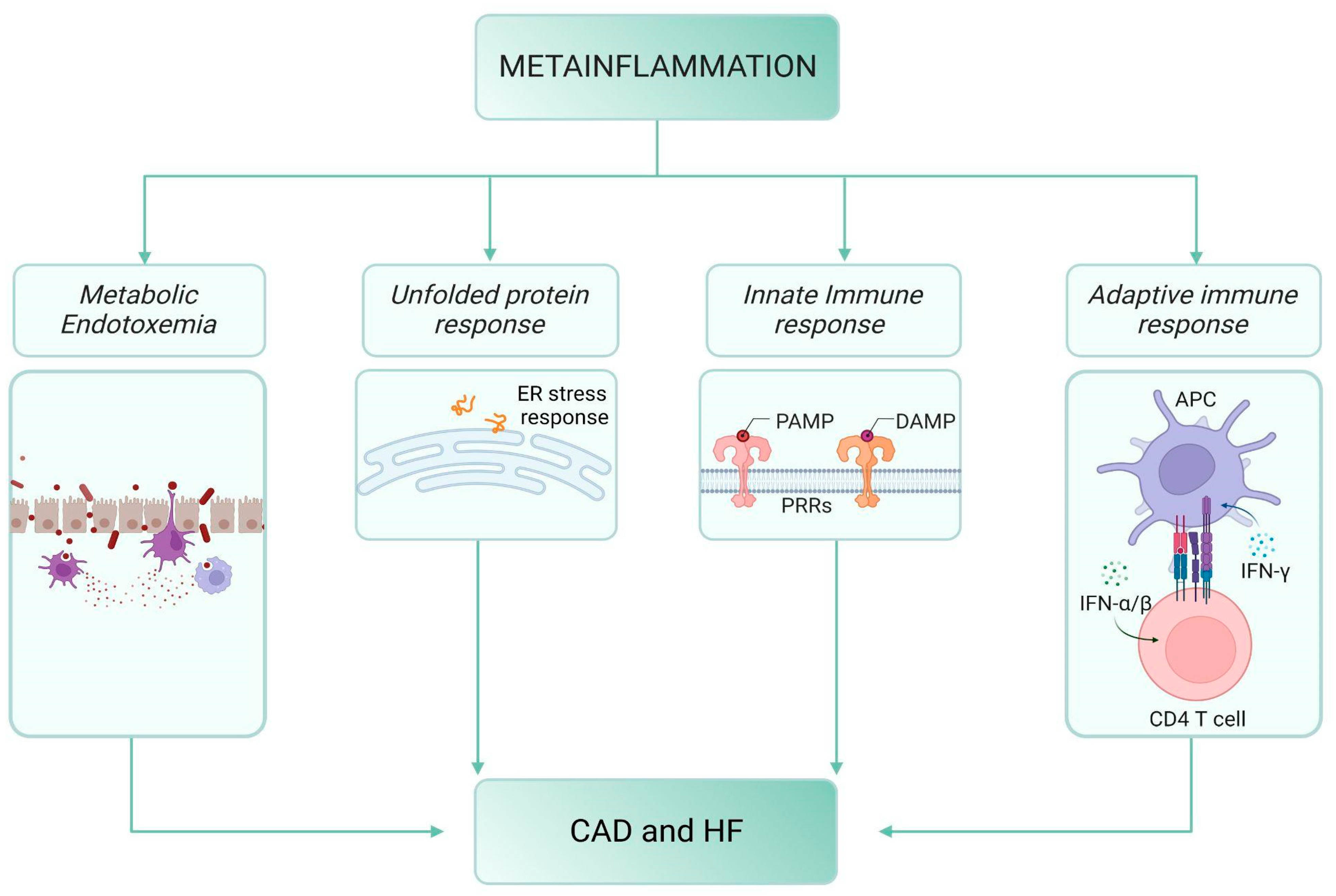
2. Meta-Inflammation, Diabetes and Cardiovascular Disease

The pathogenesis of DM shares with cardiovascular diseases (CAD) a complex landscape of risk factors, including genetic predisposition and various environmental factors like a high-fat diet, sedentary lifestyle, and chronic stress. In particular, CAD is the leading cause of morbidity and mortality in diabetic patients, determining a significant impact on life expectancy. Notably, DM is equivalent to established ischemic CAD risk, and patients with diabetes have a two- to four-fold greater risk of developing CAD than non-diabetic patients [7]. Myocardial infarction, ischemic ictus, and peripheral arterial disease are the main expression of DM progression and, often, the first event in diabetic patients. CAD and DM strictly depend on various inflammatory pathways that are able to promote the onset and development of insulin resistance, atherosclerotic plaque, and HF.
One of the mechanisms linking DM and CAD is the so-called meta-inflammation, i.e., metabolic inflammation, which is a low-grade chronic and sterile inflammatory status maintained by high nutrient intake and which is able to reshape the inflammatory milieu of metabolic cells, tissues, and organs [8]. The starting point of meta-inflammation is the physiological inflammatory trigger prompted by the intake of any meal: in particular, in the postprandial moment, remnants of chylomicrons and very low-density lipoprotein (VLDL, triglyceride-rich lipoproteins) bind to endothelial cells and circulating leukocytes causing acute cellular activation and increased expression of adhesion molecules, cytokines, and oxidative stress characters, such as thiobarbituric acid reactive substances (TBARS), leukocyte O2−, and 8-iso-prostaglandin F2alpha (8-PGF2α) [9,10,11]. Triglyceride-rich lipoproteins and free fatty acids (FFAs) raise the expression of vascular cell adhesion protein 1 (VCAM1) in human aortic endothelial cells, favoring leucocyte adhesion [12]. In parallel, an increased neutrophil count coupled with augmented levels of interleukin 6 (IL-6) and hydroperoxides was also observed after high-fat meals in healthy controls [13]. This inflammatory environment promotes insulin resistance by activation of nuclear factor kappa-light-chain-enhancer of activated B cells (NF-κB) and c-Jun N-terminal kinase (JNK) pathways that, in turn, lead to the decline of insulin secretion by pancreatic b-cells [14,15].
This complex system might be the main contributing factor to HF pathophysiology through innate and adaptive system impairment, macrophage functional level shift, accumulation of unfolded proteins [5] within a steady two-way cross-talk low-grade inflammatory state, and metabolic flexibility (Figure 1).

2.1. Meta-Inflammation and Metabolic Endotoxemia

Dietary fats can boost levels of circulating bacterial endotoxins, contributing to metabolic endotoxemia, a chronic low-grade elevation of the bacterial component lipopolysaccharide (LPS) with content 10–50-times lower than in septic conditions, which can lead to leukocyte activation, local and systemic inflammation [16]. Recent evidence has proved that metabolic endotoxemia plays a critical role in the inflammatory setting milieu, thereby predisposing to metabolic diseases via pattern recognition receptor engagement, primarily through the toll-like receptor/nod-like receptor (TLR4/NLR) signaling pathway [17,18]. Following a similar pathophysiological framework, both DM and CAD are associated with significant alterations of the gut microbiota, responsible for the increased intestinal permeability and the consequent efflux of LPS into the bloodstream, thus altering the systemic metabolic response [19]. Indeed, already in 1990, it has been demonstrated that patients with end-stage HF show elevated levels of circulating tumor necrosis factor (TNF), which could explain the cachectic condition typical of this syndrome as a consequence of innate immune dysregulation operated by endotoxin-activated monocytes [20,21].

2.2. Meta-Inflammation and the “Unfolded Protein Response” (UPR)

The liver and pancreas are the classical target organs in the physiopathology of DM, but adipose tissue is probably at the core of meta-inflammation. Nutrient excess increases adipocyte size up to critical conditions, resulting in reduced vascularization and a hypoxic environment [22], finally precipitating the inflammatory cascade.
In obese mice, adipocytes participate in endoplasmic reticulum stress, a condition accelerated by the accumulation of unfolded/misfolded proteins and able to trigger the protective unfolded protein response (UPR). This process promotes both the synthesis of endoplasmic reticulum-resident chaperone proteins, which encourages protein folding and the protein-degradation mechanism components to achieve new endoplasmic reticulum homeostasis [23]. In addition to these defensive responses, the UPR may also encourage important inflammatory signals, triggering apoptotic and cell death pathways [24]. UPR is significantly enhanced by exposure to nutrient excess, as revealed by experiments in obese and diabetic mice, and its activation promotes inflammation through several mechanisms, i.e., inducing the expression of pro-inflammatory genes by directly acting on the transcription factor activator protein 1 [25]; abnormal nutrient intake pledges the NF-κB signaling pathway [26], promoting the downstream cleavage and activation of the transcription factor cyclic-AMP responsive- element-binding protein H (CREBH), which in turn induces the production of two acute phase proteins, C-reactive protein (CRP) and serum amyloid P-component (SAP) [27]. A similar process has been recorded in HF patients and represents the ground of cardiac hypertrophy, further exacerbated by the acquired immune response.

2.3. The Innate Immune Response

An innate immune response is an essential mechanism in triggering meta-inflammation associated with DM and CAD. Pattern recognition receptors can boost meta-inflammation following recognition of both pathogens-associated molecular patterns (PAMPs) and damage-associated molecular patterns (DAMPs). Viral nucleic acids, endotoxins, and peptidoglycans are some of the PAMPs circulating in the bloodstream because of metabolic endotoxemia, while FFAs and self-nucleoproteins are some of the endogenous ligands starring as DAMPs [28]. Pattern recognition receptor activation represents the main sensor to start the inflammation cascade via NF-𝜅B and some interferon regulatory factors (IRFs), which in return trigger pro-inflammatory cytokines and type-1 interferons signaling cascade [29]. These mechanisms could lead to insulin resistance and precipitate in DM via the mediation of macrophage polarization towards the M1 phenotype [30].
In HF, adverse left ventricular (LV) remodeling and LV dysfunction are structural progressions that parallel the activity of innate immunity actors and mediators such as monocyte chemoattractant protein 1 (MCP-1) and progressive monocyte-derived macrophage generation [31].
The innate immune response could be dysregulated at multiple levels, becoming the basement of a meta-inflammatory environment that, in turn, involves multiple cellular compartments.

2.4. The Adaptive Immune Response

As far as adaptive immunity is concerned, most evidence proves that CD4+T-helper (Th) cells are crucial elements in propagating meta-inflammation [32,33]. The response started by pattern recognition receptors is transferred to T cells by the antigen-presenting cell (APC) and T cell cooperation, which determines the enrolment of these activated cells into the pancreas, adipose tissue, and other target organs, consolidating the meta-inflammation mounted by the native response. The well-known Th polarization, which shapes the adaptive arm of the immune response, is also maintained in DM patients. In particular, in the murine diet-induced obesity model, Kitschier et al. demonstrated early recruitment of Th1 cells into adipose tissue, paving the way for macrophage infiltration and insulin resistance [34]. Notably, several studies on serum cytokine profiling in DM patients reported robust Th1 polarization during the transition from DM to macrovascular complications, especially atherosclerotic coronaropathy, highlighting the role of Th1 polarization in the physiopathology of the disease [35]. Furthermore, a significant action in interfering with insulin signaling and insulin-stimulated glucose uptake is pursued by interferon-gamma (IFN-𝛾), eventually leading to insulin resistance and DM [36,37,38]. Polarized Th1 and Th17 T-cells also play a key role in cardiac fibrosis and adverse cardiac remodeling, sustaining and amplifying the local chronic inflammation leading to HF. In vivo, experimental models demonstrated that CD4+ T-cells were expanded in the failing heart, with a Th1/Th2 ratio significantly decreased, whereas the Th17/Treg ratio was increased, underling the loss of anti-inflammatory properties and the gain of the pro-inflammatory counterpart [39].

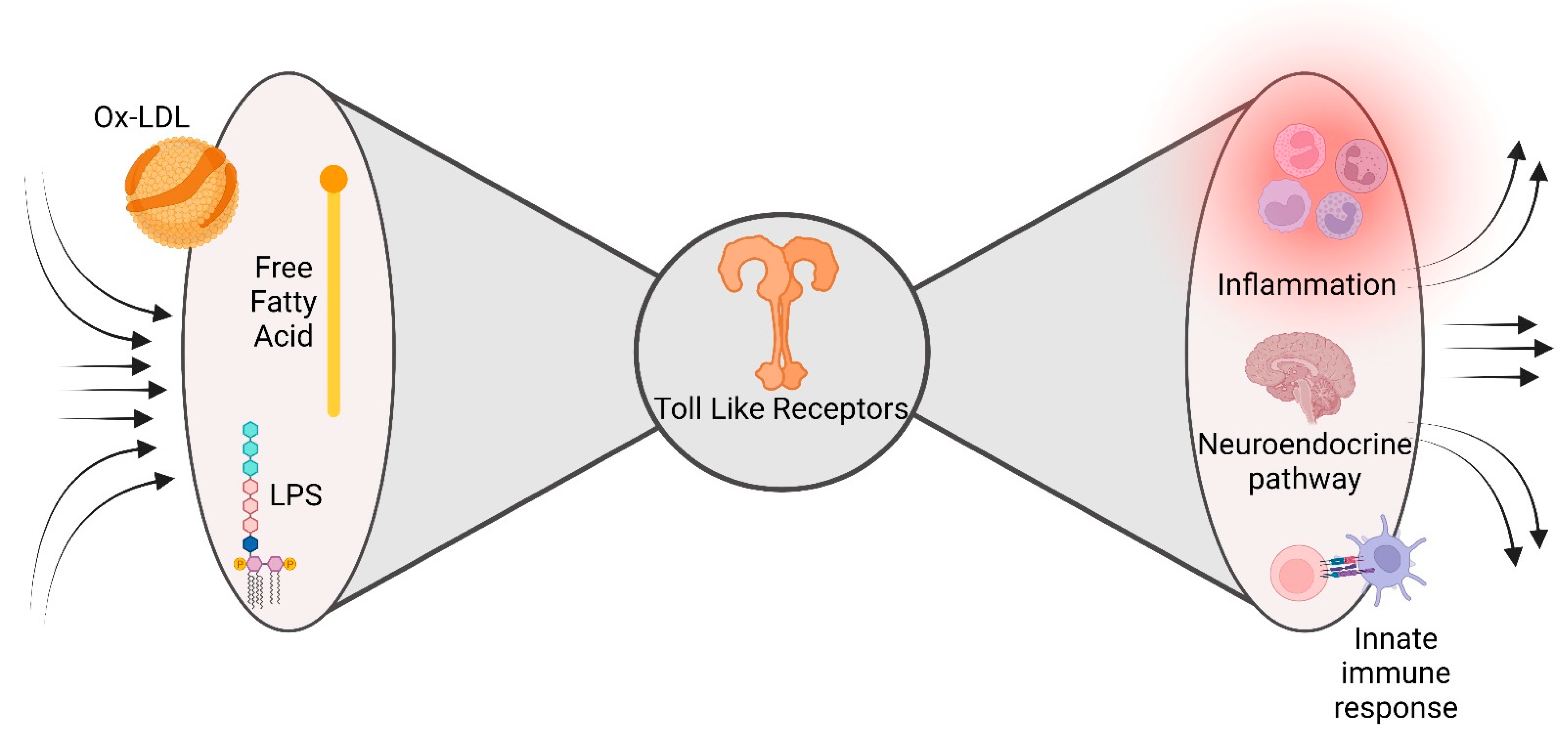
2.5. The Bow Tie Model

Metabolic and inflammatory pathways can converge at many levels, including cell-surface receptors, intracellular chaperones, or nuclear receptors. This molecular army allows robust cooperation between the nutrient-sensing pathways and the immune response aimed at maintaining homeostasis in opposite metabolic and immune circumstances. Unfortunately, this molecular rendezvous may be one of the crucial moments in DM and CAD pathophysiology. Meta-inflammation may be considered the unpredicted consequence of the evolution-driven degeneracy of damage sensors, recently described as a ‘bow tie’ architecture [40]. The main feature of bow tie architecture is the possibility to converge a vast range of inputs (fan in) on an evolutionarily reduced core of components (core), able to translate the inputs into a broad spectrum of outputs (fan out) (Figure 2). Interestingly, the inputs are represented by an extensive range of self and non-self-stimuli, i.e., free fatty acid, LPS, able to bind a restricted number of evolutionarily conserved innate immunity sensors (the bow tie core), whose activation triggers a large number of inflammatory elements [41]. A fascinating example of this promiscuity in immune response receptors is the capacity of saturated fatty acids to turn on both TLR2 and TLR4, important innate immune response receptors involved in pathogen recognition, and trigger the release of pro-inflammatory mediators [42].
This bidirectional cross-talk between metabolic alterations and immune dysregulation is also emerging as a critical component of the pathogenesis of HF, especially for the one with preserved ejection fraction (HfpEF). Indeed, all the alterations mentioned above and driven by metabolic alterations, such as macrophage polarization, accumulation of misfolded proteins, and metabolic reprogramming, contribute to structural and functional remodeling determining HF [5].
The exploration of the meta-inflammation route as a common denominator for DM and CAD might expand the knowledge of molecular pathways underpinning its pathophysiology, thus clearing the unpredicted effects of new anti-diabetic drugs on cardiovascular outcomes.

3. Newer Glucose-Lowering Medications and Cardiovascular Effects

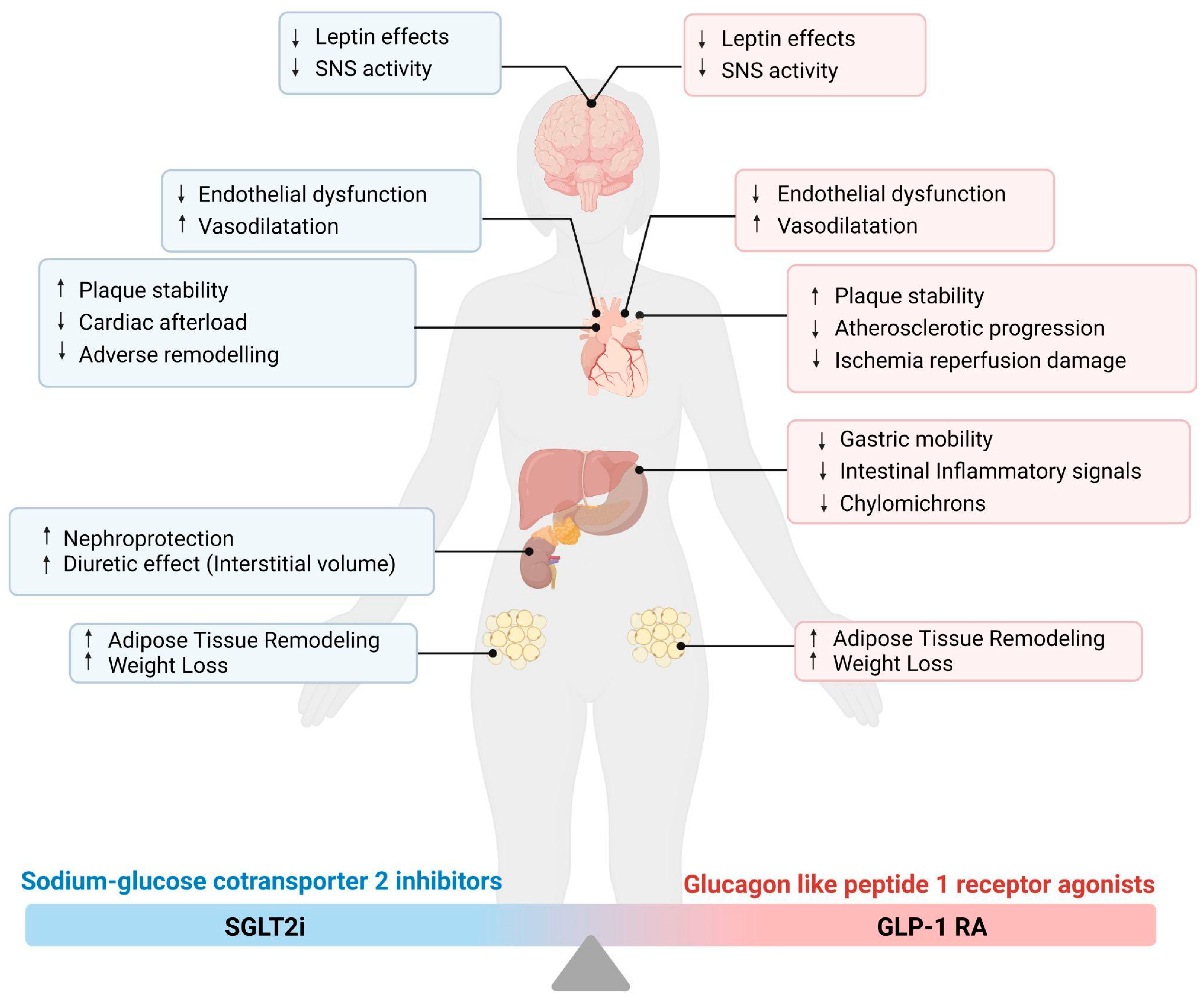
3.1. Sodium–Glucose Cotransporter 2 Inhibitors (SGLT2i)

The kidneys play a critical role in glucose homeostasis through gluconeogenesis, glucose utilization, and glucose reabsorption. In diabetic patients, renal gluconeogenesis is significantly enhanced, and the capacity to reabsorb glucose in the convoluted segment of the proximal tubule is pathologically increased through the upregulation of SGLT2 transporters [4]. In this context, the SGLT2i have been developed to inhibit the high-capacity, low-affinity SGLT2 receptors in the proximal tubule of the nephron. Interestingly, SGLT2 receptor performance is enhanced in chronic hyperglycemia, determining significant resorption of glucose and sodium while it is downregulated in lower glucose conditions, minimizing the risk of hypoglycemia during SGLT2i therapy [43], notably at lower levels of glycemia or at lower glomerular filtration rate (GFR).
Since their first approval in 2013, SGLT2i have gained a lot of success in the management of diabetic patients, improving glucose control without increasing the risk of hypoglycemia and promoting weight loss with their glucosuric effect. Several clinical trials assessed the safety and efficacy of SGLT2i in patients with diabetes and a large variety of cardiovascular and renal complications. In particular, the EMPA-REG OUTCOME trial proved that diabetic patients at high risk of CAD and treated with empagliflozin had a significant reduction in cardiovascular death and hospitalization for HF [44]. These results have been further assessed for other SGLT2i in large trials, i.e., canagliflozin in CANVAS [45] and CREDENCE [46] and dapagliflozin in DECLARE-TIMI 58 [47].
Probably the most exciting evidence is provided by the DAPA-HF trial, which demonstrated a marked reduction in worsening HF or cardiovascular death on top of HF standard-of-care therapy, with similar effects in patients with and without DM [48] (Table 1).

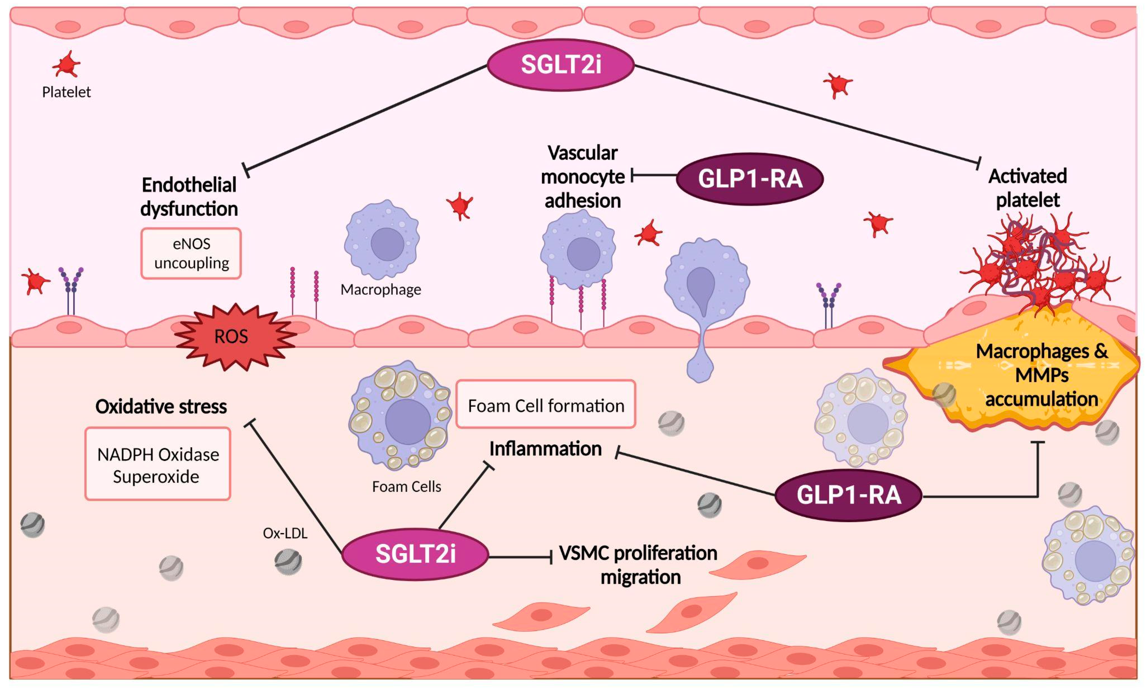
3.1.1. Possible Targets of SGLT2i Pharmacodynamics

In the last years, several experimental findings suggested a large variety of mechanisms to explain the impressive results of SGLT2i in cardiovascular outcomes. On the one hand, some authors suggested that blood pressure lowering, a “side” effect of SGLT2i, may explain improved cardiac energetics, lowering cardiac afterload and improving ventricular arterial coupling and cardiac efficiency [51] (Figure 3).
Although the precise pathophysiology for SGLT2i antihypertensive effects is not completely understood, they are probably mediated by the osmotic and diuretic effects of SGLT2i because of the inhibition of sodium reabsorption in the kidney proximal tubules. On the other hand, the capability of SGLT2i to promote natriuresis, glucosuria, and osmotic diuresis has been suggested as a mechanism that may ameliorate HF outcomes by favoring hemoconcentration [44]. This theory has been criticized because other diuretic strategies historically failed to impact HF mortality but Hallow et al. proved that dapagliflozin determined a significant reduction in interstitial versus intravascular volume, suggesting that SGLT2i may express a disparity of effect on interstitial and intravascular fluid, so limiting the neurohumoral reflex stimulus that arises in response to intravascular volume reduction with classical diuretics [52].
Interestingly, some evidence suggests that SGLT2i may improve cardiac energetics. Mitochondrial glucose oxidation decreases in the failing heart, determining a reduction in energy production and a fuel-starved heart. The uncoupling between glycolysis and glucose oxidation also leads to increased proton production and, consequently, a decrease in cardiac efficiency (cardiac work/O2 consumed) [53]. Mobilizing adipose tissue fatty acids, SGLT2i increases circulating ketone levels, which maximize cardiac energetics and efficiency, functioning as an economical fuel for the failing heart [54].
A new possible etiology of cardiorenal benefits from SGLT2i therapy is the inhibition of sympathetic nervous system (SNS) activity, indirectly suggested by the observation that SGLT2i reduces blood pressure without increasing heart rate. In fact, emerging data from animal models proved that this class of drugs might lower sympathetic nerve activity, inhibit norepinephrine metabolism in brown adipose tissue, and decrease tyrosine hydroxylase synthesis [55,56].
Adverse remodeling has been highlighted as a crucial determinant in HF pathophysiology. Remarkably, experimental models proved that SGLT2i, i.e., empagliflozin, may reduce left ventricular (LV) mass index when evaluated by cardiac magnetic resonance, compared to placebo [57]; although the molecular pathways of their anti-remodeling effect have not clarified yet, an explanation relies on a decreased fibrosis. On the other hand, in diabetic and non-diabetic rat models, SLGT2i achieved a cardioprotective effect against ischemia/reperfusion injury, determining a reduction in calmodulin kinase II activity, an increased sarcoplasmic reticulum Ca2+ flux and a stronger contractility [58].
These unexpected benefits boosted great interest in SGLT2i, and accumulating evidence have proposed a large range of pathophysiologic effects: inhibition of the cardiac Na+/H+ exchanger, reducing Ca2+ overload [59]; reduction of hyperuricemia [60]; an increase of autophagy and lysosomal degradation, favoring mitochondrial function [61]; a higher erythropoietin secretion [62]; an improved vascular function, by attenuating endothelial cell activation and dysfunction, and those molecular variations accompanying early atherogenesis [63].

3.1.2. SGLT2i and Meta-inflammation

Among all the pathways associated with SGLT2 inhibition, those related to meta-inflammation are probably the most interesting in the pathophysiology of diabetes and HF. Although major cardiovascular outcome trials did not include dosing of inflammatory markers, some insights are available from smaller pilot studies; in fact, SGLT2i therapy has been associated with a decrease in leptin concentration and lower levels of highly sensitive (hs) CRP, tumor necrosis factor-alpha (TNF-α), interleukin-6 (IL-6) and interferon-gamma (IFN-γ) [64].
As mentioned before, adipose tissue may be considered the meta-inflammation fortress. Xu et al. proved that empagliflozin promotes fat utilization and browning of white adipose tissue and reduces M1-polarized macrophage accumulation while promoting the anti-inflammatory M2 macrophage phenotype in white adipose tissue, contributing to a reduction in TNFα plasma levels and mitigation of obesity-related chronic low-grade inflammation [65]. Interestingly, recent theories proposed that leptin synthesis in adipose tissue may determine sodium retention and plasma volume expansion, meta-inflammation, and fibrosis. In this setting, the SGLT2i action on perivisceral adipose tissue may attenuate leptin secretion and, potentially, its paracrine effects, i.e., fibrosis promotion [66].
A similar effect has been described in epicardial fat tissue, considered a paracrine organ able to produce a number of bioactive molecules, i.e., leptin and TNFα, and to influence cardiac remodeling and atherosclerotic plaque in epicardial coronary arteries [67,68]. In particular, in diabetic patients with CAD, SGLT2i decreases epicardial adipose tissue (EAT) and its paracrine products [69].
As regards the potential anti-inflammatory activities of SGLT2 inhibition, animal models proved that empagliflozin therapy mitigates the enhanced expression of inflammatory genes in the diabetic kidney [70]. As proposed in other work from our group, the NLRP3 inflammasome is a milestone in the road that links inflammation and CAD, and its role has also been evaluated in HF patients [71]. Experimental data disclosed that empagliflozin might downregulate the NLRP3 inflammasome, and this result was AMPK-dependent and SGLT2/glucose-lowering independent [72]. As mentioned before, SGLT2i enhances the plasma concentration of ß-hydroxybutyrate, a potent NLRP3 inflammasome blocker, and some authors speculate that beneficial effects of new anti-diabetic drugs may occur secondary to ketone inhibition of the NLRP3 inflammasome [73].
Even if SGLT2i have a role in hyperlipidemia management in diabetic patients, the therapeutic effects on atherosclerosis may be driven by their influence on meta-inflammation. Nasiri-Ansari demonstrated that canagliflozin therapy for 5 weeks significantly reduced atherosclerosis progression in ApoE−/− mice, probably inhibiting VCAM-1 mRNA expression levels and inflammatory monocyte chemoattractant protein-1 (Mcp-1) expression [74].
Interestingly, SGLT2i may influence atherosclerotic plaque characteristics, favoring stability. In fact, animal models disclosed that treatment with dapagliflozin significantly reduced the growth of cholesterol crystals in atherosclerotic lesions in the diabetic group [75], while canagliflozin increased the collagen content by 1.6-fold in atherosclerotic lesions, leading to plaque stability [74]. Dapagliflozin treatment significantly inhibited cholesterol ester accumulation in macrophages extracted from ApoE−/−mice and reversed the atherosclerosis-associated increase in macrophage infiltration by 20%; finally, it improved the decreased production of smooth muscle cells in animal models [76].
In conclusion, SGLT2i promises to ameliorate the prognosis of patients with and without diabetes with a wide range of molecular pathways, most of them including meta-inflammation and its relationship with HF and atherosclerotic plaque progression.

3.2. Glucagon-like Peptide 1 Receptor Agonists (GLP-1 RA)

Glucagon-like peptide-1-(7-36) amide (GLP-1) is a human incretin hormone produced by the gut in response to food. It is primarily an insulin-tropic hormone, playing its role not only to promote insulin secretion but also to inhibit glucagon secretion, regulating the excessive hepatic glucose output and reducing appetite leading to weight loss. Its biological characteristics allow for avoiding hypoglycemia, making GLP-1 an attractive target molecule for anti-diabetic treatment. However, native GLP-1 is rapidly inactivated by the ubiquitous enzyme DPP4; for these reasons, DPP4-resistant GLP-1 analogs have been formulated and approved for medical use in 2005 [77].
Seven randomized control trials (ELIXA-Lixisenatide; LEADER-Liraglutide; SUSTAIN-6—Semaglutide; EXSCEL -Exenatide; HARMONY—Albiglutide; REWIND—Dulaglutide; PIONEER 6—Seraglutide) have explored cardiovascular outcomes in diabetic patients treated with GLP-1 RA [78,79,80,81,82,83,84]. They were large trials of 3000 to 14,000 patients with similar baseline patient demographics and determined to be at high risk or have established cardiovascular disease. All trials were designed to evaluate the primary outcome of MACE and to prove non-inferiority for cardiovascular safety, even if LEADER and HARMONY-OUTCOMES were powered for and demonstrated superiority versus placebo (Table 2).
To sum up, liraglutide, semaglutide, albiglutide, and dulaglutide (but not exenatide or lixisenatide) have proved superior compared to placebo for a reduction in MACE endpoint; however, to date, only liraglutide has shown a decrease in both cardiovascular and all-cause mortality [85]. A recent meta-analysis disclosed that GLP-1 RA reduced all-cause mortality by 12% and adverse renal outcomes by 17%, reporting a significant reduction in hospital admissions for HF, not evidenced by single trials [3].
As far as GLP-1 pathway inhibition is concerned, an emerging drug is drawing attention due to its effects on obesity and weight reduction: Tirzepatide. Tirzepatide is a novel, once-weekly, injectable peptide engineered from the native Glucose-dependent insulinotropic polypeptide (GIP) sequence to have dual GIP/GLP-1-RA activity, which may result in substantial weight reduction, even more than GLP-1-RA alone, by addressing several pathways involved in energy homeostasis. Tirzepatide has been approved by the FDA for the treatment of T2DM with safety and tolerability characteristics similar to other incretin-based therapies. Despite significant improvements in cardiometabolic measures have been observed, no information is available on long-term CV effects. Further studies may evaluate possible unexplored mechanisms linking these new anti-diabetic drugs to CAD and meta-inflammation [86].
Table 2. Cardiovascular Outcome Trial and GLP-1RA.
GLP-1RA NameClinical Trial Name (CVOT)Brief ReferenceNumber of PatientsDefinition of CV
Outcomes
Median Follow Up (Months)Principal Findings
LixisenatideELIXAHolman RR et al., New England Journal of Medicine, 2017 [78]6068CV death, myocardial infarction, stroke, or hospitalization for unstable angina25The primary endpoint (a composite of the first occurrence of any of the following: death from CV causes, non-fatal myocardial infarction, non-fatal stroke, or hospitalization for unstable angina) event occurred in 13.4% of the lixisenatide group and in 13.2% of the placebo group (HR, 1.02; 95% CI, 0.89 to 1.17), which showed the non-inferiority of lixisenatide to placebo (p < 0.001) but did not show superiority (p = 0.81).
LiraglutideLEADERMarso SP et al.,
New England Journal of Medicine, 2016 [79]
9340The first occurrence of death from CV causes non-fatal myocardial infarction or non-fatal stroke15.8The primary outcome (the first occurrence of death from CV causes, non-fatal myocardial infarction, or non-fatal stroke) occurred in significantly fewer patients in the liraglutide group (13.0%) than in the placebo group (14.9%) (HR, 0.87; 95% CI, 0.78 to 0.97; p < 0.001 for non-inferiority; p = 0.01 for superiority). Fewer patients died from cardiovascular causes in the liraglutide group (4.7%) than in the placebo group (6.0%) (HR, 0.78; 95% CI, 0.66 to 0.93; p = 0.007).
Mann JFE et al.,
Circulation, 2018 [87]
9340Composite of CV
death, non-fatal myocardial infarction, or non-fatal stroke
36In patients with eGFR < 60 mL/min/1.73 m2, risk reduction for the primary composite CV outcome with liraglutide was greater (HR, 0.69; 95% CI, 0.57–0.85) versus those with eGFR ≥ 60 mL/min/1.73 m2 (HR, 0.94; 95% CI, 0.83–1.07; interaction p = 0.01).
SemaglutideSUSTAIN-6Marso SP et al.,
New England Journal of Medicine, 2016 [80]
3297The first occurrence of CV death, non-fatal myocardial infarction, or non-fatal stroke24The primary outcome (composite of first occurrence of death from CV causes, non-fatal myocardial infarction, or non-fatal stroke. Occurred in 6.6% of the semaglutide group and in 8.9% of the placebo group (HR, 0.74; 95% CI, 0.58 to 0.95; p < 0.001 for non-inferiority). Non-fatal myocardial infarction occurred in 2.9% of the patients receiving semaglutide and in 3.9% of those receiving placebo (HR, 0.74; 95% CI, 0.51 to 1.08; p = 0.12); non-fatal stroke occurred in 1.6% and 2.7%, respectively (HR, 0.61; 95% CI, 0.38 to 0.99; p = 0.04).
PIONEER-6Husain M et al.,
New England Journal of Medicine, 2019 [84]
3183The first occurrence of CV death, non-fatal myocardial infarction, or non-fatal stroke15.9Major adverse CV events occurred in 3.8% of the oral semaglutide group and in 4.8% of the placebo group (hazard ratio, 0.79; 95% CI, 0.57 to 1.11; p < 0.001 for non-inferiority).
ExenatideEXSCELHolman RR et al.,
New England Journal of Medicine, 2016 [78]
14,752The first occurrence of death from CV causes non-fatal myocardial infarction or non-fatal stroke38.4A primary outcome (composite of the first occurrence of any component of the composite outcome of death from CV causes, non-fatal myocardial infarction, or non-fatal stroke) event occurred in 11.4% in the exenatide group and in 12.2% in the placebo group (HR, 0.91; 95% CI, 0.83 to 1.00)
DulaglutideREWINDGerstein HC et al.,
Lancet, 2019 [83]
9901The first occurrence of the composite endpoint of non-fatal myocardial infarction, non-fatal stroke, or CV death64.8The primary outcome (composite of first occurrence of non-fatal myocardial infarction, non-fatal stroke, and death from CV causes or unknown causes) occurred in 12.0% in the dulaglutide group and in 13.4% in the placebo group (HR 0.88, 95% CI 0.79–0.99; p = 0.026).
Confidence interval, CI; CVOT, Cardiovascular Outcome Trial; CV, cardiovascular; Hazard ratio, HR.

Possible Target of GLP-1 RA Pharmacodynamics and Their Link with Meta-Inflammation

As seen before for SGLT2i, the cardiovascular outcomes revealed in large trials are likely to be independent of glycemic control since just a modest reduction in glycosylated hemoglobin levels has been reached in the treatment groups.
As mentioned for SGLT2i, reduction in blood pressure, modification of heart rate, and weight loss have been highlighted in cardiovascular outcome trials, but their variations are probably too small to justify the significant benefits observed in clinical studies [88]. Likewise, GLP-1 RA may affect lipidic profiles in both normoglycemic and diabetic rodents and humans; in fact, they proved to rapidly decrease intestinal chylomicron production and secretion, independently of differences in gastric emptying [89]. On the other hand, preclinical data sustained meta-inflammation links antiatherogenic and plaque stabilization effects of GLP-1 RA as the leading hypothesis for the beneficial cardiovascular effects of this class of drugs (Figure 3).
Multiple evidence proved that GLP-1 secretion from enteroendocrine cells is alternatively stimulated or inhibited by pro-inflammatory stimuli describing an intricated pathway in which GLP-1 is both a target and a mediator of the inflammatory response. In some mice models, Interleukin-1 (IL-1), IL-6, and LPS all acutely increased plasma levels of GLP-1 [90], while the demonstration that Glp1r−/− mice exhibit gut microbial dysbiosis and markedly increased sensitivity to experimental colonic inflammation proved the importance of intestinal GLP-1 signaling for control of local inflammatory signals [91]. In other preclinical studies, GLP-1 reduced vascular monocyte adhesion and macrophage accumulation in blood vessels both in normoglycemic or hyperglycemic mice at high risk for the development of experimental atherosclerosis, along with other anti-inflammatory effects, i.e., reduced plaque macrophage and matrix metalloproteinase-9 (MMP-9) accumulation [92] (Figure 4). In human studies, exenatide (10 mg twice daily for 12 weeks) in obese and diabetic patients reduced circulating levels of MCP-1, serum amyloid A, MMP-9, and IL-6 [93]. Due to its complementary mechanism of action on insulin, glucagon, and appetite, GLP-1 RA therapy has been associated with lower glucose variability, i.e., fluctuations of glucose levels, either within or between days, which plays an important role in favoring the appearance of endothelial dysfunction, oxidative stress, and inflammation [94]. In this setting, native GLP-1 infusion over several hours recovers endothelial function; in particular, GLP-1 degradation products, i.e., GLP-1(9-36) or GLP-1(28-36), somewhat account for the vasodilatory actions ascribed to native GLP-1 [95]. Furthermore, therapy with twice-daily exenatide ameliorated postprandial endothelial function, assessed by measurement of digital hyperemia in diabetic patients [96]. Interestingly, preclinical data disclosed that GLP-1RA might directly inhibit mouse and human platelet aggregation and thrombus formation ex vivo [97], even if adult human platelets express a functional canonical GLP-1 receptor remains uncertain.
In the last years, GLP-1 RA raised great interest because of the hypothesis of cardioprotection in the setting of ischemia-reperfusion damage. In fact, preclinical studies revealed significant cardioprotection after the administration of GLP-1RA in the setting of permanent or transient left anterior descending coronary artery occlusion: GLP-1RA treatment was associated with reduced infarct size, improved survival, and preservation of ventricular function [98]. Additionally, in a randomized, placebo-controlled study, exenatide for 3 days in patients with ST-segment elevation myocardial infarction (STEMI) treated with percutaneous angioplasty, reduced levels of creatine kinase-MB and troponin I and ameliorated infarct size, assessed by cardiac MRI 38 days after reperfusion [99]. Furthermore, improvement in diastolic function and global longitudinal strain after transient exenatide administration have been demonstrated at the echocardiographic analysis at 6 months follow-up.
Literature has shown that EAT, a milestone of meta-inflammation, may be a modifiable and measurable cardiovascular risk factor, and that is thicker in subjects with DM, probably because of its solid relationship with visceral adiposity and insulin resistance [100]. Its anatomical position and pro-inflammatory transcriptome pave the way to the development and progression of atherosclerosis and CAD. Interestingly, Iacobellis et al. demonstrated that weekly administration of either GLP-1 RA semaglutide or dulaglutide produces a rapid, substantial, and dose-dependent reduction in EAT thickness [101], and although this action is shared with SGLT1, it is stronger [102].
In conclusion, the cardiovascular benefits of GLP-1 may be dependent on the complex relationship of this incretin hormone with the atherosclerotic pathways, in particular those having meta-inflammation and plaque stability as their intermediary.

4. From the Bed to the Bench Side: The New Glucose-Lowering Drugs and Related Cardiovascular Benefits

New anti-diabetic drugs have proven unexpected cardiovascular outcomes, which promise to significantly modify the management of this class of patients. Nevertheless, it remains doubts on the exact mechanisms through which SGLT2i and GLP-1 RA reach their exciting clinical benefits in patients with HF and cardiac ischemic disease. Clinical results indicate that these data may hardly be fully explained by glucose lowering and that the putative mechanisms may act on top of excellent background therapy, may have a rapid onset of the benefit, and may include renal protection, especially as far as SGLT2i are concerned [73].
As mentioned before, new glucose-lowering drugs proved to substantially affect inflammation pathways, in particular meta-inflammation, the low-grade chronic and sterile inflammatory status maintained by high nutrient intake, which plays a crucial role in the pathophysiology of DM and CAD. SGLT-2i have demonstrated to downregulate inflammatory cytokines; suppress cholesterol esters accumulation; decrease fat cell size; improve macrophage polarization and limit their transformation to foam cells; reduce intra-plaque macrophage infiltration; restore the reduced content of smooth muscular cells and increase collagen content in atherosclerotic lesions. Beyond the notorious effect on HF, SGLT-2i proved to slow atherosclerosis progression, favor the stability of already-formed atherosclerotic lesions, and, eventually, improve the cardiovascular-related prognosis of patients with diabetes (Figure 3). On the other hand, GLP-1 RA showed similar cardiovascular outcomes, in particular in patients affected by diabetes and CAD, acting on lipid profile, endothelial cell function, ischemia-reperfusion processes, and inflammation pathways, such as the downregulation of multiples cytokines and the restraint on local intestinal inflammation.
The never-ending “love” story between inflammation and cardiovascular diseases has its roots in the last decade of the former century [103]. Since then, growing data have supported the concept of inflammation as a milestone in heart disease pathophysiology. Recently, great emphasis has been reached because of data from the Canakinumab Anti-inflammatory Thrombosis Outcome Study (CANTOS), which supported that reducing inflammation by targeting IL-1β innate immunity pathway with canakinumab directly confers the cardiovascular benefit in a high-risk population [68]. In this framework, the connection between SGLT-2i, GLP-1 RA, and meta-inflammation rails the same path, reinforcing the strength of this relation.

5. Conclusions

A growing body of emerging data suggests that new anti-diabetic drugs may control low-grade inflammation and reduce mRNA expression of some cytokines and chemokines, significantly modifying the molecular milieu in which classical determinants of CAD act and limiting the boost imposed by meta-inflammation. For these reasons, increasing emphasis has now been placed on immunomodulatory treatments and agents that can reduce low-grade inflammation in the management of diabetes: targeting inflammation in diabetic patients may be a complementary therapeutic possibility beyond strict glycemic control.
The molecular mechanisms underpinning these results are probably multifaceted and cannot be explained by the only link with meta-inflammation. However, a decrease in adipose tissue inflammation, a switch towards a less inflammatory environment, and the attenuation of postprandial hyperglycemia, along with an improvement in renal and general hemodynamics, may play a role in the reduction of cardiovascular mortality and morbidity. Further biological and clinical studies are needed to improve our knowledge of the molecular mechanisms and off-target effects, which link meta-inflammation and diabetes to finally knock down the residual cardiovascular risk and improve the lifetime benefits in HF patients.

Author Contributions

Conceptualization, A.d., A.B. and R.V.; resources, D.P. and A.D.V.; writing—original draft preparation, A.d., S.F. and M.B.; writing—review and editing, A.d., D.P. and A.S.; supervision, G.L. All authors have read and agreed to the published version of the manuscript.

Funding

This research received no external funding.

Institutional Review Board Statement

Not applicable.

Informed Consent Statement

Not applicable.

Data Availability Statement

Not applicable.

Acknowledgments

Figures created with Biorender.com. The authors also thank the Ministry of Health (Italy) “Ricerca corrente di Rete” RCR2022 for the support.

Conflicts of Interest

The authors declare no conflict of interest.

References

  1. Sun, H.; Saeedi, P.; Karuranga, S.; Pinkepank, M.; Ogurtsova, K.; Duncan, B.B.; Stein, C.; Basit, A.; Chan, J.C.; Mbanya, J.C.; et al. IDF Diabetes Atlas: Global, regional and country-level diabetes prevalence estimates for 2021 and projections for 2045. Diabetes Res. Clin. Pract. 2022, 183, 109119. [Google Scholar] [CrossRef] [PubMed]
  2. Zelniker, T.A.; Wiviott, S.D.; Raz, I.; Im, K.; Goodrich, E.; Bonaca, M.P.; Mosenzon, O.; Kato, E.; Cahn, A.; Furtado, R.H.M.; et al. SGLT2 inhibitors for primary and secondary prevention of cardiovascular and renal outcomes in type 2 diabetes: A systematic review and meta-analysis of cardiovascular outcome trials. Lancet 2019, 393, 31–39, Erratum in: Lancet 2019, 393, 30. [Google Scholar] [CrossRef] [PubMed]
  3. Kristensen, S.L.; Rørth, R.; Jhund, P.S.; Docherty, K.F.; Sattar, N.; Preiss, D.; Køber, L.; Petrie, M.C.; McMurray, J.J.V. Cardiovascular, mortality, and kidney outcomes with GLP-1 receptor agonists in patients with type 2 diabetes: A systematic review and meta-analysis of cardiovascular outcome trials. Lancet Diabetes Endocrinol. 2019, 7, 776–785, Erratum in: Lancet Diabetes Endocrinol. 2020, 8, e2. [Google Scholar] [CrossRef]
  4. Wilcox, T.; De Block, C.; Schwartzbard, A.Z.; Newman, J.D. Diabetic Agents, From Metformin to SGLT2 Inhibitors and GLP1 Receptor Agonists: JACC Focus Seminar. J. Am. Coll. Cardiol. 2020, 75, 1956–1974, Erratum in: J. Am. Coll. Cardiol. 2020, 76, 1719–1722. [Google Scholar] [CrossRef]
  5. Schiattarella, G.G.; Rodolico, D.; A Hill, J. Metabolic inflammation in heart failure with preserved ejection fraction. Cardiovasc. Res. 2021, 117, 423–434. [Google Scholar] [CrossRef] [PubMed]
  6. McDonagh, T.A.; Metra, M.; Adamo, M.; Gardner, R.S.; Baumbach, A.; Böhm, M.; Burri, H.; Butler, J.; Čelutkienė, J.; Chioncel, O.; et al. 2021 ESC Guidelines for the diagnosis and treatment of acute and chronic heart failure: Developed by the Task Force for the diagnosis and treatment of acute and chronic heart failure of the European Society of Cardiology (ESC) With the special contribution of the Heart Failure Association (HFA) of the ESC. Rev. Esp. Cardiol. 2022, 75, 523. [Google Scholar] [CrossRef]
  7. Aronson, D.; Edelman, E.R. Coronary Artery Disease and Diabetes Mellitus. Heart Fail. Clin. 2016, 12, 117–133. [Google Scholar] [CrossRef] [PubMed]
  8. Gregor, M.F.; Hotamisligil, G.S. Inflammatory Mechanisms in Obesity. Annu. Rev. Immunol. 2011, 29, 415–445. [Google Scholar] [CrossRef]
  9. Pedicino, D.; Liuzzo, G.; Trotta, F.; Giglio, A.F.; Giubilato, S.; Martini, F.; Zaccardi, F.; Scavone, G.; Previtero, M.; Massaro, G.; et al. Adaptive Immunity, Inflammation, and Cardiovascular Complications in Type 1 and Type 2 Diabetes Mellitus. J. Diabetes Res. 2013, 2013, 184258. [Google Scholar] [CrossRef]
  10. Neri, S.; Calvagno, S.; Mauceri, B.; Misseri, M.; Tsami, A.; Vecchio, C.; Mastrosimone, G.; Di Pino, A.; Maiorca, D.; Judica, A.; et al. Effects of antioxidants on postprandial oxidative stress and endothelial dysfunction in subjects with impaired glucose tolerance and Type 2 diabetes. Eur. J. Nutr. 2010, 49, 409–416. [Google Scholar] [CrossRef]
  11. Saxena, R.; Madhu, S.V.; Shukla, R.; Prabhu, K.M.; Gambhir, J.K. Postprandial hypertriglyceridemia and oxidative stress in patients of type 2 diabetes mellitus with macrovascular complications. Clin. Chim. Acta 2005, 359, 101–108, Erratum in: Clin. Chim. Acta. 2006, 368, 203. [Google Scholar] [CrossRef] [PubMed]
  12. Wang, Y.I.; Bettaieb, A.; Sun, C.; DeVerse, J.S.; Radecke, C.E.; Mathew, S.; Edwards, C.M.; Haj, F.G.; Passerini, A.G.; Simon, S.I. Triglyceride-Rich Lipoprotein Modulates Endothelial Vascular Cell Adhesion Molecule (VCAM)-1 Expression via Differential Regulation of Endoplasmic Reticulum Stress. PLoS ONE 2013, 8, e78322. [Google Scholar] [CrossRef]
  13. van Oostrom, A.J.H.H.M.; Sijmonsma, T.P.; Verseyden, C.; Jansen, E.H.J.M.; de Koning, E.J.P.; Rabelink, T.; Cabezas, M.C. Postprandial recruitment of neutrophils may contribute to endothelial dysfunction. J. Lipid Res. 2003, 44, 576–583. [Google Scholar] [CrossRef] [PubMed]
  14. Hasegawa, Y.; Saito, T.; Ogihara, T.; Ishigaki, Y.; Yamada, T.; Imai, J.; Uno, K.; Gao, J.; Kaneko, K.; Shimosawa, T.; et al. Blockade of the Nuclear Factor-κB Pathway in the Endothelium Prevents Insulin Resistance and Prolongs Life Spans. Circulation 2012, 125, 1122–1133. [Google Scholar] [CrossRef]
  15. Solinas, G.; Becattini, B. JNK at the crossroad of obesity, insulin resistance, and cell stress response. Mol. Metab. 2016, 6, 174–184. [Google Scholar] [CrossRef]
  16. Erridge, C.; Attina, T.; Spickett, C.M.; Webb, D.J. A high-fat meal induces low-grade endotoxemia: Evidence of a novel mechanism of postprandial inflammation. Am. J. Clin. Nutr. 2007, 86, 1286–1292. [Google Scholar] [CrossRef]
  17. Pahwa, R.; Devaraj, S.; Jialal, I. The effect of the accessory proteins, soluble CD14 and lipopolysaccharide-binding protein on Toll-like receptor 4 activity in human monocytes and adipocytes. Int. J. Obes. 2016, 40, 907–911. [Google Scholar] [CrossRef] [PubMed]
  18. Vandanmagsar, B.; Youm, Y.-H.; Ravussin, A.; Galgani, J.E.; Stadler, K.; Mynatt, R.L.; Ravussin, E.; Stephens, J.M.; Dixit, V.D. The NLRP3 inflammasome instigates obesity-induced inflammation and insulin resistance. Nat. Med. 2011, 17, 179–188. [Google Scholar] [CrossRef] [PubMed]
  19. Biasucci, L.M.; La Rosa, G.; Pedicino, D.; D’aiello, A.; Galli, M.; Liuzzo, G. Where Does Inflammation Fit? Curr. Cardiol. Rep. 2017, 19, 84. [Google Scholar] [CrossRef]
  20. Levine, B.; Kalman, J.; Mayer, L.; Fillit, H.M.; Packer, M. Elevated Circulating Levels of Tumor Necrosis Factor in Severe Chronic Heart Failure. N. Engl. J. Med. 1990, 323, 236–241. [Google Scholar] [CrossRef] [PubMed]
  21. Kunkel, S.L.; Spengler, M.; May, M.A.; Spengler, R.; Larrick, J.; Remick, D. Prostaglandin E2 regulates macrophage-derived tumor necrosis factor gene expression. J. Biol. Chem. 1988, 263, 5380–5384. [Google Scholar] [CrossRef] [PubMed]
  22. Giordano, A.; Murano, I.; Mondini, E.; Perugini, J.; Smorlesi, A.; Severi, I.; Barazzoni, R.; Scherer, P.E.; Cinti, S. Obese adipocytes show ultrastructural features of stressed cells and die of pyroptosis. J. Lipid Res. 2013, 54, 2423–2436. [Google Scholar] [CrossRef] [PubMed]
  23. Gregor, M.F.; Hotamisligil, G.S. Thematic review series: Adipocyte Biology. Adipocyte stress: The endoplasmic reticulum and metabolic disease. J. Lipid Res. 2007, 48, 1905–1914. [Google Scholar] [CrossRef] [PubMed]
  24. Ron, D.; Walter, P. Signal integration in the endoplasmic reticulum unfolded protein response. Nat. Rev. Mol. Cell Biol. 2007, 8, 519–529. [Google Scholar] [CrossRef]
  25. Todd, D.J.; Lee, A.-H.; Glimcher, L.H. The endoplasmic reticulum stress response in immunity and autoimmunity. Nat. Rev. Immunol. 2008, 8, 663–674. [Google Scholar] [CrossRef]
  26. Hu, P.; Han, Z.; Couvillon, A.D.; Kaufman, R.J.; Exton, J.H. Autocrine Tumor Necrosis Factor Alpha Links Endoplasmic Reticulum Stress to the Membrane Death Receptor Pathway through IRE1α-Mediated NF-κB Activation and Down-Regulation of TRAF2 Expression. Mol. Cell. Biol. 2006, 26, 3071–3084. [Google Scholar] [CrossRef]
  27. Zhang, K.; Shen, X.; Wu, J.; Sakaki, K.; Saunders, T.; Rutkowski, D.T.; Back, S.H.; Kaufman, R.J. Endoplasmic Reticulum Stress Activates Cleavage of CREBH to Induce a Systemic Inflammatory Response. Cell 2006, 124, 587–599. [Google Scholar] [CrossRef]
  28. Kawai, T.; Akira, S. The roles of TLRs, RLRs and NLRs in pathogen recognition. Int. Immunol. 2009, 21, 317–337. [Google Scholar] [CrossRef]
  29. Pedicino, D.; Giglio, A.F.; Galiffa, V.A.; Cialdella, P.; Trotta, F.; Graziani, F.; Liuzzo, G. Infections, immunity and atherosclerosis: Pathogenic mechanisms and unsolved questions. Int. J. Cardiol. 2013, 166, 572–583. [Google Scholar] [CrossRef]
  30. Eguchi, K.; Nagai, R. Islet inflammation in type 2 diabetes and physiology. J. Clin. Investig. 2017, 127, 14–23. [Google Scholar] [CrossRef]
  31. Shioi, T.; Matsumori, A.; Kihara, Y.; Inoko, M.; Ono, K.; Iwanaga, Y.; Yamada, T.; Iwasaki, A.; Matsushima, K.; Sasayama, S. Increased Expression of Interleukin-1β and Monocyte Chemotactic and Activating Factor/Monocyte Chemoattractant Protein-1 in the Hypertrophied and Failing Heart With Pressure Overload. Circ. Res. 1997, 81, 664–671. [Google Scholar] [CrossRef] [PubMed]
  32. Pedicino, D.; Severino, A.; Ucci, S.; Bugli, F.; Flego, D.; Giglio, A.F.; Trotta, F.; Ruggio, A.; Lucci, C.; Iaconelli, A.; et al. Epicardial adipose tissue microbial colonization and inflammasome activation in acute coronary syndrome. Int. J. Cardiol. 2017, 236, 95–99. [Google Scholar] [CrossRef] [PubMed]
  33. Liuzzo, G.; Montone, R.A.; Gabriele, M.; Pedicino, D.; Giglio, A.F.; Trotta, F.; Galiffa, V.A.; Previtero, M.; Severino, A.; Biasucci, L.M.; et al. Identification of unique adaptive immune system signature in acute coronary syndromes. Int. J. Cardiol. 2013, 168, 564–567. [Google Scholar] [CrossRef]
  34. Kintscher, U.; Hartge, M.; Hess, K.; Foryst-Ludwig, A.; Clemenz, M.; Wabitsch, M.; Fischer-Posovszky, P.; Barth, T.F.; Dragun, D.; Skurk, T.; et al. T-lymphocyte infiltration in visceral adipose tissue: A primary event in adipose tissue inflammation and the development of obesity-mediated insulin resistance. Arter. Thromb. Vasc. Biol. 2008, 28, 1304–1310. [Google Scholar] [CrossRef] [PubMed]
  35. Madhumitha, H.; Mohan, V.; Deepa, M.; Babu, S.; Aravindhan, V. Increased Th1 and suppressed Th2 serum cytokine levels in subjects with diabetic coronary artery disease. Cardiovasc. Diabetol. 2014, 13, 1. [Google Scholar] [CrossRef]
  36. McGillicuddy, F.C.; Chiquoine, E.H.; Hinkle, C.C.; Kim, R.J.; Shah, R.; Roche, H.M.; Smyth, E.M.; Reilly, M.P. Interferon γ Attenuates Insulin Signaling, Lipid Storage, and Differentiation in Human Adipocytes via Activation of the JAK/STAT Pathway. J. Biol. Chem. 2009, 284, 31936–31944. [Google Scholar] [CrossRef] [PubMed]
  37. Ruggio, A.; Pedicino, D.; Flego, D.; Vergallo, R.; Severino, A.; Lucci, C.; Niccoli, G.; Trani, C.; Burzotta, F.; Aurigemma, C.; et al. Correlation between CD4+CD28null T lymphocytes, regulatory T cells and plaque rupture: An Optical Coherence Tomography study in Acute Coronary Syndromes. Int. J. Cardiol. 2019, 276, 289–292. [Google Scholar] [CrossRef]
  38. Canonico, F.; Pedicino, D.; Severino, A.; Vinci, R.; Flego, D.; Pisano, E.; D’aiello, A.; Ciampi, P.; Ponzo, M.; Bonanni, A.; et al. GLUT-1/PKM2 loop dysregulation in patients with non-ST-segment elevation myocardial infarction promotes metainflammation. Cardiovasc. Res. 2022; Online ahead of print. [Google Scholar] [CrossRef]
  39. Bansal, S.S.; Ismahil, M.A.; Goel, M.; Patel, B.; Hamid, T.; Rokosh, G.; Prabhu, S.D. Activated T Lymphocytes are Essential Drivers of Pathological Remodeling in Ischemic Heart Failure. Circ. Heart Fail. 2017, 10, e003688. [Google Scholar] [CrossRef]
  40. Tieri, P.; Grignolio, A.; Zaikin, A.; Mishto, M.; Remondini, D.; Castellani, G.C.; Franceschi, C. Network, degeneracy and bow tie. Integrating paradigms and architectures to grasp the complexity of the immune system. Theor. Biol. Med. Model. 2010, 7, 32. [Google Scholar] [CrossRef]
  41. Franceschi, C.; Garagnani, P.; Parini, P.; Giuliani, C.; Santoro, A. Inflammaging: A new immune–metabolic viewpoint for age-related diseases. Nat. Rev. Endocrinol. 2018, 14, 576–590. [Google Scholar] [CrossRef] [PubMed]
  42. Lee, J.Y.; Zhao, L.; Youn, H.S.; Weatherill, A.R.; Tapping, R.; Feng, L.; Lee, W.H.; Fitzgerald, K.A.; Hwang, D.H. Saturated Fatty Acid Activates but Polyunsaturated Fatty Acid Inhibits Toll-like Receptor 2 Dimerized with Toll-like Receptor 6 or 1. J. Biol. Chem. 2004, 279, 16971–16979. [Google Scholar] [CrossRef] [PubMed]
  43. Gallo, L.A.; Wright, E.M.; Vallon, V. Probing SGLT2 as a therapeutic target for diabetes: Basic physiology and consequences. Diabetes Vasc. Dis. Res. 2015, 12, 78–89. [Google Scholar] [CrossRef] [PubMed]
  44. Zinman, B.; Wanner, C.; Lachin, J.M.; Fitchett, D.; Bluhmki, E.; Hantel, S.; Mattheus, M.; Devins, T.; Johansen, O.E.; Woerle, H.J.; et al. Empagliflozin, Cardiovascular Outcomes, and Mortality in Type 2 Diabetes. N. Engl. J. Med. 2015, 373, 2117–2128. [Google Scholar] [CrossRef]
  45. Neal, B.; Perkovic, V.; Mahaffey, K.W.; de Zeeuw, D.; Fulcher, G.; Erondu, N.; Shaw, W.; Law, G.; Desai, M.; Matthews, D.R.; et al. Canagliflozin and Cardiovascular and Renal Events in Type 2 Diabetes. N. Engl. J. Med. 2017, 377, 644–657. [Google Scholar] [CrossRef]
  46. Perkovic, V.; Jardine, M.J.; Neal, B.; Bompoint, S.; Heerspink, H.J.L.; Charytan, D.M.; Edwards, R.; Agarwal, R.; Bakris, G.; Bull, S.; et al. Canagliflozin and Renal Outcomes in Type 2 Diabetes and Nephropathy. N. Engl. J. Med. 2019, 380, 2295–2306. [Google Scholar] [CrossRef]
  47. Wiviott, S.D.; Raz, I.; Bonaca, M.P.; Mosenzon, O.; Kato, E.T.; Cahn, A.; Silverman, M.G.; Zelniker, T.A.; Kuder, J.F.; Murphy, S.A.; et al. Dapagliflozin and Cardiovascular Outcomes in Type 2 Diabetes. N. Engl. J. Med. 2019, 380, 347–357. [Google Scholar] [CrossRef]
  48. McMurray, J.J.V.; Solomon, S.D.; Inzucchi, S.E.; Køber, L.; Kosiborod, M.N.; Martinez, F.A.; Ponikowski, P.; Sabatine, M.S.; Anand, I.S.; Bělohlávek, J.; et al. Dapagliflozin in Patients with Heart Failure and Reduced Ejection Fraction. N. Engl. J. Med. 2019, 381, 1995–2008. [Google Scholar] [CrossRef]
  49. Cannon, C.P.; Pratley, R.; Dagogo-Jack, S.; Mancuso, J.; Huyck, S.; Masiukiewicz, U.; Charbonnel, B.; Frederich, R.; Gallo, S.; Cosentino, F.; et al. Cardiovascular Outcomes with Ertugliflozin in Type 2 Diabetes. N. Engl. J. Med. 2020, 383, 1425–1435. [Google Scholar] [CrossRef]
  50. Bhatt, D.L.; Szarek, M.; Pitt, B.; Cannon, C.P.; Leiter, L.A.; McGuire, D.K.; Lewis, J.B.; Riddle, M.C.; Inzucchi, S.E.; Kosiborod, M.N.; et al. Sotagliflozin in Patients with Diabetes and Chronic Kidney Disease. N. Engl. J. Med. 2021, 384, 129–139. [Google Scholar] [CrossRef]
  51. Mazidi, M.; Rezaie, P.; Gao, H.-K.; Kengne, A.P. Effect of Sodium-Glucose Cotransport-2 Inhibitors on Blood Pressure in People With Type 2 Diabetes Mellitus: A Systematic Review and Meta-Analysis of 43 Randomized Control Trials With 22 528 Patients. J. Am. Heart Assoc. 2017, 6, e004007. [Google Scholar] [CrossRef] [PubMed]
  52. Hallow, K.M.; Helmlinger, G.; Greasley, P.J.; McMurray, J.J.V.; Boulton, D.W. Why do SGLT2 inhibitors reduce heart failure hospitalization? A differential volume regulation hypothesis. Diabetes Obes. Metab. 2018, 20, 479–487. [Google Scholar] [CrossRef]
  53. Neubauer, S. The Failing Heart—An Engine Out of Fuel. N. Engl. J. Med. 2007, 356, 1140–1151. [Google Scholar] [CrossRef] [PubMed]
  54. Ferrannini, E.; Mark, M.; Mayoux, E. CV Protection in the EMPA-REG OUTCOME Trial: A “Thrifty Substrate” Hypothesis. Diabetes Care 2016, 39, 1108–1114. [Google Scholar] [CrossRef] [PubMed]
  55. Jordan, J.; Tank, J.; Heusser, K.; Heise, T.; Wanner, C.; Heer, M.; Macha, S.; Mattheus, M.; Lund, S.S.; Woerle, H.J.; et al. The effect of empagliflozin on muscle sympathetic nerve activity in patients with type II diabetes mellitus. J. Am. Soc. Hypertens. 2017, 11, 604–612. [Google Scholar] [CrossRef]
  56. Sano, M. A new class of drugs for heart failure: SGLT2 inhibitors reduce sympathetic overactivity. J. Cardiol. 2018, 71, 471–476. [Google Scholar] [CrossRef]
  57. Verma, S.; Garg, A.; Yan, A.T.; Gupta, A.K.; Al-Omran, M.; Sabongui, A.; Teoh, H.; Mazer, C.D.; Connelly, K.A. Effect of Empagliflozin on Left Ventricular Mass and Diastolic Function in Individuals With Diabetes: An Important Clue to the EMPA-REG OUTCOME Trial? Diabetes Care 2016, 39, e212–e213. [Google Scholar] [CrossRef]
  58. Lim, V.G.; Bell, R.M.; Arjun, S.; Kolatsi-Joannou, M.; Long, D.A.; Yellon, D.M. SGLT2 Inhibitor, Canagliflozin, Attenuates Myocardial Infarction in the Diabetic and Nondiabetic Heart. JACC Basic Transl. Sci. 2019, 4, 15–26. [Google Scholar] [CrossRef]
  59. Iborra-Egea, O.; Santiago-Vacas, E.; Yurista, S.R.; Lupón, J.; Packer, M.; Heymans, S.; Zannad, F.; Butler, J.; Pascual-Figal, D.; Lax, A.; et al. Unraveling the Molecular Mechanism of Action of Empagliflozin in Heart Failure With Reduced Ejection Fraction With or Without Diabetes. JACC Basic Transl. Sci. 2019, 4, 831–840. [Google Scholar] [CrossRef]
  60. Chino, Y.; Samukawa, Y.; Sakai, S.; Nakai, Y.; Yamaguchi, J.; Nakanishi, T.; Tamai, I. SGLT2 inhibitor lowers serum uric acid through alteration of uric acid transport activity in renal tubule by increased glycosuria. Biopharm. Drug Dispos. 2014, 35, 391–404. [Google Scholar] [CrossRef]
  61. Esterline, R.L.; Vaag, A.; Oscarsson, J.; Vora, J. Mechanisms in endocrinology: SGLT2 inhibitors: Clinical benefits by restoration of normal diurnal metabolism? Eur. J. Endocrinol. 2018, 178, R113–R125. [Google Scholar] [CrossRef] [PubMed]
  62. Mazer, C.D.; Hare, G.M.; Connelly, P.W.; Gilbert, R.E.; Shehata, N.; Quan, A.; Teoh, H.; Leiter, L.A.; Zinman, B.; Jüni, P.; et al. Effect of Empagliflozin on Erythropoietin Levels, Iron Stores, and Red Blood Cell Morphology in Patients With Type 2 Diabetes Mellitus and Coronary Artery Disease. Circulation 2020, 141, 704–707. [Google Scholar] [CrossRef] [PubMed]
  63. Gaspari, T.; Spizzo, I.; Liu, H.; Hu, Y.; Simpson, R.W.; Widdop, R.E.; Dear, A.E. Dapagliflozin attenuates human vascular endothelial cell activation and induces vasorelaxation: A potential mechanism for inhibition of atherogenesis. Diabetes Vasc. Dis. Res. 2018, 15, 64–73, Erratum in: Diabetes Vasc. Dis. Res. 2018, 15, 364. [Google Scholar] [CrossRef] [PubMed]
  64. Bonnet, F.; Scheen, A.J. Effects of SGLT2 inhibitors on systemic and tissue low-grade inflammation: The potential contribution to diabetes complications and cardiovascular disease. Diabetes Metab. 2018, 44, 457–464. [Google Scholar] [CrossRef] [PubMed]
  65. Xu, L.; Ota, T. Emerging roles of SGLT2 inhibitors in obesity and insulin resistance: Focus on fat browning and macrophage polarization. Adipocyte 2017, 7, 1–8. [Google Scholar] [CrossRef]
  66. Packer, M. Do sodium-glucose co-transporter-2 inhibitors prevent heart failure with a preserved ejection fraction by counterbalancing the effects of leptin? A novel hypothesis. Diabetes Obes. Metab. 2018, 20, 1361–1366. [Google Scholar] [CrossRef]
  67. Pedicino, D.; Severino, A.; Di Sante, G.; De Rosa, M.C.; Pirolli, D.; Vinci, R.; Pazzano, V.; Giglio, A.F.; Trotta, F.; Russo, G.; et al. Restricted T-Cell Repertoire in the Epicardial Adipose Tissue of Non-ST Segment Elevation Myocardial Infarction Patients. Front. Immunol. 2022, 13, 845526. [Google Scholar] [CrossRef]
  68. Ridker, P.M.; Everett, B.M.; Thuren, T.; MacFadyen, J.G.; Chang, W.H.; Ballantyne, C.; Fonseca, F.; Nicolau, J.; Koenig, W.; Anker, S.D.; et al. Antiinflammatory Therapy with Canakinumab for Atherosclerotic Disease. N. Engl. J. Med. 2017, 377, 1119–1131. [Google Scholar] [CrossRef]
  69. Sato, T.; Aizawa, Y.; Yuasa, S.; Kishi, S.; Fuse, K.; Fujita, S.; Ikeda, Y.; Kitazawa, H.; Takahashi, M.; Sato, M.; et al. The effect of dapagliflozin treatment on epicardial adipose tissue volume. Cardiovasc. Diabetol. 2018, 17, 6. [Google Scholar] [CrossRef]
  70. Ojima, A.; Matsui, T.; Nishino, Y.; Nakamura, N.; Yamagishi, S. Empagliflozin, an Inhibitor of Sodium-Glucose Cotransporter 2 Exerts Anti-Inflammatory and Antifibrotic Effects on Experimental Diabetic Nephropathy Partly by Suppressing AGEs-Receptor Axis. Horm. Metab. Res. 2015, 47, 686–692. [Google Scholar] [CrossRef]
  71. Pedicino, D.; Giglio, A.F.; Ruggio, A.; Massaro, G.; D’Aiello, A.; Trotta, F.; Lucci, C.; Graziani, F.; Biasucci, L.M.; Crea, F.; et al. Inflammasome, T Lymphocytes and Innate-Adaptive Immunity Crosstalk: Role in Cardiovascular Disease and Therapeutic Perspectives. Thromb. Haemost. 2018, 118, 1352–1369. [Google Scholar] [CrossRef] [PubMed]
  72. Ye, Y.; Bajaj, M.; Yang, H.-C.; Perez-Polo, J.R.; Birnbaum, Y. SGLT-2 Inhibition with Dapagliflozin Reduces the Activation of the Nlrp3/ASC Inflammasome and Attenuates the Development of Diabetic Cardiomyopathy in Mice with Type 2 Diabetes. Further Augmentation of the Effects with Saxagliptin, a DPP4 Inhibitor. Cardiovasc. Drugs Ther. 2017, 31, 119–132. [Google Scholar] [CrossRef] [PubMed]
  73. Lopaschuk, G.D.; Verma, S. Mechanisms of Cardiovascular Benefits of Sodium Glucose Co-Transporter 2 (SGLT2) Inhibitors: A State-of-the-Art Review. JACC Basic Transl. Sci. 2020, 5, 632–644. [Google Scholar] [CrossRef] [PubMed]
  74. Nasiri-Ansari, N.; Dimitriadis,, G.K.; Agrogiannis, G.; Perrea, D.; Kostakis, I.D.; Kaltsas, G.; Papavassiliou, A.G.; Randeva, H.S.; Kassi, E. Canagliflozin attenuates the progression of atherosclerosis and inflammation process in APOE knockout mice. Cardiovasc. Diabetol. 2018, 17, 106. [Google Scholar] [CrossRef]
  75. Leng, W.; Ouyang, X.; Lei, X.; Wu, M.; Chen, L.; Wu, Q.; Deng, W.; Liang, Z. The SGLT-2 Inhibitor Dapagliflozin Has a Therapeutic Effect on Atherosclerosis in Diabetic ApoE−/−Mice. Mediat. Inflamm. 2016, 2016, 6305735. [Google Scholar] [CrossRef]
  76. Lee, S.-G.; Lee, S.-J.; Lee, J.-J.; Kim, J.-S.; Lee, O.-H.; Kim, C.-K.; Kim, D.; Lee, Y.-H.; Oh, J.; Park, S.; et al. Anti-Inflammatory Effect for Atherosclerosis Progression by Sodium-Glucose Cotransporter 2 (SGLT-2) Inhibitor in a Normoglycemic Rabbit Model. Korean Circ. J. 2020, 50, 443–457. [Google Scholar] [CrossRef]
  77. White, J.R. A Brief History of the Development of Diabetes Medications. Diabetes Spectr. 2014, 27, 82–86. [Google Scholar] [CrossRef]
  78. Holman, R.R.; Bethel, M.A.; Mentz, R.J.; Thompson, V.P.; Lokhnygina, Y.; Buse, J.B.; Chan, J.C.; Choi, J.; Gustavson, S.M.; Iqbal, N.; et al. Effects of Once-Weekly Exenatide on Cardiovascular Outcomes in Type 2 Diabetes. N. Engl. J. Med. 2017, 377, 1228–1239. [Google Scholar] [CrossRef]
  79. Marso, S.P.; Daniels, G.H.; Brown-Frandsen, K.; Kristensen, P.; Mann, J.F.E.; Nauck, M.A.; Nissen, S.E.; Pocock, S.; Poulter, N.R.; Ravn, L.S.; et al. Liraglutide and Cardiovascular Outcomes in Type 2 Diabetes. N. Engl. J. Med. 2016, 375, 311–322. [Google Scholar] [CrossRef]
  80. Marso, S.P.; Bain, S.C.; Consoli, A.; Eliaschewitz, F.G.; Jódar, E.; Leiter, L.A.; Lingvay, I.; Rosenstock, J.; Seufert, J.; Warren, M.L.; et al. Semaglutide and Cardiovascular Outcomes in Patients with Type 2 Diabetes. N. Engl. J. Med. 2016, 375, 1834–1844. [Google Scholar] [CrossRef]
  81. Pfeffer, M.A.; Claggett, B.; Diaz, R.; Dickstein, K.; Gerstein, H.C.; Køber, L.V.; Lawson, F.C.; Ping, L.; Wei, X.; Lewis, E.F.; et al. Lixisenatide in Patients with Type 2 Diabetes and Acute Coronary Syndrome. N. Engl. J. Med. 2015, 373, 2247–2257. [Google Scholar] [CrossRef] [PubMed]
  82. Hernandez, A.F.; Green, J.B.; Janmohamed, S.; D’Agostino, R.B.; Granger, C.B.; Jones, N.P.; Leiter, L.A.; Rosenberg, A.E.; Sigmon, K.N.; Somerville, M.C.; et al. Albiglutide and cardiovascular outcomes in patients with type 2 diabetes and cardiovascular disease (Harmony Outcomes): A double-blind, randomised placebo-controlled trial. Lancet 2018, 392, 1519–1529. [Google Scholar] [CrossRef] [PubMed]
  83. Gerstein, H.C.; Colhoun, H.M.; Dagenais, G.R.; Diaz, R.; Lakshmanan, M.; Pais, P.; Probstfield, J.; Riesmeyer, J.S.; Riddle, M.C.; Rydén, L.; et al. Dulaglutide and cardiovascular outcomes in type 2 diabetes (REWIND): A double-blind, randomised placebo-controlled trial. Lancet 2019, 394, 121–130. [Google Scholar] [CrossRef]
  84. Husain, M.; Birkenfeld, A.L.; Donsmark, M.; Dungan, K.; Eliaschewitz, F.G.; Franco, D.R.; Jeppesen, O.K.; Lingvay, I.; Mosenzon, O.; Pedersen, S.D.; et al. Oral Semaglutide and Cardiovascular Outcomes in Patients with Type 2 Diabetes. N. Engl. J. Med. 2019, 381, 841–851. [Google Scholar] [CrossRef]
  85. Newman, J.D.; Vani, A.K.; Aleman, J.O.; Weintraub, H.S.; Berger, J.S.; Schwartzbard, A.Z. The Changing Landscape of Diabetes Therapy for Cardiovascular Risk Reduction: JACC State-of-the-Art Review. J. Am. Coll. Cardiol. 2018, 72, 1856–1869. [Google Scholar] [CrossRef]
  86. Liuzzo, G.; Patrono, C. Dual glucose-dependent insulinotropic polypeptide/glucagon-like peptide-1 receptor agonism for substantial body weight reduction in obese subjects. Eur. Heart J. 2022, 43, 3288–3289. [Google Scholar] [CrossRef]
  87. Mann, J.F.E.; Fonseca, V.; Mosenzon, O.; Raz, I.; Goldman, B.; Idorn, T.; von Scholten, B.J.; Poulter, N.R.; The LEADER Publication Committee on behalf of the LEADER Trial Investigators. Effects of Liraglutide Versus Placebo on Cardiovascular Events in Patients With Type 2 Diabetes Mellitus and Chronic Kidney Disease. Circulation 2018, 138, 2908–2918. [Google Scholar] [CrossRef]
  88. Heuvelman, V.D.; Van Raalte, D.H.; Smits, M.M. Cardiovascular effects of glucagon-like peptide 1 receptor agonists: From mechanistic studies in humans to clinical outcomes. Cardiovasc. Res. 2020, 116, 916–930. [Google Scholar] [CrossRef] [PubMed]
  89. Hermansen, K.; Baekdal, T.A.; Düring, M.; Pietraszek, A.; Mortensen, L.S.; Jørgensen, H.; Flint, A. Liraglutide suppresses postprandial triglyceride and apolipoprotein B48 elevations after a fat-rich meal in patients with type 2 diabetes: A randomized, double-blind, placebo-controlled, cross-over trial. Diabetes Obes. Metab. 2013, 15, 1040–1048. [Google Scholar] [CrossRef]
  90. Drucker, D.J.; Nauck, M.A. The incretin system: Glucagon-like peptide-1 receptor agonists and dipeptidyl peptidase-4 inhibitors in type 2 diabetes. Lancet 2006, 368, 1696–1705. [Google Scholar] [CrossRef]
  91. Yusta, B.; Baggio, L.L.; Koehler, J.; Holland, D.; Cao, X.; Pinnell, L.J.; Johnson-Henry, K.C.; Yeung, W.; Surette, M.G.; Bang, K.A.; et al. GLP-1R Agonists Modulate Enteric Immune Responses Through the Intestinal Intraepithelial Lymphocyte GLP-1R. Diabetes 2015, 64, 2537–2549. [Google Scholar] [CrossRef] [PubMed]
  92. Burgmaier, M.; Liberman, A.; Möllmann, J.; Kahles, F.; Reith, S.; Lebherz, C.; Marx, N.; Lehrke, M. Glucagon-like peptide-1 (GLP-1) and its split products GLP-1(9-37) and GLP-1(28-37) stabilize atherosclerotic lesions in apoe−/− mice. Atherosclerosis 2013, 231, 427–435. [Google Scholar] [CrossRef]
  93. Chaudhuri, A.; Ghanim, H.; Vora, M.; Sia, C.L.; Korzeniewski, K.; Dhindsa, S.; Makdissi, A.; Dandona, P. Exenatide Exerts a Potent Antiinflammatory Effect. J. Clin. Endocrinol. Metab. 2012, 97, 198–207. [Google Scholar] [CrossRef]
  94. Bajaj, H.S.; Al-Jabri, B.; Verma, S. Glucagon-like peptide-1 receptor agonists and cardiovascular protection in type 2 diabetes: A pathophysiology-based review of clinical implications. Curr. Opin. Cardiol. 2018, 33, 665–675. [Google Scholar] [CrossRef]
  95. Ceriello, A.; Novials, A.; Ortega, E.; Canivell, S.; La Sala, L.; Pujadas, G.; Esposito, K.; Giugliano, D.; Genovese, S. Glucagon-Like Peptide 1 Reduces Endothelial Dysfunction, Inflammation, and Oxidative Stress Induced by Both Hyperglycemia and Hypoglycemia in Type 1 Diabetes. Diabetes Care 2013, 36, 2346–2350. [Google Scholar] [CrossRef]
  96. Koska, J.; Sands, M.; Burciu, C.; D’souza, K.M.; Raravikar, K.; Liu, J.; Truran, S.; Franco, D.A.; Schwartz, E.A.; Schwenke, D.C.; et al. Exenatide Protects Against Glucose- and Lipid-Induced Endothelial Dysfunction: Evidence for Direct Vasodilation Effect of GLP-1 Receptor Agonists in Humans. Diabetes 2015, 64, 2624–2635. [Google Scholar] [CrossRef]
  97. Cameron-Vendrig, A.; Reheman, A.; Siraj, M.A.; Xu, X.R.; Wang, Y.; Lei, X.; Afroze, T.; Shikatani, E.; El-Mounayri, O.; Noyan, H.; et al. Glucagon-Like Peptide 1 Receptor Activation Attenuates Platelet Aggregation and Thrombosis. Diabetes 2016, 65, 1714–1723. [Google Scholar] [CrossRef]
  98. Ussher, J.R.; Baggio, L.L.; Campbell, J.E.; Mulvihill, E.E.; Kim, M.; Kabir, M.G.; Cao, X.; Baranek, B.M.; Stoffers, D.A.; Seeley, R.J.; et al. Inactivation of the cardiomyocyte glucagon-like peptide-1 receptor (GLP-1R) unmasks cardiomyocyte-independent GLP-1R-mediated cardioprotection. Mol. Metab. 2014, 3, 507–517. [Google Scholar] [CrossRef] [PubMed]
  99. Woo, J.S.; Kim, W.; Ha, S.J.; Kim, J.B.; Kim, S.-J.; Kim, W.-S.; Seon, H.J.; Kim, K.S. Cardioprotective effects of exenatide in patients with ST-segment-elevation myocardial infarction undergoing primary percutaneous coronary intervention: Results of exenatide myocardial protection in revascularization study. Arter. Thromb. Vasc. Biol. 2013, 33, 2252–2260. [Google Scholar] [CrossRef] [PubMed]
  100. Iacobellis, G.; Leonetti, F. Epicardial Adipose Tissue and Insulin Resistance in Obese Subjects. J. Clin. Endocrinol. Metab. 2005, 90, 6300–6302. [Google Scholar] [CrossRef]
  101. Iacobellis, G.; Fricke, A.C.V. Effects of Semaglutide Versus Dulaglutide on Epicardial Fat Thickness in Subjects with Type 2 Diabetes and Obesity. J. Endocr. Soc. 2020, 4, bvz042. [Google Scholar] [CrossRef] [PubMed]
  102. Iacobellis, G.; Baroni, M.G. Cardiovascular risk reduction throughout GLP-1 receptor agonist and SGLT2 inhibitor modulation of epicardial fat. J. Endocrinol. Investig. 2022, 45, 489–495. [Google Scholar] [CrossRef] [PubMed]
  103. Liuzzo, G.; Biasucci, L.M.; Gallimore, J.R.; Grillo, R.L.; Rebuzzi, A.G.; Pepys, M.B.; Maseri, A. The Prognostic Value of C-Reactive Protein and Serum Amyloid A Protein in Severe Unstable Angina. N. Engl. J. Med. 1994, 331, 417–424. [Google Scholar] [CrossRef] [PubMed]
Figure 1. Mechanisms linking meta-inflammation, diabetes, and cardiovascular disease. Meta-inflammation may induce CAD onset and progression and HF through a low-grade-inflammatory state. The mechanisms involved in the onset of this metabolic-–inflammatory status start from metabolic endotoxemia, with the increase of LPS, leukocyte activation, and local and systemic inflammation. The unfolded protein response also enhances pro-inflammatory action. Finally, both innate and immune responses arouse complex pathways setting the framework for the inflammatory status. CAD, cardiovascular diseases. HF, heart failure.
Ijms 24 08643 g001
Figure 2. Bow tie model. The characteristic of bow tie architecture is the ability to draw in a wide variety of inputs, such as self- and non-self-stimuli (free fatty acids, LPS, and oxidized-LDL) into the core of components (toll-like receptors), which can convert the inputs into a wide range of outputs, such as a variety of inflammatory components.
Ijms 24 08643 g002
Figure 3. Possible effects of SGLT2i and GLP-1 RA pharmacodynamics. Both drugs act similarly on the SNS, weight loss and endothelial dysfunction, and vasodilatation mechanisms. On the other hand, major differences emerge in the effect on the cardiovascular system. Indeed, SGLT2i lower cardiac afterload and adverse remodeling, while GLP-1 RA contributes to mitigating atherosclerotic progression and ischemia-reperfusion damage. Up arrow stands for an increase in the reported action. Down arrow stands for a decrease in the reported action. SGLT2i, Sodium–glucose cotransporter 2 inhibitors; GLP-1 RA, Glucagon-Like Peptide 1 Receptor Agonists; SNS, sympathetic nervous system.
Ijms 24 08643 g003
Figure 4. Molecular mechanisms of new glucose-lowering drugs. SGLT2i proved to reduce endothelial dysfunction and oxidative stress. Furthermore, SGLT2i may decrease platelet activation and vascular smooth muscle cell proliferation and migration. On the other hand, GLP1-RA significantly limits vascular monocyte adhesion and macrophages and metalloproteinases accumulation in the atherosclerotic plaque. Remarkably, both drugs showed a reduction in inflammatory response. SGLT2i, Sodium–glucose cotransporter 2 inhibitors; GLP-1 RA, Glucagon-Like Peptide 1 Receptor Agonists; eNOS, endothelial nitric oxide synthase; NADPH, nicotinamide adenine dinucleotide phosphate; MMPs, metalloprotease; Ox-LDL, oxidized LDL; VSMC, vascular smooth muscle cells.
Ijms 24 08643 g004
Table 1. Cardiovascular Outcome Trial and SGLT-2i.
SGLT-2i NameClinical Trial Name (CVOT)Brief ReferenceNumber of PatientsDefinition of CV OutcomesMedian Follow Up (Months)Principal
Findings
EmpagliflozinEMPA-REG OUTCOMEZinman B et al., New England Journal of Medicine, 2015 [44]7020CV, non-fatal myocardial infarction, or non-fatal stroke37.2The empagliflozin group presented significantly lower rates of death from CV causes (3.7% vs. 5.9% in the placebo group; 38% relative risk reduction), and hospitalization for HF (2.7% and 4.1%, respectively; 35% relative risk reduction).
CanagliflozinCANVASNeal B et al., New England Journal of Medicine, 2017 [45]10,142CV death or hospitalization for HF29The rate of the primary outcome (composite of death from cardiovascular causes, non-fatal myocardial infarction, or non-fatal stroke) was lower with canagliflozin than with placebo (HR, 0.86; 95% CI, 0.75 to 0.97; p < 0.001 for non-inferiority; p = 0.02 for superiority).
DapagliflozinDECLARE-TIMIWiviott SD et al., New England Journal of Medicine, 2019 [47]17,160CV death or hospitalization for HF50.4Dapagliflozin did result in a lower rate of CV death or hospitalization for heart failure (4.9% vs. 5.8%; HR, 0.83; 95% CI, 0.73 to 0.95; p = 0.005), which reflected a lower rate of hospitalization for HF (hazard ratio, 0.73; 95% CI, 0.61 to 0.88)
DapagliflozinDAPA-HFMcMurray JJV et al., New England Journal of Medicine, 2019 [48]4744CV death or HF hospitalization/urgent HF visit18.2The primary outcome (composite of worsening heart failure or death from cardiovascular causes) was lower in the dapagliflozin group than in the placebo group (HR, 0.74; 95% CI, 0.65 to 0.85; p < 0.001). The first worsening HF event occurred in 10.0% of the dapagliflozin group and in 13.7% in the placebo group (HR, 0.70; 95% CI, 0.59 to 0.83). Death from cardiovascular causes occurred in 9.6% in the dapagliflozin group and in 11.5% in the placebo group (HR, 0.82; 95% CI, 0.69 to 0.98)
ErtugliflozinVERTIS-CVCannon et al., New England Journal of Medicine, 2020 [49]8246CV death or hospitalization for HF42MACE occurred in 11.9% of the ertugliflozin group and in 11.9% of the placebo group (HR, 0.97; 95.6% CI, 0.85 to 1.11; p < 0.001 for non-inferiority). Death from CV causes or hospitalization for HF occurred in 8.1% in the ertugliflozin group and in 9.1% in the placebo group (HR, 0.88; 95.8% CI, 0.75 to 1.03; p = 0.11 for superiority).
SotagliflozinSCOREDBahtt DL et al., New England Journal of Medicine, 2021 [50]10,584CV death or HF hospitalization/ urgent HF visit16The rate of primary end-point events was 5.6 events in the sotagliflozin group and 7.5 events per 100 patient-years in the placebo group (HR, 0.74; 95% [CI], 0.63 to 0.88; p < 0.001). The rate of deaths from cardiovascular causes per 100 patient-years was 2.2 with sotagliflozin and 2.4 with placebo (HR, 0.90; 95% CI, 0.73 to 1.12; p = 0.35).
Confidence interval, CI; CVOT, Cardiovascular Outcome Trial; CV, cardiovascular; Hazard ratio, HR; HF, heart failure.
Disclaimer/Publisher’s Note: The statements, opinions and data contained in all publications are solely those of the individual author(s) and contributor(s) and not of MDPI and/or the editor(s). MDPI and/or the editor(s) disclaim responsibility for any injury to people or property resulting from any ideas, methods, instructions or products referred to in the content.

